Review

Nanomaterials-Based Wound Dressing for Advanced Management of Infected Wound

Department of Cell Biology and Regenerative Medicine, School of Medicine, Ningbo University, Ningbo 315211, China
*
Author to whom correspondence should be addressed.
Antibiotics 2023, 12(2), 351; https://doi.org/10.3390/antibiotics12020351
Submission received: 30 December 2022 / Revised: 3 February 2023 / Accepted: 5 February 2023 / Published: 8 February 2023
(This article belongs to the Special Issue Nanotechnology for Antimicrobials in Medicine and Agriculture)

Abstract

:
The effective prevention and treatment of bacterial infections is imperative to wound repair and the improvement of patient outcomes. In recent years, nanomaterials have been extensively applied in infection control and wound healing due to their special physiochemical and biological properties. Incorporating antibacterial nanomaterials into wound dressing has been associated with improved biosafety and enhanced treatment outcomes compared to naked nanomaterials. In this review, we discuss progress in the application of nanomaterial-based wound dressings for advanced management of infected wounds. Focus is given to antibacterial therapy as well as the all-in-one detection and treatment of bacterial infections. Notably, we highlight progress in the use of nanoparticles with intrinsic antibacterial performances, such as metals and metal oxide nanoparticles that are capable of killing bacteria and reducing the drug-resistance of bacteria through multiple antimicrobial mechanisms. In addition, we discuss nanomaterials that have been proven to be ideal drug carriers for the delivery and release of antimicrobials either in passive or in stimuli-responsive manners. Focus is given to nanomaterials with the ability to kill bacteria based on the photo-triggered heat (photothermal therapy) or ROS (photodynamic therapy), due to their unparalleled advantages in infection control. Moreover, we highlight examples of intelligent nanomaterial-based wound dressings that can detect bacterial infections in-situ while providing timely antibacterial therapy for enhanced management of infected wounds. Finally, we highlight challenges associated with the current nanomaterial-based wound dressings and provide further perspectives for future improvement of wound healing.

1. Introduction

Every year, hundreds of millions of people around the world suffer from external (e.g., abrasions, surgery and burns) or endogenous (e.g., diabetes and blood vessels diseases) skin wounds. Studies have shown that the annual cost of treating these wounds is $25 billion in the United States alone [1,2]. These heavy health and cost burdens have therefore made wound healing a center of research focus. Normal wound healing is a complex and ordered biological process, and can be divided into the following four classical and overlapping stages: hemostasis, inflammation, proliferation and remodeling [3,4]. The Hemostasis stage usually starts as soon as the injury occurs, in which the activation of platelets contribute to the formation of a fibrin clot to be used as a hemostatic plug to minimize hemorrhage. In inflammatory stage, neutrophils and macrophages are recruited to the wound site to clear dead cells and microbes and protect the wound from infection. In addition, these inflammatory cells also induce the initiation of proliferation phase by secreting growth factors and cytokines. The proliferation stage is the phase of angiogenesis, granulation tissue formation, and re-epithelialization, which arises about 2–10 days after the injury. The remodeling stage begins 2–3 weeks after injury, in which scar tissues are formed through reorganization and rearrangement of collagen fibers. For acute wounds, defects can usually be healed in an orderly manner within 2–3 weeks. However, the healing process may be easily disturbed by internal and external factors, such as diabetes, vascular disease, poor nutrition, and microbial infections, degeneration of wounds into chronic and non-healing subtypes [5,6]. Notably, wound infections caused by the colonization of bacteria and other microorganisms at the wound site poses a major challenge to wound management, due to the induction of severe inflammatory reactions that delay wound healing and even cause wound deterioration [7,8,9]. For diabetes patients, diabetic foot ulcers represent severe chronic wounds as they are easily infected by bacteria due to the high glucose level at the wound site [10]. Previous estimates show that a diabetic patient has a 25% chance to develop a foot ulcer [11]. Studies have shown that non-healed bacterial-induced diabetic foot ulcers account for 0.21−1.37% of all diabetic patients, with a five-year mortality rate of 80% [11,12]. Most of these cases require limb amputation. Therefore, a key goal of wound management entails the prevention and treatment of bacterial infections, with a view of promoting wound repair, improving healing outcomes, and saving patients’ lives.
Wound dressings have long predisposed wounds to bacterial invasion. Studies have shown that traditional wound dressings, such as use of gauze and cottons, as well as modern techniques such as hydrogels, hydrocolloid, foams, and polymeric films can promote wound healing [13,14,15]. However, bioactive wound dressings that integrate antibiotics or other antibacterial components are not only more effective at combating bacterial infections but also accelerating the wound repair process than the above-mentioned passive wound dressings [8,16]. Traditional antibiotics, such as tetracyclines, aminoglycosides, quinolones, and cephalosporins, have proved effective in killing various bacteria by destroying the bacterial cell wall due to their effect on protein and nucleic acid synthesis [17]. However, the misuse and abuse of antibiotics has resulted in development of multiple drug-resistant bacteria and formation of biofilms [8,18]. Studies have shown that Staphylococcus aureus, methicillin-resistant S. aureus (MRSA), and Pseudomonas aeruginosa, which commonly infect wounds, exhibit differential drug resistance to more than one kind of antibiotics [19,20]. Moreover, biofilms produced by bacteria have been found to prevent antibiotic penetration thereby exacerbating drug resistance, and constraining wound infection [21,22]. Therefore, the rapid increase in the incidence of wound infections, coupled with the severe consequences such as limb amputation deaths associated with chronic wounds and the dangers of antibiotic strategies for wound infection, necessitates the urgent development of novel technologies and platforms to improve treatment efficacy and accelerate healing of wound infections.
Nanomaterials, with a diameter less than 100 nm, have been widely applied to control bacterial infections and promote wound healing due to their unique small size and surface effects, as well as tunable physiochemical properties [23,24,25]. For example, metal (silver, gold, copper, and zinc oxide) nanoparticles with intrinsic antibacterial properties, have been widely used to not only improve antibacterial efficiency but also reduce bacterial drug resistance due to their high surface-to-volume ratio attributes that allow for enough contact and interaction with bacteria [26]. In addition, the tunable shape and functional modification of nanomaterials also endow them with ideal capability for delivery of antibacterial drugs or bioactive molecules to wound sites thus promoting treatment efficacy [27]. Furthermore, special optical properties in some of the nanomaterials, such as those made of metal oxides, carbon- and polymer-based nanomaterials, have allowed them to be efficient photothermal therapy (PTT) and photodynamic therapy (PDT) agents to kill bacteria through local heating or controlled ROS productions in the infected wound without the trouble of bacteria resistance [28]. Collectively, these evidences indicate that nanomaterials-based approaches are superior to traditional antibacterial strategies for wound healing applications [29,30]. However, the application of nanomaterials alone at the wound site is constrained by safety and efficacy challenges [31]. Therefore, incorporating nanomaterials into existing wound dressings to develop nanomaterials-based wound dressing platforms may be a promising strategy for safer and more efficacious management of infected wounds.
In recent decades, various nanomaterials with intrinsic antibacterial performances and reservoir capacity of therapeutic agents have been widely developed and incorporated into wound dressing for wound repair applications [29,30,32,33]. Notably, early detection of bacteria is better than antibacterial treatment strategies, especially for the management of chronic wounds, since it offers an opportunity for the timely diagnosis of infections to guide the development of appropriate and efficacious treatment strategies [34,35]. Technological advancements in the nano-fabrication field has led to the development and wide application of functional nanomaterials in biosensing and disease diagnostics [36,37]. However, the abovementioned articles mainly focused on use of nanomaterials to control infections, with little information on their application in detection and treatment of bacterial infections. In this review, we first summarize recent research progress on the use of nanomaterials-based wound dressing to control infections via the integration of nanoparticles with intrinsic antibacterial performances (metals and metal oxide nanoparticles). Next, we describe application of nanomaterials-based antibacterial wound healing systems using nanomaterials (such as polymer, lipid and inorganic nanoparticles), as passive drug carriers to deliver and release antibacterial agents in a sustained manner or as stimuli-responsive drug carriers to release antibacterial agents in a controllable manner triggered by endogenous bacterial microenvironment or external stimuli. Focus is given to nanomaterials with the ability to kill bacteria by photo-triggered heat (PTT) or ROS (PDT). Finally, we highlight state-of-the-art nanomaterials-based wound dressings that confer all-in-one bacterial detection and infection control capacity, and further discuss the associated challenges and future perspectives.

2. Integrating Antibacterial Nanomaterials into Wound Dressings for Infection Control

The unique physicochemical properties of nanomaterials, including high surface to volume ratios, adjustable sizes, shapes and surface chemistry, has endowed them with a novel capability to combat bacteria, especially multidrug resistant (MDR) species and biofilms. Nanoparticles with intrinsic antibacterial characteristics, such as metallic and metallic oxides, represent a major class of nanomaterials used in wound healing. In addition, nanomaterials have been used as effective carriers to store and deliver antibacterial agents to wound sites. Integrating these nanomaterials into hydrogel-based wound dressings has been shown to promote antibacterial activity against bacteria, thus making them ideal candidates for wound repair.

2.1. Metal and Metal Oxide Nanoparticles as Intrinsic Antibacterial Treatment Agents

Metal NPs, such as silver nanoparticles (AgNPs), gold nanoparticles (AuNPs), copper nanoparticles (CuNPs) and zinc oxide nanoparticles (ZnO NPs), have been widely applied as intrinsic antibacterial agents. These agents have exhibited excellent bactericidal activity against both MDR bacteria and biofilms [38,39]. It has been reported that multiple mechanisms are responsible for the excellent antibacterial performances of Metal NPs [26,38,39,40]. The possible antibacterial mechanisms are illustrated in detail in Figure 1 in the previous reported work [41]. For instance, metal NPs pierce the cell membrane with their sharp edges, thereby improving permeability of the bacterial cell membrane. They also interact with bacterial membrane proteins, thereby causing the leakage of intracellular components and lysis of bacteria. Moreover, metal NPs or metallic ions released from them destroy the structure of proteins, DNA, and lipids by binding via sulfur bonds or inducing oxidative stress, thereby causing bacterial death.
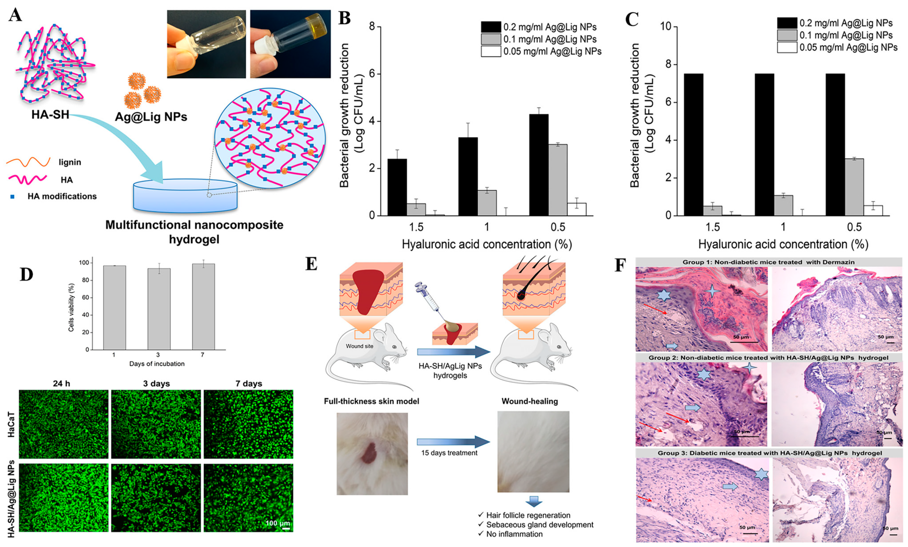
Silver nanoparticles (AgNPs). AgNPs have been widely applied as a broad-spectrum antimicrobial agent for treatment of burns and ulcers wounds for the infection control. Studies have shown that these agents accelerate wound repair by killing bacteria, destroying formation of biofilms, suppressing inflammation, and improving tissue re-epithelization [42,43,44]. AgNPs’ antibacterial activity is attributed to a release of silver ions from oxidized AgNPs under acidic conditions, which subsequently kill bacteria via the destruction of cell walls, DNA, Adenosine triphosphate (ATP), and the production of reactive oxygen (ROS) [45]. Studies have shown that AgNPs’ antibacterial activity exhibits a size-dependent trend, with a decrease in the size of AgNPs associated with an elevated release of silver ions which subsequently promote antimicrobial activity [46,47]. Considering their excellent antibacterial activity, AgNPs can be loaded into various wound dressings for the treatment of wound infections. Some commercially available AgNPs-coated wound dressings, such as “Acticoat” have been successfully used in clinical applications, and have been found to confer sustained release of silver ions thereby suppressing bacterial infections and promoting wound healing [48]. However, AgNPs-based wound dressing therapies are constrained by toxicity concerns resulting from the uncontrolled release of silver ions. Notably, some studies have shown that high AgNPs concentrations not only cause a dose-dependent DNA damage but also suppress the viability of keratinocyte and metabolism cells [44,49]. To reduce these adverse effects, a low AgNPs concentration in combination with antibiotics is recommended. A previous study demonstrated that AgNPs and tetracycline synergistically interact for enhanced antibacterial effects [50]. Additional approaches for improving antibacterial properties and minimizing the cytotoxic effects of AgNPs include coating them with biomolecules as well as incorporating them into hydrogel-based wound dressings [51,52,53,54]. Wu et al. and Pal et al. described in-situ synthesis of AgNPs on bacterial cellulose-based porous hydrogel network. Results showed that the developed wound dressings not only had high bacteria-killing activity but were also associated with low toxic events [52,53]. In another study, incorporation of collagen-coated AgNPs into collagen hydrogels resulted in efficacious and safe application of AgNPs [54]. Notably, the resulting hybrid wound healing not only retained the biocompatibility for human skin fibroblasts and keratinocytes but also exhibited remarkable antimicrobial activity against S. aureus, S. epidermidis, E. coli and P. aeruginosa. In addition, combing AgNPs with chitosan, polyvinyl alcohol (PVA), lignin and zwitterionic hydrogels successfully reduced silver cytotoxicity and improved infection control [55,56,57,58]. Researchers recently developed novel silver-lignin nanoparticles (Ag@Lig NPs) crosslinked thiolated hyaluronic acid (HA-SH) based hybrid nanocomposite hydrogels for chronic wound management, with lignin used as a reducing and capping agent of AgNPs (Figure 2) [59]. Notably, the Ag@Lig NPs-loaded hydrogel exhibited strong antibacterial activity against both Gram-negative and Gram-positive bacteria, with no toxicity to human keratinocytes after 7 days of direct contact. In vivo results showed that it effectively suppressed wound inflammation and promoted tissue remodeling in a diabetic mouse wound model, affirming its potential for the management of chronic wounds. In addition to hydrogel, electrospun nanofibers are also ideal carriers for Ag NPs nanoparticles and present promising applications for wound healing [60,61]. For example, Liu et al. developed a curcumin@β-CD/AgNPs nanoparticle and loaded the composite nanoparticle into a chitosan electrospun nanofiber [61]. The developed functionalized silver nanoparticles showed less toxicity and effective antibacterial activity against P. aeruginosa, S. aureus and E. coli. In vivo, curcumin@β-CD/AgNPs-loaded chitosan dressing showed more uniform collagen distribution, accelerated wound closure rates, and less scar formation compared to commercial AgNPs dressing.
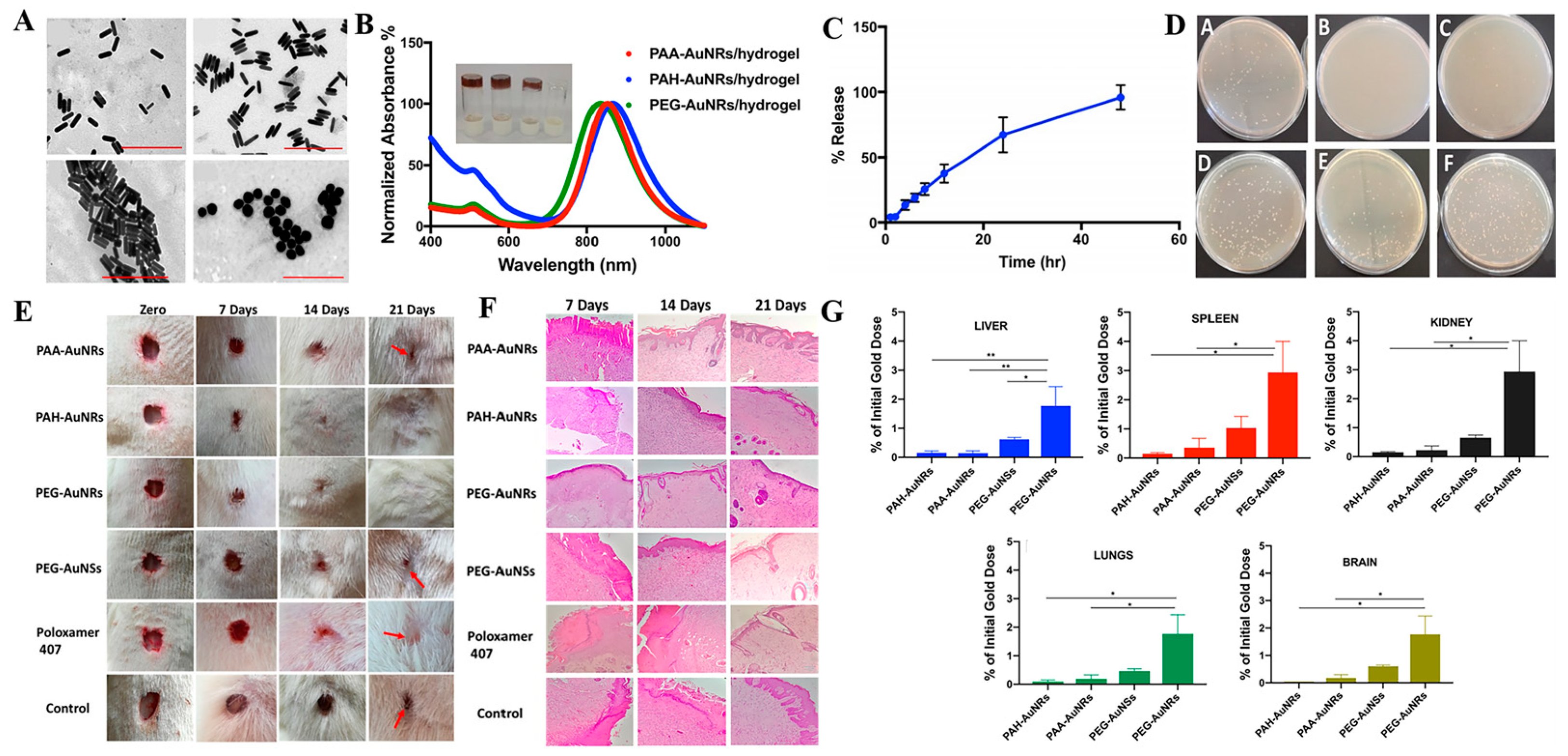
Gold nanoparticles (AuNPs). Apart from AgNPs, AuNPs have also been extensively applied in infection therapy, wound healing, and tissue regeneration due to their excellent biocompatibility, good optical and chemical stability as well as ease of surface modification [42,62,63,64]. AuNPs differ from AgNPs in that they release Ag+ which possess good bacteria-killing capability by directly targeting and destroying their cell walls to cause loss of intracellular components, inhibit ATP synthesis and interfere with DNA transcription. Studies have demonstrated that AuNPs exert broad-spectrum antibacterial activity against both Gram-negative and Gram-positive bacteria, including E. coli, P. aeruginosa, S. aureus, and MRSA [63,64]. AuNPs can also efficiently absorb near infrared light and convert it into heat, which further endows them with the photothermal bactericidal ability [65]. Moreover, AuNPs act as antioxidants and have been shown to promote wound repair by regulating anti-inflammatory cytokines, suppressing ROS levels and promoting angiogenesis [66,67]. However, using AuNPs alone at the wound site is constrained by rapid loss and the associated toxic side effects. Consequently, researchers have integrated AuNPs into a wide range of hydrogel wound dressings with the aim of improving their efficacy and reducing the associated side effect [68,69]. For example, AuNPs with different shapes (nanorods and nanospheres) and surface modifications (neutral, positive and negative charged polymers) were prepared and embedded into a thermosensitive hydrogel of poloxamer 407 (Figure 3) [69]. The resulting hydrogel exhibited the slow and prolonged release of AuNPs, thus enhancing the topical application of AuNPs at the wound site. Both neutral and positively charged polymers modified AuNPs-based hydrogels and exhibited excellent antibacterial activity against S. aureus and P. aeruginosa, in vitro and accelerated skin re-epithelization and collagen deposition in an in vivo animal model. Notably, low percentages of AuNPs were deposited on the main animal organs after 21 days of wound treatment, indicating that the combination of AuNPs with hydrogel minimized the side effects of AuNPs.
Zinc oxide nanoparticles (ZnO NPs). Just like AgNPs, ZnO NPs are also considered an excellent antibacterial agent, although their antibacterial activity depends on release of free Zn2+ from ZnO [70,71]. Numerous studies have shown that the antibacterial effect of ZnO NPs is strongly correlated with the size and concentrations of the nanoparticles [72,73]. For example, 8-nm ZnO NPs were found to kill 95% of S. aureus at a low concentration (1 mM) but those with a larger size (50−70 nm) only caused 40−50% sterilization at a high concentration (5 mM) [73]. Although the Food and Drug Administration (FDA) has confirmed safety and consequently approved most ZnO materials, studies have shown that high concentrations of nano-sized ZnOs exhibit obvious toxicity to normal cells, such as keratinocytes, by inducing mitochondrial dysfunction and causing cell membrane oxidative stress and apoptosis, which greatly limits application of ZnO NPs for wound healing treatment [23]. To solve this problem, Manuja et al. [71] loaded ZnO NPs into sodium alginate-gum acacia hydrogels (SAGA) and found a sustained release of ZnO NPs at lower doses. More importantly, the antibacterial and healing effect was retained while the toxic effects were low [74]. In addition, ZnO NPs have also been combined with other natural polysaccharides polymers-based electrospinning nanofibers such as chitosan and polyvinyl alcohol, and the resulting nanocomposite membranes demonstrated improved antibacterial and wound healing effects [75]. Overall, these reports indicate that ZnO NPs/hydrogel-based platforms hold great promise for wound healing applications.
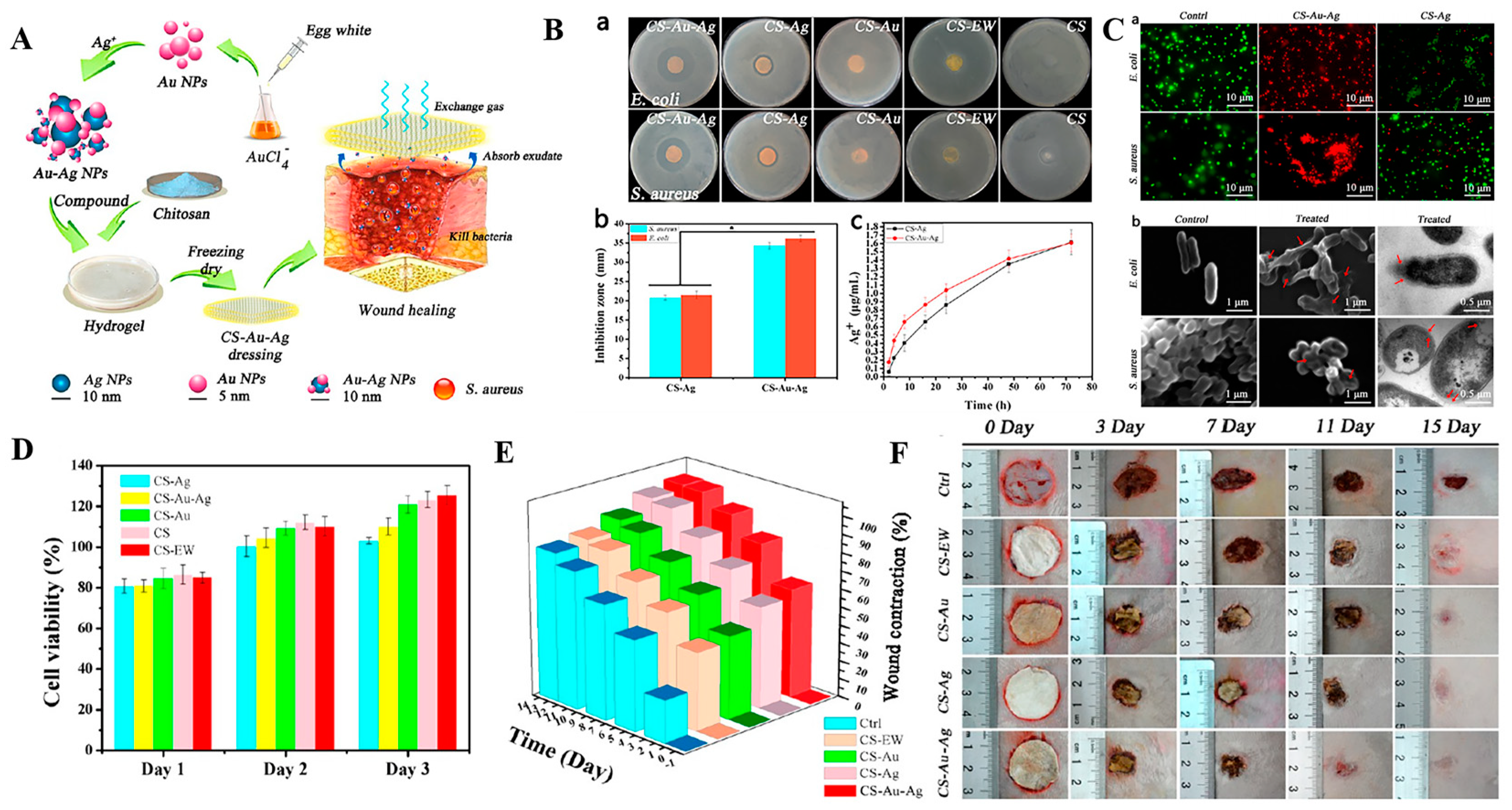
Bimetallic nanoparticles. Considering the bactericidal capacity of various metal NPs, researchers prepared bimetallic NPs (by combining two types of metals) and found that these remarkably improved bactericidal efficiency [76,77,78]. For instance, Li and coworkers synthesized bimetallic Au-Ag NPs and loaded them into a chitosan hydrogel. Results showed that the novel agent had significantly higher antibacterial activity against both S. aureus and E. coli than monometallic-loaded chitosan hydrogel (Figure 4) [79]. Apart from remarkably promoting wound healing effects, this wound dressing was not nontoxic to L929 cells. Similarly, Khan et al. covered biogenic AuNPs with a thin layer of ZnO and prepared the Au@ZnO core-shell nanocomposites. Their results revealed that these nanocomposites remarkably promoted antibacterial and anti-biofilm activity against S. aureus and methicillin resistant Staphylococcus haemolyticus (MRSH) without provoking nuclear damage and cytotoxicity to mouse fibroblasts cells [80].

2.2. Nanomaterials as Passive Carriers of Antibacterial Agents for Antibacterial Treatment

Apart from antibacterial activities, nanomaterials can also be used as effective drug delivery systems to facilitate controlled or sustained release of antibacterial agents during treatment of bacterial infections. For example, studies have shown that nanocarrier-based drug delivery systems can improve the bioavailability of poorly water-soluble drugs, prolong drug half-life, optimize the pharmacokinetics profiles, and reduce the frequency of drug administration, thereby remarkably promoting the therapeutic effect [81]. Incorporating these nanocarriers into wound dressings has emerged as an effective strategy for treatment of infected wounds. One of the most widely applied nanocarriers-based drug delivery systems is nanomaterials, which are usually fabricated from polymers, lipid-based and inorganic materials. These systems remarkably release antibacterial agents in a sustained and passive manner [82].
Polymer-based nanocarriers for antibacterial therapy. Polymer-based nanocarriers are usually synthesized from various natural or synthetic polymers through different assemble methods that are based on electrostatic interactions, hydrophilic and hydrophobic interactions or host-guest interactions. Due to their good biocompatibility, hydrophilicity, and versatility that can be tailored to satisfy the needs of delivered drugs, they are ideal drug delivery systems for wound healing applications [83,84]. For instance, natural polymers such as chitosan (CS) and synthetic polymers including polycaprolactone (PCL) and poly (lactic-co-glycolic acid) (PLGA) have been widely developed as nanocarriers for the delivery of antibacterial agents [85,86,87]. Chitosan and its derivatives are cationic polymers with intrinsic antibacterial capacities that can effectively promote the wound healing process by improving inflammatory cell functions at the wound site, stimulating fibroblasts and osteoblasts [88]. Chitosan NPs can be formed via different methods, including micro-emulsion, ionotropic gelation and emulsification-solvent diffusion as well as evaporation [85]. Antibacterial agents such as antibiotics, antibacterial peptides or small molecule drugs are loaded into CS NPs through hydrophobic hydrogen bonding or electrostatic interactions via direct encapsulation during the preparation process or soaking after CS NPs formation. In a previous study, curcumin, an antimicrobial and antioxidant agent with poor water solubility and impaired skin penetration capacities, was loaded into CS NPs and embedded in fiber-based wound dressings [89]. The dressing allowed for the gradual release and enhanced the wound healing effects of curcumin. Due to their versatile degradation kinetics and controlled drug release properties, PLGA NPs have also been widely used as drug carriers [90]. The PLGA NPs that are prepared by water/oil/water (W/O/W) double emulsion method exhibit a high drug encapsulation efficiency and relatively uniform size distribution [91]. The PLGA NPs have a 70% encapsulation efficiency of LL-37 antimicrobial peptides and are effective treatment options for polymicrobial infected wounds [91,92]. Studies have investigated the significance of NO in antibacterial and wound-healing applications, particularly in combating MRSA biofilms [93]. However, the short half-life and limited diffusion distance of NO limits its direct applications at wound sites. Hasan et al. developed an NO delivery system by doping an NO donor of polyethylenimine/diazeniumdiolate (PEI/NONOate) with PLGA NPs [94]. The PLGA-PEI/NO NPs exhibited a sustained release of NO for over 4 days and good anti-biofilm activities, resulting in accelerated healing of MRSA biofilm-infected wounds in diabetic mice. Nanoparticles that are synthesized by natural or synthetic polymers with cationic charges are also effective antibacterial agents and carriers of anionic drugs that can kill bacteria when combined with the negatively charged bacterial cell walls. In a previous study, a cationic polymer (polyethylenimine (PEI) modified clindamycin-loaded PLGA NPs (Cly/PPNPs)) bound MRSA surfaces and exhibited enhanced antibacterial abilities as well as wound re-epithelialization when compared to NPs without PEI [95]. Liu et al. fabricated a self-assembled nanofiber using the cationic poly (ethylene teraphthalate) (PET) and used it as a carrier for the delivery of anionic antibiotics, piperacillin-tazobactam (PT) [96]. Despite the advances in cationic polymeric nanoparticles in infection control, the potential cytotoxic effects of cationic polymers to normal cells and tissues have yet to be conclusively determined. Embedding these NPs into hydrophilic hydrogel networks or modifying them with hydrophilic polymers such as polyethylene glycol (PEG) and sorbitol are effective strategies for suppressing the cytotoxic effects of cationic polymer nanoparticles without affecting their therapeutic efficiency [97,98,99,100].
Lipid-based nanocarriers for antibacterial therapy. Because of their small particle sizes and good compatibility, lipid-based nanoparticles are attractive carriers of antibacterial drugs for treatment of infected wounds. Liposomes have been investigated as lipid-based nanoparticles. They are fabricated by a hydrophilic inner core and a hydrophobic shell and have good capacities for encapsulating both hydrosoluble and liposoluble antibacterial agents [101]. Liposomes can facilitate the penetration of antibacterial agents into bacterial cells, enhancing the bactericidal effects [102,103]. Various antibiotics have been loaded into different liposomes and successfully inhibited bacterial infection [102,103,104,105]. However, topical use of liposomes at wound sites is often limited by their low drug encapsulation efficiency (less than 30%) and rapid drug release [106]. To improve their efficacies, surface modification and hydrogel encapsulations of liposomes can increase the drug loading level and prolong drug release in wound sites. Thapa et al. designed collagen mimetic peptide (CMP)-modified vancomycin-loaded liposomes (CMP-Van-Lipo) and embedded the liposomes into a collagen hydrogel [107]. Vancomycin was continuously released from the wound dressing for 48 h in vitro and enhanced the antibacterial effects of wound dressing against MRSA in vitro and in vivo. Similar to liposomes, colloidal layered vesicles that are usually composed of non-ionic surfactants and amphipathic compounds (called niosomes) are also ideal carriers for the delivery of both hydrophobic and hydrophilic antibacterial agents [108,109]. Compared to liposomes, niosomes are more stable, affordable and allow better loading and higher permeability for small molecules and ions [106]. Abdelaziz et al. reported that the norfloxacin-loaded niosomes proved excellent bactericidal ability towards Pseudomonas aeruginosa biofilm [110]. Encapsulating electrosprayed cefazolin-loaded niosomes onto electrospun chitosan nanofibrous membrane also demonstrated effective antibacterial property against both Gram-negative and Gram-positive bacteria, good biocompatibility and enhanced skin regeneration by improving re-epithelialization, tissue remodeling, and angiogenesis [111]. Apart from liposomes and niosomes, lipid nanoparticles, including solid lipid NPs (SLNs) and nanostructured lipid carriers (NLCs) are effective antibacterial agent carriers for infected wound treatment [112,113]. Compared with liposomes, lipids NPs exhibit increased drug encapsulation efficiencies, decreased drug leakage and sustained drug release profiles, which are attributed to their denser structures [114]. The SLNs have a drug encapsulation efficiency of between 50% and 100% while NCLs have an encapsulation efficiency of around 85% [106]. Many studies have reported increased antibacterial effects when using antibacterial agents loaded SLNs and NCLs systems [115,116,117,118,119,120]. In one study, the LL37 antimicrobial peptide was encapsulated by SLNs, and was continuously released for over a period of 14 days and effectively killed S. aureus and E. coli [116]. In another study, the peppermint essential oil-loaded NLCs exhibited high-efficient antibacterial activities against S. epidermidis, S. aureus, E. coli as well as P. aeruginosa and accelerated wound healing effects [119]. To apply SLNs and NLCs in wound healing, these NP carriers have also been incorporated into hydrogel wound dressings. Silver sulfadiazine (SSD)-loaded SLNs was embedded into chitosan hydrogels for SSD delivery [120]. The hydrogel exhibited prolonged SSD release to wound sites and higher antibacterial efficiencies against planktonic bacteria and P. aeruginosa biofilms.
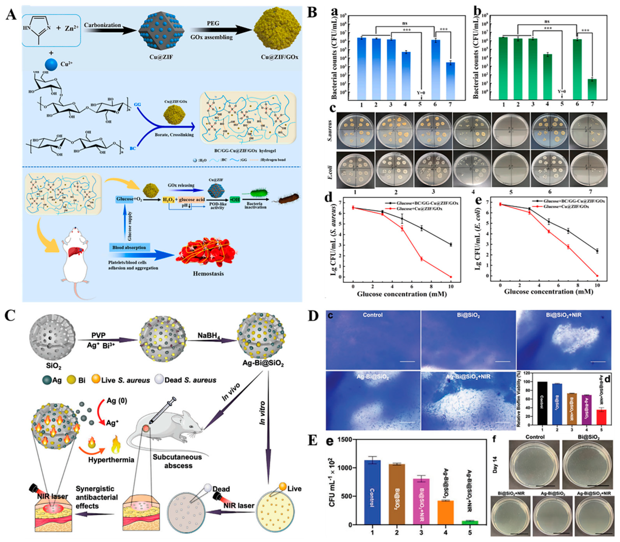
Inorganic nanocarriers for antibacterial therapy. With advanced development of chemical synthesis and preparation technologies, various types of inorganic NPs, including metallic, ceramic and carbon-based NPs have emerged as attractive drug carriers for wound healing. The inorganic NPs have a high surface area, tunable size and versatility that allow the antibacterial agents to be loaded or grafted on pore surfaces of corresponding NPs [121,122]. Among the inorganic nanoparticles, metal NPs such as AuNPs and AgNPs with intrinsic antibacterial performances have the capacity for conjugating with antibacterial agents to achieve synergistic antibacterial effects [123,124,125,126,127]. For example, conjugating ceftriaxone with AgNPs and AuNPs has been proved to enhance the bactericidal ratio against E. coli to 2- and 6-fold, respectively than that of pure ceftriaxone [123]. Moreover, peptide-coated AuNPs [124], Rauwolfia serpentina-modified AuNPs [125], tannic acid chelated Ag nanoparticles (TA-Ag NP) [126] and Zn-penicillin complex [127] have all been shown to have enhanced antibacterial activity and potential application for the healing of infected wounds. In addition, because of their ultra-high specific surface areas, excellent biocompatibility and biodegradability, porous structures and tunable pore sizes to ensure maximum drug loading and controlled release kinetics, metal-organic frameworks (MOFs)-based nanoplatforms (e.g., Zn-based MOFs, Cu-based MOFs, Fe-based MOFs and Ag-based MOFs) are ideal candidates for delivery of antibacterial agents [128]. In a proof-of-concept study, three types of MOFs, including Cu-MOFs 1, Co-MOF 2 and Zn-MOF 3 were prepared and respectively embedded in the PEG hydrogel [129]. Hydrogel@Cu-MOF 1 and hydrogel@Co-MOF 2 exhibited excellent antibacterial activities against both Gram-negative (E. coli) and Gram-positive (S. aureus) bacteria, when compared with hydrogel@Zn-MOF 3. Hydrogel@Cu-MOF 1 exhibited 99.9% antibacterial effects at the minimum bactericidal concentration without exerting cytotoxic effects to human dermal fibroblasts. The MOFs-based platform can be used as nanozyme catalysts to induce the ROS-induced bacterial killing effects [130,131]. In a typical study, glucose oxidase (GOx) was loaded into a Cu2+ doped graphited imidazolate framework (ZIF-8). Then, the as-prepared Cu@ZIF/GOx was encapsulated in a bacterial cellulose (BC) reinforced guar gum (GG) hydrogel (Figure 5A) [131]. Loading of GOx has been proven to be capable of consuming glucose to produce H2O2 and glucose acid, which induces Cu@ZIF to produce high oxidative •OH to kill bacteria. The as-prepared hydrogel exerted significant antibacterial efficacies as well as inhibitory effects on biofilm formation, implying that they have promising applications in diabetic infected wound healing (Figure 5B). In tandem with MOFs-based nanocarriers, due to their porous structures and adjustable surface modifications, ceramic-based NPs, especially mesoporous silica NPs (MSN), are used as drug carriers. Until now, various antibacterial agents including gentamicin [132], ciprofloxacin hydrochloride/Zn complex [133], AgNPs [134], and silver-bismuth nanoparticles (Ag-Bi NPs) [135] have been loaded into MSNs to treat infections in wounds. Cao et al. designed mesoporous silica supported silver-bismuth (Ag-Bi@SiO2) NPs by loading Ag-Bi NPs into porous SiO2 NPs for treatment of bacterial infected wounds (Figure 5C) [135]. The synergistic antibacterial effects against both MRSA and MRSA biofilms, which are attributed to the hyperthermia produced by Bi NPs under near-infrared light (NIR) irradiation and accelerated Ag+ release, have been observed in vitro. A 69.5% decrease in MRSA biofilm biomass was achieved using Ag-Bi@SiO2 NPs at a concentration of 100 µg mL−1 with an NIR laser, exhibiting better bactericidal effects than Bi@SiO2 NPs with laser (26.8%) or Ag-Bi@SiO2 NPs without laser treatment (30.8%) groups (Figure 5D). The in vivo test indicated that Ag-Bi@SiO2 NPs were able to kill 95.4% of bacteria in infected wounds and accelerated the wound healing process (Figure 5E). In addition, bioceramics nanoparticles such as bioactive glass nanoparticles (BG NPs), which contains the oxides of silicon, sodium, calcium, and phosphorus, exhibit good biocompability and wide applications in tissue engineering [136,137]. Using structure-forming agents, mesoporous or hollow structured BG NPs can be formed and doped with ZnO or Ag to achieve an antibacterial property for infection treatment [138,139,140]. It is worth mentioning that many reserachers have demonstrated that bioactive glass has the ability of promoting angiogenesis and skin regeneration at the wound site through promoting the production of vascular endothelial growth factor (VEGF) and fibroblast growth factor 2 (FGF2) [139]. Therefore, antibacterial BG NPs exhibit unparalleled advantage for wound healing. In addition to the above, carbon-based nanomaterials, such as graphene oxide (GO), carbon nanotubes (CNTs) and carbon-based quantum dots (CDs) are also good carriers of antibacterial drugs since their abundant oxygen-containing groups, including hydroxyl, epoxy, and carboxyl groups of carbon-based nanomaterials are active sites for the conjugation of therapeutic agents [141,142]. In their study, Patarroyo et al. grew Ag NPs in situ on graphene oxide surfaces and the as-prepared GO-Ag NPs nanoconjugates were subsequently encapsulated into gelatin-polyvinyl alcohol-hyaluronic acid hydrogels. The hydrogel exhibited 100% antibacterial effects and very low hemolytic effects (less than 5%), showing the potential for wound healing [143].

2.3. Stimuli-Responsive Nanomaterials Carriers of Antibacterial Agents for Antibacterial Treatment

Despite advances in the above nanocarriers for antibacterial therapy, passive antibacterial mechanisms of such drug delivery systems that rely on uncontrollable and untimely release of antibiotics, antibacterial ions and peroxide radicals may greatly decrease the treatment effects of nanomaterials, increasing bacterial drug resistance and occurrence of undesired side effects. Excess release of metal ions and ROS may be cytotoxic to normal tissue cells and trigger additional inflammatory reactions to impede wound healing while leading to other diseases [144,145]. Developing smart nanomaterials-based carriers that can respond to endogenous microenvironments of bacterial infections such as acidic pH, excess ROS, specific toxins and enzymes secreted by bacteria or external stimuli, including light, thermal and magnet to release antibacterial agents or trigger their own antimicrobial properties in a controllable manner may be an effective strategy [146]. Until now, such smart nanocarriers have been widely investigated and can be further integrated into wound dressings to promote wound repair. Examples of these advanced wound dressing systems for antibacterial therapy are discussed in the following sections.
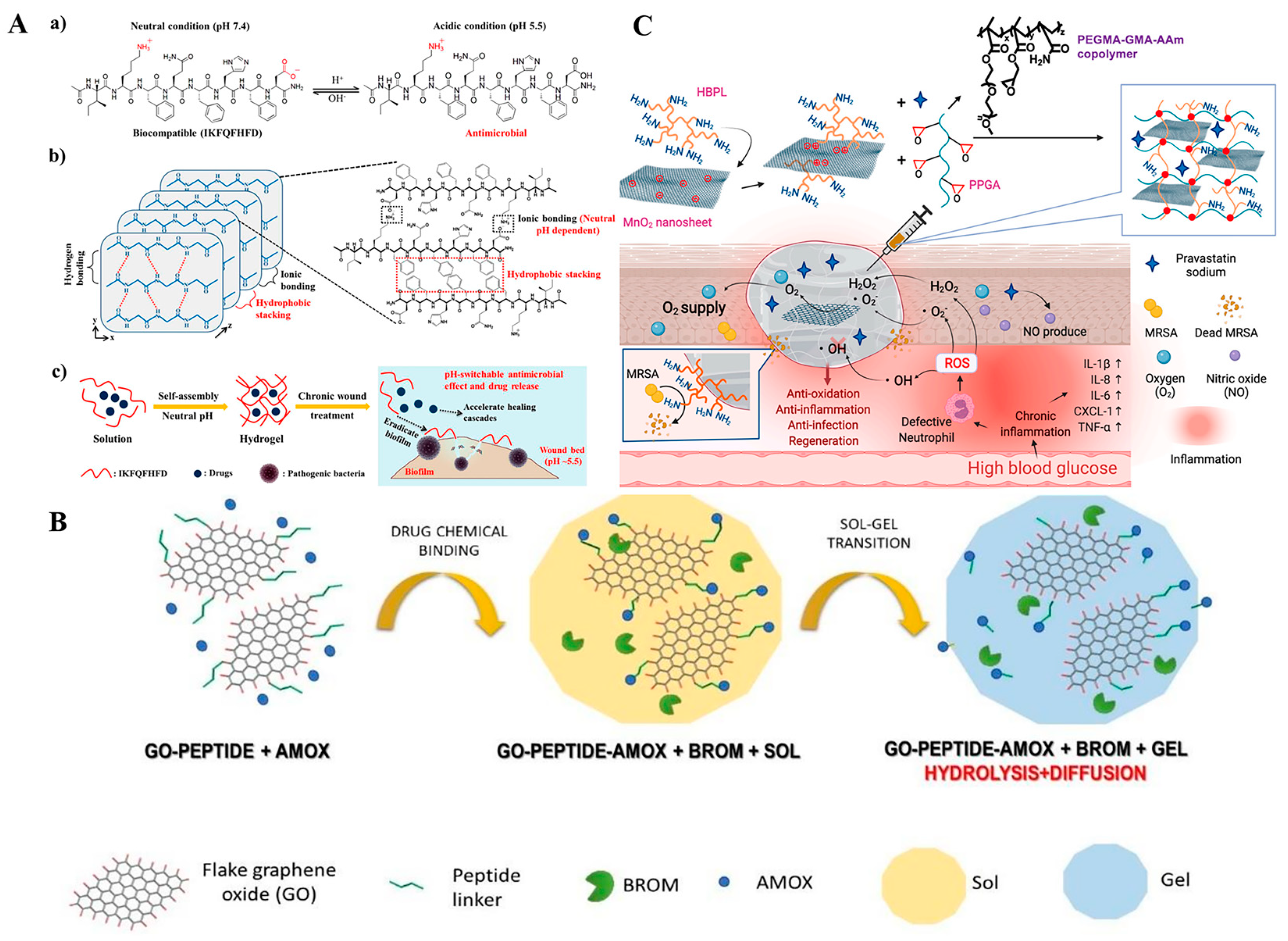
Endogenous stimuli-responsive nanocarriers for antibacterial therapy. Bacterial colonization at the wound site results in significant changes in physicochemical markers in their surrounding microenvironments. For instance, the lactic acid produced by bacteria decreases the pH value of the microenvironment and induces an acidic microenvironment [147,148]. Some novel pH-responsive antibacterial nanocarrier platforms have been fabricated by utilizing pH-cleavable chemical bonds (e.g., ester bond, Schiff base bonds and hydrazone bonds) or pH-ionizable groups (e.g., zwitterionic, chitosan, and MOFs), which allows the nanocarriers to be broken or change their surface charge at acidic pH values to release antibacterial agents or change from a negative status to a positive status to effectively kill bacteria [147,148,149,150,151,152]. Wang et al. developed a photothermal agent (cypate) and a procollagen component (proline) loaded pH-sensitive octapeptide (IKFQFHFD) nanofiber-based hydrogel network for treatment of chronically infected wounds (Figure 6A) [152]. At acidic environments, the octapeptide would change from electrically neutral to a positive antibacterial peptide, resulting in disassembly of the hydrogel and release of antibacterial IKFQFHFD and cypate. In vitro, the synergistic antibacterial effects of IKFQFHFD and cypate effectively alleviated a mature MRSA biofilm. The synergistic release of proline from the hydrogel promoted tissue cell regeneration. In vivo, the as-prepared nanofiber hydrogel enabled the complete repair of MRSA biofilm wound infections within 20 days, implying that they are potential chronic wound dressings. For chronic wounds, an alkaline pH value of 8–10 at the local wound was detected. Loading of chlorhexidine (CHX) into pH-sensitive silica nanoparticles (SiNPs) achieved a 4–5-fold release of CHX on alkaline wounds than at neutral as well as acidic environments and exhibited a 4-log reduction of bacterial cells, including both Gram-negative and -positive bacteria [153]. The as-prepared CHX-SiNPs were further encapsulated into alginate hydrogels, exhibiting good bactericidal capacities against E. coil infected chronic wounds. Apart from changes in pH values, bacteria such as S. aureus and P. aeruginosa secrete many factors, including toxins (e.g., α-toxin) and enzymes (e.g., hyaluronidase, gelatinase, and phospholipase) [154,155]. Nanocarriers that can be specially broken by these bacteria-secreted toxins and enzymes are promising strategies for inducing the release of antibacterial drugs and achieve on-demand infection treatment [156,157,158,159]. In a previous study, a gentamicin loaded mesoporous silica nanoparticle (MSN) core was modified by a bacterial toxin-cleavable lipid bilayer shell, which was further modified by a bacteria-targeting peptide ubiquicidin (UBI29-41) [157]. The final nanoparticles were capable of targeting the bacterial infection site and achieving the rapid release of gentamicin due to bacterial toxins-induced degradation of the lipid bilayer shell. In vitro and in vivo proliferation of S. aureus is effectively inhibited by nanoparticles. In another recent study, Mir et al. designed bacterial lipase-cleavable poly(ε-caprolactone) nanoparticles and loaded them with carvacrol (CAR) [158]. By incorporating CAP-PCL nanoparticles into a hydrogel matrix, there was significant bacterial-induced release of carvacrol and excellent antimicrobial activities against MRSA. Trusek et al. developed a novel enzyme-responsive graphene oxide (GO)-based nanocomposite hydrogel, achieving controlled antibiotic delivery (Figure 6B) [159]. In the hydrogel, amoxicillin (AMOX) was chemically attached to GO via a bromelain enzyme-cleavable peptide linker (Leu-Leu-Gly) and released from the hydrogel in the presence of bacteria. Despite the high specificity and catalytic abilities of enzymes in controllable drug delivery, inactivation of bacterial secreted enzymes may result in the loss of enzyme responsiveness of such smart nanocarrier platforms. Coupling enzyme responsive NPs with other stimuli-responsive antibacterial agents to create synergistic antibacterial platforms may be a possible strategy for ensuring effective antibacterial activities. Bacterial infections also result in excess wound inflammation, leading to increased ROS levels in the microenvironment, which is a potential effective trigger for antibacterial agent delivery [160,161]. Based on this, various ROS-responsive nanocarrier systems were fabricated using ROS-sensitive substances, such as poly (propylene sulfide) (PPS), phenylboronic acid, poly(thioketal) (PK) and their derivatives [162,163,164,165]. Li et al. developed ROS-responsive mesoporous silica nanoparticles (MSNs) for controlled delivery of antibiotics in infected wounds, in which vancomycin was physically loaded into the amino-modified MSNs. Then, the ROS-sensitive thioketal linker grafted methoxy poly (ethylene glycol) (mPEG-TK) was further modified on MSN surfaces as a gatekeeper [164]. In vitro, the high H2O2 levels resulted in breakage of the TK linker and induced vancomycin release. The as-prepared van-mPEG-TK-MSNs successfully killed all the bacteria and cured the in vivo tissue infection after 14 days. Some metal-containing nanomaterials have been proven to have nanoenzyme activities, which can also be used as ROS-responsive antibacterial agents [166,167]. Tu et al. fabricated a novel multifunctional nanocomposite hydrogel wound dressing platform with synergistic performances of the antibacterial polymer and ROS-sensitive manganese dioxide (MnO2) nanosheets against bacterial proliferation (Figure 6C) [166]. In this work, the antibacterial hyperbranched poly-L-lysine (HBPL) was grafted on manganese dioxide (MnO2) nanosheet surfaces and the as-prepared HBPL-MnO2 were further used as crosslink agents to crosslink hydrophilic poly (PEGMA-co-GMA-co-AAm) to prepare the nanocomposite hydrogel. The MnO2 nanosheets are used as nanoenzymes to convert excess ROS on the MRSA-infected wound into oxygen and kill bacteria. The synergistic effects of HBPL and MnO2 nanoenzymes have been shown to kill up to 94.1–99.5% of MRSA, E. coli and Pseudomonas aeruginosa even at 109 CFU/mL in vitro. In vivo, the MRSA-infected rat wounds were effectively treated by the hydrogel and the wound healing was significantly accelerated.
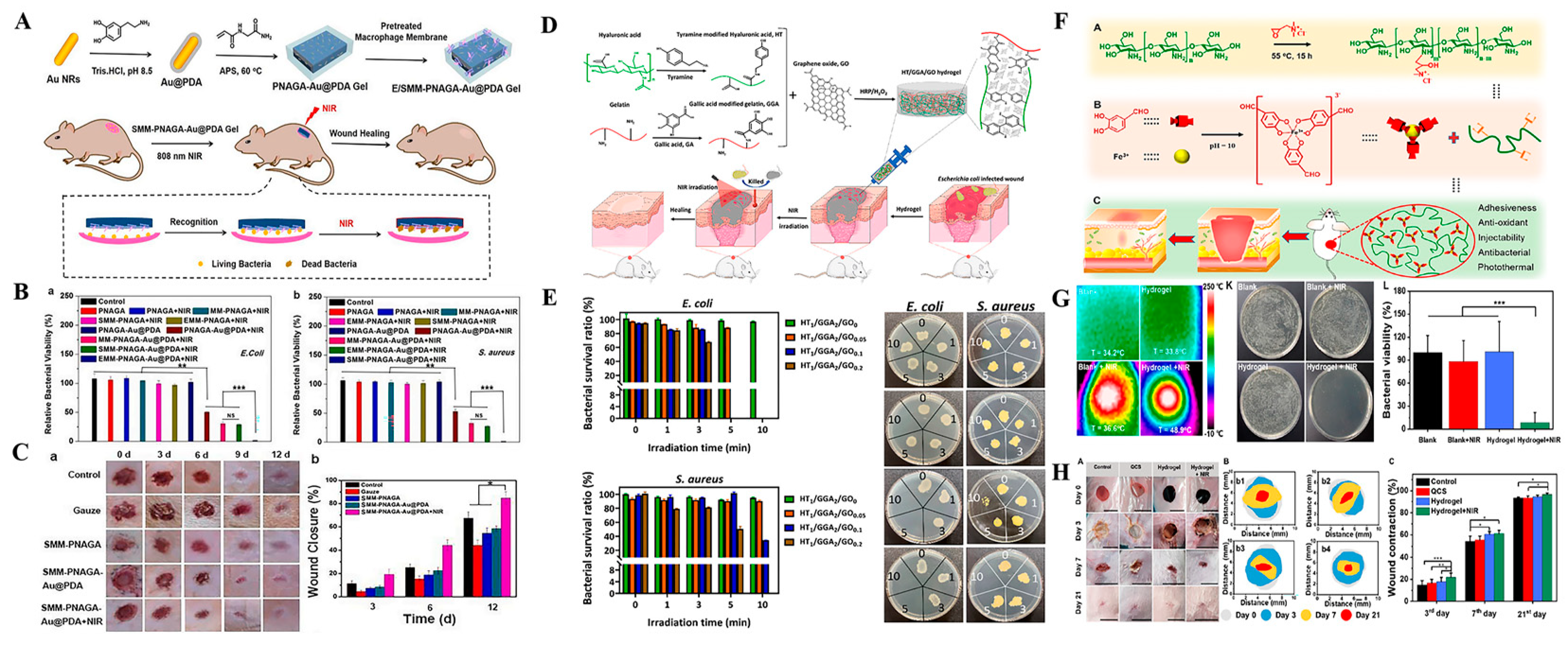
Exogenous stimuli-responsive nanocarriers for antibacterial therapy. Compared with bacterial microenvironment-responsive nanocarriers, stimuli-responsive nano-based drug delivery systems that can be induced by external stimuli, including light, magnet, and ultrasound among others possess unrivaled advantages in controlled release of antibacterial agents and infection treatment since these external stimuli can be easily acquired and precisely controlled without time and space limitations [168,169]. Among the external stimuli, because of its convenience, high-efficiency, non-invasion and the ability for spatiotemporal control, light is a particularly attractive tool that has been widely used in drug delivery, disease therapy and tissue engineering [170,171,172]. Lights, including ultraviolet (UV) light, visible light, and near-infrared (NIR) light have been investigated as triggers to combat bacterial infections by directly secreting antibacterial agents or killing bacteria through NIR-induced photothermal and photodynamic therapy [28,173]. Light-induced release of the preloaded drug from nanocarriers is one of the effective strategies for infection control [174,175,176]. Ballesteros et al. loaded AgNPs into a photosensitive nanogel and immobilized the nanogel on the surface of poly(ε-caprolactone) nanofibers mats [174]. Upon UV light irradiation (405 nm), the nanogel collapsed, resulting in responsive release of AgNPs into nanofiber mats and subsequent release of Ag+ to effectively kill bacteria. Studies have also reported on UV light-induced release of nitric oxide (NO) and visible light-induced release of carbon monoxide (CO) from nanocarriers to cure bacterial-infected wounds [175,176]. The limited penetration abilities of visible and UV light may impede their applications in deep skin tissues. Compared with visible and UV light, NIR light exhibits excellent tissue penetration abilities. Apart from the NIR-induced direct release of antibacterial drugs, the NIR-induced photothermal and photodynamic effects of nanomaterials are promising antimicrobial strategies. It has been well reported that nanomaterials including metal NPs (e.g., Au NPs [177] and Cu NPs [178]) metal sulfide/oxide nanomaterials (e.g., CuS [179], TiO2 [180] and MnO2 [181]), carbon-based nanomaterials (e.g., GO [182] and CNTs [183]), polymer NPs (e.g., polydopamine NPs [184]) and iron (III)-catechol complex (e.g., Fe3+-Tannic acid complex [185]) exhibit excellent photothermal responsive performances that can absorb NIR light and convert it into local heat to kill bacteria by destroying bacterial membranes and denaturing proteins as well as DNA. Photoactive hydrogels with attractive photothermal antibacterial capacities can be developed by combining the above-mentioned nanomaterials with hydrogels, and used as wound dressings to promote wound healing. Moorcroft et al. encapsulated gold nanorods and antimicrobial peptides (IK8) into thermal-cleavable liposomes and loaded the liposomes into a PEG hydrogel [186]. Given the photothermal effects of gold nanorods upon NIR irradiation at 860 nm, photothermal-induced release of IK8 and the intrinsic photothermal bactericidal capacity of gold nanorods exhibited excellent synergetic antibacterial effects against P. aeruginosa and S. aureus. In another study, a dopamine-coated Au nanorod (Au@PDA NR)-incorporated photothermal antibacterial hydrogel was prepared and further coated with S. aureus or E. coli pretreated macrophage membranes (Figure 7A) [187]. In vitro, due to the presence of surface-coated macrophage membranes, the as-prepared hydrogel specifically recognized and killed 98% of the bacteria via the superimposed photothermal effects of both Au nanorod and PDA within NIR irradiation for 5 min (Figure 7B). In vivo, the hydrogel exhibited positive therapeutic effects and promoted S. aureus-infected wound healing (Figure 7C). They crosslinked hyaluronic acid-grafted with tyramine (HT) and gelatin grafted with gallic acid (GGA) via a HRP/H2O2 catalytic system to form the injectable hydrogel, in which GO was loaded [188]. They crosslinked hyaluronic acid-grafted with tyramine (HT) and gelatin grafted with gallic acid (GGA) via a HRP/H2O2 catalytic system to form the injectable hydrogel, in which GO was loaded (Figure 7D). The hydrogel demonstrated a good photothermal antibacterial activity against E. coli and S. aureus and could significantly eliminate the infection of E. coli and promote the healing of E. coli-infected wound (Figure 7E). In addition, Fe3+-catechol complexes with good photothermal conversion properties have a great potential in developing NIR-responsive antibacterial hydrogel dressings [189,190,191]. In a typical study, dual dynamic-bond cross-linked hydrogels were prepared by crosslinking the protocatechualdehyde (PA)-Fe3+ complex with the quaternized chitosan (QCS) for treatment of infected wounds (Figure 7F). The inherent antibacterial activities of QCS and the excellent photothermal-antibacterial performances of the PA-Fe3+ complex endow the hydrogel with abilities to achieve high wound closure and promote MRSA-infected wound healing (Figure 7G,H) [191].
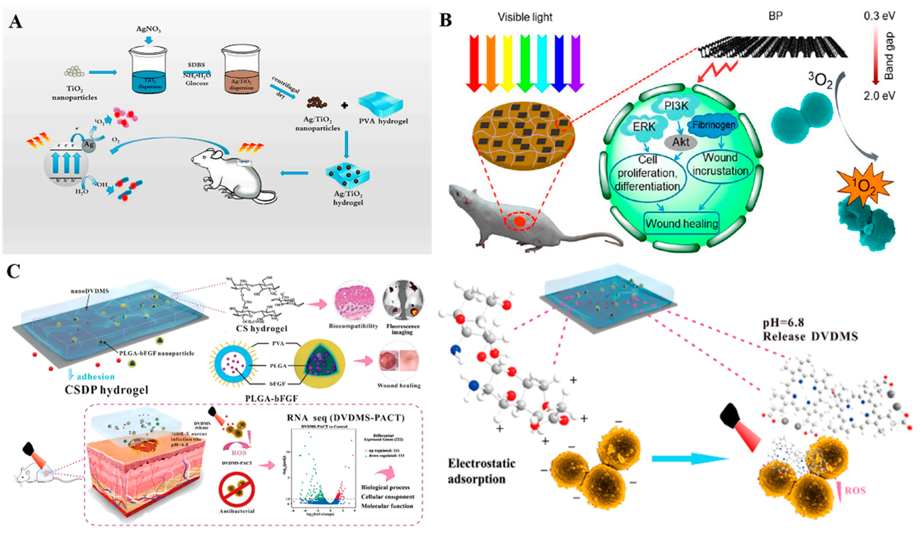
In a manner that is similar to photothermal therapy (PTT), photodynamic therapy (PDT) is an effective and minimally invasive strategy for combating bacterial infections by using some photosensitizers to secrete ROS under light irradiation and kill bacteria via ROS-induced oxidative stress of bacterial cells, and damage to intracellular components such as proteins, lipids and nucleic acids [21,192]. In recent decades, various inorganic and organic nano-based photosensitizers such as ZnO [193], TiO2 [194], CuO [195], MOFs [196,197], graphene [198], MXenes [199], black phosphorus (BP) [200], etc. have all n demonstrated excellent photothermal antibacterial activity. However, the low stability and poor water solubility of some photosensitizers inhibits their independent applications in infected wound sites. Taking advantage of hydrogels for wound healing, nano-photosensitizers loaded hydrogels with potential antibacterial and repair performances have shown inspiring applications in infected wound healing [28]. In 2019, Wang et al. incorporated Ag-doped TiO2 (Ag/TiO2) nanoparticles into PVA hydrogel to treat an infected wound (Figure 8A). The photodynamic activities of TiO2 NPs in visible wavelength range were greatly improved by the doping of Ag on TiO2 NP surfaces. Moreover, TiO2 NPs inhibited Ag+ release, thereby suppressing the potential toxicity of Ag+. Upon visible irradiation of 606 nm within 5 min, the hybrid hydrogel exhibited effective bacterial-killing abilities against both E. coli and S. aureus via light-induced production of ROS in the Ag/TiO2 system. Using this hybrid hydrogel as a wound dressing, it greatly inhibited the S. aureus-induced infection and significantly accelerated the wound healing [201]. In a similar study, due to production of singlet oxygen (1O2) of BP under visible light irradiation, black phosphorus nanosheets (BPs)-embedded chitosan hydrogels (BP@ CS) exhibited incomparable photodynamic bactericidal effects in vitro and in vivo (Figure 8B) [200]. Some porphyrin photosensitizers such as sinoporphyrin sodium (DVDMS) also have high singlet oxygen (1O2) yield and photodynamic antibacterial properties. However, under physiological conditions, DVDMS is not stable enough, which compromises its photodynamic activities. To overcome this challenge, Mai et al. incorporated nano-DVDMS into carboxymethyl chitosan (CMCS)-sodium alginate hydrogel (Figure 8C). In vitro, 10 μg/mL of the DVDMS -loaded hydrogel exhibited excellent antibacterial and anti-biofilm activities by eliminating almost 99.99% of S. aureus and MRSA under mild photoirradiation conditions (30 J/cm2, 5 min). The hydrogel has been proven to effectively inhibit bacterial growth while accelerating wound healing in burn infected models. Therefore, incorporating DVDMS into the hydrogel markedly improved the biological stability of DVDMS and enhanced the photodynamic antibacterial activities of DVDMS [202].
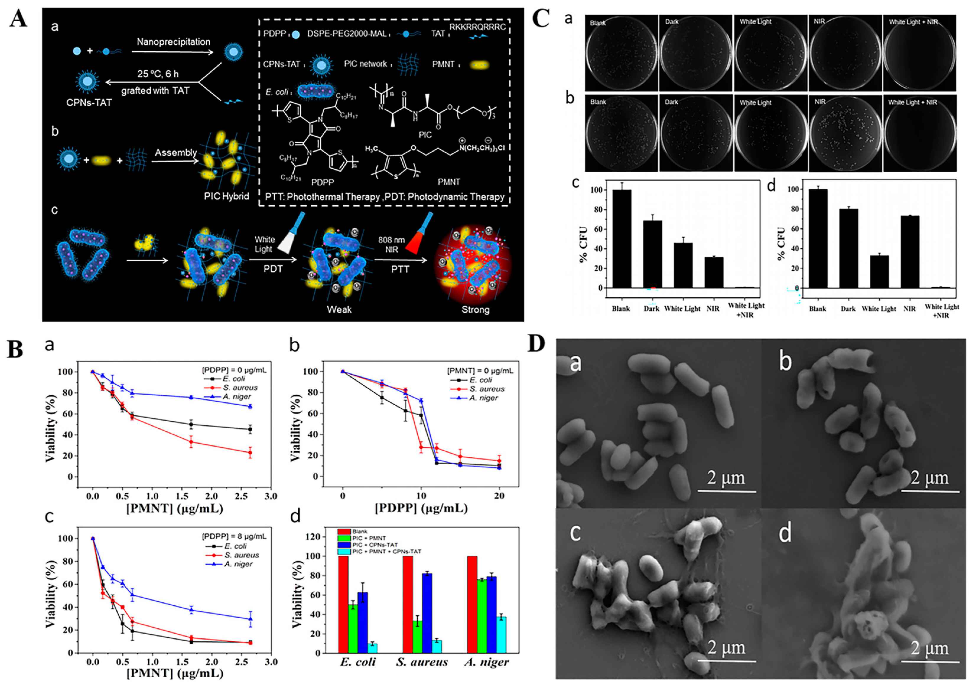
Despite mature applications of PTT and PDT on infection treatments, there are some concerns regarding the single use of PTT or PDT. For instance, to effectively alleviate bacterial growth, a high-temperature is required using PTT, which may damage normal cells and tissues around the wound [203,204]. The bactericidal effects of PDT are also dependent on elevated ROS production levels. However, excessive ROS may also induce inflammation, fibrosis, and necrosis of tissues during PDT treatment [205,206]. The heat can promote the transfer as well as ROS release by photosensitizers and increase bacterial membrane permeability to facilitate the entry of ROS into the bacterial cell and effectively kill the bacteria [207]. Moreover, a high-efficiency bacterial-killing capacity can be achieved by dual modes combination of lower temperature produced by PTT and moderate doses of ROS produced by PDT [208]. Therefore, the synergistic antibacterial therapy by combining PTT and PDT is a more effective and safer strategy than using single photothermal or photodynamic treatment [28,209]. In a recent study, a multifunctional black phosphorus (BP) hydrogel with antibacterial and antioxidant properties was developed for diabetic skin wound treatment [210]. In this study, 4-octyl itaconate (4OI) with antioxidant and anti-inflammatory properties was modified onto BP nanosheets and loaded into gelatin methacryloyl (GelMA) hydrogels (Figure 9A). In vitro, due to the photothermal effects of BP, a temperature of 52 °C was reached in the BP-based hydrogel under NIR irradiation for 5 min. This temperature has been proven to effectively kill bacteria and facilitate 1O2 permeation to enhance the PDT effects of BP. Benefiting from excellent PDT and PTT properties of BP, the as-prepared hydrogel exhibited synergistic bactericidal effects by killing 92.27% of E. coli and 96.69% of S. aureus upon 808 nm NIR irradiation for 5 min (Figure 9B) and effectively accelerated the closure of diabetic wound (Figure 9C). Similarly, Xie et al. prepared lignin-copper sulfide (LS-CuS) nanocomposites loaded PVA hydrogel, which exhibited good antibacterial and anti-biofilm capacities due to the synergistic PTT and PDT effects of CuS NPs under 808 nm laser irradiation [211]. Cui et al. fabricated a composite polyisocyanide (PIC)-based hydrogel with PDT and PTT dual antibacterial capacities in which cationic conjugated polythiophene (PMNT) was loaded and used as a PDT agent induced by white light. Water-insoluble conjugated polymer nanoparticles (CPNs-TAT) were also encapsulated and served as PTT agents triggered by NIR light (Figure 10). These complex hydrogels avoided PMNT aggregation and reduced the toxicity of CPNs-TAT, endowing the hydrogel with enhanced photodynamic and photothermal antibacterial activities [208]. Xiang et al. achieved synergistic antibacterial effects of PDT and PTT using carbon quantum dot decorated ZnO (C/ZnO) NPs and PDA, in which PDA was responsible for PTT under 808 nm NIR light and C/ZnO NPs was the source of ROS for PDT under 660 nm light. The sustained release of Zn2+ also enhanced the antibacterial properties. This system was shown to kill 99.9% of bacteria under dual-light irradiation and greatly promoted wound healing [212]. In a novel study, bacterial-targeting aggregation-induced emission (AIE) self-assembled nanoparticles (AIE NPs) with high water solubility, good biocompatibility, and good photostability were developed to achieve synergistic PTT and PDT effects against pathogens under 808 nm irradiation. Upon PTT/PDT treatment, the sterilizing efficiencies of AIE NPs were 99.9% and 99.8% for S. aureus and E. coli, respectively. In vivo, the AIE NPs-induced phototherapy efficiently eliminated S. aureus and promoted S. aureus-infected wound healing [213].

3. Intelligent Nanomaterials-Based Wound Dressings for Infection Detection and Treatment

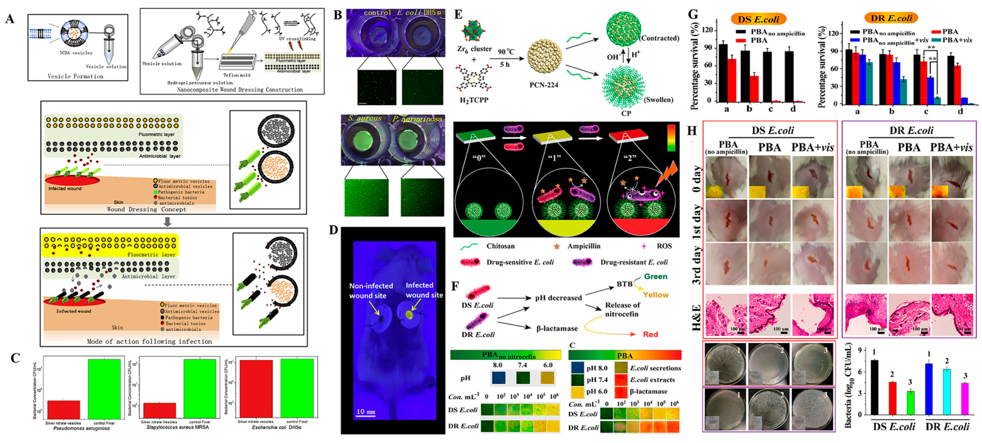
Apart from antibacterial therapy, timely detection of bacteria at the wound site is of great importance for antibacterial wound dressings to provide appropriate and efficient anti-infection treatments. Currently, clinical detection of wound infection is usually performed by removing covered dressings, scraping wound exudates for further pathogen detection and by visual diagnosis based on some obvious clinical symptoms, including redness, swelling, pustule, and heat. The infections easily induce secondary wound damage [214,215]. Frequent changes of dressings may also induce secondary wound injury and increase pain in patients. Therefore, developing intelligent wound dressings that are capable of both detecting bacterial infections in-situ and providing timely antibacterial therapies for infection control is of great significance. Such wound dressings may overcome the disadvantages of traditional clinical approaches for infection detection and markedly improve the therapeutic outcomes as well as innovate management models for wound infections. Currently, intelligent wound dressings that simultaneously integrate wearable sensors and advanced drug delivery systems have achieved tremendous advances in wound infection diagnosis and treatment [215,216,217,218]. The unique optical and physicochemical properties of nanomaterials and rapid advances in nanotechnologies have endowed nanomaterials with great potentials in sensor and disease detection [219,220]. Advanced nanomaterials-based wound dressings that possess the functions of self-reporting of bacterial infections and antibacterial therapy have been widely investigated and applied in wound healing [221,222,223,224]. Zhou et al. developed bacteria-responsive smart wound dressings for simultaneous detection and inhibition of bacterial infections by simultaneously encapsulating self-quenched fluorescent dyes and antimicrobials into bacterial secreted toxins-lytic lipid vesicles and loading the vesicles into methacrylated gelatin (GelMA) hydrogels (Figure 11A). In vitro and in vivo, the as-prepared wound dressing provided a visual warning of S. aureus- and P. aeruginosa-associated infections by color changes of the dressing upon S. aureus- and P. aeruginosa-secreted toxin-induced release of fluorescent dyes and inhibited MRSA and P. aeruginosa proliferation by the released antimicrobial (Figure 11B–D) [221]. In 2020, Qiao et al. designed a Cyanine3 (Cy3) and Cyanine5 (Cy5)-co-modified silica nanoparticles (SNP-Cy3/Cy5) and co-encapsulated a UV-cleavable antibiotic prodrug (GS-LinkerMPEG) into PVA hydrogel to monitor bacterial infections and provide on-demand treatment. In this system, SNP-Cy3/Cy5 served as a pH-responsive fluorescent probe for detection of bacterial infections in which the fluorescence resonance energy transfer (FRET) effect between Cy3 and Cy5 could be broken in an acidic bacterial microenvironment and induce fluorescence changes of SNP-Cy3/Cy5. Moreover, up-conversion nanoparticles (UCNPs) were embedded into the hydrogels, which were used to transfer NIR light to UV light and induce the release of gentamicin from the UV-cleavable prodrug (GS-LinkerMPEG) to provide on-demand antibacterial treatment [223]. This nanoparticle composited hydrogel provided a new strategy for self-reporting and effective treatment of wound infections. In another study, a colorimetric nano-based wound dressing for point-of-care sensing and treatment of both drug-sensitive (DS) bacterial and drug-resistant (DR) bacterial infections was developed, which consisted of a pH indicator dye of bromothymol blue (BTB) and a chitosan coated nitrocefin and ampicillin-coloaded porphyrin-based metal-organic framework (MOF) PCN-224 (CP) (Figure 11E–H) [224]. In such a system, bromothymol blue (BTB) is used as an indicator for drug sensitive bacteria, the color of which changes from green to yellow under acidic environments of drug sensitive bacteria. Ampicillin-loaded CS-coated PCN-224 was hydrolyzed in acidic bacterial conditions to release antibiotic and kill the drug sensitive bacteria. In the presence of drug-resistant (DR) bacteria, nitrocefin would be released from PCN-224 and interacted with β-lactamase secreted by DR bacteria, and thus the dressing color would change to red to indicate DR bacterial infection. In such a situation, PCN-224 NPs would serve as PDT antibacterial agents to generate ROS under NIR irradiation and kill DR bacteria. Such a nano-based dressing system only takes 2–4 h to realize the sensing of bacterial infections using the naked eye with a low detection limit of 104 CFU/mL for DR E. coli.

4. Conclusions and Future Perspectives

In this review, we elucidate on research progress of nanomaterials-based wound dressings in bacterial infection control and wound healing. Among the nanomaterials, metal and metal oxide nanoparticles such as AgNPs, AuNPs, ZnO NPs and bimetallic NPs that have intrinsic antibacterial properties are capable of efficiently killing bacteria and reducing bacterial drug-resistance through multiple antimicrobial mechanisms. Integrating these NPs into hydrogel-based wound dressings avoids the aggregation, prolongs the release and reduces the toxicity of NPs, which enhances the bacterial-killing performances of NPs and promotes infected wound healing. In addition, NPs, including polymer NPs, lipid NPs and inorganic NPs have been used as drug carriers to deliver and release antibacterial agents in a sustained manner. Given the specific physicochemical properties of nanomaterials and rapid nanotechnological developments, smart nanomaterials-based carriers that can respond to endogenous bacterial microenvironments such as acidic pH, specific toxins or enzymes secreted by bacteria and excess ROS, as well as external stimuli such as light to release antibacterial agents or trigger their antimicrobial properties in a controllable manner have also been widely applied in wound dressing and greatly improved antibacterial treatment outcomes. Nanomaterials with the ability to kill bacteria based on produced heat (PTT) or ROS (PDT) under light irradiation, exhibiting unparalleled advantages in infection treatment. Nano-based intelligent wound dressings with abilities for detecting bacterial infections in situ and providing timely antibacterial therapies will improve the management models for traditional wound infections and achieve better therapeutic outcomes.
Even if great progresses have been achieved in nanomaterial-based wound dressings for infected wound treatment, various challenges should be addressed before these dressings can be finally clinically applied. For example, the potential toxicity and long-term biosafety of nanomaterials should be improved. Greener synthesis of traditional nanomaterials using various bioresources or developing new nanomaterials from FDA-approved materials to yield more purified, nontoxic and even safely degradable nanomaterials can be the possible development direction of nanomaterials. Moreover, the existing smart nanomaterials with stimulus-responsive properties for infection detection and treatment require complex design and synthesis procedures, which increases the difficulty and costs for large-scale manufacturing. Developing such smart nanomaterials with simpler designs and reproducible processes by combining advanced nanotechnologies and manufacturing approaches may be a possible strategy for reducing costs and enabling clinical applications. Finally, infected wound healing is a complex issue, especially when multiple drug-resistant bacteria and biofilms occur at the wound site. New nanomaterials-based wound dressings with abilities for targeting different bacterial pathogens and personalized combinational therapies to ensure effective antibacterial treatments should be further developed. Moreover, some growth factors, genes or stem cells that greatly accelerate the wound healing can be incorporated into nano-based wound dressings and co-delivered with antibacterial agents. For such wound dressings, rational and orderly delivery of antimicrobials and repair promoting factors require careful attention. In summary, nano-based wound dressings are novel and efficient platforms for infected wound treatment. We hope that more collaborative efforts of researchers from different research fields will be devoted to development of new generations of nano-based wound dressings to solve the above challenges.

Funding

This work was supported by Natural Science Foundation of Ningbo (No. 2021J071), National Natural Science Foundation of China (No. 32000942), Natural Science Foundation of Zhejiang (No. LY20C100001), and One Health Interdisciplinary Research Project, Ningbo University (No. HY202209).

Conflicts of Interest

The authors declare no conflict of interest.

References

  1. Sen, C.K.; Gordillo, G.M.; Roy, S.; Kirsner, R.; Lambert, L.; Hunt, T.K.; Gottrup, F.; Gurtner, G.C.; Longaker, M.T. Human skin wounds: A major and snowballing threat to public health and the economy. Wound Repair Regen. 2009, 17, 763–771. [Google Scholar] [CrossRef] [PubMed]
  2. Markova, A.; Mostow, E.N. US skin disease assessment: Ulcer and wound care. Dermatol. Clin. 2012, 30, 107–111. [Google Scholar] [CrossRef] [PubMed]
  3. Gurtner, G.C.; Werner, S.; Barrandon, Y.; Longaker, M.T. Wound Repair Regen. Nature 2008, 453, 314–321. [Google Scholar] [CrossRef] [PubMed]
  4. Nosrati, H.; Khodaei, M.; Alizadeh, Z.; Banitalebi-Dehkordi, M. Cationic, anionic and neutral polysaccharides for skin tissue engineering and wound healing applications. Int. J. Biol. Macromol. 2021, 192, 298–322. [Google Scholar] [CrossRef] [PubMed]
  5. Demidova-Rice, T.N.; Hamblin, M.R.; Herman, I.M. Acute and impaired wound healing: Pathophysiology and current methods for drug delivery, part 2: Role of growth factors in normal and pathological wound healing: Therapeutic potential and methods of delivery. Adv. Ski. Wound Care 2012, 25, 349–370. [Google Scholar] [CrossRef]
  6. Frykberg, R.G.; Banks, J. Challenges in the treatment of chronic wounds. Adv. Wound Care 2011, 4, 560–582. [Google Scholar] [CrossRef]
  7. Cefalu, J.E.; Barrier, K.M.; Davis, A.H. Wound infections in critical care. Crit. Care Nurs. Clin. N. Am. 2017, 29, 81. [Google Scholar] [CrossRef]
  8. Han, D.; Li, Y.; Liu, X.; Li, B.; Han, Y.; Zheng, Y.; Yeung, K.; Li, C.; Cui, Z.; Liang, Y. Rapid bacteria trapping and killing of metal-organic frameworks strengthened photo-responsive hydrogel for rapid tissue repair of bacterial infected wounds. Chem. Eng. J. 2020, 396, 125194. [Google Scholar] [CrossRef]
  9. Negut, I.; Grumezescu, V.; Grumezescu, A.M. Treatment strategies for infected wounds. Molecules 2018, 23, 2392. [Google Scholar] [CrossRef]
  10. Spampinato, S.F.; Caruso, G.I.; Pasquale, R.D.; Sortino, M.A.; Merlo, S. The treatment of impaired wound healing in diabetes: Looking among old drugs. Pharmaceuticals 2020, 13, 60. [Google Scholar] [CrossRef] [Green Version]
  11. Singh, N.; Armstrong, D.G.; Lipsky, B.A. Preventing foot ulcers in patients with diabetes. Am. Med. Assoc. 2005, 293, 217–228. [Google Scholar] [CrossRef] [PubMed]
  12. Reiber, G.E. Epidemiology of foot ulcers and amputations in the diabetic foot. Diabet. Foot Ankle 2001, 13–32. [Google Scholar]
  13. Shi, C.; Wang, C.; Liu, H.; Li, Q.; Wang, J. Selection of appropriate wound dressing for various wounds. Front. Bioeng. Biotechnol. 2020, 8, 182. [Google Scholar] [CrossRef] [PubMed]
  14. Obagi, Z.; Damiani, G.; Grada, A.; Falanga, V. Principles of wound dressings: A review. Surg. Technol. Int. 2019, 35, 50–57. [Google Scholar]
  15. Bishop, A. Wound assessment and dressing selection: An overview. Br. J. Nurs. 2021, 30, S12–S20. [Google Scholar] [CrossRef]
  16. Simões, D.; Miguel, S.P.; Ribeiro, M.P.; Coutinho, P.; Mendonça, A.G.; Correia, I.J. Recent advances on antimicrobial wound dressing: A review. Eur. J. Pharm. Biopharm. 2018, 127, 130–141. [Google Scholar] [CrossRef]
  17. Hutchings, M.; Truman, A.W.; Wilkinson, B. Antibiotics: Past, present and future. Curr. Opin. Microbiol. 2019, 51, 72–80. [Google Scholar] [CrossRef]
  18. Percival, S.L.; Emanuel, C.; Cutting, K.F.; Williams, D.W. Microbiology of the skin and the role of biofilms in infection. Int. Wound J. 2012, 9, 14–32. [Google Scholar] [CrossRef]
  19. Friedman, N.D.; Temkin, E.; Carmeli, Y. The negative impact of antibiotic resistance. Clin. Microbiol. Infect. 2016, 22, 416–422. [Google Scholar] [CrossRef]
  20. Scudiero, O.; Brancaccio, M.; Mennitti, C.; Laneri, S.; Lombardo, B.; De Biasi, M.G.; De Gregorio, E.; Pagliuca, C.; Colicchio, R.; Salvatore, P.; et al. Human defensins: A novel approach in the fight against skin colonizing Staphylococcus aureus. Antibiotics 2020, 9, 198. [Google Scholar] [CrossRef]
  21. Jia, Q.; Song, Q.; Li, P.; Huang, W. Rejuvenated photodynamic therapy for bacterial infections. Adv. Healthc. Mater. 2019, 8, 1900608. [Google Scholar] [CrossRef]
  22. He, J.; Qiao, Y.; Zhang, H.; Zhao, J.; Zhou, M. Gold-silver nanoshells promote wound healing from drug-resistant bacteria infection and enable monitoring via surface-enhanced raman scattering imaging. Biomaterials 2020, 234, 119763. [Google Scholar] [CrossRef]
  23. Mihai, M.M.; Dima, M.B.; Dima, B.; Holban, A.M. Nanomaterials for wound healing and infection control. Materials 2019, 12, 2176. [Google Scholar] [CrossRef]
  24. Nethi, S.K.; Das, S.; Patra, C.R.; Mukherjee, S. Recent advances in inorganic nanomaterials for wound-healing applications. Biomater. Sci. 2019, 7, 2652–2674. [Google Scholar] [CrossRef]
  25. Parani, M.; Lokhande, G.; Singh, A.; Gaharwar, A.K. Engineered nanomaterials for infection control and healing acute and chronic wounds. ACS Appl. Mater. Interfaces 2016, 8, 10049–10069. [Google Scholar] [CrossRef]
  26. Wu, A.; Xu, C.; Akakuru, O.U.; Ma, X.; Zheng, J. Nanoparticle-based wound dressing: Recent progress in the detection and therapy of bacterial infections. Bioconj. Chem. 2020, 31, 1708–1723. [Google Scholar]
  27. Hska, C.; Xs, B.; Jhlc, D.; Hwkcd, E.; Xf, B.; Kwla, F. Advanced drug delivery systems and artificial skin grafts for skin wound healing. Adv. Drug Deliv. Rev. 2019, 146, 209–239. [Google Scholar]
  28. Maleki, A.; He, J.; Bochani, S.; Nosrati, V.; Shahbazi, M.A.; Guo, B. Multifunctional photoactive hydrogels for wound healing acceleration. ACS Nano 2021, 15, 18895–18930. [Google Scholar] [CrossRef]
  29. Wang, M.; Huang, X.; Zheng, H.; Tang, Y.; Li, L. Nanomaterials applied in wound healing: Mechanisms, limitations and perspectives. J. Control. Release 2021, 337, 236–247. [Google Scholar] [CrossRef]
  30. Jiang, L.; Loo, S. Intelligent Nanoparticle-based dressings for bacterial wound infections. ACS Appl. Bio Mater. 2021, 4, 3849–3862. [Google Scholar] [CrossRef]
  31. Zeng, Q.; Qi, X.; Shi, G.; Zhang, M.; Haick, H. Wound dressing: From nanomaterials to diagnostic dressings and healing evaluations. ACS Nano 2022, 16, 1708–1733. [Google Scholar] [CrossRef] [PubMed]
  32. Stoica, A.E.; Chircov, C.; Grumezescu, A.M. Nanomaterials for wound dressings: An up-to-date overview. Molecules 2020, 25, 2699. [Google Scholar] [CrossRef] [PubMed]
  33. Kang, H.J.; Chen, N.; Dash, B.C.; Hsia, H.C.; Berthiaume, F. Self-assembled nanomaterials for chronic skin wound healing. Adv. Wound Care 2020, 10, 221–233. [Google Scholar] [CrossRef] [PubMed]
  34. Pang, Q.; Lou, D.; Li, S.; Wang, G.; Wu, Z. Smart flexible electronics integrated wound dressing for real-time monitoring and on-demand treatment of infected wounds. Adv. Sci. 2020, 7, 1902673. [Google Scholar] [CrossRef]
  35. Farahani, M.; Shafiee, A. Wound healing: From passive to smart dressings. Adv. Healthc. Mater. 2021, 10, e2100477. [Google Scholar] [CrossRef]
  36. Zhou, Y.; Wang, Z.; Peng, Y.; Wang, F.; Deng, L. Gold nanomaterials as a promising integrated tool for diagnosis and treatment of pathogenic infections—A review. J. Biomed. Nanotechnol. 2021, 17, 744–770. [Google Scholar] [CrossRef]
  37. Haam, S. Application of nanomaterials as an advanced strategy for the diagnosis, prevention, and treatment of viral diseases. Pharmaceutics 2021, 13, 1570. [Google Scholar]
  38. Makabenta, J.; Nabawy, A.; Li, C.H.; Schmidt-Malan, S.; Rotello, V.M. Nanomaterial-based therapeutics for antibiotic-resistant bacterial infections. Nat. Rev. Microbiol. 2020, 19, 23–36. [Google Scholar] [CrossRef]
  39. Yang, G.; Zhang, M.; Qi, B.; Zhu, Z.; Yao, J.; Yuan, X.; Sun, D. Nanoparticle-based strategies and approaches for the treatment of chronic wounds. J. Biomater. Tissue Eng. 2018, 8, 455–464. [Google Scholar] [CrossRef]
  40. Liu, Y.; Shi, L.; Su, L.; van der Mei, H.C.; Jutte, P.C.; Ren, Y.; Busscher, H.J. Nanotechnology-based antimicrobials and delivery systems for biofilm-infection control. Chem. Soc. Rev. 2019, 48, 428–446. [Google Scholar] [CrossRef]
  41. Franco, D.; Calabrese, G.; Guglielmino, S.P.P.; Conoci, S. Metal-based nanoparticles: Antibacterial mechanisms and biomedical application. Microorganisms 2022, 10, 1778. [Google Scholar] [CrossRef]
  42. Vijayakumar, V.; Samal, S.K.; Mohanty, S.; Nayak, S.K. Recent advancements in biopolymer and metal nanoparticle-based materials in diabetic wound healing management. Int. J. Biol. Macromol. 2019, 122, 137–148. [Google Scholar] [CrossRef]
  43. Guzman, M.; Dille, J.; Godet, S. Synthesis and antibacterial activity of silver nanoparticles against gram-positive and gram-negative bacteria. Nanomedicine 2012, 8, 37–45. [Google Scholar] [CrossRef]
  44. Burdusel, A.-C.; Gherasim, O.; Grumezescu, A.M.; Mogoanta, L.; Ficai, A.; Andronescu, E. Biomedical applications of silver nanoparticles: An up-to-date overview. Nanomaterials 2018, 8, 681. [Google Scholar] [CrossRef]
  45. Xiu, Z.; Zhang, Q.; Puppala, H.L.; Colvin, V.L.; Alvarez, P.J.J. Negligible particle-specific antibacterial activity of silver nanoparticles. Nano Lett. 2012, 12, 4271–4275. [Google Scholar] [CrossRef]
  46. Ajitha, B.; Reddy, Y.A.K.; Reddy, P.S. Enhanced antimicrobial activity of silver nanoparticles with controlled particle size by pH variation. Powder Technol. 2015, 269, 110–117. [Google Scholar] [CrossRef]
  47. Martinez-Castanon, G.A.; Nino-Martinez, N.; Martinez-Gutierrez, F.; Martinez-Mendoza, J.R.; Ruiz, F. Synthesis and antibacterial activity of silver nanoparticles with different sizes. J. Nanopart. Res. 2008, 10, 1343–1348. [Google Scholar] [CrossRef]
  48. Fong, J.; Wood, F.; Fowler, B. A silver coated dressing reduces the incidence of early burn wound cellulitis and associated costs of inpatient treatment: Comparative patient care audits. Burns 2005, 31, 562–567. [Google Scholar] [CrossRef]
  49. Cierniak, A. Effect of silver nanoparticles on human primary keratinocytes. Biol. Chem. 2013, 394, 113–123. [Google Scholar]
  50. Ahmadia, M.; Adibhesamib, M. The effect of silver nanoparticles on wounds contaminated with pseudomonas aeruginosa in mice: An experimental study. Iran. J. Pharm. Res. 2017, 16, 661–669. [Google Scholar]
  51. Pangli, H.; Vatanpour, S.; Hortamani, S.; Jalili, R.; Ghahary, A. Incorporation of silver nanoparticles in hydrogel matrices for controlling wound infection. J. Burn. Care Res. 2020, 42, 785–793. [Google Scholar] [CrossRef] [PubMed]
  52. Wu, J.; Zheng, Y.; Wen, X.; Lin, Q.; Chen, X.; Wu, Z. Silver nanoparticle/bacterial cellulose gel membranes for antibacterial wound dressing: Investigation in vitro and in vivo. Biomed. Mater. 2014, 9, 035005. [Google Scholar] [CrossRef] [PubMed]
  53. Pal, S.; Nisi, R.; Stoppa, M.; Licciulli, A. Silver-functionalized bacterial cellulose as antibacterial membrane for wound-healing applications. ACS Omega 2017, 2, 3632–3639. [Google Scholar] [CrossRef] [PubMed]
  54. Alarcon, E.I.; Udekwu, K.I.; Noel, C.W.; Gagnon, L.B.P.; Taylor, P.K.; Vulesevic, B.; Simpson, M.J.; Gkotzis, S.; Islam, M.M.; Lee, C.J.; et al. Safety and efficacy of composite collagen-silver nanoparticle hydrogels as tissue engineering scaffolds. Nanoscale 2015, 7, 18789–18798. [Google Scholar] [CrossRef]
  55. Masood, N.; Ahmed, R.; Tariq, M.; Ahmed, Z.; Masoud, M.S.; Ali, I.; Asghar, R.; Andleeb, A.; Hasan, A. Silver nanoparticle impregnated chitosan-PEG hydrogel enhances wound healing in diabetes induced rabbits. Int. J. Pharm. 2019, 559, 23–26. [Google Scholar] [CrossRef]
  56. Kong, F.; Fan, C.; Yang, Y.; Lee, B.H.; Wei, K. 5-hydroxymethylfurfural-embedded poly (vinyl alcohol)/sodium alginate hybrid hydrogels accelerate wound healing. Int. J. Biol. Macromol. 2019, 138, 933–949. [Google Scholar] [CrossRef]
  57. Li, M.; Jiang, X.; Wang, D.; Xu, Z.; Yang, M. in reduction of silver nanoparticles in the lignin based hydrogel for enhanced antibacterial application. Colloids Surf. B Biointerfaces 2019, 177, 370–376. [Google Scholar] [CrossRef]
  58. Song, J.; Zhu, Y.; Zhang, J.; Yang, J.; Du, Y.; Zheng, W.; Wen, C.; Zhang, Y.; Zhang, L. Encapsulation of AgNPs within zwitterionic hydrogels for highly efficient and antifouling catalysis in biological environments. Langmuir 2019, 35, 1563–1570. [Google Scholar] [CrossRef]
  59. Pérez-Rafael, S.; Ivanova, K.; Stefanov, I.; Puiggalí, J.; Tzanov, T. Nanoparticle-driven self-assembling injectable hydrogels provide a multi-factorial approach for chronic wound treatment. Acta Biomater. 2021, 134, 131–143. [Google Scholar] [CrossRef]
  60. Alven, S.; Buyana, B.; Feketshane, Z.; Aderibigbe, B.A. Electrospun nanofibers/nanofibrous scaffolds loaded with silver nanoparticles as effective antibacterial wound dressing materials. Pharmaceutics 2021, 13, 964. [Google Scholar] [CrossRef]
  61. Liu, C.; Zhu, Y.; Lun, X.; Sheng, H.; Yan, A. Effects of wound dressing based on the combination of silver@curcumin nanoparticles and electrospun chitosan nanofibers on wound healing. Bioengineered 2022, 13, 4328–4339. [Google Scholar] [CrossRef]
  62. Abdalla, S.S.I.; Katas, H.; Azmi, F.; Busra, M.F.M. Antibacterial and anti-biofilm biosynthesised silver and gold nanoparticles for medical applications: Mechanism of action, toxicity and current status. Curr. Drug Deliv. 2020, 17, 88–100. [Google Scholar] [CrossRef]
  63. Mehravani, B.; Ribeiro, A.I.; Zille, A. Gold nanoparticles synthesis and antimicrobial effect on fibrous materials. Nanomaterials 2021, 11, 1067. [Google Scholar] [CrossRef]
  64. Mobed, A.; Hasanzadeh, M.; Seidi, F. Anti-bacterial activity of gold nanocomposites as a new nanomaterial weapon to combat photogenic agents: Recent advances and challenges. RSC Adv. 2021, 11, 34688–34698. [Google Scholar] [CrossRef]
  65. Zhao, C.; Wu, X.; Chen, G.; Wang, F.; Ren, J. AuNPs-PCL nanocomposite accelerated abdominal wound healing through photothermal effect and improving cell adhesion. J. Biomater. Sci. 2018, 29, 2035–2049. [Google Scholar] [CrossRef]
  66. Boomi, P.; Ganesan, R.; Poorani, G.P.; Jegatheeswaran, S.; Saravanan, M. Phyto-engineered gold nanoparticles (aunps) with potential antibacterial, antioxidant, and wound healing activities under in vitro and in vivo conditions. Int. J. Nanomed. 2022, 15, 7553–7568. [Google Scholar] [CrossRef]
  67. Chen, S.A.; Chen, H.M.; Yao, Y.D.; Hung, C.F.; Liang, Y.J. Topical treatment with anti-oxidants and Au nanoparticles promote healing of diabetic wound through receptor for advance glycation end-products. Eur. J. Pharm. Sci. 2012, 47, 875–883. [Google Scholar] [CrossRef]
  68. Mandapalli, P.K.; Labala, S.; Chawla, S.; Janupally, R.; Venuganti, V. Polymer-gold nanoparticle composite films for topical application: Evaluation of physical properties and antibacterial activity. Polym. Compos. 2017, 38, 2829–2840. [Google Scholar] [CrossRef]
  69. Mahmoud, N.N.; Hikmat, S.; Ghith, D.A.; Hajeer, M.; Khalil, E.A. Gold nanoparticles loaded into polymeric hydrogel for wound healing in rats: Effect of nanoparticles’ shape and surface modification. Int. J. Pharm. 2019, 565, 174–186. [Google Scholar] [CrossRef]
  70. Agarwal, H.; Menon, S.; Kumar, S.V.; Rajeshkumar, S. Mechanistic study on antibacterial action of zinc oxide nanoparticles synthesized using green route. Chem. Biol. Intereact. 2018, 286, 60–70. [Google Scholar] [CrossRef]
  71. Jamil, Z.; Tahira, S.; Naqvi, Q.; Rasul, S.; Muhammad, S.A. Antibacterial activity and characterization of zinc oxide nanoparticles synthesized by microalgae. Pak. J. Pharm. Sci. 2020, 33, 2497–2504. [Google Scholar] [PubMed]
  72. Alavi, M.; Nokhodchi, A. Synthesis and modification of bio-derived antibacterial Ag and ZnO nanoparticles by plants, fungi, and bacteria. Drug Discov. Today 2021, 26, 1953–1962. [Google Scholar] [CrossRef] [PubMed]
  73. Raghupathi, K.R.; Koodali, R.T.; Manna, A.C. Size-dependent bacterial growth inhibition and mechanism of antibacterial activity of zinc oxide nanoparticles. Langmuir 2011, 27, 4020–4028. [Google Scholar] [CrossRef] [PubMed]
  74. Manuja, A.; Raguvaran, R.; Kumar, B.; Kalia, A.; Tripathi, B.N. Accelerated healing of full thickness excised skin wound in rabbits using single application of alginate/acacia based nanocomposites of ZnO nanoparticles. Int. J. Biol. Macromol. 2020, 155, 823–833. [Google Scholar] [CrossRef] [PubMed]
  75. Ahmed, R.; Tariq, M.; Ali, I.; Asghar, R.; Noorunnisa Khanam, P.; Augustine, R.; Hasan, A. Novel electrospun chitosan/polyvinyl alcohol/zinc oxide nanofibrous mats with antibacterial and antioxidant properties for diabetic wound healing. Int. J. Biol. Macromol. 2018, 120, 385–393. [Google Scholar] [CrossRef]
  76. Arora, N.; Thangavelu, K.; Karanikolos, G.N. Bimetallic nanoparticles for antimicrobial applications. Front. Chem. 2020, 8, 412. [Google Scholar] [CrossRef]
  77. Ali, S.; Sharma, A.S.; Ahmad, W.; Zareef, M.; Chen, Q. Noble metals based bimetallic and trimetallic nanoparticles: Controlled synthesis, antimicrobial and anticancer applications. Crit. Rev. Anal. Chem. 2021, 51, 454–481. [Google Scholar] [CrossRef]
  78. Singh, C.; Mehata, A.K.; Priya, V.; Malik, A.K.; Setia, A.; Suseela, M.N.L.; Vikas; Gokul, P.; Samridhi; Singh, S.K.; et al. Bimetallic Au-Ag nanoparticles: Advanced nanotechnology for tackling antimicrobial resistance. Molecules 2022, 27, 7059. [Google Scholar] [CrossRef]
  79. Li, Q.; Fei, L.; Zhou, G.; Yu, K.; Lan, G. Silver inlaid with gold nanoparticle/chitosan wound dressing enhances antibacterial activity and porosity, and promotes wound healing. Biomacromolecules 2017, 18, 3766–3775. [Google Scholar] [CrossRef]
  80. Khan, M.I.; Behera, S.K.; Paul, P.; Das, B.; Suar, M.; Jayabalan, R.; Fawcett, D.; Poinern, G.E.J.; Tripathy, S.K.; Mishra, A. Biogenic Au@ZnO core-shell nanocomposites kill Staphylococcus aureus without provoking nuclear damage and cytotoxicity in mouse fibroblasts cells under hyperglycemic condition with enhanced wound healing proficiency. Med. Microbiol. Immunol. 2019, 208, 609–629. [Google Scholar] [CrossRef]
  81. Patra, J.K.; Das, G.; Fraceto, L.F.; Campos, E.V.R.; Rodriguez-Torres, M.D.P.; Acosta-Torres, L.S.; Diaz-Torres, L.A.; Grillo, R.; Swamy, M.K.; Sharma, S.; et al. Nano based drug delivery systems: Recent developments and future prospects. J. Nanobiotechnol. 2018, 16, 71. [Google Scholar] [CrossRef]
  82. Jahromi, M.A.M.; Zangabad, P.S.; Basri, S.M.M.; Zangabad, K.S.; Ghamarypour, A.; Aref, A.R.; Karimi, M.; Hamblin, M.R. Nanomedicine and advanced technologies for burns: Preventing infection and facilitating wound healing. Adv. Drug Deliv. Rev. 2018, 123, 33–64. [Google Scholar] [CrossRef]
  83. George, L.; Bavya, M.C.; Rohan, K.V.; Srivastava, R. A therapeutic polyelectrolyte-vitamin C nanoparticulate system in polyvinyl alcohol-alginate hydrogel: An approach to treat skin and soft tissue infections caused by Staphylococcus aureus. Colloids Surf. B Biointerfaces 2017, 160, 315–324. [Google Scholar] [CrossRef]
  84. Ehterami, A.; Salehi, M.; Farzamfar, S.; Vaez, A.; Samadian, H.; Sahrapeyma, N.; Mirzaii, M.; Ghorbani, S.; Goodarzi, A. In vitro and in vivo study of PCL/COLL wound dressing loaded with insulin-chitosan nanoparticles on cutaneous wound healing in rats model. Int. J. Biol. Macromol. 2018, 117, 601–609. [Google Scholar] [CrossRef]
  85. Rashki, S.; Asgarpour, K.; Tarrahimofrad, H.; Hashemipour, M.; Ebrahimi, M.S.; Fathizadeh, H.; Khorshidi, A.; Khan, H.; Marzhoseyni, Z.; Salavati-Niasari, M.; et al. Chitosan-based nanoparticles against bacterial infections. Carbohydr. Polym. 2021, 251, 117108. [Google Scholar] [CrossRef]
  86. Bianchera, A.; Buttini, F.; Bettini, R. Micro/nanosystems and biomaterials for controlled delivery of antimicrobial and anti-biofilm agents. Expert Opin. Ther. Pat. 2020, 30, 981–1000. [Google Scholar] [CrossRef]
  87. Zanjani, L.S.; Shapouri, R.; Dezfulian, M.; Mahdavi, M.; Ardestani, M.S. Exotoxin A-PLGA nanoconjugate vaccine against Pseudomonas aeruginosa infection: Protectivity in murine model. World J. Microbiol. Biotechnol. 2019, 35, 94. [Google Scholar] [CrossRef]
  88. Moeini, A.; Pedram, P.; Makvandi, P.; Malinconico, M.; D’Ayala, G.G. Wound healing and antimicrobial effect of active secondary metabolites in chitosan-based wound dressings: A review. Carbohydr. Polym. 2020, 233, 115839. [Google Scholar] [CrossRef]
  89. Zahiri, M.; Khanmohammadi, M.; Goodarzi, A.; Ababzadeh, S.; Sagharjoghi Farahani, M.; Mohandesnezhad, S.; Bahrami, N.; Nabipour, I.; Ai, J. Encapsulation of curcumin loaded chitosan nanoparticle within poly (ε-caprolactone) and gelatin fiber mat for wound healing and layered dermal reconstitution. Int. J. Biol. Macromol. 2020, 153, 1241–1250. [Google Scholar] [CrossRef]
  90. Chereddy, K.K.; Vandermeulen, G.; Preat, V. PLGA based drug delivery systems: Promising carriers for wound healing activity. Wound Repair Regen. 2016, 24, 223–236. [Google Scholar] [CrossRef]
  91. Chereddy, K.K.; Her, C.H.; Comune, M.; Moia, C.; Lopes, A.; Porporato, P.E.; Vanacker, J.; Lam, M.C.; Steinstraesser, L.; Sonveaux, P.; et al. PLGA nanoparticles loaded with host defense peptide LL37 promote wound healing. J. Control. Release 2014, 194, 138–147. [Google Scholar] [CrossRef] [PubMed]
  92. Duplantier, A.J.; Hoek, M.V. The human cathelicidin antimicrobial peptide ll-37 as a potential treatment for polymicrobial infected wounds. Front. Immunol. 2013, 4, 143. [Google Scholar] [CrossRef] [PubMed]
  93. Cao, J.; Lee, J.; Naeem, M.; Hlaing, S.P.; Kim, J.; Jung, Y.; Lee, B.L.; Yoo, J.W. PEI/NONOates-doped PLGA nanoparticles for eradicating methicillin-resistant Staphylococcus aureus biofilm in diabetic wounds via binding to the biofilm matrix. Mater. Sci. Eng. C 2019, 103, 109741. [Google Scholar]
  94. Hlaing, S.P.; Kim, J.; Lee, J.; Hasan, N.; Cao, J.; Naeem, M.; Lee, E.H.; Shin, J.H.; Jung, Y.; Lee, B.L.; et al. S-Nitrosoglutathione loaded poly(lactic-co-glycolic acid) microparticles for prolonged nitric oxide release and enhanced healing of methicillin-resistant Staphylococcus aureus -infected wounds. Eur. J. Pharm. Biopharm. 2018, 132, 94–102. [Google Scholar] [CrossRef]
  95. Hasan, N.; Cao, J.; Lee, J.; Hlaing, S.P.; Oshi, M.A.; Naeem, M.; Ki, M.H.; Lee, B.L.; Jung, Y.; Yoo, J.W. Bacteria-targeted clindamycin loaded polymeric nanoparticles: Effect of surface charge on nanoparticle adhesion to mrsa, antibacterial activity, and wound healing. Pharmaceutics 2019, 11, 236. [Google Scholar] [CrossRef]
  96. Liu, S.; Fukushima, K.; Venkataraman, S.; Hedrick, J.L.; Yang, Y.Y. Supramolecular nanofibers self-assembled from cationic small molecules derived from repurposed poly(ethylene teraphthalate) for antibiotic delivery. Nanomedicine 2018, 14, 165–172. [Google Scholar] [CrossRef]
  97. Dong, Y.; Zhao, S.; Wang, C.; Liu, W.; Zhang, Y.; Deng, L.; Zhang, J.; Huang, P.; Wang, W.; Dong, A. Combating drug-resistant bacterial infection using biodegradable nanoparticles assembled from comb-like polycarbonates grafted with amphiphilic polyquaternium. J. Mater. Chem. B 2021, 9, 357–365. [Google Scholar] [CrossRef]
  98. Cho, K.H.; Singh, B.; Maharjan, S.; Jang, Y.; Choi, Y.J.; Cho, C.S. Local delivery of ctgf sirna with poly(sorbitol-co-pei) reduces scar contraction in cutaneous wound healing. Tissue Eng. Regen. Med. 2017, 14, 211–220. [Google Scholar] [CrossRef]
  99. Dai, T.; Wang, C.; Wang, Y.; Xu, W.; Hu, J.; Cheng, Y. A nanocomposite hydrogel with potent and broad-spectrum antibacterial activity. ACS Appl. Mater. Interfaces 2018, 10, 15163–15173. [Google Scholar] [CrossRef]
  100. Liu, K.; Dai, L.; Li, C. A lignocellulose-based nanocomposite hydrogel with pH-sensitive and potent antibacterial activity for wound healing. Int. J. Biol. Macromol. 2021, 191, 1249–1254. [Google Scholar] [CrossRef]
  101. Akbarzadeh, A.; Rezaei-Sadabady, R.; Davaran, S.; Joo, S.W.; Zarghami, N.; Hanifehpour, Y.; Samiei, M.; Kouhi, M.; Nejati-Koshki, K. Liposome: Classification, preparation, and applications. Nanoscale Res. Lett. 2013, 8, 102. [Google Scholar] [CrossRef]
  102. Scriboni, A.B.; Couto, V.M.; de Morais Ribeiro, L.N.; Freires, I.A.; Groppo, F.C.; de Paula, E.; Franz-Montan, M.; Cogo-Muller, K. Fusogenic liposomes increase the antimicrobial activity of vancomycin against staphylococcus aureus biofilm. Front. Pharmacol. 2019, 10, 1401. [Google Scholar] [CrossRef]
  103. Sanderson, N.; Jones, M. Encapsulation of vancomycin and gentamicin within cationic liposomes for inhibition of growth of Staphylococcus epidermidis. J. Drug Target. 1996, 4, 181–189. [Google Scholar] [CrossRef]
  104. Monteiro, N.; Martins, M.; Martins, A.; Fonseca, N.A.; Moreira, J.N.; Reis, R.L.; Neves, N.M. Antibacterial activity of chitosan nanofiber meshes with liposomes immobilized releasing gentamicin. Acta Biomater. 2015, 18, 196–205. [Google Scholar] [CrossRef]
  105. Rukavina, Z.; Šegvić Klarić, M.; Filipović-Grčić, J.; Lovrić, J.; Vanić, Ž. Azithromycin-loaded liposomes for enhanced topical treatment of methicillin-resistant Staphyloccocus aureus (MRSA) infections. Int. J. Pharm. 2018, 553, 109–119. [Google Scholar] [CrossRef]
  106. Matei, A.-M.; Caruntu, C.; Tampa, M.; Georgescu, S.R.; Matei, C.; Constantin, M.M.; Constantin, T.V.; Calina, D.; Ciubotaru, D.A.; Badarau, I.A.; et al. Applications of nanosized-lipid-based drug delivery systems in wound care. Appl. Sci. 2021, 11, 4915. [Google Scholar] [CrossRef]
  107. Rkt, A.; Klk, B.; Mos, A. Encapsulation of collagen mimetic peptide-tethered vancomycin liposomes in collagen-based scaffolds for infection control in wounds. Acta Biomater. 2020, 103, 115–128. [Google Scholar]
  108. Moghassemi, S.; Hadjizadeh, A. Nano-niosomes as nanoscale drug delivery systems: An illustrated review. J. Control. Release 2014, 185, 22–36. [Google Scholar] [CrossRef]
  109. Bartelds, R.; Nematollahi, M.H.; Pols, T.; Stuart, M.C.A.; Pardakhty, A.; Asadikaram, G.; Poolman, B. Niosomes, an alternative for liposomal delivery. PLoS ONE 2018, 13, e0194179. [Google Scholar] [CrossRef] [Green Version]
  110. Abdelaziz, A.A.; Elbanna, T.E.; Sonbol, F.I.; Gamaleldin, N.M.; El Maghraby, G.M. Optimization of niosomes for enhanced antibacterial activity and reduced bacterial resistance: In vitro and in vivo evaluation. Expert Opin. Drug Deliv. 2015, 12, 163–180. [Google Scholar] [CrossRef]
  111. Mansouri, M.; Boroujeni; Barzi, S.M.; Zafari, M.; Chiani, M.; Chehrazi, M.; Nosrati, H.; Shams Nosrati, M.S.; Nayyeri, S.; Khodaei, M.; et al. Electrosprayed cefazolin-loaded niosomes onto electrospun chitosan nanofibrous membrane for wound healing applications. J. Biomed. Mater. Res. Part B Appl. Biomater. 2022, 110, 1814–1826. [Google Scholar] [CrossRef] [PubMed]
  112. Czajkowska-Konik, A.; Szekalska, M.; Winnicka, K. Nanostructured lipid carriers: A potential use for skin drug delivery systems. Pharmacol. Rep. 2019, 71, 156–166. [Google Scholar] [CrossRef] [PubMed]
  113. Muller, R.H.; Shegokar, R.; Keck, C.M. 20 years of lipid nanoparticles (sln & nlc): Present state of development & industrial applications. Curr. Drug Discov. Technol. 2011, 8, 207–227. [Google Scholar] [PubMed]
  114. de Souza, M.L.; Dos Santos, W.M.; de Sousa, A.L.M.D.; de Albuquerque Wanderley Sales, V.; Nóbrega, F.P.; de Oliveira, M.V.G.; Rolim-Neto, P.J. lipid nanoparticles as a skin wound healing drug delivery system: Discoveries and advances. Curr. Pharm. Des. 2020, 26, 4536–4550. [Google Scholar] [CrossRef] [PubMed]
  115. Moglad, E.H.; Fatima, F.; Mohammed, M.A.; Devanathad, V.; Aldawsa, M.F. Development of topical antibacterial gel loaded with cefadroxil solid lipid nanoparticles: In vivo wound healing activity and epithelialization study. Int. J. Pharmacol. 2020, 16, 298–309. [Google Scholar] [CrossRef]
  116. Fumakia, M.D.; Ho, E.A. Nanoparticles encapsulated with LL37 and serpin A1 promotes wound healing and synergistically enhances antibacterial activity. Mol. Pharm. 2016, 13, 2318–2331. [Google Scholar] [CrossRef]
  117. Ghaffari, S.; Alihosseini, F.; Rezayat Sorkhabadi, S.M.; Arbabi Bidgoli, S.; Mousavi, S.E.; Haghighat, S.; Afshar Nasab, A.; Kianvash, N. Nanotechnology in wound healing; semisolid dosage forms containing curcumin-ampicillin solid lipid nanoparticles, in vitro, ex vivo and in vivo characteristics. Adv. Pharm. Bull. 2018, 8, 395–400. [Google Scholar] [CrossRef]
  118. Garcia-Orue, I.; Gainza, G.; Girbau, C.; Alonso, R.; Hernandez, R.M. LL37 Loaded nanostructured lipid carriers (NLC): A new strategy for the topical treatment of chronic wounds. Eur. J. Pharm. Biopharm. 2016, 108, 310–316. [Google Scholar] [CrossRef]
  119. Mg, A.; Mrf, B.; Hh, C. Encapsulation of peppermint essential oil in nanostructured lipid carriers: In-vitro antibacterial activity and accelerative effect on infected wound healing. Colloids Surf. A Physicochem. Eng. Asp. 2019, 564, 161–169. [Google Scholar]
  120. Patel, K.K.; Surekha, D.B.; Tripathi, M.; Anjum, M.M.; Singh, S. Anti-biofilm potential of silver sulfadiazine loaded nanoparticle formulations: A study on the role of DNase-I in microbial biofilm and wound healing activity. Mol. Pharm. 2019, 16, 3916–3925. [Google Scholar] [CrossRef]
  121. Kashapov, R.; Ibragimova, A.; Pavlov, R.; Gabdrakhmanov, D.; Sinyashin, O. Nanocarriers for biomedicine: From lipid formulations to inorganic and hybrid nanoparticles. Int. J. Mol. Sci. 2021, 22, 7055. [Google Scholar] [CrossRef] [PubMed]
  122. Gulin-Sarfraz, T.; Kalantzopoulos, G.N.; Pettersen, M.K.; Asli, A.W.; Tho, I.; Axelsson, L.; Sarfraz, J. Inorganic nanocarriers for encapsulation of natural antimicrobial compounds for potential food packaging application: A comparative study. Nanomaterials 2021, 11, 379. [Google Scholar] [CrossRef] [PubMed]
  123. Shah, M.R.; Ali, S.; Ateeq, M.; Perveen, S.; Ahmed, S.; Bertino, M.F.; Ali, M. Morphological analysis of the antimicrobial action of silver and gold nanoparticles stabilized with ceftriaxone on Escherichia coli using atomic force microscopy. New J. Chem. 2014, 38, 5633–5640. [Google Scholar] [CrossRef]
  124. Qiu, L.; Wang, C.; Lan, M.; Guo, Q.; Xia, J. Antibacterial photodynamic gold nanoparticles for skin infection. ACS Appl. Bio Mater. 2021, 4, 3124–3132. [Google Scholar] [CrossRef]
  125. Alshahrani, M.Y.; Rafi, Z.; Alabdallah, N.M.; Shoaib, A.; Ahmad, I.; Asiri, M.; Zaman, G.S.; Wahab, S.; Saeed, M.; Khan, S. A comparative antibacterial, antioxidant, and antineoplastic potential of Rauwolfia serpentina (L.) leaf extract with its biologically synthesized gold nanoparticles (R-AuNPs). Plants 2021, 10, 2278. [Google Scholar] [CrossRef]
  126. Jia, Z.; Lv, X.; Hou, Y.; Wang, K.; Ren, F.; Xu, D.; Wang, Q.; Fan, K.; Xie, C.; Lu, X. Mussel-inspired nanozyme catalyzed conductive and self-setting hydrogel for adhesive and antibacterial bioelectronics. Bioact. Mater. 2021, 6, 2676–2687. [Google Scholar] [CrossRef]
  127. Refat, M.S.; Elsabawy, K.M.; Alhadhrami, A.; Almalki, A.; El-Sayed, M.Y.; Hassan, R.F. Development of medical drugs: Synthesis and in vitro bio-evaluations of nanomedicinal zinc-penicillins polymeric hydrogel membranes for wound skin dressing by new chemical technology. J. Mol. Liq. 2018, 255, 462–470. [Google Scholar] [CrossRef]
  128. Liu, Y.; Zhou, L.; Dong, Y.; Wang, R.; Pan, Y.; Zhuang, S.; Liu, D.; Liu, J. Recent developments on MOF-based platforms for antibacterial therapy. RSC Med. Chem. 2021, 12, 915–928. [Google Scholar] [CrossRef]
  129. Gwon, K.; Han, I.; Lee, S.; Kim, Y.; Lee, D.N. Novel metal-organic framework-based photocrosslinked hydrogel system for efficient antibacterial applications. ACS Appl. Mater. Interfaces 2020, 12, 20234–20242. [Google Scholar] [CrossRef]
  130. Liu, X.; Yan, Z.; Zhang, Y.; Liu, Z.; Sun, Y.; Ren, J.; Qu, X. Two-dimensional metal-organic framework/enzyme hybrid nanocatalyst as a benign and self-activated cascade reagent for in vivo wound healing. ACS Nano 2019, 13, 5222–5230. [Google Scholar] [CrossRef]
  131. Zhang, S.; Ding, F.; Liu, Y.; Ren, X. Glucose-responsive biomimetic nanoreactor in bacterial cellulose hydrogel for antibacterial and hemostatic therapies. Carbohydr. Polym. 2022, 292, 119615. [Google Scholar] [CrossRef]
  132. Tariq, S.; Rahim, A.; Muhammad, N.; Rahman, S.U.; Azhar, U.; Sultana, K.; Sharif, F.; Siddiqi, S.A.; Zaman, M.; Rehman, F. Controllable delivery from gentamicin loaded polycaprolactone/grafted silica nanoparticles composite mats. J. Mol. Liq. 2019, 290, 111205. [Google Scholar] [CrossRef]
  133. Zhang, Y.; Chang, M.; Bao, F.; Xing, M.; Wang, E.; Xu, Q.; Huan, Z.; Guo, F.; Chang, J. Multifunctional Zn doped hollow mesoporous silica/polycaprolactone electrospun membranes with enhanced hair follicle regeneration and antibacterial activity for wound healing. Nanoscale 2019, 11, 6315–6333. [Google Scholar] [CrossRef]
  134. Abduraimova, A.; Molkenova, A.; Duisembekova, A.; Mulikova, T.; Kanayeva, D.; Atabaev, T.S. Cetyltrimethylammonium bromide (CTAB)-loaded SiO2-Ag mesoporous nanocomposite as an efficient antibacterial agent. Nanomaterials 2021, 11, 477. [Google Scholar] [CrossRef]
  135. Cao, C.; Ge, W.; Yin, J.; Yang, D.; Dong, X. Mesoporous silica supported silver-Bismuth nanoparticles as photothermal agents for skin infection synergistic antibacterial therapy. Small 2020, 16, e2000436. [Google Scholar] [CrossRef]
  136. Naseri, S.; Lepry, W.C.; Nazhat, S.N. Bioactive glasses in wound healing: Hope or hype? J. Mater. Chem. B 2017, 5, 6167–6174. [Google Scholar] [CrossRef]
  137. Zheng, K.; Boccaccini, A.R. Sol-gel processing of bioactive glass nanoparticles: A review. Adv. Colloid Interface Sci. 2017, 249, 363–373. [Google Scholar] [CrossRef]
  138. Xu, B.; Cao, Q.; Zhang, Y.; Yu, W.; Zhu, J.; Liu, D.; Jiang, G. Microneedles integrated with ZnO quantum dots capped mesoporous bioactive glasses for glucose-mediated insulin delivery. ACS Biomater. Sci. Eng. 2018, 4, 2473–2483. [Google Scholar] [CrossRef]
  139. Zhu, J.; Jiang, G.; Song, G.; Liu, T.; Cao, C.; Yang, Y.; Zhang, Y.; Hong, W. Incorporation of ZnO/bioactive glass nanoparticles into alginate/chitosan composite hydrogels for wound closure. ACS Appl. Bio Mater. 2019, 2, 5042–5052. [Google Scholar] [CrossRef]
  140. Ju, Q.; Zenji, T.; Maçon, A.L.B.; Norris, E.; Poologasundarampillai, G.; Obata, A.; Jones, J.R.; Kasuga, T. Silver-doped calcium silicate sol-gel glasses with a cotton-wool-like structure for wound healing. Biomater. Adv. 2022, 134, 112561. [Google Scholar] [CrossRef]
  141. Qi, X.; Shah, H.; Nawaz, A.; Xie, W.; Akram, M.Z. Antibacterial carbon-based nanomaterials. Adv. Mater. 2019, 31, e1804838. [Google Scholar]
  142. Alavi, M.; Jabari, E.; Jabbari, E. Functionalized carbon-based nanomaterials and quantum dots with antibacterial activity: A review. Expert Rev. Anti-Infect. Ther. 2021, 19, 35–44. [Google Scholar] [CrossRef] [PubMed]
  143. Patarroyo, J.L.; Cifuentes, J.; Muoz, L.N.; Cruz, J.C.; Reyes, L.H. Novel antibacterial hydrogels based on gelatin/polyvinyl-alcohol and graphene oxide/silver nanoconjugates: Formulation, characterization, and preliminary biocompatibility evaluation. Heliyon 2022, 8, e09145. [Google Scholar] [CrossRef] [PubMed]
  144. Liao, C.; Li, Y.; Tjong, S.C. Bactericidal and cytotoxic properties of silver nanoparticles. Int. J. Mol. Sci. 2019, 20, 449. [Google Scholar] [CrossRef]
  145. Sánchez-López, E.; Gomes, D.; Esteruelas, G.; Bonilla, L.; Lopezmachado, A.L.; Galindo, R.; Cano, A.; Espina, M.; Ettcheto, M.; Camins, A. Metal-based nanoparticles as antimicrobial agents: An overview. Nanomaterials 2020, 10, 292. [Google Scholar] [CrossRef]
  146. Zhong, Y.; Zheng, X.T.; Zhao, S.; Su, X.; Loh, X.J. Stimuli-activable metal-bearing nanomaterials and precise on-demand antibacterial strategies. ACS Nano 2022, 16, 19840–19872. [Google Scholar] [CrossRef]
  147. Lv, X.; Zhang, J.; Yang, D.; Shao, J.; Wang, W.; Zhang, Q.; Dong, X. Recent advances in pH-responsive nanomaterials for anti-infective therapy. J. Mater. Chem. B 2020, 8, 10700–10711. [Google Scholar] [CrossRef]
  148. Ding, H.; Tan, P.; Fu, S.; Tian, X.; Zhang, H.; Ma, X.; Gu, Z.; Luo, K. Preparation and application of pH-responsive drug delivery systems. J. Control. Release 2022, 348, 206–238. [Google Scholar] [CrossRef]
  149. Mou, J.; Liu, Z.; Liu, J.; Lu, J.; Zhu, W.; Pei, D. Hydrogel containing minocycline and zinc oxide-loaded serum albumin nanopartical for periodontitis application: Preparation, characterization and evaluation. Drug Deliv. 2019, 26, 179–187. [Google Scholar] [CrossRef]
  150. Zhao, Z.; Ding, C.; Wang, Y.; Tan, H.; Li, J. pH-Responsive polymeric nanocarriers for efficient killing of cariogenic bacteria in biofilms. Biomater. Sci. 2019, 7, 1643–1651. [Google Scholar] [CrossRef]
  151. Mazloom-Jalali, A.; Shariatinia, Z.; Tamai, I.A.; Pakzad, S.R.; Malakootikhah, J. Fabrication of chitosan-polyethylene glycol nanocomposite films containing ZIF-8 nanoparticles for application as wound dressing materials. Int. J. Biol. Macromol. 2020, 153, 421–432. [Google Scholar] [CrossRef]
  152. Wang, J.H.; Zhao, Y.; Chen, X.Y.; Yang, Y.; Li, Y.Q. pH-switchable antimicrobial nanofiber networks of hydrogel eradicate biofilm and rescue stalled healing in chronic wound. ACS Nano 2019, 13, 11686–11697. [Google Scholar] [CrossRef]
  153. Pan, F.; Giovannini, G.; Zhang, S.; Altenried, S.; Zuber, F.; Chen, Q.; Boesel, L.F.; Ren, Q. pH-responsive silica nanoparticles for the treatment of skin wound infections. Acta Biomater. 2022, 145, 172–184. [Google Scholar] [CrossRef]
  154. Sharma, A.K.; Dhasmana, N.; Dubey, N.; Kumar, N.; Gangwal, A.; Gupta, M.; Singh, Y. Bacterial virulence factors: Secreted for survival. Indian J. Microbiol. 2017, 57, 1–10. [Google Scholar] [CrossRef]
  155. Liu, P.; Hao, Y.; Ding, Y.; Yuan, Z.; Liu, Y.; Cai, Y. Fabrication of enzyme-responsive composite coating for the design of antibacterial surface. J. Mater. Sci. Mater. Med. 2018, 29, 160. [Google Scholar] [CrossRef]
  156. Wu, Y.; Song, Z.; Wang, H.; Han, H. Endogenous stimulus-powered antibiotic release from nanoreactors for a combination therapy of bacterial infections. Nat. Commun. 2019, 10, 4464. [Google Scholar] [CrossRef]
  157. Yang, S.; Han, X.; Yang, Y.; Qiao, H.; Yu, Z.; Liu, Y.; Wang, J.; Tang, T. Bacteria-targeting nanoparticles with microenvironment-responsive antibiotic release to eliminate intracellular staphylococcus aureus and associated infection. ACS Appl. Mater. Interfaces 2018, 10, 14299–14311. [Google Scholar] [CrossRef]
  158. Mir, M.; Ahmed, N.; Permana, A.D.; Rodgers, A.M.; Donnelly, R.F.; Rehman, A.U. Enhancement in site-specific delivery of carvacrol against methicillin resistant staphylococcus aureus induced skin infections using enzyme responsive nanoparticles: A proof of concept study. Pharmaceutics 2019, 11, 606. [Google Scholar] [CrossRef]
  159. Trusek, A.; Kijak, E. Drug carriers based on graphene oxide and hydrogel: Opportunities and challenges in infection control tested by amoxicillin release. Materials 2021, 14, 3182. [Google Scholar] [CrossRef]
  160. Fan, Z.; Xu, H. Recent progress in the biological applications of reactive oxygen species-responsive polymers. Polym. Rev. 2020, 60, 114–143. [Google Scholar] [CrossRef]
  161. Liang, J.; Liu, B. ROS-responsive drug delivery systems. Bioeng. Transl. Med. 2016, 1, 239–251. [Google Scholar] [CrossRef] [PubMed]
  162. Vanderburgh, J.P.; Kwakwa, K.A.; Werfel, T.A.; Merkel, A.R.; Rhoades, J.A. Systemic delivery of a Gli inhibitor via polymeric nanocarriers inhibits tumor-induced bone disease. J. Control. Release 2019, 311, 257–272. [Google Scholar] [CrossRef] [PubMed]
  163. Wang, J.; Liu, L.G.; Jiao, W.Q.; Yang, H.; Liu, D. Phenylboronic acid-conjugated chitosan nanoparticles for high loading and efficient delivery of curcumin. Carbohydr. Polym. 2020, 256, 117497. [Google Scholar] [CrossRef] [PubMed]
  164. Li, J.; Ding, Z.; Li, Y.; Miao, J.; Chen, S. Reactive oxygen species-sensitive thioketal-linked mesoporous silica nanoparticles as drug carrier for effective antibacterial activity. Mater. Des. 2020, 195, 109021. [Google Scholar] [CrossRef]
  165. Wang, Y.; Yuan, Q.; Feng, W.; Pu, W.; Zhang, D. Targeted delivery of antibiotics to the infected pulmonary tissues using ROS-responsive nanoparticles. J. Nanobiotechnol. 2019, 17, 103. [Google Scholar] [CrossRef]
  166. Tu, C.; Lu, H.; Zhou, T.; Zhang, W.; Deng, L.; Cao, W.; Yang, Z.; Wang, Z.; Wu, X.; Ding, J.; et al. Promoting the healing of infected diabetic wound by an anti-bacterial and nano-enzyme-containing hydrogel with inflammation-suppressing, ROS-scavenging, oxygen and nitric oxide-generating properties. Biomaterials 2022, 286, 121597. [Google Scholar] [CrossRef]
  167. Wang, B.B.; Yan, L.X.; Chen, L.J.; Zhao, X.; Yan, X.P. Responsive nanoplatform for persistent luminescence “turn-on” imaging and “on-demand” synergistic therapy of bacterial infection. J. Colloid Interface Sci. 2022, 610, 687–697. [Google Scholar] [CrossRef]
  168. Devnarain, N.; Osman, N.; Fasiku, V.O.; Makhathini, S.; Salih, M.; Ibrahim, U.H.; Govender, T. Intrinsicstimuli-responsivenanocarriers for smart drug delivery of antibacterial agents-Anin-depthreview of the last two decades. Wiley Interdiscip. Rev. Nanomed. Nanobiotechnol. 2021, 13, 1664. [Google Scholar] [CrossRef]
  169. Huang, Y.; Zou, L.; Wang, J.; Jin, Q.; Ji, J. Stimuli-responsive nanoplatforms for antibacterial applications. Wiley Interdiscip. Rev. Nanomed. Nanobiotechnol. 2022, 14, 1775. [Google Scholar] [CrossRef]
  170. Zhao, W.; Zhao, Y.; Wang, Q.; Liu, T.; Zhang, R. Remote light-responsive nanocarriers for controlled drug delivery: Advances and perspectives. Small 2019, 15, 1903060. [Google Scholar] [CrossRef]
  171. Tao, Y.; Chan, H.F.; Shi, B.; Li, M.; Leong, K.W. Light: A magical tool for controlled drug delivery. Adv. Funct. Mater. 2020, 30, 2005029. [Google Scholar] [CrossRef]
  172. Biscari, G.; Pitarresi, G.; Fiorica, C.; Schillaci, D.; Catania, V.; Palumbo, F.S.; Giammona, G. Near-infrared light-responsive and antibacterial injectable hydrogels with antioxidant activity based on a dopamine-functionalized Gellan Gum for wound healing. Int. J. Pharm. 2022, 627, 122257. [Google Scholar] [CrossRef]
  173. Wang, Y.; Yang, Y.; Shi, Y.; Song, H.; Yu, C. Antibiotic-free antibacterial strategies enabled by nanomaterials: Progress and perspectives. Adv. Mater. 2020, 32, 1904106. [Google Scholar] [CrossRef]
  174. Ballesteros, C.A.S.; Correa, D.S.; Zucolotto, V. Polycaprolactone nanofiber mats decorated with photoresponsive nanogels and silver nanoparticles: Slow release for antibacterial control. Mater. Sci. Eng. 2020, 107, 110334. [Google Scholar] [CrossRef]
  175. Zhao, Z.; Li, H.; Tao, X.; Xie, Y.; Xia, W. Light-triggered nitric oxide release photosensitizer to combat bacterial biofilm infections. Chemistry 2021, 27, 5453–5460. [Google Scholar] [CrossRef]
  176. Hu, J.; Cheng, J.; Gan, G.; Shen, Z.; Gao, L.; Zhang, G. Red light-triggered intracellular carbon monoxide release enables selective eradication of MRSA infection. Angew. Chem. Int. Ed. 2021, 60, 13513–13520. [Google Scholar]
  177. Wu, S.; Li, A.; Zhao, X.; Zhang, C.; Yu, B.; Zhao, N.; Xu, F.J. Silica coated gold-silver nanocages as photothermal antibacterial agents for combined anti-infective therapy. ACS Appl. Mater. Interfaces 2019, 11, 17177–17183. [Google Scholar] [CrossRef]
  178. Kumar, S.; Karmacharya, M.; Joshi, S.R.; Gulenko, O.; Park, J.; Kim, G.-H.; Cho, Y.-K. Photoactive antiviral face mask with self-sterilization and reusability. Nano Lett. 2021, 21, 337–343. [Google Scholar] [CrossRef]
  179. Xiao, Y.; Peng, J.; Liu, Q.; Chen, L.; Shi, K.; Han, R.; Yang, Q.; Zhong, L.; Zha, R.; Qu, Y. Ultrasmall CuS@BSA nanoparticles with mild photothermal conversion synergistically induce MSCs-differentiated fibroblast and improve skin regeneration. Theranostics 2020, 10, 1500–1513. [Google Scholar] [CrossRef]
  180. Wang, X.; Ma, B.; Xue, J.; Wu, J.; Chang, J.; Wu, C. Defective black nano-titania thermogels for cutaneous tumor-induced therapy and healing. Nano Lett. 2019, 19, 2138–2147. [Google Scholar] [CrossRef]
  181. Wang, Q.; Qiu, W.; Li, M.; Li, N.; Li, X.; Qin, X.; Wang, X.; Yu, J.; Li, F.; Huang, L.; et al. Mussel-inspired multifunctional hydrogel dressing with hemostasis, hypoglycemic, photothermal antibacterial properties on diabetic wounds. Biomater. Sci. 2022, 10, 4796–4814. [Google Scholar] [CrossRef] [PubMed]
  182. Chen, Y.; Wu, W.; Xu, Z.; Jiang, C.; Han, S.; Ruan, J.; Wang, Y. Photothermal-assisted antibacterial application of graphene oxide-Ag nanocomposites against clinically isolated multi-drug resistant Escherichia coli. R. Soc. Open Sci. 2020, 7, 192019. [Google Scholar] [CrossRef] [PubMed]
  183. Li, B.; Yang, Y.; Wu, N.; Zhao, S.; Jin, H.; Wang, G.; Li, X.; Liu, W.; Liu, J.; Zeng, Z. Bicontinuous, high-strength, and multifunctional chemical-cross-linked mxene/superaligned carbon nanotube film. ACS Nano 2022, 16, 19293–19304. [Google Scholar] [CrossRef] [PubMed]
  184. Fu, Y.; Yang, L.; Zhang, J.; Hu, J.; Duan, G.; Liu, X.; Li, Y.; Gu, Z. Polydopamine antibacterial materials. Mater. Horiz. 2021, 8, 1618–1633. [Google Scholar] [CrossRef]
  185. Yeo, Y.H.; Chathuranga, K.; Lee, J.S.; Koo, J.; Park, W.H. Multifunctional and thermoresponsive methylcellulose composite hydrogels with photothermal effect. Carbohydr. Polym. 2022, 277, 118834. [Google Scholar] [CrossRef]
  186. Moorcroft, S.C.T.; Roach, L.; Jayne, D.G.; Ong, Z.Y.; Evans, S.D. Nanoparticle-loaded hydrogel for the light-activated release and photothermal enhancement of antimicrobial peptides. ACS Appl. Mater. Interfaces 2020, 12, 24544–24554. [Google Scholar] [CrossRef]
  187. Li, J.; Wang, Y.; Yang, J.; Liu, W. Bacteria activated-macrophage membrane-coated tough nanocomposite hydrogel with targeted photothermal antibacterial ability for infected wound healing. Chem. Eng. J. 2020, 420, 127638. [Google Scholar] [CrossRef]
  188. Ren, Y.; Ma, S.; Zhang, D.; Guo, S.; Chang, R.; He, Y.; Yao, M.; Guan, F. Functionalized injectable hyaluronic acid hydrogel with antioxidative and photothermal antibacterial activity for infected wound healing. Int. J. Biol. Macromol. 2022, 210, 218–232. [Google Scholar] [CrossRef]
  189. Deng, H.; Yu, Z.; Chen, S.; Fei, L.; Sha, Q.; Zhou, N.; Chen, Z.; Xu, C. Facile and eco-friendly fabrication of polysaccharides-based nanocomposite hydrogel for photothermal treatment of wound infection. Carbohydr. Polym. 2020, 230, 115565. [Google Scholar] [CrossRef]
  190. Ma, M.; Zhong, Y.; Jiang, X. An injectable photothermally active antibacterial composite hydroxypropyl chitin hydrogel for promoting the wound healing process through photobiomodulation. J. Mater. Chem. B 2021, 9, 4567–4576. [Google Scholar] [CrossRef]
  191. Liang, Y.; Li, Z.; Huang, Y.; Yu, R.; Guo, B. Dual-dynamic-bond cross-linked antibacterial adhesive hydrogel sealants with on-demand removability for post-wound-closure and infected wound healing. ACS Nano 2021, 15, 7078–7093. [Google Scholar] [CrossRef]
  192. Qi, M.; Chi, M.; Sun, X.; Xie, X.; Xu, H. Novel nanomaterial-based antibacterial photodynamic therapies to combat oral bacterial biofilms and infectious diseases. Int. J. Nanomed. 2019, 14, 6937–6956. [Google Scholar] [CrossRef] [Green Version]
  193. Mao, C.; Xiang, Y.; Liu, X.; Cui, Z.; Yang, X.; Yeung, K.W.K.; Pan, H.; Wang, X.; Chu, P.K.; Wu, S. Photo-inspired antibacterial activity and wound healing acceleration by hydrogel embedded with Ag/Ag@AgCl/ZnO nanostructures. ACS Nano 2017, 11, 9010–9021. [Google Scholar] [CrossRef]
  194. Yang, G.; Yin, H.; Liu, W.; Yang, Y.; Zou, Q.; Luo, L.; Li, H.; Huo, Y.; Li, H. Synergistic Ag/TiO2-N photocatalytic system and its enhanced antibacterial activity towards Acinetobacter baumannii. Appl. Catal. B Environ. 2018, 224, 175–182. [Google Scholar] [CrossRef]
  195. Karim, M.N.; Singh, M.; Weerathunge, P.; Bian, P.; Zheng, R.; Dekiwadia, C.; Ahmed, T.; Walia, S.; Gaspera, E.D.; Singh, S. Visible light-triggered ros-mediated antibacterial activity of peroxidase-mimic CuO nanorods. ACS Appl. Nano Mater. 2018, 1, 1694–1704. [Google Scholar] [CrossRef]
  196. Han, D.; Liu, X.; Wu, S. Metal organic framework-based antibacterial agents and their underlying mechanisms. Chem. Soc. Rev. 2022, 51, 7138–7169. [Google Scholar] [CrossRef]
  197. Chen, M.; Long, Z.; Dong, R.; Wang, L.; Jiang, X. Titanium incorporation into ZrPorphyrinic metal-organic frameworks with enhanced antibacterial activity against multidrug-resistant pathogens. Small 2020, 16, 1906240. [Google Scholar] [CrossRef]
  198. Xia, M.Y.; Xie, Y.; Yu, C.H.; Chen, G.Y.; Peng, Q. Graphene-based nanomaterials: The promising active agents for antibiotics-independent antibacterial applications. J. Control. Release 2019, 307, 16–31. [Google Scholar] [CrossRef]
  199. Iravani, P.; Iravani, S.; Varma, R.S. MXene-chitosan composites and their biomedical potentials. Micromachines 2022, 13, 1383. [Google Scholar] [CrossRef]
  200. Mao, C.; Xiang, Y.; Liu, X.; Cui, Z.; Yang, X.; Li, Z.; Zhu, S.; Zheng, Y.; Yeung, K.W.K.; Wu, S. Repeatable photodynamic therapy with triggered signaling pathways of fibroblast cell proliferation and differentiation to promote bacteria-accompanied wound healing. ACS Nano 2018, 12, 1747–1759. [Google Scholar] [CrossRef]
  201. Wang, J.; Zhang, C.; Yang, Y.; Fan, A.; Chi, R.; Shi, J.; Zhang, X. Poly (vinyl alcohol) (PVA) hydrogel incorporated with Ag/TiO2 for rapid sterilization by photoinspired radical oxygen species and promotion of wound healing. Appl. Surf. Sci. 2019, 494, 708–720. [Google Scholar] [CrossRef]
  202. Mai, B.; Jia, M.; Liu, S.; Sheng, Z.; Wang, P. Smart hydrogel-based DVDMS/bFGF nanohybrids for antibacterial phototherapy with multiple damaging sites and accelerated wound healing. ACS Appl. Mater. Interfaces 2020, 12, 10156–10169. [Google Scholar] [CrossRef] [PubMed]
  203. Zhu, Y.; Ramasamy, M.; Dong, K.Y. Antibacterial activity of ordered gold nanorod arrays. ACS Appl. Mater. Interfaces 2014, 6, 15078. [Google Scholar] [CrossRef]
  204. Mao, C.; Xiang, Y.; Liu, X.; Zheng, Y.; Yeung, K.W.K.; Cui, Z.; Yang, X.; Li, Z.; Liang, Y.; Zhu, S.; et al. Local photothermal/photodynamic synergistic therapy by disrupting bacterial membrane to accelerate reactive oxygen species permeation and protein leakage. ACS Appl. Mater. Interfaces 2019, 11, 17902–17914. [Google Scholar] [CrossRef] [PubMed]
  205. Zou, J.; Yin, Z.; Wang, P.; Chen, D.; Shao, J.; Zhang, Q.; Sun, L.; Huang, W.; Dong, X. Photosensitizer synergistic effects: D–A–D structured organic molecule with enhanced fluorescence and singlet oxygen quantum yield for photodynamic therapy. Chem. Sci. 2018, 9, 2188–2194. [Google Scholar] [CrossRef]
  206. Li, Y.; Liu, X.; Tan, L.; Cui, Z.; Yang, X.; Zheng, Y.; Yeung, K.W.K.; Chu, P.K.; Wu, S. Rapid sterilization and accelerated wound healing using Zn2+ and graphene oxide modified G-C3N4 under dual light irradiation. Adv. Funct. Mater. 2018, 28, 1800299. [Google Scholar] [CrossRef]
  207. Zhang, X.; Zhang, G.; Zhang, H.; Liu, X.; Zhang, X. A bifunctional hydrogel incorporated with CuS@MoS2 microspheres for disinfection and improved wound healing. Chem. Eng. J. 2019, 382, 122849. [Google Scholar] [CrossRef]
  208. Cui, Q.; Yuan, H.; Bao, X.; Ma, G.; Xing, C. Synergistic photodynamic and photothermal antibacterial therapy based on conjugated polymer nanoparticles doped hydrogel. ACS Appl. Bio Mater. 2020, 3, 4436–4443. [Google Scholar] [CrossRef]
  209. Xu, Y.; Chen, H.; Fang, Y.; Wu, J. Hydrogel combined with phototherapy in wound healing. Adv. Healthc. Mater. 2022, 11, e2200494. [Google Scholar] [CrossRef]
  210. Ding, Q.; Sun, T.; Su, W.; Jing, X.; Ye, B.; Su, Y.; Zeng, L.; Qu, Y.; Yang, X.; Wu, Y.; et al. Bioinspired multifunctional black phosphorus hydrogel with antibacterial and antioxidant properties: A stepwise countermeasure for diabetic skin wound healing. Adv. Healthc. Mater. 2022, 11, e2102791. [Google Scholar] [CrossRef]
  211. Xie, Y.; Gan, C.; Li, Z.; Liu, W.; Yang, D.; Qiu, X. Fabrication of a lignin-copper sulfide-incorporated pva hydrogel with near-infrared-activated photothermal/photodynamic/peroxidase-like performance for combating bacteria and biofilms. ACS Biomater. Sci. Eng. 2022, 8, 560–569. [Google Scholar] [CrossRef]
  212. Xiang, Y.; Mao, C.; Liu, X.; Cui, Z.; Jing, D.; Yang, X.; Liang, Y.; Li, Z.; Zhu, S.; Zheng, Y.; et al. Rapid and superior bacteria killing of carbon quantum dots/zno decorated injectable folic acid-conjugated pda hydrogel through dual-light triggered ros and membrane permeability. Small 2019, 15, 1900322. [Google Scholar] [CrossRef]
  213. Wang, W.; Wu, F.; Zhang, Q.; Zhou, N.; Zhang, M.; Zheng, T.; Li, Y.; Tang, B.Z. Aggregation-induced emission nanoparticles for single near-infrared light-triggered photodynamic and photothermal antibacterial therapy. ACS Nano 2022, 16, 7961–7970. [Google Scholar] [CrossRef]
  214. Dargaville, T.R.; Farrugia, B.L.; Broadbent, J.A.; Pace, S.; Upton, Z.; Voelcker, N.H. Sensors and imaging for wound healing: A review. Biosens. Bioelectron. 2013, 41, 30–42. [Google Scholar] [CrossRef]
  215. Davis, J.; Mclister, A. Wound diagnostics and diagnostic dressings. In Smart Bandage Technologies: Design and Application; Academic Press: Cambridge, MA, USA, 2016; pp. 145–193. [Google Scholar]
  216. Derakhshandeh, H.; Kashaf, S.S.; Aghabaglou, F.; Ghanavati, I.O.; Tamayol, A. Smart Bandages: The Future of Wound Care. Trends Biotechnol. 2018, 36, 1259–1274. [Google Scholar] [CrossRef]
  217. Tang, N.; Zheng, Y.; Cui, D.; Haick, H. Multifunctional dressing for wound diagnosis and rehabilitation. Adv. Healthc. Mater. 2021, 10, e2101292. [Google Scholar] [CrossRef]
  218. Wang, Y.; Guo, M.; He, B.; Gao, B. Intelligent patches for wound management: In situ sensing and treatment. Anal. Chem. 2021, 93, 4687–4696. [Google Scholar] [CrossRef]
  219. Karaman, D.E.; Pamuku, A.; Karakaplan, M.B.; Kocaoglu, O.; Rosenholm, J.M. Recent advances in the use of mesoporous silica nanoparticles for the diagnosis of bacterial infections. Int. J. Nanomed. 2021, 16, 6575–6591. [Google Scholar] [CrossRef]
  220. Maduraiveeran, G.; Sasidharan, M.; Ganesan, V. Electrochemical sensor and biosensor platforms based on advanced nanomaterials for biological and biomedical applications. Biosens. Bioelectron. 2018, 103, 113–129. [Google Scholar] [CrossRef]
  221. Zhou, J.; Yao, D.; Qian, Z.; Hou, S.; Li, L.; Jenkins, A.T.A.; Fan, Y. Bacteria-responsive intelligent wound dressing: Simultaneous In situ detection and inhibition of bacterial infection for accelerated wound healing. Biomaterials 2018, 161, 11–23. [Google Scholar] [CrossRef]
  222. Zhou, Q.; Wang, J.; Liang, Y.L.; Yang, H.; Li, Q.L.; Li, Q.Y.; Liao, D.K.; Liu, Y.Y.; Liu, H.B. Development of an intelligent photosensitive antibacterial wound dressing: Simultaneous detection and treatment of bacterial infection for accelerated wound healing. ChemNanoMat 2020, 6, 516–523. [Google Scholar] [CrossRef]
  223. Qiao, B.; Pang, Q.; Yuan, P.; Luo, Y.; Ma, L. Smart wound dressing for infection monitoring and NIR-triggered antibacterial treatment. Biomater. Sci. 2020, 8, 1649–1657. [Google Scholar] [CrossRef] [PubMed]
  224. Sun, Y.; Zhao, C.; Niu, J.; Ren, J.; Qu, X. Colorimetric band-aids for point-of-care sensing and treating bacterial infection. ACS Cent. Sci. 2020, 6, 207–212. [Google Scholar] [CrossRef] [PubMed]
Figure 1. Schematic description of the antibacterial mechanisms of metal and metal oxide nanoparticles. (A) Antibacterial mechanisms due to a physical interaction mediated by nanoparticles (NPs). (B) Antibacterial mechanisms due to the release of metal ions (+) by NPs. (C) Antibacterial mechanisms due to the ROS produced by NPs. Reproduced with permission from ref [41], Copyright 2022 MDPI.
Antibiotics 12 00351 g001
Figure 2. (A) Schematic representation of the preparation process and possible self-assembly mechanism of Ag@Ligin NPs-loaded hyaluronic acid (HA) nanocomposite hydrogel. Profile of the hydrogel’s antibacterial activities against (B) Staphylococcus aureus and (C) Pseudomonas aeruginosa. (D) Viability of keratinocytes after 1, 3 and 7 days coculture with the Ag@Lig NPs-loaded hydrogel. (E) Representative images of full-thickness skin wounds in a mouse model before and after application of the nano-enabled hydrogels. (F) Hematoxylin-eosin-stained sections of wound tissues across different treatments. Reproduced with permission from ref [59], Copyright 2021 Elsevier.
Antibiotics 12 00351 g002
Figure 3. (A) TEM images of AuNPs showing different sizes and surface modifications. (B) UV-vis absorption spectra and photos of AuNRs-hydrogels with different surface modifications. (C) In vitro release curve of AuNRs from the PEG-AuNRs loaded poloxamer 407 hydrogel. (D) Overnight bacterial cultures of wound-contacted swaps after 14 days of wound treatment by different hydrogels. (E) Photographs and (F) H&E histological examination of rat wounds after treatment by hydrogels of PAA-AuNRs, PAH-AuNRs, PEG-AuNRs, PEG-AuNSs, poloxamer 407 hydrogel and control for different time. (G) Deposition percentages of AuNPs into different rat’s organ. Reproduced with permission from ref [69], Copyright 2019 Elsevier.
Antibiotics 12 00351 g003
Figure 4. (A) Conceptual illustration of the preparation process for bimetallic Au-Ag NPs loaded chitosan antibacterial hydrogel wound dressing. (B) Antibacterial activities of different hydrogel samples against E. coli and S. aureus and Ag+ release kinetics of CS-Ag and CS-Au-Ag hydrogel. (C) Representative fluorescent images and SEM and TEM micrographs of S. aureus and E. coli treated with CS-Au-Ag and CS-Ag hydrogel. (D) Viability of cells incubated with different hydrogel samples. (E) Wound contraction ratio and (F) macrographs of wounds covered with different hydrogel samples at days 0, 3, 7, 11, and 15. Reproduced with permission from ref [79], Copyright 2017 American Chemical Society.
Antibiotics 12 00351 g004
Figure 5. Inorganic nanocarriers for antibacterial therapy. (A) Schematic illustration of the preparation procedure of BC/GG-Cu@ZIF/GOx hydrogel and its catalytic cascaded reaction for bacteria inactivation. (B) In vitro antibacterial efficacy of various samples against (a) S. aureus and (b) E. coli after 6 h incubation; (c) Representative photographs of bacterial colonies of each set. 1. PBS, 2. Glucose, 3. Cu@ZIF/GOx + PBS, 4. GOx + glucose, 5. Cu@ZIF/GOx + glucose, 6. BC/GG-Cu@ZIF/GOx + PBS, 7. BC/GG-Cu@ZIF/GOx +glucose; (d,e) Antibacterial efficacy of Cu@ZIF/GOx treated with different concentrations of glucose after 6 h incubation. Reproduced with permission from ref [131], Copyright 2022 Elsevier. (C) Schematic description for the preparation of Ag-Bi@SiO2 NPs and its synergistic antibacterial effects. (D) In vitro anti-biofilms analysis of Ag-Bi@SiO2 NPs with different treatments by (c) staining the biofilms with crystal violet and (d) quantification of the biofilm activity using XTT assay. (E) Determination of the in vivo anti-infection efficacy by (e) based on the numbers of survived MRSA in infected skin after 14 d of therapy and (f) spreading the bacteria on plate and taking corresponding photographs. Reproduced with permission from ref [135], Copyright 2020 Wiley-VCH.
Antibiotics 12 00351 g005
Figure 6. Endogenous stimuli-responsive nanocomposite hydrogel wound dressings for antibacterial therapy. (A) Schematic illustration of pH-switchable antimicrobial peptide (IKFQFHFD)-based nanofiber hydrogel wound dressing. (a) Chemical structure of IKFQFHFD under neutral and acidic conditions; (b) Proposed molecular arrangement of IKFQFHFD to form self-assembled supramolecular nanofiber networks of hydrogel at neutral pH; (c) Conceptual illustration of IKFQFHFD during the formation of hydrogel at neutral pH and response to the released peptides for biofilm clearance at acidic conditions to promote healing of chronic wound healing. Reproduced with permission from ref [152], Copyright 2019 American Chemical Society. (B) Schematic of an enzyme-responsive nanocomposite hydrogel for the controllable release of antibiotic to treat bacterial infection, in which amoxicillin (AMOX) was chemically attached to graphene oxide (GO) through a bromelain enzyme-cleavable peptide linker (Leu-Leu-Gly) and then loaded into sodium alginate hydrogel. Reproduced with permission from ref [159], Copyright 2021 MDPI. (C) A ROS-sensitive hyperbranched poly-lysine (HBPL) modified manganese dioxide (MnO2) nanosheets composite hydrogel for antibacterial treatment of MRSA-infected diabetic skin wound, in which MnO2 nanosheets were used as nanoenzymes to convert the excessive ROS production by bacterial infection into oxygen and synergistically kill bacteria with HBPL. Reproduced with permission from ref [166], Copyright 2022 Elsevier.
Antibiotics 12 00351 g006
Figure 7. Nanocomposite hydrogel wound dressings with efficient photothermal bactericidal performances for the treatment of infected wound. (A) Schematic illustration of a dopamine-coated Au nanorod (Au@PDA NR)-incorporated nanocomposite hydrogel with a targeted binding to specific bacteria by coating with S. aureus or E. coli pretreated macrophage membrane and efficient photothermal antibacterial activity for promoting wound healing. (B) In vitro antibacterial activity of different samples and (C) in vivo wound healing activity of S. aureus infected wound on the dorsal area of rats after the treatment of hydrogels under NIR irradiation. Reproduced with permission from ref [187], Copyright 2020 Elsevier. (D) Preparation and therapeutic mechanism of graphene oxide (GO) composited hyaluronic acid (HT)/gallic acid-grafted gelatin (GGA) hydrogel for E. coli infected wound healing. (E) In vitro photothermal bactericidal activities of the as-prepare hydrogel against E. coli and S. aureus after NIR irradiation for 0, 1, 3, 5, and 10 min. Reproduced with permission from ref [188], Copyright 2022 Elsevier. (F) A photoactive antibacterial hydrogel wound dressing for the treatment of infected wound by crosslinking protocatechualdehyde (PA)-Fe3+ complex with the quaternized chitosan (QCS). (G) In vitro photo-triggered antibacterial property and (H) in vivo MRSA-infected wound healing evaluation of the hydrogel. Reproduced with permission from ref [191], Copyright 2021 American Chemical Society.
Antibiotics 12 00351 g007
Figure 8. Nanocomposite hydrogel wound dressings with efficient photodynamic bactericidal performance in the treatment of infected wound. (A) The schematic illustration of the synthesis route of Ag/TiO2-incorporated polyvinyl alcohol hydrogel and its application in the prevention of bacterial infection during wound healing through the photodynamic antibacterial activity of TiO2 upon visible irradiation at 606 nm. Reproduced with permission from ref [201], Copyright 2019 Elsevier. (B) A black phosphorus nanosheets (BPs)-embedded chitosan hybrid hydrogel (BP@CS) for visible light-triggered photodynamic therapy to accelerate bacteria-accompanied wound healing by BP-induced singlet oxygen (1O2) production and activation of signaling pathways. Reprinted from ref [200]. Copyright 2018 American Chemical Society. (C) Schematic illustration of a photosensitizer (DVDMS) loaded multifunctional hydrogel for synergizes bacterial clearance and skin regeneration of the infected burn wounds. Reprinted from ref [202]. Copyright 2020 American Chemical Society.
Antibiotics 12 00351 g008
Figure 9. (A) Schematic illustration of the preparation procedure of the 4-octyl itaconate (4OI)-modified black phosphorus (BP) nanosheets loaded gelatin methacrylamide hydrogel to produce a new photothermal therapy (PTT) and photodynamic therapy (PDT) system under single NIR irradiation (808 nm) with antibacterial properties for diabetic wound healing. (B) In vitro antibacterial effects of various hydrogels against E. coli and S. aureus with or without NIR irradiation. (C) In vivo wound closure rate by the hydrogel during the healing process of diabetic infected wound. Reprinted from ref [210]. Copyright 2022 Wiley-VCH.
Antibiotics 12 00351 g009
Figure 10. (A) Schematic illustration of the polyisocyanide (PIC)-based hybrid hydrogel for synergistic antibacterial application of photodynamic therapy (PDT) and photothermal therapy (PTT) under both white and NIR light irradiation. (B) In vitro growth inhibition of various hybrid hydrogel against E. coli, S. aureus, and A. niger (a) under white light irradiation; (b) under NIR light irradiation; (c) under white light and NIR light irradiation; and (d) histogram of the inhibition effects. (C) In vitro antibacterial effect of the PIC hybrid hydrogel against E. coli and S. aureus under different light sources using standard plate counting method. (D) SEM images of E. coli after different treatments: (a) incubated with the PIC hydrogel without any light irradiation; (b) incubated with the PIC/CPNs-TAT complex without any light irradiation; (c) incubated with the PIC/PMNT complex with white light irradiation; (d) incubated with the PIC hybrid with white light and NIR light irradiation. Reprinted from ref [208]. Copyright 2020 American Chemical Society.
Antibiotics 12 00351 g010
Figure 11. Nano-based intelligent wound dressings for the all-in-one infection detection and treatment. (A) The schematic illustration of bacteria-responsive vesicles loaded with hydrogel wound dressing for fluorescent detection of pathogenic bacteria and antibacterial treatment, in which the self-quenched fluorescent dyes and antimicrobials are encapsulated into the bacteria-cleavable vesicles. (B) In vitro fluorescence response of the as-prepared wound dressing when cocultured with different bacteria. (C) In vitro antimicrobial behaviors of the as-prepared wound dressing against three bacterial strains. (D) Representative images of showing the colorimetric sensing property of the wound dressing when applied to the infected wounds. Reprinted from ref [221]. Copyright 2018 Elsevier. (E) The schematic illustration of the colorimetric nano-based wound dressing for point-of-care sensing and treatment of both drug-sensitive (DS) bacteria and drug-resistant (DR) bacteria based on a pH indicator dye of bromothymol blue (BTB) and a chitosan coated nitrocefin and ampicillin-coloaded porphyrin-based metal-organic framework (MOF) PCN-224 (CP). (F) The schematic diagram showing the wound dressing in sensing bacterial infection and drug resistance. (G) Viability of drug-sensitive (DR) E. coli and drug-resistant (DR) E. coli incubated on the hydrogel with or without light irradiation. (H) In vivo disinfection effects of the intelligent wound dressing in mice wound model. Reprinted from ref [224]. Copyright 2019 American Chemical Society.
Antibiotics 12 00351 g011
Disclaimer/Publisher’s Note: The statements, opinions and data contained in all publications are solely those of the individual author(s) and contributor(s) and not of MDPI and/or the editor(s). MDPI and/or the editor(s) disclaim responsibility for any injury to people or property resulting from any ideas, methods, instructions or products referred to in the content.