Article

The RNA Polymerase Inhibitor Corallopyronin A Has a Lower Frequency of Resistance Than Rifampicin in Staphylococcus aureus

1
Institute for Medical Microbiology, Immunology and Parasitology, University Hospital Bonn, 53127 Bonn, Germany
2
German Center for Infection Research (DZIF), Partner Site Bonn-Cologne, 53127 Bonn, Germany
3
Institute of Pharmaceutical Biology, University of Bonn, 53115 Bonn, Germany
4
Department of Microbial Drugs, Helmholtz Centre for Infection Research, 38124 Braunschweig, Germany
5
German Center for Infection Research (DZIF), Partner Site Hannover-Braunschweig, 38124 Braunschweig, Germany
6
Translational Project Management Office (TPMO), German Center for Infection Research (DZIF), 38124 Braunschweig, Germany
*
Author to whom correspondence should be addressed.
Antibiotics 2022, 11(7), 920; https://doi.org/10.3390/antibiotics11070920
Submission received: 13 June 2022 / Revised: 28 June 2022 / Accepted: 5 July 2022 / Published: 8 July 2022
(This article belongs to the Section Novel Antimicrobial Agents)

Abstract

:
Corallopyronin A (CorA) is active against Gram-positive bacteria and targets the switch region of RNA polymerase. Because of the high frequency of mutation (FoM) leading to rifampicin resistance, we determined the CorA FoM in S. aureus using fluctuation analysis at 4 × minimum inhibitory concentration (MIC). Resistant mutants were characterized. S. aureus strains HG001, Mu50, N315, and USA300 had an MIC of 0.25 mg/L. The median FoM for CorA resistance was 1.5 × 10⁻⁸, 4.5-fold lower than the median FoM of 6.7 × 10⁻⁸ for rifampicin, and was reflected in a 4-fold lower mutation rate for CorA than rifampicin (6 × 10⁻⁹ for CorA vs. 2.5 × 10⁻⁸ for rifampicin). In CorA-resistant/rifampicin-sensitive strains, the majority of amino acid exchanges were S1127L in RpoB or K334N in RpoC. S. aureus Mu50, a rifampicin-resistant clinical isolate, yielded two further exchanges targeting amino acids L1131 and E1048 of the RpoB subunit. The plating of >1011 cells on agar containing a combination of 4 × MIC of rifampicin and 4 × MIC of CorA did not yield any growth. In conclusion, with proper usage, e.g., in combination therapy and good antibiotic stewardship, CorA is a potential antibiotic for treating S. aureus infections.

Graphical Abstract

1. Introduction

Corallopyronin A (CorA) is an α-pyrone antibiotic (MW 527.7 Da) produced by the myxobacterium Corallococcus coralloides Cc c127 [1]. CorA is active against Gram-positive organisms including Staphylococcus aureus and some Gram-negatives such as E. coli DH21tolC, a mutant strain with increased permeability of the outer membrane; Neisseria flava [1]; and the intracellular bacteria Chlamydia trachomatis [2], Wolbachia spp. [3], Rickettsia typhi, and Orientia tsutsugamushi [4]. CorA is in preclinical development for the anti-wolbachial treatment of filarial diseases [5].
CorA inhibits bacterial DNA-dependent RNA polymerase (RNAP) by blocking the switch region and thus prevents the opening and closing of the RNA clamp [6]. CorA binds to residues in the β (RpoB) and β’ (RpoC) subunits of RNAP. In a saturated mutagenesis experiment with the related pyrone myxopyronin and E. coli, exchanges of two amino acids in RpoC (K345 and V1351) and four amino acids in RpoB (V1275, E1279, S1322 and L1326) increased the CorA MIC at least eightfold [7]. Furthermore, the crystal structure of a derivative of the related compound myxopyronin bound to the RNAP of Thermus thermophilus showed that of these sites, K345, E1279, and S1322 directly interact with myxopyronin [6]. The CorA and myxopyronin-resistance-conferring mutations are not located in the active site of RNAP and do not confer cross-resistance to rifampicin [7].
After the selection of resistant S. aureus SH1000 mutants with CorA, five mutated amino acids were identified: S1127, E1084, and L1131 in S. aureus RpoB and K334 and L1165 in S. aureus RpoC [8]. Four of these residues directly correspond to the above-mentioned residues in E. coli (S1322, E1279, and L1326 in E. coli RpoB and K345 in E. coli RpoC) [8]. Staphylococcus aureus L1165 is located next to K1166 (K1348 in E. coli RpoC), which also shows an interaction with myxopyronin [6]. Additionally, this publication reported that the frequency of mutation (FoM) for resistance to CorA is high, and about equal to that of rifampicin. However, the MIC values for CorA (2 mg/L) reported in that study were higher than those published previously [1,9]. Today, preparations of CorA with increased purity are available from a recently developed heterologous producer strain [10]. In addition, CorA tends to isomerize when stored at room temperature and loses activity in stored agar plates [11]. Therefore, we chose to repeat the investigation of the mutation frequency with a >91% pure preparation of CorA, fresh agar plates, and S. aureus HG001, a closely related strain that, however, does not harbor the UV-treatment-induced SNPs of S. aureus SH1000 [12]. In this study, we show that the FoM of CorA is indeed lower than that of rifampicin and present resistance mutations obtained with different S. aureus clinical strains.

2. Results

2.1. Stability of CorA in Agar Plates

Measured mutation rates are influenced by the concentration of the selective agent in the agar [13]. In a preliminary plating experiment, an agar plate with 0.5 mg/mL CorA (2 × MIC) showed more resistant colonies than agar plates that contained 1 mg/mL CorA or 2 mg/mL, indicating that the CorA concentration in the agar plate has an influence on the FoM. Therefore, before plating bacteria, the stability of CorA was tested using GC agar (Becton-Dickinson, Heidelberg, Germany). At least 75% CorA was still present after 48 h of incubation at 37 °C (Supplemental Figure S1A). Longer incubation times resulted in a decrease in the CorA content (Supplemental Figure S1B). Therefore, agar plates were prepared fresh before plating, dried for only a short time, and stored at 4 °C for a maximum of a week. In addition, CorA is sensitive to light and, therefore, the agar plates were always stored in the dark and protected from light when handled.

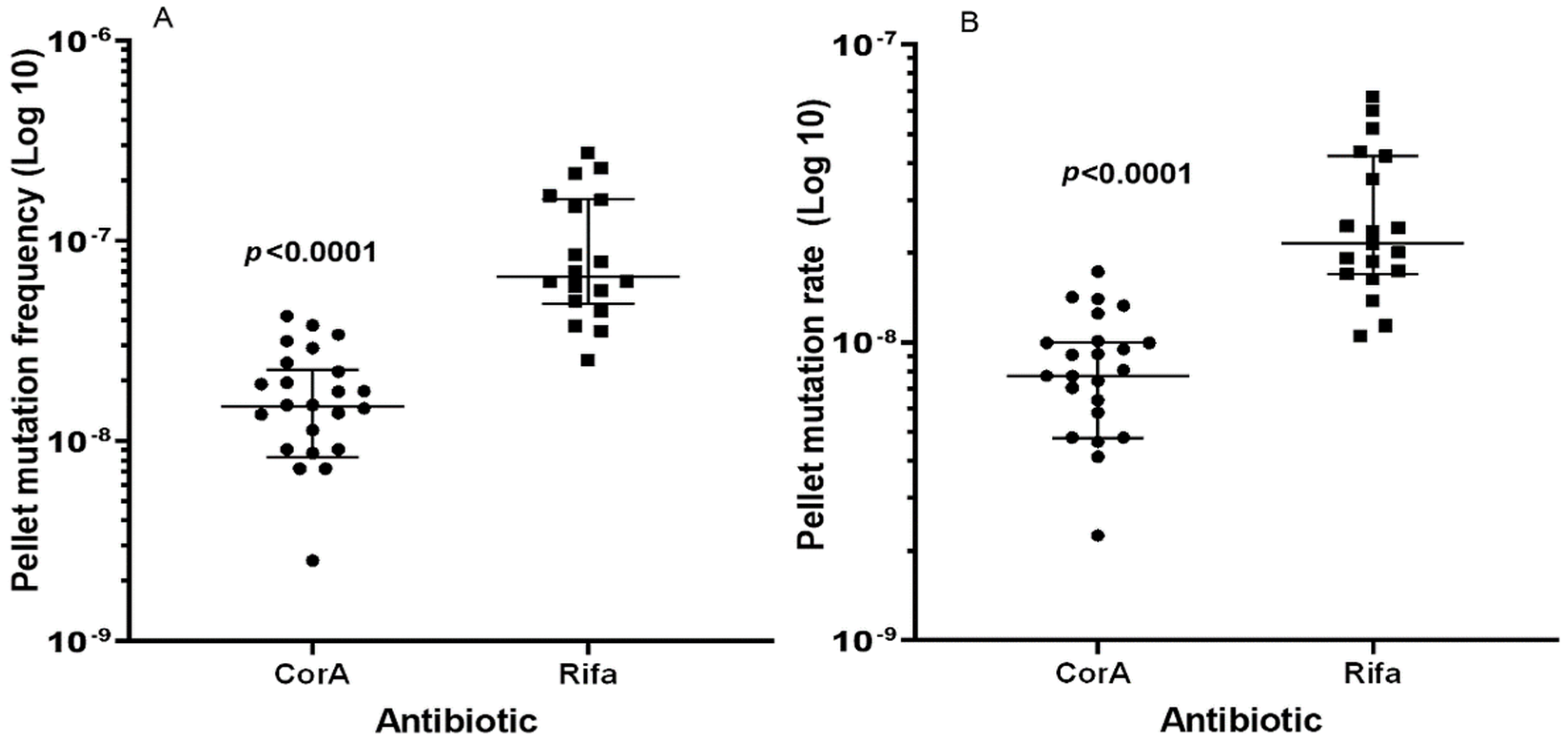
2.2. Determination of Mutation Frequency

CorA had an MIC of 0.25 mg/l against S. aureus strains HG001, Mu50, N315, and USA300 (Table 1). To determine the spontaneous FoM (resistant cells per total cells in the culture) leading to CorA resistance, forty parallel cultures were assayed on four different days on agar plates containing 1 mg/L CorA (4 × MIC). The FoM leading to resistance to rifampicin (MIC vs. S. aureus HG001, 0.0019 mg/L) was determined in parallel, using rifampicin (4 × MIC, 0.0076 mg/L) for the preparation of selective plates. Employing directly plated 100 µL culture aliquots, the percentage of agar plates that did not show the growth of resistant colonies was significantly higher for CorA (37.5%) than for rifampicin (2.9%) (p = 0.004, Fisher’s Exact Test). Therefore, 250 or 1000 µL of culture was pelleted, resuspended in 100 µL of buffer, and plated. Using this method, the median FoM to rifampicin resistance was 4.5-fold higher than that to CorA resistance (Table 2) and the differences between mutation frequencies were statistically significant (p < 0.0001, Mann–Whitney U-test) (Figure 1A; raw data sets are shown in Supplementary Tables S1 and S2).
The mutation rates (Table 2) were 2.8- and 4.2-fold higher for rifampicin than for CorA, according to the Lea–Coulson Method of the Median and the Ma–Sandri–Sarkar Maximum Likelihood Estimation (Figure 1B), respectively, and were also significantly different (p < 0.0001, Mann–Whitney U-test). The plating of 1.2 × 10¹¹ cells of S. aureus HG001 on Iso-Sensitest agar containing a combination of both agents, 4 × MIC of rifampicin and 4 × MIC of CorA, did not yield any growth after 24 h of incubation.

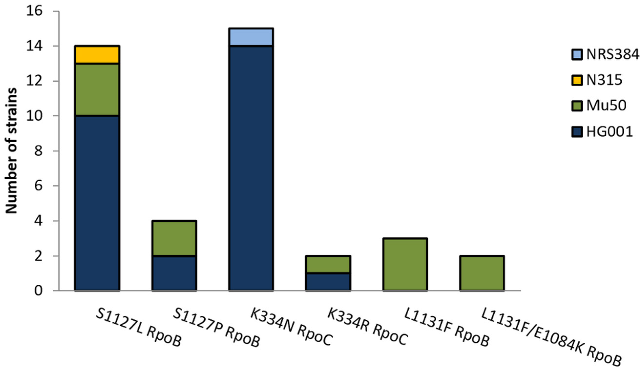
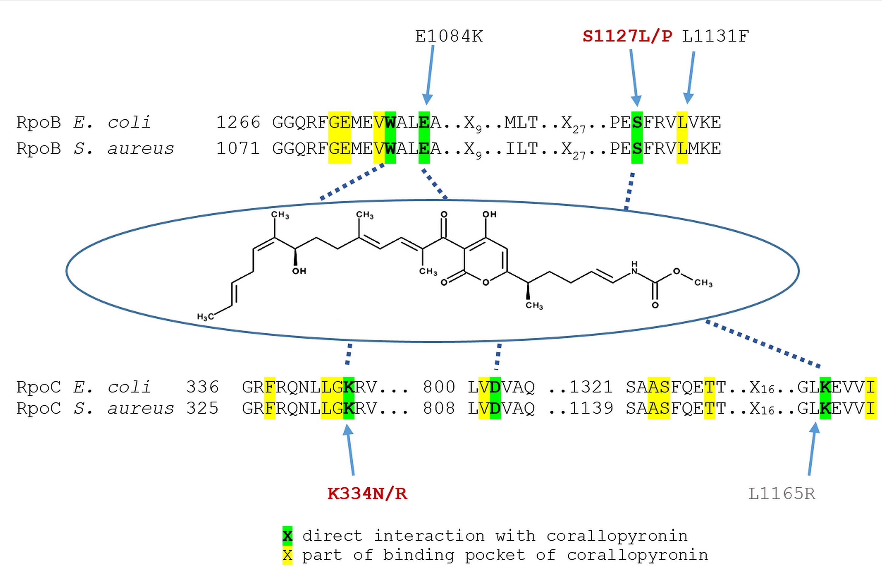
2.3. Mutations in CorA Resistant Strains

Twenty-seven resistant mutants from nine independent cultures of S. aureus HG001 were sequenced and were found to harbor exchanges in two mutational hotspots, i.e., position 1127 in RpoB (favoring the exchange S1127L) and position 334 in RpoC (favoring the exchange K334N) (Figure 2). Rifampicin-resistant strains have mutations in rpoB, which might influence the sites available for mutations to CorA resistance and the degree of resistance that can be reached. Therefore, resistance to CorA in a rifampicin-resistant background was tested using eight independent cultures of S. aureus Mu50, a clinical isolate that is resistant to methicillin and rifampicin and intermediately resistant to vancomycin. Eleven CorA-resistant colonies were sequenced. Here, additional exchanges of amino acids in RpoB at position L1131 and E1048 were found. Mutations were also sequenced in the clinically relevant, rifampicin-susceptible strains, S. aureus N315 and S. aureus USA300 NRS384, with exchanges in the same hotspots as in S. aureus HG001 (S1127 in RpoB and in K334 in RpoC). All sites that were affected in different strains are shown in Figure 2. The MICs of the clinical wildtype strains (Mu50, N315, and USA300 NRS384) were 0.25 mg/L CorA, whereas the MICs of resistant mutants ranged from 4 to 128 mg/L (Table 1).

3. Discussion

In a previous publication, FoM values of 7.2 × 10⁻⁸ ± 2.4 × 10⁻⁸ for resistance to CorA and of 1.4 × 10⁻⁷ ± 2.2 × 10⁻⁷ for resistance to rifampicin at 4 × MIC were determined, and the authors concluded that both FoMs were “comparable”. However, the exact methodologies of the experiment and calculations were not described [8]. In the current study, we repeated these experiments and took care to use agar plates with fresh CorA, because, as a neat active pharmaceutical ingredient, CorA is unstable and tends to isomerize at room temperature [11]. This resulted in significant differences in the median FoMs. The mutation rates of resistance to CorA were, depending on the method of calculation, 2.8-4.2-fold lower than those of resistance to rifampicin. In addition, 30% of the CorA plates that had received only 0.1 mL inoculum showed no growth, which was significantly higher than that for rifampicin plates. For the related antibiotic myxopyronin, mutation rates of 6 × 10⁻⁸ (calculated using the MSS-MLE method) were reported at 4 × MIC, which is higher than the rates of 6 × 10⁻⁹ to 9 × 10⁻⁹ obtained here using the same calculation method. Potentially unstable features have also been identified in myxopyronin. However, chemical modifications of these functional groups led to decreases in antibacterial activity [15,16].
The spontaneous mutation rates of growing bacterial cultures are strain-specific and constant in the absence of agents that influence the mutation rates. Therefore, the lower FoM and mutation rates measured with CorA might depend on a lower number of different mutations that convey resistance against CorA compared to rifampicin. For rifampicin, 16 different exchanges have been described in S. aureus 8325-4 after the plating of saturated cultures on agar containing 4 × MIC, and further mutations have been found in clinical isolates [17]. Saturation mutagenesis and a subsequent selection of mutants with CorA yielded seven different exchanges in six sites in E. coli. However, using myxopyronin, nine mutations were detected, which also increased CorA MICs eightfold or more [7]. In this study, the sequencing of 27 resistant mutants of S. aureus HG001 picked from nine different cultures yielded only four different exchanges targeting mainly two amino acids, S1127 in RpoB and K334 in RpoC, with S1127L RpoB and K334N RpoC being the most abundant exchanges (Figure 2). Identical exchanges were also present in the clinical strain and the community-associated MRSA (S. aureus N315 and S. aureus NRS384, respectively). We also tested the mutational changes in a rifampicin-resistant strain, S. aureus Mu50, an MRSA with intermediate susceptibility to vancomycin [18] that already harbors the H481Y exchange in RpoB [19]. Eleven sequenced resistant S. aureus Mu50 yielded two additional mutation sites at L1131 and E1084 in RpoB. Mariner et al. found exchanges in the same positions in eight sequenced mutants, including an additional exchange in L1165 [8]. In a similar study with myxopyronin, 11 different mutations from 17 independent mutants and a higher mutation rate in the range of rifampicin were reported [13]. Six of these eleven mutants, however, exhibited relatively low resistance levels (20 × MIC of the wildtype), comparable to that of the S. aureus Mu50 CSz1 mutant in this study. Therefore, it is possible that in these experiments relatively more mutants with a lower resistance level were able to survive the selection [13].
In conclusion, six possible amino acid positions were identified that yield resistance against CorA in S. aureus in direct selection experiments, which might explain the lower mutation rates compared to rifampicin. A stable formulation is now available [11]; therefore, with proper usage, e.g., in combination therapy and good antibiotic stewardship, CorA is a potential antibiotic for treating S. aureus infections. The efficacy of CorA as a chemotherapeutic agent in combination with different antibiotics for the treatment of staphylococcal disease will be investigated in the near future.

4. Materials and Methods

4.1. Strains

Staphylococcus aureus HG001, a derivative of the standard strain S. aureus NCTC 8325 (clonal complex (CC) 8) that has been repaired in rsbU [12], was used for the determination of mutation frequency and the characterization of mutants. S. aureus Mu50 (ATCC700699, rifampicin-resistant, methicillin-resistant (MRSA), vancomycin-intermediate susceptibility, CC5 [18]) was employed to test the influence of a rifampicin-resistant background on the generation of amino acid exchanges in RpoB and RpoC. The MICs and mutations of S. aureus USA300 (NRS384) (community-associated MRSA, source NARSA, CC8) and S. aureus N315 (NRS70, MRSA, rifampicin-susceptible, CC5 [18]) were also determined. Mutant strains are available from the authors.

4.2. Corallopyronin

CorA with a purity of >91% was produced by the Helmholtz Centre for Infection Research, Department of Microbial Drugs, using the heterologous producer strain Myxococcus xanthus carrying the CorA biosynthesis gene cluster, and was purified as reported previously [10].

4.3. Minimal Inhibitory Concentration

The determination of MICs was performed in Iso-Sensitest broth (Oxoid, Thermo Scientific, Waltham, MA, USA) with an inoculum of 1–5 × 10⁵ cells per ml, as specified by EUCAST [20]. Rifampicin and CorA stock solutions in DMSO (dimethyl sulfoxide) were stored in the dark at −20 °C. DMSO stock solutions were diluted with broth directly before the preparation of microtiter plates (1 µg/mL for rifampicin, 8 µg/mL for CorA as the final concentration in the stock solutions for susceptible strains and up to 512 µg/mL CorA for resistant strains). A solvent control of 12.8% of DSMO was included. Plates were read after 24 h of incubation at 37 °C.

4.4. Determination of Mutation Frequency and Mutation Rates

Iso-Sensitest agar (Oxoid, Thermo Scientific, Waltham, MA, USA) was prepared as advised by the manufacturer, autoclaved, and equilibrated at 50 °C before the addition of antibiotics (final concentration: 4 × MIC) for the preparation of selective agar plates. Agar plates (20 mL) were poured shortly before plating, wrapped in aluminum foil, and stored at 4 °C and never longer than for one week. All steps that involved the handling of selective agar plates were performed in the absence of artificial light, since both antibiotics are sensitive to light. For preculture, 5 mL broth were inoculated with one colony of the test strain S. aureus HG0001 from a fresh Columbia blood agar plate and incubated overnight. In the morning, 9–15 main cultures (9.9 mL Iso-Sensitest broth in a 100 mL Erlenmeyer flask) were inoculated with 0.1 mL preculture and incubated at 37 °C and 170 rpm until the cultures had reached an optical density OD600 of 1. Then, 1 mL of a culture of OD 1 (or 0.25 mL for most agar plates with rifampicin) was centrifuged (8000 rpm, 3 min) and the pellets were resuspended in 100 µL of broth. An aliquot of 100 µL of this resuspended pellet and 100 µL of undiluted culture were plated onto selective plates with CorA (1 µg/mL) and rifampicin (0.076 µg/mL). In addition, serial tenfold dilutions of the cultures were plated onto Iso-Sensitest agar without antibiotics for the determination of colony forming units (CFU). CFU were counted after 24 h of incubation at 37 °C in the dark. All culture plates that did not yield colony smears were evaluated, i.e., for CorA 26 of 40 agar plates with plated pellets were evaluated. For rifampicin, 34 main cultures were tested and it was possible to evaluate 19 of 34 plated pellets.
A two-tailed Mann–Whitney-U-Test was used for the statistical comparison of the median mutation frequencies and median mutation rates. Fisher’s exact test was employed for the comparison of agar plates without growth (GraphPad Prims version 9.2 for Windows, GraphPad Prism Software, San Diego, CA, USA). Mutation rates were analyzed using the FALCOR Fluctuation AnaLysis CalculatOR (https://lianglab.brocku.ca/FALCOR/, accessed on 18 March 2022) [14].
For the determination of FoM against a combination of both agents, rifampicin and CorA, fresh agar plates with 4 × MIC of both antibiotics were prepared two days before plating. Eight different overnight cultures were grown from eight different colonies of S. aureus HG001 in 5 mL of Iso-Sensitest broth. A total of 4.9 mL of culture was pelleted, and the cells were resuspended in 500 µL of Iso-Sensitest broth. These suspensions were plated in 150 µL aliquots on agar plates containing the combination of both antibiotics. For the determination of the total cell counts, a series of tenfold dilutions was plated on Iso-Sensitest agar without antibiotics. All agar plates were incubated at 37 °C and evaluated after 24 h.

4.5. Identification of Mutations in rpoB and rpoC

Resistant colonies were selected on agar with CorA (4 × MIC) employing 19 independent cultures (nine cultures of S. aureus HG001 (rifampicin-susceptible), eight cultures of S. aureus Mu50 (rifampicin-resistant), and one culture each of S. aureus USA300 and S. aureus N315 (clinical isolates that are rifampicin-susceptible)). Resistant colonies were picked and purified by streaking on Iso-Sensitest agar (4 × MIC CorA). For the DNA preparation, strains were grown overnight on 4 × MIC CorA-containing agar, and two to three colonies were resuspended in 60 µL of sterile distilled water and heated for 5 min to 100 °C. Cells were removed by centrifugation and the supernatant served as a template for the amplification of rpoA, rpoB, and rpoC (for primers see Table 3). S. aureus Mu50 DNA was purified from 1.5 mL of culture using the Molzym Presto-SpinD Bug Kit, including an additional lysis step with 1 mg/mL lysostaphin in RS buffer at 37 °C, 1 h. After agarose gel electrophoresis, bands with PCR products were excised and purified using the Monarch PCR & DNA Cleanup Kit (New England Biolabs, Frankfurt, Germany). Sanger sequencing was performed by Microsynth Seqlab (Göttingen, Germany) using the primers in Table 3. Sequences were evaluated with the Geneious software (Geneious R10 [21]).

Supplementary Materials

The following are available online at https://www.mdpi.com/article/10.3390/antibiotics11070920/s1, Table S1: Raw data set for the determination of mutation frequency and mutation rates shown in Table 2 in the manuscript using 1 mL of culture. Table S2: Raw data set for the determination of mutation frequency and mutation rates shown in Table 2 in the manuscript using 0.1 mL of culture, Figure S1: Decrease in the CorA isomer in agar plates during storage at different temperatures.

Author Contributions

Conceptualization, G.B.; Data curation, G.B. and K.P.; Funding acquisition, K.P. and A.H.; Investigation, J.B., C.S., S.K., T.A., M.G. and M.S.; Resources, M.G., R.J., G.M.K. and M.S.; Supervision, G.B.; Validation, K.P.; Writing—original draft, G.B.; Writing—review and editing, G.B., K.P., T.H., A.S. and A.H. All authors have read and agreed to the published version of the manuscript.

Funding

This research was funded by the German Center for Infection Research (DZIF). Translational Thematic Unit: Novel Antibiotics, grant numbers #09.807 and #09.806 to A.H., and the German Federal Ministry of Education and Research (BMBF) grant number 16GW0227K to M.S.

Institutional Review Board Statement

Not applicable.

Informed Consent Statement

Not applicable.

Data Availability Statement

All data are contained within the article or Supplementary Material.

Acknowledgments

Expert technical assistance from Kerstin Schober and Silke Reinecke is gratefully acknowledged.

Conflicts of Interest

The authors declare no conflict of interest. The funders had no role in the design of the study; in the collection, analysis, or interpretation of data; in the writing of the manuscript; or in the decision to publish the results.

References

  1. Irschik, H.; Jansen, R.; Höfle, G.; Gerth, K.; Reichenbach, H. The corallopyronins, new inhibitors of bacterial RNA synthesis from Myxobacteria. J. Antibiot. 1985, 38, 145–152. [Google Scholar] [CrossRef] [PubMed] [Green Version]
  2. Loeper, N.; Graspeuntner, S.; Ledig, S.; Kaufhold, I.; Hoellen, F.; Schiefer, A.; Henrichfreise, B.; Pfarr, K.; Hoerauf, A.; Shima, K.; et al. Elaborations on Corallopyronin A as a novel treatment strategy against genital chlamydial infections. Front. Microbiol. 2019, 10, 943. [Google Scholar] [CrossRef] [PubMed] [Green Version]
  3. Schiefer, A.; Schmitz, A.; Schäberle, T.F.; Specht, S.; Lämmer, C.; Johnston, K.L.; Vassylyev, D.G.; König, G.M.; Hoerauf, A.; Pfarr, K. Corallopyronin A specifically targets and depletes essential obligate Wolbachia endobacteria from filarial nematodes in vivo. J. Infect. Dis. 2012, 206, 249–257. [Google Scholar] [CrossRef] [PubMed] [Green Version]
  4. Kock, F.; Hauptmann, M.; Osterloh, A.; Schäberle, T.F.; Poppert, S.; Frickmann, H.; Menzel, K.-D.; Peschel, G.; Pfarr, K.; Schiefer, A.; et al. Orientia tsutsugamushi is highly susceptible to the RNA polymerase switch region inhibitor Corallopyronin A in vitro and in vivo. Antimicrob. Agents Chemother. 2018, 62, e01732-17. [Google Scholar] [CrossRef] [PubMed] [Green Version]
  5. Schiefer, A.; Hübner, M.P.; Krome, A.; Lämmer, C.; Ehrens, A.; Aden, T.; Koschel, M.; Neufeld, H.; Chaverra-Muñoz, L.; Jansen, R.; et al. Corallopyronin A for short-course anti-wolbachial, macrofilaricidal treatment of filarial infections. PLoS Negl. Trop. Dis. 2020, 14, e0008930. [Google Scholar] [CrossRef] [PubMed]
  6. Belogurov, G.A.; Vassylyeva, M.N.; Sevostyanova, A.; Appleman, J.R.; Xiang, A.X.; Lira, R.; Webber, S.E.; Klyuyev, S.; Nudler, E.; Artsimovitch, I.; et al. Transcription inactivation through local refolding of the RNA polymerase structure. Nature 2009, 457, 332–335. [Google Scholar] [CrossRef] [PubMed] [Green Version]
  7. Mukhopadhyay, J.; Das, K.; Ismail, S.; Koppstein, D.; Jang, M.; Hudson, B.; Sarafianos, S.; Tuske, S.; Patel, J.; Jansen, R.; et al. The RNA polymerase “switch region” is a target for inhibitors. Cell 2008, 135, 295–307. [Google Scholar] [CrossRef] [PubMed] [Green Version]
  8. Mariner, K.; McPhillie, M.; Trowbridge, R.; Smith, C.; O’Neill, A.J.; Fishwick, C.W.G.; Chopra, I. Activity of and development of resistance to corallopyronin A, an inhibitor of RNA polymerase. Antimicrob. Agents Chemother. 2011, 55, 2413–2416. [Google Scholar] [CrossRef] [PubMed] [Green Version]
  9. Schäberle, T.F.; Schmitz, A.; Zocher, G.; Schiefer, A.; Kehraus, S.; Neu, E.; Roth, M.; Vassylyev, D.G.; Stehle, T.; Bierbaum, G.; et al. Insights into structure-activity relationships of bacterial RNA polymerase inhibiting Corallopyronin derivatives. J. Nat. Prod. 2015, 78, 2505–2509. [Google Scholar] [CrossRef] [PubMed]
  10. Pogorevc, D.; Panter, F.; Schillinger, C.; Jansen, R.; Wenzel, S.C.; Müller, R. Production optimization and biosynthesis revision of corallopyronin A, a potent anti-filarial antibiotic. Metab. Eng. 2019, 55, 201–211. [Google Scholar] [CrossRef] [PubMed]
  11. Krome, A.K.; Becker, T.; Kehraus, S.; Schiefer, A.; Steinebach, C.; Aden, T.; Frohberger, S.J.; López Mármol, Á.; Kapote, D.; Jansen, R.; et al. Solubility and stability enhanced oral formulations for the anti-infective Corallopyronin A. Pharmaceutics 2020, 12, 1105. [Google Scholar] [CrossRef] [PubMed]
  12. Herbert, S.; Ziebandt, A.-K.; Ohlsen, K.; Schäfer, T.; Hecker, M.; Albrecht, D.; Novick, R.; Götz, F. Repair of global regulators in Staphylococcus aureus 8325 and comparative analysis with other clinical isolates. Infect. Immun. 2010, 78, 2877–2889. [Google Scholar] [CrossRef] [PubMed] [Green Version]
  13. Srivastava, A.; Degen, D.; Ebright, Y.W.; Ebright, R.H. Frequency, spectrum, and nonzero fitness costs of resistance to myxopyronin in Staphylococcus aureus. Antimicrob. Agents Chemother. 2012, 56, 6250–6255. [Google Scholar] [CrossRef] [Green Version]
  14. Hall, B.M.; Ma, C.-X.; Liang, P.; Singh, K.K. Fluctuation analysis CalculatOR: A web tool for the determination of mutation rate using Luria-Delbruck fluctuation analysis. Bioinformatics 2009, 25, 1564–1565. [Google Scholar] [CrossRef] [Green Version]
  15. Doundoulakis, T.; Xiang, A.X.; Lira, R.; Agrios, K.A.; Webber, S.E.; Sisson, W.; Aust, R.M.; Shah, A.M.; Showalter, R.E.; Appleman, J.R.; et al. Myxopyronin B analogs as inhibitors of RNA polymerase, synthesis and biological evaluation. Bioorg. Med. Chem. Lett. 2004, 14, 5667–5672. [Google Scholar] [CrossRef]
  16. Lira, R.; Xiang, A.X.; Doundoulakis, T.; Biller, W.T.; Agrios, K.A.; Simonsen, K.B.; Webber, S.E.; Sisson, W.; Aust, R.M.; Shah, A.M.; et al. Syntheses of novel myxopyronin B analogs as potential inhibitors of bacterial RNA polymerase. Bioorg. Med. Chem. Lett. 2007, 17, 6797–6800. [Google Scholar] [CrossRef]
  17. O’Neill, A.J.; Huovinen, T.; Fishwick, C.W.G.; Chopra, I. Molecular genetic and structural modeling studies of Staphylococcus aureus RNA polymerase and the fitness of rifampin resistance genotypes in relation to clinical prevalence. Antimicrob. Agents Chemother. 2006, 50, 298–309. [Google Scholar] [CrossRef] [PubMed] [Green Version]
  18. Kuroda, M.; Ohta, T.; Uchiyama, I.; Baba, T.; Yuzawa, H.; Kobayashi, I.; Cui, L.; Oguchi, A.; Aoki, K.; Nagai, Y.; et al. Whole genome sequencing of meticillin-resistant Staphylococcus aureus. Lancet 2001, 357, 1225–1240. [Google Scholar] [CrossRef]
  19. Katayama, Y.; Sekine, M.; Hishinuma, T.; Aiba, Y.; Hiramatsu, K. Complete reconstitution of the vancomycin-intermediate Staphylococcus aureus phenotype of strain Mu50 in vancomycin-susceptible S. aureus. Antimicrob. Agents Chemother. 2016, 60, 3730–3742. [Google Scholar] [CrossRef] [PubMed] [Green Version]
  20. The European Committee on Antimicrobial Susceptibility Testing (EUCAST). Breakpoint Tables for Intrepretation of MICs and Zone Diameters. Version 12.0. 2022. Available online: http://www.eucast.org (accessed on 23 June 2022).
  21. Kearse, M.; Moir, R.; Wilson, A.; Stones-Havas, S.; Cheung, M.; Sturrock, S.; Buxton, S.; Cooper, A.; Markowitz, S.; Duran, C.; et al. Geneious Basic: An integrated and extendable desktop software platform for the organization and analysis of sequence data. Bioinformatics 2012, 28, 1647–1649. [Google Scholar] [CrossRef] [PubMed]
Figure 1. Spontaneous mutations leading to resistance against CorA and Rifa using plated pellets. (A) FoM to CorA and Rifa resistance, (B) corresponding mutation rates calculated using the Lea–Coulson Method of the Median in FALCOR [14]. Three CorA plates did not show growth and a value of zero cannot be depicted using a logarithmic axis, but these values were included in the calculation of the median values and significance. P-values were computed by the Mann–Whitney-U-Test (GraphPad Prism 9.4 for Windows, GraphPad Prism Software, San Diego, CA, USA). The whiskers indicate 25 and 75 percentiles.
Antibiotics 11 00920 g001
Figure 2. Statistics of amino acid exchanges identified in RpoB and RpoC in 40 CorA-resistant S. aureus mutants with different strain and resistance backgrounds (S. aureus HG001, N315, Mu50, and NRS384).
Antibiotics 11 00920 g002
Table 1. CorA MICs for different S. aureus strains and their resistant mutants.
StrainExchange in Resistant MutantCorA MIC
S. aureus HG001Wildtype0.25 mg/L
S. aureus HG001CSz1K334N RpoC64 mg/L
S. aureus Mu50Wildtype0.25 mg/L
S. aureus Mu50CSz1L1131F RpoB4 mg/L
S. aureus Mu50R1.1S1127P RpoB32 mg/L
S. aureus Mu50R3.1L1131F/E1084K RpoB128 mg/L
S. aureus Mu50R4.1S1127L RpoC64 mg/L
S. aureus Mu50R5.1K334R RpoC64 mg/L
S. aureus N315Wildtype0.25 mg/L
S. aureus N315CSz1S1127L RpoB64 mg/L
S. aureus USA300Wildtype0.25 mg/L
S. aureus USA300CSz1K334N RpoC128 mg/L
Table 2. Frequency of mutations (FoM) leading to resistance and mutation rates of S. aureus selected on 4 × MIC CorA or rifampicin using 1 mL or 0.1 mL of culture and two different methods (LC and MSS-MLE) for the calculation of the mutation rates.
1 mL Culture on CorA Agar
(+/− 95% CI 1)
1 mL Culture on Rifa Agar
(+/− 95% CI)
Ratio Rifa/CorA
Median of FoM1.48 × 10−8 (4.7 × 10⁻⁹, 5.7 × 10⁻⁹)6.67 × 10⁻⁸ (8.2 × 10⁻⁸, 1.0 × 10⁻⁸)4.5
Mutation rate (LC) 27.72 × 10⁻⁹ (2.3 × 10⁻⁹, 2.9 × 10⁻⁹)2.15 × 10⁻⁸ (1.4 × 10⁻⁸, 4.1 × 10⁻⁹)2.8
Mutation rate (MSS-MLE) 35.93 × 10⁻⁹ (2.0 × 10⁻⁹, 1.79 × 10⁻⁹)2.49 × 10⁻⁸ (7.3 × 10⁻⁹, 6.6 × 10⁻⁹)4.2
0.1 mL culture on CorA agar (+/− 95% CI)0.1 mL culture on rifampicin agar (+/− 95% CI)Ratio Rifa/CorA
Median of FoM2.27 × 10−8 (1.5 × 10⁻⁷, 2.3 × 10⁻⁷)1.27 × 10⁻⁷ (1.0 × 10⁻⁷, 4.0 × 10⁻⁸)5.6
Mutation rate (LC) 28.77 × 10⁻⁹ (5.8 × 10⁻⁹, 8.8 × 10⁻⁹)3.49 × 10⁻⁸ (2.0 × 10⁻⁸, 9.0 × 10⁻⁹)4.0
Mutation rate (MSS-MLE) 39.5 × 10⁻⁹ (3.1 × 10⁻⁹, 2.8 × 10⁻⁹)3.51 × 10⁻⁸ (8.6 × 10⁻⁹, 7.8 × 10⁻⁹)3.7
1 CI: 95% Confidence intervals of the medians (+/−). 2 LC: Lea–Coulson Method of the Median. 3 MSS-MLE: Ma–Sandri–Sarkar Maximum Likelihood Estimator Method.
Table 3. Primers used for the PCR amplification and sequencing of RNAP.
PrimerGeneSequenceRef.
rpoAIrpoA5′-TAACTGCGATCAGAGACGTTACTCC-3′[17]
rpoAIIrpoA5′-GCTGCATTACGACGAGAAGCTAAAT-3′[17]
rpoBFrpoB5′-ATTAGTGTTGCCGTTTTCTTTT-3′[17]
rpoBRrpoB5′-AGTATCTTTTTGCCTGTTTTG-3′[17]
rpoCIrpoC5′-GACGATGATGTTGTAGAACGCAAAG-3′[17]
rpoCIIrpoC5′-TGTTGTTTGTTAAAGCGTGCAACTT-3′[17]
rpoCfor2rpoC5′-GGACGATTTGCAACAAGTGA-3′This study
rpoCrev2rpoC5′-CCAACTGCTTCACCAACTTC-3′This study
F3rpoB5′-AGTCTATCACACCTCAACAA-3′This study
F4rpoB5′-TAATAGCCGCACCAGAATCA-3′This study
2955FrpoB5′-GATCGATATCATGTTAAATCCTCTTGGTGTACCATC-3′This study
3824RrpoB5′-CTATTTTCATTAATGGAAATTATTT-ACATCAATCA-3′This study
955FrpoB5′-ACTGAAACTGGTGAAATTGTAGTTG-3′This study
2706RrpoB5′-GTGAGGTACACGTAATGAAGTATCT-3′This study
653FrpoC5′-TTTTGATGTACTTCCAATCATCCCACCAGAAATTC-3′This study
1552RrpoC5′-CATATGCTTTTAATACTTCATTTGTATTATTAAAGA-3′This study
Publisher’s Note: MDPI stays neutral with regard to jurisdictional claims in published maps and institutional affiliations.

