Review

Does Chlorination Promote Antimicrobial Resistance in Waterborne Pathogens? Mechanistic Insight into Co-Resistance and Its Implication for Public Health

by
Martins A. Adefisoye
* and
Ademola O. Olaniran
Discipline of Microbiology, School of Life Sciences, College of Agriculture, Engineering and Science, Westville Campus, University of KwaZulu-Natal, Private Bag X54001, Durban 4000, South Africa
*
Author to whom correspondence should be addressed.
Antibiotics 2022, 11(5), 564; https://doi.org/10.3390/antibiotics11050564
Submission received: 31 March 2022 / Revised: 18 April 2022 / Accepted: 20 April 2022 / Published: 22 April 2022

Abstract

:
Chemical agents including chlorine and antibiotics are used extensively to control infectious microorganisms. While antibiotics are mainly used to treat bacterial infections, chlorine is widely used for microbial inactivation in the post-secondary disinfection steps of water treatment. The extensive use of these agents has been acknowledged as a driving force for the expansion of antimicrobial resistance (AMR) and has prompted discourse on their roles in the evolution and proliferation of resistant pathogens in the aquatic milieus. We live in a possible “post-antibiotic” era when resistant microbes spread at startling levels with dire predictions relating to a potential lack of effective therapeutic antibacterial drugs. There have been reports of enhancement of resistance among some waterborne pathogens due to chlorination. In this context, it is pertinent to investigate the various factors and mechanisms underlying the emergence and spread of resistance and the possible association between chlorination and AMR. We, therefore, reflect on the specifics of bacterial resistance development, the mechanisms of intrinsic and acquired resistance with emphasis on their environmental and public health implications, the co-selection for antibiotic resistance due to chlorination, biofilm microbiology, and multidrug efflux activity. In-depth knowledge of the molecular basis of resistance development in bacteria will significantly contribute to the more rational utilization of these biocidal agents and aid in filling identified knowledge gap toward curbing resistance expansion.

1. Introduction

Antimicrobial-resistant waterborne pathogens are a major global health concern. There are rising reports of resistance selection in microorganisms due to inappropriate use of antimicrobial agents [1,2]. Upon antibiotic usage, a small portion of the microbial cells, even <10−9 within a susceptible wild-type population, is not affected by the antimicrobial agent. This subgroup retains antimicrobial impeding mutations, which favor their selection during treatment. Thus, antibiotic resistance is known to emerge during the frame of a selective compartment described as a mutant selection window (MSW) [3].
The MSW hypothesis was initially proposed based on a study investigating resistance to quinolones in Gram-negative bacteria in the 1990s [4]. The study noted the existence of a “dangerous” concentration range where mutant selection occurs most frequently and suggested that resistance prevention against norfloxacin in strains carrying a “first mutation” should employ older quinolones and emphasizes the need for early detection of such strains [5]. Accordingly, the MSW postulates that, for every antimicrobial drug-microorganisms combination, there exists a drug concentration range where resistant-mutants selective amplification occurs in a single step, leading to reduced susceptibility [6]. This range extends from the minimum inhibitory concentration of the drug, which prevents colony growth by 99% (MIC99), to the mutant prevention concentration (MPC), at which the least-susceptible single-step mutant subpopulation is inhibited. Within these limits, selective enrichment of resistant mutants frequently occurs [7]. Upon mutation acquisition, the MIC99 and the MPC increase, shifting up the MSW of an antimicrobial and making it more challenging to prevent the emergence of new mutant strains [8].
Some reports have documented correlations between antibiotics and chlorine resistance from aquatic environments [9,10], while other studies have reported the co-selection for antibiotic resistance among bacteria due to chlorination [11,12]. Although chlorination has proved efficient in reducing waterborne diseases, little effort has been devoted to exploring the potential impacts of chlorine disinfection in enhancing antibiotic resistance genes (ARGs). The apprehension about the contribution of chlorination to an upsurge of AMR in waterborne pathogens presents a potential threat to human health and the aquatic ecosystem. Albeit, the reasons for this phenomenon and the factors triggering AMR expansion during chlorination remain poorly understood [13].
To this end, this review describes the implications of intrinsic and acquired bacterial resistance with emphasis on aquatic and human health, connections between antibiotics and chlorine resistance, the roles of microbial biofilms and multidrug efflux activity in AMR, and identifies knowledge gap in the existing literature on the molecular biology of AMR. A comprehensive understanding of the molecular basis of resistance development in bacteria will significantly contribute to these agents’ more rational selection and utilization toward curbing AMR proliferation and advancing the global antimicrobial stewardship effort.

2. Water Quality and Human Health

Easy access to improved water sources is vital to human health and development. Contaminated water predisposes individuals to hazards and has been linked to disease outbreaks. Despite progress in global access to improved water quality in recent years, over two billion people still lack access to safely managed water sources [14]. Climate change, rising human population and urbanization pose challenge for water supplies and important strategies such as wastewater recycling is becoming vital in water reclamation. Inadequately treated recycled wastewater poses an unacceptable risk of infectious disease and a threat to public health [15].
Concerns about the microbiological quality of water have previously focused on the occurrence of pathogens. However, the prevalence of antimicrobial-resistant bacteria (ARB) and resistance genes in water sources is an emerging issue of public health globally [16]. Different studies have presented varying opinions on the selection or enrichment of resistant strains during chlorination [10,13]. Jin et al. [13] reported that chlorination promotes the emergence and exchange of resistance genes across bacterial genera. Murray [17] observed a significant increase in the level of multiple drug-resistant (MDR) bacteria in sewage, after treatment with chlorine, and during a laboratory-scale chlorination investigation. Contrarily, other studies have suggested that disinfection does not contribute to resistant strain selection but rather induces the development of antimicrobial resistance [18,19]. Another study yet has noted that the selection of stress-tolerant strains by chlorination might lead to more antimicrobial resistance [9].

3. Fundamentals of Chlorine Disinfection

Public water supply chlorination is one of the most significant advancements of the last century. Chlorine-based disinfectants have proven highly effective, safe, economical and widely accepted. Water hydrolysis, upon the addition of chorine, produces hypochlorous acid (HOCl), which dissociates yielding hydrogen and hypochlorite ions (Equations (1) and (2)).
Chlorine disinfection chemistry:
C l 2 + H 2 O   H O C l + H + +   C l
H O C l     H + +   O C l
Chlorine first reacts with organic and inorganic matters before pathogens inactivation. The amount of chlorine consumed in this process is termed chlorine demand. The combined chlorine that forms together with any free available chlorine in the water is called the chlorine residual (Figure 1). This is the component of the added chlorine that disinfects the water. Free available chlorine is formed by differences in the concentrations of hypochlorous and hypochlorite ions, a process that depends on the pH of the water [20]. Even though the chlorination procedure is well-researched, establishing an appropriate chlorine dose remains a difficult task for many field applications. Nevertheless, the effective chlorine dose should be sufficient to destroy pathogens and oxidize the organic contaminants as well as maintain sufficient free available chlorine in the water distribution system, post-chlorination [21].

3.1. Breakpoint Chlorination and Factors Influencing Disinfection

Breakpoint chlorination is the continuous addition of chlorine until the chlorine demand is satisfied, combined chlorine compounds are oxidized and only free chlorine remains. The four phases involved in breakpoint chlorination is depicted in Figure 2.
Complete oxidation of chlorine occurs in the first phase due to low chlorine concentration and lack of free chlorine residual. The phase is also known as the initial chlorine demand. Combined chlorine residual is formed between Cl and NH4+ in the second phase. The concentration of the added chlorine in this phase is proportional to the total concentration of chlorine residual. Continuous addition of chlorine in the third phase causes oxidation of the combined residues causing a reduced concentration of the residual chlorine up to “dip” or “breakpoint”. Beyond this point, the concentration of the residual chlorine increases [22].
Temperature, pH, contact time, density and the inherent nature of microorganisms present determine the efficacy of the chlorination. Keeping all other factors constant, higher microbial load will demand higher chlorine dose and contact time. The physiological state of the microbes plays an important role in their chlorine sensitivity [13]. Generally, autochthonous microbes in natural water environments are more resistant to chlorination than laboratory-grown strains.

3.2. Mechanisms of Chlorine Disinfection

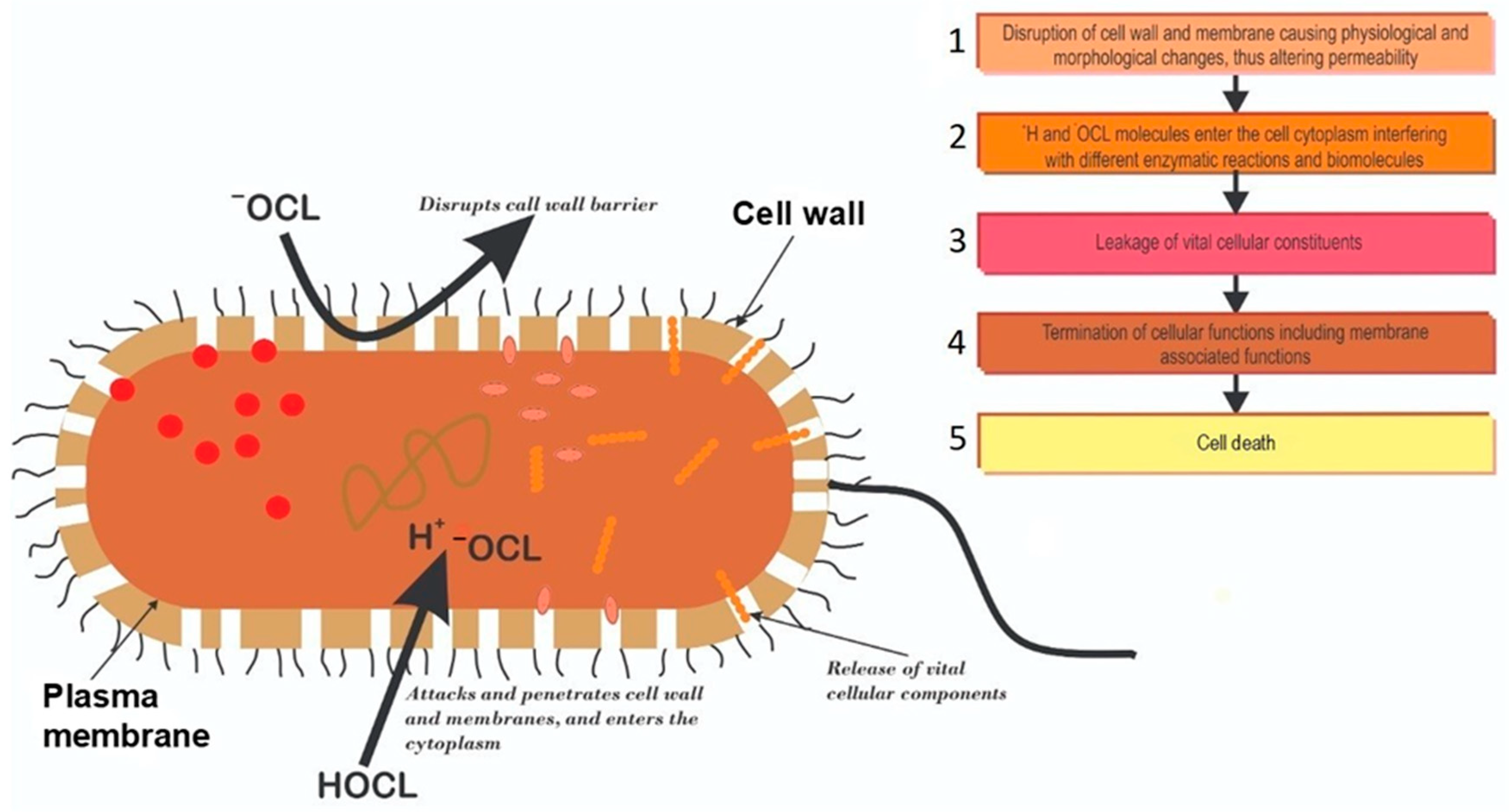
The biocidal mechanism of chlorination remains poorly understood. Some investigators opined that HOCl and OCl formed when chlorine is added to water destroy microbial macromolecules. Further work on this supposition led to the “multiple hit” concept, which asserts that bacterial kill by chlorination is probably due to the attack and damage of microbial biomolecules [23]. Venkobachar et al. [24] and Haas [25] reported leakage of cellular macromolecules when bacteria were treated with chlorine. Venkobachar [24] also observed that chlorine significantly inhibits oxidative phosphorylation and the uptake of oxygen, and that, the inhibition of respiratory enzyme was responsible for the phosphorylation inhibition phenomenon, rather than a deficiency in phosphate uptake. In a different study, Chang [26] suggested that the extensive destruction of bacterial enzymatic systems is responsible for the rapid destruction of pathogens during chlorination. Various other investigators have also recognized that chlorine destroys microorganisms through changes in membrane permeability, nucleic acid damage and leakage of intracellular biomolecules [27,28]. Generally, it appears that chlorination causes physiological and morphological changes to the bacterial cell wall, altering its permeability. The chlorine molecules subsequently enter the cytoplasm, interfering with different enzymatic reactions, in a possible event cascade depicted in Figure 3.

3.3. Incidences of Chlorine Tolerant Microorganisms from Treated Water Sources

Various international and local authorities have regulations to ensure the protection of water sources from chemical and microbiological contaminants. At a global level, for instance, the WHO recommends chlorine residual of 0.2 to 0.5 mg/L in water supplies [29], while the South African Department of Water and Sanitation stipulates chlorine residual of 0.25 mg/L for discharged wastewater effluents released into surface water environment [30,31]. Although some concerns have emerged in recent years about the limitations of chlorine usage in water treatments, there is no doubt, however, that chlorination remains one of the most significant advances in water purification and public health protection [32]. Reports on chlorine surviving microorganisms from chlorinated water sources continue to emanate [33,34,35]. Various investigators have proposed different mechanisms by which microorganisms may develop tolerance to disinfectants. These include (i) cell surface modifications or encapsulation [36], (ii) attachment to surfaces or suspended particulate matter [37], (iii) microbial aggregation in biofilms [38], (iv) expression of multidrug efflux pump activity [9] and (v) spore formation [39]. These mechanisms conceivably favor the survival of opportunistic waterborne pathogens, thereby contributing to their persistence in water [40]. Table 1 lists some published studies on chlorine-resistant microorganisms from different sources and their reported mechanism of resistance.

4. Major Drivers of Antimicrobial Resistance (AMR) in Aquatic Environments

The prevalence of resistance genes in the aquatic environments is the result of a complex interplay of numerous factors propelling evolution by natural selections and variations, through cascades of mutational events. The buildup of chemical pollutants including antimicrobials, disinfectants, heavy metals, detergents, pharmaceuticals and xenobiotics contribute to the evolution and spread of resistance in the aquatic environment. Huge selective pressure resulting from anthropogenic activities escalates and enriches resistance determinants in microbial populations [51,52]. Efforts to reduce resistance development would include limiting the amount of resistant microbial loads as well as optimization of disinfection protocols in water treatment. It has been argued that AMR action plans may not achieve their anticipated objectives if all AMR drivers and pathways in the environment are not appropriately appraised.

4.1. Stepwise Accumulation of Drug Resistance Mutations

During antimicrobial administration, the application of doses within an MSW can allow the enrichment of a mutant fraction of the microbial population resulting in increased genetic and phenotypic resistance [53]. Genetic resistance is generally classified as acquired or de novo. Acquired resistance often occurs in a single step through the incorporation of resistance determinants frequently borne on mobile genetic elements from an external source. De novo resistance, on the other hand, arises either as a single step or as an accumulation of a series of mutations that discretely reduce susceptibility by a modest increment in a step-by-step manner (e.g., intermediate susceptibility). Usually, the next mutational step is more readily achieved with the occurrence of a previous mutation in a strain, since numerous alleles can accumulate [54]. In the stepwise process, a microbial cell is only considered resistant due to the presence of more than one genetic mutation [55]. This typical example is seen in Streptococcus pneumonia in the development of resistance against fluoroquinolones, where at least seven different alleles are identified [56].

4.2. Contribution of Chlorination to AMR Expansion via Horizontal Gene Transfer

Although chlorination is a vital step in water treatment, its contribution to the expansion and dissemination of AMR has been noted. Increased natural transformation rates were observed across different bacterial genera through the transfer of antimicrobial resistance genes (ARGs) and other mobile genetic elements (MGEs), leading to the enrichment of ARG after chlorine treatment [13]. The study also noted that the release of naked DNA (including plasmids, ARGs and integrons) from “killed” donor bacteria during the chlorination process contributed to an increased natural transformation of chlorine-injured-but-culturable strains at a rate >550 folds in comparison to untreated bacteria. In a related study, Guo and colleagues [57] reported that low chlorine doses ranging between 5 and 40 mg Cl min/L enhanced the rate of conjugative transfer of ARGs by factors between 2 and 5 due to increased induction of pilus on the surface of conjugative bacterial cells, facilitating the exchange of genetic materials. They, however, noted that high chlorine doses (>80 mg Cl min/L) significantly suppressed the rate of ARG transfer. In another study, a retrieval of chlorination-driven enrichment of ARGs between 18.1 and 102% was observed through metagenomics binning strategies [58]. In general, horizontal gene transfer plays a significant role in AMR expansion during water chlorination.

5. Association between AMR and Disinfectant Resistance in Microorganisms

Putative resistance factors are thought to have existed in the natural environment, primarily for other functions apart from resistance conferment. However, the versatility of microbial pathogens has benefitted by recruiting and incorporating those genes into their own genome to evade the biocide effect of antimicrobials [59]. It should be noted that AMR genes are concurrently found with other genes that promote resistance to varieties of potentially harmful chemicals, including disinfectants in bacterial strains. The unfortunate historical convention, however, is the labelling of all these AMR genes; whereas, in actual fact, they can potentially promote resistance against different classes of chemicals, a phenomenon known as co-selection [60]. Co-selection of resistance has serious health implications and occurs either as co-resistance or cross-resistance. Cross-resistance allows a single resistance gene to foster protection against multiple biocides while co-resistance allows one gene to promote the maintenance of another resistance gene. Co-resistance phenomenon is exemplified as a “toolbox” in which one might only need one or two tools; however, the availability of many tools in the same box makes it easier to select any other tool for use should the need arise. Genomic architecture such as plasmids and transposons are the toolbox while the tools are the resistance genes co-existing on them [61]. The survival of some waterborne pathogens in chlorinated water and the co-selection for AMR raises concerns. Although there are conflicting reports on the role of chlorination in promoting AMR [9,62], it is noteworthy that a huge evidence-based gap still exists in substantiating the precise links between chlorination and antibiotic resistance in waterborne pathogens.

6. Proteome Mediated Chlorine Tolerance

During water treatment, the interactions of reactive chlorine with microbial biomolecules trigger several adaptive responses, which serve to prevent severe cellular damage, promoting resistance. Some of these include the release of detoxifying enzymes, chaperones activation, protein and DNA repair mechanisms, and changes in membrane conformation, among others. The activation of chaperone (heat-shock proteins, e.g., Hsp33, CnoX and RidA) for instance, prevents the misfolding of other proteins and the formation of lethal protein aggregation in response to the accumulation of protein unfolding intermediates caused by chlorine disinfection [63]. Hsp33 is the most studied chaperone, which is specifically activated as a response to the production of unfolding proteins by chlorination, although initiated to a lesser degree by other oxidizing agents [64,65]. Similarly, a number of transcriptional regulators, including HypT, NemR and RclR, which are crucial for microbial survival, are activated by oxidizing agents [66]. However, the specific roles of the genes regulated by these factors remain unclear. Overall, chlorine resistance is believed to be mediated by oxidative stress regulons, universal stress response rpoS-regulon and heat shock proteins. Wang and colleagues [67] noted that there is overlap between the putative mechanisms of heat resistance and chlorine resistance in E. coli, although the extent to which this overlap occurs has not been confirmed experimentally.

7. Biofilm and Pathogen Survival in Water Treatment

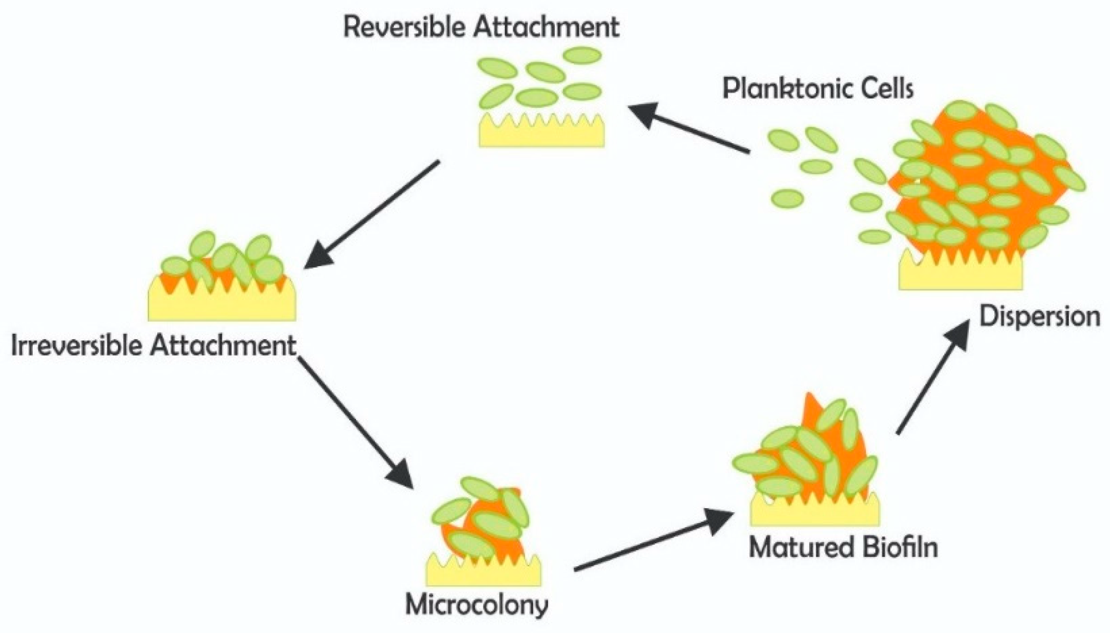
Biofilm is an adaptive mechanism of disinfection tolerance and has a long history in water treatment. The cells in a biofilm constantly interact and coordinate their activities efficiently by cell-to-cell contact or indirectly, via the release of signaling molecules, a process known as quorum sensing (QS) [68]. The biofilm microbial consortia are shielded by the extracellular polymeric substances (EPSs), thereby protecting them from chlorine, antimicrobials, increased temperatures and other stressful conditions. Upon maturation, portions of the biofilm can detach and be delivered to the end-users, which may permit a possible outbreak of waterborne disease [69]. Unfortunately, it is almost impossible to totally eradicate biofilm formation in water treatment and distribution systems; therefore, a number of factors to control their growth should be carefully considered. Some of these factors include nutrient availability in water, surface topography and pipe materials, hydrodynamics of the distribution network, pH, temperature, and disinfectant residual [70]. Figure 4 illustrates the various stages involved in the biofilm microbiological buildup.

Resistance and Pathogen Protection in Biofilms

Microbial biofilm resistance has numerous health, environmental and economic implications, such as water distribution system clogging, biofouling, food-processing disruptions, oil recovery, decay of water pipes and clinical implants, among others [71]. Microbial cells embedded in biofilms often show varying phenotypic characteristics, such as increased resistance to biocidal agents compared to planktonic cells. The exact mechanism of biofilm resistance remains unclear. The process, however, has been described as multifactorial involving the spatial distribution and organization of the biofilm architecture [38]. While numerous studies have concentrated on biofilm resistance to antibiotics [72,73,74], very minimal information exists on biofilm resistance to other biocides including disinfectants such as chlorine. Generally, resistance to biocides may be phenotypic (tolerance), genotypic or intrinsic in nature. For instance, the multilayered EPSs and microbial cells in a biofilm may constitute a very complex and compact barrier, making it difficult for the biocide to penetrate and reach the inner cells, thereby hampering its efficacy [75]. Additionally, glycocalyx or capsule, found in both Gram-positive and Gram-negative bacteria, can accumulate biocide molecules up to 25% of its weight [74], and can provide protection against antimicrobials in a biofilm [73].
Apart from the physical and structural barriers which biofilm provides, the preferential expression of some specific genes within biofilms can contribute to resistance. The upregulation of the ndvB gene that codes for a glucosyltransferase, an enzyme that catalyzes the production of periplasmic β-(1→3)-cyclic glucans, for example, is specifically noted in biofilms [76]. The glucans presumably sequester antibiotics away from their cellular target in the periplasm [77]. Moreover, ndvB upregulation is involved in the expression of some biofilm-specific genes associated with ethanol oxidation, and these genes may additionally play specific roles in biofilm resistance. Even though the upregulation of the ndvB gene is associated with biofilm, the mechanisms involved in its control and regulations are not known [78]. Understanding the genetic and physiological requirements for this biofilm-specific gene expression may serve as a potential tool in reducing biocide and antimicrobial resistance.

8. Multidrug Efflux Pumps Induction and Biocide Resistance

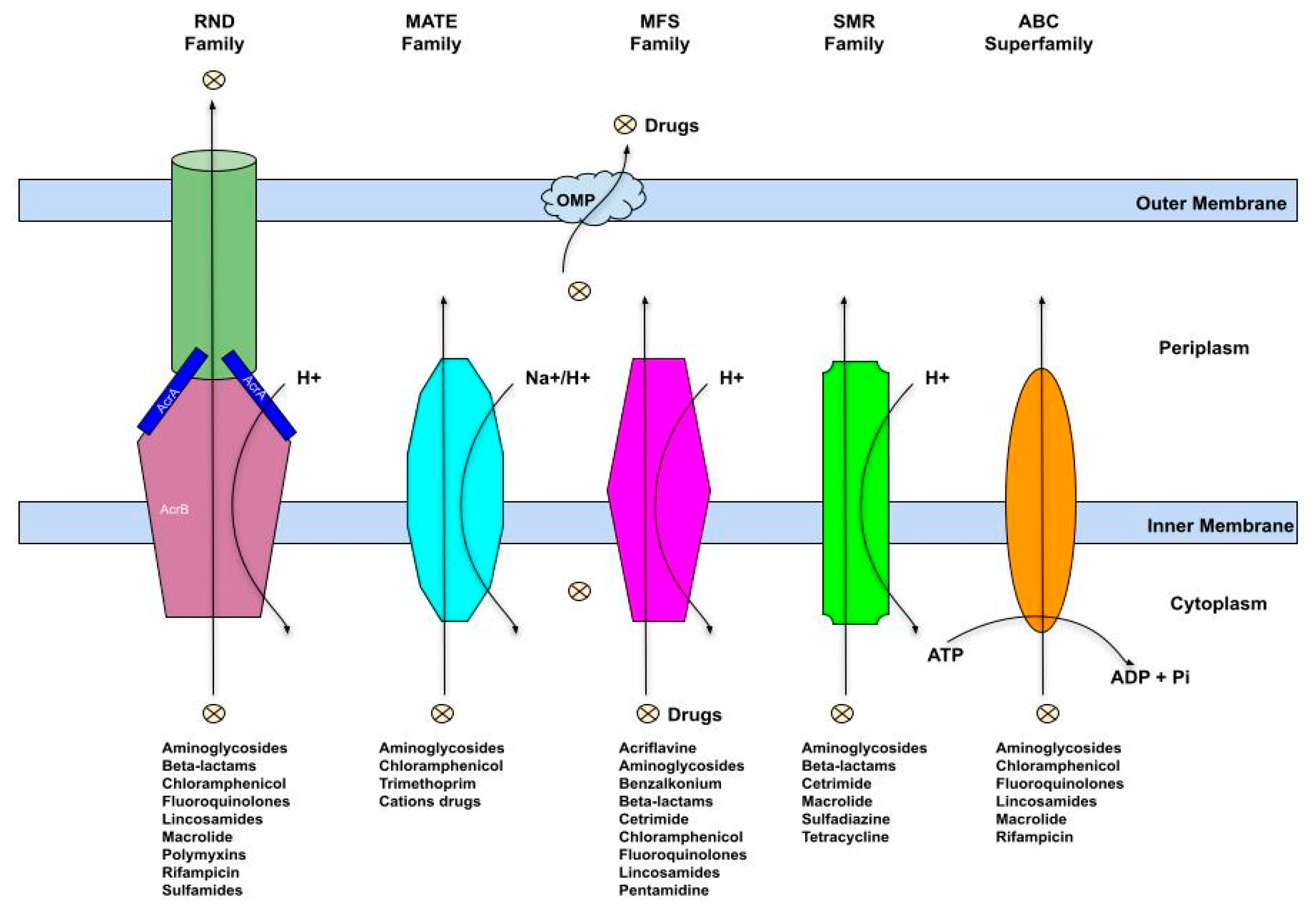
Some efflux pumps are expressed constitutively at low levels, thereby contributing to intrinsic resistance in microbial cells. Active efflux pumps may also contribute to high levels of resistance due to their overexpression in the presence of an effector molecule. Overexpression of multidrug efflux pumps may, however, be transient in nature [79]. While efflux pumps have mostly been associated with antibiotic resistance in pathogens, it is worthy to note that they can non-selectively extrude diverse compounds, including heavy metals, antiseptics, disinfectants, dyes, toxins, organic pollutants, metabolites, quorum sensing signaling molecules, neurotransmitters and quaternary ammonium compounds [80,81]. Structurally, an efflux pump is made up of four major components of an outer membrane protein, a middle periplasmic protein, an inner membrane protein and a transmembrane duct. As depicted in Figure 5, the outer and the inner membranes stabilize the channel (duct) in a closed state through their interaction with the periplasmic membrane protein [82]. The system is activated when a substance is transported, e.g., drug binds to the inner membrane protein, which then activates a cascade of biochemical events leading to the opening of the channel and eventual expulsion of the drug molecules. The protein-protein interaction generates the required energy by exchanging the molecule for a hydrogen ion (H+) [83]. Depending on the secondary structure, energy source, homology of amino acids and size, active efflux systems are classified into five different families (Figure 5). These include the ATP-binding cassette (ABC), the major facilitator superfamily (MFS), the resistance-nodulation division superfamily (RND), the small multidrug resistance family (SMR) and the multi antimicrobial and toxic compound extrusion protein family (MATE) [84].
The MFS transporters are also referred to as uniporter-symporter-antiporter (USA) family and represent the largest efflux family with an extraordinary broad-spectrum substrate affinity [85]. The SMR proteins are one of the smallest efflux systems with lengths ranging from about 100 to 140 amino acids. They are often inherited chromosomally but have also been found encoded by prophages, conjugative plasmids as well as Class I and Class III integrons. E. coli ethidium multidrug resistance protein E (EmrE) is one of the most characterized members of this protein family [80]. The most intensively characterized RND protein efflux system is the AcrAB-TolC, described in Gram-negative bacteria, including E. coli K-12 and Salmonella enterica serovar Typhimurium SH5014. It is made up of a periplasmic protein, AcrA, anchored to the inner membrane, twelve transmembrane α-helices and two large hydrophilic loops AcrB transporters, and the TolC outer membrane protein, which forms a channel allowing substrates to diffuse across the outer membrane. Most members of the RND family contain large polypeptides with 700–1300 amino acid residues [86]. The MATE proteins were initially identified in Vibrio parahaemolyticus and, subsequently, in many other cells, including humans [87]. MATE majorly transports cationic substrates, including clinically important drugs. The ABC transporters are energy-dependent and are found in all life forms. Seven distinct subfamilies of these transporters have been classified phylogenetically from ABCA to ABCG [88]. The proteins can transport a wide range of substrates and are characterized by two nucleotide-binding domains and two transmembrane domains. Overexpression of the ABC transporter proteins has been associated with increased extrusion of chemotherapeutic agents and multidrug resistance cases [89].
The expression of efflux is tightly regulated, and its induction can be achieved either in the presence of a right inducer molecule transiently or by mutations of the genes which downregulate their expression constitutively [90]. While a single efflux system is capable of conferring resistance to multiple biocidal agents, simultaneous induction of multiple efflux pumps has been identified in bacteria strains such as P. aeruginosa and Stenotrophomonas maltophilia strains, thus conferring both unique and overlapping biocide and drug selectivity [91,92]. Efflux systems are a growing clinical concern due to the possible co-existence of an assortment of diverse pump systems in clinically relevant microbial pathogens. The systems can also be easily spread on conjugative plasmids and other mobile genetic elements further underlining their importance in resistance biology. The redundant (overlapping) substrate recognition and drug polyspecificity nature of the efflux pump systems, coupled with their cross-resistance activities to varieties of biocides and antimicrobials are particularly prominent in Gram-negative pathogens such as the Enterobacteriaceae family. These group of enteric bacteria can survive and persist in soil, and aquatic environments, and are able to acquire biocide adaptation through chronic sub-lethal concentration exposure [80]. Due to the important role active efflux mechanisms play in resistance, the development of antimicrobials that target efflux systems and effectively inhibit their activities has been considered a potential control strategy to tackle the problem of resistance in microorganisms.

9. Conclusions and Future Directives

The endless problem of resistance proliferation among clinically relevant waterborne pathogens is a worrisome topic that necessitates the improvement of contemporary and next-generation treatment procedures. Since the turn of the century, large-scale chlorination has helped in maintaining the microbiological quality of public water supplies. Even though this has recorded many gains, conflicting reports continue to emanate on the role chlorination plays in the enhancement of AMR genes and resistant microorganisms in aquatic environments. While chlorine has been effective in reducing outbreaks of waterborne diseases, the molecular mechanisms underlining its roles in promoting the evolution and spread of resistance remain a grey area, with its attendant ecological and public health implications. Cross-resistance and co-resistance are important phenomena in the associations between chlorine and AMR. Similarly, proteome-mediated chlorine resistance has been described in some bacterial species while non-specific expression of multidrug efflux mechanisms and biofilm formation are other processes identified in waterborne pathogen resistance. Despite the various efforts toward understanding disinfection resistance among waterborne pathogens, a huge evidence-based gap at substantiating the exact links between chlorine disinfection and AMR spread exists. Unfortunately, there is still no standardized method to evaluate microbial chlorine resistance making the comparison of chlorine resistance challenging. In light of the above and the widespread observation of reduced disinfection efficacy of chlorine at the recommended dose, further investigation is crucial to more accurately explicate chlorine resistance mechanisms and the relationship between AR and chlorine resistance/tolerance for more rational water disinfection approaches with a view to improve its efficacy or develop novel strategies.

Author Contributions

Conceptualization, M.A.A.; methodology, M.A.A.; writing—original draft preparation, M.A.A.; writing—review and editing, M.A.A. and A.O.O.; supervision, A.O.O.; project administration, A.O.O. All authors have read and agreed to the published version of the manuscript.

Funding

This research was funded by the National Research Foundation, grant number UID: 129616.

Institutional Review Board Statement

Not applicable.

Informed Consent Statement

Not applicable.

Data Availability Statement

Not applicable.

Acknowledgments

We appreciate the National Research Foundation for providing M.A.A. with postdoctoral funding (UID: 129616).

Conflicts of Interest

The authors declare no conflict of interest.

References

  1. DeNegre, A.A.; Mbah, M.L.N.; Myers, K.; Fefferman, N.H. Emergence of antibiotic resistance in immunocompromised host populations: A case study of emerging antibiotic resistant tuberculosis in AIDS patients. PLoS ONE 2019, 14, e0212969. [Google Scholar] [CrossRef] [PubMed]
  2. León-Buitimea, A.; Garza-Cárdenas, C.R.; Garza-Cervantes, J.A.; Lerma-Escalera, J.A.; Morones-Ramírez, J.R. The demand for new antibiotics: Antimicrobial peptides, nanoparticles, and combinatorial therapies as future strategies in antibacterial agent design. Front. Microbiol. 2020, 2020, 1669. [Google Scholar] [CrossRef] [PubMed]
  3. Sumi, C.D.; Heffernan, A.J.; Lipman, J.; Roberts, J.A.; Sime, F.B. What antibiotic exposures are required to suppress the emergence of resistance for Gram-negative bacteria? A systematic review. Clin. Pharmacokinet. 2019, 58, 1407–1443. [Google Scholar] [CrossRef] [PubMed]
  4. Zhao, X.; Drlica, K. Restricting the selection of antibiotic-resistant mutants: A general strategy derived from fluoroquinolone studies. Clin. Infect. Dis. 2001, 33 (Suppl. S3), S147–S156. [Google Scholar] [CrossRef] [Green Version]
  5. Baquero, F. Resistance to quinolones in gram-negative microorganisms: Mechanisms and prevention. Eur. Urol. 1990, 17, 3–12. [Google Scholar] [CrossRef]
  6. Zhang, N.; Ye, X.; Wu, Y.; Huang, Z.; Gu, X.; Cai, Q.; Shen, X.; Jiang, H.; Ding, H. Determination of the mutant selection window and evaluation of the killing of Mycoplasma gallisepticum by danofloxacin, doxycycline, tilmicosin, tylvalosin and valnemulin. PLoS ONE 2017, 12, e0169134. [Google Scholar] [CrossRef]
  7. Huang, Z.; Mao, C.; Wei, Y.; Gu, X.; Cai, Q.; Shen, X.; Ding, H. Analysis of the mutant selection window and killing of Mycoplasma hyopneumoniae for doxycycline, tylosin, danofloxacin, tiamulin, and valnemulin. PLoS ONE 2020, 15, e0220350. [Google Scholar] [CrossRef]
  8. García-León, G.; Sánchez, M.B.; Martínez, J.L. The inactivation of intrinsic antibiotic resistance determinants widens the mutant selection window for quinolones in Stenotrophomonas maltophilia. Antimicrob. Agents Chemother. 2012, 56, 6397–6399. [Google Scholar] [CrossRef] [Green Version]
  9. Khan, S.; Beattie, T.K.; Knapp, C.W. Relationship between antibiotic-and disinfectant-resistance profiles in bacteria harvested from tap water. Chemosphere 2016, 152, 132–141. [Google Scholar] [CrossRef] [Green Version]
  10. Yuan, Q.B.; Guo, M.T.; Yang, J. Fate of antibiotic resistant bacteria and genes during wastewater chlorination: Implication for antibiotic resistance control. PLoS ONE 2015, 10, e0119403. [Google Scholar] [CrossRef]
  11. Liu, S.S.; Qu, H.M.; Yang, D.; Hu, H.; Liu, W.L.; Qiu, Z.G.; Hou, A.M.; Guo, J.; Li, J.W.; Shen, Z.Q.; et al. Chlorine disinfection increases both intracellular and extracellular antibiotic resistance genes in a full-scale wastewater treatment plant. Water Res. 2018, 136, 131–136. [Google Scholar] [CrossRef] [PubMed] [Green Version]
  12. Xu, L.; Ouyang, W.; Qian, Y.; Su, C.; Su, J.; Chen, H. High-throughput profiling of antibiotic resistance genes in drinking water treatment plants and distribution systems. Environ. Pollut. 2016, 213, 119–126. [Google Scholar] [CrossRef] [PubMed]
  13. Jin, M.; Liu, L.; Wang, D.N.; Yang, D.; Liu, W.L.; Yin, J.; Yang, Z.W.; Wang, H.R.; Qiu, Z.G.; Shen, Z.Q.; et al. Chlorine disinfection promotes the exchange of antibiotic resistance genes across bacterial genera by natural transformation. ISME J. 2020, 114, 1847–1856. [Google Scholar] [CrossRef]
  14. World Health Organisation. Drinking-Water. 2019. Available online: https://www.who.int/news-room/fact-sheets/detail/drinking-water (accessed on 12 July 2021).
  15. World Health Organisation. Water. 2020. Available online: https://www.afro.who.int/health-topics/water (accessed on 11 July 2021).
  16. Sabri, N.A.; Schmitt, H.; Van der Zaan, B.; Gerritsen, H.W.; Zuidema, T.; Rijnaarts, H.H.M.; Langenhoff, A.A.M. Prevalence of antibiotics and antibiotic resistance genes in a wastewater effluent-receiving river in the Netherlands. J. Environ. Chem. Eng. 2020, 8, 102245. [Google Scholar] [CrossRef]
  17. Murray, G.E.; Tobin, R.S.; Junkins, B.; Kushner, D.J. Effect of chlorination on antibiotic resistance profiles of sewage-related bacteria. Appl. Environ. Microbiol. 1984, 48, 73–77. [Google Scholar] [CrossRef] [PubMed] [Green Version]
  18. Rutala, W.A.; Stiegel, M.M.; Sarubbi, F.A.; Weber, D.J. Susceptibility of antibiotic-susceptible and antibiotic-resistant hospital bacteria to disinfectants. Infect. Control Hosp. Epidemiol. 1997, 18, 417–421. [Google Scholar] [CrossRef]
  19. Maertens, H.; De Reu, K.; Meyer, E.; Van Coillie, E.; Dewulf, J. Limited association between disinfectant use and either antibiotic or disinfectant susceptibility of Escherichia coli in both poultry and pig husbandry. BMC Vet. Res. 2019, 15, 310. [Google Scholar] [CrossRef] [Green Version]
  20. Al-Abri, M.; Al-Ghafri, B.; Bora, T.; Dobretsov, S.; Dutta, J.; Castelletto, S.; Rosa, L.; Boretti, A. Chlorination disadvantages and alternative routes for biofouling control in reverse osmosis desalination. NPJ Clean Water 2019, 2, 2. [Google Scholar] [CrossRef] [Green Version]
  21. Goyal, R.V.; Patel, H.M. Analysis of residual chlorine in simple drinking water distribution system with intermittent water supply. Appl. Water Sci. 2015, 5, 311–319. [Google Scholar] [CrossRef] [Green Version]
  22. Musz-Pomorska, A.; Widomski, M.K. Variant analysis of the chlorination efficiency of water in a selected water supply network. Proc. ECOpole 2020, 14, 19–28. [Google Scholar]
  23. Calomiris, J.; Christman, K. How Does Chlorine Added to Drinking Water Kill Bacteria and Other Harmful Organisms? Why Doesn’t It Harm Us? Scientific American. Available online: https://www.scientificamerican.com/article/how-does-chlorine-added-t/ (accessed on 5 August 2021).
  24. Venkobachar, C.; Iyengar, L.; Rao, A.P. Mechanism of disinfection: Effect of chlorine on cell membrane functions. Water Res. 1977, 11, 727–729. [Google Scholar] [CrossRef]
  25. Haas, C.N. Mechanisms of Inactivation of New Indicators of Disinfection Efficiency by Free Available Chlorine. Ph.D. Thesis, Department of Civil Engineering, the University of Illinois at Urbana-Champaign, Champaign, IL, USA, 1978; p. 158. [Google Scholar]
  26. Chang, S.L. Modern concept of disinfection. JSEDA 1971, 97, 689–707. [Google Scholar] [CrossRef]
  27. Mizozoe, M.; Otaki, M.; Aikawa, K. The mechanism of chlorine damage using enhanced green fluorescent protein-expressing Escherichia coli. Water 2019, 11, 2156. [Google Scholar] [CrossRef] [Green Version]
  28. Bridges, D.F.; Lacombe, A.; Wu, V.C. Integrity of the Escherichia coli O157: H7 cell wall and membranes after chlorine dioxide treatment. Front. Microbiol. 2020, 11, 888. [Google Scholar] [CrossRef] [PubMed]
  29. Reed, B.; Shaw, R.; Chatterton, K. Technical Notes on Drinking-Water, Sanitation and Hygiene in Emergencies; WHO, Water, Engineering and Development Centre: Loughborough, UK, 2013. [Google Scholar]
  30. Adefisoye, M.A.; Okoh, A.I. Ecological and public health implications of the discharge of multidrug-resistant bacteria and physicochemical contaminants from treated wastewater effluents in the Eastern Cape, South Africa. Water 2017, 9, 562. [Google Scholar] [CrossRef] [Green Version]
  31. National Water Act. Revision of General Authorization in Terms of Section 39 of the National Water Act, 1998 (Act No. 36 of 1998) (The Act); Gazette No. 19182, Notice No. 1091; National Water Act: 1998; South African Government Gazette: Pretoria, South Africa, 1998; Volume 26187.
  32. Yang, L.; Chen, X.; She, Q.; Cao, G.; Liu, Y.; Chang, V.W.C.; Tang, C.Y. Regulation, formation, exposure, and treatment of disinfection by-products (DBPs) in swimming pool waters: A critical review. Environ. Int. 2018, 121, 1039–1057. [Google Scholar] [CrossRef]
  33. Al-Berfkani, M.I.; Zubair, A.I.; Bayazed, H. Assessment of chlorine resistant bacteria and their susceptibility to antibiotic from water distribution system in Duhok province. J. Appl. Biol. Biotechnol. 2014, 2, 010–013. [Google Scholar]
  34. Owoseni, M.C.; Olaniran, A.O.; Okoh, A.I. Chlorine tolerance and inactivation of Escherichia coli recovered from wastewater treatment plants in the Eastern Cape, South Africa. Appl. Sci. 2017, 7, 810. [Google Scholar] [CrossRef]
  35. Jathar, S.; Shinde, D.; Dakhni, S.; Fernandes, A.; Jha, P.; Desai, N.; Jobby, R. Identification and characterization of chlorine-resistant bacteria from water distribution sites of Mumbai. Arch. Microbiol. 2021, 203, 5241–5248. [Google Scholar] [CrossRef]
  36. Martin, D.J.; Wesgate, R.L.; Denyer, S.P.; McDonnell, G.; Maillard, J.Y. Bacillus subtilis vegetative isolate surviving chlorine dioxide exposure: An elusive mechanism of resistance. J. Appl. Microbiol. 2015, 119, 1541–1551. [Google Scholar] [CrossRef] [Green Version]
  37. Momba, M.N.B.; Kfir, R.; Venter, S.N.; Cloete, T.E. An overview of biofilm formation in distribution systems and its impact on the deterioration of water quality. Water SA 2000, 26, 59–66. [Google Scholar]
  38. Bridier, A.; Briandet, R.; Thomas, V.; Dubois-Brissonnet, F. Resistance of bacterial biofilms to disinfectants: A review. Biofouling 2011, 27, 1017–1032. [Google Scholar] [CrossRef] [PubMed]
  39. Du, Z.; Nandakumar, R.; Nickerson, K.W.; Li, X. Proteomic adaptations to starvation prepare Escherichia coli for disinfection tolerance. Water Res. 2015, 69, 110–119. [Google Scholar] [CrossRef] [PubMed] [Green Version]
  40. Falkinham, J.O.; Pruden, A.; Edwards, M. Opportunistic premise plumbing pathogens: Increasingly important pathogens in drinking water. Pathogens 2015, 4, 373–386. [Google Scholar] [CrossRef] [Green Version]
  41. Shrivastava, R.; Upreti, R.K.; Jain, S.R.; Prasad, K.N.; Seth, P.K.; Chaturvedi, U.C. Suboptimal chlorine treatment of drinking water leads to selection of multidrug-resistant Pseudomonas aeruginosa. Ecotoxicol. Environ. Saf. 2004, 58, 277–283. [Google Scholar] [CrossRef]
  42. Karumathil, D.P.; Yin, H.B.; Kollanoor-Johny, A.; Venkitanarayanan, K. Effect of chlorine exposure on the survival and antibiotic gene expression of multidrug-resistant Acinetobacter baumannii in water. Int. J. Environ. Res. Public Health 2014, 11, 1844–1854. [Google Scholar] [CrossRef]
  43. Martins, S.C.S.; Júnior, G.D.S.F.; de Melo Machado, B.; Martins, C.M. Chlorine and antibiotic-resistant bacilli isolated from an effluent treatment plant. Acta Sci. Technol. 2013, 35, 3–9. [Google Scholar] [CrossRef] [Green Version]
  44. Owoseni, M.; Okoh, A. Assessment of chlorine tolerance profile of Citrobacter species recovered from wastewater treatment plants in Eastern Cape, South Africa. EMA 2017, 189, 201. [Google Scholar] [CrossRef]
  45. Ridgway, H.F.; Olson, B.H. Chlorine resistance patterns of bacteria from two drinking water distribution systems. Appl. Environ. Microbiol. 1982, 44, 972–987. [Google Scholar] [CrossRef] [Green Version]
  46. LeChevallier, M.W.; Hassenauer, T.S.; Camper, A.K.; McFETERS, G.A. Disinfection of bacteria attached to granular activated carbon. Appl. Environ. Microbiol. 1984, 48, 918–923. [Google Scholar] [CrossRef] [Green Version]
  47. King, C.H.; Shotts, E.B.; Wooley, R.E.; Porter, K.G. Survival of coliforms and bacterial pathogens within protozoa during chlorination. Appl. Environ. Microbiol. 1988, 54, 3023–3033. [Google Scholar] [CrossRef] [PubMed] [Green Version]
  48. Owoseni, M.; Okoh, A. Evidence of emerging challenge of chlorine tolerance of Enterococcus species recovered from wastewater treatment plants. Int. Biodeterior. Biodegrad. 2017, 120, 216–223. [Google Scholar] [CrossRef]
  49. Dupuy, M.; Mazoua, S.; Berne, F.; Bodet, C.; Garrec, N.; Herbelin, P.; Ménard-Szczebara, F.; Oberti, S.; Rodier, M.H.; Soreau, S.; et al. Efficiency of water disinfectants against Legionella pneumophila and Acanthamoeba. Water Res. 2011, 45, 1087–1094. [Google Scholar] [CrossRef] [PubMed]
  50. Martínez-Hernández, S.; Vázquez-Rodríguez, G.A.; Beltrán-Hernández, R.I.; Prieto-García, F.; Miranda-López, J.M.; Franco-Abuín, C.M.; Álvarez-Hernández, A.; Iturbe, U.; Coronel-Olivares, C. Resistance and inactivation kinetics of bacterial strains isolated from the non-chlorinated and chlorinated effluents of a WWTP. Int. J. Environ. Res. Public Health 2013, 10, 3363–3383. [Google Scholar] [CrossRef] [PubMed]
  51. Adefisoye, M.A.; Okoh, A.I. Identification and antimicrobial resistance prevalence of pathogenic Escherichia coli strains from treated wastewater effluents in Eastern Cape, South Africa. Microbiologyopen 2016, 5, 143–151. [Google Scholar] [CrossRef] [Green Version]
  52. Buelow, E.; Ploy, M.C.; Dagot, C. Role of pollution on the selection of antibiotic resistance and bacterial pathogens in the environment. Curr. Opin. Microbiol. 2021, 64, 117–124. [Google Scholar] [CrossRef]
  53. Cantón, R.; Morosini, M.I. Emergence and spread of antibiotic resistance following exposure to antibiotics. FEMS Microbiol. Rev. 2011, 35, 977–991. [Google Scholar] [CrossRef] [Green Version]
  54. Li, X.; Zhao, X.; Drlica, K. Selection of Streptococcus pneumoniae mutants having reduced susceptibility to levofloxacin and moxifloxacin. Antimicrob. Agents Chemother. 2002, 46, 522–524. [Google Scholar] [CrossRef] [Green Version]
  55. Drlica, K. The mutant selection window and antimicrobial resistance. J. Antimicrob. Chemother. 2003, 52, 11–17. [Google Scholar] [CrossRef] [Green Version]
  56. Urban, C.; Rahman, N.; Zhao, X. Fluoroquinolone resistant Streptococcus pneumoniae associated with levofloxacin therapy. J. Infect. Dis. 2001, 184, 794–798. [Google Scholar] [CrossRef] [Green Version]
  57. Guo, M.T.; Yuan, Q.B.; Yang, J. Distinguishing effects of ultraviolet exposure and chlorination on the horizontal transfer of antibiotic resistance genes in municipal wastewater. Environ. Sci. Technol. 2015, 49, 5771–5778. [Google Scholar] [CrossRef] [PubMed]
  58. Ma, L.; Yang, H.; Guan, L.; Liu, X.; Zhang, T. Risks of antibiotic resistance genes and antimicrobial resistance under chlorination disinfection with public health concerns. Environ. Int. 2022, 158, 106978. [Google Scholar] [CrossRef] [PubMed]
  59. Ojemaye, M.O.; Adefisoye, M.A.; Okoh, A.I. Nanotechnology as a viable alternative for the removal of antimicrobial resistance determinants from discharged municipal effluents and associated watersheds: A review. J. Environ. Manag. 2020, 275, 111234. [Google Scholar] [CrossRef] [PubMed]
  60. Reyes-Fernández, E.Z.; Schuldiner, S. Acidification of cytoplasm in Escherichia coli provides a strategy to cope with stress and facilitates development of antibiotic resistance. Sci. Rep. 2020, 10, 9954. [Google Scholar] [CrossRef]
  61. Bourély, C.; Cazeau, G.; Jarrige, N.; Haenni, M.; Lupo, A.; Madec, J.Y.; Leblond, A.; Gay, E. Co-resistance to amoxicillin and tetracycline as an indicator of multidrug resistance in Escherichia coli isolates from animals. Front. Microbiol. 2019, 10, 2288. [Google Scholar] [CrossRef] [Green Version]
  62. Landecker, H. Antimicrobials before antibiotics: War, peace, and disinfectants. Palgrave Commun. 2019, 5, 1–11. [Google Scholar] [CrossRef] [Green Version]
  63. Voth, W.; Jakob, U. Stress-activated chaperones: A first line of defense. Trends Biochem. Sci. 2017, 42, 899–913. [Google Scholar] [CrossRef]
  64. Goemans, C.V.; Collet, J.F. Stress-induced chaperones: A first line of defense against the powerful oxidant hypochlorous acid. F1000Research 2019, 8, 1678. [Google Scholar] [CrossRef]
  65. da Cruz Nizer, W.S.; Inkovskiy, V.; Overhage, J. Surviving reactive chlorine stress: Responses of gram-negative bacteria to hypochlorous acid. Microorganisms 2020, 8, 1220. [Google Scholar] [CrossRef]
  66. Cornelis, P.; Wei, Q.; Andrews, S.C.; Vinckx, T. Iron homeostasis and management of oxidative stress response in bacteria. Metallomics 2011, 3, 540–549. [Google Scholar] [CrossRef]
  67. Wang, Z.; Fang, Y.; Zhi, S.; Simpson, D.J.; Gill, A.; McMullen, L.M.; Neumann, N.F.; Gänzle, M.G. The locus of heat resistance confers resistance to chlorine and other oxidizing chemicals in Escherichia coli. Appl. Environ. Microbiol. 2020, 86, e02123-19. [Google Scholar] [CrossRef] [PubMed]
  68. Butler, C.S.; Boltz, J.P. Biofilm processes and control in water and wastewater treatment. In Comprehensive Water Quality and Purification; Ahuja, S., Ed.; Elsevier: Waltham, MA, USA, 2014; pp. 90–107. [Google Scholar]
  69. Simões, L.C.; Simões, M. Biofilms in drinking water: Problems and solutions. Rsc Adv. 2013, 3, 2520–2533. [Google Scholar] [CrossRef] [Green Version]
  70. Sehar, S.; Naz, I. Role of the biofilms in wastewater treatment. In Microbial Biofilms-Importance and Applications; Dhanasekaran, D., Thajuddin, N., Eds.; IntechOpen: Rijeka, Croatia, 2016; pp. 121–144. [Google Scholar]
  71. Muhammad, M.H.; Idris, A.L.; Fan, X.; Guo, Y.; Yu, Y.; Jin, X.; Qiu, J.; Guan, X.; Huang, T. Beyond risk: Bacterial biofilms and their regulating approaches. Front. Microbiol. 2020, 11, 928. [Google Scholar] [CrossRef] [PubMed]
  72. Cepas, V.; López, Y.; Muñoz, E.; Rolo, D.; Ardanuy, C.; Martí, S.; Xercavins, M.; Horcajada, J.P.; Bosch, J.; Soto, S.M. Relationship between biofilm formation and antimicrobial resistance in gram-negative Bacteria. Microb. Drug Resist. 2019, 25, 72–79. [Google Scholar] [CrossRef] [Green Version]
  73. Hall, C.W.; Mah, T.F. Molecular mechanisms of biofilm-based antibiotic resistance and tolerance in pathogenic bacteria. FEMS Microbiol. Rev. 2017, 41, 276–301. [Google Scholar] [CrossRef]
  74. Singh, S.; Singh, S.K.; Chowdhury, I.; Singh, R. Understanding the mechanism of bacterial biofilms resistance to antimicrobial agents. Open Microbiol. J. 2017, 11, 53–62. [Google Scholar] [CrossRef] [Green Version]
  75. Bas, S.; Kramer, M.; Stopar, D. Biofilm surface density determines biocide effectiveness. Front. Microbiol. 2017, 8, 2443. [Google Scholar] [CrossRef]
  76. Cai, Y. Molecular Mechanism of Nitric Oxide-Mediated Regulation of Intracellular Cyclic-di-GMP in Pseudomonas Aeruginosa Biofilms. Ph.D. Dissertation, University of Southampton, England, UK, 2018. [Google Scholar]
  77. Mah, T.F. Biofilm-specific antibiotic resistance. Future Microbiol. 2012, 7, 1061–1072. [Google Scholar] [CrossRef] [Green Version]
  78. Hall, C.W.; Hinz, A.J.; Gagnon, L.B.P.; Zhang, L.; Nadeau, J.P.; Copeland, S.; Saha, B.; Mah, T.F. Pseudomonas aeruginosa biofilm antibiotic resistance gene ndvB expression requires the RpoS stationary-phase sigma factor. Appl. Environ. Microbiol. 2018, 84, e02762-17. [Google Scholar] [CrossRef] [Green Version]
  79. Alcalde-Rico, M.; Hernando-Amado, S.; Blanco, P.; Martínez, J.L. Multidrug efflux pumps at the crossroad between antibiotic resistance and bacterial virulence. Front. Microbiol. 2016, 7, 1483. [Google Scholar] [CrossRef] [Green Version]
  80. Slipski, C.J.; Zhanel, G.G.; Bay, D.C. Biocide selective TolC-independent efflux pumps in Enterobacteriaceae. J. Membr. Biol. 2018, 251, 15–33. [Google Scholar] [CrossRef] [PubMed]
  81. Abdi, S.N.; Ghotaslou, R.; Ganbarov, K.; Mobed, A.; Tanomand, A.; Yousefi, M.; Asgharzadeh, M.; Kafil, H.S. Acinetobacter baumannii efflux pumps and antibiotic resistance. Infect. Drug. Resist. 2020, 13, 423. [Google Scholar] [CrossRef] [PubMed] [Green Version]
  82. Ughachukwu, P.O.; Unekwe, P.C. Efflux pump-mediated resistance in chemotherapy. Ann. Med. Health Sci. Res. 2012, 2, 191–198. [Google Scholar] [CrossRef] [PubMed] [Green Version]
  83. Delmar, J.A.; Su, C.C.; Yu, E.W. Bacterial multidrug efflux transporters. Annu. Rev. Biophys. 2014, 43, 93–117. [Google Scholar] [CrossRef] [PubMed] [Green Version]
  84. Rahman, T.; Yarnall, B.; Doyle, D.A. Efflux drug transporters at the forefront of antimicrobial resistance. Eur. Biophys. J. 2017, 46, 647–653. [Google Scholar] [CrossRef] [PubMed]
  85. Perland, E.; Fredriksson, R. Classification systems of secondary active transporters. TIPS 2017, 38, 305–315. [Google Scholar] [CrossRef] [PubMed]
  86. Cacciotto, P.; Ramaswamy, V.K.; Malloci, G.; Ruggerone, P.; Vargiu, A.V. Molecular modeling of multidrug properties of resistance nodulation division (RND) transporters. In Bacterial Multidrug Exporters; Yamaguchi, A., Nishino, K., Eds.; Humana Press: New York, NY, USA, 2018; pp. 179–219. [Google Scholar]
  87. Morita, Y.; Tomida, J.; Kawamura, Y. Responses of Pseudomonas aeruginosa to antimicrobials. Front Microbiol. 2014, 4, 422. [Google Scholar] [CrossRef] [Green Version]
  88. Van der Deen, M.; De Vries, E.G.; Timens, W.; Scheper, R.J.; Timmer-Bosscha, H.; Postma, D.S. ATP-binding cassette (ABC) transporters in normal and pathological lung. Respir. Res. 2005, 6, 59. [Google Scholar] [CrossRef] [Green Version]
  89. Wilkens, S. Structure and mechanism of ABC transporters. F1000prime Rep. 2015, 7, 14. [Google Scholar] [CrossRef]
  90. Blanco, P.; Hernando-Amado, S.; Reales-Calderon, J.A.; Corona, F.; Lira, F.; Alcalde-Rico, M.; Bernardini, A.; Sanchez, M.B.; Martinez, J.L. Bacterial multidrug efflux pumps: Much more than antibiotic resistance determinants. Microorganisms 2016, 4, 14. [Google Scholar] [CrossRef] [Green Version]
  91. Gould, V.C.; Okazaki, A.; Avison, M.B. Coordinate hyperproduction of SmeZ and SmeJK efflux pumps extends drug resistance in Stenotrophomonas maltophilia. Antimicrob. Agents Chemother. 2013, 57, 655–657. [Google Scholar] [CrossRef] [PubMed] [Green Version]
  92. Poonsuk, K.; Tribuddharat, C.; Chuanchuen, R. Simultaneous overexpression of multidrug efflux pumps in Pseudomonas aeruginosa non-cystic fibrosis clinical isolates. Can. J. Microbiol. 2014, 60, 437–443. [Google Scholar] [CrossRef] [PubMed]
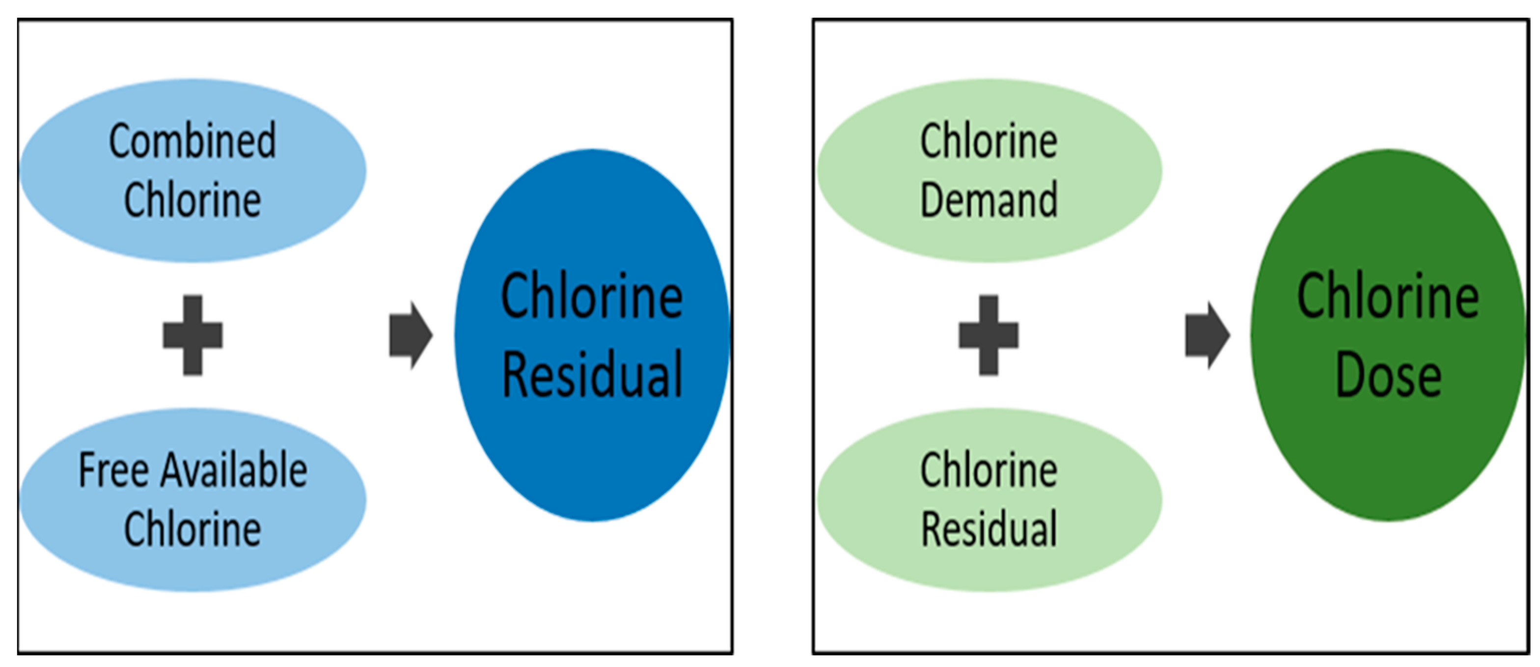
Figure 1. Schematic illustration of chlorine disinfection.
Antibiotics 11 00564 g001
Figure 2. Graphical representation of breakpoint chlorination.
Antibiotics 11 00564 g002
Figure 3. Schematic presentation of the possible events cascade of chlorine disinfection leading to microbial cell death. 1: Dissociating chlorine molecules attack and destroy bacterial cell walls altering permeability; 2: Entry of chlorine molecules into the cytoplasm interfering with biomolecules and enzymatic reactions; 3: Altered permeability causes vital cellular components leakage; 4: The series of events lead to the loss of cellular constituents and functions; 5: The loss of cellular constituents and function lead to eventual cell death.
Antibiotics 11 00564 g003
Figure 4. Schematic illustration of different phases in biofilm microbiology.
Antibiotics 11 00564 g004
Figure 5. Different classes of efflux systems.
Antibiotics 11 00564 g005
Table 1. Summary of some reports of disinfectant-resistant microbes isolated from diverse aquatic sources.
SourceMicroorganismDisinfectant Concentration/TimeMechanism(s) of ResistanceReference
Drinking waterP. aeruginosa≤0.5 mg/L ClNatural resistance due to the permeability barrier caused by outer membrane lipopolysaccharides; biofilm formation[41]
Experimental isolatesAcinetobacter
baumannii
0.2–4 mg/LIncreased expression of efflux pumps other antibiotic resistance genes[42]
Drinking water reservoirAcinetobater species, Serratia species 2 mg/LNot determined[35]
SewageBacillus species 0.1 mg/L NaOClProbable spore formation[43]
Secondary effluentCitrobacter species 0.5 mg/L Ca(OCl)2 for 30 minNot determined[44]
Drinking waterBacillus species, Actinomycete10 mg/L NaOCl for 2 minCellular aggregation or adhesion to suspended particulate. Production of extracellular slime or capsular material[45]
Drinking water and experimental isolatesHeterotrophic bacteria, faecal coliforms, E. coli, Salmonella typhimurium, Yersinia enterocolitica, Shigella sonnie2.0 mg/L free chlorine for 1 hBacterial attachment to surface and production of extracellular slime layer[46]
Chlorine-demand–free buffer solutionColiform isolated from drinking water systems and Enteric bacterial from culture collections cocultured with protozoa (Ciliates and amoebae).2–4 mg/L free chlorine for 1–2 hShielding of bacteria from chlorine by ingesting protozoans (cysts) and, thus, enhancing resistance[47]
Treated drinking waterS. aureus, Micrococcus varians, Aeromonas hydrophila1–100 mg/L Ca(OCl)2 solution for 30 minPossible synthesis of unique proteins or aggregation of bacteria or encapsulation[33]
Environmental isolates (Wastewater clarifier effluent) suspended in phosphate buffer salineEnterococcus species0.5 mg/L Ca(OCl)2 for 30 minNot determined[48]
Environmental strains cultured in sterile phosphate buffer solutionLegionella pneumophila from environmental water cocultured with Acanthamoeba species2–3 mg/L Cl2 for 1 h with a residual Cl2 of 1 mg/L after 1 hPossible phenotypic modification of Legionella pneumophila due to intra-cellular growth with Acanthamoeba sp.[49]
Environmental isolates from wastewater treatment plants suspended in salineBacillus species, Citrobacter freundii, Enterobacter species, Kluyvera cryocrescens, Kluyvera intermedia0.5 mg/L NaOCl for 30 minAuthors suggested the possible expression of certain stress factor genes which may reduce bacterial metabolism or change the permeability of cell membranes[50]
Environmental isolates (Wastewater clarifier effluent) suspended in phosphate buffer salineE. coli0.5 mg/L NaOCl for 30 minNot determined[34]
Publisher’s Note: MDPI stays neutral with regard to jurisdictional claims in published maps and institutional affiliations.