Article

Gelidiales Are Not Just Agar—Revealing the Antimicrobial Potential of Gelidium corneum for Skin Disorders

1
MARE—Marine and Environmental Sciences Centre, Polytechnic of Leiria, 2520-630 Peniche, Portugal
2
BioISI—Biosystems and Integrative Sciences Institute, Faculty of Sciences, University of Lisbon, 1749-016 Lisboa, Portugal
3
MARE—Marine and Environmental Sciences Centre, ESTM, Polytechnic of Leiria, 2520-630 Peniche, Portugal
*
Authors to whom correspondence should be addressed.
Antibiotics 2022, 11(4), 481; https://doi.org/10.3390/antibiotics11040481
Submission received: 12 March 2022 / Revised: 30 March 2022 / Accepted: 1 April 2022 / Published: 5 April 2022
(This article belongs to the Special Issue Green Antimicrobials)

Abstract

:
In recent decades, seaweeds have proven to be an excellent source of bioactive molecules. Presently, the seaweed Gelidium corneum is harvested in a small area of the Portuguese coast exclusively for agar extraction. The aim of this work was to fully disclosure Gelidium corneum as a sustainable source of antimicrobial ingredients for new dermatological formulations, highlighting its potential to be explored in a circular economy context. For this purpose, after a green sequential extraction, these seaweed fractions (F1–F5) were chemically characterized (1H NMR) and evaluated for their antimicrobial potential against Staphylococcus aureus, Staphylococcus epidermidis and Cutibacterium acnes. The most active fractions were also evaluated for their effects on membrane potential, membrane integrity and DNA damage. Fractions F2 and F3 displayed the best results, with IC50 values of 16.1 (7.27–23.02) μg/mL and 51.04 (43.36–59.74) μg/mL against C. acnes, respectively, and 53.29 (48.75–57.91) μg/mL and 102.80 (87.15–122.30) μg/mL against S. epidermidis, respectively. The antimicrobial effects of both fractions seem to be related to membrane hyperpolarization and DNA damage. This dual mechanism of action may provide therapeutic advantages for the treatment of skin dysbiosis-related diseases.

1. Introduction

In recent decades, marine organisms have proven to be an excellent source of bioactive molecules with a wide range of applications. However, few examples have reached the industrial sector, particularly due to the limitations of biomass availability, solvent suitability, low yields of extraction, and specific legal requirements, among others. The seaweed Gelidium corneum (former Gelidium sesquipedale) is a Rhodophyta belonging to the order Gelidiales, and several species belonging to this order, including Gelidium corneum, have the characteristic of being rich in agarans, highly valued in the food industry [1,2]. Presently, this seaweed is harvested in a small area of the Portuguese coast São Martinho do Porto for agar extraction. However, Gelidium species are rich in bioactive compounds such as mycosporine-like amino acids, flavonoids, pigments, phycobiliproteins, fatty acids, etc., with relevant biotechnological potential [3,4,5,6]. Within a circular economy approach it is highly relevant to understand the full potential of this biomass, to take the maximum advantage of one resource for the development of multiple new products, enhancing economic revenues and, consequently, boosting the local economy.
Within this framework, the present study targeted the red seaweed Gelidium corneum as a source of bioactive ingredients with antimicrobial activity with relevance for inclusion in new dermatological formulations.
Skin is the most effective barrier of the human body against external aggressions. Covered by a highly complex microbiome composed of bacteria, fungi and viruses, this living barrier is an effective defence against invading pathogens and heavily contributes to modulating the immune system [7]. Some of the most representative species of skin microbes include Cutibacterium acnes (former Propionibacterium acnes), Staphylococcus spp., Streptococcus spp., Corynebacterium spp., and Malassezia spp., which are distributed according to their specific affinity for a determined microenvironment—moist, dry or sebaceous [7]. While in normal conditions these microorganisms are fundamental for a healthy skin barrier, changes in their normal balance (dysbiosis) may lead to the development of skin pathologies such as acne vulgaris, dermatitis, eczema, and chronic wounds, among others [7]. Even though the skin’s microbiome is composed of thousands of different microorganisms, recent studies have focussed on the relationships between two commensal bacteria, Staphylococcus epidermidis and Cutibacterium acnes [8,9]. The dysbiosis between these two organisms results in a series of complications for the human host. Oily skin or excessive sebum production by the oil glands, may boost the growth of C. acnes that, combined with a decreased S. epidermidis population, may lead to a skin pathological condition known as acne vulgaris [10,11]. Acne vulgaris is a chronic inflammation of the skin, beginning in the pilosebaceous unit. Not only it is triggered by the increase in sebum production, but also by the hypercornification of the pilosebaceous glands due to the hyperproliferation of keratinocytes on the upper part of the follicle.
The homeostasis of the skin microbiota is considered to be the objective of any acne treatment [10,12]. As a consequence of microbial imbalance, the treatments available are mostly provided by topical antibiotic prescription; however, severe cases require oral treatment. The most common antibiotics for acne topical treatment are tetracycline, clindamycin, and erythromycin, sometimes combined with benzoyl peroxide and zinc acetate [13,14,15]. A prescription of oral antibiotics is mostly avoided due to the possibility of increased antibiotic resistance. However, C. acnes infections can also occur in other organs due to wounds and, in these cases, there is not any other treatment option.
When the balance of these two commensals is disrupted there can also occur an excess of S. epidermidis growth, which may result in nosocomial infections [16]. Although S. epidermidis rarely lead to severe life-threatening diseases, their infections are extremely common and difficult to treat. This microorganism is frequently involved in vascular graft, prosthetic joint, and cardiac device infections, among others, being the second infectious driver after Staphylococcus aureus [16].
The skin microbiome has a vast influence on individuals’ wellbeing, thus, finding natural derivatives that can not only deter pathogens but also maintain a microbial balance by targeting key microbes is critical [7].
Knowing that marine seaweeds are a propelling source of new chemical structures with an array of bioactivities, including antimicrobial activity [17,18], this work aims to disclose the antimicrobial potential of G. corneum-derived components against microorganisms that are frequently associated with skin disorders, namely Staphylococcus aureus, Staphylococcus epidermidis and Cutibacterium acnes.

2. Results

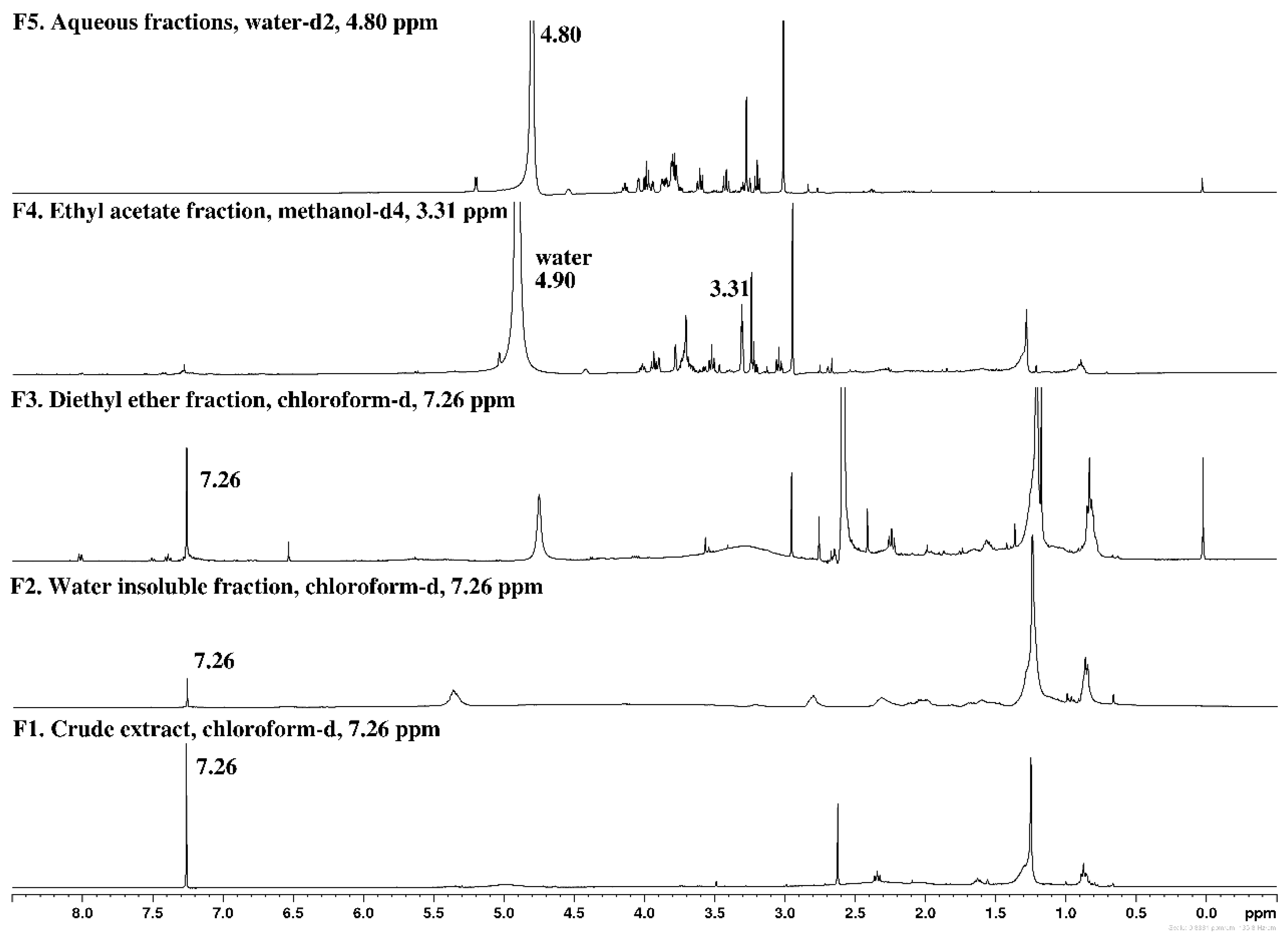
2.1. Chemical Screening by 1H NMR

The chemical profile of the fractions obtained from Gelidium corneum was evaluated by 1H NMR and the corresponding spectra are depicted in Figure 1.
Crude extract (F1) evidenced signals in the region of 0.8–2.8 ppm, characteristic of less polar lipophilic compounds such as fatty acids, sterols, terpenes, pigments, and other lipids [19,20]. The presence of these classes of compounds was also evidenced in the insoluble fraction retained in the filter (F2), with the intensity of signals being more pronounced in this fraction due to their higher concentration. More intense proton signals in the region of 0.8–3.1 ppm were also observed in diethyl ether fraction (F3), also suggesting the richness of lipophilic compounds in this fraction. Fraction F3 also evidenced low-intensity signals of aromatic protons (7.5–8.2 ppm), which can be attributed to phenolic compounds. The signals of these compounds are also visible in the spectrum of ethyl acetate fraction (F4), although with less intensity. In this fraction, the most intense signals were observed at 2.7–3.9 ppm, which can be attributed to the presence of amino compounds such as amino acids and proteins [20,21]. However, signals between 3.2 and 4.1 ppm were observed in fractions F4 and F5, which can denote the presence of alcohols, sugars, and esters [20]. Signals between 3.0 and 4.4 ppm are characteristic of the ring hydrogens of polysaccharides [22], supporting the richness of the aqueous fraction (F5) in this group of molecules.

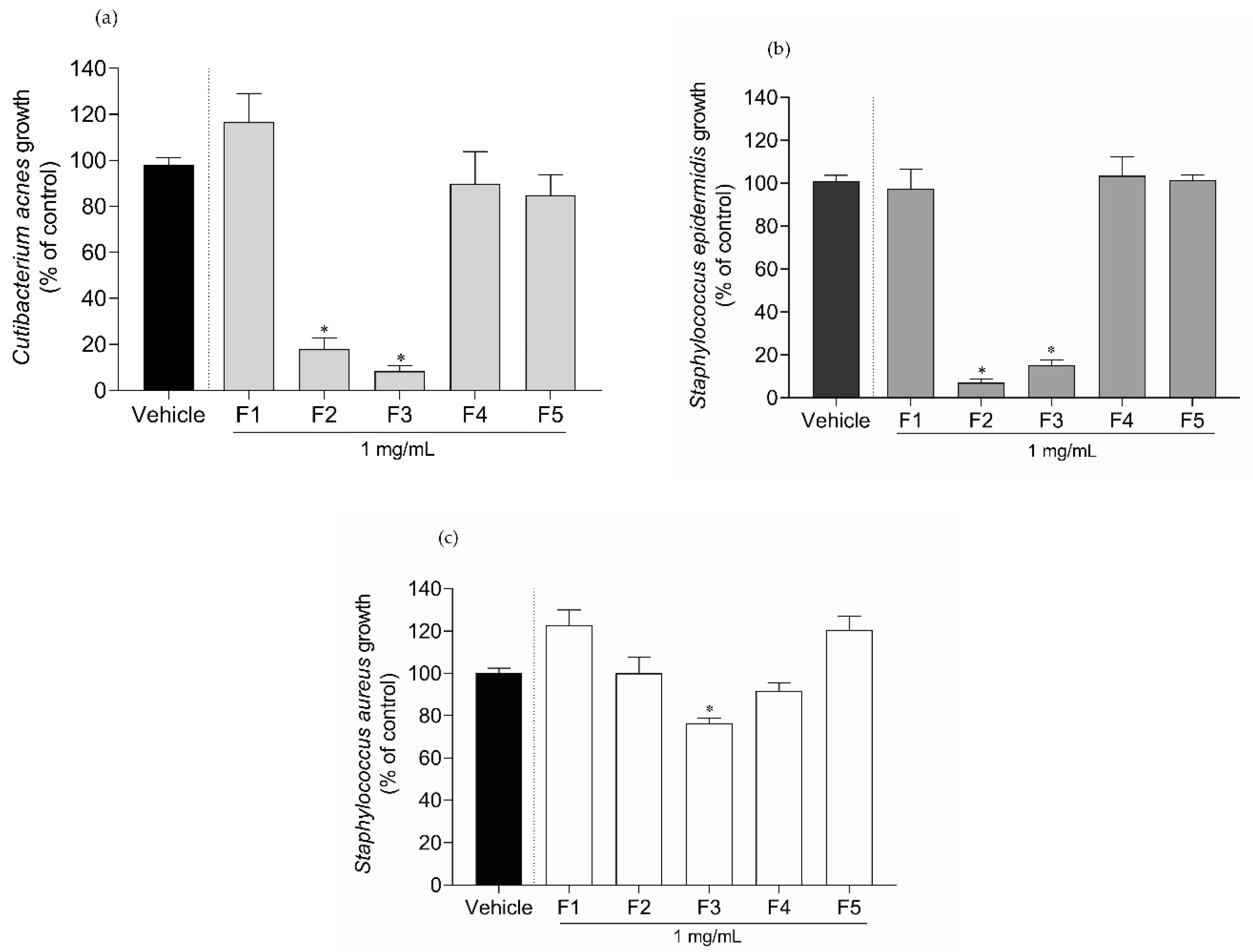
2.2. Antimicrobial Activity of Gelidium corneum Fractions

The antimicrobial potential of G. corneum fractions was tested against C. acnes, S. epidermidis and S. aureus growth. The results are shown in Figure 2.
Only the water-insoluble fraction (F2) and diethyl ether fraction (F3) inhibited C. acnes and S. epidermidis growth by more than 50%. In the case of S. aureus, only the F3 fraction promoted inhibition (≈25%). Since fractions F2 and F3 showed high inhibitory activity, a dose–response analysis was conducted and the IC50 determined. The results are shown in Table 1.
Gelidium corneum F2 and F3 fractions exhibited IC50 values of 53.29 (48.75–57.91) μg/mL and 102.8 (87.15–122.30) μg/mL, respectively, against S. epidermidis. Concerning C. acnes, fraction F2 revealed the highest potency with an IC50 of 16.10 (7.27–23.02) μg/mL, followed by fraction F3 with an IC50 of 51.04 (43.36–59.74) μg/mL.

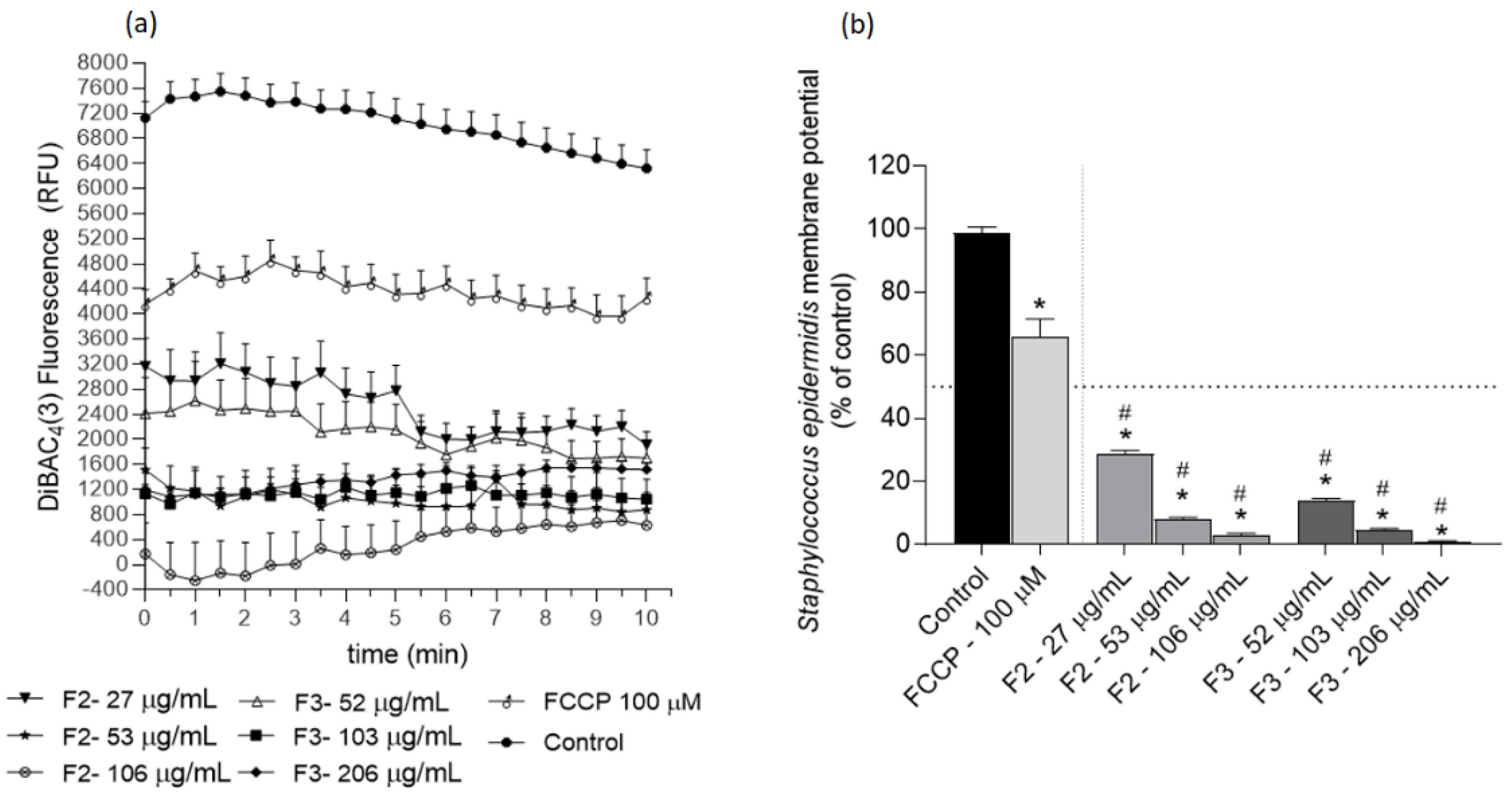
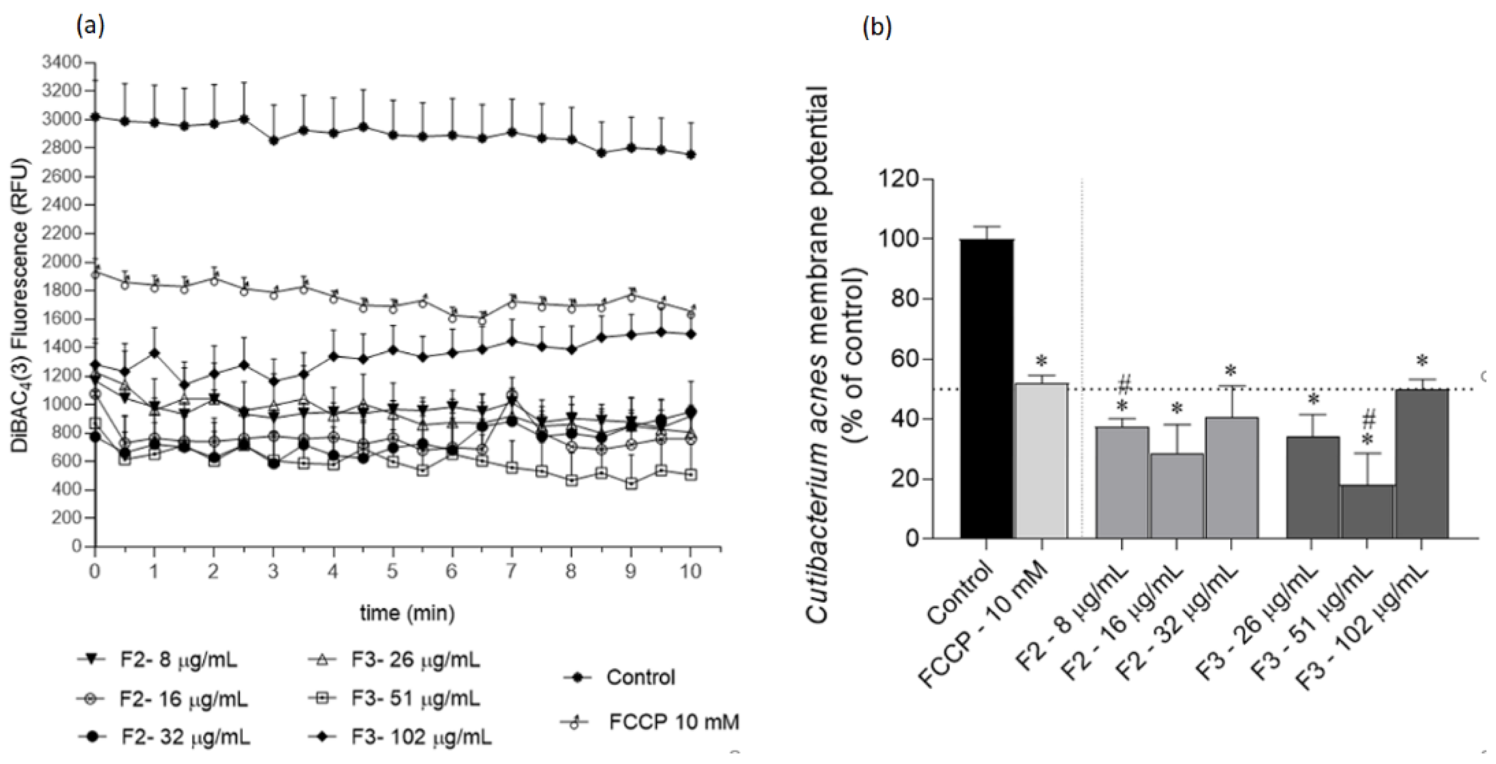
2.3. Effects of Gelidium corneum Fractions on Cutibacterium acnes and Staphylococcus epidermidis Membrane Potential

Several assays were conducted to understand the possible mechanisms underlying the F2 and F3 fractions’ antimicrobial effects. The effects on C. acnes and S. epidermidis membrane potential are depicted in Figure 3 and Figure 4, respectively.
The results obtained with the DiBAC4(3) method suggest that F2 and F3 fractions promote a membrane hyperpolarization in C. acnes and S. epidermidis, which is noticeable due to the lower fluorescence emission when compared with the control. Additionally, the fractions exhibited a more marked effect than the positive control FCCP in both bacteria. In C. acnes, both fractions decreased the membrane potential in more than 50%, except for F3 at 2 × IC50 after 10 min (Figure 3b). This profile was similar against S. epidermidis; however, the hyperpolarization was more pronounced, with a reduction in membrane polarization ranging from ≈70% (fraction F2 at ½ IC50) to ≈100% reduction (fraction F3 at 2 × IC50).

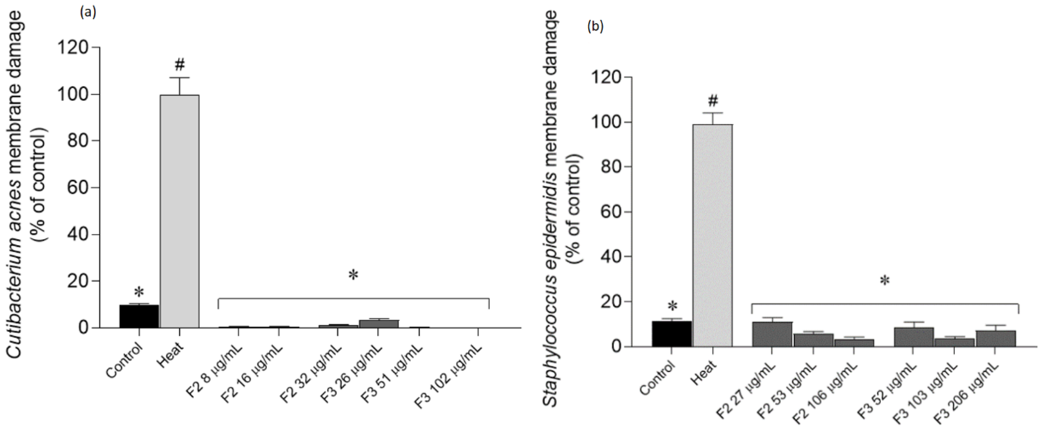
2.4. Effects of Gelidium corneum Fractions on Cutibacterium acnes and Staphylococcus epidermidis Membrane Integrity

To further understand if fractions could impair bacterial growth by affecting the membrane integrity, Sytox Green—a green-fluorescent nuclear probe—was used. This probe is impermeant to cells; however, when the membrane is disrupted, this probe links to nucleic acids emitting fluorescence, thus being an extremely useful tool to monitor membrane damage (Figure 5).
After an incubation with the most active fractions, it was possible to verify that they did not promote membrane damage, since there were no significant differences in relation to the untreated control.

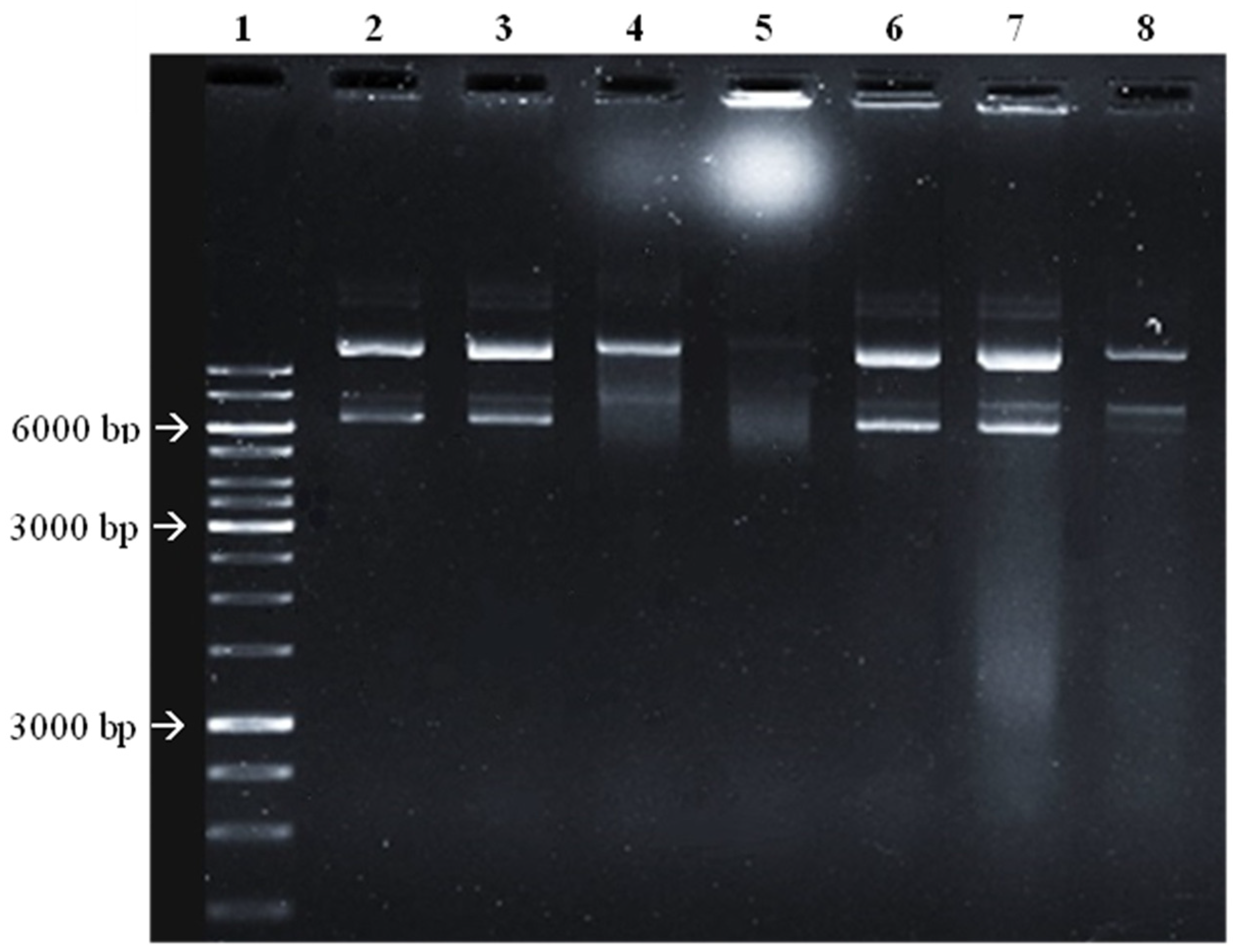
2.5. Potential of Gelidium corneum Fractions to Promote DNA Damage

Since different mechanisms can be behind the antimicrobial effects, the ability of the most active fractions to link and damage DNA was evaluated. The results are shown in Figure 6.
In Figure 6, it is possible to observe a clear smear of the DNA bars in the G. corneum F2 and F3 (lanes 7 and 8) when compared to the DMSO controls. Additionally, in lane 8 (F3), both linear and supercoiled DNA have a lower intensity, suggestive of DNA degradation. Ciprofloxacin promoted extensive DNA damage at 30 µg/mL and, at 10 µg/mL, the degradation is particularly evident in the supercoiled DNA (lane 4).

3. Discussion

In the present work, Gelidium corneum was evaluated regarding its potential as a source of antimicrobial compounds through a green extraction approach, aiming to develop new therapeutic alternatives for dysbiosis-related skin disorders. It was verified that two fractions—F2 (water insoluble fraction) and F3 (diethyl ether fraction)—were particularly active against S. epidermidis and C. acnes.
One of the main causes behind dermatology appointments is acne vulgaris. This disease is the most common skin problem worldwide, occurring in adulthood in about 50% of the population in occidental countries [23,24,25]. On the other hand, S. epidermidis can be an important opportunistic pathogen, with treatment options posing a challenge. Most infections caused by this microorganism start with the introduction of skin bacteria during the insertion of a medical device into the patient [16]. When looking at therapeutical options targeting both microorganisms, the conventionally prescribed topical and oral antibiotics can have some serious side effects, such as disrupting gut health and increasing skin dryness, impacting patients’ quality of life. On the other hand, even when the side effects are minimal, bacterial resistance might still occur [26]. This makes seeking new antibiotic options urgent. There are several natural products that could be possible candidates for novel drugs targeting multiple pathogenic factors [27]. Although in the present work the antimicrobial activity against S. aureus was low, previous studies with other Gelidium species have shown the capacity to deter the growth of S. aureus [28,29,30,31]. Additionally, a review gathered by Pérez, Falqué and Domínguez [31] cited G. attenatum, G. micropterum, G. pulchellum, G. pusillum, G. robustum, and G. spinosum with antimicrobial activity against different bacteria, including Escherichia coli, Klebsiella pneumoniae, Vibrio spp. and Enterococcus faecalis. However, no studies were found with Gelidium species against C. acnes or S. epidermidis. Choi et al. [32] reported 15 seaweed species with antimicrobial activity against C. acnes, with the red seaweed Symphyocladia latiuscula displaying the highest inhibitory potential (MIC = 160 µg/mL). Comparatively, the results obtained with F2 and F3 fractions from G. corneum are quite exciting due to their high potency, particularly against C. acnes; thus, they are promising candidates for use in new formulations aiming to treat skin diseases, where C. acnes and S. epidermidis play a key role. Scientific evidence suggests that S. epidermidis has a suppressive effect against C. acnes, and thus extracts with a higher antimicrobial effect against C. acnes than against S. epidermidis may bring additional advantages for acne vulgaris therapeutics [33]. This evidence highlights the relevance of the results obtained with G. corneum F2 and F3 fractions.
Antibiotics can work in a synergic way to effectively decrease bacterial resistance. Yet, their mechanism and range of action should be complementary. This is already exploited specifically in acne vulgaris treatment with the combination of different oral antibiotics and benzoyl peroxide [15]. To better understand the antibacterial effect, it is necessary to evaluate the possible mechanisms of action which can be underlying the bacterial growth inhibition. Since fractions F2 and F3 revealed promising results, the antimicrobial effects due to membrane potential disruption, membrane rupture, and/or DNA damage were evaluated.
Membrane potential is central to bacterial development; therefore, disruptions to it might induce an antimicrobial effect [34]. Across the cellular membrane, there is an electrochemical potential that is involved in several functions of bacterial cells, such as intra- and intercellular signalling mediation, which in turn regulates important physiological processes, namely mechano-sensation, spore formation, and biofilm dynamics [35,36,37,38]. In addition to its role in bioelectrical signalling, membrane potential is also central to cellular proliferation since it provides the essential driving force for ATP synthesis [39], which is crucial for cell division [40]. Although this potential may fluctuate depending on cells’ physiologies, abrupt changes may lead to cellular death. In particular, several studies have shown that a hyperpolarized membrane is associated with bacterial death [34,41]. It was verified that the most active extracts (F2 and F3) promoted a hyperpolarization of bacteria membrane potential (Figure 3 and Figure 4). Although studies surrounding the mechanisms of action of seaweed derivatives are scarce, Patra et al. [42] evaluated the mechanisms underlying the antimicrobial effects of essential oils extracted from Ulva linza (former Enteromorpha linza) against Escherichia coli and verified that the antimicrobial effects were related to changes in the membrane potential. Concerning phytochemical sources, Wu et al. [43] evaluated the antimicrobial effects of a natural compound (2R,3R-dihydromyricetin) obtained from the pine needles of Cedrus deodara against S. aureus and verified that this compound promoted a high membrane hyperpolarization, resulting in bactericidal effects. Although the results shown in the present work suggest that G. corneum fractions did not promote membrane damage, other seaweed species have shown antimicrobial activity mediated by membrane rupture. Patra and Baek [44] evaluated the antimicrobial mechanisms of oil extracted from the seaweed U. linza against Bacillus cereus and S. aureus and verified that the antimicrobial effects were related to membrane injury. Additionally, El Shafay et al. [45] evaluated the antimicrobial activity of Sargassum species against S. aureus and Klebsiella pneumoniae and concluded that the antimicrobial activity was mediated by cells’ membranes rupture and physical distortions. A work conducted with the green seaweed U. linza showed that essential oil from this seaweed promotes membrane damage in the foodborne pathogen Escherichia coli [44].
It has been suggested that antimicrobial compounds can reach DNA through membranes, with or without membrane rupture [46,47]. Thus, although it was not possible to verify membrane damage, a simple test was conducted to understand if the seaweed fractions could impact DNA. It was shown that both fractions F2 and F3 can promote DNA damage. In accordance with these results, Pinteus et al. [48] also showed that extracts derived from the red seaweed Asparagopsis armata had potential to promote DNA damage. Additionally, seaweed polysaccharides have shown antimicrobial activity mediated by DNA damage [49]. Other natural products from various works have also shown the ability to target DNA. Subramanian et al. [50] verified that resveratrol inhibited the growth of E. coli, probably by inducing DNA damage. Da et al. [51] evaluated the antimicrobial activity of an extract obtained from Scutellaria baicalensis root and verified that it had antimicrobial activity against fungi, possible mediated by DNA damage. Antimicrobial peptides extracted from different sources have also shown potential to target bacteria and fungi DNA [52]. He et al. [53] isolated a novel polysaccharide from Streptomyces virginia and verified that this compound exhibited antimicrobial activity against several microorganisms, possibly mediated by DNA damage.
Fractions 2 and 3 from Gelidium corneum are rich in lipophilic compounds, as evidenced by their 1H NMR spectra. Lipophilic compounds from different algae species have shown to display antimicrobial effects [31,49,54,55,56] and within this group of compounds, different terpenes sourced from red algae show great potential. Rodrigues et al. [57] studied the antimicrobial activity of terpenes isolated from Sphaerococcus coronopifolius and one sphaerane bromoditerpene had a great effect on S. aureus with an IC50 of 6.35 µM. Xu et al. [55] reported that tetracyclic brominated diterpenes from the same algae exhibited a bactericidal effect with MIC values of 16 and 128 μg/mL against multi-resistant Staphylococcus aureus. Amongst lipophilic compounds, fatty acids from different species of seaweeds have a reported antimicrobial effect in several microorganisms. For example, Dunaliella salina fatty acids are described for their effect over Listeria monocytogenes and B. cereus with a MIC value of 2.5 mg/mL, and over Salmonella enteritidis with a MIC of 1.25 mg/mL [22]. There are no studies regarding compounds isolated from red algae affecting the growth of S. epidermidis and C. acnes, highlighting the importance of G. corneum as a source of compounds with antimicrobial effects against these bacteria. The chemical characterization here presented constitutes the first approach concerning the evaluation of G. corneum ingredients for dermatological applications, also validating the effectiveness of the fractionation methodology here reported. Yet, it is important to proceed with a deeper chemical characterization to identify the compound(s) responsible for the observed antimicrobial activities acting individually and/or synergistically.

4. Materials and Methods

4.1. Seaweed Harvest and Sampling

Gelidium corneum (Hudson) J.V.Lamouroux 1813 was collected on October 2020 at Praia dos Barcos (39°22′35.9″ N 9°20′23.7″ W), Peniche, Portugal (identification number: GC.PB.2020-10). It was identified by Professor Teresa Mouga, a botanical expert, and immediately transported to the laboratory facilities (MARE-Polytechnic of Leiria). Samples were washed, firstly with sea water and then with distilled water, to remove invertebrate organisms and debris, and dried at 70 °C with air circulation (Universal laboratory oven UF450, Memmert, Buchenbach, Germany). Dried samples were ground (Moulinex Food processor, Paris, France) and stored in a dry dark place until extraction processing.

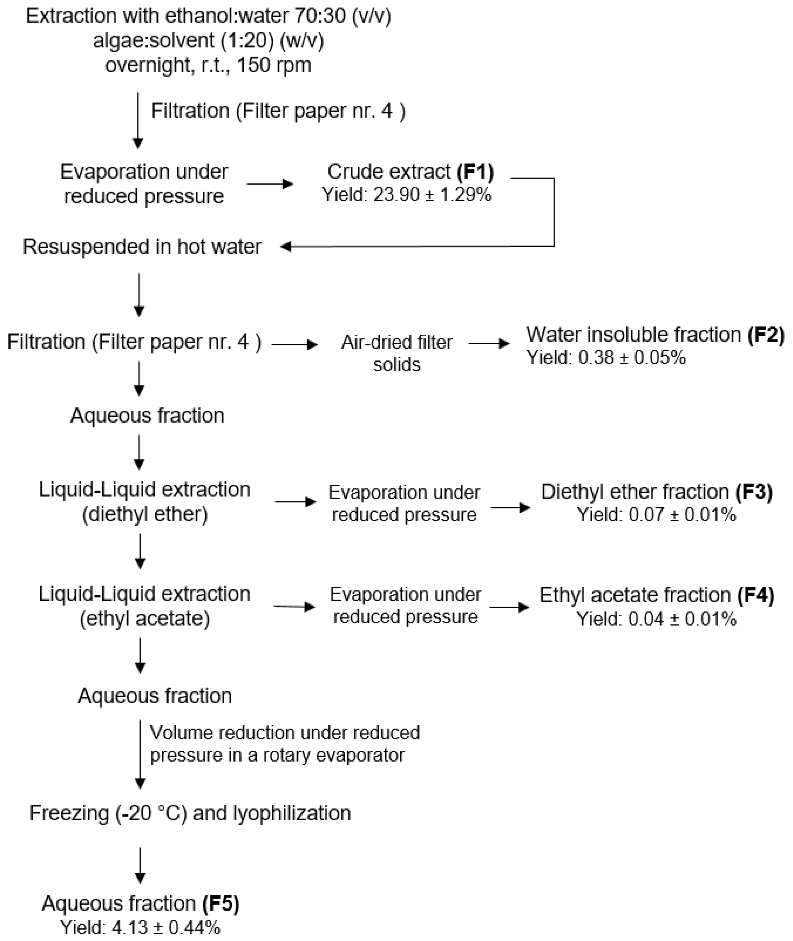
4.2. Extraction Procedure

To obtain different fractions from G. corneum, a sequential extraction methodology was performed (Figure 1). Solvents were selected according to EU Regulation No. 1223/2009 for cosmetic application and were obtained from VWR-BDH Chemicals (Fontenay-sous-Bois, France). Powdered seaweed (100 g) was extracted with ethanol: water (70:30) under constant stirring (150 rpm), over 17 h, protected from light. The hydroethanolic solution was posteriorly filtered (qualitative filter paper (FP) nr. 4, VWR International, Alcabideche, Portugal) and concentrated under vacuum at low temperature (<40 °C) in a rotary evaporator. In total, 20 g of the dried crude extract (F1) was resuspended in Milli-Q water (Advantage A10 Milli-Q lab, Merck, Darmstadt, Germany) previously warmed to 75 °C and filtered (FP nr. 4), affording a solid insoluble fraction (F2) and an aqueous fraction. After cooling to room temperature (r.t.), this last one was subjected to a liquid–liquid partition, firstly with diethyl ether (F3) and then, with ethyl acetate (F4), which were evaporated until dryness in a rotary evaporator. The remaining aqueous fraction (F5) was also evaporated until a half volume was obtained, and then it was frozen (−20 °C) and lyophilized. The extraction procedure flowchart is depicted in Figure 7.

4.3. Chemical Screening by 1H NMR

A preliminary chemical screening of all fractions (F1–F5) obtained from Gelidium corneum was attained by proton nuclear magnetic resonance (1H NMR) spectroscopy. Samples (c.a. 5–6 mg) were dissolved in 0.5 mL of deuterated solvents (CDCl3, MeOD, or D2O) (Sigma-Aldrich, St. Louis, MO, USA) and the 1H NMR spectra were recorded at 400.13 MHz on a Bruker AMX400 spectrometer (Bremen, Germany), at 25 °C. Chemical shifts (δ) are expressed in ppm and referenced to the residual solvent signal (δH 7.26 CDCl3, δH 3.31 MeOD, δH 4.79 D2O).

4.4. Antimicrobial Activity of Gelidium corneum Fractions against Microorganisms Associated with Skin Disorders

The antimicrobial activity of G. corneum fractions was evaluated against three microorganisms that belong to the skin’s natural microflora, namely, three Gram (+) bacteria, Staphylococcus epidermidis (DSM: 1798), Staphylococcus aureus (ATCC: 25923) and Cutibacterium acnes (DSM: 1897) obtained from the German Collection of Microorganisms and Cell Cultures (DSMZ). The growth conditions and media used for each microorganism were: trypticase soy broth (VWR-BDH Chemicals-Prolabo, Leuven, Belgium) + 0.3 % (w/w) of yeast extract (VWR-BDH Chemicals-Prolabo, Leuven, Belgium) at 37 °C for S. epidermidis; tryptic soy broth (VWR-BDH Chemicals-Prolabo, Leuven, Belgium) in anaerobic conditions at 37 °C for C. acnes, and lysogeny broth (LB) medium (VWR-BDH Chemicals-Prolabo, Leuven, Belgium) at 37 °C for S. aureus. Fractions were suspended in dimethyl sulfoxide (DMSO) at 100 mg/mL, and the assays were conducted on 96-well microplates with the fractions at a maximum concentration of 1000 μg/mL. Antimicrobial activity was evaluated in the exponential phase of each microorganism. Microorganisms’ growth was accompanied spectrophotometrically at 600 nm, and the results were expressed as a percentage of growth inhibition relative to the control (growth medium with microorganism and vehicle (DMSO)). For the fractions that affected the microorganisms’ growth by more than 50%, a dose–response was conducted (10–1000 μg/mL) and the IC50 was determined. Oxytetracycline was used as a positive control.

4.5. Mechanisms of Action Underlying the Antimicrobial Activity

To understand the mechanisms that could be underlying the antimicrobial effects, further tests were conducted with the fractions that exhibited the highest activity (inhibition >50% at 1 mg/mL).

4.5.1. Membrane Potential Analysis

The membrane potential variation assay was adapted from Clementi et al. [58], by combining the potentiometric dye bis-(1,3-dibutylbarbituric acid) trimethine oxonol (DiBAC4(3)) (Thermo Fisher Scientific, Waltham, MA, USA) with the DNA-staining dye propidium iodide (PI) (Sigma Aldrich, Darmstadt, Germany). The extracts with the highest activity were tested at ½ IC50, IC50 and 2 × IC50.
Freshly grown bacteria were centrifuged at 1000× g and washed twice with 1× PBS (Sigma-Aldrich, Darmstadt, Germany). The pellet was resuspended in 10 mL PBS buffer at 1.5 MacFarland, and 0.25% (v/v) of a stock glucose solution (1 M) was added to the mix. A blank was prepared with the same proportions but lacking the microbial solution. The microbial solution was then incubated at 37 °C for 30 min. A solution of carbonyl cyanide 4-(trifluoromethoxy) phenylhydrazone (FCCP) (Sigma-Aldrich, Darmstadt, Germany) was used as the positive control for hyperpolarization. A positive control for total membrane permeabilization was conducted with heat-treated (100 °C, 10 min) bacteria.
After incubation, 45 μL of DiBAC4(3) (25 μM) + 90 μL of PI (1 mg/mL) was added to the microbial solution, distributed through 96-well black plates (99 μL per well), and then incubated in the dark at 37 °C, over one hour. The samples were quickly added (1 μL per well) and the fluorescence read at 490/516 nm excitation/emission (DIBAC) and 535/617 nm (PI) in 30 s intervals for 10 min (Multimodal Synergy H1, BioTek® Instruments, Winooski, VT, USA). The results are presented in DiBAC4(3) relative fluorescence units (RFU).

4.5.2. Membrane Damage Assay

Membrane damage analysis was performed according to Pinteus et al. [48], as follows: A freshly overnight grown culture was centrifuged (4000 rpm, 5 min) and resuspended in sterile saline solution (0.85%) at an optical density of Abs600 = 0.5. Seaweed fractions were added at a concentration of ½ IC50, IC50 and 2 × IC50, and incubated over 4 h at 37 °C for S. epidermidis, and 6 h for C. acnes. DMSO was used as a negative control. Blanks were prepared with samples without microorganism. A positive control was prepared with a thermic treatment (100 °C, 10 min) to induce total membrane permeability. All suspensions were transferred to a black microplate and incubated with 2 μM Sytox Green (Thermo Fisher Scientific, Waltham, MA, USA) for 10 min in the microplate reader. The resultant fluorescence of the DNA-bound dye was quantified on a fluorescence microplate reader (Multimodal Synergy H1, BioTek® Instruments, Winooski, VT, USA). The membrane damage was determined in % of control (DMSO).

4.5.3. DNA Damaging Potential

DNA damaging potential was assessed following the methodology described by Hu et al. [59]. Plasmid (pGADT7—7987 bp) DNA (5 µL; 100 ng) was mixed with seaweed fractions (2 µL; 10 mg/mL) and ultrapure water (13 μL). The reaction mixture was incubated at 37 °C for 1 h before being loaded onto a 0.8% agarose gel containing 1% RedSafeTM. Gene Ruler 1 Kb DNA Ladder (5 µL) was also loaded onto the gel. Electrophoresis was then performed for 60 min under 85 V. DMSO was used in the same conditions as a negative control and ciprofloxacin (10 and 30 µg/mL) was used as positive control.

4.6. Data and Statistical Analysis

To determine possible significant differences relative to the control, a one-way analysis of variance (ANOVA) with Dunnett’s multiple comparison test was used. All data were checked for normality (Shapiro–Wilk test) and homoscedasticity (Levene´s test). When requirements for an ANOVA were not met, a non-parametric Kruskal–Wallis and Dunn’s multiple comparison test were applied. The IC50 values were determined using the GraphPad v9.3.1 software through the equation y = 100/(1 + 10 (X−Log IC50)). Calculations were carried out and final graphical representations were made using GraphPad v9.3.1 (GraphPad Software, La Jolla, CA, USA). All data were obtained from at least three independent experiments carried out in triplicate and are presented as standard error of the mean (SEM), with a significance level of 0.05 (p < 0.05).

5. Conclusions

With the increasing awareness on the importance of sustainable strategies for the development of new nature-based health and wellness products, this work addressed, for the first time, the potential of Gelidium corneum as a source of antimicrobial compounds for skin dysbiosis-related diseases. This seaweed is particularly interesting since it is already explored industrially for agar extraction. Thus, within a circular economy concept, it would be of high economic relevance to define a biorefinery concept to extract both antimicrobial compounds and agar, thus taking the maximum advantage of the same natural resource for maximum environmental and economic sustainability.
The results reveal that G. corneum contains compounds with high antimicrobial activity against two important skin opportunistic pathogens, C. acnes and S. epidermidis. The compounds concentrated in F2 (water insoluble fraction) and F3 (diethyl ether fraction) seem to affect both microorganisms’ growth by inducing changes in membrane polarization and by binding to DNA, promoting damage. This dual mechanism of action may provide therapeutic advantages for the treatment of skin dysbiosis-related diseases.

Author Contributions

Conceptualization, S.P., C.A., J.S., A.M. and R.P.; methodology, M.M., S.P., J.S., C.A., A.M. and H.G.; validation, S.P., C.A., A.M., T.M. and R.P.; formal analysis, M.M., S.P., C.A., A.M. and H.G.; investigation, M.M., S.P., J.S., C.A., A.M., T.M. and H.G.; resources, S.P., J.S., C.A., A.M., H.G. and R.P.; writing—original draft preparation, M.M., S.P., A.M. and C.A.; writing—review and editing, all authors; supervision, S.P., C.A., J.S., A.M. and R.P.; project administration, R.P; funding acquisition, S.P., C.A., J.S., A.M. and R.P. All authors have read and agreed to the published version of the manuscript.

Funding

This work was supported by the Portuguese Foundation for Science and Technology (FCT) through the POCI project—01-0247-FEDER-070155—ORCHESTRA—add value to ORChards through thE full valoriSaTion of macroalgae and by FEDER—European Regional Development Fund of the European Union, under the Portugal 2020 Programme, through COMPETE 2020—Competitiveness and Internationalization Operational Program. Additionally, we were also supported through the FCT projects: UIDP/04292/2020 and UIDB/04292/2020 granted to MARE—Marine and Environmental Sciences Centre; UIDP/Multi/04046/2020 and UIDB/04046/2020 granted to BioISI—Biosystems and Integrative Sciences Institute; CROSS-ATLANTIC (PTDC/BIA-OUT/29250/2017), co-financed by COMPETE (POCI-01-0145-FEDER-029250); and Molecules for Health (PTDC/BIA-BQM/28355/2017); National NMR Network (PTNMR), partially supported by Infrastructure Project N° 022161 (co-financed by FEDER through COMPETE 2020, POCI and PORL and FCT through PIDDAC). Additional support was provided by the Project Operational Programme MAR2020 through the project MAR-04.03.01-FEAMP-0101–Limo do Cais.

Institutional Review Board Statement

Not applicable.

Informed Consent Statement

Not applicable.

Data Availability Statement

Not applicable.

Conflicts of Interest

The authors declare no conflict of interest.

References

  1. Williams, P.A.; Phillips, G.O. 11—The use of hydrocolloids to improve food texture. In Texture in Food; McKenna, B.M., Ed.; Woodhead Publishing: Cambridge, UK, 2003; pp. 251–274. ISBN 185573673X. [Google Scholar] [CrossRef]
  2. Jiao, G.; Yu, G.; Zhang, J.; Ewart, H.S. Chemical Structures and Bioactivities of Sulfated Polysaccharides from Marine Algae. Mar. Drugs 2011, 9, 196–223. [Google Scholar] [CrossRef] [PubMed] [Green Version]
  3. El-Din, S.M.M.; Alagawany, N.I. Phytochemical Constituents and Anticoagulation Property of Marine Algae Gelidium crinale, Sargassum hornschuchii and Ulva linza. Thalass. Int. J. Mar. Sci. 2019, 35, 381–397. [Google Scholar] [CrossRef]
  4. Öztürk, B.Y.; Gürsu, B.Y.; Dağ, I. Antibiofilm and antimicrobial activities of green synthesized silver nanoparticles using marine red algae Gelidium corneum. Process Biochem. 2019, 89, 208–219. [Google Scholar] [CrossRef]
  5. Martínez-Sanz, M.; Gómez-Mascaraque, L.G.; Ballester, A.R.; Martinez-Abad, A.; Brodkorb, A.; López-Rubio, A. Production of unpurified agar-based extracts from red seaweed Gelidium sesquipedale by means of simplified extraction protocols. Algal Res. 2019, 38, 101420. [Google Scholar] [CrossRef]
  6. Castejón, N.; Parailloux, M.; Izdebska, A.; Lobinski, R.; Fernandes, S.C.M. Valorization of the Red Algae Gelidium sesquipedale by Extracting a Broad Spectrum of Minor Compounds Using Green Approaches. Mar. Drugs 2021, 19, 574. [Google Scholar] [CrossRef] [PubMed]
  7. Byrd, A.L.; Belkaid, Y.; Segre, J.A. The human skin microbiome. Nat. Rev. Microbiol. 2018, 16, 143–155. [Google Scholar] [CrossRef]
  8. Dagnelie, M.; Corvec, S.; Timon-David, E.; Khammari, A.; Dréno, B. Cutibacterium acnes and Staphylococcus epidermidis: The unmissable modulators of skin inflammatory response. Exp. Dermatol. 2021, 31, 406–412. [Google Scholar] [CrossRef] [PubMed]
  9. Fournière, M.; Latire, T.; Souak, D.; Feuilloley, M.G.J.; Bedoux, G. Staphylococcus epidermidis and Cutibacterium acnes: Two Major Sentinels of Skin Microbiota and the Influence of Cosmetics. Microorganisms 2020, 8, 1752. [Google Scholar] [CrossRef]
  10. Claudel, J.-P.; Auffret, N.; Leccia, M.-T.; Poli, F.; Corvec, S.; Dréno, B. Staphylococcus epidermidis: A Potential New Player in the Physiopathology of Acne? Dermatology 2019, 235, 287–294. [Google Scholar] [CrossRef] [PubMed]
  11. Sfriso, R.; Egert, M.; Gempeler, M.; Voegeli, R.; Campiche, R. Revealing the secret life of skin—With the microbiome you never walk alone. Int. J. Cosmet. Sci. 2019, 42, 116–126. [Google Scholar] [CrossRef]
  12. Williams, H.C.; Dellavalle, R.; Garner, S. Acne vulgaris. Lancet 2011, 379, 361–372. [Google Scholar] [CrossRef]
  13. Blaskovich, M.A.T.; Elliott, A.G.; Kavanagh, A.M.; Ramu, S.; Cooper, M.A. In vitro Antimicrobial Activity of Acne Drugs Against Skin-Associated Bacteria. Sci. Rep. 2019, 9, 14658. [Google Scholar] [CrossRef] [PubMed] [Green Version]
  14. Dréno, B.; Pécastaings, S.; Corvec, S.; Veraldi, S.; Khammari, A.; Roques, C. Cutibacterium acnes (Propionibacterium acnes) and acne vulgaris: A brief look at the latest updates. J. Eur. Acad. Dermatol. Venereol. 2018, 32 (Suppl. S2), 5–14. [Google Scholar] [CrossRef] [PubMed] [Green Version]
  15. Zaenglein, A.L.; Pathy, A.L.; Schlosser, B.J.; Alikhan, A.; Baldwin, H.E.; Berson, D.S.; Bowe, W.P.; Graber, E.M.; Harper, J.C.; Kang, S.; et al. Guidelines of care for the management of acne vulgaris. J. Am. Acad. Dermatol. 2016, 74, 945–973.e33. [Google Scholar] [CrossRef] [PubMed] [Green Version]
  16. Otto, M. Staphylococcus epidermidis—The ‘accidental’ pathogen. Nat. Rev. Genet. 2009, 7, 555–567. [Google Scholar] [CrossRef] [PubMed] [Green Version]
  17. Costantino, V.; Fattorusso, E.; Menna, M.; Taglialatela-Scafati, O. Chemical diversity of bioactive marine natural products: An illustrative case study. Curr. Med. Chem. 2004, 11, 1671–1692. [Google Scholar] [CrossRef]
  18. Pinteus, S.; Lemos, M.F.; Alves, C.; Neugebauer, A.; Silva, J.; Thomas, O.; Botana, L.M.; Gaspar, H.; Pedrosa, R. Marine invasive macroalgae: Turning a real threat into a major opportunity—The biotechnological potential of Sargassum muticum and Asparagopsis armata. Algal Res. 2018, 34, 217–234. [Google Scholar] [CrossRef]
  19. Valentão, P.; Trindade, P.; Gomes, D.; de Pinho, P.G.; Mouga, T.; Andrade, P.B. Codium tomentosum and Plocamium cartilagineum: Chemistry and antioxidant potential. Food Chem. 2009, 119, 1359–1368. [Google Scholar] [CrossRef]
  20. Rodrigues, D.; Costa-Pinto, A.R.; Sousa, S.; Vasconcelos, M.W.; Pintado, M.M.; Pereira, L.; Rocha-Santos, T.A.P.; Da Costa, J.P.; Silva, A.M.S.; Duarte, A.C.; et al. Sargassum muticum and Osmundea pinnatifida Enzymatic Extracts: Chemical, Structural, and Cytotoxic Characterization. Mar. Drugs 2019, 17, 209. [Google Scholar] [CrossRef] [Green Version]
  21. Date, Y.; Sakata, K.; Kikuchi, J. Chemical profiling of complex biochemical mixtures from various seaweeds. Polym. J. 2012, 44, 888–894. [Google Scholar] [CrossRef] [Green Version]
  22. Cakmak, Y.S.; Kaya, M.; Ozusaglam, M.A. Biochemical composition and bioactivity screening of various extracts from Dunaliella salina, a green microalga. EXCLI J. 2014, 13, 679–690. [Google Scholar] [CrossRef] [PubMed]
  23. Patel, D.J.; Bhatia, N. Oral Antibiotics for Acne. Am. J. Clin. Dermatol. 2020, 22, 193–204. [Google Scholar] [CrossRef] [PubMed]
  24. Adamski, Z.; Gornowicz-Porowska, J.; Sobkowska, D.; Kaszuba, K.; Czajkowski, R. Acne—Therapeutic challenges to the cooperation between a dermatologist and a cosmetologist. Adv. Dermatol. Allergol. 2021, 38, 21–31. [Google Scholar] [CrossRef] [PubMed]
  25. Baldwin, H. Oral Antibiotic Treatment Options for Acne Vulgaris. J. Clin. Aesthetic Dermatol. 2020, 13, 26–32. [Google Scholar]
  26. Karadag, A.S.; Kayıran, M.A.; Wu, C.; Chen, W.; Parish, L.C. Antibiotic resistance in acne: Changes, consequences and concerns. J. Eur. Acad. Dermatol. Venereol. 2020, 35, 73–78. [Google Scholar] [CrossRef] [PubMed]
  27. Yang, J.H.; Yoon, J.Y.; Kwon, H.H.; Min, S.; Moon, J.; Suh, D.H. Seeking new acne treatment from natural products, devices and synthetic drug discovery. Derm. Endocrinol. 2017, 9, e1356520. [Google Scholar] [CrossRef] [PubMed] [Green Version]
  28. Samri, N.; Hsaine, L.; Elkafhi, S.; Khlifi, S.; Etahiri, S. Antimicrobial properties of seven brown algae harvested from the coast of sidi bouzid (El Jadida-Morocco). Int. J. Pharm. Pharm. Sci. 2020, 5, 57–62. [Google Scholar] [CrossRef]
  29. Metidji, H.; Dob, T.; Mohamed, T.; Krimat, S.; Ksouri, A.; Nouasri, A. In vitro screening of secondary metabolites and evalu-ationof antioxidant, antimicrobial and cytotoxic properties of Gelidium sesquipedale Thuret et Bornet red seaweed from Algeria. J. Mater. Environ. Sci. 2015, 6, 3184–3196. [Google Scholar]
  30. El Wahidi, M.; El Amraoui, B.; El Amraoui, M.; Bamhaoud, T. Screening of antimicrobial activity of macroalgae extracts from the Moroccan Atlantic coast. Ann. Pharm. Fr. 2015, 73, 190–196. [Google Scholar] [CrossRef]
  31. Pérez, M.J.; Falqué, E.; Domínguez, H. Antimicrobial Action of Compounds from Marine Seaweed. Mar. Drugs 2016, 14, 52. [Google Scholar] [CrossRef] [Green Version]
  32. Choi, J.-S.; Bae, H.-J.; Kim, S.-J.; Choi, I.S. In vitro antibacterial and anti-inflammatory properties of seaweed extracts against acne inducing bacteria, Propionibacterium acnes. J. Environ. Biol. 2011, 32, 313–318. [Google Scholar] [PubMed]
  33. Januário, A.P.; Félix, R.; Félix, C.; Reboleira, J.; Valentão, P.; Lemos, M.F.L. Red Seaweed-Derived Compounds as a Potential New Approach for Acne Vulgaris Care. Pharmaceutics 2021, 13, 1930. [Google Scholar] [CrossRef]
  34. Benarroch, J.; Asally, M. The Microbiologist’s Guide to Membrane Potential Dynamics. Trends Microbiol. 2020, 28, 304–314. [Google Scholar] [CrossRef] [PubMed] [Green Version]
  35. Lee, D.-Y.D.; Prindle, A.; Liu, J.; Süel, G.M. SnapShot: Electrochemical Communication in Biofilms. Cell 2017, 170, 214–214.e1. [Google Scholar] [CrossRef]
  36. Prindle, A.; Liu, J.; Asally, M.; Ly, S.; Garcia-Ojalvo, J.; Süel, G.M. Ion channels enable electrical communication in bacterial communities. Nature 2015, 527, 59–63. [Google Scholar] [CrossRef] [PubMed] [Green Version]
  37. Bruni, G.N.; Weekley, R.A.; Dodd, B.J.T.; Kralj, J.M. Voltage-gated calcium flux mediates Escherichia coli mechanosensation. Proc. Natl. Acad. Sci. USA 2017, 114, 9445–9450. [Google Scholar] [CrossRef] [PubMed] [Green Version]
  38. Sirec, T.; Benarroch, J.M.; Buffard, P.; Garcia-Ojalvo, J.; Asally, M. Electrical Polarization Enables Integrative Quality Control during Bacterial Differentiation into Spores. iScience 2019, 16, 378–389. [Google Scholar] [CrossRef] [PubMed] [Green Version]
  39. Kaim, G.; Dimroth, P. ATP synthesis by F-type ATP synthase is obligatorily dependent on the transmembrane voltage. EMBO J. 1999, 18, 4118–4127. [Google Scholar] [CrossRef] [PubMed] [Green Version]
  40. Strahl, H.; Hamoen, L.W. Membrane potential is important for bacterial cell division. Proc. Natl. Acad. Sci. USA 2010, 107, 12281–12286. [Google Scholar] [CrossRef] [Green Version]
  41. Lee, D.-Y.D.; Galera-Laporta, L.; Bialecka-Fornal, M.; Moon, E.C.; Shen, Z.; Briggs, S.P.; Garcia-Ojalvo, J.; Süel, G.M. Magnesium Flux Modulates Ribosomes to Increase Bacterial Survival. Cell 2019, 177, 352–360.e13. [Google Scholar] [CrossRef] [Green Version]
  42. Patra, J.K.; Das, G.; Baek, K.-H. Antibacterial mechanism of the action of Enteromorpha linza L. essential oil against Escherichia coli and Salmonella Typhimurium. Bot. Stud. 2015, 56, 13. [Google Scholar] [CrossRef] [PubMed] [Green Version]
  43. Wu, Y.; Bai, J.; Zhong, K.; Huang, Y.; Gao, H. A dual antibacterial mechanism involved in membrane disruption and DNA binding of 2R,3R-dihydromyricetin from pine needles of Cedrus deodara against Staphylococcus aureus. Food Chem. 2017, 218, 463–470. [Google Scholar] [CrossRef] [PubMed]
  44. Patra, J.K.; Baek, K.-H. Antibacterial Activity and Action Mechanism of the Essential Oil from Enteromorpha linza L. against Foodborne Pathogenic Bacteria. Molecules 2016, 21, 388. [Google Scholar] [CrossRef] [PubMed] [Green Version]
  45. El Shafay, S.M.; Ali, S.S.; El-Sheekh, M. Antimicrobial activity of some seaweeds species from Red sea, against multidrug resistant bacteria. Egypt. J. Aquat. Res. 2016, 42, 65–74. [Google Scholar] [CrossRef] [Green Version]
  46. Tang, Y.-L.; Shi, Y.-H.; Zhao, W.; Hao, G.; Le, G.-W. Interaction of MDpep9, a novel antimicrobial peptide from Chinese tra-ditional edible larvae of housefly, with Escherichia coli genomic DNA. Food Chem. 2009, 115, 867–872. [Google Scholar] [CrossRef]
  47. Brogden, K.A. Antimicrobial peptides: Pore formers or metabolic inhibitors in bacteria? Nat. Rev. Microbiol. 2005, 3, 238–250. [Google Scholar] [CrossRef]
  48. Pinteus, S.; Lemos, M.F.; Simões, M.; Alves, C.; Silva, J.; Gaspar, H.; Martins, A.; Rodrigues, A.; Pedrosa, R. Marine invasive species for high-value products’ exploration—Unveiling the antimicrobial potential of Asparagopsis armata against human pathogens. Algal Res. 2020, 52, 102091. [Google Scholar] [CrossRef]
  49. Shannon, E.; Abu-Ghannam, N. Antibacterial Derivatives of Marine Algae: An Overview of Pharmacological Mechanisms and Applications. Mar. Drugs 2016, 14, 81. [Google Scholar] [CrossRef] [PubMed]
  50. Subramanian, M.; Soundar, S.; Mangoli, S. DNA damage is a late event in resveratrol mediated inhibition of Escherichia coli. Free Radic. Res. 2016, 50, 708–719. [Google Scholar] [CrossRef] [PubMed]
  51. Da, X.; Nishiyama, Y.; Tie, D.; Hein, K.Z.; Yamamoto, O.; Morita, E. Antifungal activity and mechanism of action of Ougon (Scutellaria root extract) components against pathogenic fungi. Sci. Rep. 2019, 9, 1683. [Google Scholar] [CrossRef] [PubMed]
  52. Lei, J.; Sun, L.; Huang, S.; Zhu, C.; Li, P.; He, J.; Mackey, V.; Coy, D.H.; He, Q. The antimicrobial peptides and their potential clinical applications. Am. J. Transl. Res. 2019, 11, 3919–3931. [Google Scholar] [PubMed]
  53. He, F.; Yang, Y.; Yang, G.; Yu, L. Studies on antibacterial activity and antibacterial mechanism of a novel polysaccharide from Streptomyces virginia H03. Food Control 2010, 21, 1257–1262. [Google Scholar] [CrossRef]
  54. Shanmughapriya, S.; Manilal, A.; Sujith, S.; Selvin, J.; Kiran, G.S.; Natarajaseenivasan, K. Antimicrobial activity of seaweeds extracts against multiresistant pathogens. Ann. Microbiol. 2008, 58, 535–541. [Google Scholar] [CrossRef]
  55. Xu, N.; Fan, X.; Yan, X.; Li, X.; Niu, R.; Tseng, C. Antibacterial bromophenols from the marine red alga Rhodomela confervoides. Phytochemistry 2003, 62, 1221–1224. [Google Scholar] [CrossRef]
  56. Manilal, A.; Sujith, S.; Kiran, G.S.; Selvin, J.; Shakir, C.; Gandhimathi, R.; Lipton, A.P. Antimicrobial potential and seasonality of red algae collected from the southwest coast of India tested against shrimp, human and phytopathogens. Ann. Microbiol. 2009, 59, 207–219. [Google Scholar] [CrossRef]
  57. Rodrigues, D.; Alves, C.; Horta, A.; Pinteus, S.; Silva, J.; Culioli, G.; Thomas, O.P.; Pedrosa, R. Antitumor and Antimicrobial Potential of Bromoditerpenes Isolated from the Red Alga, Sphaerococcus coronopifolius. Mar. Drugs 2015, 13, 713–726. [Google Scholar] [CrossRef] [Green Version]
  58. Clementi, E.A.; Marks, L.; Roche-Håkansson, H.; Håkansson, A.P. Monitoring Changes in Membrane Polarity, Membrane Integrity, and Intracellular Ion Concentrations in Streptococcus pneumoniae Using Fluorescent Dyes. J. Vis. Exp. 2014. [Google Scholar] [CrossRef] [PubMed] [Green Version]
  59. Hu, Q.-P.; Cao, X.-M.; Hao, D.-L.; Zhang, L.-L. Chemical Composition, Antioxidant, DNA Damage Protective, Cytotoxic and Antibacterial Activities of Cyperus rotundus Rhizomes Essential Oil against Foodborne Pathogens. Sci. Rep. 2017, 7, 45231. [Google Scholar] [CrossRef] [PubMed] [Green Version]
Figure 1. 1H NMR (400 MHz) spectra of Gelidium corneum fractions (F1–F5).
Antibiotics 11 00481 g001
Figure 2. Antimicrobial activity of Gelidium corneum fractions (1000 μg/mL) against (a) Cutibacterium acnes, (b) Staphylococcus aureus, and (c) Staphylococcus epidermidis. Values in each column represent the mean ± SEM of three independent experiments carried out in triplicate. Symbol (*) represents significant differences when compared to vehicle (ANOVA, Dunnett’s test, p < 0.05).
Antibiotics 11 00481 g002
Figure 3. Cutibacterium acnes membrane potential when exposed to Gelidium corneum F2 (water insoluble) and F3 (diethyl ether) fractions at ½ IC50; IC50 and 2 × IC50, labelled with DiBAC4(3) probe; (a) 30 s interval readings; (b) after 10 min. FCCP (10 mM) was used as positive control and DMSO as negative control. Each value and bars represent the average of three independent experiments. Vertical lines represent the standard error of the mean (SEM). Symbol (*) represents significant differences when compared to the control. Symbol (#) represents significant differences when compared to FCCP (ANOVA, Dunnett’s test, p < 0.05).
Antibiotics 11 00481 g003
Figure 4. Staphylococcus epidermidis membrane potential when exposed to Gelidium corneum F2 (water insoluble) and F3 (diethyl ether) fractions at ½ IC50; IC50 and 2 × IC50, labelled with DiBAC4(3) probe; (a) 30 s interval readings; (b) after 10 min. FCCP (100 µM) was used as positive control and DMSO as negative control. Each value and bars represent the average of three independent experiments. Vertical lines represent the standard error of the mean (SEM). Symbol (*) represents significant differences when compared to the control. Symbol (#) represents significant differences when compared to FCCP (ANOVA, Dunnett’s test, p < 0.05).
Antibiotics 11 00481 g004
Figure 5. Cutibacterium acnes (a) and Staphylococcus epidermidis (b) membrane integrity when exposed to Gelidium corneum F2 and F3 fractions at ½ IC50; IC50 and 2 × IC50, labelled with Sytox Green probe. Bacterial cells exposed to a heat treatment were used as a positive control. DMSO was used as negative control. Each value represents the average of three independent experiments. Bars represent the standard error of the mean (SEM). Symbols represent significant differences when compared to the heat-treated cells (*) and (#) to control (ANOVA, Dunnett’s test, p < 0.05).
Antibiotics 11 00481 g005
Figure 6. Electrophoresis gel. 1—Marker; 2—DNA; 3—negative control (DMSO); 4—10 µg/mL ciprofloxacin; 5—30 µg/mL ciprofloxacin; 6—negative control (DMSO); 7—F2 (water insoluble) fraction (1 mg/mL); 8—F3 (diethyl ether) fraction (1 mg/mL). Agarose at 0.8%, ran at 85 V for 1 h. Image was obtained through gel imaging system (Gel doc). This figure is representative of three independent experiments.
Antibiotics 11 00481 g006
Figure 7. Extraction methodology to obtain Gelidium corneum fractions (F1–F5).
Antibiotics 11 00481 g007
Table 1. Dose–response analysis of Gelidium corneum F2 (water insoluble) and F3 (diethyl ether) fractions against Staphylococcus epidermidis and Cutibacterium acnes.
FractionStaphylococcus epidermidisCutibacterium acnes
IC50 (µg/mL)
F253.29 (48.75–57.91)16.10 (7.27–23.02)
F3102.80 (87.15–122.30)51.04 (43.36–59.74)
Oxytetracycline12.40 (11.22–16.13)0.07 (0.05–0.09)
Publisher’s Note: MDPI stays neutral with regard to jurisdictional claims in published maps and institutional affiliations.

