Article

Evidence of Linezolid Resistance and Virulence Factors in Enterococcus spp. Isolates from Wild and Domestic Ruminants, Italy

by
Camilla Smoglica
1,*,
Alberto Vergara
1,
Simone Angelucci
1,2,
Anna Rita Festino
1,
Antonio Antonucci
2,
Fulvio Marsilio
1 and
Cristina Esmeralda Di Francesco
1
1
Faculty of Veterinary Medicine, University of Teramo, Loc. Piano D’Accio, 64100 Teramo, TE, Italy
2
Maiella National Park, Wildlife Research Center, Viale del Vivaio, 65023 Caramanico Terme, PE, Italy
*
Author to whom correspondence should be addressed.
Antibiotics 2022, 11(2), 223; https://doi.org/10.3390/antibiotics11020223
Submission received: 21 January 2022 / Revised: 5 February 2022 / Accepted: 8 February 2022 / Published: 10 February 2022
(This article belongs to the Special Issue Antimicrobial and Antimicrobial Resistance in the Environment)

Abstract

:
The aim of this study was to evaluate the resistance patterns against selected critically and highly important antibiotics (quinupristin/dalfopristin, vancomycin, and linezolid) in 48 Enterococcus isolates obtained from wild (red deer and Apennine chamois) and domestic (cattle, sheep, and goats) ruminants living with varying degrees of sympatry in the protected area of Maiella National Park (central Italy). According to CLSI breakpoints, 9 out of 48 isolates (18.8%) showed resistance to at least one antibiotic. One Apennine chamois isolate was resistant to all tested antibiotics. The PCR screening of related resistance genes highlighted the occurrence of msrC or cfrD in seven Enterococcus resistant isolates. In addition, msrC and vanC genes were amplified in susceptible isolates. Specific sequences of virulence genes (gelE, ace, efa, asa1, and esp) related to pathogenic enterococci in humans were amplified in 21/48 isolates (43.75%), belonging mostly to wild animals (15/21; 71.42%). This is the first report of linezolid-resistant enterococci harboring virulence genes in Italian wildlife with special regard to the red deer and Apennine chamois species. The results allow us to evaluate the potential role of wild animals as indicators of antibiotic resistance in environments with different levels of anthropic pressure.

1. Introduction

Antimicrobial resistance (AMR) is a significant public health threat related to many factors, including the overuse or misuse of antibiotics in veterinary and human medicine. The gastrointestinal tracts of animals are considered the largest reservoir of bacteria potentially involved in the spread of antibiotic resistance [1]. Among these bacteria, the genus Enterococcus includes commensal Gram-positive bacteria that colonize the gastrointestinal tracts of many animal species and humans. These bacteria are important nosocomial pathogens characterized by intrinsic and acquired virulence determinants and resistance mechanisms against different antibiotics [2]. In addition, enterococci are considered responsible for several animal diseases, such as mastitis, endocarditis, diarrhea, and septicemia in bovine, pets, pigs, and poultry [3]. The pathogenicity of these bacteria is related to antibiotic resistance mechanisms and virulence factors that promote colonization in host cells and damage of tissues by means of protein and peptides [4]. Due to their adaptability to the environment and their genomic plasticity, these ubiquitous microorganisms can be employed as sentinel bacteria suitable for antibiotic resistance surveillance systems in humans, domestic animals, and wildlife. In recent years, the emergence and rapid spread of vancomycin-resistant enterococci (VRE) has resulted in the increased use of alternative molecules, such as linezolid (LNZ) and quinupristin/dalfopristin (QD), to treat VRE-related infections. These drugs are considered as the last resort in human medicine and are included in the WHO list of critically (LNZ) and highly (QD) important antibiotics [1,5]. Linezolid is an oxazolidinone that is highly effective in treating serious infections caused by multidrug-resistant Gram-positive bacteria in humans [2]. Although this molecule is not approved worldwide for use in veterinary medicine, the mobile genetic elements involved in the oxazolidinone resistance have been detected in livestock during the last few years [2,5]. Indeed, LNZ-resistant enterococci have been obtained from food-producing animals in the USA, Europe, Asia, and Africa [5,6,7,8,9,10,11]. Quinupristin/dalfopristin is a streptogramins B and streptogramins A compound that is particularly useful in treating nosocomial infections caused by resistant E. faecium. After the first detection in 1997, other evidence of QD-resistant isolates has been highlighted in several countries in both humans and food-producing animals [5,12]. These data suggest that the emergence of LNZ- and QD-resistant enterococci may reduce therapeutic options for the successful treatment of VRE infections in humans. Therefore, the monitoring of environmental sources (i.e., soil, water, and wild animals) that are potentially able to harbor resistant bacteria and their relative genetic elements, including pathogens hard to treat with currently available antibiotics, is considered relevant for AMR surveillance inspired by a “One Health” approach [13]. Wildlife is generally less or not at all treated with antimicrobials but may acquire resistant bacteria from the environment [14], especially where a co-existence of domestic animals, livestock, agriculture, and other human activities is widely established. In this regard, the aim of this study was to investigate the AMR against quinupristin/dalfopristin, vancomycin, and linezolid in Enterococcus spp. isolates from wild and domestic ungulates living in Maiella National Park (Central Italy) with varying levels of sympatry established using georeferencing data. In addition, in order to evaluate the potential pathogenicity of microorganisms under study, specific virulence genes were investigated.

2. Results

2.1. Bacterial Isolation and Antibiotic Susceptibility Test

One or more Enterococcus species were isolated from fecal pool samples with a total of 48 bacterial strains. In detail, 17 isolates were obtained from the group of sympatric populations (four chamois and three goats; five red deer and five sheep) and 31 from the non-sympatric animals (16 goats, 8 chamois, 7 cattle). Based on Vitek 2 analysis, 15 E. gallinarum, 12 E. faecium, 11 E. faecalis, 6 E. hirae, and 4 E. casseliflavus species were identified from both wild and domestic animals, as reported in Table 1.
In greater detail, five E. faecalis, four E. faecium, three E. gallinarum, three E. hirae, and two E. casseliflavus were recovered from sympatric animals. Twelve E. gallinarum, eight E. faecium, six E. faecalis, three E. hirae, and two E. casseliflavus were isolated from non-sympatric animals.
Out of 48 isolates, 11 acquired/transferable resistant bacteria were identified, while the intrinsic resistance for QD (E. faecalis) and VAN (E. casseliflavus and E. gallinarum) was not considered. Nine enterococci were resistant to QD, seven to LNZ, and one to VAN. In detail, phenotypic resistance isolates were detected in three wild sympatric, two domestic sympatric, and four domestic non-sympatric animals. Only one E. faecium isolate, detected from an Apennine chamois belonging to the sympatric animal group, showed multiple resistance to QD, LNZ, and VAN.

2.2. Antibiotic Resistance Genes and Virulence Determinants

Among the QD resistance genes investigated, the msrC gene was detected in 20/48 (41.66%) isolates, including one QD intrinsically resistant isolate and six QD acquired/transferable resistant bacteria. The remaining 13 enterococci results were considered sensitive to QD by the antibiotic susceptibility analysis. The VAN resistance genes were not amplified, except for the vanC1 and vanC2 observed in nine intrinsically resistant enterococci and in one sensitive isolate as reported in Table 1. All enterococci under study were negative for the LNZ resistance genes optrA, cfr, cfr(B), and poxtA, but the cfr(D) gene was amplified in all the LNZ-resistant isolates.
In 21/48 (43.75%) isolates, the virulence gene fragments under study were amplified, except for the hyl gene. The most representative gene was gelE (17/48, 35.41%), followed by asa1 (12/48, 25%), efa (11/48, 22.91%), ace (4/48, 0.08%), and esp (2/48, 0.04%). In detail, gelE and efa genes were predominant in wild animals (94.11% and 81.81%, respectively), and all four ace-positive isolates were obtained from Apennine chamois populations (Table 1). Finally, the asa1 fragment was amplified in both wild and domestic animals. Co- occurrence of virulence genes was observed in several isolates. The gelE/efa and gelE/asa1 genes were observed in three isolates, while eight isolates showed multiple positivity for gelE/efa/asa1, gelE/efa/esp, gelE/efa/ace, or esp/efa/asa1, along with two isolates being positive for gelE/efa/ace/asa1. Table 1 shows the details of gene occurrence in the samples under study.
For both antibiotic resistance and virulence genes, the analysis of amplicons confirmed the specificity of PCR reactions, showing an identity between 98–99% (coverage 93–96%) with homologous sequences deposited in GenBank.

3. Discussion

In this study, the resistance against critically and highly important antibiotics in enterococci isolated from wild and domestic ruminants was tested in order to provide new information about the spread of resistance to these antibiotics in the environment. Among them, VAN was selected for the relevance of VRE in human health, while LNZ and QD were investigated because they are considered alternative molecules to treat VRE-infections.
In Europe, several authors have tested enterococci recovered from free-ranging terrestrial wild mammals against different antibiotics. Isolates from chamois have shown resistant results to tetracyclines, erythromycin, and VAN in Poland and Slovakia [15,16]. During the last few years in Spain and Portugal, the enterococci obtained from carnivores, ungulates, and wild rabbits have shown a phenotypic or genetic resistance against QD and LNZ in wild boars and Iberian wolves; VAN in rabbits, wild boars, and roe deer; and both QD and VAN in foxes [17,18,19,20,21,22,23,24]. A similar investigation was carried out in Italy (Tuscany region) involving different species, but only VAN resistance in three enterococci sourced from a wolf, a mouflon, and a wild boar was observed [14]. Compared to the aforementioned papers, the present study described for the first time enterococci resistant to LNZ and QD in Italian free-ranging Apennine chamois and red deer.
LNZ resistance was only described in one isolate from sympatric Apennine chamois, two isolates from sympatric red deer, and four bacteria from non-sympatric goats. While LNZ is not licensed for food-producing animals, LNZ-resistant isolates have been reported in European countries in domestic animals, and recent data in wildlife have aroused interest on environmental contamination [6,7,8,9,10,11,22]. Furthermore, molecular investigations have allowed the detection of the LNZ resistance gene, cfr(D), in all phenotypical resistant E. faecium and E. gallinarum. This gene has recently been described as a new variant of the cfr gene in E. faecium from human clinical isolates in France, Ireland, the Netherlands, and Australia [25,26,27,28], and in E. faecalis from Spanish hospital isolates [11].
The detection of VAN resistance genes was mostly related to intrinsically resistant isolates, but, interestingly, a positive result was obtained from a sensitive E. faecium, while in the resistant E. faecium none of the investigated VAN genes was detected. Other factors may be involved in the expression of genetic information or in the phenotypic resistance. As suggested by other authors, these factors may be related to environmental conditions and may modify the expression of a specific gene and the phenotypic profile. Specifically, the modulation role of collective resistance was described, including mechanisms where the phenotypic behavior of a bacterium was modified by communities of bacteria (i.e., biofilm formation and indirect resistance). Additionally, a low, host-related growth rate or the presence of specific metabolites, such as oxygens radicals, may have modified resistance [29].
The msrC gene was amplified in both QD-resistant and sensitive isolates, suggesting that this gene may be silent or involved in resistance to other antibiotics not tested in this study. Indeed, this gene has been related to erythromycin-resistant enterococci derived from domestic animals and different wild mammals [5,14].
Furthermore, the patterns of resistance observed in this study showed some differences in relation to the settings and the populations sampled. The resistant isolates were found in sympatric wild and domestic animals (Apennine chamois and goats; red deer and sheep) and in non-sympatric livestock (goats), rather than in non-sympatric Apennine chamois living in territories where human activities are limited and extensive livestock is not admitted. This evidence suggests the potential role of interactions with livestock and a shared environment as sources of antibiotic resistance determinants for wildlife: using the same land may provide an opportunity to share the resistome.
The virulence genes detected in this study are relevant in nosocomial infections and they have a direct effect on host colonization and immune response escapement [30]. The encoding gene of the aggregation substance (gelE) was detected mostly in resistant bacteria from red deer and, along with the genes encoding the endocarditis antigen (efa), the collagen-binding protein (ace), and the aggregation substance (asa1), in isolates from Apennine chamois. The gelE gene appears to increase the biofilm formation or aggregation, and it was previously detected in wild animals in Poland, along with the efa gene that provides a similar function [31]. In Italy, the detection of gelE, ace, and asa1 was reported in different species of wild mammals [14]. Interestingly, isolates with three or four virulence genes found in this study were mainly distributed in Apennine chamois samples.
In detail, out of twelve isolates from sympatric and non-sympatric Apennine chamois, eleven bacteria showed occurrence of virulence genes. As suggested from other authors, these multi-virulent profiles may be related to the adaptation of the hosts rather than to an increase in virulence [31]. However, some strains harboring virulence factors also showed resistance to highly or critically important antibiotics, such as two isolates from Apennine chamois, enhancing their potential pathogenic roles. In addition, the virulence factors found in E. gallinarum, E. casseliflavus, and E. hirae species have been poorly investigated in previous studies [4].
In the future, a whole-genome analysis of these isolates could improve the evaluation of the relationships among the bacteria, environmental sources, and animals investigated. Indeed, the coexistence in commensal enterococci of resistant profiles against medically important antibiotics, along with several virulence genes, highlights the need to investigate the impact of human activities and, in particular, the food-producing livestock industry on the environment and its role as a potential source of AMR determinants [13,32].

4. Materials and Methods

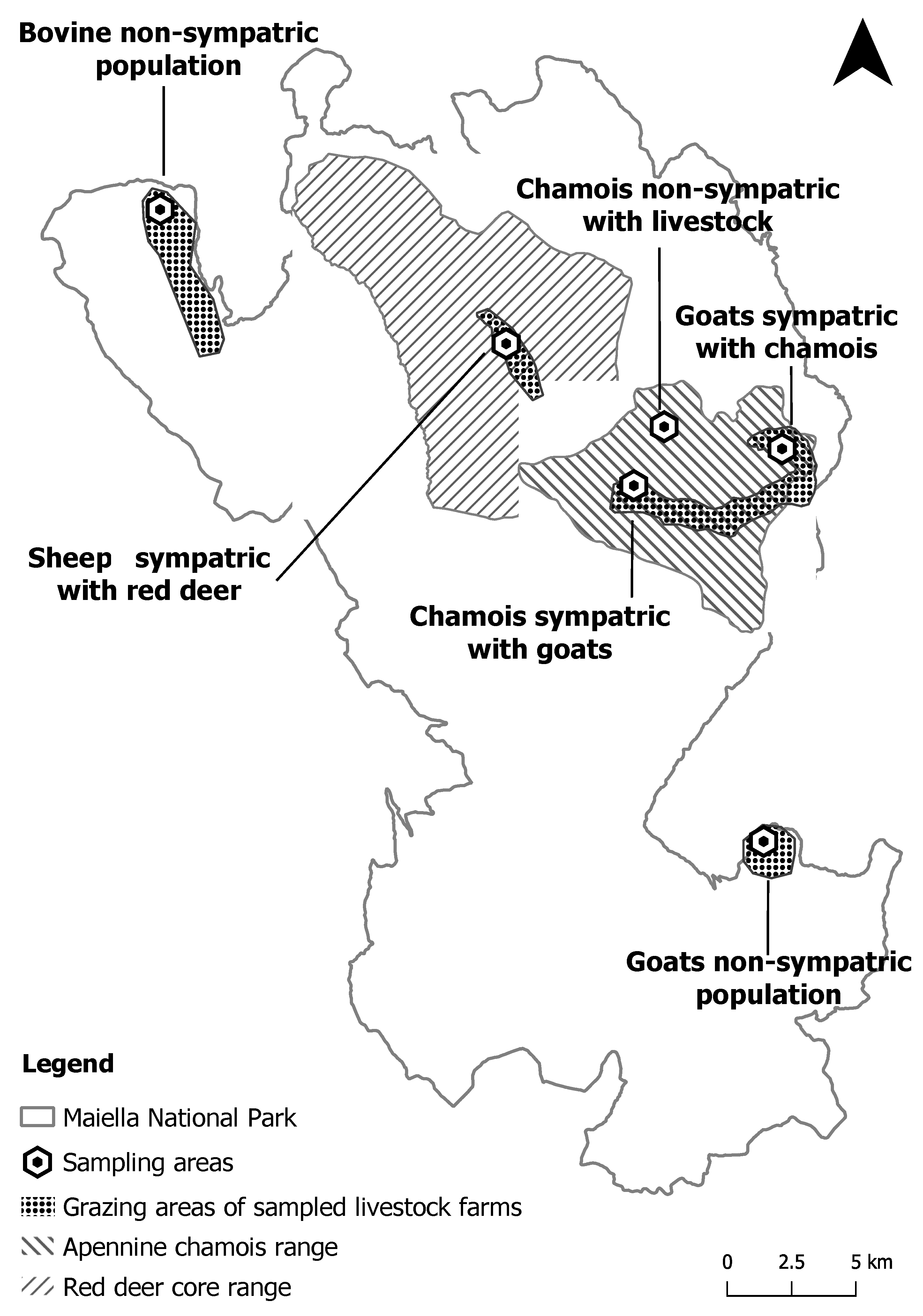
4.1. Study Area and Sampling Design

The Maiella National Park (MNP) covers a vast, mountainous area of about 740 km2 in the central Apennine Mountains and is home to several diversified vertebrate fauna, including mammalian species relevant at national and international levels listed in the Habitats Directive (92/43/EEC). Among them, the Apennine chamois (Rupicapra pyrenaicaornata) is a rare subspecies of chamois living in extremely limited areas of central Italy, including the territories of MNP, where the current population size is approximately 1300 individuals as a result of a reintroduction program carried out in the past. This species coexists with more widespread wild ungulates, such as red deer (Cervus elaphus, approximately 1500 individuals), and domestic livestock (cattle, sheep, and goats) traditionally raised on small farms with extensive grazing systems. In this regard, the distribution areas of wild and domestic animals were previously determined by georeferencing data in order to define the level of grazing land sharing. The populations under study are routinely monitored by the MNP technical staff. The Apennine chamois population (size and distribution area) is defined using a census block technique, consistently performed in summer and autumn over the last twenty years. The information regarding demographic structure and territory occupancy of the main herds is obtained by GPS collars, ear tags, and direct visual observations that have been conducted in a systematic design during summer and autumn of the last ten years. The red deer population is defined by estimating the minimum number of reproductive males during the rutting season and combining this information with data on population demographic structure that is established by recurrent visual observations during all the seasons. Additionally, the farms of domestic animals require a specific authorization every year to use the grazing lands, and each livestock group is assigned an area defined by GPS coordinates. Based on that, the first group of animals, including sympatric populations (100 Apennine chamois coexisting with a farm of 120 goats, and 50 red deer with a farm of 300 sheep), and the second group of non-sympatric populations (70 cattle, 210 goats, and 100 Apennine chamois) were each distributed in different areas of the MNP (Figure 1).
From October to November 2019, fecal samples from red deer, Apennine chamois, and domestic ruminants were collected. In order to obtain fresh feces suitable for microbiological investigations, the different groups of grazing animals were followed and observed with no disturbance to their physiological activities, and the fecal samples were collected while limiting as much as possible any contamination with the soil. Based on the sampling design, a total of 132 fecal samples were recovered and considered suitable for laboratory analysis. Subsequently, the specimens were grouped in 33 fecal pools with 4 fecal samples for each pool (Table 2) and were stored at 4 °C and analyzed within 24 h after the collection.

4.2. Bacteria Isolation and Antibioticssusceptibility Test

Enterococcus spp. isolates were obtained by a preliminary non-selective enrichment of fecal samples in buffered peptone water (24 h at 37 °C), followed by subculturing on Slanetz and Bartley agar (Liofilchem, Italy) at 37 °C for 48 h. From each plate, 1 or 2 representative colonies, morphologically referred to Enterococcus genus and adequately isolated from other microorganisms, were selected to obtain pure subcultures. The species identification of typical colonies and the antimicrobial susceptibility tests for QD, VAN, and LNZ were performed using a Vitek 2 system (Biomerieux, France) following the instructions given by the manufacturer. The minimum inhibitory concentration (MIC) values were evaluated based on the Clinical and Laboratory Standards Institute (CLSI) breakpoints considered relevant for human health [33].

4.3. Detection of Antibiotic Resistance and Virulence Genes

The genes related to QD (vgA, msrC, VatD, vgbB, vgbA, ermB, and vatE), VAN (vanA, vanB, vanC1, vanC2, vanD, vanM, and vanN), and LNZ (cfr, cfr(B), cfr(D), optrA, and poxtA) resistance and those encoding some virulence factors (gelE, esp, efa, ace, hyl, and asa1) in enterococci were amplified by previously described protocols of end-point PCR (Table 3). The amplicons with the expected size were purified, sequenced, and compared with those included in the GenBank database using Chromas software, FASTA (http://www.ebi.ac.uk/fasta33, accessed on 20 January 2022), Clustal Omega (http://www.ebi.ac.uk/Tools/msa/clustalo, accessed on 20 January 2022), and the Basic Local Alignment Search Tool (BLAST) to confirm the specificity of protocols applied. The sequences obtained were submitted to the GenBank database with accession numbers from OM525845 to OM525851.

5. Conclusions

In conclusion, the combined analysis of laboratory and georeferenced data applied in this study provided new information about emerging resistance patterns in commensal and potentially pathogenic enterococci and suggests the importance of a multidisciplinary approach to the AMR challenge. The wild species investigated in this study were strictly linked to the territory of the MNP, which is characterized by complex ecosystems with different levels of human–animal–environment interactions. Therefore, they may be considered sentinel species of emerging antibiotic resistance patterns. Additional studies should include both human and ungulate-derived isolates in order to assess the relationships and to confirm the importance of animal–human interactions in the transmission of antibiotic resistance organisms.

Author Contributions

Conceptualization, C.S., C.E.D.F., A.V., S.A. and F.M.; methodology, C.S., C.E.D.F., S.A., A.A. and A.R.F.; software, C.S., C.E.D.F. and A.R.F.; validation, C.S., C.E.D.F. and A.R.F.; formal analysis, C.S., C.E.D.F. and A.R.F.; investigation, C.S., A.R.F., S.A. and A.A.; resources, A.V., C.E.D.F. and F.M.; data curation, C.S., C.E.D.F. and A.R.F.; writing—original draft preparation, C.S. and C.E.D.F.; writing—review and editing, C.S, C.E.D.F., A.V., S.A. and F.M.; supervision, C.E.D.F. and F.M.; funding acquisition, F.M. All authors have read and agreed to the published version of the manuscript.

Funding

This research was funded by the Italian Ministry for Education, University and Research, in the framework of Project Demetra (Dipartimenti di Eccellenza 2018–2022, CUP_C46C18000530001).

Institutional Review Board Statement

Ethical review and approval were waived for this study because access to the samples was gained through the regular management plan for the investigated species by environmental collecting activities without direct handling of the animals. Therefore, this research did not cause any harm or suffering to any animal.

Informed Consent Statement

Not applicable.

Data Availability Statement

Data will be made available upon reasonable request to the corresponding author.

Conflicts of Interest

The authors declare no conflict of interest.

References

  1. Prieto, A.M.G.; van Schaik, W.; Rogers, M.R.C.; Coque, T.M.; Ebaquero, F.; Ecorander, J.; Willems, R. Global Emergence and Dissemination of Enterococci as Nosocomial Pathogens: Attack of the Clones? Front. Microbiol. 2016, 7, 788. [Google Scholar] [CrossRef] [Green Version]
  2. Wang, Y.; Lv, Y.; Cai, J.; Schwarz, S.; Cui, L.; Hu, Z.; Zhang, R.; Li, J.; Zhao, Q.; Dacheng, W.; et al. A novel gene, optrA, that confers transferable resistance to oxazolidinones and phenicols and its presence in Enterococcus faecalisand Enterococcus faeciumof human and animal origin. J. Antimicrob. Chemother. 2015, 70, 2182–2190. [Google Scholar] [CrossRef] [Green Version]
  3. Torres, R.T.; Carvalho, J.; Cunha, M.V.; Serrano, E.; Palmeira, J.D.; Fonseca, C. Temporal and geographical research trends of antimicrobial resistance in wildlife—A bibliometric analysis. One Health 2020, 11, 100198. [Google Scholar] [CrossRef]
  4. Chajecka-Wierzchowska, W.; Zadernowska, A.; Łaniewska-Trokenheim, Ł. Virulence factors of Enterococcus spp. presented in food. LWT 2017, 75, 670–676. [Google Scholar] [CrossRef]
  5. Zaheer, R.; Cook, S.R.; Barbieri, R.; Goji, N.; Cameron, A.; Petkau, A.; Polo, R.O.; Tymensen, L.; Stamm, C.; Song, J.; et al. Surveillance of Enterococcus spp. reveals distinct species and antimicrobial resistance diversity across a One-Health continuum. Sci. Rep. 2020, 10, 3937, Corrected in Sci. Rep. 2020, 10, 13401. [Google Scholar] [CrossRef] [PubMed]
  6. Brenciani, A.; Fioriti, S.; Morroni, G.; Cucco, L.; Morelli, A.; Pezzotti, G.; Paniccià, M.; Antonelli, A.; Magistrali, C.F.; Rossolini, G.M.; et al. Detection in Italy of a porcine Enterococcus faecium isolate carrying the novel phenicol-oxazolidinone-tetracycline resistance gene poxtA. J. Antimicrob. Chemother. 2018, 74, 817–818. [Google Scholar] [CrossRef] [PubMed]
  7. Elghaieb, H.; Freitas, A.; Abbassi, M.S.; Novais, C.; Zouari, M.; Hassen, A.; Peixe, L. Dispersal of linezolid-resistant enterococci carrying poxtA or optrA in retail meat and food-producing animals from Tunisia. J. Antimicrob. Chemother. 2019, 74, 2865–2869. [Google Scholar] [CrossRef] [PubMed]
  8. Huang, J.; Wang, M.; Gao, Y.; Chen, L.; Wang, L. Emergence of plasmid-mediated oxazolidinone resistance gene poxtA from CC17 Enterococcus faecium of pig origin—authors’ response. J. Antimicrob. Chemother. 2020, 75, 1359–1361. [Google Scholar] [CrossRef] [PubMed]
  9. Lei, C.-W.; Kang, Z.-Z.; Wu, S.-K.; Chen, Y.-P.; Kong, L.-H.; Wang, H.-N. Detection of the phenicol–oxazolidinone–tetracycline resistance gene poxtA in Enterococcus faecium and Enterococcus faecalis of food-producing animal origin in China. J. Antimicrob. Chemother. 2019, 74, 2459–2461. [Google Scholar] [CrossRef] [PubMed]
  10. Na, S.-H.; Moon, D.-C.; Kim, M.-H.; Kang, H.-Y.; Kim, S.-J.; Choi, J.-H.; Mechesso, A.-F.; Yoon, S.-S.; Lim, S.-K. Detection of the Phenicol–Oxazolidinone Resistance Gene poxtA in Enterococcus faecium and Enterococcus faecalis from Food-Producing Animals during 2008–2018 in Korea. Microorganisms 2020, 8, 1839. [Google Scholar] [CrossRef]
  11. Ruiz-Ripa, L.; Feßler, A.T.; Hanke, D.; Eichhorn, I.; Azcona-Gutiérrez, J.M.; Pérez-Moreno, M.O.; Seral, C.; Aspiroz, C.; Alonso, C.A.; Torres, L.; et al. Mechanisms of Linezolid Resistance Among Enterococci of Clinical Origin in Spain—Detection of optrA- and cfr(D)-Carrying E. faecalis. Microorganisms 2020, 8, 1155. [Google Scholar] [CrossRef] [PubMed]
  12. Torres, C.; Alonso, C.A.; Ruiz-Ripa, L.; León-Sampedro, R.; Del Campo, R.; Coque, T.M. Antimicrobial Resistance in Enterococcus spp. of animal origin. Microbiol. Spectr. 2018, 6. [Google Scholar] [CrossRef] [PubMed]
  13. Ramey, A.M.; Ahlstrom, C.A. Antibiotic Resistant Bacteria in Wildlife: Perspectives on Trends, Acquisition and Dissemination, Data Gaps, and Future Directions. J. Wildl. Dis. 2020, 56, 1–15. [Google Scholar] [CrossRef] [PubMed]
  14. Dec, M.; Stępień-Pyśniak, D.; Gnat, S.; Fratini, F.; Urban-Chmiel, R.; Cerri, D.; Winiarczyk, S.; Turchi, B. Antibiotic Susceptibility and Virulence Genes in Enterococcus Isolates from Wild Mammals Living in Tuscany, Italy. Microb. Drug Resist. 2020, 26, 505–519. [Google Scholar] [CrossRef] [PubMed]
  15. Jánošková, A.; Kmet, V. Vancomycin Resistance Genes in Enterococcus spp. Strains Isolated from Alpine Accentor and Chamois. Acta Vet. Brno 2004, 73, 211–214. [Google Scholar] [CrossRef] [Green Version]
  16. Vandžurová, A.; Hrašková, I.; Jüdová, J.; Javorskỳ, P.; Pristaš, P. Antibiotic resistance and restriction endonucleases in fecal enterococci of chamois (Rupicapra rupicapra Linnaeus, 1758). Folia Microbiol. 2012, 57, 355–358. [Google Scholar] [CrossRef]
  17. Poeta, P.; Costa, D.; Igrejas, G.; Rodrigues, J.; Torres, C. Phenotypic and genotypic characterization of antimicrobial resistance in faecal enterococci from wild boars (Sus scrofa). Vet. Microbiol. 2007, 125, 368–374. [Google Scholar] [CrossRef]
  18. Figueiredo, N.; Radhouani, H.; Gonçalves, A.; Rodrigues, J.; Carvalho, C.; Igrejas, G.; Poeta, P. Genetic characterization of vancomycin-resistant enterococci isolates from wild rabbits. J. Basic Microbiol. 2009, 49, 491–494. [Google Scholar] [CrossRef]
  19. Radhouani, H.; Igrejas, G.; Carvalho, C.; Pinto, L.; Gonçalves, A.; Lopez, M.; Sargo, R.; Cardoso, L.; Martinho, A.; Rego, V.; et al. Clonal Lineages, Antibiotic Resistance and Virulence Factors in Vancomycin-Resistant Enterococci Isolated from Fecal Samples of Red Foxes (Vulpes Vulpes). J. Wildl. Dis. 2011, 47, 769–773. [Google Scholar] [CrossRef]
  20. Radhouani, H.; Igrejas, G.; Gonçalves, A.; Pacheco, R.; Monteiro, R.; Sargo, R.; Brito, F.; Torres, C.; Poeta, P. Antimicrobial resistance and virulence genes in Escherichia coli and enterococci from red foxes (Vulpes vulpes). Anaerobe 2013, 23, 82–86. [Google Scholar] [CrossRef]
  21. Gonçalves, A.; Igrejas, G.; Radhouani, H.; Correia, S.; Pacheco, R.; Santos, T.; Monteiro, R.; Guerra, A.; Petrucci-Fonseca, F.; Brito, F.; et al. Antimicrobial resistance in faecal enterococci and Escherichia coli isolates recovered from Iberian wolf. Lett. Appl. Microbiol. 2013, 56, 268–274. [Google Scholar] [CrossRef]
  22. Navarro-Gonzalez, N.; Casas-Díaz, E.; Porrero, C.M.; Mateos, A.; Domínguez, L.; Lavín, S.; Serrano, E. Food-borne zoonotic pathogens and antimicrobial resistance of indicator bacteria in urban wild boars in Barcelona, Spain. Vet. Microbiol. 2013, 167, 686–689. [Google Scholar] [CrossRef] [PubMed]
  23. Lozano, C.; González-Barrio, D.; García, J.T.; Ceballos, S.; Olea, P.P.; Ruiz-Fons, F.; Torres, C. Detection of vancomycin-resistant Enterococcus faecalis ST6-vanB2 and E. faecium ST915-vanA in faecal samples of wild Rattus rattus in Spain. Vet. Microbiol. 2015, 177, 168–174. [Google Scholar] [CrossRef] [PubMed]
  24. Guerrero-Ramos, E.; Cordero, J.; Molina, D.; Poeta, P.; Igrejas, G.; Alonso-Calleja, C.; Capita, R. Antimicrobial resistance and virulence genes in enterococci from wild game meat in Spain. Food Microbiol. 2016, 53, 156–164. [Google Scholar] [CrossRef] [PubMed]
  25. Sassi, M.; Guerin, F.; Zouari, A.; Beyrouthy, R.; Auzou, M.; Fines-Guyon, M.; Potrel, S.; Dejoies, L.; Collet, A.; Boukthir, S.; et al. Emergence of optrA-mediated linezolid resistance in enterococci from France, 2006–16. J. Antimicrob. Chemother. 2019, 74, 1469–1472. [Google Scholar] [CrossRef]
  26. Almeida, L.M.; Gaca, A.; Bispo, P.M.; Lebreton, F.; Saavedra, J.T.; Silva, R.A.; Basílio-Júnior, I.D.; Zorzi, F.M.; Filsner, P.H.; Moreno, A.M.; et al. Coexistence of the Oxazolidinone Resistance—Associated Genes cfr and optrA in Enterococcus faecalis From a Healthy Piglet in Brazil. Front. Public Health 2020, 8, 518. [Google Scholar] [CrossRef]
  27. Guerin, F.; Sassi, M.; Dejoies, L.; Zouari, A.; Schutz, S.; Potrel, S.; Auzou, M.; Collet, A.; Lecointe, D.; Auger, G.; et al. Molecular and functional analysis of the novel cfr(D) linezolid resistance gene identified in Enterococcus faecium. J. Antimicrob. Chemother. 2020, 75, 1699–1703. [Google Scholar] [CrossRef]
  28. Pang, S.; Boan, P.; Lee, T.; Gangatharan, S.; Tan, S.J.; Daley, D.; Lee, Y.T.; Coombs, G.W. Linezolid-resistant ST872 Enteroccocus faecium harbouring optrA and cfr (D) oxazolidinone resistance genes. Int. J. Antimicrob. Agents 2019, 55, 105831. [Google Scholar] [CrossRef]
  29. Hughes, D.; I Andersson, D. Environmental and genetic modulation of the phenotypic expression of antibiotic resistance. FEMS Microbiol. Rev. 2017, 41, 374–391. [Google Scholar] [CrossRef]
  30. Ben Yahia, H.; Chairat, S.; Hamdi, N.; Gharsa, H.; Ben Sallem, R.; Ceballos, S.; Torres, C.; Ben Slama, K. Antimicrobial resistance and genetic lineages of faecal enterococci of wild birds: Emergence of vanA and vanB2 harbouring Enterococcus faecalis. Int. J. Antimicrob. Agents 2018, 52, 936–941. [Google Scholar] [CrossRef]
  31. Nowakiewicz, A.; Zięba, P.; Gnat, S.; Trościańczyk, A.; Osińska, M.; Łagowski, D.; Kosior-Korzecka, U.; Puzio, I. A significant number of multi-drug resistant Enterococcus faecalis in wildlife animals; long-term consequences and new or known reservoirs of resistance? Sci. Total Environ. 2019, 705, 135830. [Google Scholar] [CrossRef] [PubMed]
  32. Drobni, M.; Bonnedahl, J.; Hernandez, J.; Haemig, P.; Olsen, B. Vancomycin-Resistant Enterococci, Point Barrow, Alaska, USA. Emerg. Infect. Dis. 2009, 15, 838–839. [Google Scholar] [CrossRef]
  33. Wayne, P.A. Clinical and Laboratory Standards Institute: Performance Standards for Antimicrobial Susceptibility Testing. 30th ed. CLSI Supplement M100. Available online: https://clsi.org/media/3481/m100ed30_sample.pdf (accessed on 9 February 2022).
  34. Nomura, T.; Hashimoto, Y.; Kurushima, J.; Hirakawa, H.; Tanimoto, K.; Zheng, B.; Ruan, G.; Xue, F.; Liu, J.; Hisatsune, J.; et al. New colony multiplex PCR assays for the detection and discrimination of vancomycin-resistant enterococcal species. J. Microbiol. Methods 2018, 145, 69–72. [Google Scholar] [CrossRef]
  35. Bender, J.K.; Fleige, C.; Klare, I.; Werner, G. Development of a multiplex-PCR to simultaneously detect acquired linezolid resistance genes cfr, optrA and poxtA in enterococci of clinical origin. J. Microbiol. Methods 2019, 160, 101–103. [Google Scholar] [CrossRef]
  36. Lee, S.-M.; Huh, H.J.; Song, D.J.; Shim, H.J.; Park, K.S.; Kang, C.-I.; Ki, C.-S.; Lee, N.Y. Resistance mechanisms of linezolid-nonsusceptible enterococci in Korea: Low rate of 23S rRNA mutations in Enterococcus faecium. J. Med. Microbiol. 2017, 66, 1730–1735. [Google Scholar] [CrossRef] [PubMed]
  37. Shaw, T.D.; Fairley, D.J.; Schneiders, T.; Pathiraja, M.; Hill, R.L.R.; Werner, G.; Elborn, J.S.; McMullan, R. The use of high-throughput sequencing to investigate an outbreak of glycopeptide-resistant Enterococcus faecium with a novel quinupristin-dalfopristin resistance mechanism. Eur. J. Clin. Microbiol. 2018, 37, 959–967. [Google Scholar] [CrossRef] [PubMed] [Green Version]
  38. Martín-Platero, A.M.; Valdivia, E.; Maqueda, M.; Martínez-Bueno, M. Characterization and safety evaluation of enterococci isolated from Spanish goats’ milk cheeses. Int. J. Food Microbiol. 2009, 132, 24–32. [Google Scholar] [CrossRef]
Figure 1. Maiella National Park boundaries, geographic distribution of sympatric and non-sympatric animals, and sampling areas.
Antibiotics 11 00223 g001
Table 1. Details of bacterial species, animal sources, MIC values, and genes detected in this study.
GroupsStrainAnimal SourceMIC (μg/mL) *Genes
QDVANLNZResistanceVirulence
Sympatric populationsE. faecalisApennine Chamois412 gelE, efa, asa1
E. faecalisApennine Chamois412 gelE, efa, asa1
E. faecalisGoats412vanC1/C2, msrCgelE, efa, asa1
E. hiraeApennine Chamois142 gelE
E. hiraeGoats10.52
E. faeciumApennine Chamois16328msrC, cfrDgelE, efa
E. casselliflavusGoats142vanC1/C2
E. faecalisRed deer412 gelE
E. faeciumRed deer412msrCgelE
E. faeciumRed deer0.50.52
E. faeciumSheep10.52msrC
E. gallinarumRed deer16328vanC1, msrC, cfrDgelE
E. gallinarumSheep16328vanC1/C2, cfrD
E. gallinarumSheep16328vanC1, msrC, cfrDgelE, esp, efa
E. hiraeSheep10.52
E. casselliflavusSheep141
Non-sympatric populationsE. gallinarumCattle120.5
E. casseliflavusCattle112vanC2
E. faeciumCattle121msrC
E. faeciumCattle120.5msrC
E. gallinarumGoats120.5
E. gallinarumGoats16832msrC, vanC1/C2, cfrD
E. gallinarumGoats16832msrC, cfrD
E. gallinarumGoats0.520.5msrC
E. gallinarumGoats120.5
E. gallinarumGoats120.5vanC1/C2, msrCasa1
E. gallinarumGoats120.5msrC
E. gallinarumGoats0.50.52msrC
E. gallinarumGoats412msrCesp, efa, asa1
E. gallinarumGoats16328vanC2, cfrD
E. gallinarumGoats120.5
E. faeciumGoats0.520.5msrC
E. faeciumGoats0.524msrC
E. faeciumGoats0.520.5
E. hiraeGoats120.5 asa1
E. faecalisApennine Chamois221 gelE, asa1
E. faecalisApennine Chamois221 gelE, efa, ace
E. faecalisApennine Chamois421 gelE, efa, ace
E. faecalisApennine Chamois421 gelE, asa1
E. faecalisApennine Chamois421 gelE, efa, ace, asa1
E. faecalisApennine Chamois421 gelE, efa, asa1
E. casseliflavusApennine Chamois124vanC2gelE, efa, ace, asa1
E. hiraeApennine Chamois120.5
* QD: quinupristin/dalfopristin; VAN: vancomycin; LNZ: linezolid. The MIC values related to the resistance based on the CLSI breakpoints are indicated in bold.
Table 2. Number of fecal pools and colonies selected for testing from each population under study. The number of collected samples for each population is reported in parentheses.
GroupsAnimalsN. Fecal PoolsN. Colonies
Sympatric populationsApennine chamois (8)24
Goat (12)33
Red deer (16)45
Sheep (12)35
Non-sympatric populationsCattle (20)57
Goat (32)816
Red deer (12)30
Apennine chamois (20)58
Total 3348
Table 3. Details of single and multiplex PCR protocols applied for antibiotic resistance and virulence gene detection.
PrimerSequence 5’- 3’Size (bp)References
VanD_F1TGGAATCACAAAATCCGGCG311[34]
VanD_R2TWCCCGCATTTTTCACAACS
VanM_F1GGCAGAGATTGCCAACAACA425
VanM _R1AGGTAAACGAATCTGCCGCT
VanC2_F1GCAAACGTTGGTACCTGATG523
VanC2_R4 GGTGATTTTGGCGCTGATCA
VanB_F1GATGTGTCGGTAAAATCCGC640
VanB_R1CCACTTCGCCGACAATCAAA
VanA_F1GCAAGTCAGGTGAAGATGGA721
VanA_R1GCTAATACGATCAAGCGGTC
VanC1_5GTATCAAGGAAACCTCGCGA836
VanC1_6CGTAGGATAACCCGACTTCC
VanN_F1CCTCAAATCAGCAGCTAGTG941
VanN_R1GCTCCTGATAAGTGATACCC
Cfr_fwTGAAGTATAAAGCAGGTTGGGAGTCAAC746[35]
Cfr_revCATATAATTGACCACAAGCAGC
optrA_fwTACTTGATGAACCTACTAACCA422
optrA_revCCTTGAACTACTGATTCTCGG
poxtAfwAAAGCTACCCATAAAATATC533
poxtArevTCATCAAGCTGTTCGAGTTC
cfr(B) fwTGAGCATATACGAGTAACCTCAAGA293[36]
cfr(B) revCGCAAGCAGCGTCTATATCA
cfr(D) fwAGAAGTCGCAACAAGTGAGGA595[11]
cfr(D) revGCAACTGCATGAGTCAAAGAA
Vat D FTCCAGCTAACATGTATGGCG271[37]
Vat D RGCTCAATAGGACCAGGTGTA
vgaA FAGTGGTGGTGAAGTAACACG659
vgaA RCTTGTCTCCTCCGCGAATAC
vgaB FTGACAATATGAGTGGTGGTG576
vgaB RGCGACCATGAAATTGCTCTC
vgbB FCAGCAGTCTAGATCAGAGTGG728[37]
vgbB RCATACGGATCCATCTTTTCC
msrC FAAGGAATCCTTCTCTCTCCG343
msrC RGTAAACAAAATCGTTCCCG
vgbA FTACAGAGTACCCACTACCGA569
vgbA RTCAATTCCTGCTCCAGCAGT
ermB FCATTTAACGACGAAACTGGC424
ermB RGGAACATCTGTGGTATGGCG
vatE FACTATACCTGACGCAAATGC511[37]
vatE RGGTTCAAATCTTGGTCCG
gelE FTATGACAATGCTTTTTGGGAT213[38]
gelE RAGATGCACCCGAAATAATATA
esp FAGATTTCATCTTTGATTCTTGG510
esp RAATTGATTCTTTAGCATCTGG
ace FGAATTGAGCAAAAGTTCAATCG1008
ace RGTCTGTCTTTTCACTTGTTTC
efa FGCCAATTGGGACAGACCCTC688
efa RCGCCTTCTGTTCCTTCTTTGGC
asa1 FGCACGCTATTACGAACTATGA375[38]
asa1 RTAAGAAAGAACATCACCACGA
hyl FACAGAAGAGCTGCAGGAAATG276
hyl RGACTGACGTCCAAGTTTCCAA
cylA FACTCGGGGATTGATAGGC688
cylA RGCTGCTAAAGCTGCGCTT
Publisher’s Note: MDPI stays neutral with regard to jurisdictional claims in published maps and institutional affiliations.

