Article

Staphylococcus aureus-Cure-Associated Antigens Elicit Type 3 Immune Memory T Cells

1
Veterinary Clinical Immunology Research Group, Departamento de Clínica Médica, Faculdade de Medicina Veterinária e Zootecnia, Universidade de São Paulo, São Paulo 05508-270, Brazil
2
Programa de Pós-Graduação em Ciência Animal, Universidade Federal da Paraíba, Areia 58397-000, Brazil
3
Laboratório de Soroloepidemiologia e Imunobiologia, Instituto de Medicina Tropical, Universidade de São Paulo, São Paulo 05403-000, Brazil
4
Departamento de Salud Publica, Facultad de Ciencias de La Salud, Universidad Nacional Torino Rodriguez de Mendonza de Amazonas, Chachapoyas 01001, Peru
5
Laboratório de Genética, Instituto Butantã, Universidade de São Paulo, São Paulo 05503-900, Brazil
6
Departamento de Medicina Veterinária Preventiva e Saúde Animal, Faculdade de Medicina Veterinária e Zootecnia, Universidade de São Paulo, São Paulo 05508-270, Brazil
7
Departamento de Tecnologia e Inspeção de Produtos de Origem Animal, Escola de Veterinária, Universidade Federal de Minas Gerais, Belo Horizonte 31270-010, Brazil
8
Departamento de Parasitologia, Instituto de Ciências Biológicas, Universidade Federal de Minas Gerais, Belo Horizonte 31270-901, Brazil
9
Laboratório de Parasitologia Clínica, Faculdade de Ciências Farmacêuticas, Universidade Federal de Alfenas, Alfenas 37130-000, Brazil
10
Departamento de Imunologia, Instituto de Ciências Biomédicas, Universidade de São Paulo, São Paulo 05508-900, Brazil
11
Departamento de Medicina Preventiva, Faculdade de Medicina, Universidade de São Paulo, São Paulo 01246-903, Brazil
*
Author to whom correspondence should be addressed.
Antibiotics 2022, 11(12), 1831; https://doi.org/10.3390/antibiotics11121831
Submission received: 2 October 2022 / Revised: 20 November 2022 / Accepted: 22 November 2022 / Published: 16 December 2022
(This article belongs to the Special Issue Mastitis: Causative Agents, Drug Resistance, and Treatment Approaches)

Abstract

:
Background: Staphylococcus aureus is one of the most frequently major mastitis pathogens that cause clinical and subclinical mastitis worldwide. Current antimicrobial treatments are usually ineffective, and the commercially available vaccines lack proven effectiveness. The immunological response elicited by the recombinant S. aureus-cure-associated proteins phosphoglycerate kinase (PGK), enolase (ENO), and elongation factor-G (EF-G) in combination with the granulocyte-macrophage colony-stimulating factor (GM-CSF) DNA vaccination was studied in this work. Methods: Here, twenty-three C57BL/6 mice were divided into four groups and vaccinated with: G1: none (control); G2: GM-CSF DNA plasmid DNA vaccine; G3: the combination of EF-G+ENO+PGK; and G4: the combinations of EF-G+ENO+PGK proteins plus GM-CSF plasmid DNA vaccine. After 44 days, spleen cells were collected for immunophenotyping and lymphocyte proliferation evaluation by flow cytometry upon S. aureus stimulus. Results: Immunization with the three S. aureus recombinant proteins alone resulted in a higher percentage of IL-17A+ cells among CD8+ T central memory cells, as well as the highest intensity of IL-17A production by overall lymphocytes indicating that the contribution of the combined lymphocyte populations is crucial to sustaining a type 3 cell immunity environment. Conclusion: The immunization with three S. aureus-cure-associated recombinant proteins triggered type 3 immunity, which is a highly interesting path to pursue an effective bovine S. aureus mastitis vaccine.

1. Introduction

Bovine mastitis is the costliest disease in any dairy farm, causing a pronounced reduction in milk production and quality. Furthermore, mastitis implies approximately 70% of the antimicrobial usage in dairy farms, raising serious public health concerns [1]. Staphylococcus aureus is one of the most frequently associated bacteria that cause clinical and subclinical mastitis worldwide [2,3]. Because of its pathogenicity, contagiousness, likely persistence of intramammary infections (IMIs), and refractoriness to the current antimicrobial therapy, this mastitis pathogen remains a major challenge for the dairy industry worldwide [3]. Besides that, S. aureus is a multi-host bacterial pathogen and may infect a distinct host-species population, constituting a major menace to public health and food security [4,5]. In this context, S. aureus mastitis points to an urgent need for innovation and searches for effective alternative approaches to its treatment and prevention.
Thus, it would be valuable to have an alternative approach that increases the likelihood of the cure of an existing infection in an integrated approach with the antimicrobial treatment [6] or of the prevention of new S. aureus infections. We would emphasize that considering the contagious behavior of this pathogen [2], any alternative measure that increases the quarter cure of an IMI may result in lower exposure, decreasing the likelihood of a healthy quarter becoming infected, and consequently reducing the transmission rates and the prevalence of this pathogen in the herd [1].
In a previous study, we searched S. aureus-derived proteins [7] using sera from S. aureus IMI-cured animals, aiming selection of proteins likely related to cure. With this immunoproteomic approach, we revealed three staphylococcal proteins: enolase (ENO), phosphoglycerate kinase (PGK) and elongation factor-G (EF-G). In this latter study, we assumed that antibodies from chronically infected dairy cows clinically and bacteriologically cured S. aureus IMIs, after immunization with a commercial inactivated vaccine, recognized S. aureus proteins used in the present study that may translate into a therapeutic and/or preventive vaccine of relevance. The first S. aureus protein, the so-called ENO, is a conserved moonlighting protein that is expressed in situations when a critical nutrient (e.g., iron) becomes extremely scarce [8], which is the case in the mammary gland environment, especially during mastitis [9]. This protein is essential for bacterial growth and plays an important role in several steps of the infection cycle, with a likely major role in glycolysis, a critical pathway for bacterial energy supply [10,11]. Furthermore, ENO mediates the binding of S. aureus to laminin and collagen I guiding the mechanism of invasion and dissemination. Initially, it enables S. aureus to bind to the extracellular matrix, initiating tissue colonization, followed by plasminogen activation and laminin degradation, which cause the extracellular matrix destruction, favoring bacterial invasion and spread [8,12,13]. Further, ENO is involved in cell wall biosynthesis and resistance to antimicrobials and had a critical role in biofilm formation [8,11,14]. Similarly, PGK is also a moonlighting protein that plays an important role in S. aureus energy metabolism. Its inhibition negatively impacts the bacterial population during biofilm formation. In addition, this staphylococcal protein may be involved in antimicrobial resistance, acts as an adhesion protein [15] and is a potent neutrophil activator with the ability to trigger degranulation. Besides, it dampens the classical and alternative complement pathways [16]. Lastly, the EF-G belongs to the GTPase superfamily that catalyzes two different steps of protein synthesis, and it is used as an energy source, for protein synthesis and cell growth [17,18,19,20]. Even though the molecular functions of these proteins are not precisely understood, it is reasonable to hypothesize that the induction of the host immune response by these proteins may represent an exciting and promising approach for vaccine development. The animals that have successfully eliminated the chronic IMIs by S. aureus produce specific antibodies against the abovementioned S. aureus proteins showing induction of functioning adaptive humoral responses. This observation encouraged us to study their effect on T-cell-mediated immunity which is considered more and more crucial for solid protection against S. aureus infection [21]. Furthermore, some studies have encouraged the use of a granulocyte-macrophage colony-stimulating factor (GM-CSF) DNA vaccine as a powerful immunoadjuvant [22,23] that could be related to T-cell-derived IL-17 production [24,25].
It should be highlighted that there is growing evidence that T cell immunity, especially T-cell-derived interleukin (IL)-17, plays a pivotal role in protection against S. aureus infections [24,26,27]; therefore, a fine-tuned characterization of T cell immunity elicited by the S. aureus proteins above is proposed here. Thus, in the present study, we investigate the immune response induced by three recombinant proteins of S. aureus, EF-G, ENO and PGK in combination with GM-CSF DNA vaccine in mice for future development of use in the cow.

2. Materials and Methods

2.1. Production of S. aureus Recombinant Proteins

The amino acid sequences encoding to the EF-G (Genbank gi|395759321), PGK (Genbank gi|446997488), and ENO; (Genbank gi|447044500) S. aureus antigens were codon-optimized for Escherichia coli expression and synthesized by Genscript (Piscataway, NJ, USA). The synthetic genes were cloned into the pUC57 vector and subsequently were sub-cloned into the pET28a expression vector [24]. Recombinant plasmids were used to transform the expression strain E. coli BL21-Star™ (DE3) as previously described [24]. Concisely, kanamycin plates were used to select transformed E. coli BL21-StarTM (DE3) transformations. Then, three bacterial colonies harboring each expression plasmid were cultured overnight in Luria-Bertani medium (LB) supplemented with kanamycin (0.05 mg mL−1) on a rotating shaker at 37 °C until they reached an optical density (OD) of 0.4 at 600 nm. Isopropyl-ß-D-1-thiogalactopyranoside (IPTG, a final concentration of 0.4 mM; Sigma, St. Louis, MO, USA) was then added to the culture, and the induced cultures were allowed to grow for 4 h. Cells were ruptured on ice using ultrasonic sonication, and debris was eliminated through centrifugation (20,000× g for 30 min at 4 °C). Under denaturing conditions and per the manufacturer’s instructions, the recombinant proteins were purified using Ni Sepharose High Performance immobilized metal ion affinity chromatography columns (HisTrap Hp, cat. n. GE17-5248-02, GE Healthcare, Logan, UT, USA) coupled to an ÄKTA Pure (GE Healthcare, Chicago, IL, USA).

2.2. GM-CSF-Based DNA Vaccine

The GM-CSF amino acid sequence was sent to FastBio (Ribeiro Preto, Brazil) for eukaryotic cell optimization and gene synthesis. The synthetic genes were cloned in the pUC57 vector before being sub-cloned into the pCI-neo mammalian expression vector insert (Promega Incorporation, Madison, WI, USA). Escherichia coli DH5α was transformed using pCI-GM-CSF, and the immunization plasmid was purified according to the manufacturer’s instructions using the ZR plasmid Gigaprep Kit (Zymo Research, Irvine, CA, USA), as previously described [24].

2.3. Liposome Preparation and Entrapment of Plasmid DNA

In the present study, we employed a cationic liposome to deliver the GM-CSF DNA plasmid DNA effectively, as previously described by Fotoran [28] with minor modifications [24]. Briefly, dimethyl-di-octadecyl-ammonium (DDAB), 1,2-distearoyl-sn-glycero-3-phosphoethanolamine-N-[amino (polyethylene glycol)-2000 (DSPE + PEG2000), cholesterol (molar ratio 1:4), 5% of the total lipids were used to produce liposomes. This lipid solution was dissolved in 1 mL of chloroform before being exposed to a steady flow of N2 to evaporate the chloroform and generate a phospholipid layer on the tube walls. To remove all traces of chloroform, this film was then kept under vacuum for at least an hour. Afterward, the film was rehydrated in 5 mM Tris-HCl (pH 7.5) at 60 °C for 1 h while being stirred vigorously every 10 min. After obtaining an opaque solution, we subjected it to high-energy sonication until it approached complete translucence, and then the solution was centrifuged at 100,000× g for 1 h. Any remaining pellet was eliminated, and the supernatant containing unilamellar lipid vesicles was employed to produce the liposome. The genetic material was subsequently added to the liposomes using a molar stoichiometry of 8 nM DDAB per 1 g of nucleic acids, with an estimated molarity of 0.2 pmol.

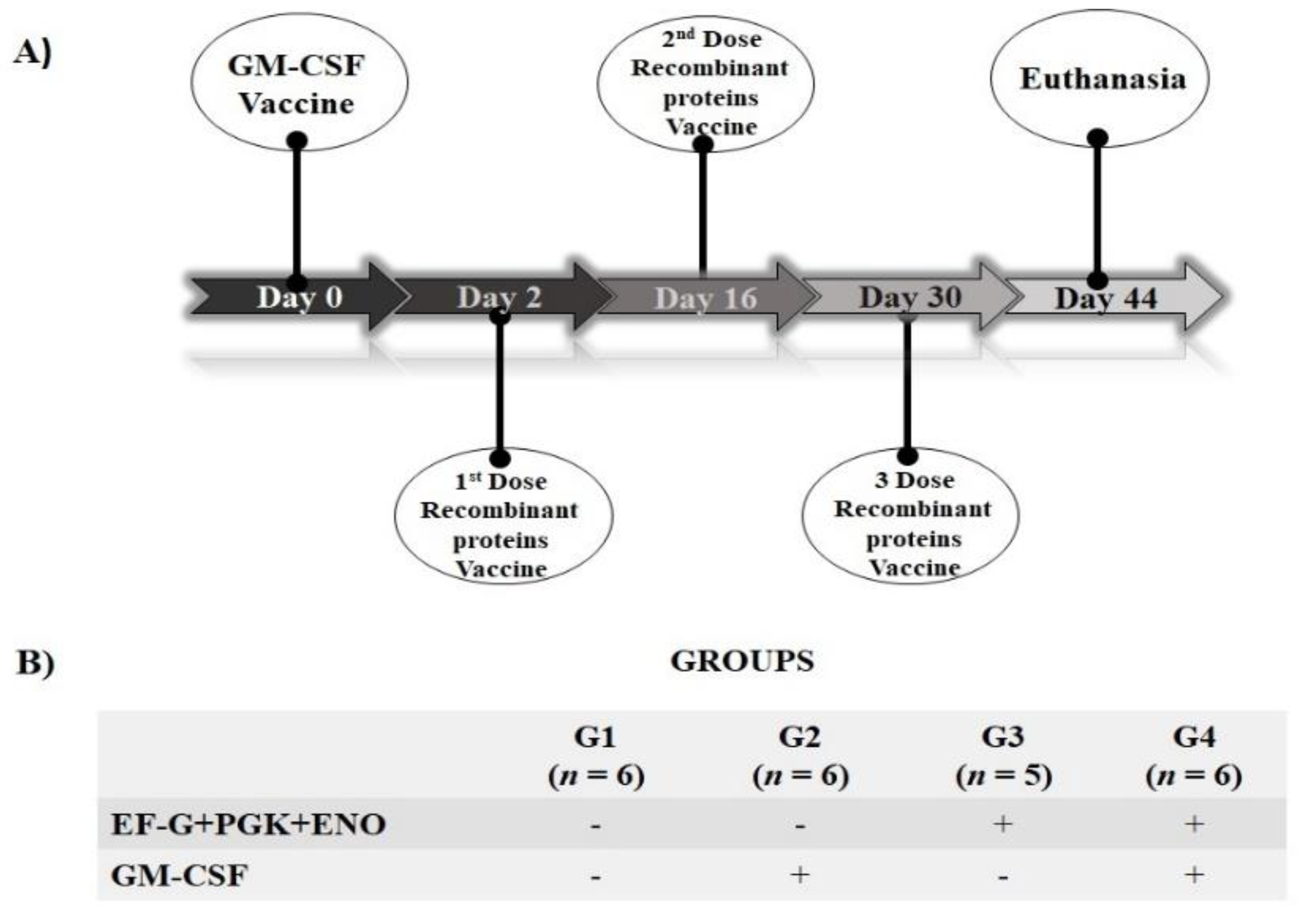
2.4. S. aureus Antigens Candidates: Immunological Evaluation in Mice

In the present study, twenty-three female, six-week-old C57BL/6J mice purchased from the Centro de Bioterismo, of Faculdade de Medicina, Universidade de São Paulo, were divided, after three weeks of adaptation, into four groups and immunized as described in Figure 1. In the present study, 50 µg of pCI-GM-CSF plasmid DNA resuspended in 100 µL of sterile liposomal formulation were injected subcutaneously near the draining lymph nodes in the interscapular region. The recombinant EF-G, PGK, and ENO proteins (20 g each) were injected intramuscularly into the deltoid muscles in 100 µL of saponin adjuvant (Quil-A®, cat. n. 8047-15-2, Invivogen, San Diego, CA, USA) (Figure 1).
On day 44 (14 days following the last dose of S. aureus recombinant proteins), the mice were anesthetized intramuscularly with a solution of xylazine (10 mg kg−1) and ketamine (50 mg kg−1) and euthanized by cervical dislocation.

2.5. Spleen and Cell Recovery

The spleen was then excised to obtain splenic cells for further immunophenotyping and lymphocyte proliferation studies, as previously described [24]. The spleens were mechanically disrupted and homogenized with a syringe plunger into a cell strainer (cat. n. Z742102, Sigma Aldrich, St. Louis, MO, USA) on top of a 50 mL tube containing 10 mL of RPMI 1640 medium (cat. n. R7638, Sigma Aldrich, St. Louis, MO, USA) supplemented with 5% heat-inactivated fetal bovine serum (Gibco, Waltham, MA, USA), 100 μg mL−1 streptomycin, 100 U mL−1 penicillin and Fungizone 0.25 μg mL−1 (cat. n. 15240-096, Gibco, Waltham, MA, USA) (complete medium).
The spleen and lymph node cells were centrifuged for 10 min at 4 °C at 380× g. Erythrocytes were lysed hypotonically by introducing 1000 μL of 0.2% NaCl for 20 s, and afterwards, isotonicity was reestablished by adding 1000 μL of 1.6% NaCl. The cells were centrifuged for 10 min at 4 °C at 380× g and then resuspended in 1 mL of the complete medium. The viability of cells was initially determined using the trypan blue exclusion test (cat. n. T8154-100ML, Sigma Aldrich, St. Louis, MO, USA).

2.6. Preparation of S. aureus Inoculum

Staphylococcus aureus (spa typing t605) isolated from a case of persistent subclinical IMI was used, and the bacterial inoculum dose (2 × 10⁸ staphylococci mL−1) was prepared as previously described [29] with minor modifications [24]. Briefly, bacteria were resuspended in a complete medium after measuring the colony-forming unit, adjusting the concentration of the bacterial inoculum to achieve a ratio of 10 bacteria per cell, and inactivating the bacteria for 1 h at 60 °C.

2.7. Immunophenotyping and Lymphocyte Proliferation Evaluation of Cultured Splenic Cells

The lymphocyte proliferation of splenic cells with or without (control) 10 µL of heat-inactivated S. aureus (2 × 10⁸ CFU mL−1; 10 bacteria per cell) was measured using 2 × 10⁵ viable cells per well (96-wells flat-bottom plates) at 37 °C and 5% CO2 atmosphere for 96 h. The immunophenotyping and lymphocyte proliferation were performed as previously described [24]. The supernatant of the cell culture was collected and frozen at −80 ℃ for cytokines quantification. Briefly, the splenic cells were immunophenotyped using fluorescent-conjugated monoclonal antibodies (mAbs; Table 1). Then, the cells were fixed and permeabilized for assessment of intracellular production of interferon (IFN)-γ and interleukin (IL)-17A, besides the evaluation of in vitro antigen-specific lymphocyte proliferation using ki67 [24,30,31]. The samples were analyzed by flow cytometry (BD LSRFortessaTM X-20 flow cytometer, Becton Dickinson Immunocytometry SystemTM, San Diego, CA, USA). Here, 100,000 events were assessed per sample. The data were analyzed using Flow Jo Tree Star software (FlowJo–Treestar 10.5.3 for Windows, Tree Star Inc., Ashland, OR, USA). As compensation controls, an unstained control, and single-stained samples were also prepared. Conjugated isotype control antibodies were also used in negative control samples. Furthermore, splenic cells were stained with fluorescence minus-one (FMO) controls. The forward scatter (FSC) area versus FSC height was used to exclude doublets.

2.8. Cytokine Measurement

The production rates of cytokines IFN-γ, tumor necrosis factor (TNF)-α, IL-17A, IL-2, IL-4, IL-6 and IL-10 were measured in the supernatant of control or S. aureus-stimulated spleen cell culture by the BD Cytometric Bead Array Mouse TH1, TH2 and TH17 Cytokine (cat. n. 560485, BD BioscienceTM, San Jose, CA, USA) using a flow cytometer (BD LSR FortessaTM X-20 flow cytometer, Becton Dickinson Immunocytometry SystemTM, San Diego, CA, USA) following the manufacturer’s instructions. The data were analyzed using the FCAP ArrayTM v3.0 software (SoftflowTM, Pécs, Hungary).

2.9. Statistical Analysis

To accurately measure the intensity of T cell response upon S. aureus stimulation, the data were normalized by dividing the percentage of proliferative cells (ki67+) or the geometric mean fluorescence (GMFI) upon S. aureus stimulation by the percentage of proliferative cells (ki67+) or the GMFI of unstimulated control, thereby generating a stimulation index (SI) [24,32,33,34,35,36]. The GMFI values of IL-17A or IFN-γ were measured among IL-17A+ or IFN-γ+ cells to determine the intensity of cytokine production per cell. The Gaussian distribution was verified using the Kolmogorov–Smirnov and Shapiro–Wilk tests. A one-way ANOVA was performed on the parametric data, followed by the Tukey test. The Kruskal–Wallis test, followed by Dunn’s test, was used to assess variables with non-parametric distributions. Results are reported as violin plots (median, interquartile range, and adjacent values). p ≤ 0.05 was set as significant unless otherwise indicated. GraphPad Prism 9 (GraphPad Software, Inc., San Diego, CA, USA) was used to perform the statistical analysis.

3. Results

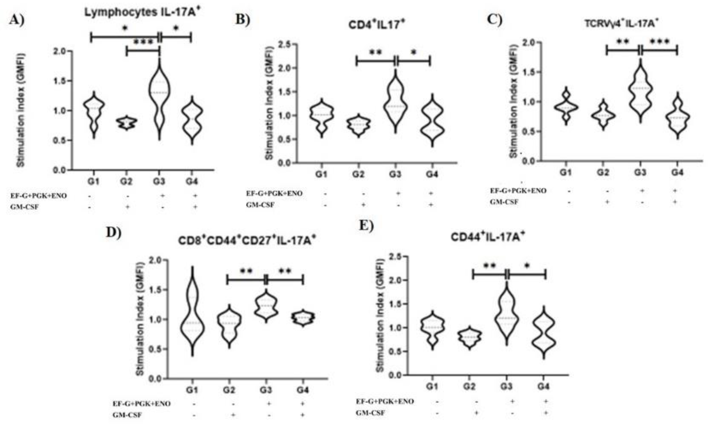
3.1. Type 3 Immunity Is Triggered by αβ and γδ T Memory Cells in Immunized Animals with the Three Recombinant Proteins

Immunization with the three S. aureus recombinant proteins alone resulted in a higher SI of the IL-17A+ cells among CD8+ TCM cells (p = 0.04) compared with the non-immunized control group and with the GM-CSF DNA vaccine group (Figure 2). Further, animals immunized with the three S. aureus recombinant proteins alone had a higher SI of IL-17A+ cells among proliferative (Ki67+) TCRVγ4+ compared with those that received the GM-CSF DNA vaccine (Figure 2). In contrast, GM-CSF DNA vaccination alone negatively impacted the SI of IL-17A+ cells among γδ T cells (p = 0.005) and γδ central memory T cells (TCM) (p = 0.005) compared with non-immunized control (Figure 2). No effect of immunization on type 1 (IFN-γ+) immune cells was observed.
Importantly, we observed the highest SI values in the intensity of IL-17A production by overall lymphocytes in animals immunized with the combination of recombinant proteins compared with other groups (p = 0.05; Figure 3). These data suggest that the contribution of the combined lymphocyte populations is crucial to sustaining a type 3 cell immunity environment. However, analyzing distinct populations, we only detected a tendency to higher SI in the intensity of IL-17A production in animals immunized with the combination of recombinant proteins in T CD4+ lymphocytes, TCRVγ4+ cells, T CD8+ central memory cells and in overall memory T cells when compared with both groups that received the GM-CSF DNA vaccine (Figure 3).
The animals that received the combination of three S. aureus recombinant proteins and the GM-CSF DNA vaccine had a lower SI of T CD4+ cells than the other vaccinated groups (p = 0.03), although it did not significantly differ from the unvaccinated control animals (Figure 4). The GM-CSF DNA vaccination favors an increase in the SI of TCRVγ4+ effector memory cells (p = 0.06) while dampening TCRVγ4+ central memory cells (p = 0.001; Figure 4). No B cell response was observed upon immunization (data not shown).

3.2. Animals Immunized with Three Recombinant Proteins in Association with the GM-CSF DNA Vaccine Was Associated an Anti-Inflammatory Cytokines Production

No effect of immunization on the levels of IL-2 and IL-4 cytokines was observed. The levels of IL-6 (p = 0.06) tend to be lower upon stimulation with S. aureus than unstimulated control just in immunized animals with the three recombinant proteins and the GM-CSF DNA vaccine (Supplementary Figure S1); however, no significant difference among groups was observed under unstimulated control (p = 0.79) and S. aureus (p = 0.68) stimulation conditions. The concentrations of IL-10, IL-17A, TNF-α and IFN-γ were below the limit of the detection.

4. Discussion

We should highlight that our previous study has already shown that specific antibodies (humoral immunity) against the three S. aureus proteins used here are related to the cure of existing IMIs by S. aureus after vaccination with a commercial (bacterin) vaccine [7]. However, humoral immunity alone may not provide adequate protection against S. aureus infections [26]. In this context, there is an emerging knowledge appointing that memory T cells critically contribute to the S. aureus infection control [37]. Indeed, deficiencies in T cells enhance susceptibility to S. aureus infections [37,38]. Interestingly, protective immunity against recurrent S. aureus skin and soft tissue infection in a human was associated with both antibody and T-cell mediated (i.e., IL-17) immunities, suggesting that a multimechanistic approach targeting both humoral and T-cell-mediated immunities may be an important strategy for providing protective immunity and preventing new S. aureus infections [26]. Furthermore, Interleukin-17 is associated with antigen-specific inflammation, neutrophil recruitment, and modulating the communication between the immune system and mammary epithelial cells [39]. Puzzling, the investigation on T-cell-mediated immunity, especially on type 3 immunity, in S. aureus vaccine development against bovine mastitis had not received much attention so far. In the present study, the data suggest that both αβ and γδ T cells contribute to type 3 immunity, which has been regarded as the major mechanism of mammary gland immune defense [39]. Type 3 immunity is mediated by a variety of cells, including γδ T cells, T CD4+ helper (Th17), T CD8+ (Tc17) and innate lymphoid cells 3 [39]. A population of highly IL-17A-producing memory TCRVγ4+ cells has recently been recognized as the most important population that confers protective immunity against subsequent infections by S. aureus [40,41]. In the present study, we observed a tendency for higher SI in this subpopulation. Therefore, we assumed that overall IL-17A highly producing αβ and γδ T cells are required for spontaneous recovery from IMI caused by S. aureus, rather than a specific γδ T cell subpopulation, as we found here. Thus, our study focusing on three targets of S. aureus recombinant protein immunization places the basis for the increases in the likelihood of the cure of an existing S. aureus IMI in an integrated approach with antimicrobial treatment or a non-antibiotic approach to deal with existing S. aureus IMIs, especially regarding the refractoriness of this pathogen to the current antimicrobial therapies, although further studies in dairy cows are still needed.
Although GM-CSF is an important mediator of the immune response [42], our outcomes did not show robust improvement in T-cell mediated immunity of the three S. aureus-cure-associated antigens boosted by the GM-CSF DNA vaccine. In this concern, animals that were vaccinated with the GM-CSF DNA vaccine had a lower SI of IL-17A+ cells among overall memory (CD44+) proliferative lymphocytes and TCRVγ4+ proliferative cells, which could have a critical implication for vaccine efficacy.
Although we do not have any clear idea of this paradox, it has been postulated that it depends on numerous factors, including the immunogen(s), the dose and the timing of GM-CSF administration [43]. In this regard, it has been proposed that great amounts of GM-CSF may expand myeloid cells in secondary lymphoid organs, which in turn may recruit T regulatory cells leading to an immunosuppressive effect [43]. One factor that may result in this effect is the use of a cationic liposome-based approach to efficiently deliver the GM-CSF DNA plasmid DNA.

5. Conclusions

The immunization with three S. aureus-cure-associated recombinant proteins triggered the type 3 immune response in splenocytes stimulated with S. aureus by overall T cells, which is a highly interesting path to pursue an effective control strategy against bovine S. aureus mastitis. Therefore, although our results are encouraging, further studies are required to corroborate our findings about the protective immunity against S. aureus in ruminants triggered by these recombinant S. aureus proteins.

6. Patent

The authors have a patent titled “Vaccine composition, kit for diagnosis of Staphylococcus aureus infections in ruminants, method and use.”

Supplementary Materials

The following supporting information can be downloaded at: https://www.mdpi.com/article/10.3390/antibiotics11121831/s1, Figure S1: Interleukin-6 (IL-6) concentration in the supernatant of the immunized and non-immunized mouse spleen cells culture under unstimulated (basal) and Staphylococcus aureus stimulated conditions. G1: unvaccinated control group; G2: GM-CSF DNA plasmid DNA vaccination; G3: EF-G + ENO + PGK S. aureus recombinant proteins vaccination; G4: EF-G + ENO+ PGK S. aureus recombinant proteins vaccination associated with GM-CSF DNA plasmid DNA vaccine; EF-G: elongation factor-G; ENO: enolase; and PGK: phosphoglycerate kinase. [Student paired t-test].

Author Contributions

Conceptualization, F.N.S., E.M.R.-S., H.G. and A.F.C.; methodology, K.R.S., F.N.S., E.M.R.-S., L.C.R., M.C.R., W.L.F., A.R.F., H.M.A. and C.F.B.; resources, C.F.B., A.M.M.P.D.L., M.B.H. and H.G.; writing—original draft preparation, K.R.S., F.N.S., H.G. and M.G.; writing—review and editing, C.F.B., A.M.M.P.D.L., E.M.R.-S., L.C.R., W.L.F., A.R.F., H.M.A., M.B.H., A.F.C., M.C.R. and M.M.O.P.C.; project administration, K.R.S.; funding acquisition, C.F.B., A.M.M.P.D.L., H.G. and F.N.S. All authors have read and agreed to the published version of the manuscript.

Funding

This work was supported by the São Paulo Research Foundation–FAPESP (FAPESP grant no 2018/23572-3), and by the Coordenação de Aperfeiçoamento de Pessoal de Nível Superior–Brasil (CAPES)–Finance Code 001.

Institutional Review Board Statement

The study was conducted according to the guidelines of the Declaration of Helsinki and approved by the Animal Research Ethics Committee of the UNIVERSIDADE DE SÃO PAULO, Brazil (protocol number 6276100519; 23 June 2020).

Informed Consent Statement

Not applicable.

Data Availability Statement

The data presented in this study are available in the manuscript and the Supplemental File. Additional data are available upon reasonable request from the corresponding author.

Acknowledgments

A.M.M.P.D.L., H.G., M.G. and M.B.H. thank the National Council for Scientific and Technological Development (CNPq) for their fellowships. K.R.S. thanks CAPES for her fellowship. F.N.S. (Process FAPESP n. 2014/23189-4) thanks FAPESP for his fellowship. H.G. and A.M.M.P.D.L. should be regarded as co-last authors. K.R.S. and F.N.S. should be regarded as co-first authors.

Conflicts of Interest

The authors have a patent titled “Vaccine composition, kit for diagnosis of Staphylococcus aureus infections in ruminants, method and use”. The funders had no role in the design of the study; in the collection, analyses, or interpretation of data; in the writing of the manuscript, or in the decision to publish the results.

References

  1. Stevens, M.; Piepers, S.; De Vliegher, S. Mastitis prevention and control practices and mastitis treatment strategies associated with the comsuption of (critically important) antimicrobials o dairy herds in Flanders, Belgium. J. Dairy Sci. 2016, 99, 2896–2903. [Google Scholar] [CrossRef] [PubMed] [Green Version]
  2. Santos, R.P.; Souza, F.N.; Oliveira, A.C.D.; de Souza Filho, A.F.; Aizawa, J.; Moreno, L.Z.; da Cunha, A.F.; Cortez, A.; Della Libera, A.M.M.P.; Heinemann, M.B.; et al. Molecular typing and antimicrobial susceptibility profile of Staphylococcus aureus isolates recovered from bovine mastitis and nasal samples. Animals 2020, 10, 2143. [Google Scholar] [CrossRef] [PubMed]
  3. Rainard, P.; Foucras, G.; Fitzgerald, J.R.; Watts, J.L.; Koop, G.; Middleton, J.R. Knowledge gaps and research priorities in Staphylococcus aureus mastitis control. Transbound. Emerg. Dis. 2018, 65, 149–165. [Google Scholar] [CrossRef] [Green Version]
  4. Richardson, E.J.; Bacigalupe, R.; Harrison, E.M.; Weinert, L.A.; Lycett, S.; Vrieling, M.; Robb, K.; Hoskisson, P.A.; Holden, M.T.G.; Feil, E.J.; et al. Gene exchange drives the ecological success of a multi-host bacterial pathogen. Nat. Ecol. Evol. 2018, 2, 1468–1478. [Google Scholar] [CrossRef]
  5. Campos, B.; Pickering, A.C.; Rocha, L.S.; Aguilar, A.P.; Fabres-Klein, M.H.; de Oliveira Mendes, T.A.; Fitzgerald, J.R.; de Oliveira Barros Ribon, A. Diversity and pathogenesis of Staphylococcus aureus from bovine mastitis: Current understanding and future perspectives. BMC Vet. Res. 2022, 18, 115. [Google Scholar] [CrossRef]
  6. Poolman, J.T. Expanding the role of bacterial vaccines into life-course vaccination strategies and prevention of antimicrobial-resistant infections. Npj Vaccines 2020, 5, 84. [Google Scholar] [CrossRef]
  7. Cunha, A.F.; Andrade, H.M.; Souza, F.N.; Fialho Júnior, L.C.; Rosa, D.L.S.O.; Ramos Sanchez, E.M.; Gidlund, M.; Goto, H.; Brito, M.A.V.P.; Guimarães, A.S.; et al. Comparison of antibody repertories against Staphylococcus aureus in healthy and infected dairy cows with a distinct mastitis history and vaccinated with a polyvalent mastitis vaccine. J. Dairy Sci. 2020, 103, 4588–4605. [Google Scholar] [CrossRef] [PubMed]
  8. Hemmadi, V.; Biswas, M. An overview of moonlighting proteins in Staphylococcus aureus infection. Arch. Microbiol. 2021, 203, 481–498. [Google Scholar] [CrossRef]
  9. Le Maréchal, C.; Jardin, J.; Briard-Bion, V.; Rault, L.; Berkova, N.; Vautor, E.; Thiéry, R.; Even, S.; Le Loir, Y. Staphylococcus aureus proteins differentially produced in ewe gangrenous mastitis or ewe milk. Vet. Microbiol. 2013, 164, 150–157. [Google Scholar] [CrossRef]
  10. Bergmann, S.; Rohde, M.; Chhatwal, G.S.; Hammerschmidt, S. α-Enolase of Streptococcus pneumoniae is a plasmin(ogen)-binding protein displayed on the bacterial cell surface. Mol. Microbiol. 2001, 40, 1273–1287. [Google Scholar] [CrossRef]
  11. Yu, X.; Zheng, L.; Yang, J.; Lei, T.; Ji, Y. Characterization of essential enolase in Staphylococcus aureus. World J. Microbiol. Biotechnol. 2011, 27, 897–905. [Google Scholar] [CrossRef]
  12. Carneiro, C.R.W.; Postol, E.; Nomizo, R.; Reis, L.F.L.; Brentani, R.R. Identification of enolase as a laminin-binding protein on the surface of Staphylococcus aureus. Microbes Infect. 2004, 6, 604–608. [Google Scholar] [CrossRef]
  13. Adamczyk-Poplawska, M.; Markowicz, S.; Jagusztyn-Krynicka, E.K. Proteomics for development of vaccine. J. Proteom. 2011, 74, 2596–2616. [Google Scholar] [CrossRef] [PubMed]
  14. Foulston, L.; Elsholz, A.K.W.; DeFrancesco, A.S.; Losick, R. The extracellular matrix of Staphylococcus aureus biofilms comprises cytoplasmic proteins that associate with the cell surface in response to decreasing pH. MBio 2014, 5, e01667-14. [Google Scholar] [CrossRef] [PubMed] [Green Version]
  15. Becker, P.; Hufnagle, W.; Peters, G.; Herrmann, M. Detection of Differential Gene Expression in Biofilm-Forming versus Planktonic Populations of Staphylococcus aureus Using Micro-Representational-Difference Analysis. Appl. Environ. Microbiol. 2001, 67, 2958–2965. [Google Scholar] [CrossRef] [PubMed] [Green Version]
  16. Uhlmann, J. Neutrophil Interactions with Streptococcus Pyogenes and Staphylococcus aureus. PhD. Thesis, Karolinska Institutet, Stockholm, Sweden, 2017. [Google Scholar]
  17. Chen, Y.; Koripella, R.K.; Sanyal, S.; Selmer, M. Staphylococcus aureus elongation factor G- Structure and analysis of a target for fusidic acid. FEBS J. 2010, 277, 3789–3803. [Google Scholar] [CrossRef] [Green Version]
  18. Guo, X.; Peisker, K.; Bäckbro, K.; Chen, Y.; Koripella, R.K.; Mandava, C.S.; Sanyal, S.; Selmer, M. Structure and function of FusB: An elongation factor G-binding fusidic acid resistance protein active in ribosomal translocation and recycling. Open Biol. 2012, 2, 120016. [Google Scholar] [CrossRef] [PubMed] [Green Version]
  19. Koripella, R.K.; Chen, Y.; Peisker, K.; Koh, C.S.; Selmer, M.; Sanyal, S. Mechanism of elongation factor-G-mediated fusidic acid resistance and fitness compensation in Staphylococcus aureus. J. Biol. Chem. 2012, 287, 30257–30267. [Google Scholar] [CrossRef] [PubMed] [Green Version]
  20. Nyfeler, B.; Hoepfner, D.; Palestrant, D.; Kirby, C.A.; Whitehead, L.; Yu, R.; Deng, G.; Caughlan, R.E.; Woods, A.L.; Jones, A.K.; et al. Identification of elongation factor g as the conserved cellular target of argyrin B. PLoS ONE 2012, 7, e42657. [Google Scholar] [CrossRef] [Green Version]
  21. Bekeredjian-Ding, I. Deciphering the significance of the T-cell response to Staphylococcus aureus. Future Microbiol. 2017, 12, 1023–1026. [Google Scholar] [CrossRef]
  22. Kehrli, M.E.; Cullor, J.S.; Nickerson, S.C. Immunobiology of Hematopoietic Colony-Stimulating Factors: Potential Application to Disease Prevention in the Bovine. J. Dairy Sci. 1991, 74, 4399–4412. [Google Scholar] [CrossRef]
  23. Shi, Y.; Liu, C.H.; Roberts, A.I.; Das, J.; Xu, G.; Ren, G.; Zhang, Y.; Zhang, L.; Zeng, R.Y.; Tan, H.S.W.; et al. Granulocyte-macrophage colony-stimulating factor (GM-CSF) and T-cell responses: What we do and don’t know. Cell Res. 2006, 16, 126–133. [Google Scholar] [CrossRef]
  24. Santos, K.R.; Souza, F.N.; Ramos-Sanchez, E.M.; Batista, C.F.; Reis, L.C.; Fotoran, W.F.; Heinemann, M.B.; Goto, H.; Gidlund, M.; Cunha, A.F.; et al. Staphylococcus aureus protection-related type 3 cell-mediated immune response elicited by recombinant proteins and gm-csf dna vaccine. Vaccines 2021, 9, 899. [Google Scholar] [CrossRef]
  25. Mahdavi, M.; Tajik, A.H.; Ebtekar, M.; Rahimi, R.; Adibzadeh, M.M.; Moozarmpour, H.R.; Beikverdi, M.S.; Olfat, S.; Hassan, Z.M.; Choopani, M.; et al. Granulocyte-macrophage colony-stimulating factor, a potent adjuvant for polarization to Th-17 pattern: An experience on HIV-1 vaccine model. Apmis 2017, 125, 596–603. [Google Scholar] [CrossRef] [PubMed]
  26. Montgomery, C.P.; Daniels, M.; Zhao, F.; Alegre, M.L.; Chong, A.S.; Daum, R.S. Protective immunity against recurrent Staphylococcus aureus skin infection requires antibody and interleukin-17A. Infect. Immun. 2014, 82, 2125–2134. [Google Scholar] [CrossRef] [PubMed] [Green Version]
  27. Zhang, F.; Ledue, O.; Jun, M.; Goulart, C.; Malley, R.; Lu, Y.J. Protection against Staphylococcus aureus colonization and infection by B-and T-cell-mediated mechanisms. MBio 2018, 9, e01949-18. [Google Scholar] [CrossRef] [Green Version]
  28. Fotoran, W.L.; Santangelo, R.; de Miranda, B.N.M.; Irvine, D.J.; Wunderlich, G. DNA-Loaded Cationic Liposomes Efficiently Function as a Vaccine against Malarial Proteins. Mol. Ther. Methods Clin. Dev. 2017, 7, 1–10. [Google Scholar] [CrossRef] [Green Version]
  29. Souza, F.N.; Piepers, S.; Della Libera, A.M.M.P.; Heinemann, M.B.; Cerqueira, M.M.O.P.; De Vliegher, S. Interaction between bovine-associated coagulase-negative staphylococci species and strains and bovine mammary epithelial cells reflects differences in ecology and epidemiological behavior. J. Dairy Sci. 2016, 99, 2867–2874. [Google Scholar] [CrossRef]
  30. Soares, T.C.S.; Santos, K.R.; Lima, D.M.; Maia, R.B.M.; Ramos-Sanchez, E.M.; Reis, L.C.; Gidlund, M.; da Cunha, A.F.; Ordinola-Ramirez, C.M.; Cerqueira, M.M.O.P.; et al. Memory CD4+ and CD8+ T lymphocyte proliferation in vaccinated dairy cows with different histories of Staphylococcus aureus mastitis. Vet. Immunol. Immunopathol. 2022, 53, 110508. [Google Scholar] [CrossRef] [PubMed]
  31. Souza, F.N.; Santos, K.R.; Ferronatto, J.A.; Ramos-Sanchez, E.M.; Toledo-Silva, B.; Heinemann, M.B.; De Vliegher, S.; Della Libera, A.M.M.P. Bovine-associated staphylococci and mammaliicocci trigger T-lymphocyte proliferative response and cytokine production differently. J. Dairy Sci. 2022. accepted for publication. [Google Scholar]
  32. Giunchetti, R.C.; Corrêa-Oliveira, R.; Martins-Filho, O.A.; Teixeira-Carvalho, A.; Roatt, B.M.; de Oliveira Aguiar-Soares, R.D.; de Souza, J.V.; das Dores Moreira, N.; Malaquias, L.C.C.; Mota e Castro, L.L.; et al. Immunogenicity of a killed Leishmania vaccine with saponin adjuvant in dogs. Vaccine 2007, 25, 7674–7686. [Google Scholar] [CrossRef]
  33. Abdeladhim, M.; Ahmed, M.; Marzouki, S.; Hmida, N.B.; Boussoffara, T.; Hamida, N.B.; Salah, A.; Louzir, H. Human cellular immune response to the saliva of Phlebotomus papatasi is mediated by IL-10-producing CD8+ T cells and TH1-Polarized CD4+ lymphocytes. PLoS Negl. Trop. Dis. 2011, 5, e1345. [Google Scholar] [CrossRef] [Green Version]
  34. Duz, A.L.C.; Vieira, P.M.D.A.; Roatt, B.M.; Aguiar-Soares, R.D.O.; Cardoso, J.M.D.O.; de Oliveira, F.C.B.; Reis, L.E.S.; Tafuri, W.L.; Veloso, V.M.; Reis, A.B.; et al. The TcI and TcII Trypanosoma cruzi experimental infections induce distinct immune responses and cardiac fibrosis in dogs. Mem. Inst. Oswaldo Cruz 2014, 109, 1005–1013. [Google Scholar] [CrossRef]
  35. Mackroth, M.S.; Abel, A.; Steeg, C.; Schulze zur Wiesch, J.; Jacobs, T. Acute Malaria Induces PD1+CTLA4+ Effector T Cells with Cell-Extrinsic Suppressor Function. PLoS Pathog. 2016, 12, e1005909. [Google Scholar] [CrossRef] [Green Version]
  36. Mann, S.E.; Zhou, Z.; Landry, L.G.; Anderson, A.M.; Alkanani, A.K.; Fischer, J.; Peakman, M.; Mallone, R.; Campbell, K.; Michels, A.W.; et al. Multiplex T Cell Stimulation Assay Utilizing a T Cell Activation Reporter-Based Detection System. Front. Immunol. 2020, 11, 9–23. [Google Scholar] [CrossRef]
  37. Armentrout, E.I.; Liu, G.Y.; Martins, G.A. T cell immunity and the quest for protective vaccines against Staphylococcus aureus infection. Microorganisms 2020, 8, 1936. [Google Scholar] [CrossRef]
  38. Bröker, B.; Mrochen, D.; Péton, V. The T Cell Response to Staphylococcus aureus. Pathogens 2016, 5, 31. [Google Scholar] [CrossRef] [Green Version]
  39. Rainard, P.; Cunha, P.; Martins, R.P.; Gilbert, F.B.; Germon, P.; Foucras, G. Type 3 immunity: A perspective for the defense of the mammary gland against infections. Vet. Res. 2020, 51, 129. [Google Scholar] [CrossRef] [PubMed]
  40. Murphy, A.G.; O’Keeffe, K.M.; Lalor, S.J.; Maher, B.M.; Mills, K.H.G.; McLoughlin, R.M. Staphylococcus aureus Infection of Mice Expands a Population of Memory γδ T Cells That Are Protective against Subsequent Infection. J. Immunol. 2014, 192, 3697–3708. [Google Scholar] [CrossRef] [PubMed] [Green Version]
  41. Marchitto, M.C.; Dillen, C.A.; Liu, H.; Miller, R.J.; Archer, N.K.; Ortines, R.V.; Alphonse, M.P.; Marusina, A.I.; Merleev, A.A.; Wang, Y.; et al. Clonal Vγ6+Vδ4+ T cells promote IL-17–mediated immunity against Staphylococcus aureus skin infection. Proc. Natl. Acad. Sci. USA 2019, 166, 10917–10926. [Google Scholar] [CrossRef] [PubMed] [Green Version]
  42. Warren, T.L.; Weiner, G.J. Uses of granulocyte-macrophage colony-stimulating factor in vaccine development. Curr. Opin. Hematol. 2000, 7, 168–173. [Google Scholar] [CrossRef]
  43. Kumar, A.; Taghi Khani, A.; Sanchez Ortiz, A.; Swaminathan, S. A double-edged sword in cancer immunotherapy. Front. Immunol. 2022, 13, 901277. [Google Scholar] [CrossRef]
Figure 1. Scheme of the timeline of immunization (A) and experimental groups (B). EF-G: elongation factor g recombinant S. aureus protein; PGK: transmembrane protein phosphoglycerate kinase recombinant S. aureus protein; ENO: enolase recombinant S. aureus protein; GM-CSF: pCI-granulocyte and macrophage colony-stimulating factor plasmid DNA. The unvaccinated group was merely administrated saponin adjuvant to liposome.
Antibiotics 11 01831 g001
Figure 2. Percentage of IL-17A+ T cells in mice vaccinated with three S. aureus-cure-associated recombinant proteins. T cell responses are normalized by dividing the percentage of IL-17A+ cells upon S. aureus stimulation per the percentage of IL-17A+ cells under unstimulated control a stimulation index. (I) Representative gating of IL-17A production by different lymphocyte populations. (II) Stimulation index of (A) T CD8+ CD44+ CD27+ (central memory) cells, (B) overall γδ TCR+ cells, (C) γδ TCR+ CD44+ CD27+ (central memory) cells, and (D) TCRVγ4+ Ki67+ cells. Granulocyte-macrophage colony-stimulating factor; EF-G: elongation factor-G, ENO: enolase; and PGK: phosphoglycerate kinase. * indicate p ≤ 0.05, ** indicate p ≤ 0.01. (One-way ANOVA followed by Tukey test).
Antibiotics 11 01831 g002
Figure 3. Geometric mean fluorescence intensity (GMFI) of IL-17A+ production by T cells in mice vaccinated with three-antigens S. aureus-cure-associated recombinant proteins. T cell responses are normalized by dividing the GMFI of IL-17A+ cells upon S. aureus stimulation per the percentage of IL-17A+ cells under unstimulated control condition, thereby creating a stimulation index. Here, we show the GMFI of stimulation index of (A) overall lymphocytes, (B) T CD4+, (C) TCRVγ4+ and (D) T CD8+ CD44+ CD27+ (central memory) and (E) T CD44+ lymphocytes. Granulocyte-macrophage colony-stimulating factor; EF-G: elongation factor-G, ENO: enolase; and PGK: phosphoglycerate kinase. * indicate p ≤ 0.05, ** indicate p ≤ 0.01, *** indicate p ≤ 0.001 (One-way ANOVA followed by Tukey test).
Antibiotics 11 01831 g003
Figure 4. Percentage of T CD4+ cells in immunized animals, and the antidromic trend of the percentage TCRVγ4+ effector and central memory cells in mice vaccinated with GM-CSF DNA vaccine. (A) Overall T CD4+, (B) TCRVγ4+ CD44+ CD27 (effector memory cells), and (C) TCRVγ4+ CD44+ CD27+ (central memory cells). GM-CSF: Granulocyte-macrophage colony-stimulating factor; EF-G: elongation factor-G, ENO: enolase; and PGK: phosphoglycerate kinase. * Indicate p ≤ 0.05, ** indicate p ≤ 0.01. (One-way ANOVA followed by Tukey test).
Antibiotics 11 01831 g004
Table 1. Monoclonal antibodies (mAbs) used for immunophenotyping of spleen lymphocytes by flow cytometry.
mAbsFluorescent ProbesCloneHostCat. n.
Anti-IL-17A 1Alexa 700TC11-18H10Rat554,412
Anti-IFN-γ 1PEXMG1.2Rat554,412
Anti-TCRVγ4 1FITCGL2Hamster552,143
Anti- γδ TCR 1BV650GL3Hamster563,993
Anti-CD4 1APC-Cy7GK1.5Rat552,051
Anti-CD8 1BV51053-6.7Rat563,068
Anti-CD44 1BV421IM7Rat563,970
Anti-CD27 1BV750LG.3A10Hamster747,399
Anti-CD19 1PE-Cy71D3Rat552,854
1 BD Pharmingen™ (San Diego, CA, USA); IL-17A: interleukin-17A; IFN-γ: interferon-γ; FITC: Fluorescein isothiocyanate; APC-Cy7: Allophycocyanin-cyanine 7; PE: R-Phycoerythrin; PE-Cy7: R-Phycoerythrin-cyanine 7; BV510: Brilliant Violet 510; BV421: Brilliant Violet 421; BV650: Brilliant Violet 650; BV750: Brilliant Violet 750.
Publisher’s Note: MDPI stays neutral with regard to jurisdictional claims in published maps and institutional affiliations.

