Article

Irrigation Ponds as Sources of Antimicrobial-Resistant Bacteria in Agricultural Areas with Intensive Use of Poultry Litter

1
Laboratório de Genética Microbiana, Instituto de Microbiologia Paulo de Góes, Universidade Federal do Rio de Janeiro (UFRJ), Rio de Janeiro 21941-902, Brazil
2
Laboratório de Radioisótopos Eduardo Penna Franca, Instituto de Biofísica Carlos Chagas Filho, Universidade Federal do Rio de Janeiro (UFRJ), Rio de Janeiro 21941-902, Brazil
3
Laboratório de Investigação em Microbiologia Médica, Instituto de Microbiologia Paulo de Góes, Universidade Federal do Rio de Janeiro (UFRJ), Rio de Janeiro 21941-902, Brazil
*
Author to whom correspondence should be addressed.
Antibiotics 2022, 11(11), 1650; https://doi.org/10.3390/antibiotics11111650
Submission received: 11 October 2022 / Revised: 3 November 2022 / Accepted: 15 November 2022 / Published: 18 November 2022

Abstract

:
Poultry litter is widely used worldwide as an organic fertilizer in agriculture. However, poultry litter may contain high concentrations of antibiotics and/or antimicrobial-resistant bacteria (ARB), which can be mobilized through soil erosion to water bodies, contributing to the spread of antimicrobial resistance genes (ARGs) in the environment. To better comprehend this kind of mobilization, the bacterial communities of four ponds used for irrigation in agricultural and poultry production areas were determined in two periods of the year: at the beginning (low volume of rainfall) and at the end of the rainy season (high volume of rainfall). 16S rRNA gene sequencing revealed not only significantly different bacterial community structures and compositions among the four ponds but also between the samplings. When the DNA obtained from the water samples was PCR amplified using primers for ARGs, those encoding integrases (intI1) and resistance to sulfonamides (sul1 and sul2) and β-lactams (blaGES, blaTEM and blaSHV) were detected in three ponds. Moreover, bacterial strains were isolated from CHROMagar plates supplemented with sulfamethoxazole, ceftriaxone or ciprofloxacin and identified as belonging to clinically important Enterobacteriaceae. The results presented here indicate a potential risk of spreading ARB through water resources in agricultural areas with extensive fertilization with poultry litter.

1. Introduction

Antimicrobial resistance (AMR) is a global public health problem that generates social and economic impacts [1,2]. Although AMR is a natural evolutionary phenomenon, its main driver is the widespread use of antimicrobials in human and veterinary medicine [3].
In animal husbandry, it is estimated that the use of antimicrobials will increase by 70% between 2010 and 2030 with the intensification of production to meet the growing demand for cheap animal protein [4,5,6]. The poultry industry draws attention to the wide use of antimicrobials, such as β-lactams, sulfonamides, and fluoroquinolones, which are widely used due to their broad spectrum against Gram-positive and Gram-negative pathogens [7,8,9]. The use of antimicrobial agents as growth promoters is controlled in animal husbandry in Brazil. However, many molecules may still be used for therapeutic, prophylactic and metaphylactic purposes [4,10]. This constant exposure to antimicrobials, especially in subtherapeutic doses, may result in selective pressure on the poultry microbiota. Therefore, increased expression and transfer of antimicrobial resistance genes (ARGs) from one organism to another, triggering a reduction in the sensitivity of microorganisms to these drugs, can be observed [11].
Additionally, most antimicrobials are not completely metabolized by the animal organism, and bioactive compounds are excreted without changes. These bioactive compounds are able to reach the environment mainly through the application of poultry litter in agricultural soils [12,13]. Poultry litter, mainly composed of chicken manure, spilled feed, feathers, and bedding materials, is the main waste of poultry farming, and millions of tons are generated annually worldwide, providing macronutrients and improving the physical, chemical and biological characteristics of soils [14]. However, considering the risks to public health, it is necessary to comprehensively verify the environmental fate of contaminants due to poultry litter fertilization in agricultural environments [15].
Previous studies have reported that antimicrobials can accumulate in soils and reach surface and groundwater through erosive processes, such as leaching and surface runoff, increasing selective pressure in these environments [12,16,17,18,19]. Contaminated water can be an important route of transmission of pathogens through animal watering, human supply, and irrigation of agricultural crops [20,21].
In this context, to better understand the influence of the use of poultry litter on the spread of antimicrobial-resistant bacteria (ARB) and antimicrobial resistance genes (ARGs) and to predict possible risks to human health, the bacterial communities found in the water of four irrigation ponds surrounded by agricultural areas with intensive use of poultry litter were characterized. Moreover, the presence of clinically important ARB and the occurrence of β-lactam, sulfonamide and fluoroquinolone resistance genes were also investigated. As the study area is situated in a highly weathered tropical region (average annual rainfall greater than 2500 mm [22,23], and considering that the wet season is the period of the greatest mobilization (soil–water) of contaminants (through soil leaching and surface water runoff), two periods of the year—high and low rainfall (end and beginning of rainy season, respectively)—were evaluated to understand seasonal influences on the evaluated bacterial communities.

2. Materials and Methods

2.1. Study Area and Sampling Design

The study area comprises agricultural areas in the municipality of São José do Vale do Rio Preto (SJVRP; 220 km2 and 615 m.a.s.l. mean elevation), located in the upland region of Rio de Janeiro state, in southeastern Brazil (Figure S1, [24,25]). The climate of the region is classified as Altitude Tropical, with dry winters and humid summers [22,23].
The municipality stands out as the largest poultry producer in Rio de Janeiro state (RJ), housing approximately one hundred poultry farms surrounded by agricultural areas that supply fresh products to the RJ metropolitan region [24]. In SJVRP, agricultural soils are periodically fertilized with poultry litter contaminated with veterinary antimicrobials [24,25] and irrigated with water from surrounding ponds. The relief features with slopes and agricultural valleys can favor erosive processes, such as leaching and surface runoff along drainage basins, reaching irrigation ponds. The wet season is considered the critical period for the mobilization (soil–water) of contaminants. Therefore, two samplings (denoted S1 and S2) were carried out in the wet season to assess periods with high (S1; on 14 March 2019) and low monthly rainfall (S2; on 25 November 2019). The amount of rain was significantly different between samplings: S1 (187 mm) and S2 (107 mm) [26].
Water samples were collected in four artificial ponds used for crop irrigation: LA (22°11′35.4″ S, 42°55′11.0″ W) with 245 m2; LB (22°11′31.8″ S, 42°55′06.3″ W) with 147 m2; LC (22°06′52.7″ S, 42°57′04.2″ W) with 665 m2 and LD (22°08′57.4″ S, 42°52′54.4″ W) with 294 m2 (Figure S2). The main crops in the surroundings were chayote, tomato, zucchini, bell pepper, among others.
The water samples (1 L) were collected (up to 30 cm deep) in triplicate with distances ranging from 1 to 10 m between each replicate in sterile glass bottles. They were identified by capital letters followed by the number of replicates: LA (LA1, LA2 and LA3), LB (LB1, LB2 and LB3), LC (LC1, LC2 and LC3) and LD (LD1, LD2 and LD3).
The physicochemical parameters of the pond waters—pH, salinity, and temperature—were measured using a portable meter (Mettler Toledo, Inc., Schwerzenbach, Switzerland). These data are shown in Table S1.

2.2. DNA Extraction from Water Samples

For the extraction of total DNA, 250 mL of each water sample was filtered through Millipore filters with 0.45 µm pore size membranes using a Kitassato connected to a vacuum pump. DNA extraction from water samples was performed from the membranes using the FastDNA Spin kit (MP Biomedicals, Santa Ana, CA, USA) according to the protocol provided by the manufacturer. After extraction, the concentration of the obtained DNA samples was evaluated using a Qubit 4 fluorometer (Thermo Fisher Scientific, Waltham, MA, USA).

2.3. Detection of β-Lactam, Sulfonamide and Fluoroquinolone Resistance Genes

Simplex and multiplex PCRs were performed using the DNA extracted from the four ponds to detect genes encoding class 1 and 2 integrases (intI1 and intI2) and genes encoding resistance to β-lactams (blaCTX-M, blaTEM, blaSHV, blaGES, blaFOX-like, blaMOX-like, blaCIT-like, blaEBC-like, blaDHA-like, blaACC-like, blaMIR and blaACT), sulfonamides (sul1 and sul2) and fluoroquinolones (qnrA, qnrS, qnrC, qnrD, qnrB, qnrVC and qepA). The primer sequences used and the amplification conditions are presented in Table S2 [27,28,29,30,31,32,33,34]. The PCR products were visualized after electrophoresis in a 1.4% agarose gel in 1X TBE buffer [35]. The presence or absence of common bands among the different ponds was compared and presented in a Venn diagram.

2.4. Amplicon Sequencing of 16S rRNA Genes from Water Samples

Total DNA extracted (approximately 10–20 ng/μL) from the 24 samples (four ponds in triplicate and two samplings) was sequenced using the MiSeq system (Illumina, USA) paired-end by Novogene Corporation (Beijing, China). The primers used for PCR amplification of the V4 region of the rrs gene (encoding 16S rRNA) were 515F (GTGCCAGCMGCCGCGGTAA) and 806R (GGACTACHVGGGTWTCTAAT) [36,37], which generated fragments of approximately 250 bp.

2.5. Bioinformatics Analysis

The sequences obtained from sequencing were analyzed using Mothur v.1.44.3 software [38]. The forward and reverse sequences were paired in contigs, and sequences with sizes incompatible with these fragments, ambiguities and homopolymers (>8) were removed from the analysis, while similar sequences were grouped to eliminate redundancies. Virtual PCR was performed using primers 515F and 806R [36,37] to align the consensus sequences based on the Silva v138 database [39]. Sequences with insufficient alignments and columns without nucleotides were removed. A precluster was performed for error correction in rare sequences. The sequences were classified using the Ribosomal Database Project (RDP) [40] to remove contaminants such as mitochondrial DNA, chloroplasts, Archaea, Eukarya, chimeric sequences and nontaxon organisms. The Bayesian method was used for the classification of sequences based on the RDP dataset [40]. Using a cutoff of 97% similarity, the sequences were grouped into operational taxonomic units (OTUs), and singletons were removed. Finally, the data related to α-diversity and β-diversity indexes, rarefaction curves, and taxonomic relative abundance were used in further statistical analyses.
Raw sequence data were deposited in the NCBI Sequence Read Archive (SRA) and are available under Bioproject accession number (PRJNA895046).

2.6. Statistical Analyses

The statistical analyses of sequencing data were performed in Past v4.03 software [41] based on two independent variables: the sampling period (March and November 2019, corresponding to the first (S1)—high rainfalls—and second (S2)—low rainfalls—samplings, respectively) and the area (the four studied ponds). The richness and diversity indexes, as well as the relative abundance of the taxa (phylum, class and genus), were checked for distribution normality by the Shapiro–Wilk test or homoscedasticity among samples by the Levene test. The resulting data were submitted to normalization by Box Cox [42] when necessary. Student’s t test was applied to parametric data, while the Mann–Whitney test was applied to nonparametric data to compare the bacterial communities between the two samplings from each pond. Parametric data were submitted to one-way analysis of variance (ANOVA), and the nonparametric data were submitted to the Kruskal–Wallis test to compare the bacterial communities of the four ponds at each sampling. In addition, richness and diversity indexes were also submitted to ANOVA.
Finally, nonmetric multidimensional scaling (nMDS) was performed with the Bray–Curtis dissimilarity index using the OTU distribution matrix followed by two-way PERMANOVA to test the distribution of OTUs considering the sampling periods and the ponds studied. The physical–chemical variables (pH, salinity and temperature) were integrated into nMDS as metadata.

2.7. Isolation of ARB from Water Samples

Bacterial strains resistant to different antimicrobials were isolated from water samples in the second sampling campaign (S2, low rainfall sampling). The triplicates were mixed (750 mL of water from each replicate), and 1000 mL of water from each pond was filtered through 0.45 μm pore size Millipore® (Barueri, SP, Brazil) membranes using a Kitassato connected to a vacuum pump. The four membranes were washed individually in 20 mL of saline (NaCl 0.85%).
Aliquots of 100 µL of each sample and their respective dilutions (10−1 and 10−2) were plated in Petri dishes containing CHROMagar Orientation culture medium (BD Diagnostics) supplemented with 50 μg/mL ciprofloxacin (Sigma®, Saint Louis, MI, USA), 60 μg/mL sulfamethoxazole (Sigma®, Buchs, Switzerland) or 8 μg/mL ceftriaxone (Sigma® Saint Louis, MI, USA) [43,44]. The plates were incubated at 37 °C for 24 h. After determination of colony forming units per milliliter (CFU/mL), three colonies of each morphology were reinoculated on CHROMagar and further identified at the genus level by MALDI-TOF (Bruker Daltonics Bremen, Germany). All isolated strains were maintained at −80 °C in tryptic soy broth medium (TSB) supplemented with the antibiotic used for their isolation, and 20% glycerol.

3. Results

3.1. PCR Amplification of Antimicrobial Resistance Genes in Water Samples

After DNA extraction from the water samples of the four ponds studied here in two periods of the year corresponding to high (S1) and low (S2) rainfall samplings, β-lactams resistance genes were detected in two ponds (LB and LC). The genes blaGES and blaTEM were detected in LB in S1 and S2, respectively, while both genes and the blaSHV gene were also observed in LC, but only in S2 (Figure 1 and Figure S3A–C).
Sulfonamide resistance genes were detected in three ponds (LB, LC and LD). Bands corresponding to sul1 were observed in LB and LD (S1) and in LC and LD (S2), while bands corresponding to sul2 were observed in LC (S1) and in LB, LC and LD (S2) (Figure 1 and Figure S4A,B).
Genes encoding integrases were also detected in three of the four ponds (LB, LC and LD). Bands corresponding to the intI1 gene were observed in the LB, LC and LD (S1) and in the LC and LD (S2) (Figure 1 and Figure S5A).
Only in one pond (LA) was the presence of the abovementioned genes not observed. Furthermore, amplification of genes encoding resistance to fluoroquinolones (qnrA, qnrS, qnrC, qnrD, qnrB, qnrVC and qepA) was not detected in any of the studied ponds.

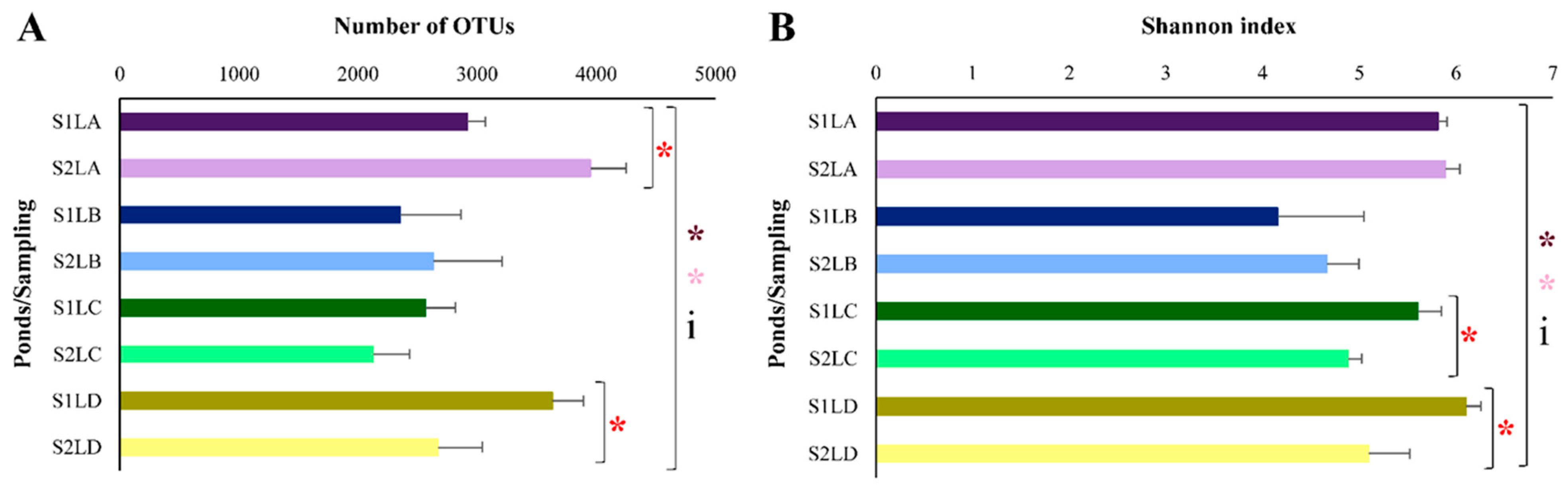
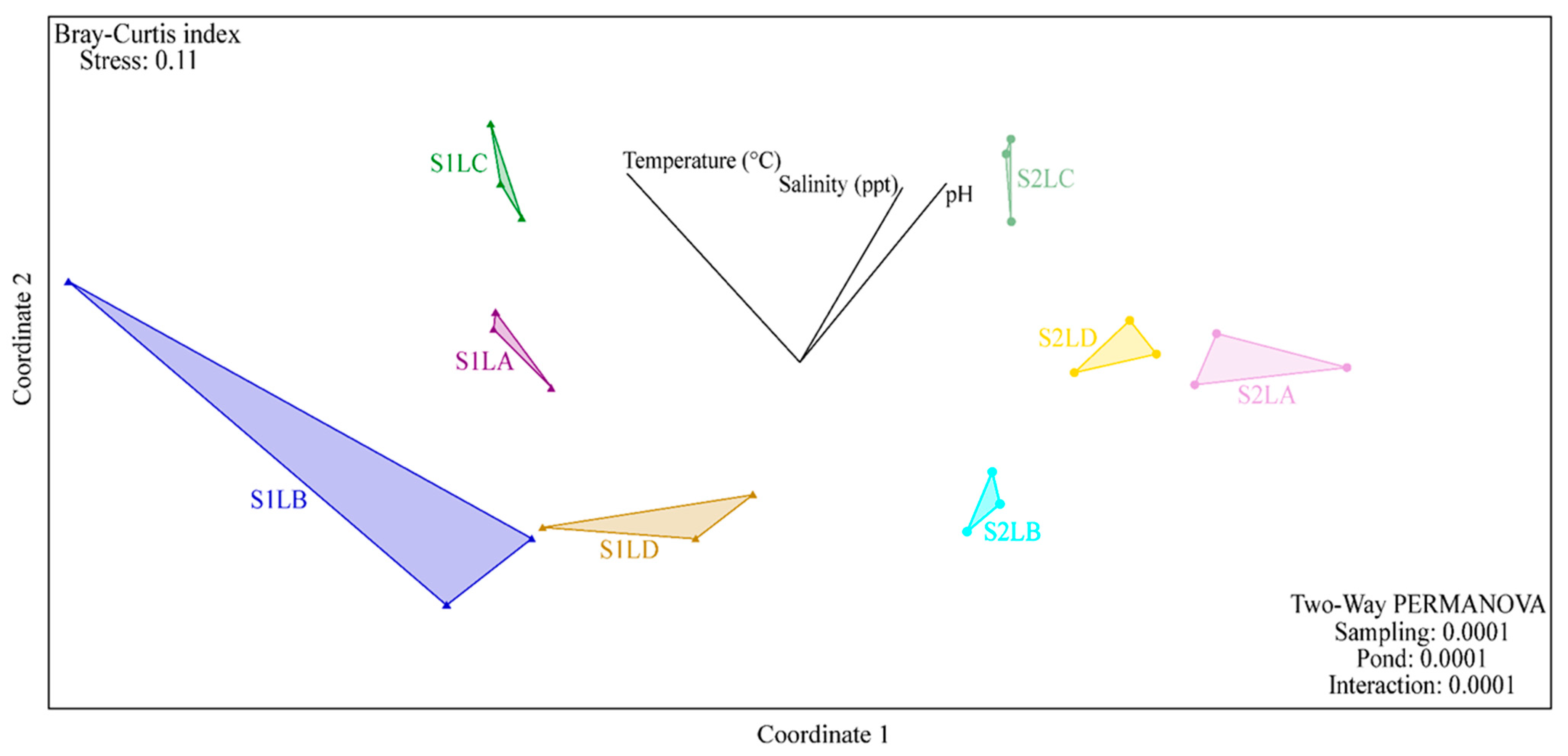
3.2. Structure and Composition of the Bacterial Communities of Water Samples

After sequencing the V4 region of the gene encoding 16S rRNA of the 24 DNA samples obtained from the four ponds, the sequences were analyzed and normalized to 47,990 sequences per sample. The number of sequences represented a sufficient coverage of the OTUs present in the bacterial communities, which can be observed by the tendency of the rarefaction curves to reach a plateau (Figure S6).
A significant difference was observed for richness (number of OTUs) between S1 and S2 for ponds LA (p = 0.0058288) and LD (p = 0.021009) (Figure 2A). Statistical differences were also observed in either S1 (p = 0.00584) or S2 (p = 0.003146) among the four ponds (Figure 2A). Considering the Shannon diversity index (Figure 2B), significant differences between S1 and S2 for LC (p = 0.011584) and for LD (p = 0.018024) were observed among the four ponds in S1 (p = 0.004127) and S2 (p = 0.004021). Moreover, an interaction between sampling periods (S1 and S2) and ponds (four water samples), related to both richness (p = 0.001341) (Figure 2A) and diversity (p = 0.01374) (Figure 2B), was observed using a two-way ANOVA.
Beta diversity was evaluated by nonmetric multidimensional scaling (nMDS) of OTUs based on the Bray–Curtis dissimilarity index (Figure 3). From a two-way PERMANOVA, it was observed that the structure of the bacterial communities of each pond was different between samplings (S1 and S2) (p = 0.0001). Additionally, a clear dispersion among the four ponds (p = 0.0001) was observed (Figure 3). With the integration of the physicochemical data to the NMDs, the temperature positively influenced the first sampling (S1) of LC, while pH and salinity positively influenced the second sampling (S2) of this pond (Figure 3).
The relative abundance of bacterial taxa in the four water samples was determined in S1 and S2. All phyla with at least 1% of the total relative abundance are shown in Figure 4. A significant difference was observed among the ponds in the relative abundance of Proteobacteria, Verrucomicrobia and Planctomycetes in S1 and S2. For Firmicutes, a significant difference among the ponds was observed only in S1, while for Actinobacteria and Bacteroidetes, a significant difference was observed only in S2. In addition, the following can be observed in Figure 4: (i) a significant difference in the relative abundance of Proteobacteria between S1 and S2 in LC and LD; (ii) a significant difference in the abundances of Actinobacteria and Verrucomicrobia between S1 and S2 in all water samples except in LB; (iii) the predominance of Actinobacteria and Verrucomicrobia in LC and LD (in S1) and in LA (in S2); and (iv) the predominance of Firmicutes in the four ponds in S1 and Bacteroidetes in LA (S1).
At the class level, the relative abundance of the taxa also varied among the four ponds and between the two samplings (Figure S7). Significant differences were observed in S1 and S2 in the relative abundance of classes Betaproteobacteria, Alphaproteobacteria, Clostridia, Sphingobacteria, Gammaproteobacteria, Cytophaga, Deltaproteobacteria and Planctomycetia. The predominance of Actinobacteria and Bacilli varied among the ponds in S2 and S1, respectively. In addition, between the two samplings of the four ponds, Clostridia was the only class that predominated in S1. Ten classes showing relative abundances of at least 1% are represented in Figure S7.
More than 40% of the sequences were taxonomically classified at the genus level. A significant difference was observed among the four ponds in the relative abundance of the genera Clostridium, Sediminibacterium, Pseudarcicella and Polynucleobacter in both samplings. Clostridium was predominant in the four ponds in S1. The genera Paenibacillus and Methylocystis showed significant differences only in S1. The genus Paenibacillus was more prevalent in LC and LD (in S1). In S2, Rhodobacter was more abundant in LC, and the relative abundance of Acidovorax, Novosphingobium and Rhodobacter varied in the four ponds (Figure S8).

3.3. Strains of ARB Identified by MALDI-TOF

A total of 135 bacterial strains isolated from CHROMagar plates supplemented with the different antimicrobials were submitted to MALDI-TOF identification. From this total, 102 strains showed score values between 1700 and 2448.
No growth was observed from LC in cultures supplemented with ciprofloxacin. The highest number of colonies was observed in LA (8 × 10² CFU/mL). Three colonies of each morphology observed in the different plates (from LA, LB and LD) were reinoculated in ciprofloxacin-supplemented plates, but only three of these strains (isolated from LD) could be identified as belonging to the genus Escherichia, with score values between 1961 and 2238 (Table S3).
Fifty-four bacterial strains recovered from cultures supplemented with ceftriaxone were identified with score values between 1700 and 2402: 10 strains of LA; 11 strains of LB; 23 strains of LC and 10 strains of LD. Strains belonging to the genus Chryseobacterium were identified from samples from all four ponds. On the other hand, strains belonging to the genus Elizabethkingia were observed only in LB, Acinetobacter sp. and Stenotrophomonas sp. only in LC, while Enterobacter sp. and Proteus sp. only in LD (Table S4).
Forty-five strains recovered from sulfamethoxazole-supplemented cultures were identified with score values between 1700 and 2438: 14 of LA; 8 of LB; 14 of LC and 9 of LD. Strains belonging to the genera Escherichia and Klebsiella were isolated from all ponds, while strains of Aeromonas and Cronobacter were identified only in LA, and those belonging to the genus Serratia were identified only in LB. Strains belonging to the genera Enterobacter and Proteus were found in LA, LC and LD (Table S5).

3.4. Comparison of Bacterial Community Composition with the Prevalence of ARB

By sequencing the gene encoding 16S rRNA, different OTUs could be related to genera attributed to bacterial strains resistant to the different antimicrobials tested and identified by MALDI-TOF (Figure S9).
In general, isolated strains identified as belonging to the genera Pseudomonas, Bacillus, Acinetobacter, Stenotrophomonas, Chryseobacterium, Aeromonas, Pantoea, Escherichia and Citrobacter were also found in the taxonomic classification of the different OTUs in all ponds. Bacterial strains from other genera belonging to the Enterobacteriaceae family were also identified, such as Cronobacter, Enterobacter, Proteus and Klebsiella.
A significant difference was observed among the ponds (one-way ANOVA/Kruskal–Wallis) in the relative abundance of OTUs associated with the genera Pseudomonas and Bacillus in S1 and with Aeromonas and Enterobacteriaceae in S2. Statistical differences were also observed between S1 and S2 in the relative abundance of Bacillus and Pantoea in LC and of Enterobacteriaceae and Bacillus in LD (Student’s t test/Mann–Whitney).

3.5. ARGs in Isolated Strains

Forty-four isolated strains were PCR amplified for the presence of ARGs using specific primers. Table 1 shows all strains where a PCR product was detected. Among the strains grown on ceftriaxone plates, Escherichia strains (isolated from LA) were positive for the blaTEM gene. Strains of Klebsiella (isolated from LA) were positive for blaTEM and blaSHV (Table 1, Figure S3B). Among the strains previously isolated on sulfamethoxazole-containing plates, the sul1 gene was detected in an Enterobacter strain (isolated from LA) (Table 1, Figure S4A) and the sul2 gene was detected in strains belonging to the genera Escherichia, Proteus and Aeromonas (isolated from LA and LD) (Table 1, Figure S4B). The intI1 gene was observed in Escherichia and Enterobacter strains (isolated from LA and LD) (Table 1, Figure S5), and the intI2 gene was observed in Enterobacter and Pantoea strains (isolated from LA and LB) (Table 1, Figure S3).

4. Discussion

It is well known that surface waters act as a sink for pollutants from terrestrial environments. Therefore, there is a great concern regarding the fate of antimicrobials and the role of bacterial communities in the dissemination of antimicrobial resistance genes in water sources. Several studies have demonstrated the presence of antibiotics, families of ARGs and ARB in aquatic ecosystems, including wastewaters, sea water, surface water (rivers, ponds, and lakes), recreational or drinking water [45,46,47,48,49,50,51]. This confirms the widespread dissemination of resistance in aquatic systems and the importance of the problem worldwide [49]. However, studies with this approach are still incipient not only in Brazil but also in other countries, especially when surface water in agricultural producing areas with the use of poultry litter is considered [52,53].
In this study, β-lactams resistance genes (blaGES, blaTEM and blaSHV) were detected in two ponds (LB and LC). Furlan and Stehling (2018) [54] also detected the blaSHV gene in water, feces, and soil from a pig farm in Brazil. However, the blaGES gene could not be detected in the water samples analyzed. Indeed, GES β-lactamases (encoded by blaGES) are less frequently found than other ESBLs (Extended Spectrum β-lactamases) and carbapenemases, except for bacterial strains isolated from clinical settings [54,55]. Therefore, the presence of the blaGES gene in the ponds studied here corroborates the data obtained by Jurelevicius et al. (2021) [51], where this gene and the blaTEM gene were observed in marine waters.
Sulfonamide resistance genes and genes encoding integrases were simultaneously detected in the three ponds. The detection of sul1 and intI1 genes in the same sample is not a surprise, as the sul1 gene is often found in the conserved regions of class 1 integrons. Therefore, in addition to the potential risk of accumulation of sul1 genes, this result may indicate the risk of dissemination of the sul genes by the frequent association of integrons with transposons and plasmids [56,57]. Lai et al. (2021) [50] also observed similar results in surface water sources in Sweden and highlighted the importance of the intI1 and sul1 genes as genetic markers of anthropogenic pollution. The sul1 and sul2 genes were also the most prevalent genes detected by PCR in river water samples from Germany and Australia, showing the environmental spread of these genes worldwide [45].
Although fluoroquinolone resistance genes were not detected on the water surface of the ponds in this study, we cannot exclude the possibility of their presence in a small amount, below the detection limit of the PCR technique. The occurrence of the qnrS gene and high concentrations of ciprofloxacin and enrofloxacin (fluoroquinolones) have already been reported in agricultural soils from the same region [24]. Moreover, fluoroquinolones have a low leaching potential and are usually detected in water at low concentrations, as they are strongly adsorbed to soils and sediments [58,59]. In contrast, sulfonamides have a high leaching potential and have been detected in high concentrations on water surfaces [60,61]. Vollú et al. (2018) [14] did not detect the qnrA, qnrB and qnrS genes in poultry litter samples in the presence of enrofloxacin and ciprofloxacin (fluoroquinolones) residues. The authors suggested that either these genes were not present or were present in low copy numbers, below the detection limit of the PCR technique.
The differences observed in the structure and composition of the bacterial communities in the four ponds may be related to soil fertilization and the amount of precipitation (which was significantly different between samplings), in addition to the physicochemical characteristics of the water samples. The phyla Firmicutes, Clostridia (class) and Clostridium (genus) were significantly more abundant in S1. Likewise, Clostridia has been described as an abundant class in the microbiota of poultries that are treated with penicillins [62,63]. Considering that S1 occurred in the wet season in a period of high rainfall volume, it is possible that bacteria of these taxa were mobilized from the soil fertilized with the poultry litter to the ponds by the surface runoff of water and leaching. Brooks et al. (2009) [62] reported that the species Clostridium perfringens can be an indicator of fecal contamination and of mobilization of poultry litter by surface runoff.
When ARB were isolated from the different ponds and further characterized, strains identified as enterobacteria (Escherichia sp., Enterobacter sp., Aeromonas sp., Proteus sp. and Pantoea sp.) were isolated from all sulfamethoxazole-containing plates. One of the most common mechanisms of sulfonamide resistance in enterobacteria is the acquisition of sul genes, which encode resistant variants of the dihydropteroate synthase (DHPS) enzyme. Usually, sul1 and sul2 genes are found among sulfonamide-resistant Enterobacteriaceae with the same frequency [64,65]. Considering the results obtained here using primers for sul1, sul2, intI1 and intI2 genes, there is a potential risk of dissemination of sulfonamide resistance among clinically important bacteria in the studied ponds.
In the same way, as genes encoding ESBLs were detected in water samples (blaGES, blaSHV and blaTEM) and in Klebsiella and Escherichia strains, there is a potential risk of dissemination of β-lactam resistance in these environments, especially considering that the genes blaSHV and blaTEM are plasmid-mediated and often reported in these genera. These genes encode narrow-spectrum β-lactamases or ESBLs that hydrolyze broad spectrum cephalosporins and monobactams [66]. The production of these enzymes by pathogenic strains of E. coli and K. pneumoniae has been reported by researchers worldwide, and their prevalence has been increasing from 6% to 88% in healthcare settings [66,67,68]. The presence of these ARGs in surface water sources indicates the dissemination of AMR in the environment. Elshafiee et al. (2022) [69] also detected the presence of blaSHV and blaTEM genes in K. pneumoniae isolates in irrigation waters from fresh produce farms in Egypt. Likewise, Amato et al. (2021) [49] also detected a high prevalence of blaTEM in E. coli strains isolated from irrigation water surface sources in Spain. These studies indicate the risk of transmission of resistant β-lactams for farmers and through the consumption of commercialized products. Bacteria of the Enterobacteriaceae family are often associated with intra-abdominal infections and can be transmitted by contaminated food and water, such as pathogenic strains of E. coli and K. pneumoniae that can cause gastrointestinal and urinary tract infections (UTIs) [69,70].
Finally, some bacterial genera isolated here, such as those of the CESP group (Citrobacter, Enterobacter, Serratia and Providencia), are producers of inducible AmpC-type β-lactamases, which mediate intrinsic resistance to most third-generation cephalosporins [71]. Intrinsic resistance is increasingly considered relevant mainly when it is detected in opportunistic pathogens. Stenotrophomonas sp. has been described as a reservoir and as a vector of acquired ARGs that encode carbapenemases, such as the blaKPC gene, especially in aquatic environments [72].
Indeed, ESBL- and carbapenemase-producing Gram-negative bacteria of the Enterobacteriaceae family represent a global threat to human and animal health. These bacteria are the main propagators of the AMR pandemic and are on the World Health Organization priority pathogens list to guide research, discovery, and development of new antimicrobials [73,74].
According to Lapidot and Yaron (2009) [75], contaminated water can increase the ability of pathogens to colonize agricultural crops, favoring resistant bacteria dissemination in the environment and resistant pathogen transmission to humans and animals [76]. Water used in irrigation has already been identified as the likely source of several outbreaks of diseases transmitted by fresh and raw foods, such as fruits and vegetables, which are the most common agricultural crops associated with outbreaks [77].

5. Conclusions

This study demonstrates that surface water sources can be considered reservoirs of resistant bacteria and resistance genes in agricultural areas with extensive use of poultry litter as an organic fertilizer. The presence of sul1, sul2, intI1 and intI2 genes and in strains isolated from the studied ponds may indicate the accumulation of resistance to sulfonamides in the environment. The detection of genes encoding ESBLs, mainly blaGES, which are found less frequently in the environment, also suggests the spread of resistance to β-lactams in the environment. In addition, seasonal influences on the evaluated bacterial communities (mainly periods of high rainfall) are an additional aggravating factor for antimicrobial-resistant bacteria mobilization (soil–water). Considering the extensive use of surface water sources, this study suggests the potential risk of dissemination and transmission of AMR, through different forms of water use, mainly in the irrigation of agricultural crops, which favors the entry of pathogens into the food chain. Therefore, monitoring and understanding of aquatic resistomes under anthropic influence is of great importance to control the environmental spread of AMR. It is still necessary to contain the irrational use of antimicrobials in human medicine, veterinary medicine, and especially in livestock and agriculture.

Supplementary Materials

The following supporting information can be downloaded at: https://www.mdpi.com/article/10.3390/antibiotics11111650/s1.

Author Contributions

L.S., E.S.L., C.E.T.P. and R.C.P. conceived the study. L.S. was responsible for project administration and funding acquisition. E.S.L. and C.E.T.P. collected the water samples. E.S.L. analyzed data from 16S rRNA amplicon metagenomic sequencing, isolated, identified and characterized the AMR bacteria. L.S., E.S.L. and C.E.T.P. drafted the manuscript. All authors have read and agreed to the published version of the manuscript.

Funding

This study was supported by grants from Conselho Nacional de Desenvolvimento Científico e Tecnológico (CNPq), Coordenação de Aperfeiçoamento de Pessoal de Nível Superior (CAPES, financial code 001) and Fundação de Amparo à Pesquisa do Estado do Rio de Janeiro (FAPERJ).

Institutional Review Board Statement

Not applicable.

Informed Consent Statement

Not applicable.

Data Availability Statement

The datasets used and/or analyzed under the current study are available from the corresponding author upon reasonable request.

Conflicts of Interest

The authors declare no conflict of interest.

References

  1. World Health Organization. Antimicrobial Resistance-Global Report on Surveillance; World Health Organization: Geneva, Switzerland, 2014; Volume 61, pp. 383–394. [Google Scholar]
  2. Chow, L.K.M.; Ghaly, T.M.; Gillings, M.R. A Survey of Sub-Inhibitory Concentrations of Antibiotics in the Environment. J. Environ. Sci. 2021, 99, 21–27. [Google Scholar] [CrossRef] [PubMed]
  3. Maillard, J.Y.; Bloomfield, S.F.; Courvalin, P.; Essack, S.Y.; Gandra, S.; Gerba, C.P.; Rubino, J.R.; Scott, E.A. Reducing Antibiotic Prescribing and Addressing the Global Problem of Antibiotic Resistance by Targeted Hygiene in the Home and Everyday Life Settings: A Position Paper. Am. J. Infect. Control 2020, 48, 1090–1099. [Google Scholar] [CrossRef] [PubMed]
  4. Rabello, R.F.; Bonelli, R.R.; Penna, B.A.; Albuquerque, J.P.; Souza, R.M.; Cerqueira, A.M.F. Antimicrobial Resistance in Farm Animals in Brazil: An Update Overview. Animals 2020, 10, 552. [Google Scholar] [CrossRef] [PubMed] [Green Version]
  5. van Boeckel, T.P.; Brower, C.; Gilbert, M.; Grenfell, B.T.; Levin, S.A.; Robinson, T.P.; Teillant, A.; Laxminarayan, R. Global Trends in Antimicrobial Use in Food Animals. Proc. Natl. Acad. Sci. USA 2015, 112, 5649–5654. [Google Scholar] [CrossRef] [PubMed] [Green Version]
  6. Gouvêa, R.; Dos Santos, F.F.; De Aquino, M.H.C. Fluoroquinolones in Industrial Poultry Production, Bacterial Resistance and Food Residues: A Review. Braz. J. Poult. Sci. 2015, 17, 1–10. [Google Scholar] [CrossRef] [Green Version]
  7. Meletis, G. Carbapenem Resistance: Overview of the Problem and Future Perspectives. Ther. Adv. Infect. Dis. 2016, 3, 15–21. [Google Scholar] [CrossRef] [Green Version]
  8. Morales-Gutiérrez, F.J.; Barbosa, J.; Barrón, D. Metabolic Study of Enrofloxacin and Metabolic Profile Modifications in Broiler Chicken Tissues after Drug Administration. Food Chem. 2015, 172, 30–39. [Google Scholar] [CrossRef]
  9. van Duijkeren, E.; Schink, A.-K.; Roberts, M.C.; Wang, Y.; Schwarz, S. Mechanisms of Bacterial Resistance to Antimicrobial Agents. Microbiol. Spectr. 2018, 6, 52–82. [Google Scholar] [CrossRef]
  10. Cardoso, M. Antimicrobial Use, Resistance and Economic Benefits and Costs to Livestock Producers in Brazil. OECD Food Agric. Fish. Pap. 2019, 135, 1–44. [Google Scholar]
  11. Palma, E.; Tilocca, B.; Roncada, P. Antimicrobial Resistance in Veterinary Medicine: An Overview. Int. J. Mol. Sci. 2020, 21, 1914. [Google Scholar] [CrossRef] [Green Version]
  12. Sarmah, A.K.; Meyer, M.T.; Boxall, A.B.A. A Global Perspective on the Use, Sales, Exposure Pathways, Occurrence, Fate and Effects of Veterinary Antibiotics (VAs) in the Environment. Chemosphere 2006, 65, 725–759. [Google Scholar] [CrossRef] [PubMed]
  13. Kemper, N. Veterinary Antibiotics in the Aquatic and Terrestrial Environment. Ecol. Indic. 2008, 8, 1–13. [Google Scholar] [CrossRef]
  14. Vollú, R.E.; Cotta, S.R.; Jurelevicius, D.; Leite, D.C.D.A.; Parente, C.E.T.; Malm, O.; Martins, D.C.; Resende, Á.V.; Marriel, I.E.; Seldin, L. Response of the Bacterial Communities Associated with Maize Rhizosphere to Poultry Litter as an Organomineral Fertilizer. Front. Environ. Sci. 2018, 6, 118. [Google Scholar] [CrossRef] [Green Version]
  15. Ljubojevic, D.; Puvača, N.; Pelic, M.; Todorovic, D.; Pajic, M.; Milanov, D.; Velhner, M. Epidemiological Significance of Poultry Litter for Spreading the Antibiotic-Resistant Strains of Escherichia coli. Worlds Poult. Sci. J. 2016, 72, 485–494. [Google Scholar] [CrossRef]
  16. Lamshöft, M.; Sukul, P.; Zühlke, S.; Spiteller, M. Behaviour of 14C-Sulfadiazine and 14C-Difloxacin during Manure Storage. Sci. Total Environ. 2010, 408, 1563–1568. [Google Scholar] [CrossRef]
  17. Jechalke, S.; Heuer, H.; Siemens, J.; Amelung, W.; Smalla, K. Fate and Effects of Veterinary Antibiotics in Soil. Trends Microbiol. 2014, 22, 536–545. [Google Scholar] [CrossRef]
  18. Xie, W.Y.; Shen, Q.; Zhao, F.J. Antibiotics and Antibiotic Resistance from Animal Manures to Soil: A Review. Eur. J. Soil Sci. 2018, 69, 181–195. [Google Scholar] [CrossRef] [Green Version]
  19. Muhammad, J.; Khan, S.; Su, J.Q.; Hesham, A.E.L.; Ditta, A.; Nawab, J.; Ali, A. Antibiotics in Poultry Manure and Their Associated Health Issues: A Systematic Review. J. Soils Sediments 2020, 20, 486–497. [Google Scholar] [CrossRef]
  20. Wright, G.D. Antibiotic Resistance in the Environment: A Link to the Clinic? Curr. Opin. Microbiol. 2010, 13, 589–594. [Google Scholar] [CrossRef]
  21. Jones, L.A.; Worobo, R.W.; Smart, C.D. Plant-Pathogenic Oomycetes, Escherichia coli Strains, and Salmonella spp. Frequently Found in Surface Water Used for Irrigation of Fruit and Vegetable Crops in New York State. Appl. Environ. Microbiol. 2014, 80, 4814–4820. [Google Scholar] [CrossRef] [Green Version]
  22. Dourado, F.; Arraes, T.C.; Silva, M.F. The “Megadesastre” in the Mountain Region of Rio de Janeiro State-Causes, Mechanisms of Mass Movements and Spatial Allocation of Investments for Reconstruction Post Disaster. Anuário Inst. Geociências-UFRJ 2012, 35, 43–54. [Google Scholar] [CrossRef]
  23. Rosi, A.; Canavesi, V.; Segoni, S.; Dias Nery, T.; Catani, F.; Casagli, N. Landslides in the Mountain Region of Rio de Janeiro: A Proposal for the Semi-Automated Definition of Multiple Rainfall Thresholds. Geosciences 2019, 9, 203. [Google Scholar] [CrossRef] [Green Version]
  24. Parente, C.E.T.; Azeredo, A.; Vollú, R.E.; Zonta, E.; Azevedo-Silva, C.E.; Brito, E.M.S.; Seldin, L.; Torres, J.P.M.; Meire, R.O.; Malm, O. Fluoroquinolones in Agricultural Soils: Multi-Temporal Variation and Risks in Rio de Janeiro Upland Region. Chemosphere 2019, 219, 409–417. [Google Scholar] [CrossRef] [PubMed]
  25. Parente, C.E.T.; Brusdzenski, G.S.; Zonta, E.; Lino, A.S.; Azevedo-Silva, C.E.; Dorneles, P.R.; Azeredo, A.; Torres, J.P.M.; Meire, R.O.; Malm, O. Fluoroquinolones and Trace Elements in Poultry Litter: Estimation of Environmental Load Based on Nitrogen Requirement for Crops. J. Environ. Sci. Health B 2020, 55, 1087–1098. [Google Scholar] [CrossRef] [PubMed]
  26. Instituto Nacional de Meteorologia Histórico de Dados Meteorológicos. Available online: https://portal.inmet.gov.br/ (accessed on 5 March 2021).
  27. Xu, X.; Kong, F.; Cheng, X.; Yan, B.; Du, X.; Gai, J.; Ai, H.; Shi, L.; Iredell, J. Integron Gene Cassettes in Acinetobacter spp. Strains from South China. Int. J. Antimicrob. Agents 2008, 32, 441–445. [Google Scholar] [CrossRef] [PubMed]
  28. Campana, E.H.; Xavier, D.E.; Petrolini, F.V.B.; Cordeiro-Moura, J.R.; de Araujo, M.R.E.; Gales, A.C. Carbapenem-Resistant and Cephalosporin-Susceptible: A Worrisome Phenotype among Pseudomonas aeruginosa Clinical Isolates in Brazil. Braz. J. Infect. Dis. 2017, 21, 57–62. [Google Scholar] [CrossRef] [Green Version]
  29. Picão, R.C.; Poirel, L.; Gales, A.C.; Nordmann, P. Diversity of β-Lactamases Produced by Ceftazidime-Resistant Pseudomonas aeruginosa Isolates Causing Bloodstream Infections in Brazil. Antimicrob. Agents Chemother. 2009, 53, 3908–3913. [Google Scholar] [CrossRef] [Green Version]
  30. Pérez-Pérez, F.J.; Hanson, N.D. Detection of Plasmid-Mediated AmpC β-Lactamase Genes in Clinical Isolates by Using Multiplex PCR. J. Clin. Microbiol. 2002, 40, 2153–2162. [Google Scholar] [CrossRef] [Green Version]
  31. Toleman, M.A.; Bennett, P.M.; Bennett, D.M.C.; Jones, R.N.; Walsh, T.R. Global Emergence of Trimethoprim/Sulfamethoxazole Resistance in Stenotrophomonas maltophilia Mediated by Acquisition of sul Genes. Emerg. Infect. Dis. 2007, 13, 559–565. [Google Scholar] [CrossRef]
  32. Cattoir, V.; Poirel, L.; Rotimi, V.; Soussy, C.J.; Nordmann, P. Multiplex PCR for Detection of Plasmid-Mediated Quinolone Resistance Qnr Genes in ESBL-Producing Enterobacterial Isolates. J. Antimicrob. Chemother. 2007, 60, 394–397. [Google Scholar] [CrossRef] [Green Version]
  33. Kraychete, G.B.; Botelho, L.A.B.; Campana, E.H.; Picão, R.C.; Bonelli, R.R. Updated Multiplex PCR for Detection of All Six Plasmid-Mediated Qnr Gene Families. Antimicrob. Agents Chemother. 2016, 60, 7524–7526. [Google Scholar] [CrossRef] [PubMed]
  34. Yamane, K.; Wachino, J.I.; Suzuki, S.; Kimura, K.; Shibata, N.; Kato, H.; Shibayama, K.; Konda, T.; Arakawa, Y. New Plasmid-Mediated Fluoroquinolone Efflux Pump, QepA, Found in an Escherichia coli Clinical Isolate. Antimicrob. Agents Chemother. 2007, 51, 3354–3360. [Google Scholar] [CrossRef] [PubMed] [Green Version]
  35. Sambrook, J.; Fritsch, E.F.; Maniatis, T. Molecular Cloning: A Laboratory Manual, 2nd ed.; Cold Spring Harbor Laboratory Press: New York, NY, USA, 1989. [Google Scholar]
  36. Apprill, A.; Mcnally, S.; Parsons, R.; Weber, L. Minor Revision to V4 Region SSU rRNA 806R Gene Primer Greatly Increases Detection of SAR11 Bacterioplankton. Aquatic. Microbial. Ecol. 2015, 75, 129–137. [Google Scholar] [CrossRef] [Green Version]
  37. Caporaso, J.G.; Lauber, C.L.; Walters, W.A.; Berg-Lyons, D.; Lozupone, C.A.; Turnbaugh, P.J.; Fierer, N.; Knight, R. Global Patterns of 16S rRNA Diversity at a Depth of Millions of Sequences per Sample. Proc. Natl. Acad. Sci. USA 2011, 108, 4516–4522. [Google Scholar] [CrossRef] [PubMed] [Green Version]
  38. Schloss, P.D.; Westcott, S.L.; Ryabin, T.; Hall, J.R.; Hartmann, M.; Hollister, E.B.; Lesniewski, R.A.; Oakley, B.B.; Parks, D.H.; Robinson, C.J.; et al. Introducing Mothur: Open-Source, Platform-Independent, Community-Supported Software for Describing and Comparing Microbial Communities. Appl. Environ. Microbiol. 2009, 75, 7537–7541. [Google Scholar] [CrossRef] [Green Version]
  39. Quast, C.; Pruesse, E.; Yilmaz, P.; Gerken, J.; Schweer, T.; Yarza, P.; Peplies, J.; Glöckner, F.O. The SILVA Ribosomal RNA Gene Database Project: Improved Data Processing and Web-Based Tools. Nucleic Acids Res. 2013, 41, 590–596. [Google Scholar] [CrossRef]
  40. Cole, J.R.; Wang, Q.; Cardenas, E.; Fish, J.; Chai, B.; Farris, R.J.; Kulam-Syed-Mohideen, A.S.; McGarrell, D.M.; Marsh, T.; Garrity, G.M.; et al. The Ribosomal Database Project: Improved Alignments and New Tools for rRNA Analysis. Nucleic Acids Res. 2009, 37, 141–145. [Google Scholar] [CrossRef] [Green Version]
  41. Hammer, R.; Harper, D.A.T.; Ryan, P.D. PAST: Paleontological Statistics Software Package for Education and Data Analysis. Palaeontol. Electron. 2001, 4, 9. [Google Scholar]
  42. Box, G.E.P.; Cox, D.R. An Analysis of Transformations. J. R. Stat. Soc. Ser. B 1964, 26, 211–243. [Google Scholar] [CrossRef]
  43. Suzuki, S.; Ogo, M.; Miller, T.W.; Shimizu, A.; Takada, H.; Siringan, M.A.T. Who Possesses Drug Resistance Genes in the Aquatic Environment?: Sulfamethoxazole (SMX) Resistance Genes among the Bacterial Community in Water Environment of Metro-Manila, Philippines. Front. Microbiol. 2013, 4, 102. [Google Scholar] [CrossRef] [Green Version]
  44. Röderova, M.; Halova, D.; Papousek, I.; Dolejska, M.; Masarikova, M.; Hanulik, V.; Pudova, V.; Broz, P.; Htoutou-Sedlakova, M.; Sauer, P.; et al. Characteristics of Quinolone Resistance in Escherichia coli Isolates from Humans, Animals, and the Environment in the Czech Republic. Front. Microbiol. 2017, 7, 2147. [Google Scholar] [CrossRef] [PubMed] [Green Version]
  45. Stoll, C.; Sidhu, J.P.S.; Tiehm, A.; Toze, S. Prevalence of Clinically Relevant Antibiotic Resistance Genes in Surface Water Samples Collected from Germany and Australia. Environ. Sci. Technol. 2012, 46, 9716–9726. [Google Scholar] [CrossRef] [PubMed]
  46. Christou, A.; Agüera, A.; Bayona, J.M.; Cytryn, E.; Fotopoulos, V.; Lambropoulou, D.; Manaia, C.M.; Michael, C.; Revitt, M.; Schröder, P.; et al. The Potential Implications of Reclaimed Wastewater Reuse for Irrigation on the Agricultural Environment: The Knowns and Unknowns of the Fate of Antibiotics and Antibiotic Resistant Bacteria and Resistance Genes—A Review. Water Res. 2017, 123, 448–467. [Google Scholar] [CrossRef] [PubMed] [Green Version]
  47. Niu, Z.G.; Zhang, K.; Zhang, Y. Occurrence and Distribution of Antibiotic Resistance Genes in the Coastal Area of the Bohai Bay, China. Mar. Pollut. Bull. 2016, 107, 245–250. [Google Scholar] [CrossRef]
  48. Proia, L.; von Schiller, D.; Sànchez-Melsió, A.; Sabater, S.; Borrego, C.M.; Rodríguez-Mozaz, S.; Balcázar, J.L. Occurrence and Persistence of Antibiotic Resistance Genes in River Biofilms after Wastewater Inputs in Small Rivers. Environ. Pollut. 2016, 210, 121–128. [Google Scholar] [CrossRef]
  49. Amato, M.; Dasí, D.; González, A.; Ferrús, M.A.; Castillo, M.Á. Occurrence of Antibiotic Resistant Bacteria and Resistance Genes in Agricultural Irrigation Waters from Valencia City (Spain). Agric. Water Manag. 2021, 256, 107097. [Google Scholar] [CrossRef]
  50. Lai, F.Y.; Muziasari, W.; Virta, M.; Wiberg, K.; Ahrens, L. Profiles of Environmental Antibiotic Resistomes in the Urban Aquatic Recipients of Sweden Using High-Throughput Quantitative PCR Analysis. Environ. Pollut. 2021, 287, 117651. [Google Scholar] [CrossRef]
  51. Jurelevicius, D.; Cotta, S.R.; Montezzi, L.F.; Dias, A.C.F.; Mason, O.U.; Picão, R.C.; Jansson, J.K.; Seldin, L. Enrichment of Potential Pathogens in Marine Microbiomes with Different Degrees of Anthropogenic Activity. Environ. Pollut. 2021, 268, 115757. [Google Scholar] [CrossRef]
  52. Amarasiri, M.; Sano, D.; Suzuki, S. Understanding Human Health Risks Caused by Antibiotic Resistant Bacteria (ARB) and Antibiotic Resistance Genes (ARG) in Water Environments: Current Knowledge and Questions to Be Answered. Crit. Rev. Environ. Sci. Technol. 2020, 50, 2016–2059. [Google Scholar] [CrossRef]
  53. Liu, X.; Wang, Z.; Zhang, L.; Fan, W.; Yang, C.; Li, E.; Du, Y.; Wang, X. Inconsistent Seasonal Variation of Antibiotics between Surface Water and Groundwater in the Jianghan Plain: Risks and Linkage to Land Uses. J. Environ. Sci. 2021, 109, 102–113. [Google Scholar] [CrossRef]
  54. Furlan, J.P.R.; Stehling, E.G. Detection of β-Lactamase Encoding Genes in Feces, Soil and Water from a Brazilian Pig Farm. Environ. Monit. Assess. 2018, 190, 76. [Google Scholar] [CrossRef] [PubMed]
  55. Queenan, A.M.; Bush, K. Carbapenemases: The Versatile β-Lactamases. Clin. Microbiol. Rev. 2007, 20, 440–458. [Google Scholar] [CrossRef]
  56. Yang, Y.; Song, W.; Lin, H.; Wang, W.; Du, L.; Xing, W. Antibiotics and Antibiotic Resistance Genes in Global Lakes: A Review and Meta-Analysis. Environ. Int. 2018, 116, 60–73. [Google Scholar] [CrossRef] [Green Version]
  57. Poirel, L.; Madec, J.-Y.; Lupo, A.; Schink, A.-K.; Kieffer, N.; Nordmann, P.; Schwarz, S. Antimicrobial Resistance in Escherichia coli. Microbiol. Spectr. 2018, 6, 289–316. [Google Scholar] [CrossRef] [PubMed] [Green Version]
  58. Riaz, L.; Mahmood, T.; Khalid, A.; Rashid, A.; Ahmed Siddique, M.B.; Kamal, A.; Coyne, M.S. Fluoroquinolones (FQs) in the Environment: A Review on Their Abundance, Sorption and Toxicity in Soil. Chemosphere 2018, 191, 704–720. [Google Scholar] [CrossRef] [PubMed]
  59. van Doorslaer, X.; Dewulf, J.; van Langenhove, H.; Demeestere, K. Fluoroquinolone Antibiotics: An Emerging Class of Environmental Micropollutants. Sci. Total Environ. 2014, 500–501, 250–269. [Google Scholar] [CrossRef] [PubMed]
  60. Tang, J.; Wang, S.; Fan, J.; Long, S.; Wang, L.; Tang, C.; Tam, N.F.; Yang, Y. Predicting Distribution Coefficients for Antibiotics in a River Water–Sediment Using Quantitative Models Based on Their Spatiotemporal Variations. Sci. Total Environ. 2019, 655, 1301–1310. [Google Scholar] [CrossRef]
  61. Leal, R.M.P.; Alleoni, L.R.F.; Tornisielo, V.L.; Regitano, J.B. Sorption of Fluoroquinolones and Sulfonamides in 13 Brazilian Soils. Chemosphere 2013, 92, 979–985. [Google Scholar] [CrossRef] [Green Version]
  62. Brooks, J.P.; Adeli, A.; Read, J.J.; McLaughlin, M.R. Rainfall Simulation in Greenhouse Microcosms to Assess Bacterial-Associated Runoff from Land-Applied Poultry Litter. J. Environ. Qual. 2009, 38, 218–229. [Google Scholar] [CrossRef]
  63. Singh, P.; Karimi, A.; Devendra, K.; Waldroup, P.W.; Cho, K.K.; Kwon, Y.M. Influence of Penicillin on Microbial Diversity of the Cecal Microbiota in Broiler Chickens. Poult. Sci. 2013, 92, 272–276. [Google Scholar] [CrossRef]
  64. Shin, H.W.; Lim, J.; Kim, S.; Kim, J.; Kwon, G.C.; Koo, S.H. Characterization of Trimethoprim-Sulfamethoxazole Resistance Genes and Their Relatedness to Class 1 Integron and Insertion Sequence Common Region in Gram-Negative Bacilli. J. Microbiol. Biotechnol. 2015, 25, 137–142. [Google Scholar] [CrossRef] [PubMed] [Green Version]
  65. Eliopoulos, G.M.; Huovinen, P. Resistance to Trimethoprim-Sulfamethoxazole. Antimicrob. Resist. 2001, 32, 1608–1614. [Google Scholar] [CrossRef] [PubMed]
  66. Ugbo, E.N.; Anyamene, C.O.; Moses, I.B.; Iroha, I.R.; Babalola, O.O.; Ukpai, E.G.; Chukwunwejim, C.R.; Egbule, C.U.; Emioye, A.A.; Okata-Nwali, O.D.; et al. Prevalence of blaTEM, blaSHV, and blaCTX-M Genes among Extended Spectrum Beta-Lactamase-Producing Escherichia coli and Klebsiella pneumoniae of Clinical Origin. Gene Rep. 2020, 21, 100909. [Google Scholar] [CrossRef]
  67. Fadil Saedii, A.A.; Abdelraheim, A.R.; Abdel Aziz, A.A.; Swelam, S.H. ESBL-Producing E. coli and Klebsiella among Patients Treated at Minia University Hospitals. J. Infect. Dis. Prev. Med. 2017, 5, 156. [Google Scholar] [CrossRef]
  68. Zongo, K.J.; Dabire, A.M.; Compaore, L.G.; Sanou, I.; Sangare, L.; Simpore, J.; Zeba, B. First Detection of bla TEM, SHV and CTX-M among Gram Negative Bacilli Exhibiting Extended Spectrum -Lactamase Phenotype Isolated at University Hospital Center, Yalgado Ouedraogo, Ouagadougou, Burkina Faso. Afr. J. Biotechnol. 2015, 14, 1174–1180. [Google Scholar] [CrossRef] [Green Version]
  69. Elshafiee, E.A.; Kadry, M.; Nader, S.M.; Ahmed, Z.S. Extended-Spectrum-Beta-Lactamases and Carbapenemase-Producing Klebsiella pneumoniae Isolated from Fresh Produce Farms in Different Governorates of Egypt. Vet. World 2022, 15, 1191–1196. [Google Scholar] [CrossRef]
  70. Schuetz, A.N. Emerging Agents of Gastroenteritis: Aeromonas, Plesiomonas, and the Diarrheagenic Pathotypes of Escherichia coli. Semin. Diagn Pathol. 2019, 36, 187–192. [Google Scholar] [CrossRef]
  71. Meurant, A.; Guérin, F.; le Hello, S.; Saint-Lorant, G.; de La Blanchardière, A. Cefepime Use: A Need for Antimicrobial Stewardship. Infect. Dis. Now 2021, 51, 445–450. [Google Scholar] [CrossRef]
  72. Mançano, S.M.C.N.; Campana, E.H.; Felix, T.P.; Barrueto, L.R.L.; Pereira, P.S.; Picão, R.C. Frequency and Diversity of Stenotrophomonas Spp. Carrying blaKPC in Recreational Coastal Waters. Water Res. 2020, 185, 116210. [Google Scholar] [CrossRef]
  73. Falgenhauer, L.; Schwengers, O.; Schmiedel, J.; Baars, C.; Lambrecht, O.; Heß, S.; Berendonk, T.U.; Falgenhauer, J.; Chakraborty, T.; Imirzalioglu, C. Multidrug-Resistant and Clinically Relevant Gram-Negative Bacteria Are Present in German Surface Waters. Front. Microbiol. 2019, 10, 2779. [Google Scholar] [CrossRef] [Green Version]
  74. WHO. Prioritization of Pathogens to Guide Discovery, Research and Development of New Antibiotics for Drug-Resistant Bacterial Infections, Including Tuberculosis; WHO/EMP/IAU/2017.12; World Health Organization: Geneva, Switzerland, 2017. [Google Scholar]
  75. Lapidot, A.; Yaron, S. Transfer of Salmonella enterica Serovar Typhimurium from Contaminated Irrigation Water to Parsley Is Dependent on Curli and Cellulose, the Biofilm Matrix Components. J. Food Prot. 2009, 72, 618–623. [Google Scholar] [CrossRef] [PubMed]
  76. Marques, R.Z.; Wistuba, N.; Brito, J.C.M.; Bernardoni, V.; Rocha, D.C.; Gomes, M.P. Crop Irrigation (Soybean, Bean, and Corn) with Enrofloxacin-Contaminated Water Leads to Yield Reductions and Antibiotic Accumulation. Ecotoxicol. Environ. Saf. 2021, 216, 112193. [Google Scholar] [CrossRef] [PubMed]
  77. Uyttendaele, M.; Jaykus, L.A.; Amoah, P.; Chiodini, A.; Cunliffe, D.; Jacxsens, L.; Holvoet, K.; Korsten, L.; Lau, M.; McClure, P.; et al. Microbial Hazards in Irrigation Water: Standards, Norms, and Testing to Manage Use of Water in Fresh Produce Primary Production. Compr. Rev. Food Sci. Food Saf. 2015, 14, 336–356. [Google Scholar] [CrossRef]
Figure 1. Presence of resistance genes in the studied ponds. Resistance genes are represented by geometric figures—dark pink in S1 and light pink in S2—in a Venn diagram. S1 and S2 correspond to the sampling period: high (S1; on 14 March 2019) and low monthly rainfall (S2; on 25 November 2019).
Antibiotics 11 01650 g001
Figure 2. Alpha diversity evaluated by massive sequencing of the gene encoding 16S rRNA in water samples collected in LA (purple), LB (blue), LC (green) and LD (yellow) in S1 (high rainfall period—dark tones) and in S2 (low rainfall period—light tones). The bars represent the standard deviation. Species richness and alpha diversity showed significant differences between the ponds in the two samplings (parametric data submitted to a one-way ANOVA) represented by asterisks (dark pink for S1 and light pink for S2). A red asterisk represents the significant difference among the ponds at each sampling (parametric data submitted to the t test). The data were also submitted to a two-way ANOVA, and the interaction between the factors is represented by the letter i. (A) Richness evaluated by the number of OTUs; (B) Shannon diversity index.
Antibiotics 11 01650 g002
Figure 3. Nonmetric multidimensional scaling (nMDS) with the Bray–Curtis dissimilarity index. Representation of LA (purple), LB (blue), LC (green) and LD (yellow). The replicates are represented by triangles in dark tones (S1) and circles in light tones (S2). The physical–chemical data evaluated from the water samples from the ponds were integrated as metadata.
Antibiotics 11 01650 g003
Figure 4. Relative abundance of bacterial phyla in water samples collected in LA (purple), LB (blue), LC (green) and LD (yellow) in S1 (high rainfall period—dark tones) and in S2 (low rainfall period—light tones). The bars represent the standard deviation. Asterisks (dark pink for S1 and light pink for S2) represent the significant differences among the ponds in the two samplings (parametric data submitted to a one-way ANOVA and nonparametric data submitted to Kruskal–Wallis). A red asterisk corresponds to the significant difference between the samples of each pond (parametric data submitted to the t test and nonparametric data submitted to Mann–Whitney). (D) next to the bacterial taxon means that the classification was at the domain level, and no statistical analysis was applied. The data represented by “others” were also not statistically analyzed because they are formed by more than one taxon.
Antibiotics 11 01650 g004
Table 1. Presence of antimicrobial resistance genes in the isolated strains.
Ceftriaxone Resistant StrainsAntimicrobial Resistance Genes
blaTEMblaSHV
Escherichia sp. (CCA 1.2) *10
Escherichia sp. (CCA 1.3)10
Klebsiella sp. (CCA 2.1)11
Klebsiella sp. (CCA 2.2)11
Klebsiella sp. (CCA 2.3)11
Sulfamethoxazole resistant strainssul1sul2intI1intI2
Escherichia sp. (CSA 1.1) *0110
Proteus sp. (CSA 2.1A)0100
Aeromonas sp. (CSA 2.2)0100
Enterobacter sp. (CSA 2.3)1010
Enterobacter sp. (CSA 5.1)0001
Enterobacter sp. (CSA 5.2)0001
Pantoea sp. (CSB 1.1)0001
Escherichia sp. (CSD 1.2)0100
Escherichia sp. (CSD 1.3)0110
* The name of the strains corresponds to the origin of their isolation: CCA (CHROMagar containing ceftriaxone and LA), CSA, CSB and CSD (CHROMagar containing sulfamethoxazole and LA, LB and LD, respectively).
Publisher’s Note: MDPI stays neutral with regard to jurisdictional claims in published maps and institutional affiliations.

