
Chemical Composition and Biological Activities of the Leaf Essential Oils of Curcuma longa, Curcuma aromatica and Curcuma angustifolia

 ,
,  ,
,  , , ,
, , ,  and
and
Abstract
:1. Introduction
2. Results
2.1. Determination of the Yield and Chemical Composition of Leaf Essential Oils by GC-MS
2.2. Antioxidant Activities of Leaf Essential Oils of Different Curcuma spp.
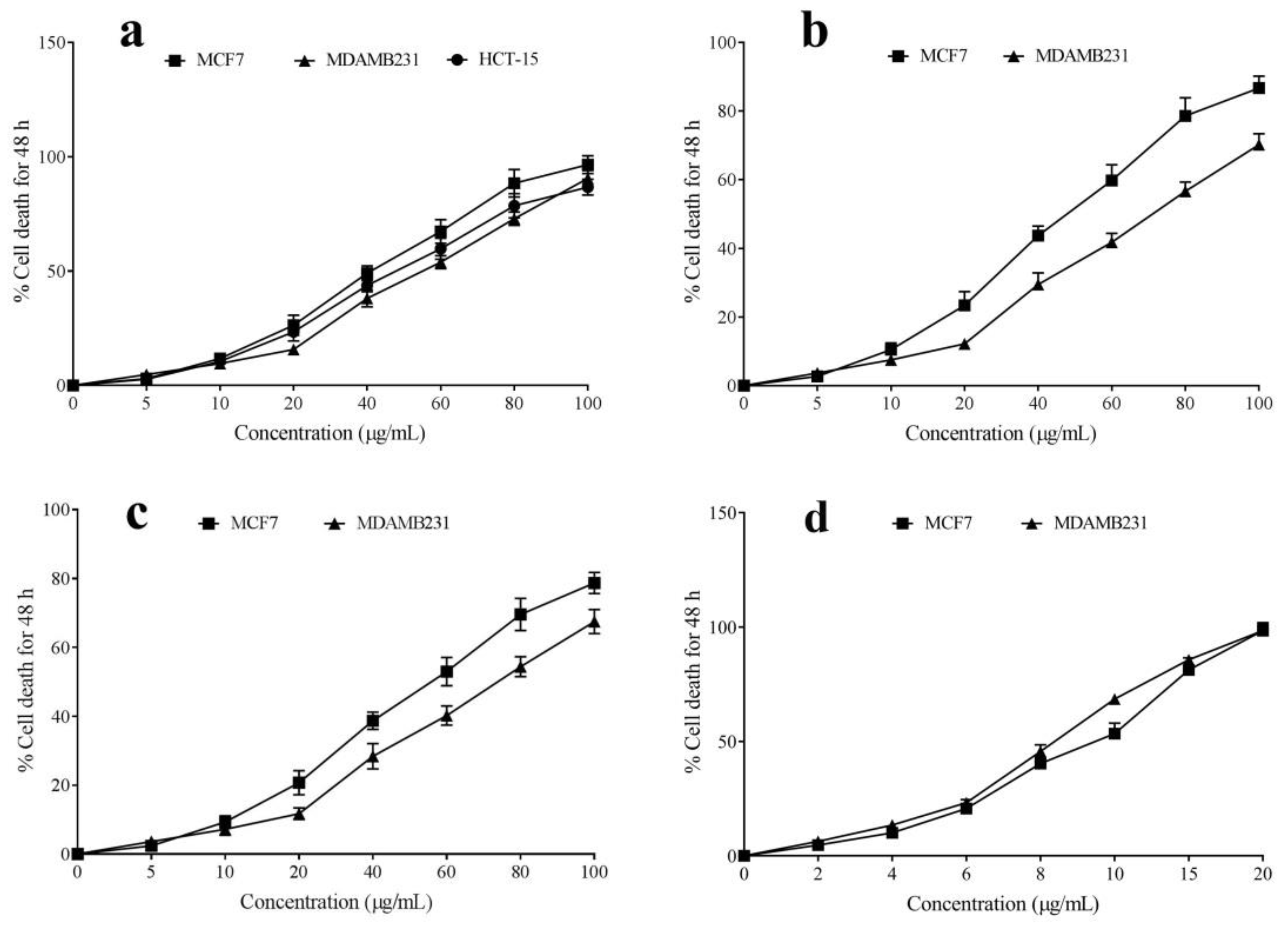
2.3. Cytotoxic Activity of Leaf Essential Oil of Different Curcuma spp.
2.4. Antibacterial Activity of Leaf Essential Oil of Different Curcuma spp.
3. Discussion
4. Materials and Methods
4.1. Materials and Chemicals
4.2. Collection of Curcuma Leaves and Extraction of Essential Oil
4.3. Chemical Component Analysis by GC-MS Analysis
4.4. In Vitro Antioxidant Activity as Scavenging of DPPH, Hydrogen Peroxide and ABTS Radicals of the Essential Oils of Different Curcuma spp.
4.4.1. Anti-DPPH Radical Assay
4.4.2. Curcuma Essential Oils and ABTS Radical Quenching Ability
4.4.3. Hydrogen Peroxide Neutralization Assay
4.5. Cytotoxic Activity of the Leaf Essential Oils of Different Curcuma spp.
4.6. Antibacterial Activity of Leaf Essential Oil of Different Curcuma spp. by Disc Diffusion Method
4.7. Minimum Inhibitory Concentration (MIC) of Essential Oil of Different Curcuma spp.
4.8. Statistical Analysis
5. Conclusions
Supplementary Materials
Author Contributions
Funding
Institutional Review Board Statement
Informed Consent Statement
Data Availability Statement
Acknowledgments
Conflicts of Interest
References
- Yuan, H.; Ma, Q.; Ye, L.; Piao, G. The Traditional Medicine and Modern Medicine from Natural Products. Molecules 2016, 21, 559. [Google Scholar]
- Oreopoulou, A.; Choulitoudi, E.; Tsimogiannis, D.; Oreopoulou, V. Six Common Herbs with Distinctive Bioactive, Antioxidant Components. A Review of Their Separation Techniques. Molecules 2021, 26, 2920. [Google Scholar] [CrossRef] [PubMed]
- Khan, S.; Khan, H.U.; Khan, F.A.; Shah, A.; Wadood, A.; Ahmad, S.; Almehmadi, M.; Alsaiari, A.A.; Shah, F.U.; Kamran, N. Anti-Alzheimer and Antioxidant Effects of Nelumbo nucifera L. Alkaloids, Nuciferine and Norcoclaurine in Alloxan-Induced Diabetic Albino Rats. Pharmaceuticals 2022, 15, 1205. [Google Scholar] [CrossRef] [PubMed]
- Khurram, M.; Lawton, L.A.; Edwards, C.; Iriti, M.; Hameed, A.; Khan, M.A.; Khan, F.A.; Rahman, S.U. Rapid Bioassay-Guided Isolation of Antibacterial Clerodane Type Diterpenoid from Dodonaea viscosa (L.) Jaeq. Int. J. Mol. Sci. 2015, 16, 20290–20307. [Google Scholar] [CrossRef] [PubMed] [Green Version]
- Khan, F.A.; Khan, N.M.; Ahmad, S.; Khan, N.; Aziz, R.; Ullah, I.; Almehmadi, M.; Allahyani, M.; Alsaiari, A.A.; Aljuaid, A. Phytochemical Profiling, Antioxidant, Antimicrobial and Cholinesterase Inhibitory Effects of Essential Oils Isolated from the Leaves of Artemisia scoparia and Artemisia absinthium. Pharmaceuticals 2022, 15, 1221. [Google Scholar] [CrossRef] [PubMed]
- Khan, F.A.; Jan, A.K.; Khan, N.M.; Khan, N.A.; Khan, S. GC/MS analysis, antimicrobial and in vitro anti-cholinesterase activities of the essential oil from Buddleja asiatica. Bangladesh J. Pharmacol. 2015, 10, 891–895. [Google Scholar] [CrossRef] [Green Version]
- Arct, J.; Ratz-Łyko, A.; Mieloch, M.; Witulska, M. Evaluation of skin colouring properties of Curcuma longa extract. Indian J. Pharm. Sci. 2014, 76, 374–378. [Google Scholar]
- Pujimulyani, D.; Suryani, C.L.; Setyowati, A.; Handayani, R.A.S.; Arumwardana, S.; Widowati, W.; Maruf, A. Cosmeceutical potentials of Curcuma mangga Val. extract in human BJ fibroblasts against MMP1, MMP3, and MMP13. Heliyon 2020, 6, e04921. [Google Scholar] [CrossRef]
- Pabuprapap, W.; Nakyai, W.; Chaichompoo, W.; Pheedee, N.; Phetkeereerat, S.; Viyoch, J.; Yingyongnarongkul, B.-E.; Ajavakom, V.; Chompoosor, A.; Piyachaturawat, P.; et al. Curcuma aromatica and Curcuma comosa Extracts and Isolated Constituents Provide Protection against UVB-Induced Damage and Attenuate Matrix Metalloproteinase-1 Expression in HaCaT Cells. Cosmetics 2022, 9, 23. [Google Scholar] [CrossRef]
- Ma, X.W.; Guo, R.Y. Dose-dependent effect of Curcuma longa for the treatment of Parkinson’s disease. Exp. Ther. Med. 2017, 13, 1799–1805. [Google Scholar] [CrossRef] [Green Version]
- Voulgaropoulou, S.D.; van Amelsvoort, T.A.M.J.; Prickaerts, J.; Vingerhoets, C. The effect of curcumin on cognition in Alzheimer’s disease and healthy aging: A systematic review of pre-clinical and clinical studies. Brain Res. 2019, 1725, 146476. [Google Scholar] [CrossRef]
- Ringman, J.M.; Frautschy, S.A.; Teng, E.; Begum, A.N.; Bardens, J.; Beigi, M.; Gylys, K.H.; Badmaev, V.; Heath, D.D.; Apostolova, L.G.; et al. Oral curcumin for Alzheimer’s disease: Tolerability and efficacy in a 24-week randomized, double blind, placebo-controlled study. Alzheimer’s Res. Ther. 2012, 4, 43. [Google Scholar] [CrossRef]
- Jarhahzadeh, M.; Alavinejad, P.; Farsi, F.; Husain, D.; Rezazadeh, A. The effect of turmeric on lipid profile, malondialdehyde, liver echogenicity and enzymes among patients with nonalcoholic fatty liver disease: A randomized double blind clinical trial. Diabetol. Metab. Syndr. 2021, 13, 112. [Google Scholar] [CrossRef]
- White, C.M.; Lee, J.Y. The impact of turmeric or its curcumin extract on nonalcoholic fatty liver disease: A systematic review of clinical trials. Pharm. Pract. 2019, 17, 4. [Google Scholar] [CrossRef] [Green Version]
- Maithilikarpagaselvi, N.; Sridhar, M.G.; Swaminathan, R.P.; Sripradha, R.; Badhe, B. Curcumin inhibits hyperlipidemia and hepatic fat accumulation in high-fructose-fed male Wistar rats. Pharm. Biol. 2016, 54, 2857–2863. [Google Scholar] [CrossRef] [Green Version]
- Jena, S.; Ray, A.; Banerjee, A.; Sahoo, A.; Nasim, N.; Sahoo, S.; Kar, B.; Patnaik, J.; Panda, P.C.; Nayak, S. Chemical composition and antioxidant activity of essential oil from leaves and rhizomes of Curcuma angustifolia Roxb. Nat. Prod. Res. 2017, 31, 2188–2191. [Google Scholar] [CrossRef]
- Dosoky, N.S.; Setzer, W.N. Chemical Composition and Biological Activities of Essential Oils of Curcuma Species. Nutrients 2018, 10, 1196. [Google Scholar] [CrossRef] [Green Version]
- Liju, V.B.; Jeena, K.; Kuttan, R. Chemopreventive activity of turmeric essential oil and possible mechanisms of action. Asian Pac. J. Cancer Prev. 2014, 15, 6575–6580. [Google Scholar] [CrossRef] [Green Version]
- Jacob, J.N.; Toloue, M. Biological Studies of Turmeric Oil, Part 1: Selective In Vitro Anticancer Activity of Turmeric Oil (TO) and TO-Paclitaxel Combination. Nat. Prod. Commun. 2013, 8, 1934578X1300800632. [Google Scholar] [CrossRef] [Green Version]
- Al-Reza, S.M.; Rahman, A.; Sattar, M.A.; Rahman, M.O.; Fida, H.M. Essential oil composition and antioxidant activities of Curcuma aromatica Salisb. Food Chem. Toxicol. 2010, 48, 1757–1760. [Google Scholar] [CrossRef]
- Sharma, K.; Garg, V.K. Vermicomposting of Waste: A Zero-Waste Approach for Waste Management. In Sustainable Resource Recovery and Zero Waste Approaches; Taherzadeh, M.J., Bolton, K., Wong, J., Pandey, A., Eds.; Elsevier: Amsterdam, The Netherlands, 2019; pp. 133–164. [Google Scholar] [CrossRef]
- Gomes-Araújo, R.; Martínez-Vázquez, D.G.; Charles-Rodríguez, A.V.; Rangel-Ortega, S.; Robledo-Olivo, A. Bioactive Compounds from Agricultural Residues, Their Obtaining Techniques, and the Antimicrobial Effect as Postharvest Additives. Int. J. Food Sci. 2021, 2021, 9936722. [Google Scholar] [CrossRef]
- Duque-Acevedo, M.; Belmonte-Ureña, L.J.; Cortés-García, F.J.; Camacho-Ferre, F. Agricultural waste: Review of the evolution, approaches and perspectives on alternative uses. Glob. Ecol. Conserv. 2020, 22, e00902. [Google Scholar] [CrossRef]
- Sarangi, P.K.; Subudhi, S.; Bhatia, L.; Saha, K.; Mudgil, D.; Shadangi, K.P.; Srivastava, R.K.; Pattnaik, B.; Arya, R.K. Utilization of agricultural waste biomass and recycling toward circular bioeconomy. Environ. Sci. Pollut. Res. Int. 2022, 13, 022–20669. [Google Scholar]
- Ravindran, R.; Hassan, S.S.; Williams, G.A.; Jaiswal, A.K. A Review on Bioconversion of Agro-Industrial Wastes to Industrially Important Enzymes. Bioengineering 2018, 5, 93. [Google Scholar] [CrossRef] [PubMed] [Green Version]
- Baliyan, S.; Mukherjee, R.; Priyadarshini, A.; Vibhuti, A.; Gupta, A.; Pandey, R.P.; Chang, C.-M. Determination of Antioxidants by DPPH Radical Scavenging Activity and Quantitative Phytochemical Analysis of Ficus religiosa. Molecules 2022, 27, 1326. [Google Scholar] [CrossRef]
- Manasa, V.; Vaishnav, S.R.; Tumaney, A.W. Physicochemical characterization and nutraceutical compounds of the selected spice fixed oils. J. Food Sci. Technol. 2021, 58, 3094–3105. [Google Scholar] [CrossRef]
- Fuloria, S.; Mehta, J.; Chandel, A.; Sekar, M.; Rani, N.N.I.M.; Begum, M.Y.; Subramaniyan, V.; Chidambaram, K.; Thangavelu, L.; Nordin, R.; et al. A Comprehensive Review on the Therapeutic Potential of Curcuma longa Linn. in Relation to Its Major Active Constituent Curcumin. Front. Pharmacol. 2022, 13, 820806. [Google Scholar] [CrossRef]
- Jyotirmayee, B.; Mahalik, G. A review on selected pharmacological activities of Curcuma longa L. Int. J. Food Prop. 2022, 25, 1377–1398. [Google Scholar] [CrossRef]
- Gounder, D.K.; Lingamallu, J. Comparison of chemical composition and antioxidant potential of volatile oil from fresh, dried and cured turmeric (Curcuma longa) rhizomes. Ind. Crops Prod. 2012, 38, 124–131. [Google Scholar] [CrossRef]
- Hong, S.L.; Lee, G.S.; Rahman, S.N.S.A.; Hamdi, O.A.A.; Awang, K.; Nugroho, N.A.; Abd Malek, S.N. Essential oil content of the rhizome of Curcuma purpurascens Bl. (Temu Tis) and its antiproliferative effect on selected human carcinoma cell lines. Sci. World J. 2014, 2014, 397430. [Google Scholar] [CrossRef] [Green Version]
- Sindhu, S.; Chempakam, B.; Leela, N.K.; Bhai, R.S. Chemoprevention by essential oil of turmeric leaves (Curcuma longa L.) on the growth of Aspergillus flavus and aflatoxin production. Food Chem. Toxicol. 2011, 49, 1188–1192. [Google Scholar] [CrossRef]
- Sharma, R.K.; Misra, B.P.; Sarma, T.C.; Bordoloi, A.K.; Pathak, M.G.; Leclercq, P.A. Essential Oils of Curcuma longa L. from Bhutan. J. Essent. Oil Res. 1997, 9, 589–592. [Google Scholar] [CrossRef]
- Avanço, G.B.; Ferreira, F.D.; Bomfim, N.S.; Peralta, R.M.; Brugnari, T.; Mallmann, C.A.; de Abreu Filho, B.A.; Mikcha, J.M.; Machinski, M., Jr. Curcuma longa L. essential oil composition, antioxidant effect, and effect on Fusarium verticillioides and fumonisin production. Food Control 2017, 73, 806–813. [Google Scholar] [CrossRef]
- Sahoo, A.; Jena, S.; Ray, A.; Dash, K.T.; Nayak, S.; Panda, P.C. Chemical Constituent Analysis and Antioxidant Activity of Leaf Essential Oil of Curcuma xanthorrhiza. J. Essent. Oil Bear. Plants 2021, 24, 736–744. [Google Scholar] [CrossRef]
- Roberto, D.; Micucci, P.; Sebastian, T.; Graciela, F.; Anesini, C. Antioxidant activity of limonene on normal murine lymphocytes: Relation to H2O2 modulation and cell proliferation. Basic Clin. Pharmacol. Toxicol. 2010, 106, 38–44. [Google Scholar] [CrossRef]
- Wang, C.-Y.; Chen, Y.-W.; Hou, C.-Y. Antioxidant and antibacterial activity of seven predominant terpenoids. Int. J. Food Prop. 2019, 22, 230–238. [Google Scholar] [CrossRef] [Green Version]
- Del Prado-Audelo, M.L.; Cortés, H.; Caballero-Florán, I.H.; González-Torres, M.; Escutia-Guadarrama, L.; Bernal-Chávez, S.A.; Giraldo-Gomez, D.M.; Magaña, J.J.; Leyva-Gómez, G. Therapeutic Applications of Terpenes on Inflammatory Diseases. Front. Pharmacol. 2021, 12, 704197. [Google Scholar] [CrossRef]
- Yang, W.; Chen, X.; Li, Y.; Guo, S.; Wang, Z.; Yu, X. Advances in Pharmacological Activities of Terpenoids. Nat. Prod. Commun. 2020, 15, 1934578X20903555. [Google Scholar] [CrossRef] [Green Version]
- Parida, R.; Mohanty, S.; Nayak, S. Chemical Composition and Anti-proliferative Activity of Essential Oil from Rhizomes of Micropropagated Curcuma aromatica in Eastern India. J. Biol. Act. Prod. Nat. 2020, 10, 1–7. [Google Scholar] [CrossRef]
- Ma, J.-W.; Tsao, T.C.-Y.; Hsi, Y.-T.; Lin, Y.-C.; Chen, Y.; Chen, Y.; Ho, C.-T.; Kao, J.-Y.; Way, T.-D. Essential oil of Curcuma aromatica induces apoptosis in human non-small-cell lung carcinoma cells. J. Funct. Foods 2016, 22, 101–112. [Google Scholar] [CrossRef]
- Chen, C.-C.; Chen, Y.; Hsi, Y.-T.; Chang, C.-S.; Huang, L.-F.; Ho, C.-T.; Way, T.-D.; Kao, J.-Y. Chemical Constituents and Anticancer Activity of Curcuma zedoaria Roscoe Essential Oil against Non-Small Cell Lung Carcinoma Cells in Vitro and in Vivo. J. Agric. Food Chem. 2013, 61, 11418–11427. [Google Scholar] [CrossRef] [PubMed]
- Li, Y.; Shi, X.; Zhang, J.; Zhang, X.; Martin, R.C. Hepatic protection and anticancer activity of curcuma: A potential chemopreventive strategy against hepatocellular carcinoma. Int. J. Oncol. 2014, 44, 505–513. [Google Scholar] [CrossRef]
- Hsieh, S.L.; Li, Y.C.; Chang, W.C.; Chung, J.G.; Hsieh, L.C.; Wu, C.C. Induction of necrosis in human liver tumor cells by α-phellandrene. Nutr. Cancer 2014, 66, 970–979. [Google Scholar] [CrossRef] [PubMed]
- Lin, J.-J.; Yu, C.-C.; Lu, K.-W.; Chang, S.-J.; Yu, F.-S.; Liao, C.-L.; Lin, J.-G.; Chung, J.-G. α-Phellandrene Alters Expression of Genes Associated with DNA Damage, Cell Cycle, and Apoptosis in Murine Leukemia WEHI-3 Cells. Anticancer. Res. 2014, 34, 4161–4180. [Google Scholar] [PubMed]
- Moayedi, Y.; Greenberg, S.A.; Jenkins, B.A.; Marshall, K.L.; Dimitrov, L.V.; Nelson, A.M.; Owens, D.M.; Lumpkin, E.A. Camphor white oil induces tumor regression through cytotoxic T cell-dependent mechanisms. Mol. Carcinog. 2019, 58, 722–734. [Google Scholar] [CrossRef]
- Li, J.; Bian, W.H.; Wan, J.; Zhou, J.; Lin, Y.; Wang, J.R.; Wang, Z.X.; Shen, Q.; Wang, K.M. Curdione inhibits proliferation of MCF-7 cells by inducing apoptosis. Asian Pac. J. Cancer Prev. 2014, 15, 9997–10001. [Google Scholar] [CrossRef] [Green Version]
- Wei, C.; Li, D.; Liu, Y.; Wang, W.; Qiu, T. Curdione Induces Antiproliferation Effect on Human Uterine Leiomyosarcoma via Targeting IDO1. Front. Oncol. 2021, 11, 637024. [Google Scholar] [CrossRef]
- Rahaman, A.; Chaudhuri, A.; Sarkar, A.; Chakraborty, S.; Bhattacharjee, S.; Mandal, D.P. Eucalyptol targets PI3K/Akt/mTOR pathway to inhibit skin cancer metastasis. Carcinogenesis 2022, 43, 571–583. [Google Scholar] [CrossRef]
- Sampath, S.; Veeramani, V.; Krishnakumar, G.S.; Sivalingam, U.; Madurai, S.L.; Chellan, R. Evaluation of in vitro anticancer activity of 1,8-Cineole-containing n-hexane extract of Callistemon citrinus (Curtis) Skeels plant and its apoptotic potential. Biomed. Pharmacother. 2017, 93, 296–307. [Google Scholar] [CrossRef]
- Aydin, E.; Türkez, H.; Taşdemir, S. Anticancer and antioxidant properties of terpinolene in rat brain cells. Arch. Ind. Hyg. Toxicol. 2013, 64, 415–424. [Google Scholar] [CrossRef]
- Salehi, B.; Upadhyay, S.; Orhan, I.E.; Jugran, A.K.; Jayaweera, S.L.D.; Dias, D.A.; Sharopov, F.; Taheri, Y.; Martins, N.; Baghalpour, N.; et al. Therapeutic Potential of α- and β-Pinene: A Miracle Gift of Nature. Biomolecules 2019, 9, 738. [Google Scholar] [CrossRef] [Green Version]
- Jo, H.; Cha, B.; Kim, H.; Brito, S.; Kwak, B.M.; Kim, S.T.; Bin, B.H.; Lee, M.G. α-Pinene Enhances the Anticancer Activity of Natural Killer Cells via ERK/AKT Pathway. Int. J. Mol. Sci. 2021, 22, 656. [Google Scholar] [CrossRef]
- Xiao, Y.; Yang, F.Q.; Li, S.P.; Hu, G.; Lee, S.M.; Wang, Y.T. Essential oil of Curcuma wenyujin induces apoptosis in human hepatoma cells. World J. Gastroenterol. 2008, 14, 4309–4318. [Google Scholar] [CrossRef]
- Neto, A.G.M.; Lo, K.B.; Wattoo, A.; Salacup, G.; Pelayo, J.; DeJoy, R., 3rd; Bhargav, R.; Gul, F.; Peterson, E.; Albano, J.; et al. Bacterial infections and patterns of antibiotic use in patients with COVID-19. J. Med. Virol. 2021, 93, 1489–1495. [Google Scholar] [CrossRef]
- Murray, C.J.L.; Ikuta, K.S.; Sharara, F.; Swetschinski, L.; Aguilar, G.R.; Gray, A.; Han, C.; Bisignano, C.; Rao, P.; Wool, E.; et al. Global burden of bacterial antimicrobial resistance in 2019: A systematic analysis. Lancet 2022, 399, 629–655. [Google Scholar] [CrossRef]
- Kebede, B.H.; Forsido, S.F.; Tola, Y.B.; Astatkie, T. Free radical scavenging capacity, antibacterial activity and essential oil composition of turmeric (Curcuma domestica) varieties grown in Ethiopia. Heliyon 2021, 7, e06239. [Google Scholar] [CrossRef]
- Parveen, Z.; Nawaz, S.; Siddique, S.; Shahzad, K. Composition and Antimicrobial Activity of the Essential Oil from Leaves of Curcuma longa L. Kasur Variety. Indian J. Pharm. Sci. 2013, 75, 117–122. [Google Scholar] [CrossRef] [Green Version]
- Xiang, H.; Zhang, L.; Yang, Z.; Chen, F.; Zheng, X.; Liu, X. Chemical compositions, antioxidative, antimicrobial, anti-inflammatory and antitumor activities of Curcuma aromatica Salisb. essential oils. Ind. Crops Prod. 2017, 108, 6–16. [Google Scholar] [CrossRef]
- Revathi, S.; Malathy, N.S. Antibacterial Activity of Rhizome of Curcuma aromatica and Partial Purification of Active Compounds. Indian J. Pharm. Sci. 2013, 75, 732–735. [Google Scholar]
- Jena, S.; Ray, A.; Sahoo, A.; Panda, P.C.; Nayak, S. Deeper insight into the volatile profile of essential oil of two Curcuma species and their antioxidant and antimicrobial activities. Ind. Crops Prod. 2020, 155, 112830. [Google Scholar] [CrossRef]
- Lee, K.H.; Kim, B.S.; Keum, K.S.; Yu, H.H.; Kim, Y.H.; Chang, B.S.; Ra, J.Y.; Moon, H.D.; Seo, B.R.; Choi, N.Y.; et al. Essential oil of Curcuma longa inhibits Streptococcus mutans biofilm formation. J. Food Sci. 2011, 76, 1750–3841. [Google Scholar] [CrossRef] [PubMed]
- Septama, A.W.; Tasfiyati, A.N.; Kristiana, R.; Jaisi, A. Chemical profiles of essential oil from Javanese turmeric (Curcuma xanthorrhiza Roxb.), evaluation of its antibacterial and antibiofilm activities against selected clinical isolates. South Afr. J. Bot. 2022, 146, 728–734. [Google Scholar] [CrossRef]
- Zhang, J.H.; Sun, H.L.; Chen, S.Y.; Zeng, L.; Wang, T.T. Anti-fungal activity, mechanism studies on α-Phellandrene and Nonanal against Penicillium cyclopium. Bot. Stud. 2017, 58, 017–0168. [Google Scholar] [CrossRef] [PubMed]
- İşcan, G.; Kirimer, N.; Demirci, F.; Demirci, B.; Noma, Y.; Başer, K.H. Biotransformation of (-)-(R)-α-phellandrene: Antimicrobial activity of its major metabolite. Chem. Biodivers. 2012, 9, 1525–1532. [Google Scholar] [CrossRef] [PubMed]
- Carvalho, M.F.N.N.; Leite, S.; Costa, J.P.; Galvão, A.M.; Leitão, J.H. Ag(I) camphor complexes: Antimicrobial activity by design. J. Inorg. Biochem. 2019, 199, 110791. [Google Scholar] [CrossRef]
- Chen, W.; Vermaak, I.; Viljoen, A. Camphor—A fumigant during the Black Death and a coveted fragrant wood in ancient Egypt and Babylon—A review. Molecules 2013, 18, 5434–5454. [Google Scholar] [CrossRef] [Green Version]
- Wang, L.; Zhang, K.; Zhang, J.; Fu, J.; Li, J.; Wang, G.; Qiu, Z.; Wang, X. Antibacterial Activity of Cinnamomum camphora Essential Oil on Escherichia coli during Planktonic Growth and Biofilm Formation. Front. Microbiol. 2020, 11, 561002. [Google Scholar] [CrossRef]
- Hendry, E.R.; Worthington, T.; Conway, B.R.; Lambert, P.A. Antimicrobial efficacy of eucalyptus oil and 1,8-cineole alone and in combination with chlorhexidine digluconate against microorganisms grown in planktonic and biofilm cultures. J. Antimicrob. Chemother. 2009, 64, 1219–1225. [Google Scholar] [CrossRef]
- Li, L.; Shi, C.; Yin, Z.; Jia, R.; Peng, L.; Kang, S.; Li, Z. Antibacterial activity of α-terpineol may induce morphostructural alterations in Escherichia coli. Braz. J. Microbiol. Publ. Braz. Soc. Microbiol. 2015, 45, 1409–1413. [Google Scholar] [CrossRef] [Green Version]
- Da Silva, A.C.R.; Lopes, P.M.; de Azevedo, M.M.B.; Costa, D.C.; Alviano, C.S.; Alviano, D.S. Biological activities of α-pinene and β-pinene enantiomers. Molecules 2012, 17, 6305–6316. [Google Scholar] [CrossRef] [Green Version]
- Wang, Y.H.; Zhang, Y.R. Variations in compositions and antioxidant activities of essential oils from leaves of Luodian Blumea balsamifera from different harvest times in China. PLoS ONE 2020, 15, e0234661. [Google Scholar] [CrossRef] [PubMed]
- Visakh, N.U.; Pathrose, B.; Narayanankutty, A.; Alfarhan, A.; Ramesh, V. Utilization of Pomelo (Citrus maxima) Peel Waste into Bioactive Essential Oils: Chemical Composition and Insecticidal Properties. Insects 2022, 13, 480. [Google Scholar] [CrossRef] [PubMed]
- NIST. National Institute of Standards and Technologies, Mass Spectra Libraries. Available online: http://www.sisweb.com/software/nist-gc-library.htm (accessed on 15 September 2022).
- Raina, V.K.; Srivastava, S.K.; Jain, N.; Ahmad, A.; Syamasundar, K.V.; Aggarwal, K.K. Essential oil composition of Curcuma longa L. cv. Roma from the plains of northern India. Flavour Fragr. J. 2002, 17, 99–102. [Google Scholar] [CrossRef]
- Salehi, P.; Sonboli, A.; Khaligh, P.; Mirzajani, F. Essential oil composition and antioxidant activity of different extracts of Nepeta betonicifolia C.A. Meyer and Nepeta saccharata Bunge. Nat. Prod. Res. 2012, 26, 736–743. [Google Scholar] [CrossRef]
- Priya, R.; Prathapan, A.; Raghu, K.G.; Menon, A.N. Chemical composition and in vitro antioxidative potential of essential oil isolated from Curcuma longa L. leaves. Asian Pac. J. Trop. Biomed. 2012, 2, S695–S699. [Google Scholar] [CrossRef]
- Munteanu, I.G.; Apetrei, C. Analytical Methods Used in Determining Antioxidant Activity: A Review. Int. J. Mol. Sci. 2021, 22, 3380. [Google Scholar] [CrossRef]
- Al-Amiery, A.A.; Al-Majedy, Y.K.; Kadhum, A.A.; Mohamad, A.B. Hydrogen Peroxide Scavenging Activity of Novel Coumarins Synthesized Using Different Approaches. PLoS ONE 2015, 10, e0132175. [Google Scholar] [CrossRef] [Green Version]
- Narayanankutty, A.; Gopinath, M.K.; Vakayil, M.; Ramavarma, S.K.; Babu, T.D.; Raghavamenon, A.C. Non-enzymatic conversion of primary oxidation products of Docosahexaenoic acid into less toxic acid molecules. Spectrochim. Acta Part A Mol. Biomol. Spectrosc. 2018, 203, 222–228. [Google Scholar] [CrossRef]
- Walia, S.; Mukhia, S.; Bhatt, V.; Kumar, R.; Kumar, R. Variability in chemical composition and antimicrobial activity of Tagetes minuta L. essential oil collected from different locations of Himalaya. Ind. Crops Prod. 2020, 150, 112449. [Google Scholar] [CrossRef]
- European Committee for Antimicrobial Susceptibility Testing (EUCAST) of the European Society of Clinical Microbiology and Infectious Dieases (ESCMID). Determination of minimum inhibitory concentrations (MICs) of antibacterial agents by agar dilution. Clin. Microbiol. Infect. 2000, 6, 509–515. [Google Scholar] [CrossRef] [Green Version]
- Campana, R.; Tiboni, M.; Maggi, F.; Cappellacci, L.; Cianfaglione, K.; Morshedloo, M.R.; Frangipani, E.; Casettari, L. Comparative Analysis of the Antimicrobial Activity of Essential Oils and Their Formulated Microemulsions against Foodborne Pathogens and Spoilage Bacteria. Antibiotics 2022, 11, 447. [Google Scholar] [CrossRef]
- Aljeldah, M.M. Antioxidant and Antimicrobial Potencies of Chemically-Profiled Essential Oil from Asteriscus graveolens against Clinically-Important Pathogenic Microbial Strains. Molecules 2022, 27, 3539. [Google Scholar] [CrossRef]


| Species | Extraction Time (Hour) | Fresh Weight (kg) | Yield% (v/w) | Color |
|---|---|---|---|---|
| Curcuma longa | 5 | 1.8 | 1.62 ± 0.34 | Light brown |
| Curcuma aromatica | 5 | 1.1 | 0.51 ± 0.15 | Light brown |
| Curcuma angustifolia | 5 | 1.0 | 0.37 ± 0.02 | Light brown |
| Curcuma spp. Essential Oil | RT a | Component | RI b | RI c | %RA d |
|---|---|---|---|---|---|
| C. longa | 5.56 | β-Pinene | 981 | 980 | 4.76 |
| 6.68 | α-Phellandrene | 1006 | 1004 | 31.27 | |
| 6.76 | o-Cymene | 1029 | 1030 | 5.45 | |
| 7.10 | Eucalyptol | 1050 | 1052 | 13.54 | |
| 8.00 | 2-Carene | 1148 | 1168 | 21.73 | |
| C. aromatica | 5.80 | Camphene | 956 | 955 | 4.80 |
| 7.09 | Camphor | 1135 | 1134 | 19.82 | |
| 8.93 | 2-Bornanone | 1144 | 1145 | 12.25 | |
| 9.15 | Isoborneol | 1154 | 1153 | 4.56 | |
| 20.59 | Curdione | 1679 | 1680 | 15.31 | |
| 22.14 | 1-heptatriacotanol | 1683 | 1688 | 4.70 | |
| C. angustifolia | 7.09 | Eucalyptol | 1027 | 1029 | 11.58 |
| 13.62 | α-Curcumene | 1470 | 1462 | 5.12 | |
| 15.49 | Curzerenone | 1499 | 1488 | 25.32 | |
| 18.15 | Boldenone | 1570 | 1574 | 6.45 | |
| 20.17 | α-Elemenone | 1670 | 1670 | 13.59 | |
| 21.20 | Longiverbenone | 1676 | 1678 | 9.37 |
| DPPH Radical Scavenging | ABTS Radical Scavenging | H2O2 Radical Scavenging | |
|---|---|---|---|
| C. longa (LEO) | 8.62 ± 0.18 | 9.21 ± 0.29 | 4.35 ± 0.16 |
| C. aromatica (REO) | 15.23 ± 0.35 | 13.28 ± 0.51 | 8.38 ± 0.24 |
| C. angustifolia (NEO) | 16.08 ± 0.22 | 12.81 ± 0.43 | 8.08 ± 0.31 |
| Ascorbic acid | 9.72 ± 0.15 | 10.97 ± 0.36 | 15.55 ± 0.29 |
| MCF-7 | MDA-MB-231 | |
|---|---|---|
| C. longa (LEO) | 40.74 ± 2.19 | 45.17 ± 2.36 |
| C. aromatica (REO) | 55.75 ± 1.39 | 67.11 ± 3.07 |
| C. angustifolia (NEO) | 64.17 ± 1.95 | 70.31 ± 1.59 |
| Cyclophosphamide | 9.46 ± 0.20 | 8.52 ± 0.22 |
| Bacteria | Zone of Inhibition (mm) | |||
|---|---|---|---|---|
| LEO | REO | NEO | Gentamicin | |
| Escherichia coli | 20.6 ± 0.3 | 19.1 ± 0.2 | 17.5 ± 0.2 | 22.4 ± 0.3 |
| Pseudomonas aeruginosa | 22.2 ± 0.3 | 18.0 ± 0.3 | 16.8 ± 0.2 | 19.7 ± 0.1 |
| Staphylococcus aureus | 20.4 ± 0.2 | 16.3 ± 0.3 | 16.1 ± 0.2 | 22.5 ± 0.3 |
| Salmonella enterica | 17.6 ± 0.2 | 16.1 ± 0.1 | 15.5 ± 0.2 | 19.1 ± 0.5 |
| Bacteria | MIC Concentration (mg/mL) | |||
|---|---|---|---|---|
| LEO | REO | NEO | Gentamicin | |
| Escherichia coli | 0.625 ± 0.02 | 1.000 ± 0.02 | 1.000 ± 0.01 | 0.0312 ± 0.00 |
| Pseudomonas aeruginosa | 0.625 ± 0.03 | 0.625 ± 0.02 | 0.750 ± 0.03 | 0.0312 ± 0.00 |
| Staphylococcus aureus | 0.500 ± 0.01 | 0.750 ± 0.04 | 1.000 ± 0.02 | 0.0625 ± 0.01 |
| Salmonella enterica | 0.625 ± 0.02 | 1.250 ± 0.03 | 1.250 ± 0.03* | 0.0312 ± 0.00 |
Publisher’s Note: MDPI stays neutral with regard to jurisdictional claims in published maps and institutional affiliations. |
© 2022 by the authors. Licensee MDPI, Basel, Switzerland. This article is an open access article distributed under the terms and conditions of the Creative Commons Attribution (CC BY) license (https://creativecommons.org/licenses/by/4.0/).
Share and Cite
Albaqami, J.J.; Hamdi, H.; Narayanankutty, A.; Visakh, N.U.; Sasidharan, A.; Kuttithodi, A.M.; Famurewa, A.C.; Pathrose, B. Chemical Composition and Biological Activities of the Leaf Essential Oils of Curcuma longa, Curcuma aromatica and Curcuma angustifolia. Antibiotics 2022, 11, 1547. https://doi.org/10.3390/antibiotics11111547
Albaqami JJ, Hamdi H, Narayanankutty A, Visakh NU, Sasidharan A, Kuttithodi AM, Famurewa AC, Pathrose B. Chemical Composition and Biological Activities of the Leaf Essential Oils of Curcuma longa, Curcuma aromatica and Curcuma angustifolia. Antibiotics. 2022; 11(11):1547. https://doi.org/10.3390/antibiotics11111547
Chicago/Turabian StyleAlbaqami, Jawaher J., Hamida Hamdi, Arunaksharan Narayanankutty, Naduvilthara U. Visakh, Anju Sasidharan, Aswathi Moothakoottil Kuttithodi, Ademola C. Famurewa, and Berin Pathrose. 2022. "Chemical Composition and Biological Activities of the Leaf Essential Oils of Curcuma longa, Curcuma aromatica and Curcuma angustifolia" Antibiotics 11, no. 11: 1547. https://doi.org/10.3390/antibiotics11111547
APA StyleAlbaqami, J. J., Hamdi, H., Narayanankutty, A., Visakh, N. U., Sasidharan, A., Kuttithodi, A. M., Famurewa, A. C., & Pathrose, B. (2022). Chemical Composition and Biological Activities of the Leaf Essential Oils of Curcuma longa, Curcuma aromatica and Curcuma angustifolia. Antibiotics, 11(11), 1547. https://doi.org/10.3390/antibiotics11111547