Article

Exploring the Relationships between Structure and Antimicrobial Potency of Quinolinequinones

by
Emel Mataracı-Kara
1,
Nilüfer Bayrak
2,
Mahmut Yıldız
3,
Hatice Yıldırım
2 and
Amaç Fatih TuYuN
4,*
1
Pharmaceutical Microbiology Department, Pharmacy Faculty, Istanbul University, Beyazit, Istanbul 34116, Turkey
2
Department of Chemistry, Faculty of Engineering, Istanbul University-Cerrahpasa, Avcilar, Istanbul 34320, Turkey
3
Chemistry Department, Gebze Technical University, Gebze, Kocaeli 41400, Turkey
4
Department of Chemistry, Faculty of Science, Istanbul University, Fatih, Istanbul 34126, Turkey
*
Author to whom correspondence should be addressed.
Antibiotics 2022, 11(10), 1397; https://doi.org/10.3390/antibiotics11101397
Submission received: 17 August 2022 / Revised: 18 September 2022 / Accepted: 26 September 2022 / Published: 12 October 2022
(This article belongs to the Special Issue Synthesis and Biological Activity of Antimicrobial Agents, 2nd Volume)

Abstract

:
Microorganisms are responsible for hospital infections, and methicillin-resistant Staphylococcus aureus is one of them. In looking for the most effective lead structures to cope with the rise of antimicrobial (antibiotic) resistance, we evaluated the antimicrobial profile of quinolinequinones for potential antimicrobial applications. 1,4-quinone molecules fused with heteroatom have been studied extensively for many years as a source of drugs and lead structures. The aims of this study were to evaluate the antimicrobial activity of quinolinequinones against bacterial and fungal strains, and to probe for potential lead structures. For this reason, the activity of these compounds against three different strains of Candida fungi (C. albicans, C. parapsilosis, and C. tropicalis) and Gram-positive and Gram-negative pathogenic bacteria were investigated, searching for potential lead compounds. Five of nine quinolinequinones showed activity mainly against the Gram-positive strains with a minimal inhibitory concentration within the Clinical and Laboratory Standards Institute (CLSI) levels. The results revealed that quinolinequinones have significant activity against bacteria including Staphylococcus aureus and Staphylococcus epidermidis, and fungi including Candida albicans and Candida parapsilosis. QQ1, QQ2, QQ3, QQ5, and QQ6 exhibited the highest growth inhibition against two essential species of the Gram-positive strains (Staphylococcus epidermidis and Staphylococcus aureus). Among these, four molecules (QQ2, QQ3, QQ5, and QQ6) were also active against Enterococcus faecalis, the other member of the Gram-positive strains. The antifungal profile of two quinolinequinones (QQ7 and QQ8) indicated that they were as effective as the reference drug Clotrimazole against Candida albicans. The same molecules also have potential inhibitory antifungal activity against Candida tropicalis. For better understanding, the most active two quinolinequinones (QQ2 and QQ6) were examined for biofilm inhibition and a time-kill kinetic study.

1. Introduction

Nature is a crucial source of drugs with an ample supply of versatile active pharmaceutical ingredients (APIs), obtained from natural sources [1]. An impressive number of APIs, especially antibiotics used in human medicine, are natural molecules produced by microorganisms, showing the ability to inhibit the growth of bacteria or fungi. However, bacterial resistance due to adaptations of microorganisms caused by overuse and/or abuse of antibiotics has appeared in a group of patients less than ten years after the appearance of the molecule in the market [2,3]. As an inexhaustible source of drug discovery, natural products especially defined as small molecule secondary metabolites are privileged structures to develop new antimicrobial lead compounds in response to the growing antibiotic resistance problem [4]. Bacteria produce antimicrobial substances that kill or inhibit the replication of other antimicrobials. In other words, scientists can search for new antibiotics wherever bacteria originate, even in the human body. Indeed, the isolation of API from natural sources, especially from Streptomyces, has brought with many drugs currently used in fighting the treatment of various diseases [5,6]. Streptomycin, a marketed antibiotic, has been derived from bacteria that grow on artificial solid or liquid media [7]. Daunorubicin and doxorubicin, a hydroxylated analog of daunorubicin, are the most prominent members isolated from diverse Streptomyces strains [8].
Nowadays, tremendous efforts are required to cope with emerging antimicrobial resistance that constantly infects the human population and threatens global public health. Since most drugs commercially available in the market or approved worldwide are related to a natural product origin, natural products have had a majority as templates or direct impacts of treatment in health care [9]. Despite the growing demand for new therapeutic compounds from natural sources, their isolation, scale-up, and structure elucidation remain intensive processes [10,11]. Thus, a rational and hopeful strategy targeting microorganisms such as bacteria and/or fungi with molecules from natural sources is required. As an alternative, scientists in medicinal chemistry and biological chemistry generally use a concept, namely, the natural products derivatization method, as a method of drug development by modifying the pharmacophore of natural products to construct libraries of natural product analogs and natural product-like compounds [12].
Quinone compounds are a kind of cyclic molecule with two ketone moieties widely found in natural products with extensive biological activity in biology and medicine. For example, thymoquinone is the vital component of Nigel sativa L. seeds [13], showing high antibacterial activity against methicillin-resistant Staphylococcus aureus (MRSA) [14] and H. influenzae [15] and moderate activity against S. pneumoniae [15]. The promising quinones from nature are ubiquinones and plastoquinones, responsible for the electron transfer chain in plant cells during photosynthesis in plant growth and development [16]. The literature shows that the heterocyclic quinones containing one or more heteroatoms, especially nitrogen [17], called azaquinones, represent one of the most abundant derivatives of quinone molecules [18]. Azaquinones are an important family with diverse biological activities [18,19]. The azaquinone moiety is known to afford antimicrobial and/or anticancer activity in a number of clinically used streptonigrin [20] or mitomycins drugs [21]. In consideration of the importance of azaquinones, we recently synthesized a series of quinolinequinones containing aminophenyl rings with different substituents via a previously reported two-step route [22] and evaluated their in vitro cytotoxic activity against four different cancer cell lines (K562, Jurkat, MT-2, and HeLa) and human peripheral blood mononuclear cells (PBMCs) by MTT assay with further mechanistic studies assisting in understanding the mainly apoptotic effects of these analogs in addition to the ABL1 kinase inhibitory activity and DNA cleavage activity [22]. In this study, the activity and mode of action of these compounds against seven different pathogenic bacterial and three fungal strains were investigated in order to determine the antibacterial and antifungal profile of quinolinequinones. In order to provide greater insight into the chemical features that might enhance antimicrobial activity, the aminophenyl ring was substituted with a variety of chemically diverse but pharmacologically important groups in different positions. Towards this direction, a bactericidal time-kill kinetic study, antibiofilm activity, and potential antimicrobial activity (20 different clinically obtained strains) were investigated. Structure–activity relationship models (SAR) were generated in an effort to provide a putative explanation of the correlation among key parameters of the quinolinequinones that might influence their activity.

2. Materials and Methods
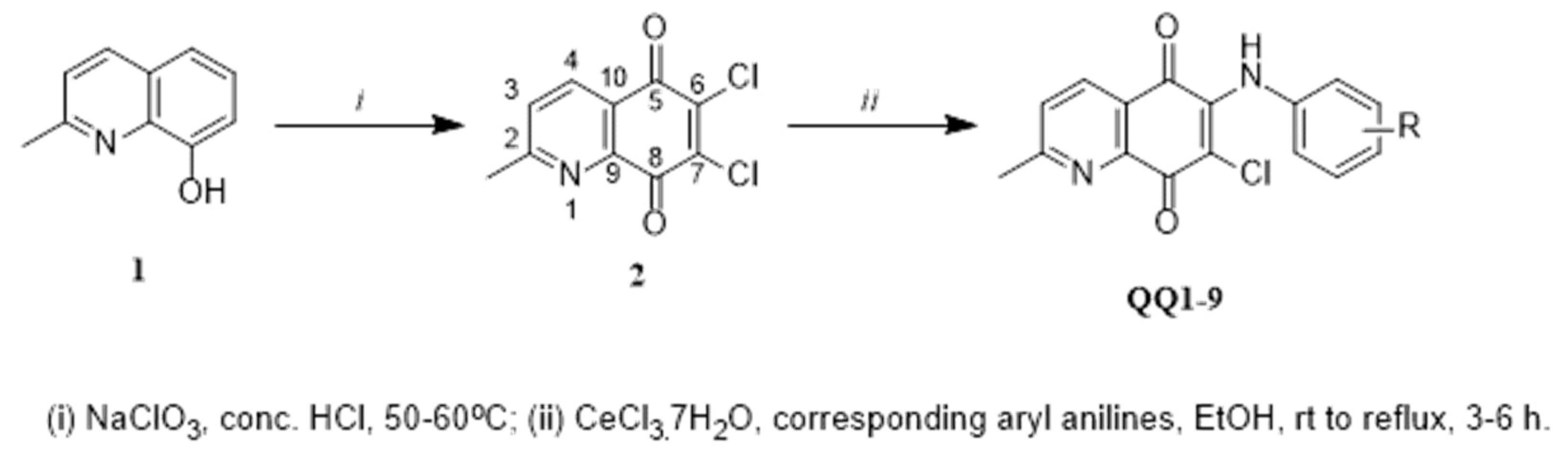
2.1. Chemistry

Synthesis of the quinolinequinones (QQ19) has been reported previously, and references are cited therein [22].

2.2. In Vitro Antimicrobial Activity

2.2.1. Determination of Minimum Inhibitory Concentrations (MICs)

Antimicrobial studies were determined by the broth microdilution technique using the Clinical Laboratory Standards Institute (CLSI) recommendations [23,24]. Mueller–Hinton broth (BD Difco, Detroit, MI, USA catalog number: DF0757-17-6) for bacteria ((Staphylococcus aureus (ATCC 29213), Staphylococcus epidermidis (ATCC 12228), Enterococcus faecalis (ATCC 29212), Pseudomonas aeruginosa (ATCC 27853), Escherichia coli (ATCC 25922), Klebsiella pneumoniae ATCC 4352), and Proteus mirabilis (ATCC 14153)), and RPMI-1640 medium buffered to pH 7.0 with MOPS (Sigma, St. Louis, MO, USA, R6504, M1254) for the yeast strains ((Candida albicans (ATCC 10231), Candida tropicalis (ATCC 750), and Candida parapsilosis (ATCC 22019)) were used as the test media. Then 10 mg/mL stock solution of each molecule was prepared using DMSO (Merck, Catalog number: 67685). Then, serial twofold dilutions ranging from 1250 µg/mL to 0.6 µg/mL were prepared in the medium. The inoculum was diluted in broth media to give a final concentration of 5 × 10⁵ CFU/mL for bacteria and 0.5 × 10³ to 2.5 × 10³ CFU/mL for yeast in the test tray. The MIC was defined as the lowest concentration of compound producing complete inhibition of visible growth [23,24]. The antimicrobial effects of the solvents were also investigated against the test microorganisms as controls, and the results were evaluated according to the values of the controls. According to the results, in vitro activities of QQ2 and QQ6 were investigated against each of the 31 resistant Staphylococcus aureus strains obtained clinically by the broth microdilution technique recommended in CLSI [23,24]. Thirty-one nonduplicates, nosocomially acquired methicillin-resistant Staphylococcus spp. isolated from blood specimens between June and December 2018, were obtained from the Department of Infectious Diseases and Clinical Microbiology, Faculty of Medicine, Istanbul Medipol University. API STAPH (bioM’erieux) was used to identify the studied clinical strains. Additionally, all the tested Staphylococcus spp. were selected by using oxacillin susceptibility to examine the methicillin-resistant isolates, approved by CLSI (MIC ≥ 4 µg/mL).

2.2.2. Determination of Bactericidal Effects by Time-Kill Curve Kinetics (TKC)

Time-kill studies were performed according to NCCLS guidelines [25]. To investigate the synergistic bactericidal effects against clinically resistant S. aureus and S. epidermidis strains, the selected molecules (QQ2 and QQ6) and levofloxacin (Sigma, catalog number: 28266), QQ2 + levofloxacin, and QQ6 + levofloxacin were combined at a concentration of 1 × MIC for each strain. According to NCCLS [25], after adding antimicrobials, the starting inoculum was 1 × 10⁶ to 5 × 10⁶ CFU/mL. The test tubes containing MHB with and without (growth control) antimicrobials in a final volume of 10 mL were incubated in a 37 °C calibrated shaking water bath (Nukleon, catalog number: UM-NCS30L; 180 rpm), and viability counts were performed at 0, 2, 4, 6, and 24 h intervals after inoculation by subculturing 0.1 mL serial dilutions onto TSA (BD Difco, catalog number: 236950) plates. All tests were performed in duplicate. Time-kill curves were determined by plotting mean colony counts (log10 CFU/mL) versus time. The lower limit of detection for time-kill assays was one log10 CFU/mL. Antimicrobial carry-over was controlled by the inhibition of colonial growth at the site of the initial streak according to NCCLS guidelines [25]. According to NCCLS [25], bactericidal activity was defined as a decrease of ≥3 log10 CFU/mL from the initial inoculum at 24 h. The results were interpreted by the effect of the combination in comparison with its more active constituent. Synergy and antagonism were defined as a two log10 decrease or increase in the viable count of the combination at 24 h compared to the more active of the two agents alone by using NCCLS criteria [25], respectively.

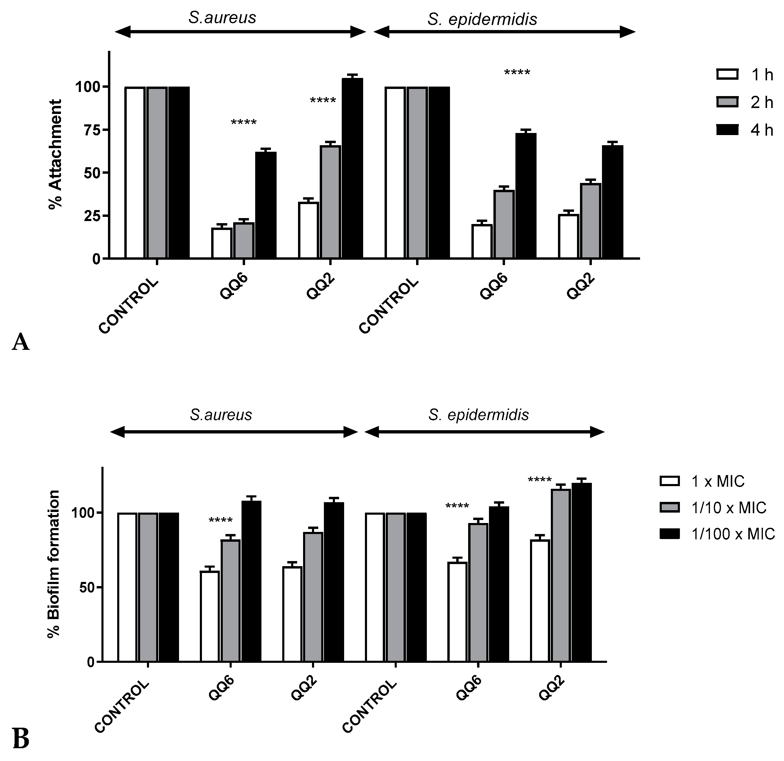
2.2.3. Biofilm Attachment and Inhibition of Biofilm Formation Assays

Biofilm attachment and inhibition of biofilm formation assays were performed as per the previously described method with some modifications [26]. According to the previously reported method by Yıldız et al. and Mataracı Kara et al. [27] for biofilm attachment assays, an overnight culture of S. epidermidis ATCC 35984 and S. aureus ATCC 43300 were added to each well of a 96-well tissue culture microtiter plate (Greiner, catalog number: M0812) with 1/10 the MICs of QQ2 and QQ6 compounds, respectively. The plates were incubated for 1, 2, and 4 h for the studied isolates at 37 °C [26,27].
We also determined the inhibition of biofilm formation methods by using those previously reported by Yıldız et al. and Mataracı-Kara et al. [26,27]. According to their experiments [26,27] for inhibition of biofilm formation, TSB-glucose (BD Difco, catalog number: DF0370-17-3) for S. epidermidis and S. aureus were incubated at 37 °C, 24 h, with the tested QQ compounds at 1 × MIC, 1/10 the MIC, and 1/100 the MIC in 96-well tissue culture, respectively. Six wells were used for each molecule. The positive controls were microorganisms in TSB-glucose for S. epidermidis and S. aureus without molecules, respectively. After the incubation, wells were washed with PBS solutions and measured at OD595 nm (BioTek Eon Microplate Spectrophotometer).

2.3. Statistical Analysis

Two-way ANOVA-Tukey’s multiple comparison tests (Prism 8.0, GraphPad, San Diego, CA, USA) were used to compare differences between control and antimicrobial-treated biofilms, and experiments were performed using two independent assays. A p-value < 0.0001 was considered statistically significant.

3. Results

3.1. Chemistry

The quinolinequinones used in the present study were prepared by the nucleophilic substitution reaction of the 6,7-dichloro-5,8-quinolinequinone synthesized by using Shaikh’s methodology starting from the commercially available 8-hydroxyquinoline (1) with sodium chlorate in concentrated HCl solution according to the procedure [20] with aryl amines in ethanol in the presence of CeCl3·7H2O as the catalyst, as shown in Scheme 1.
We found that the growth inhibition of dimethylquinone containing aryl amines having different substituents at various positions was substantially active [26]. We also designed and synthesized the quinolinequinones by linking the aryl amino group to azaquinone [22]. To develop improved antimicrobial molecules, we adopted a pharmacophore approach to merge the quinolinequinone and aryl amines containing an ester moiety. The present work extends our ongoing efforts toward developing biologically active molecules with high efficiency [26,27].

3.2. Library Screening

The genus Staphylococcus, one of the Gram-positive strains, is a member of the Micrococcaceae family from the phylum Actinobacteria. In addition to being resistant to most disinfecting agents, they can survive in harsh conditions, such as outside the body [28,29]. Regarding the development of effective antimicrobial agents, particularly against methicillin-resistant Staphylococcus spp. strains, quinolinequinones are classified as having the potential for developing novel antibacterial candidates [27,30,31]. Other noteworthy examples are sulfanyl and amino analogs of quinolinequinones, which play a significant role against MRSA, as published by Yildirim and Bayrak [32,33].
For these studies, each member of the library was studied for in vitro antimicrobial activity against a panel of Gram-positive bacteria and Gram-negative bacteria by using the broth microdilution technique to obtain the minimal inhibitory concentrations (MICs) according to the Clinical Laboratory Standards Institute (CLSI) recommendations [23,24]. The minimal inhibitory concentrations (MICs) determined against the standard strains of clinical importance used in this study were from the American Type Culture Collection (ATCC®) of the quinolinequinones (QQ19) and are summarized in Table 1 and Table 2.
Primarily, the quinolinequinones (QQ19) were evaluated for in vitro antibacterial profile detection against Gram-positive and Gram-negative bacteria. All quinolinequinones showed no acceptable inhibitory activity (with the MIC values at 625–1250 μg/mL) against four reference strains, namely, all Gram-negative bacteria. Interestingly, the activity profiles were mainly against the Gram-positive strains. The results showed that the Gram-positive strains were the most susceptible to the quinolinequinones. The screening showed that five quinolinequinones were active molecules (QQ1, QQ2, QQ3, QQ5, and QQ6) against two important species of the Gram-positive strains (Staphylococcus aureus and Staphylococcus epidermidis). Among these, four molecules (QQ2, QQ3, QQ5, and QQ6) were also active against Enterococcus faecalis, the other member of the Gram-positive strains (Table 1). Remarkably, three quinolinequinones (QQ1, QQ5, and QQ6) displayed the most significant antibacterial activity against S. aureus with a MIC value of 1.22 μg/mL, which was equal to the reference drug cefuroxime-Na. Among all the tested molecules, two quinolinequinones (QQ2 and QQ3) gave low MIC values of 2.44 μg/mL against S. aureus. These results indicated that these two molecules had the potency to be further studied. Furthermore, the quinolinequinones (QQ1, QQ2, QQ3, QQ5, and QQ6) showed 8-fold inhibitory potency against S. epidermidis than cefuroxime with a MIC value of 1.22 μg/mL. Noticeably, QQ6 exhibited the best (27-fold more potent) anti-E. faecalis activity with a MIC value of 4.88 μg/mL, which was superior to the standard drug amikacin (MIC = 128 μg/mL). On the other hand, QQ3 was the second most active one of this series against E. faecalis (13-fold more potent than amikacin with a MIC value of 9.76 μg/mL). Another two quinolinequinones (QQ2 and QQ5) gave the best activity against E. faecalis with MIC values of 19.53 μg/mL, which was 6-fold more potent when compared with the standard drug amikacin.
Regarding the antifungal activity, Table 2 revealed that most quinolinequinones exhibited an inversely inhibitory tendency to their antibacterial activity, though weaker than the antibacterial activity. Predominantly, two quinolinequinones (QQ7 and QQ8) showed a profound effect against C. albicans with a MIC value of 4.88 μg/mL, equal to the reference drug clotrimazole [34]. In addition, when we investigated the inhibitory activity of the same quinolinequinones (QQ7 and QQ8) against C. parapsilosis strains, QQ7 and QQ8 had acceptable activity compared with the reference drug amphotericin B, which had 5-fold and 10-fold less inhibitory potency, respectively.
Following our findings, we objected to specifying the potential antibacterial effects of QQ2 and QQ6, which have prolonged activity analogs. They were selected for further analysis against clinically resistant isolates of Staphylococcus spp. isolates. Therefore, we explored their eventual antimicrobial activities against each of 31 clinically obtained resistant strains of Staphylococcus spp. The results indicated in Figure 1 from the MIC assessment showed that the MIC ranges for QQ2 and QQ6 were 1.22–9.76 and 0.66–19.53 μg/mL, respectively. In the present study, the in vitro antimicrobial activity of QQ2 was up to 100% (MIC range 1.22–9.76 μg/mL) among all the studied isolates of clinically resistant Staphylococcus spp. and exceeded the susceptibilities to reference antimicrobial cefuroxime (MIC = 9.8 μg/mL). Overall, for the clinically resistant Staphylococcus spp. isolates, both QQ2 (MIC50/90 = 2.44/9.76 μg/mL) and QQ6 (MIC50/90 = 2.44/9.76 μg/mL) were the most active agents compared with reference antimicrobial cefuroxime.

3.3. Structure–Activity Relationships (SARs) Study for Biological Evaluation

The SAR study was conducted to analyze the effect of the substituent with various positions [35]. The structures of the small library of the obtained quinolinequinones (QQ19) are shown in Figure 2. The SAR study demonstrated that the ester group, in particular unbranched alkyl chain (–COOCH3 and –COOCH2CH3) at all positions put forth a significant impact on biological activities for the antibacterial activity against S. aureus, S. epidermidis, and E. faecalis strains. Moreover, when the unbranched alkyl chain was replaced by a branched alkyl chain (COOC(CH3)3), the corresponding quinolinequinones (QQ7, QQ8, and QQ9) displayed weak or no prominent antibacterial activity. This fact pointed out that the branched alkyl chain was unfavorable for the antibacterial activity, and only a suitable length of the alkyl chain (methyl and ethyl) in the ester group was necessary for the antibacterial activity. The replacement of the ester group from ortho to meta or meta to para remarkably improved the antibacterial activity against E. faecalis.
The antifungal evaluation showed that most quinolinequinones, especially containing an ester group with an unbranched alkyl chain, showed weak to good bioactivity against most of the tested fungi. In contrast to antibacterial activity, two quinolinequinones (QQ7 and QQ8) displayed better antifungal activity against C. albicans and C. parapsilosis.
In a previous study, our group demonstrated that the incorporation of dimethylquinone with aryl amines containing an ester group was not an efficient strategy to increase antibacterial and antifungal activity [26]. When aryl amines containing the ester group are coordinated with quinolinequinone, they induce structural changes, ultimately altering the molecule’s ability to interact with biomolecules, which may have a positive impact on the desired biological activity. Among nine active quinolinequinones against the bacteria and fungi, SAR analysis of the nine quinolinequinones tested indicated that the two quinolinequinones (QQ2 and QQ6) presented remarkable activity against all Gram-positive bacteria, including S. aureus, S. epidermidis, and E. faecalis. Furthermore, these two quinolinequinones are responsible for the promising increase in antibacterial activity (Table 1). Thus, strikingly, they were selected for further studies to understand the mode of action, since they had a wide range of activity on the studied strains.

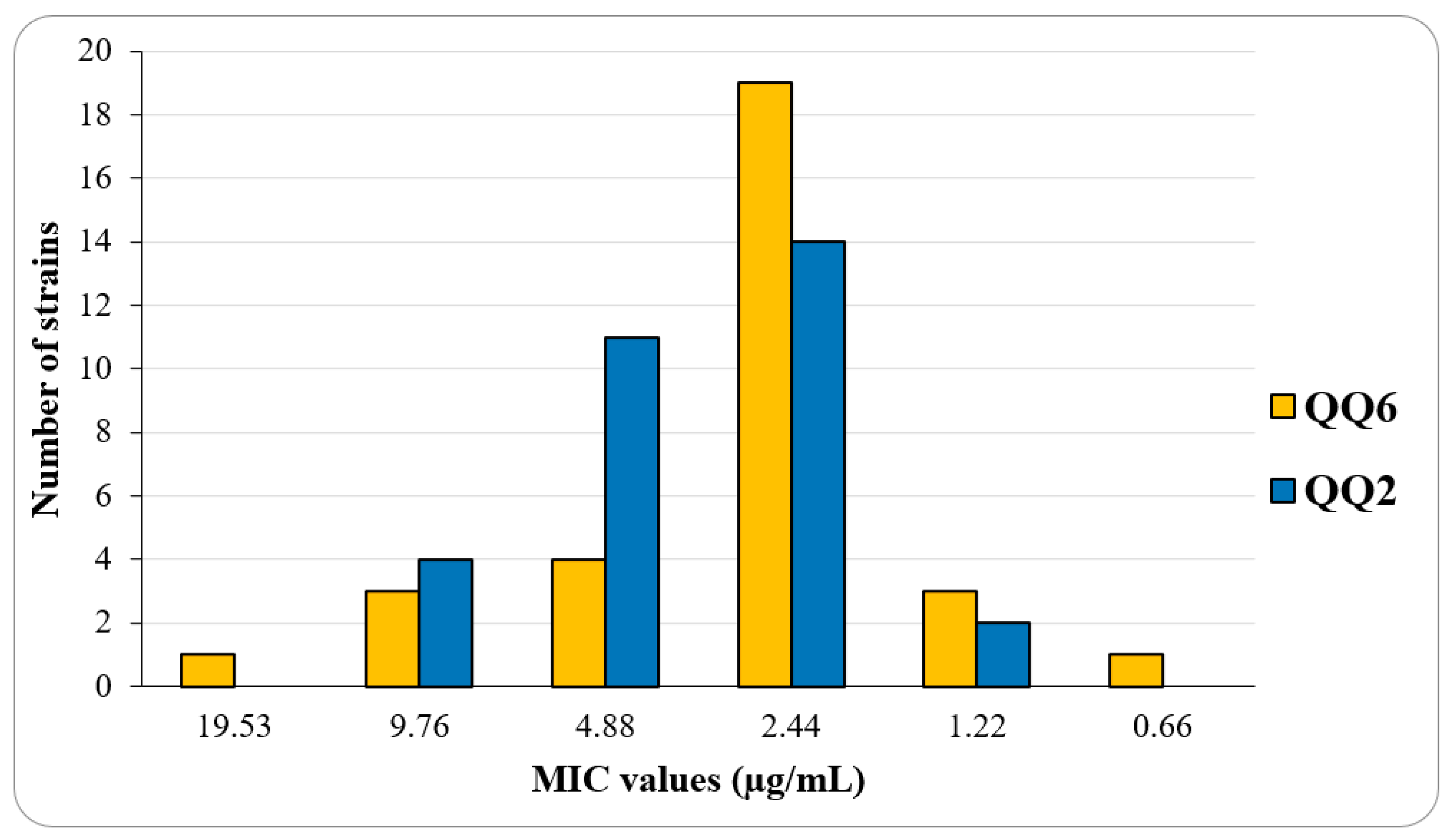
3.4. Bactericidal Effects by Time-Kill Curve Kinetic Study

The bactericidal activity of the most active two quinolinequinones (QQ2 and QQ6) alone or in combination with levofloxacin was also examined from the library for further investigation using time-kill assays against clinically resistant Staphylococcus aureus and Staphylococcus epidermidis isolates at the respective MICs, and the results are given in Figure 3. Levofloxacin antibiotic, which shows high activity against Gram-positive bacteria containing S. aureus, has a fluoroquinolone structure [36]. Experimental studies have revealed that fluoroquinolones combined with standard antistaphylococcal therapy in severe S. aureus infections have an additive effect [37].
The results of the time-kill curve studies generally showed that neither QQ2 nor QQ6 displayed bactericidal activity (with a 3-log10 killing) in accordance with the 1 × MIC concentration used within 24 h. However, synergistic interaction was achieved in all strains studied with the combination of QQ6 and levofloxacin used in 1 × MIC, and different antagonisms were not observed in different combinations.

3.5. Effect of Quinolinequinones on Biofilm Formation

Most bacterial species prefer to live as microcolony communities rather than single cells, since this formation behaves as a shield that keeps away the host immune response and prevents antibiotic effects. The biofilm formation of bacterial cells has highly increased the resistance to antibiotics as well as to host immune responses compared to the planktonic form of life [38,39]. Two important members of Staphylococci, in particular S. epidermidis and S. aureus strains, are responsible for most invasive infectious diseases because of bacterial biofilms [40,41]. For that reason, strong demand for searching the novel lead structures has increased in order to overcome resistance to biofilm-associated infectious diseases [42]. To date, as mentioned in the literature, the quinone scaffolds have shown the ability to disrupt formed bacterial biofilms [38,39] against S. aureus [43] and S. epidermidis [44]. On these bases, the potential therapeutic application of two highly active quinolinequinones (QQ2 and QQ6) to disrupt bacterial biofilms was analyzed. When we incubated 1/10 MICs of QQ2 and QQ6 with MRSA and S. epidermidis for adhesion to the wells of microtiter plates, it was observed that neither compound alone inhibited biofilm attachment in relation to incubation time (Figure 4). QQ2 and QQ6 showed less than 50% inhibitory activity against MRSA and S. epidermidis biofilm formation at 24 h, respectively, based on their concentrations (Figure 4).

4. Discussion

The present investigation effectively showed that the quinolinequinones could be considered promising molecules against Gram-positive strains. SAR clearly suggested a correlation between Gram-positive bacteria with the antibacterial profiles. Structural analysis showed that the presence of the ester group is an important moiety for activity. On the other hand, our data also showed that the presence of the ester group at the para position improved the antibacterial activity. In addition, two active quinolinequinones (QQ2 and QQ6) against S. aureus were studied for their antimicrobial mechanism via a time-kill kinetic assay and antibiofilm efficacy. S. aureus is a significant pathogen in both hospitals and in the community. Methicillin-resistant S. aureus isolates are often resistant to other classes of antibiotics (through different mechanisms), making treatment options limited. This has led to the search for new compounds active against these strains [45]. The present study has improved our knowledge of the properties of QQ molecules by showing the extended activity of QQ2 and QQ6 against clinically resistant Staphylococcus spp. isolates. Additionally, in our study, QQ6 demonstrated synergistic interaction with levofloxacin against both S. aureus and S. epidermidis clinically resistant isolates. Synergistic combination therapy applications using existing antibiotics have gained importance, increasing the possibilities of providing active initial therapy, preventing further resistance development, and treating infections by clinically resistant Staphylococcus spp. isolates. The compounds QQ2 and QQ6 selected for the study of antibiofilm activity among the QQ compounds did not exhibit significant antibiofilm activity. However, to the best of our knowledge, this is the first study to examine the antibiofilm and bactericidal activities of quinolinequinones against clinically resistant strains, which adds special value to this study. In addition, the activity of levofloxacin, when combined with QQ6, shows promise as an alternative to treatments for infections caused by clinically resistant Staphylococcus spp. As a result, in addition to the promising activity of the combination of levofloxacin and QQ6 compound against clinically resistant Staphylococcus spp. isolates, further clinical studies would be beneficial to determine the clinical effect of this combination and to investigate and develop the efficacy of this regimen in the treatment of infections with clinically resistant Staphylococcus spp. isolates.

Author Contributions

Conceptualization, E.M.-K. and A.F.T.; Data curation, E.M.-K.; Formal analysis, E.M.-K.; Funding acquisition, A.F.T.; Investigation, E.M.-K., N.B., M.Y., H.Y. and A.F.T.; Methodology, E.M.-K. and A.F.T.; Project administration, A.F.T.; Resources, E.M.-K. and A.F.T.; Software, E.M.-K.; Supervision, A.F.T.; Validation, N.B., M.Y. and H.Y.; Visualization, E.M.-K. and A.F.T.; Writing—original draft, E.M.-K. and A.F.T.; Writing—review and editing, E.M.-K., N.B., M.Y., H.Y. and A.F.T. All authors have read and agreed to the published version of the manuscript.

Funding

This work was financially funded by the Scientific and Technological Research Council of Turkey (TÜBİTAK), Grant No.: 118Z620, for the supply of equipment and materials.

Institutional Review Board Statement

Not applicable.

Informed Consent Statement

Not applicable.

Conflicts of Interest

The authors declare no conflict of interest.

Abbreviations

APIsActive pharmaceutical ingredients
ATCC®American Type Culture Collection
CLSIClinical Laboratory Standards Institute
MRSAMethicillin-resistant Staphylococcus aureus
MICsMinimum inhibitory concentrations
NCCLSNational Committee for Clinical Laboratory Standards
PBMCsPeripheral blood mononuclear cells
SARStructure–activity relationship models
TKCTime-kill Curve

References

  1. Sharifi-Rad, J.; Ozleyen, A.; Tumer, T.B.; Adetunji, C.O.; El Omari, N.; Balahbib, A.; Taheri, Y.; Bouyahya, A.; Martorell, M.; Martins, N.; et al. Natural Products and Synthetic Analogs as a Source of Antitumor Drugs. Biomolecules 2019, 9, 679. [Google Scholar] [CrossRef] [PubMed]
  2. Duval, R.E.; Grare, M.; Demore, B. Fight Against Antimicrobial Resistance: We Always Need New Antibacterials but for Right Bacteria. Molecules 2019, 24, 3152. [Google Scholar] [CrossRef] [PubMed] [Green Version]
  3. De Oliveira, J.F.A.; Saito, A.; Bido, A.T.; Kobarg, J.; Stassen, H.K.; Cardoso, M.B. Defeating Bacterial Resistance and Preventing Mammalian Cells Toxicity Through Rational Design of Antibiotic-Functionalized Nanoparticles. Sci. Rep. 2017, 7, 1326. [Google Scholar] [CrossRef] [Green Version]
  4. Pan, L.; Chai, H.B.; Kinghorn, A.D. Discovery of new anticancer agents from higher plants. Front. Biosci. 2012, 4, 142–156. [Google Scholar] [CrossRef] [PubMed]
  5. Durand, G.A.; Raoult, D.; Dubourg, G. Antibiotic discovery: History, methods and perspectives. Int. J. Antimicrob. Ag. 2019, 53, 371–382. [Google Scholar] [CrossRef]
  6. Nain-Perez, A.; Barbosa, L.C.A.; Maltha, C.R.A.; Forlani, G. First total synthesis and phytotoxic activity of Streptomyces sp. metabolites abenquines. Tetrahedron Lett. 2016, 57, 1811–1814. [Google Scholar] [CrossRef]
  7. Schatz, A.; Bugie, E.; Waksman, S.A. Streptomycin, a Substance Exhibiting Antibiotic Activity Against Gram-Positive and Gram-Negative Bacteria. Proc. Soc. Exp. Biol. Med. 1944, 55, 66–69. [Google Scholar] [CrossRef]
  8. Zhang, Z.; Yu, X.; Wang, Z.; Wu, P.; Huang, J. Anthracyclines potentiate anti-tumor immunity: A new opportunity for chemoimmunotherapy. Cancer Lett. 2015, 369, 331–335. [Google Scholar] [CrossRef]
  9. Ravelo, A.G.; Estevez-Braun, A.; Chavez-Orellana, H.; Perez-Sacau, E.; Mesa-Siverio, D. Recent Studies on Natural Products as Anticancer Agents. Curr. Top. Med. Chem. 2004, 4, 241–265. [Google Scholar] [CrossRef]
  10. Cordell, G.A. Changing Strategies in Natural-Products Chemistry. Phytochemistry 1995, 40, 1585–1612. [Google Scholar] [CrossRef]
  11. Silverman, L.; Campbell, R.; Broach, J.R. New assay technologies for high-throughput screening. Curr. Opin. Chem. Biol. 1998, 2, 397–403. [Google Scholar] [CrossRef]
  12. Liu, W.J.; Li, Q.S.; Hu, J.H.; Wang, H.X.; Xu, F.B.; Bian, Q. Application of natural products derivatization method in the design of targeted anticancer agents from 2000 to 2018. Bioorg. Med. Chem. 2019, 27, 115150. [Google Scholar] [CrossRef] [PubMed]
  13. Gali-Muhtasib, H.; Roessner, A.; Schneider-Stock, R. Thymoquinone: A promising anti-cancer drug from natural sources. Int. J. Biochem. Cell B 2006, 38, 1249–1253. [Google Scholar] [CrossRef] [PubMed]
  14. Hariharan, P.; Paul-Satyaseela, M.; Gnanamani, A. In vitro profiling of antimethicillin-resistant Staphylococcus aureus activity of thymoquinone against selected type and clinical strains. Lett. Appl. Microbiol. 2016, 62, 283–289. [Google Scholar] [CrossRef] [PubMed] [Green Version]
  15. Houdkova, M.; Rondevaldova, J.; Doskocil, I.; Kokoska, L. Evaluation of antibacterial potential and toxicity of plant volatile compounds using new broth microdilution volatilization method and modified MTT assay. Fitoterapia 2017, 118, 56–62. [Google Scholar] [CrossRef] [PubMed]
  16. Kawamukai, M. Biosynthesis and applications of prenylquinones. Biosci. Biotech. Bioch. 2018, 82, 963–977. [Google Scholar] [CrossRef]
  17. Ryu, C.K.; Park, R.E.; Ma, M.Y.; Nho, J.H. Synthesis and antifungal activity of 6-arylamino-phthalazine-5,8-diones and 6,7-bis(arylthio)-phthalazine-5,8-diones. Bioorg. Med. Chem. Lett. 2007, 17, 2577–2580. [Google Scholar] [CrossRef]
  18. Kruschel, R.D.; Buzid, A.; Khandavilli, U.B.R.; Lawrence, S.E.; Glennon, J.D.; McCarthy, F.O. Isoquinolinequinone N-oxides as anticancer agents effective against drug resistant cell lines. Org. Biomol. Chem. 2020, 18, 557–568. [Google Scholar] [CrossRef]
  19. Dias Flaviana, R.F.; Novais, J.S.; Devillart, S.; da Silva, W.A.; Fereirra, M.O.; Loureiro, R.d.S.; Campos, V.R.; Fereirra, V.F.; de Souza, M.C.B.V.; Castro, H.C.; et al. Synthesis and antimicrobial evaluation of amino sugar-based naphthoquinones and isoquinoline-5,8-diones and their halogenated compounds. Eur. J. Med. Chem. 2018, 156, 1–12. [Google Scholar] [CrossRef]
  20. Shaikh, I.A.; Johnson, F.; Grollman, A.P. Streptonigrin.1. Structure-Activity-Relationships among Simple Bicyclic Analogs—Rate Dependence of DNA-Degradation on Quinone Reduction Potential. J. Med. Chem. 1986, 29, 1329–1340. [Google Scholar] [CrossRef]
  21. Lown, J.W.; Sim, S.K.; Chen, H.H. Hydroxyl radical production by free and DNA-bound aminoquinone antibiotics and its role in DNA degradation. Electron spin resonance detection of hydroxyl radicals by spin trapping. Can. J. Biochem. 1978, 56, 1042–1047. [Google Scholar] [CrossRef] [PubMed]
  22. Bayrak, N.; Ciftci, H.I.; Yildiz, M.; Yildirim, H.; Sever, B.; Tateishi, H.; Otsuka, M.; Fujita, M.; Tuyun, A.F. Structure based design, synthesis, and evaluation of anti-CML activity of the quinolinequinones as LY83583 analogs. Chem. Biol. Interact. 2021, 345, 109555. [Google Scholar] [CrossRef] [PubMed]
  23. Clinical and Laboratory Standards Institute, 950 West Valley Road, Suite 2500, Wayne, Pennsylvania 19087, USA, 2020. Available online: https://www.nih.org.pk/wp-content/uploads/2021/02/CLSI-2020.pdf?utm_medium=email&utm_source=transaction (accessed on 1 August 2022).
  24. Clinical and Laboratory Standards Institute. Reference Method for Broth Dilution Antifungal Susceptibility Testing of Yeasts, 2nd ed.; Approved Standard; Clinical and Laboratory Standards Institute: Wayne, PA, USA, 1997. [Google Scholar]
  25. National Committee for Clinical Laboratory Standards. Methods for Determining Bactericidal Activity of Antimicrobial Agents; NCCLS: Wayne, PA, USA, 1999. [Google Scholar]
  26. Bayrak, N.; Yildiz, M.; Yildirim, H.; Mataraci-Kara, E.; Tuyun, A.F. Novel plastoquinone analogs containing benzocaine and its analogs: Structure-based design, synthesis, and structural characterization. Res. Chem. Intermediat. 2021, 47, 2125–2141. [Google Scholar] [CrossRef]
  27. Mataraci-Kara, E.; Bayrak, N.; Yildiz, M.; Yildirim, H.; Ozbek-Celik, B.; Tuyun, A.F. Discovery and structure-activity relationships of the quinolinequinones: Promising antimicrobial agents and mode of action evaluation. Drug Dev. Res 2021, 83, 628–636. [Google Scholar] [CrossRef]
  28. Pulverer, G. Taxonomy of Staphylococcus-Aureus. Zbl. Bakt. Int. J. Med. M. 1986, 262, 425–437. [Google Scholar] [CrossRef]
  29. Melter, O.; Radojevic, B. Small colony variants of Staphylococcus aureus – review. Folia Microbiol. 2010, 55, 548–558. [Google Scholar] [CrossRef]
  30. Tong, S.Y.C.; Davis, J.S.; Eichenberger, E.; Holland, T.L.; Fowler, V.G. Staphylococcus aureus Infections: Epidemiology, Pathophysiology, Clinical Manifestations, and Management. Clin. Microbiol. Rev. 2015, 28, 603–661. [Google Scholar] [CrossRef] [Green Version]
  31. Wu, X.W.; Wu, Z.P.; Wang, L.X.; Zhang, H.B.; Chen, J.W.; Zhang, W.; Gu, L.Q.; Huang, Z.S.; An, L.K. Synthesis, antimicrobial activity and possible mechanism of action of 9-bromo-substituted indolizinoquinoline-5,12-dione derivatives. Eur. J. Med. Chem. 2011, 46, 4625–4633. [Google Scholar] [CrossRef]
  32. Yildirim, H. Arylthio analogs of 5,8-quinolinedione: Synthesis, characterization, antibacterial, and antifungal evaluations. Phosphorus Sulfur 2020, 195, 516–522. [Google Scholar] [CrossRef]
  33. Bayrak, N. Novel azanaphtoquinone compounds with aromatic amino moiety: Synthesis, structural characterization, and antimicrobial features. J. Mol. Struct. 2019, 1195, 411–416. [Google Scholar] [CrossRef]
  34. Joshi, K.M.; Shelar, A.; Kasabe, U.; Nikam, L.K.; Pawar, R.A.; Sangshetti, J.; Kale, B.B.; Singh, A.V.; Patil, R.; Chaskar, M.G. Biofilm inhibition in Candida albicans with biogenic hierarchical zinc-oxide nanoparticles. Biomater. Adv. 2022, 134, 112592. [Google Scholar] [CrossRef] [PubMed]
  35. Singh, A.V.; Kayal, A.; Malik, A.; Maharjan, R.S.; Dietrich, P.; Thissen, A.; Siewert, K.; Curato, C.; Pande, K.; Prahlad, D.; et al. Interfacial Water in the SARS Spike Protein: Investigating the Interaction with Human ACE2 Receptor and In Vitro Uptake in A549 Cells. Langmuir 2022, 38, 7976–7988. [Google Scholar] [CrossRef] [PubMed]
  36. Fu, K.P.; Lafredo, S.C.; Foleno, B.; Isaacson, D.M.; Barrett, J.F.; Tobia, A.J.; Rosenthale, M.E. In vitro and in vivo antibacterial activities of levofloxacin (l-ofloxacin), an optically active ofloxacin. Antimicrob. Agents Chemother. 1992, 36, 860–866. [Google Scholar] [CrossRef] [PubMed] [Green Version]
  37. Rohner, P.; Herter, C.; Auckenthaler, R.; Pechere, J.C.; Waldvogel, F.A.; Lew, D.P. Synergistic effect of quinolones and oxacillin on methicillin-resistant Staphylococcus species. Antimicrob. Agents Chemother. 1989, 33, 2037–2041. [Google Scholar] [CrossRef] [PubMed] [Green Version]
  38. Rabin, N.; Zheng, Y.; Opoku-Temeng, C.; Du, Y.X.; Bonsu, E.; Sintim, H.O. Agents that inhibit bacterial biofilm formation. Future Med. Chem. 2015, 7, 647–671. [Google Scholar] [CrossRef]
  39. Di Marco, N.I.; Paez, P.L.; Lucero-Estrada, C.S.M.; Pungitore, C.R. Naphthoquinones inhibit formation and viability of Yersinia enterocolitica biofilm. World J. Microbiol. Biotechnol. 2021, 37, 30. [Google Scholar] [CrossRef]
  40. Vuong, C.; Otto, M. Staphylococcus epidermidis infections. Microbes Infect. 2002, 4, 481–489. [Google Scholar] [CrossRef]
  41. Wang, R.; Khan, B.A.; Cheung, G.Y.; Bach, T.H.; Jameson-Lee, M.; Kong, K.F.; Queck, S.Y.; Otto, M. Staphylococcus epidermidis surfactant peptides promote biofilm maturation and dissemination of biofilm-associated infection in mice. J. Clin. Invest. 2011, 121, 238–248. [Google Scholar] [CrossRef]
  42. Novais, J.S.; Moreira, C.S.; Silva, A.C.J.A.; Loureiro, R.S.; Figueiredo, A.M.S.; Ferreira, V.F.; Castro, H.C.; da Rocha, D.R. Antibacterial naphthoquinone derivatives targeting resistant strain Gram-negative bacteria in biofilms. Microb. Pathog. 2018, 118, 105–114. [Google Scholar] [CrossRef]
  43. Bayrak, N.; Yildirim, H.; Tuyun, A.F.; Kara, E.M.; Celik, B.O.; Gupta, G.K.; Ciftci, H.I.; Fujita, M.; Otsuka, M.; Nasiri, H.R. Synthesis, Computational Study, and Evaluation of In Vitro Antimicrobial, Antibiofilm, and Anticancer Activities of New Sulfanyl Aminonaphthoquinone Derivatives. Lett. Drug Des. Discov. 2017, 14, 647–661. [Google Scholar] [CrossRef]
  44. Tuyun, A.F.; Yıldız, M.; Bayrak, N.; Yıldırım, H.; Kara, E.M.; Jannuzzi, A.T.; Celik, B.O. Discovery of a new family of heterocyclic amine linked plastoquinone analogs for antimicrobial evaluation. Drug Dev. Res. 2019, 80, 1098–1109. [Google Scholar] [CrossRef] [PubMed]
  45. Stapleton, P.D.; Taylor, P.W. Methicillin resistance in Staphylococcus aureus: Mechanisms and modulation. Sci. Prog. 2002, 85, 57–72. [Google Scholar] [CrossRef] [PubMed]
Scheme 1. Preparation of the aminated QQs (QQ19).
Antibiotics 11 01397 sch001
Figure 1. MIC distribution of QQ2 and QQ6 against 31 clinically obtained resistant strains of Staphylococcus spp.
Antibiotics 11 01397 g001
Figure 2. The structures of quinolinequinones with aryl amine containing different ester groups in different positions.
Antibiotics 11 01397 g002
Figure 3. Time-kill determinations for clinically resistant (A) S. aureus and (B) S. epidermidis isolates after treatment with QQ2 and QQ6 alone or in combination with levofloxacin at 1 × MIC. The x-axis represents the killing time, and the y-axis represents the logarithmic S. aureus and S. epidermidis survival, respectively. Experiments were performed in duplicate.
Antibiotics 11 01397 g003
Figure 4. Inhibition of S. aureus and S. epidermidis. (A): surface attachment to the wells contained 1/10 × MIC of molecules and an inoculum of 1 × 10⁶ CFU/200 μL, incubated for 1, 2, and 4 h at 37 °C for S. aureus and S. epidermidis; (B): biofilm formation in each well contained 1 ×, 1/10 ×, and 1/100 × MIC of molecules and an inoculum of 5 × 10⁶ CFU/200 μL, incubated for 24 h at 37 °C for S. aureus and S. epidermidis. Control bars indicate microorganisms without molecules accepted as 100%. Six wells were used for the tested molecules. Each experiment is representative of two independent tests. All differences between the control and molecule-treated biofilms were statistically significant (**** p < 0.0001).
Antibiotics 11 01397 g004
Table 1. In vitro antibacterial activity results from the quinolinequinones (QQ19).
IDMicroorganisms
Gram-Negative Bacteria
(MIC, μg/mL)
Gram-Positive Bacteria
(MIC, μg/mL) a
P. aeruginosaE. coliK. pneumoniaeP. mirabilisS. aureusS. epidermidisE. faecalis
QQ1- b625125012501.22
(6.84)
1.22
(6.84)
625
(3503.76)
QQ212501250125012502.44
(13.69)
1.22
(6.84)
19.53
(109.49)
QQ31250125062512502.44
(13.69)
1.22
(6.84)
9.76
(54.75)
QQ41250125012501250-625
(3371.21)
1250
(6742.43)
QQ512501250-12501.22
(6.58)
1.22
(6.58)
19.53
(105.35)
QQ612506256256251.22
(6.58)
1.22
(6.58)
4.88
(26.34)
QQ7-----156.25
(784.97)
-
QQ8625----156.25
(784.97)
-
QQ9----1250
(6279.78)
1250
(6279.78)
-
Ceftazidime2.44
(8.93)
------
Cefuroxime-Na-4.88
(21.88)
4.88
(21.88)
2.44
(10.94)
1.22
(5.47)
--
Cefuroxime-----9.76
(46.02)
-
Amikacin------128
(223.84)
a = The reported MIC values in parenthesis are presented in µM for each analog. b = >1250 µg/mL.
Table 2. In vitro antifungal activity results from the quinolinequinones (QQ19).
IDMicroorganisms
Fungi
(MIC, μg/mL) a
C. albicansC. parapsilosisC. tropicalis
QQ11250
(7007.52)
156.25
(875.94)
625
(3503.76)
QQ2156.25
(875.94)
156.25
(875.94)
1250
(7007.52)
QQ31250
(7007.52)
156.25
(875.94)
625
(3503.76)
QQ41250
(6742.43)
625
(3371.21)
1250
(6742.43)
QQ51250
(6742.43)
78.12
(421.40)
625
(3371.21)
QQ61250
(6742.43)
156.25
(842.80)
625
(3371.21)
QQ74.88
(24.53)
4.88
(24.53)
- b
QQ84.88
(24.53)
2.44
(12.27)
-
QQ978.12
(392.49)
312.50
(1569.94)
-
Clotrimazole4.88
(14.50)
--
Amphotericin B-0.5
(0.83)
1
(1.35)
a = The reported MIC values in parenthesis are presented in µM for each analog. b = >1250 µg/mL.
Publisher’s Note: MDPI stays neutral with regard to jurisdictional claims in published maps and institutional affiliations.

