Review

Multifunctional Therapeutic Potential of Phytocomplexes and Natural Extracts for Antimicrobial Properties

by
Md. Mominur Rahman
1,
Md. Saidur Rahaman
1,
Md. Rezaul Islam
1,
Md. Emon Hossain
1,
Faria Mannan Mithi
1,
Muniruddin Ahmed
1,
Marianela Saldías
2,
Esra Küpeli Akkol
3 and
Eduardo Sobarzo-Sánchez
2,4,*
1
Department of Pharmacy, Faculty of Allied Health Sciences, Daffodil International University, Dhaka 1207, Bangladesh
2
Instituto de Investigación y Postgrado, Facultad de Ciencias de la Salud, Universidad Central de Chile, Santiago 8330507, Chile
3
Department of Pharmacognosy, Faculty of Pharmacy, Gazi University, 06330 Ankara, Turkey
4
Department of Organic Chemistry, Faculty of Pharmacy, University of Santiago de Compostela, 15782 Santiago de Compostela, Spain
*
Author to whom correspondence should be addressed.
Antibiotics 2021, 10(9), 1076; https://doi.org/10.3390/antibiotics10091076
Submission received: 1 August 2021 / Revised: 30 August 2021 / Accepted: 3 September 2021 / Published: 6 September 2021
(This article belongs to the Special Issue Antimicrobial Properties of Phytocomplexes and Isolated Compounds)

Abstract

:
Natural products have been known for their antimicrobial factors since time immemorial. Infectious diseases are a worldwide burden that have been deteriorating because of the improvement of species impervious to various anti-infection agents. Hence, the distinguishing proof of antimicrobial specialists with high-power dynamic against MDR microorganisms is central to conquer this issue. Successful treatment of infection involves the improvement of new drugs or some common source of novel medications. Numerous naturally occurring antimicrobial agents can be of plant origin, animal origin, microbial origin, etc. Many plant and animal products have antimicrobial activities due to various active principles, secondary metabolites, or phytochemicals like alkaloids, tannins, terpenoids, essential oils, flavonoids, lectins, phagocytic cells, and many other organic constituents. Phytocomplexes’ antimicrobial movement frequently results from a few particles acting in cooperative energy, and the clinical impacts might be because of the direct effects against microorganisms. The restorative plants that may furnish novel medication lead the antimicrobial movement. The purpose of this study is to investigate the antimicrobial properties of the phytocomplexes and natural extracts of the plants that are ordinarily being utilized as conventional medications and then recommended the chance of utilizing them in drugs for the treatment of multiple drug-resistant disease.

Graphical Abstract

1. Introduction

Conventional medication has generally utilized an assortment of medicinal plants to treat drug-resistant diseases [1]. Old healers habitually joined helpful spices with odd spells, with plans and proprietary advantages passed down through the ages. Oral transmission of information about ancient restorative practices included modern conventions about plant gathering, readiness strategies, applications, portions, unique eating regimens, and linkages with other mystery tales about the idea of disease [2]. Plant species on the earth are assessed to number somewhere between 250,000 and 500,000 [3]. People and animals consume a limited quantity of them (1 to 10%). Quite possibly, significantly more are utilized for clinical purposes [4]. Hippocrates referenced 300 to 400 beneficial florae in the late 5th century Before Common Era (BCE) [5]. In the first century of the Common Era(CE), Dioscorides wrote De Materia Medica, a medicinal plant inventory that established the model for current pharmacopeias. Approximately 30 medicinal plants are described in the Bible. Indeed, the therapeutic virtues of frankincense and myrrh likely contributed to their high value. They were also used as mouthwashes because they were said to have antibacterial characteristics.
Western progression in the appreciation of helpful plants was hampered by the obliteration of urban foundations, with a critical record of plant drugs obliterated or lost. All through the Dark Ages, the Arab world proceeded to uncover and develop its previous signs. Asian societies, then again, were occupied with building their pharmacopeia. The Renaissance years in the West saw a resurgence of old medication, which depended for the most part on medicinal plants. Diseases have tormented people since the dawn of time, going from minor urinary parcel contaminations to massive pandemics. They utilized a broad scope of substances in their mending, including spices and other regular materials accessible around. For instance, a new report [6] found that nectar, a notable familiar substance, had antibacterial activity in vitro against Staphylococcus aureus and Streptococcus pyogenes. Numerous prescriptions have additionally been distinguished [7] because of ethnobotanical leads. The spice Filipendula ulmaria (sovereign of the knoll) was utilized to make the most significant pain-relieving and mitigating drug ibuprofen [8].
Psoralea corylifolia, Catunaregum spinosa, Solanum virginianum, Woodfordia fructicosa, and Syzygium cumin acetone extracts were proficient against the standard fungal culture of Alternaria alternate and Fusarium oxysporum. Inhibitory peptides for microbes were discovered in 1942 [9]. They have disulfide bonds and are frequently positively charged [10]. There is a scarcity of knowledge on antibacterial chemicals found in mushrooms [11]. Few chemicals that give antibacterial action to a variety of mushrooms have been discovered. The bulk of these chemicals are classified as terpenes. Phytotherapy is one of the oldest methods for the treatment of infectious disorders. Herbal medicine has a long history in many countries [12,13,14,15]. In flavonoid compounds, catechins, the most decreased sort of the C3 unit found, are especially significant. Due to their presence in oolong green teas, these flavonoids have been intensively studied. Teas have for quite some time been known to have antibacterial properties [9] and to contain a variety of catechin chemicals. Vibrio cholerae [16] Streptococcus mutans [17], Shigella [18], and other microbes and pathogens [19] were all suppressed in vitro by these compounds. Catechins hindered disconnected bacterial glucosyltransferases in S. freaks [20] and inactivated cholera poison in Vibrio, presumably because of the complexing exercises examined above for quinones.
This exploration addresses the feasible execution of natural products in the treatment of infectious diseases. However, most of the previous studies have been performed on specific natural sources for screening potential antibacterial and/or antifungal properties. Considering the fact, this study was designed to assemble all the possible natural phytocomplexes, their chemical structures along with their potential bioactivity, and the mechanism of action against bacteria, fungi viruses, or protozoa, which could facilitate the approach of developing new antimicrobials from these inherent phytocomplexes [21].

2. Significant Gatherings of Antimicrobial Mixtures from Plants

Plants can create a practically unlimited number of sweet-smelling compounds, most of which are phenols or their oxygen-subbed subsidiaries [22]. The more significant part is secondary metabolites (Figure 1), of which 12,000 have been recognized, representing under 10% of the total [23]. Plant smells are given by a few substances, for example, terpenes and terpenoids, although plant scent can be provided by others. Numerous atoms are liable for plant seasoning (for instance, the terpenoid capsaicin found in stew peppers). A few comparable plants and flavors used to prepare food by people likewise contain remedial substances [24].

2.1. Phenolics and Polyphenols

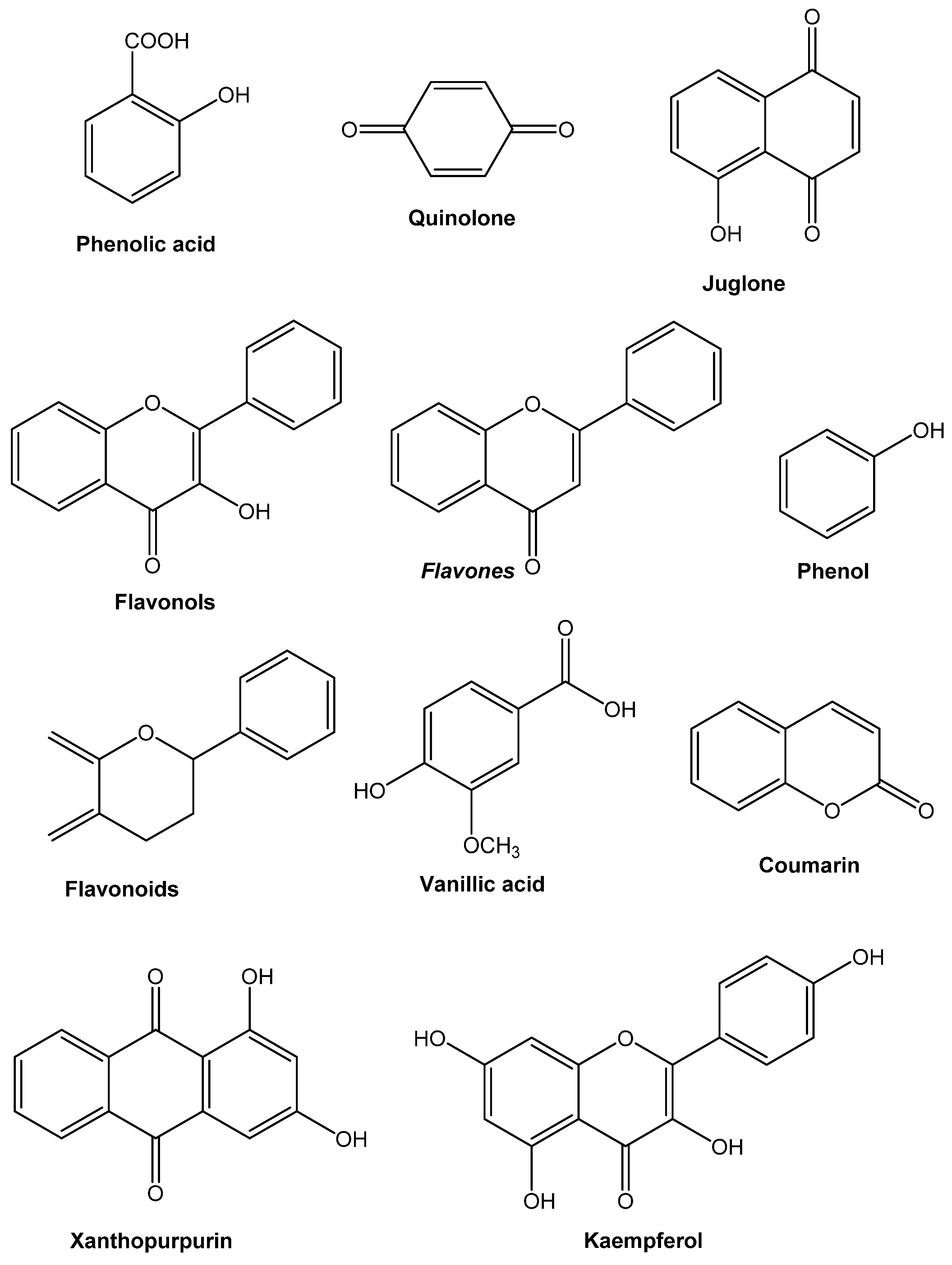
2.1.1. Simple Phenols and Phenolic Acids

Verifiably the most un-problematic bioactive phytochemicals involve a single subbed phenolic ring. Caffeic and cinnamic acids are occurrences of various phenylpropane-construed manufactured subordinates with the best oxidation state. Caffeic acid, found in the common herbs tarragon and thyme, is efficient against viruses, bacteria [25,26], and fungi [27]. Pyrogallol and catechol are both hydroxylated phenols that have been demonstrated to be dangerous to minute organic entities. Pyrogallol contains three OH gatherings, while catechol has two. The measure of hydroxyl bunches on the phenol bunch and their location(s) on the phenyl bunch are thought to be associated with their overall harmfulness to microorganisms, with proof showing higher hydroxylation prompts expanded poisonousness [28].
Moreover, a few analysts have found that phenols that have been seriously oxidized are more inhibitory [29]. Compound restraint by oxidized synthetics, maybe by reactivity with sulfhydryl gatherings, or more vague communications with proteins are proposed to be liable for phenolic harmfulness to microorganisms. Essential oils with a C3 side chain are phenolic compounds which is less oxidized and have no oxygen, and they are now and again utilized as antimicrobials. Eugenol is present in clove oil, which is an outstanding delegate. The two growths [30] and microorganisms are considered bacteriostatic by eugenol. Phenol increases intracellular leakage, which includes the release of K+, which is the first sign of membrane breakdown [31] and radioactivity from 14C-labeled Escherichia coli [32]. According to Pulvertaft and Lumb [7], low dosages of phenols (0.032%, 320 g/mL) and other (nonphenolic) agents destroyed fast-developing cultures of E. coli, staphylococci, and streptococci, showing that autolytic enzymes were not included. According to Srivastava and Thompson [33,34], phenol only acts at the point where two daughter cells divide and that young bacterial cells are more vulnerable to phenol than older cells. Phenolics have antifungal and antiviral properties. The breakdown of the plasma membrane, which permits intracellular substances to spill out, is thought to be responsible for their antifungal action. Phenol has little effect on Pseudomonas aeruginosa PAO transduction by bacteriophage F116 has no impact on phage DNA within the capsid and has no impact on a variety of phage band proteins unless the treatment is 20 min or longer [35].

2.1.2. Quinones

Quinones are shred rings that have two ketone replacements. They are discovered all through the world and are known for being incredibly receptive. Since these fabricated compounds are pigmented, they cause caramelizing in cut or injured foods from the ground and are a stage in the melanin arrangement passage in human skin [36]. They are available in henna, which gives it shading abilities [37]. The advancement from diphenol (or hydroquinone) to diketone (or quinone) through oxidation and diminishing reactions is essential. In various typical structures, the individual redox capacity of the quinone–hydroquinone pair is essential. Nutrient K is a naphthoquinone with a scramble construction. Its ability to oxidize in actual tissues may clarify its antihemorrhagic properties. Inside seeing fitting impetuses, for example, a polyphenol oxidase, hydroxylated amino acids can be changed over to quinones [37]. Quinones have been found to frame irreversible edifices with nucleophilic amino acids in proteins [38], leading to protein inactivation and function loss. As a result, quinone antibacterial effects have a broad range of applications. Surface-uncovered adhesins, cell divider polypeptides, and film-bound chemicals are generally expected targets in the microbial cell. Quinones may also make it difficult for bacteria to obtain substrates.

2.1.3. Flavones, Flavonoids, and Flavonols

Flavones are widely distributed phenolic compounds with just a single carbonyl gathering (as opposed to quinones, which have two). A flavonol is created by adding a 3-hydroxyl group [37]. Flavonoids are hydroxylated phenolic compounds that are connected to an aromatic ring by a C6-C3 unit. It ought not to shock anyone that they have been discovered in vitro to be powerful antibacterial blends against a broad extent of microorganisms, given that they are notorious for being created by plants due to microbial contamination [39,40]. Their action is undoubtedly identified with their capacity to tie to extracellular and dissolvable proteins, just as to tie to bacterial cell dividers, as quinones do. Microbial films might be disturbed by more lipophilic flavonoids [41]. A catechin is a group of flavonoids that is present in a high concentration in green tea. Dental caries (created by Streptococcus mutans) were diminished by 40% when rodents were taken care of an eating regimen containing 0.1% tea catechins [42].
Flavonoid compounds inhibit multiple viruses. Flavonoids, including swertifrancheside [43], glycyrrhizin (from licorice) [44], and chrysin [45], are effective against HIV in numerous studies. Flavone derivatives have been shown to suppress respiratory syncytial virus (RSV) in several studies [46]. In vitro cell culture monolayers, Kaul et al. sum up the exercises and methods of quercetin, naringin, catechin, and hesperetin. While naringin was absent against herpes simplex ailment type 1 (HSV-1), poliovirus type 1, parainfluenza corruption type 3, and RSV, the other three flavonoids were successful. Hesperetin checked infectivity but not intracellular replication of the sum of the four pollutions; catechin hindered infectivity yet not intracellular replication of RSV and HSV-1; and quercetin bound infectivity yet not intracellular replication of the entire of the four ailments. Minimal hidden changes in the combinations, according to the makers, are vital for their ability, and many plant subordinates have an unimportant risky potential. The ordinary Western regular eating routine contains around 1 g of flavonoids and pharmacologically unique sums are presumably not going to hurt human hosts. The mechanism of action of flavones and flavonoids is challenging to pin down due to conflicting findings. Flavonoids with hydroxyl bunches on their rings are more dynamic against microorganisms than those without [47], suggesting that the membrane is their microbiological target. Lipophilic substances have a higher chance of disrupting this structure.
On the other hand, other authors have discovered that the higher the hydroxylation, the higher the antibacterial activity [48]. In a study with radioactive precursors, Mori and colleagues observed that flavonoids substantially restrict DNA synthesis in Proteus vulgaris, while RNA synthesis was most affected in S. aureus. The flavonoids that showed this activity were robinetin, myricetin, and (−)-epigallocatechin. Protein and lipid synthesis were also affected, but to a lesser extent. The B ring of flavonoids could be involved in intercalation or hydrogen bonding with the stacking of nucleic acid bases, according to the researchers, which could explain their inhibitory effect on DNA and RNA synthesis [49].

2.1.4. Tannins

The explanation “tannin” infers a mix of polymeric phenolic builds that can tan calfskin or rush gelatin from the arrangement, a quality known as astringency. Their sub-nuclear loads an area from 500 to 3000, and they may be found in basically all pieces of the plant, including the bark, wood, leaves, ordinary things, and roots. Tannins are of two sorts: hydrolyzable and consolidated tannins. Hydrolyzable tannins are two or three esters of gallic damaging with glucose. Nonetheless, thick tannins (regardless, called proanthocyanidins) are incorporated flavonoid monomers. Tannins are comprised of flavan subordinates that have been moved to plant woody tissues and afterward consolidated. Tannins can likewise be delivered utilizing the polymerization of quinone units [50]. Since it was conjectured that drinking refreshments containing tannin, especially red wines and green teas, [51] would recuperate or forestall various afflictions, this gathering of synthetic substances has gotten a great deal of consideration as of late [52]. Tannins have been linked to multiple human physiological functions, including phagocytic cell activation, host-mediated tumor activity, and anti-infective capabilities [53]. Its synthetic capacities incorporate the creation of covalent securities with proteins by mysterious powers, for example, hydrogen holding and hydrophobic impacts, just as the development of covalent bonds [54]. As a result, their ability to inactivate microbial adhesins, enzymes, cell envelope transport proteins, and other proteins could be linked to their antimicrobial mode of action. They also form polysaccharide complexes [55]. The antibacterial potential of this activity has yet to be determined. Low tannin concentrations cause Crinipellis perniciosa germ tubes to change their form, indicating that bacteria are killed directly [56]. Tannins in plants impede insect development and ruminant animals’ digestive processes [57].

2.1.5. Coumarins

Another phenolic intensifies, coumarins that contain intertwined benzene and pyrone rings [58]. At least 1300 had been identified as of 1996 [59]. Their antithrombotic, anti-inflammatory, and vasodilatory properties have made them famous. Warfarin is a prominent coumarin utilized as an oral anticoagulant comparatively as, oddly, a rodenticide. It could have antiviral effects. Coumarins are pretty dangerous in rodents. Hence, the clinical area around them is extremely cautious. The latest examination has uncovered an “articulated species-subordinate digestion,” which implies that numerous in vivo creature discoveries cannot be applied to people. In people, noxious coumarin subordinates give off an impression of being wiped out securely in urine [60]. A few more coumarins have antimicrobial properties. R.D. Thornes, a physician at Boston Lying-In Hospital, was looking for a way to cure vaginal candidiasis in his pregnant patients in 1954. Candida albicans was addressed to be curbed by coumarin in vitro. (The coumarin-spiked water supply was incidentally given to the entirety of the organisms in the assessment area achieving in vivo centers around rabbits. It also was shown to be an excellent defense mechanism when replicating endeavors began to come up short). Its estrogenic properties were found later [61]. The pharmacological effects of coumarins have been extensively described, and several of them have been discovered to have antibacterial activities. More recently, investigations have emphasized the potential importance of coumarins as alternative therapeutic methods, citing their capacity to reduce biofilm formation in clinically relevant bacteria and impede QS signaling systems (an intricate cell–cell communication system known as quorum sensing). In addition to human ailments, coumarins effectively manage plant pathogens, aquaculture infections, food spoilage, and lower biofouling created by eukaryotic organisms [62].

2.2. Terpenoids and Essential Oils

The Quinta essential, or fundamental oil division, conveys the aroma of plants. These oils are auxiliary metabolites with a high concentration of isoprene-based chemicals. Terpenes are diterpenes, triterpenes, and tetraterpenes (C20, C30, and C40), just as hemiterpenes (C5) and sesquiterpenes (C5), with an overall substance construction of C10H16 (C15). Terpenoids are synthetic substances that contain additional components, most normally oxygen. Terpenoids are comprised of acetic acid derivation units and consequently have a comparable beginning to unsaturated fats. They contrast with unsaturated fats in that they are cyclized and have a great deal of spreading. Methanol and camphor (monoterpenes) are instances of normal terpenoids, as are farnesol and artemisinin (polyterpenes) (sesquiterpenoids). Artemisin and its subsidiary α-arteether, also called by qinghaosu, discovered the present use as antimalarials [63]. In 1985, the controlling board of the logical working gathering of the World Health Organization chose to foster the last medication as a therapeutic for cerebral intestinal sickness. Bacteria [64], fungi viruses, and protozoa [65] are all susceptible to terpenes or terpenoids. In 1977, it was asserted that 60% of fundamental oil subordinates tried so far were growth inhibitors, while 30% were microorganism inhibitors [7]. Another triterpenoid betulinic corrosive was found to smother HIV. The exact mode of action of terpenes is unknown. However, it is thought that lipophilic molecules disrupt membranes. As a result, Mendoza et al. [66] discovered that adding a methyl group to the hydrophilicity of kaurene diterpenoids dramatically lowered their antibacterial action. Food experts have found that terpenoids found in plant essential oils can help control Listeria monocytogenes [67]. In the sterilization of lettuce leaves, oil of basil, a monetarily accessible natural, was discovered to be just about as powerful as 125 ppm chlorine [68]. The antibacterial activity of most terpenoids is linked to their functional groups, and the hydroxyl group of phenolic terpenoids and the presence of delocalized electrons are essential. Carvacrol methyl ether and p-cymene, for example, had much lower antibacterial activity than carvacrol. When the hydroxyl group in carvacrol is replaced with methyl ether, the hydrophobicity, antibacterial activity, and how it interacts with the membrane are all changed. Carvacrol’s antibacterial activity is equivalent to 2-amino-p-cymene, implying that the hydroxyl group is essential but unnecessary for carvacrol’s function. Various theories have been proposed to explain essential oils’ antibacterial properties. Essential oils destabilize cellular architecture and increase permeability, disrupting multiple cellular operations such as energy synthesis (membrane-coupled), membrane transport, and other metabolic regulatory functions. Many cellular processes, including energy synthesis (membrane-coupled), membrane transport, and other metabolic regulatory functions, are disrupted as a result. Energy conversion, nutrition processing, structural macromolecule synthesis, and growth regulator secretion may benefit from essential oils’ cell membrane disruption. Essential oils have the potential to alter both the cell’s external envelope [69] and its cytoplasm. Because essential oils are lipophilic, they can easily penetrate through bacterial cell membranes. Essential oils from various MAPs have increased bacterial cell membrane permeability, causing cellular component leakage and ion loss. Essential oils’ antibacterial activities are connected to reduced membrane potentials, disruption of proton pumps, and ATP depletion. This change in cell architecture could set off a chain reaction that affects other cell organelles. According to Cox et al., tea tree oil inhibits the growth of S. aureus and E. coli via altering cell permeability, increasing intracellular K+ ion leakage, and disturbing cell respiration. Essential oils disturb the arrangement of various fatty acids, phospholipid bilayers, and polysaccharides molecules as they move across the cell membrane and cytoplasmic membrane. The cytoplasmic coagulation of inner cellular components and the disintegration of the connections between the lipid and protein layers could be caused by any of these events [10].

2.3. Alkaloids

Alkaloids are basic nitrogenous compounds that are heterocyclic. In 1805, morphine was derived from the opium plant Papaver somniferum, the main therapeutically persuading alkaloid; the term morphine derives from the Greek word Morpheus, meaning leader of dreams. Both codeine and heroin are morphine aids. Antimicrobial effects have been found in diterpenoid alkaloids, customarily removed from plants in the Ranunculaceae, or buttercup family Solamargine, a glycoalkaloid present in the berries of Solanum khasianum, and various alkaloids have been exhibited to be faltering against HIV illness and AIDS-related intestinal contaminations. In contrast, alkaloids have been shown to have micro-biocidal properties (especially against Giardia and Entamoeba species, their significant antidiarrheal improvement is undoubtedly related to their impact on the little stomach-related bundle travel time. The alkaloid berberine is a critical individual from the alkaloid family. It can kill trypanosomes and plasmodia. The capacity of profoundly fragrant planar quaternary alkaloids like berberine and harmane to intercalate with DNA is believed to be the activity system. The antibacterial mode of action of alkaloids varies depending on the kind. The antibacterial activity of the indolizine alkaloids pergularinine and tylophorinidine are due to the inhibition of nucleic acid synthesis by the enzyme dihydrofolate reductase. Two mechanisms of bacterial inhibition have been discovered within the isoquinoline class: the Ungereminea phenanthridine isoquinoline inhibits nucleic acid synthesis, whereas studies with benzophenanthridine and protoberberine isoquinolines suggested that those agents act by disrupting the Z-ring and inhibiting cell division, which was investigated further. Agelasines alkaloids have an antibacterial action by inhibiting the dioxygenase enzyme BCG 3185c, which impairs bacterial hemostasis. This was discovered using overexpression and binding affinity experiments on the anti-mycobacterial alkaloid agelasine D. Saqualamine, a polyamine alkaloid, affects bacterial membrane integrity.

2.4. Lectins and Polypeptides

Inhibitory peptides for microbes were initially discovered in 1942. It is containing disulfide bonds and is frequently positively charged. The improvement of particle directs in the microbial film or cutthroat concealment of microbial protein bond to have polysaccharide receptors [70] could be their activity component. Against HIV, peptides and lectins have gotten a great deal of consideration as of late. However, these macromolecules, like those found in the herbaceous Amaranthus, have for some time been known to stifle microscopic organisms and parasites [71]. Thionins are peptides with corrosive amino deposits that are frequently identified in grain and wheat. Yeasts, just as Gram-negative and Gram-positive microscopic organisms, are noxious to them. Sugar beet thionins AX1 and AX2 are dynamic against improvement in any case not small life structures. Fabatin, a 47-advancement peptide from fava beans that suppresses P. aeruginosa, E. coli, Enterococcus hirae yet not Saccharomyces or Candida, shows up to be on a very basic level related with thionins from grains that limit P. aeruginosa, E. coli, and Enterococcus hirae at any rate, not Candida or Saccharomyces. Larger lectin molecules, such as mannose-specific lectins from various plants MAP30 from bitter melon GAP31 from Gelonium multiflorum and jacalin inhibit viral development (HIV, CMV) by inhibiting [9] viral interaction with key host cell components. Most general plant antimicrobial screening approaches, such as bioassay-guided fractionation procedures used by natural-products chemists, will overlook molecules and compounds like these whose mode of action could be to inhibit adhesion [16].

3. Some Phytochemicals That Have Antimicrobial Activity
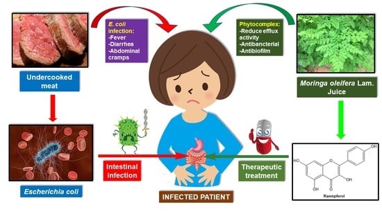
3.1. Kaempferol

The juice of Moringa oleifera Lam. Adenanthera pavonina L. and Annona squamosa L. contain flavonoids, kaempferol, and quercetin, which possess potent antimicrobial properties [72]. Kaempferol is a small molecule with an anti-biofilm activity found to suppress the formation of S. aureus biofilms. Kaempferol inhibits bacteria’s adhesion to fibrinogen, which is the initial step in forming S. aureus biofilms. Kaempferol decreases surface protein anchoring through reducing SrtA function, according to one theory. Another theory is that kaempferol suppresses the expression of specific surface proteins. As kaempferol affects the attachment phase of biofilm development specifically, we identified genes connected to adhesion protein expression. Clumping factor, A and clumping factor B are the essential proteins for S. aureus to bind to fibrinogen, and they are encoded by the genes clfA and clfB. Fibrinogen-binding proteins ClfA and ClfB are up-regulated during biofilm formation in S. aureus. In addition to fibrinogen-binding proteins, S. aureus has two fibronectin-binding proteins, FnBPA and FnBPB, encoded by fnbA and fnbB, respectively. As they induce biofilm formation through a self-association process separate from ligand binding, FnBPs are multifunctional in the S. aureus biofilm life cycle [73].

3.2. Juglone

For a long time, society’s medication has utilized the juice of newly macerated unripe bodies of the dark pecan (Juglans nigra) to remedy confined, parasitic skin infections like ringworm. It has been proposed that the organic movement of pecan bodies is identified with the presence of juglone (5-hydroxy-1,4 naphthoquinone), a basic naphthoquinone disconnected by sublimation from the unripe hulls. S. aureus is restrained by this anti-microbial compound, which ties to DNA and disturbs cell divider arrangement, putting bacterial cells under higher peroxidative pressure [74]. Juglone is antimicrobial, and it is thought to inhibit Gram-positive bacteria like S. aureus. Juglone has a particular antibacterial effect against distinct types of S. aureus. In the same bacterium, juglone has been postulated as a natural source of antibiotic resistance-modifying activity and a phytochemical element with antibiotic resistance-modifying activity. It is also probable that redox cycling is involved in juglone’s antifungal and antibacterial properties. According to bioinformatics analysis, juglone inhibits bacterial proteins involved in DNA, RNA, and protein production, as well as the tricarboxylic acid cycle. Juglone nanoparticles have also been demonstrated to have better antifungal and antibacterial activity than free juglone, showing that nanoparticle production could be a viable technique for enhancing juglone’s beneficial effects [75].

3.3. Xanthopurpurin and Vanillic Acid

In children under the age of five, rotavirus infections are the most common cause of dehydrating gastroenteritis. Despite universal rotavirus immunizations, it remains a leading cause of death, particularly in poverty-stricken countries (94). These infections contaminate enterocytes and cause diarrhea by reducing enterocyte absorption capacity, expanding intestinal emission set off by viral non-underlying protein 4, and initiating the enteric sensory system [76].
In MA104 cells, Rubia cordifolia L. extracts and identified chemicals such as vanillic acid and xanthopurpurin were very efficient against rotavirus. By enhancing virus-mediated apoptosis, they were able to suppress its proliferation [77]—vanillic acid (VA) antibacterial activity and mechanisms of action against carbapenem-resistant E. hormaechei (CREH). VA’s antibacterial effectiveness against CREH was investigated using variations in intracellular ATP concentration, intracellular pH, membrane potential, and cell shape. The usefulness of VA in reducing biofilm formation and inflicting VA damage to CREH cells trapped in biofilms was also examined. A decline in intracellular ATP, pH, and membrane potential, as well as specific alterations in cell shape, suggest that VA may affect CREH’s cell membrane integrity. VA also demonstrated a considerable inhibitory effect on CREH biofilm formation and killed CREH cells within biofilms. VA disrupts the cell membrane of carbapenem-resistant E. hormaechei. Carbapenem-resistant VA inhibits the formation of E. hormaechei biofilms and kills carbapenem-resistant E. hormaechei cells within biofilms [78].

4. Antimicrobial Activity of Medicinal Plants

Plant-determined antimicrobials have amazing supportive assurance. They are capable of treating powerful sicknesses while also avoiding countless horrible effects that fabricated antimicrobials are known. Plant materials’ positive therapeutic effects are, for the most part, a direct result of a mix of assistant blends found in the plant. Auxiliary metabolites in plants and fundamental oils like alkaloids, tannins, steroids, phenol compounds, flavonoids, steroids, unsaturated fats, gums, and tars are suitable for conveying a specific physiological outcome to the living being (Table 1 and Table 2). Mixtures obtained from various segments of the plants can be utilized to treat looseness of the bowels, diarrhea, and different diseases. Blends from different segments of the plants can be used to deal with ailments like looseness of the bowels, diarrhea, hack, cold, cholera, and fever. B. cereus, S. aureus, E. coli, C. albicans, and P. aeruginosa were utilized to determine the antimicrobial activity of unrefined ethanolic concentrates of 10 medicinal plants used in conventional medication against five microorganisms B. cereus, S. aureus, E. coli, C. albicans, and P. aeruginosa. Five out of these ten plants analyzed and expressed antibacterial activity against at least one organism animal category. Sanguisorba officinalis, Chelidonium majus, and Tussilago farfara were potent antibacterial herbs. Nine plants were assessed for antibacterial properties. Hibiscus rosasinensis, Sapindus emarginatus, Mirabilis jalapa, Nyctanthes arbortristis, Rheo discolor, Colocasia esculenta, Gracilaria corticata, Dictyota sp., and Pulicaria wightiana were among the plants examined. Pseudomonas testosteroni, Staphylococcus epidermidis, Klebsiella pneumoniae, Bacillus subtilis, Proteus morganii, and Micrococcus flavus were evaluated for antibacterial activity. The activity of these plants was investigated following two methods: Agar disc diffusion and Agar plug diffusion. The bacterium strains P. testosteroni and K. pneumoniae were expressed as the most resistant.
Several diseases, including asthma, gastrointestinal complaints, skin disorders, respiratory and urinary troubles, and hepatic and cardiovascular illness, have long been treated with medicinal herbs [79,80]. These plants create various physiologically active compounds [81,82] that are required for them to survive and prosper in the environment, including defensive responses to abiotic challenges such as temperature, water availability, mineral nutrition, and insect pests [81,82,83]. Physiologically active compounds in medicinal plants vary widely depending on plant species, soil type, and microbial interaction [84,85]. The bioactive secondary metabolites of medicinal plants have been shown to have a considerable impact on plant-associated microbial populations and physiological activities [86,87,88,89]. Growth promotion, nutrient acquisition, induced systemic resistance, and abiotic stress tolerance are all traits and actions for which plants rely on their microbiome [89,90,91,92,93,94]. Despite substantial research into the phytochemical contents and pharmacological activity of a wide range of therapeutic plants, the microbiome and physiological interactions between the host and microbes remain poorly understood. Several microbial communities dwell in the roots, shoots, and endospheric of plants, forming the microbiome [94,95]. The rhizospheres of many plants have been studied and found to be a good source for choosing beneficial bacteria that can improve plant health [87,96,97]. Understanding how microbial communities adapt to changes in the physiochemical environment of the rhizosphere could help researchers better understand the microbial ecology of plant-associated microorganisms. [89] Researchers observed a significant density of antagonistic bacteria in the rhizosphere of the therapeutic plants Matricaria chamomilla, Calendula officinalis, and Solanum distichum. The root-associated bacteria of Ajuga bracteosa boosted plant growth in a variety of ways by producing siderophores and indole acetic acid, as well as displaying antioxidant activity [98]. Plant phytochemical constituents are thought to be linked to endophytic bacteria and their interactions with host plants, either directly or indirectly [86,99,100]; it is thought that plant phytochemical constituents are linked to endophytic bacteria and their interactions with host plants, either directly or indirectly [101].
Table 1. Plants with antimicrobial characteristics have been utilized in traditional medicine mL [102].
Scientific Name Plant Parts Active Principle Antimicrobial Activity MIC Value
Aloe veraSLatex Salmonella and Streptococcus spp. P. aeruginosa: ≤400 μg/mL [103]
Malus sylvestrisFR ‘Phloretin’ a flavonoid General antimicrobial P. aeruginosa: 7.81 μg/mL [104]
Withania somniferaR and L ‘Withaferin A’ a lactoneAntibacterial
Antifungal
S. aureus: 250 μg/mL [105]
Aegel marmelosL, FR and R Essential oils/terpenoidsAntifungal A. fumigatus: 15.625 μg/mL
Barberis vulgarisR and SB‘Barberine’ an alkaloid M. tuberculosis,
Vibrio cholera,
Plasmodium
and Trypanosomes
P. aeruginosa: 16 μg/mL
Proteus vulgaris: 32 μg/mL
E. coli: 32 μg/mL [106]
Ocimum sanctuma. L and Sb. Ra. Essential oils
b. Root extract
a. Salmonella, Ringworm,
common cold virus
b. In malarial fever to bring sweating
S. aureus: 128 μg/mL [107]
Laurus nobilisLeaves Essential oils Antibacterial and antifungal E. coli: >22.5 mg/mL,
P. aeruginosa: 22 mg/mL [108]
Piper nigrumS ‘Piperine’ an alkaloid Fungi, Micrococci, E. coli C. albicans: 3.125 mg/mL [109]
Bacopa monnieriWP‘Brahmine’ an alkaloid Anthelmintic property UTI and RTI bacteria: 2.5 mg/mL
E. coli: 2.5 μg/mL [110]
Acorus calamusRH and L Volatile oils Enteric bacteria
Insecticidal
S. aureus and E. coli: 5–10 mg/mL
Ricinus communisSCastor oil Antifungal (in dermatitis) S. aureus: 62.5 μg/mL [111]
Cinnamomum verumBA and L Essential oils General antimicrobial S. aureus: 0.5 μL/disc [112]
Cinchona officinalisBA ‘Quinine’ an alkaloid Antimalarial Helicobacter pylori: 0.1 ng/ML [113]
Capsicum annumFR‘Capsaicin’ a terpenoid Antibacterial From 10 to 20 μg/mL [114]
Hydnocarpus kurzii Essential oilMycobacterium lepraeThyme: 1.25 mg/mL [115]
Coriandrum sativumWP, L, S Antibacterial
Antifungal
Candida albicans:
0.02 mg/mL,
E. coli: 0.64 mg/mL [116]
Eucalyptus globulusL Tannins and terpenoids /essential oils Antibacterial
Antiviral
Antifungal
S. aureus: 64 mg/mL,
S. pyogenes: 32 mg/mL,
S. pneumoniae: 16 mg/mL,
Haemophilus influenzae:
16 mg/mL [117]
Allium sativumB Sulfated terpenoids General antimicrobial(Methanolic extract)
S. aureus: 1.25 mg/mL
S. pneumonia: 0.312 mg/mL
P. aeruginosa: 1.25 mg/mL
K. pneumoniae: 0.312 mg/mL(Ethanolic extract)
S. aureus: 2.5 mg/mL
S. pneumonia: 0.312 mg/mL
P. aeruginosa: 0.625 mg/mL
K. pneumoniae: 0.156 mg/mL [118]
Piper longumFR and LPiperinAntibacterial B. cereus and E. coli: 12.5 mg/mL [102]
Glycyrrhiza glabra 1. ‘Glycyrrhizin’ a terpenoid
2. ‘Glabrol’ an alcohol
1. HIV virus and other viruses
2. M. tuberculosis
S. aureus
S. aureus: 50 mg/mL [119]
Calendula officinalisL
FL
Essential oils/Terpenoids General antimicrobial S. mutans: 3.12 μg/mL [120]
Mentha arvensisL ‘Menthol’ an alcohol AntisepticMouth wash Acinetobacter baumannii: 23.5 μg/mL [121]
Azadirachtus indicaR, LB, FR, FL1.‘Azadirachtin’
2. ‘Nimbin’
3. ‘Nimbidin’
4. ‘Gedunin’
5. ‘Salannin’
6. ‘Quercetin’
1. Repellant and antifeedant
2. Antifungal
3. Antibacterial, antifungal
4. Anti-malarial, antifungal
5. Repellant
6. Antibacterial, antiprotozoal
Streptococcus sp.: 125 μg/mL
S. aureus: 250 μg/mL
Enterococcus faecalis: 500 μg/mL [122]
Oleo europoea ‘Hexanal’ an aldehyde General antimicrobial S. cerevisiae: 24 μg/mL, [123]
Allium cepaB‘Allicin’ a sulfoxide and other sulfated terpenoids Antibacterial
Antifungal
S. aureus: 7 μg/mL [124]
Citrus sinensisFP, L Terpenoids Antifungal Streptococcus sp.:12.4 mg/mL
Carica papayaLA, FR Terpenoids, organic acids, and alkaloids General antimicrobial S. aureus: 1250 μg/mL
Butea monospermaS, L Tannins Round worm, Ring worm, Dhobi-itchAcinetobacter sp: 2.62 mg/mL [125]
Mentha piperitaWP 1. ‘Menthol’ an alcohol 2. Peppermint oil terpenoid 1. General antimicrobial
2. Mouth freshener
Klebsiella pneumonia:
0.4 ± 0.02(v/v) [126]
Papaver somniferum ‘Opium’ an alkaloid General antimicrobial K. pneumonia: 2.2 mg/mL
C. albicans: 1.1 mg/mL [127]
Solanum tuberosumT Potato starch Antibacterial
Antifungal
S. aureus: 0.62 mg/mL,
S. pyogenes:1.25 mg/mL [101]
Rauwolfia serpentineR ‘Reserpine’ an alkaloid General antimicrobial S. aureus: 30 mg/mL [101]
Pterocarpus santalinusW Terpenoids Antibacterial, Antiseptic
Against skin infections and inflammations
S. aureus: 4 mg/mL
Catharanthus roseus Ajmalicine, serpentine, reserpine
(alkaloids)
General antimicrobial
Anti-cancer
E. coli: 12.5 µg/mL
Santalum albumW Terpenoids, saponins, phenolic compounds Antibacterial
Skin infections
TB of gallbladder
S. aureus: 0.078 µg/mL [128]
Centratherum anthelmintiumS Anthelmintic E. coli: 0.0020 µg/mL
P. aeruginosa: 0.006 µg/mL
Sida cardifoliaWP and R with ginger Antimicrobial C. albicans: 8.33 µg/mL
Thymus vulgaris ‘Caffeic acid’,
‘thymol’ and tannins
Antibacterial, antiviral, antifungal S. aureus: 0.312 mg/mL
Tamarindus indicaPF GIT infections and toxicity E. coli: 15 mg/mL and Shigella flexnerri: 10 mg/mL [129]
Curcuma longaR, RH and L‘Curcumin’, turmeric oil, terpenoids Antibacterial, antiprotozoal, AnthelminticS. aureus: 190 mg/mL
Salix alba ‘Salicin’, tannins, and essential oils General antimicrobial S. aureus: 100 mg/mL
Gaultheria fragrantissima Tannins and polyphenols Hook worms, mosquito and fly repellant, anticancer drug Inhibited growth and aflatoxin B1: 7 at 1.0 and 0.7 µL/mL, respectively [130]
Artemisia maritimeImmature F and L Anthelmintic (worms and round worms), GIT infections B. subtilis, S. aureus, Salmonella sp: 0.09mg/mL [131]
Terminalia chebula
Terminalia belerica Embilica officinalis
S and FR S. aureus
E. coli
P. aeroginosa
M. tuberculosis
Common cold virus
S. typhimirium: 1 mg/mL,
MRSA: 0.25 mg/mL [132]
AE: Aerial part; B: Bulb; BA: Bark; FL: Flower; FP: Fruit peels; FR: Fruit; L: Leave; LA: Latex; LB: leaves bark; R: Root; RH: Rhizome; S: Seed; SB: Stem bark; PF: Pulp of fruit; W: Wood; WP: Whole plant; T: Tuber.
Table 2. Effect of essential oil on the pathogenic microorganisms [133].
Plant Part Used Chemical Compounds Inhibited Microorganisms
Cymbopogon citrates
Allium sativum
FR
B
Ethanolic compounds
Isothiocyanate
Enterobacteriaceae, S. aureus
Enteriobacteriacae, Candida spp. [134]
Thymus vulgarisAE Thymol,
Linalol,
Carvacrol
L. monocytogens, E. coli,
S. typhimirium, S. aureus [134]
Pimpinella anisumSTrans-anethole S. typhimirium, E. coli [135]
Origanum vulgareAECarvacrol,
Thymol, γ-Terpinene
L. monocytogens, E. coli, Adeno virus, Polio virus [136]
Feoniculum vulgareSTrans-anethole Alternaria alternata, Fusarium oxysporium, Aspergillus flavus [137]
Cinnamomum zeylanicumBACinnamaldehyde Enterobacteriacae [138]
Amomum kerervanhS Ethanolic compounds Enteriobacteriacae [139]
Syzygium aromaticum
Zingiber officinale
FB
RH
Eugenol, Eugenylacetate
Ethanolic compounds
Enteriobactericae
A. fumigatus, Candida spp.,
Adeno virus, Polio virus [140]
Artemisa arborescensL β-Triketone Herpes simplex virus [141]
Rosmarinus officinalisFL Benzaylacetate, Linalool,
α-pipene
E. coli, S. typhimirium,
B. cerus, S. aureus [142]
Thymus vulgaris,
Mentha piperita
AE 1,8- Cineole, Eugenol S. aureus, S. typhimirium
Vibrio parahaemolyticus [143]
Salvia officinalisAE 1,8-Cineole, α-pipene S. aureus, E. coli [144]
Verbana officinalisAE Borneol, Geranoil S. aureus, E. coli, S. typhimirium,
L. monocytogens [145]
AE: Aerial part; B: Bulb; BA: Bark; FB: Flower bud; FL: Flower; L: Leave; RH: Rhizome; S: Seed.

5. Antifungal Activity of Medicinal Plants

Catunaregum spinosa, Woodfordia fructicosa, Psoralea corylifolia, Solanum virginianum, and Syzygium cumin (CH3)2CO extricates were discovered to be efficacious against the standard infectious culture of Alternaria substitute and Fusarium oxysporum. The growth of Fusarium oxysporum was inhibited by all of the plant extracts tested. Psoralea corylifolia has the most current inhibition, followed by Calunaregum spinosa. Except for Syzygium cumini, all plant extracts were shown to suppress Alternaria growth. Catunaregum spinosa and Solanum virginianum extracts showed the most inhibition [146]. Himalayans utilized lipophilic (Dichloromethane) leaf extracts of medicinal plants, which were studied. In bioautography, Alternaria alternata and Curvularia lunata were utilized as test living things. It was discovered that 5 of the 12 plant species expressed antifungal activity. To deliver silica gel TLC plates, CH3OH (1:9, v/v) was utilized as a dissolvable. Lipophilic concentrates of Vitex negundo (RF esteem 0.85), Ipomea carnea (RF esteem 0.86), Thuja orientalis (RF esteem 0.80), and Cinnamomum camphora (RF esteem 0.80) developed clear inhibitory zones (RF esteem 0.89). Thuja orientalis lipophilic leaf extricate has the best antifungal action. The agar strong dispersion strategy was utilized to describe distinctive natural and watery concentrates of leaves of Indigofera suffruticose (Fabaceae) for 17 contagious strains. Except for the liquid concentrate of leaves of Indigofera suffruticosa delivered by imbuement, most of the concentrates were sans antifungal. Dermatophyle strains had MIC upsides of 2500 mg/mL against Trichophyton rubrum and Microsporum canis.

6. Antimicrobial Fractions or Compounds Isolated from Agaricus bisporus

There is a scarcity of knowledge on antibacterial chemicals found in mushrooms [11]. Few chemicals that give antibacterial action to a variety of mushrooms have been discovered. The bulk of these chemicals are classified as terpenes. However, just a little research has been done to look into the chemicals responsible for A. bisporus’ antibacterial activity. In recent years, intriguing results on potential elements that play a role in antibacterial activity in A. bisporus have been added to the current knowledge. Antibacterial efficacy of aqueous total protein extracts of grown A. bisporus was demonstrated, notably against methicillin-resistant S. aureus (MRSA) and S. aureus [147]. The entire protein was fractionated into three fractions, one of which has antibacterial properties. A DEAE-A50 ion-exchange column with a stepwise salt slop elution was used to purify this fraction further. SDS-PAGE revealed that the fraction produced an almost pure protein fraction. This protein’s molecular weight was calculated to be 22,500 Dalton. The MIC50 of this pure peptide against S.aureus and MRSA was 100 g/mL [147] 2,4-dihydroxybenzoic, and protocatechuic acids, which have previously been identified from numerous wild mushroom species, including A. bisporus, exhibited significant antibacterial activity in the search among phenolic compounds. 2,4-dihydroxybenzoic and protocatechuic acids were found to have antibacterial activity (MIC = 1 mg/mL) against Gram-negative bacteria such as E. coli, Pasteurella multocida, and Neisseria gonorrhoeae in clinical isolates [148]. However, no clear link between these phenolic chemicals and A. bisporus has been shown. It has been recently reported that fractionation of the grown A. bisporus methanol-dichloromethane (1:1) extract. Over 200 initial eluates were eluted using step-wise gradient elution, with six distinct fractions produced depending on their thin layer chromatography band patterns. Antibacterial activity was measurable and dose-dependent in one of the fractions (eluted by two eluent systems: ethyl acetate and ethyl acetate/methanol 1:1). While the crude extract of A. bisporus exclusively inhibited Gram-positive bacteria, the fraction inhibited both Gram-positive and Gram-negative bacteria, especially E. coli, with an MIC50 of 8 mg/mL. These discoveries might be a first step in determining the chemical structures of components that give A. bisporus its antibacterial properties [149,150]. Antifungal activity has been found in 52 species, the majority of which are edible mushrooms (A. bisporus), as far as we know. Even though researchers are less interested in non-edible mushrooms than edible mushrooms, they have different metabolites used in pharmaceutical products. As the majority of antifungal mushrooms are wild, there is more variance among the species evaluated. Mushroom cultivation demands a lot of unusual and difficult-to-find circumstances. Several factors influence mushroom cultivation, including meteorological and physiological circumstances and the prevalence of epidemics. Soil conditions affect the phenotype of all mushrooms, according to Pinna et al. [151], but each species has a different reaction. Soil moisture can either stimulate (Boletus edulis and Lactarius deterrimus) or delay (Cortinarius caperatus and Catathelasma ventricosum) the initial fructification stage. Insects, mites, crustaceans, and other arthropods that breakdown synthetic substrates or wood used in mushroom cultivation have also been found to stymie mushroom growth. Another study discovered that adding sodium carbonate precipitate (CaCO3) to the substrate enhances shiitake mushroom (Lentinus edodes) yield and size. Only a few species are created as a result of the aforementioned concerns, and the only way to obtain a varied range of mushrooms is to pick them in their natural habitats. Only twenty-one species are saprotrophic, which means they digest dead organic matter and are essential to the ecosystem’s balance. Sixteen species are mycorrhizal parasites, which infect a host and profit from a symbiotic relationship with the roots of plants and trees. Six are biotrophic parasites, which infect a host and benefit from a symbiotic relationship with the roots of plants and trees. The remaining five species are saprotrophic but also mycorrhizal [152]. Four species are necrotrophic parasite fungi that kill the host and then feed on the dead materials, finally becoming saprotrophic. Biologically active natural compounds can be found in plants, mushrooms, and other natural sources. Mushrooms require antibacterial and antifungal compounds to survive in their natural environments. As a result, antifungal compounds of varied potencies might be isolated from a range of mushroom species, potentially beneficial to humans [153]. Pathogenic fungus damage humans, agricultural animals, crops, and other species. Fungal infections can be life-threatening, wreaking havoc on one’s health and finances. The organism has an intrinsic ability to fight fungal invasions by producing antifungal compounds; however, this ability is weakened in immunocompromised individuals, and fungal infections become more common. In addition, fungal invasion in agriculture causes considerable crop quality and productivity reductions, as well as large economic losses. Antifungal chemical research could lead to remedies, such as introducing genes expressing antifungal proteins into crops to boost their resistance to fungal infections [154,155].

7. Phytotherapy

Phytotherapy is one of the oldest methods for the treatment of infectious disorders. Herbal medicine has a long history in many countries [14,156,157]. Phytotherapy makes use of whole plants or portions of plants that have been prepared in various ways. Oils, colors, plant eliminates, mother tone (TM), essential oils, suppositories, syrups, inhalants, and different mixes are among the many plant things available today. There have been considered that not just show that specific plants have antibacterial properties but also pinpoint the possible objective of the activity. Juglans regia (walnut) related to Camellia sinensis (tea bush) hinders numerous opposition microbes (MDR) by acting synergistically with diverse antimicrobial mixtures, most likely assaulting the cell wall of bacteria [158].

7.1. Phytotherapy in Bacterial Infection

7.1.1. Respiratory Tract Infection

Cinnamomum zeylanicum (cinnamon), Daucus carota (wild carrot), Eucalyptus globulus (eucalyptus), and Rosmarinus officinalis (rosemary) essential oil mixes are additionally valuable in treating influenza sickness and bacterial challenges [159]. Chemical substances such as carvacrol, cinnamaldehyde, eugenol, and camphor have been identified as the key chemical components responsible for antibacterial activity in their essential oils [160,161,162].
The antibacterial actions of Eos are thought to work by inflicting structural and functional damage to the bacterial cell membrane [163].

7.1.2. Urinary Tract Infection

Most uropathogenic strains are inhibited or prevented by aqueous extracts of Calluna vulgaris (common heather) and Vaccinium vitis-idaea (lingonberry, cowberry, or bearberry [164]. The antibacterial effects of various C. vulgaris extracts revealed that phenolic compounds and flavonoids were responsible for bacterial strain growth inhibition.
The aqueous extract of C. vulgaris showed significant antibacterial activity against different strains of E. coli, E. faecalis, and P. vulgaris in an in vitro test. The MIC values for this extract ranged from 2.5 mg/mL to 20 mg/mL [165].

7.1.3. Cutaneous Infection

The utilization of skin therapeutics with various plants or plant–drug blends is typical. A couple of in vivo and in vitro examinations demonstrated that different plant and fundamental oil removes repress bacterial species found in cutaneous diseases [159]. In vitro, chamomile essential oil and -bisabolol were found to have bactericidal and fungicidal activity (mostly against Gram-positive bacteria, S. aureus, B. subtilis, and the fungus C. albicans) [166].
Infections of the skin can also be treated with A. cepa and R. officinalis. Malassezia furfur (25 strains), C. albicans (18 strains), other Candida sp. (12 strains), and 35 strains of other dermatophyte species were investigated for antifungal activity using aqueous extracts from A. cepa (onion; AOE) and A. sativum (garlic; AGE). The findings revealed that onions and garlic could help treat fungal infections caused by pathogenic genera like Candida, Malassezia, and dermatophytes [167].

7.1.4. Digestive Infection

Vaccinium myrtillus is, without a doubt, the most commonly suggested plant species for stomach illnesses (bilberry). A clinical preliminary demonstrates the adequacy of Jiechang combination, a conventional Chinese medicine herb, in the treatment of juvenile mycosis enteritis [159].

7.2. Phytotherapy in Viral Infection

The flexible immunomodulator Echinacea angustifolia (tight leaf echinacea) is doubtlessly the best choice for treating the ordinary cold and hindering influenza disarrays. Other recurring viral diseases include infectious herpes simplex, and the appearance of acyclovir has made treatment more troublesome. Birch bark has been demonstrated to hinder acyclovir-sensitive and acyclovir-resistant bacteria [3,168].

7.3. Phytotherapy in Parasitosis

Numerous parasite contaminations are dangerous to individuals’ wellbeing, but helminth infections are the most prevalent [169]. Ramson (Allium ursinum TM) accelerates digestion and aids in the elimination of intestinal worms.
Suppositories, on the other hand, may be suggested in pediatrics. Nigella sativa L. seeds (black cumin, fennel flower, and negrilla) have been utilized to treat a variety of clinical conditions, including helminth infections, epilepsy [170], and oral malodor prevention [171,172].
In helminth infections, promising disclosures have been obtained; another formula of significantly antiparasitic substance silver doped titanium dioxide nanoparticles (TiAgNps) and Nigella sativa L. basic oil is particularly incredible against Cutaneous Leishmaniasis [173].

8. Active Compounds from Medicinal Plants
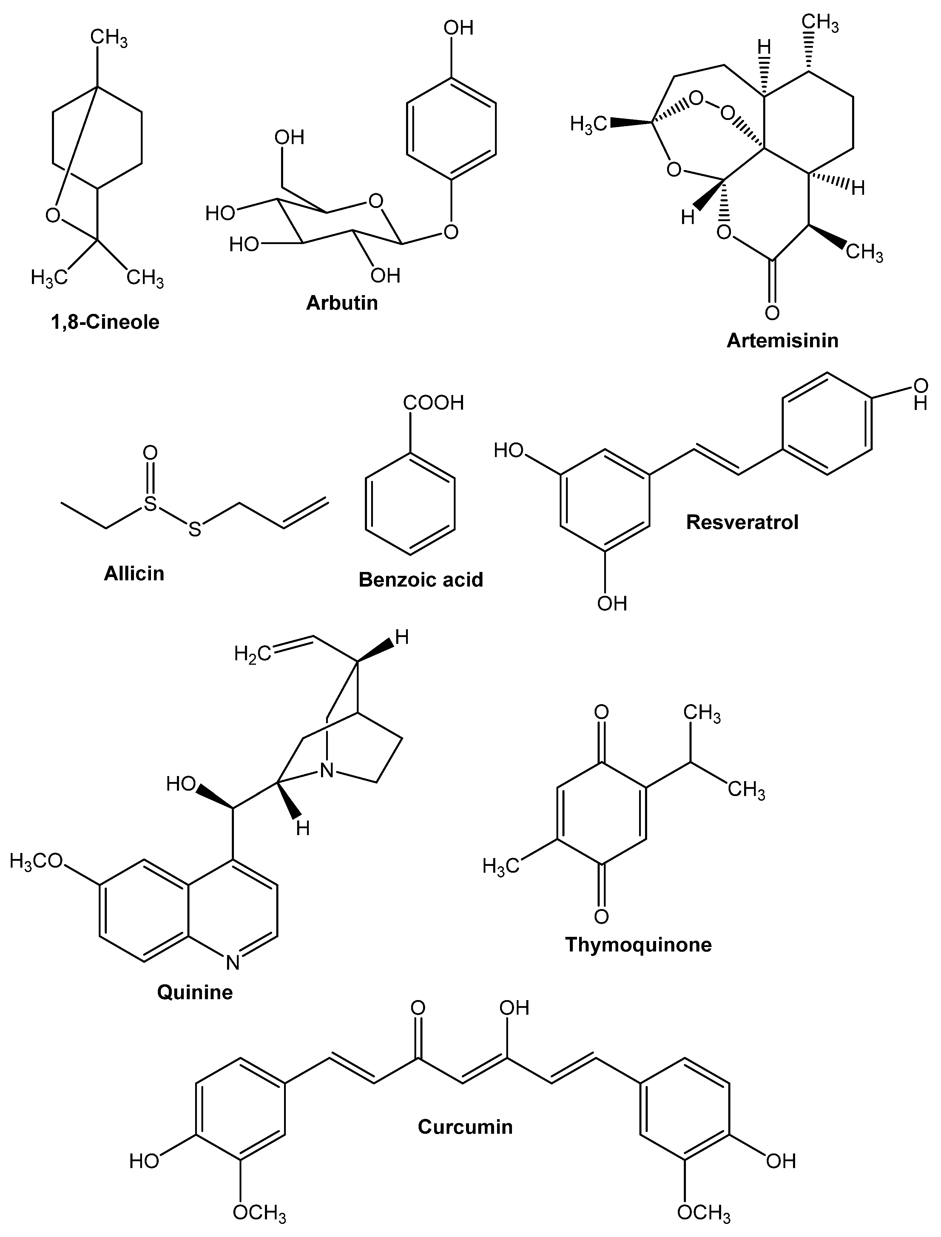
Some other secondary metabolites from medicinal plants show some major antimicrobial properties that are significant active compounds (Figure 2).

8.1. 1,8-Cineole

Numerous fundamental oils incorporate (ethyl-dimethyl-(3-sulfopropyl)azanium) (C7H18NO3S+) (PubChem CID 448830, for example, E. globulus (eucalyptus) oil, Melaleuca alternifolia (tea tree) oil, or R. officinalis oil (rosemary). It is one of the fundamental unique combinations in key oils, which clarifies their antibacterial properties [2]. It functions as an accelerator of penetration for topical application to the skin [16]. By temporarily disrupting the intercellular lipids in the skin’s stratum corneum, 1,8-cineole enables the penetration of otherwise less penetrable substances into the skin [174].

8.2. Arbutin

Plants from the Ericaceae, Asteraceae, and Rosaceae families contain arbutin ((2R,3S,4S,5R,6S)-2-(hydroxymethyl)-6-(4-hydroxyphenoxy)oxane-3,4,5-triol) (C12H16O7) (PubChem CID 440936). It explains the antiseptic qualities, although its interactions with bacteria, particularly the carcinogenic impact of its metabolite’s hydroquinone, are not well understood [175,176].

8.3. Allicin

(3-prop-2-enylsulfinylsulfanylprop-1-ene) (C6H10OS2) (PubChem CID 65036) belongs to the Alliaceae family and contains antibacterial and immunomodulatory properties, as well as numerous other health benefits [175].
The antibiotic activity of allicin was initially being thought to be mediated mainly through the inhibition of specific thiol-containing enzymes in bacteria via the fast interaction of thiosulfinates with thiol groups [3]. Allicin’s capacity to react with a model thiol molecule (L-cysteine) to create the S-thiolation product S-allyl mercapto cysteine was recently validated in a study [176].

8.4. Artemisinin

Artemisinin (C15H22O5) (PubChem CID 68826) is a semi-synthetic plant-derived chemical with a prolonged usage history in infectious illnesses. The dynamic part of Artemisia annua (sweet wormwood) is artemisinin, which has given the medical community new expectation [177,178]. The development of artemisinin-resistant Plasmodium falciparum explains why novel antimalarial is so essential. Lupane triterpenes, derived from Buxus sempervirens (common boxwood), have just been identified as the next generation of antimalarials, according to a recent study [179,180].

8.5. Benzoicacid

The antibacterial action of benzoic acid (C7H6O2) (PubChem CID 243), which is present in the fruits of Vaccinium vitis-idaea (lingonberry, cowberry, or bearberry), explains jam preservation [2].

8.6. Curcumin

Curcumin ((1E,6E)-1,7-bis(4-hydroxy-3-methoxyphenyl)hepta-1,6-diene-3,5-dione) (C21H20O6) (PubChem CID 969516), a functioning blend extricated from fragrant Curcuma aromatica (wild turmeric), not only an amazing malignant growth avoidance agent, calming, and anticancer fixing, yet it furthermore has antiparasitic action in vitro [2]. Curcumin is effective at suppressing both acute and chronic inflammation. It inhibits inflammation by decreasing histamine levels and possibly increasing adrenal glands’ natural cortisone production [8]. Additionally, curcumin demonstrated anti-inflammatory activity in vitro against human vascular cells. Curcumin exerts anti-inflammatory activity by inhibiting the inflammatory response of TNF-α stimulated human endothelial cells by interfering with NF-κB. Additionally, curcumin inhibits platelet-derived growth factor (PDGF) [181].

8.7. Quinine

(R)-[(2S,4S,5R)-5-ethenyl-1-azabicyclo[2.2.2]octan-2-yl]quinine(R)-[(2S,4S,5R)-5-ethenyl-1-azabicyclo[2.2.2]octan-2-yl]quinine(R)-[(2 Cinchona produces-(6-methoxyquinolin-4-yl)methanol) (C20H24O2N2 or C20H24N2O2) (PubChem CID 3034034). This compound was considered the primary treatment for malaria for quite a long time, although alternative plant-based active chemicals have now proven their worth [2]. Additionally, numerous investigations have indicated that quinine exhibits antibacterial effects. Quinine was discovered to be bactericidal when E. coli, K. pneumonia, and S. aureus were inhibited [182].

8.8. Resveratrol

Resveratrol(5-[(E)-2-(4-hydroxyphenyl)ethenyl]benzene-1,3-diol) (C14H12O3)(PubChem CID 445154) is an antioxidant that also possesses antibacterial effects [183]. Resveratrol inhibits the ATP synthase enzyme in a variety of bacteria [17].

8.9. Thymoquinone

Thymoquinone (2-methyl-5-propane-2-ylcyclohexa-2,5-diene-1,4-dione) (C10H12O2) (PubChem CID 1028) is an essential oil, and is also the major active component in N. sativa L. seeds. It has anti-inflammatory properties as well as being a flexible immunomodulator with unique pathways recently revealed. The valuable results of N. sativa L. seeds essential oil in viral diseases can be clarified by its contribution in initiating cell invulnerability by rousing CD4+ T cells and producing -interferon [184,185].

9. Chemical Compounds Having Antimicrobial Properties Derived from Marine Source

9.1. Terpenoids

Terpenoids are bountiful in higher plants and are widely spread in nature. Uncommon terpenoids can likewise be found in bounty in marine species. Terpenoids are considered phytoalexins, bug antifeedants and anti-agents, fertilization attractants, herbivore protection specialists, pheromones, allelochemicals, plant chemicals, and sign atoms, and they have ruled the subject of synthetic biology. As per current artificial information, the elementary classes of antibacterial and antiviral terpenoids found in the sea climate incorporate sesterterpenoids, sesquiterpenoids, and meroterpenoids. Marine sesterterpenoids are plentiful, particularly in wipes, and they have a wide scope of biological activities, for instance, antibacterial and antiviral impacts [186]. Lee et al. [187] secluded seven sesterterpene sulfates from the tropical wipe Dysidea sp. likewise contemplated their prohibiting activity against C. albicans isocitrate lyase. A large portion of blends was discovered to be incredible isocitrate lyase inhibitors with the antibacterial movement against B. subtilis and P. vulgaris. Hyrtiosal (Figure 3) got from the marine wipe Hyrtios erectus, is another bioactive sesterterpenoid that represses HIV integrase (IN) confining to viral DNA through a novel inhibitor restricting area [188]. The way that hyrtiosal could tie HIV N-terminal space at Ser17, Trp19, and Lys34 created such hyrtiosal-instigated viral DNA/IN hindrance, as indicated by a unique sub-atomic investigation matched with a site-coordinated mutagenesis strategy. Since hyrtiosal was as recently demonstrated to be a protein tyrosine phosphatase 1B inhibitor, this exploration could give data on numerous objectives to this marine regular substance. Many fascinating dynamic sesquiterpene–quinones/–hydroquinones are being found in marine wipes. The 1,4-benzoquinone moiety is found in a wide scope of accumulates that have gotten a great deal of interest because of their wide scope of natural exercises, including antibacterial and antiviral attributes. Puupehanol, alongside the known synthetic substances puupehenone and chloropuupehenone, is a novel sesquiterpene–dihydroquinone subordinate discovered from ocean wipe Hyrtios sp. that is liable for the antifungal action found in the wipe separate [189]. Puupehenone has the most grounded suppressing development against Cryptococcus neoformans and Candida krusei of the overall large number of substances reviewed, with MICs going from 1.25 to 2.50 g/mL, independently. Other antimicrobial sesquiterpenoid-quinones found in marine wipes include nakijiquinones G-I, isolated from Okinawan marine wipes of the Spongilidae family, and new sesquiterpenoid–hydroquinones from the marine wipe Dysidea Arenaria, which had a moderate inhibitory effect on HIV pivot transcriptase (RT) [190].
Other antimicrobial terpenoids isolated from ocean wipes included meroterpenoids, according to reports. Zhang et al. extracted fascioquinols A–F as naturally dynamic meroterpenes from significant water southern Australian ocean wipes during a study to discover new antimicrobial experts from marine living things. Fasciospongia sp. is a species of Fasciospongia. Fascioquinols B, C, and D result from fascioquinol A’s destructive interceded hydrolysis/cyclization delayed consequences. Two of these combinations, facioloquinol A and B, showed promising Gram (+) unequivocal antibacterial activity against S. aureus (IC50 0.9–2.5 M) and B. subtilis (IC50 0.3–7 M). Novel Caledonia’s distant ocean wipe yielded four new meroterpenes, alisiaquinones A-C, and alisiaquinol. The blends inhibited the plasmodial kinase Pfnek-1 and a protein farnesyltransferase in the M reach, as well as different chloroquine-sensitive and –safe Plasmodium falciparum strains. Tropical ocean diterpenes and diterpene isonitriles wipe Another antimicrobial terpenoid found in marine wipes is Cymbastela hooperi [191].

9.2. Phenolic Compounds

Phenols are the most widely perceived sort of auxiliary metabolite found in plants. They range from straightforward designs with one aromatic ring to exceptionally complex polymeric substances and can be found in numerous classes of typical blends with sweet-smelling moieties. In the marine climate, phenolic compounds with halogen can be found frequently. An enormous number of studies on the antimicrobial action of phenolic compounds disconnected from ocean wipes, principally antibacterial movement, have been led as of late (Figure 3). 2-(2′,4′-dibromophenoxy)-4,6-dibromophenol from an ocean wipe in vitro antibacterial movement was found in Dysidea granulosa gathered off the shore of the Lakshadweep Islands in the Indian Ocean, especially against methicillin-safe and touchy S. aureus; vancomycin-safe, and delicate Enterococci; and Bacillus sp. A tale polybrominated diphenyl ether, which was recognized from another Dysideaspecies from the Federated States of Micronesia [192]. In the hyphae arrangement hindrance measure, these medications showed inhibitory movement against Streptomyces 85E. The Indonesian wipe Lamellodysidea herbacea was additionally displayed to contain these synthetics [193]. Antimicrobial viability against Bacillus subtilis was exhibited by these metabolites. Incorporating two phenolic hydroxyl gatherings and bromines at C-2 and C-5 is vital for presenting antibacterial movement, as indicated by investigations of construction action communications. Other marine creatures and microorganisms, like red–green growth and microbes, have been found to contain bromophenol synthetics. In the wake of distinguishing movement in marine concentrates, a portion of these synthetics were secluded utilizing bioassay-directed fractionation. Gracious et al. [194] gathered Odonthalia corymbifera in the journey for naturally dynamic fixings in marine green growth and found that its unrefined concentrates have antibacterial activity against an assortment of organisms. The crude concentrate was isolated utilizing a bioassay-directed strategy, yielding various bromophenol compounds. The 2,2′,3,3′-tetrabromo-4,4′,5,5′-tetrahydroxy diphenylmethane subsidiary was the most dynamic against Candida albicans, Aspergillus fumigatus, Trichophyton rubrum, and Trichophyton mentagrophytes among the secluded normal items. 4,4′,6-tribromo-2,2′-biphenol, obtained from a concentrate of a marine Pseudoalteromonas sp. CMMED 290 showed noteworthy antibacterial development against methicillin-safe Staphylococcus aureus. Isnansetyo and Kamei recovered 2,2′,3-tribromo-biphenyl-4,4′-dicarboxylic destructive from another Pseudoalteromonas sp. the marine bacterium Pseudoalteromonas phenolica O-BC30T. This was found to threaten methicillin-safe Staphylococcus aureus action against all ten clinical isolates, with MIC regards going from 1 to 4 g/mL. The combination was convincing against B. subtilis and Enterococcus serolicida, yet not Gram (−) minuscule life forms or development. These disclosures demonstrated that this bromophenyl compound has strong in vitro movement against methicillin-safe S. aureus and could be used as a lead particle to improve novel antimicrobials. Other antibacterial bromophenyl compounds have been found from the INH strain of the marine bacterium Pseudoalteromonas haloplanktis [195].

9.3. Alkaloids

Alkaloids with particular synthetic highlights and conspicuous substance action have been found in marine organic entities and microorganisms, recommending that they could be helpful as lead structures to improve novel prescriptions (Figure 4). Antimicrobial and antiviral exercises are among the expected pharmacological impacts of a few of these substances. Many intriguing antimicrobial dynamic nitrogen-containing heterocyclic mixtures, such as alkylpiperidine, bromopyrrole, and pyrroloiminoquinone alkaloids, are found in marine wipes. Halicyclamine A was rediscovered as a lead for hostile to tuberculosis specialist from a marine wipe of Haliclona sp. on the direction of the created biological assay in the journey for antimicrobial medications against torpid Mycobacterium tuberculosis [196]. Halicyclamine A repressed the improvement of Mycobacterium smegmatis, Mycobacterium Bovis, and Mycobacterium TB with MICs going from 1 to 5 g/mL in both vigorous and hypoxic conditions, causing lethargy. Halicyclamine A’s development inhibitory movement was bactericidal, and it did not cross-oppose isoniazid, ethambutol, rifampicin, or streptomycin, which are right now utilized as anti-tubercular drugs. This wipe, as of late, gave 22-hydroxyhaliclonacyclamine B, a novel tetracyclic alkyl piperidine alkaloid, just as two familiar alkaloids, haliclonacyclamine An and B, as against lethargic mycobacterial compounds [197]. Since 22-hydroxyhaliclonacyclamine B has decreased antibacterial activity against M. tuberculosis, it may be extrapolated that the 22-hydroxy gathering in position one was found to decrease hostile to mycobacterial action in examinations of construction action associations. Haliclonin A, another antimicrobial alkaloid from Haliclona sp., was found to have antibacterial activity against an assortment of microbial strains [198].
Bromopyrrole alkaloids, which are familiar among the most continuous metabolites found in these species, are additionally antibacterial alkaloids found in marine wipes. These antimicrobial segments, for example, nagelamides Q, R, J, K, L, M, and N, have been recognized from the wipe Agelas sp. [199,200,201]. Monanchora unguifera has been displayed to contain various polycyclic guanidine alkaloids with antiviral and antibacterial properties [202]. Batzelladine alkaloids such as 16-hydroxycrambescidin 359; batzelladines K, L, M, and N; ptilomycalin A; crambescidine 800; batzelladine C; and dehydrobatzelladine C were recovered from this Caribbean plant. The mixtures were found to have antiretroviral action against HIV and AIDS’s shrewd diseases microbes. Merobatzelladines An and B have, as of late, been distinguished as antibacterial constituents from this marine wipe [203,204].

9.4. Polysaccharides

The immense number of polysaccharides are taken from marine plants and creatures, or made by ocean microbes and parasites, which implies that the sector of marine polysaccharides is continually expanding [205]. The antimicrobial and antiviral movement was seen in a portion of these marine polysaccharides (Figure 4). The antiviral part (hostile to HSV-1) of the acidic polysaccharide nostoflan was separated from the scrumptious blue-green algae Nostoc flagelliforme [206]. In time-of-addition studies, early events such as virus binding and/or penetration were revealed to be the most sensitive stage of viral replication to nostoflan. Separate virus binding and penetration assays were performed to see how much nostoflan is involved in these processes.
The findings suggest that the antiherpetic activity of nostoflan is due to the suppression of virus binding to, but not penetration into, host cells. Infection restricting and infiltration tests were done independently to determine the degree to which nostoflan might be engaged with these cycles. Another antiviral polysaccharide of marine origin is a lectin obtained from the filamentous cyanobacterium Oscillatoria agardhii NIES-204, which smothers HIV propagation in MT-4 cells [207]. The antifungal movement was found in marine polysaccharides, for example, a chitinase segregated from an ocean Streptomyces sp. DA11 connected with the South China ocean wipe Craniella australiensis [173], which expressed antifungal action against Aspergillus niger and C. albicans. The wipe’s microbial symbiont might support chitin breakdown and antifungal safeguard with chitinase action [173].

9.5. Fatty Acids

Unsaturated fats containing at least two methylene-intruded on twofold securities are significant for excellent cell work. Their organic pertinence in explicit clinical issues normal in Western culture, like heftiness and cardiovascular sickness, has prompted their utilization in biomedical and nutraceutical research. Marine unsaturated fats are intriguing due to the different jobs and natural highlights they play in marine animals’ cells. A portion of these unsaturated fats has been displayed to have significant natural highlights, like antibacterial and antiviral action. The calcareous wipe Paragrantia cf. waguensis has yielded a novel acetylenic unsaturated fat [208]. With MICs of 64 and 128 g/mL, the substance exhibited antibacterial advancement against S. aureus and E. coli, autonomously. Brominated unsaturated fats from an ocean wipe gathered in Papua New Guinea [209] and motualevic acids A–F separated from the wipe Siliquaria spongia sp., which stifles the improvement of S. aureus and its methicillin-safe strains are instances of other antimicrobial unsaturated fats from seawater wipes [210]. Marine algae have also been used to isolate antimicrobial fatty acids. Extracts from the marine diatom Phaeodactylum tricornutum have been reported to have antibacterial properties. Desbois et al. [211] antibacterial compounds like the monounsaturated fatty acid (9Z)—hexadecenoic acid and the relatively uncommon polyunsaturated fatty acid (6Z,9Z,12Z)—hexadecatrienoic acid were discovered and recognized as being responsible for this activity. Both compounds are effective against Gram (+) bacteria, with Listonella anguillarum, a Gram (−) marine pathogen, showing an extra inhibitory impact. The first chemical kills bacteria quickly at μM concentrations and is extremely effective against multidrug-resistant S. aureus. Eicosapentaenoic acid, a novel antibacterial fatty acid produced by this diatom, is efficacious against a wide spectrum of Gram (+) and Gram (−) bacteria, including multi resistant S. aureus [212].
The culture extract of A. niger EN-13, an endophytic fungus isolated from the marine brown algae Colpomenia sinuosa [213], yielded Asperamides A and B, a sphingolipid and its related glycosphingolipid with a previously unreported 9-methyl-C20-sphingosine moiety. Asperamide A showed antifungal activity against C. albicans in the antifungal assay.

9.6. Fungi

Comazaphilones C–E (30–32), azaphilone subsidiaries, were distinguished from Penicillium cooperative QSD-17, which was recuperated from oceanic residue in the southern China Sea. Antibacterial action was seen in intensifies 30–32, with MIC esteems going from 16 to 64 lg/mL [214]. A marine-determined parasite strain, Penicillium sp., was utilized to create 7-O-acetylsecopenicillide C (33) MA-37 was segregated from the marine mangrove plant Bruguiera gymnorrhiza’s rhizospheric soil test. Compound 33 hindered the development of Micrococcus luteus and E. coli, individually, having MIC upsides of 64 and 16 L g/mL [215,216]. Penicillium sp. ML226 was found in a silt test taken in the Fu Gong mangrove zone of Long Hai, Taiwan Strait, China, and created penicitrinols J (34) and K. (35). The two mixtures (34 and 35) were antibacterial against S. aureus CMCC26003, with inhibitory zones of 4 and 3 mm, separately, at 20 mg/plate [216]. P. spinophilin SD-272 was detached from a dregs test in the Pearl River, South China Sea, and it is anything but A (36), which restrained E. coli with a 10 mm restraint zone at 20 L g/circle [217]. Herqueidiketal (37), another synthetic with an exceptionally oxidized naphthoquinone fraction confined from the growth Penicillium sp. F011, showed antibacterial movement against S. aureus with an IC50 of 23.6 IM. Pinophilin G (38), an azaphilone subordinate separated from the marine-determined growth P. spinophilin, was found to have antibacterial action against Vibrio anguillarum with an MIC worth of 25.0 mol/L (Figure 4) (172). Penicillium sp. SCSIO of 101 was separated from an oceanic dregs test acquired from the South China Sea and was displayed for penicacid D (39), an antibacterial specialist with an MIC worth of 641 g/mL against E. coli [218].

9.7. Actinobacteria

Actinobacteria, a phylum of germs, is found in an assortment of traditional settings. The sort Streptomyces creates maximum microbial-determined biologically active mixtures combined by agents of the request Actinomycetes, which are Gram-positive, non-motile, oxygen- consuming microorganisms with high G C substance in their DNA (70–80 percent) and high phenotypic variety (Figure 5). Low natural matter focus, high hydrostatic pressing factor, high NaCl fixation, and low temperature are, for the most part, present in a profound marine living space. Thus, it is conceivable that marine Actinomycetes have interesting qualities that have not been seen in earthly Actinomycetes [219]. A few marine actinobacteria have effectively been displayed to make unmistakable auxiliary metabolites [220]. Actinomycetes have been broadly examined as antibacterial substance producers such as synthetic antimicrobial compounds found in marine strains. Streptomyces sp. was segregated from ocean residue and displayed to create the anti-toxin synthetics fijimycins A–C (intensifies 1–3) and etamycin A (4), which had considerable antibacterial action against MRSA strains (86). In the circle dispersion examine, the unprecedented neosidomycin metabolite kahakamide A (5) has antibacterial activity against Bacillus subtilis [221]. Heronamycin is delivered by other Streptomyces species gathered from ocean garbage (6, a benzothazine ansamycin). This synthetic displayed having unobtrusive antibacterial action against two strains of B. subtilis [222]. Lynamicins A–E (7–11), chlorinated bisindole pyrroles were distinguished from sea silt-related actinomycetes. Medication safe microbes, including MRSA and vancomycin-safe Enterococcus faecium (VRE) are helpless to these mixtures [223]. Novel antimicrobial synthetic substances, including three benzopyrone subordinates, 7-methyl coumarin, and two flavonoids, rhamnazin and cirsimaritin, were distinguished from a Streptomyces sp. disconnected from a sea silt [224]. Essramycin (12), for instance, has been exhibited to have antibacterial viability against P. aeruginosa, E. coli, and Micrococcus luteus [224,225].

9.8. Cyanobacteria

Cyanophyta is a solitary bacterial phylum that gets energy through photosynthesis and is the lone photosynthetic prokaryotes equipped for creating oxygen and engrossing CO2 [226]. Although some Cyanobacteria are hard to develop, this gathering of microorganisms accumulates consideration in essential exploration because of its shortage. Unicellular and filamentous cyanobacteria have been assembled throughout the long term, portrayed morphologically and microscopically, and developed in axenic societies utilizing lumbering techniques. The present metagenomics structure may help accomplish unadulterated culture necessities [227] for additional therapeutic property research (Figure 5). Cyanobacteria are an incredible non-customary wellspring of mixtures with a splendid and cheerful future in prescription advancement for various illnesses [228]. Indeed, compounds confined from these microorganisms have been displayed to have antibacterial, antifungal, antiviral, anticancer, antiplasmodium, and antialgae activity. It additionally seems to have immunosuppressive properties [229]. Ambiguine-K, Ambiguine K-O isonitriles, and M isonitriles have been segregated from the oceanic cyanobacterium Fischerella ambigua (UTEX 1903) and show critical antibacterial action against Mycobacterium TB [230]. Lyngbyoic corrosive is a cyclopropane-containing unsaturated fat that has been separated from the marine cyanobacterium Lyngbya and can disturb P. aeruginosa majority detecting. This particle envelops over 260 marine synthetic compounds and is right now going through preclinical testing, including certain macrolactins created by Bacillus spp., napyradiomycins, and dixiamycins delivered by Streptomyces spp. [71,225].

10. Conclusions and Future Prospects

Antimicrobials have become important in contemporary life; the wonders of these drugs display the usefulness of the natural world. However, the inappropriate use and over-prescription of medications leads to multi-drug resistance (MDR) and cross-resistance to other drugs, which have become worldwide concerns. This emerging problem has ushered in the Era of Responsibility. Warning bells can be heard from every quarter. There is an urgent need to discover some novel alternatives to help combat this significant problem. Many studies are currently in process about the antimicrobial properties of natural products. The current literature in this area indicates an urgent need for a coordinated effort for meaningful research and the discovery of novel alternatives by exploring natural products.
This investigation shares a plethora of information regarding the phytocomplexes and the mode of action of pure antimicrobials isolated and purified from the potential medicinal plants. These constituents also unlock the opportunities to design new nutraceuticals or other effective drugs. However, future investigation should explore the additional bioactivity of the corresponding genus and species to discover new therapeutics.

Author Contributions

Conceptualization, M.M.R., M.S.R. and M.R.I.; drafting the manuscript, M.M.R., M.S.R., M.R.I., M.E.H., F.M.M. and M.A.; review and editing the paper: E.K.A. and E.S.-S.; revising, M.M.R., M.S.R., M.R.I., M.E.H., E.K.A., F.M.M., M.A., M.S. and E.S.-S. All authors have read and agreed to the published version of the manuscript.

Funding

This research received no external funding.

Data Availability Statement

Not applicable.

Acknowledgments

The authors are grateful to the Department of Pharmacy, Daffodil International University for providing many different supports to conduct this research. E.S.-S. is thankful to Proyecto Interno I+D+I UCEN (CIP2020036) for financial support.

Conflicts of Interest

The authors declare no conflict of interest.

References

  1. Homsy, J.; King, R.; Balaba, D.; Kabatesi, D. Traditional health practitioners are key to scaling up comprehensive care for HIV/AIDS in sub-Saharan Africa. Aids 2004, 18, 1723–1725. [Google Scholar] [CrossRef]
  2. Ionescu, M.I. Are herbal products an alternative to antibiotics? In Bacterial Pathogenesis and Antibacterial Control; IntechOpen: London, UK, 2017. [Google Scholar]
  3. Di Pierro, F.; Rapacioli, G.; Ferrara, T.; Togni, S. Use of a standardized extract from Echinacea angustifolia (Polinacea [R]) for the prevention of respiratory tract infections. Altern. Med. Rev. 2012, 17, 36–42. [Google Scholar] [PubMed]
  4. Chapman, A.D. Numbers of Living Species in Australia and the World; Department of the Environment, Water, Heritage and the Arts: Canberra, ACT, Australia, 2009. [Google Scholar]
  5. Smith, J.P., Jr. Medical Plants-General References; Humboldt State University: Arcata, CA, USA, 2017. [Google Scholar]
  6. Bahmani, M.; Zargaran, A.; Rafieian-Kopaei, M.; Saki, K. Ethnobotanical study of medicinal plants used in the management of diabetes mellitus in the Urmia, Northwest Iran. Asian Pac. J. Trop. Med. 2014, 7, S348–S354. [Google Scholar] [CrossRef] [Green Version]
  7. Biswasroy, P.; Pradhan, D.; Kar, B.; Ghosh, G.; Rath, G. Recent advancement in topical nanocarriers for the treatment of psoriasis. AAPS PharmSciTech 2021, 22, 164. [Google Scholar] [CrossRef] [PubMed]
  8. Kharat, M.; Du, Z.; Zhang, G.; McClements, D.J. Physical and chemical stability of curcumin in aqueous solutions and emulsions: Impact of pH, temperature, and molecular environment. J. Agric. Food Chem. 2017, 65, 1525–1532. [Google Scholar] [CrossRef] [PubMed]
  9. Khare, T.; Anand, U.; Dey, A.; Assaraf, Y.G.; Chen, Z.-S.; Liu, Z.; Kumar, V. Exploring phytochemicals for combating antibiotic resistance in microbial pathogens. Front. Pharmacol. 2021, 12, 720726. [Google Scholar] [CrossRef] [PubMed]
  10. Salem, M.A.; Ezzat, S.M. The use of aromatic plants and their therapeutic potential as antiviral agents: A hope for finding anti-COVID 19 essential oils. J. Essent. Oil Res. 2021, 33, 105–113. [Google Scholar] [CrossRef]
  11. Alves, M.; Young, C.; Bozzetto, K.; Poole-Warren, L.; Martens, P. Degradable, click poly (vinyl alcohol) hydrogels: Characterization of degradation and cellular compatibility. Biomed. Mater. 2012, 7, 024106. [Google Scholar] [CrossRef]
  12. Ramić, A.; Skočibušić, M.; Odžak, R.; Čipak Gašparović, A.; Milković, L.; Mikelić, A.; Sović, K.; Primožič, I.; Hrenar, T. Antimicrobial activity of quasi-enantiomeric cinchona alkaloid derivatives and prediction model developed by machine learning. Antibiotics 2021, 10, 659. [Google Scholar] [CrossRef]
  13. Al Ramahi, M.; Keszthelyi-Szabó, G.; Beszédes, S. Corrigendum: Water Science and Technology 81 (6), 1231–1241: Improving biogas production performance of dairy activated sludge via ultrasound disruption prior to microwave disintegration. Water Sci. Technol. 2020, 82, 1720. [Google Scholar] [CrossRef]
  14. Wang, X.; Liu, Z. Prevention and treatment of viral respiratory infections by traditional Chinese herbs. Chin. Med. J. 2014, 127, 1344–1350. [Google Scholar] [PubMed]
  15. Yesilada, E. Contribution of traditional medicine in the healthcare system of the Middle East. Chin. J. Integr. Med. 2011, 17, 95–98. [Google Scholar] [CrossRef]
  16. Ghirga, F.; Quaglio, D.; Mori, M.; Cammarone, S.; Iazzetti, A.; Goggiamani, A.; Ingallina, C.; Botta, B.; Calcaterra, A. A unique high-diversity natural product collection as a reservoir of new therapeutic leads. Org. Chem. Front. 2021, 8, 996–1025. [Google Scholar] [CrossRef]
  17. Liu, L.; Yu, J.; Shen, X.; Cao, X.; Zhan, Q.; Guo, Y.; Yu, F. Resveratrol enhances the antimicrobial effect of polymyxin B on Klebsiella pneumoniae and Escherichia coli isolates with polymyxin B resistance. BMC Microbiol. 2020, 20, 306. [Google Scholar] [CrossRef]
  18. Kang, J.; Liu, L.; Liu, M.; Wu, X.; Li, J. Antibacterial activity of gallic acid against Shigella flexneri and its effect on biofilm formation by repressing mdoH gene expression. Food Control 2018, 94, 147–154. [Google Scholar] [CrossRef]
  19. Fahrenholtz, C.G.; Bonanno, L.S.; Martin, J.B. Tranexamic acid as adjuvant treatment for postpartum hemorrhage: A systematic review protocol. JBI Evid. Synth. 2019, 17, 1565–1572. [Google Scholar] [CrossRef] [Green Version]
  20. Lee, M.-J.; Maliakal, P.; Chen, L.; Meng, X.; Bondoc, F.Y.; Prabhu, S.; Lambert, G.; Mohr, S.; Yang, C.S. Pharmacokinetics of tea catechins after ingestion of green tea and (−)-epigallocatechin-3-gallate by humans: Formation of different metabolites and individual variability. Cancer Epidemiol. Prev. Biomark. 2002, 11, 1025–1032. [Google Scholar]
  21. Stephenson, J.; Nutma, E.; van der Valk, P.; Amor, S. Inflammation in CNS neurodegenerative diseases. Immunology 2018, 154, 204–219. [Google Scholar] [CrossRef] [PubMed] [Green Version]
  22. Dai, J.; Mumper, R.J. Plant phenolics: Extraction, analysis and their antioxidant and anticancer properties. Molecules 2010, 15, 7313–7352. [Google Scholar] [CrossRef] [PubMed]
  23. Goud, J.V.; Suryam, A.; Charya, M.S. Biomolecular and phytochemical analyses of three aquatic angiosperms. Afr. J. Microbiol. Res. 2009, 3, 418–421. [Google Scholar]
  24. Vojtovič, D.; Luhová, L.; Petřivalský, M. Something smells bad to plant pathogens: Production of hydrogen sulfide in plants and its role in plant defence responses. J. Adv. Res. 2020, 27, 199–209. [Google Scholar] [CrossRef] [PubMed]
  25. Chojnacka, K.; Witek-Krowiak, A.; Skrzypczak, D.; Mikula, K.; Młynarz, P. Phytochemicals containing biologically active polyphenols as an effective agent against Covid-19-inducing coronavirus. J. Funct. Foods 2020, 73, 104146. [Google Scholar] [CrossRef] [PubMed]
  26. Guzman, J.D. Natural cinnamic acids, synthetic derivatives and hybrids with antimicrobial activity. Molecules 2014, 19, 19292–19349. [Google Scholar] [CrossRef]
  27. Duke, J.A. Handbook of Medicinal Herbs; CRC Press: Boca Raton, FL, USA, 2002. [Google Scholar]
  28. Katerere, D.R.; Luseba, D. Ethnoveterinary Botanical Medicine: Herbal Medicines for Animal Health; CRC Press: Boca Raton, FL, USA, 2010. [Google Scholar]
  29. Khandelwal, K.R. Practical Pharmacognosy; Pragati Books Private Limited: Pune, India, 2008. [Google Scholar]
  30. Saran, S.; Menon, S.; Shailajan, S.; Pokharna, P. Validated RP-HPLC method to estimate eugenol from commercial formulations like Caturjata Churna, Lavangadi Vati, Jatiphaladi Churna, Sitopaladi Churna and clove oil. J. Pharm. Res. 2013, 6, 53–60. [Google Scholar] [CrossRef]
  31. McDonnell, G.; Russell, A.D. Antiseptics and disinfectants: Activity, action, and resistance. Clin. Microbiol. Rev. 2001, 14, 227. [Google Scholar] [CrossRef] [Green Version]
  32. Marcó, M.B.; Mercanti, D.J. Bacteriophages in dairy plants. In Advances in Food and Nutrition Research; Elsevier: Amsterdam, The Netherlands, 2021; Volume 97, pp. 1–54. [Google Scholar]
  33. Solano, F. Melanins: Skin pigments and much more—Types, structural models, biological functions, and formation routes. N. J. Sci. 2014, 2014, 28. [Google Scholar] [CrossRef] [Green Version]
  34. Motley, T.J. The ethnobotany of sweet flag, Acorus calamus (Araceae). Econ. Bot. 1994, 48, 397–412. [Google Scholar] [CrossRef]
  35. El-Najjar, N.; Gali-Muhtasib, H.; Ketola, R.A.; Vuorela, P.; Urtti, A.; Vuorela, H. The chemical and biological activities of quinones: Overview and implications in analytical detection. Phytochem. Rev. 2011, 10, 353–370. [Google Scholar] [CrossRef]
  36. Ahuja, I.; Kissen, R.; Bones, A.M. Phytoalexins in defense against pathogens. Trends Plant Sci. 2012, 17, 73–90. [Google Scholar] [CrossRef]
  37. Crini, G. Historical review on chitin and chitosan biopolymers. Environ. Chem. Lett. 2019, 17, 1623–1643. [Google Scholar] [CrossRef]
  38. Chandra, A.K.; De, N. Goitrogenic/antithyroidal potential of green tea extract in relation to catechin in rats. Food Chem. Toxicol. 2010, 48, 2304–2311. [Google Scholar] [CrossRef]
  39. Chinsembu, K.C. Chemical diversity and activity profiles of HIV-1 reverse transcriptase inhibitors from plants. Rev. Bras. Farmacogn. 2019, 29, 504–528. [Google Scholar] [CrossRef]
  40. Matsumoto, M.; Hamada, S.; Ooshima, T. Molecular analysis of the inhibitory effects of oolong tea polyphenols on glucan-binding domain of recombinant glucosyltransferases from Streptococcus mutans MT8148. FEMS Microbiol. Lett. 2003, 228, 73–80. [Google Scholar] [CrossRef] [Green Version]
  41. De Oliveira, D.R.; Tintino, S.R.; Braga, M.F.B.M.; Boligon, A.A.; Athayde, M.L.; Coutinho, H.D.M.; de Menezes, I.R.A.; Fachinetto, R. In vitro antimicrobial and modulatory activity of the natural products silymarin and silibinin. Biomed. Res. Int. 2015, 2015, 292797. [Google Scholar] [CrossRef] [PubMed] [Green Version]
  42. Shahid, M.; Shahzad, A.; Malik, A.; Sahai, A. Recent Trends in Biotechnology and Therapeutic Applications of Medicinal Plants; Springer: Berlin/Heidelberg, Germany, 2013. [Google Scholar]
  43. Pasetto, S.; Pardi, V.; Murata, R.M. Anti-HIV-1 activity of flavonoid myricetin on HIV-1 infection in a dual-chamber in vitro model. PLoS ONE 2014, 9, e115323. [Google Scholar] [CrossRef] [PubMed]
  44. Pengsuparp, T.; Cai, L.; Constant, H.; Fong, H.H.; Lin, L.-Z.; Kinghorn, A.D.; Pezzuto, J.M.; Cordell, G.A.; Ingolfsdöttir, K.; Wagner, H. Mechanistic evaluation of new plant-derived compounds that inhibit HIV-1 reverse transcriptase. J. Nat. Prod. 1995, 58, 1024–1031. [Google Scholar] [CrossRef]
  45. Watanbe, H.; Miyaji, C.; Makino, M.; Abo, T. Therapeutic effects of glycyrrhizin in mice infected with LP-BM5 murine retrovirus and mechanisms involved in the prevention of disease progression. Biotherapy 1996, 9, 209–220. [Google Scholar] [CrossRef] [PubMed]
  46. Saleh, A.M.; Madany, M. Coumarin pretreatment alleviates salinity stress in wheat seedlings. Plant Physiol. Biochem. 2015, 88, 27–35. [Google Scholar] [CrossRef]
  47. Jaafar, H.Z.; Ibrahim, M.H.; Karimi, E. Phenolics and flavonoids compounds, phenylanine ammonia lyase and antioxidant activity responses to elevated CO2 in Labisia pumila (Myrisinaceae). Molecules 2012, 17, 6331–6347. [Google Scholar] [CrossRef] [Green Version]
  48. Scervino, J.; Ponce, M.; Erra-Bassells, R.; Bompadre, M.; Vierheilig, H.; Ocampo, J.; Godeas, A. Glycosidation of apigenin results in a loss of its activity on different growth parameters of arbuscular mycorrhizal fungi from the genus Glomus and Gigaspora. Soil Biol. Biochem. 2006, 38, 2919–2922. [Google Scholar] [CrossRef]
  49. Cushnie, T.T.; Lamb, A.J. Antimicrobial activity of flavonoids. Int. J. Antimicrob. Agents 2005, 26, 343–356. [Google Scholar] [CrossRef]
  50. Yadav, H.; Yadav, M.; Jain, S.; Bhardwaj, A.; Singh, V.; Parkash, O.; Marotta, F. Antimicrobial property of a herbal preparation containing Dalbergia sissoo and Datura stramonium with cow urine against pathogenic bacteria. Int. J. Immunopathol. Pharmacol. 2008, 21, 1013–1020. [Google Scholar] [CrossRef]
  51. Savic, I.; Nikolic, V.; Savic-Gajic, I.; Nikolic, L.B.; Moder, K.; Hopkins, M. Optimization of quercetin extraction from green tea (Camellia sinensis) using central composite design, and the pharmacological activity of the extract. Chem. Biochem. Eng. Q. 2016, 30, 103–115. [Google Scholar] [CrossRef]
  52. Kirsch, G.; Abdelwahab, A.B.; Chaimbault, P. Natural and synthetic coumarins with effects on inflammation. Molecules 2016, 21, 1322. [Google Scholar] [CrossRef]
  53. Polassi, M.R.; Oliveira, T.d.S.; Carvalho, A.C.d.; Medeiros, L.S.d.M.; Veiga, T.A.M.; Graeff, C.F.d.O.; González, A.H.M.; Marcucci, M.C.; Grecco, S.d.S.; D’Alpino, P.H.P. Influence of dentin priming with tannin-rich plant extracts on the longevity of bonded composite restorations. Sci. World J. 2021, 2021, 1614643. [Google Scholar] [CrossRef] [PubMed]
  54. Chen, Z.; Farag, M.A.; Zhong, Z.; Zhang, C.; Yang, Y.; Wang, S.; Wang, Y. Multifaceted role of phyto-derived polyphenols in nanodrug delivery systems. Adv. Drug Deliv. Rev. 2021, 113870. [Google Scholar] [CrossRef]
  55. Prabhu, S.; Molath, A.; Choksi, H.; Kumar, S.; Mehra, R. Classifications of polyphenols and their potential application in human health and diseases. Int. J. Physiol. Nutr. Phys. Educ. 2021, 6, 293–301. [Google Scholar] [CrossRef]
  56. Jubair, N.; Rajagopal, M.; Chinnappan, S.; Abdullah, N.B.; Fatima, A. Review on the antibacterial mechanism of plant-derived compounds against multidrug-resistant bacteria (MDR). Evid.-Based Complement. Altern. Med. 2021, 2021, 3663315. [Google Scholar] [CrossRef] [PubMed]
  57. Curto, M.Á.; Butassi, E.; Ribas, J.C.; Svetaz, L.A.; Cortés, J.C. Natural products targeting the synthesis of β (1, 3)-D-glucan and chitin of the fungal cell wall. Existing drugs and recent findings. Phytomedicine 2021, 88, 153556. [Google Scholar] [CrossRef]
  58. Bilgin, H.M.; Atmaca, M.; Obay, B.D.; Özekinci, S.; Taşdemir, E.; Ketani, A. Protective effects of coumarin and coumarin derivatives against carbon tetrachloride-induced acute hepatotoxicity in rats. Exp. Toxicol. Pathol. 2011, 63, 325–330. [Google Scholar] [CrossRef]
  59. Hoult, J.; Payá, M. Pharmacological and biochemical actions of simple coumarins: Natural products with therapeutic potential. Gen. Pharmacol. Vasc. Syst. 1996, 27, 713–722. [Google Scholar] [CrossRef]
  60. Tripathi, R.; Sen, M.; Dutta, G. The Use of Artemisinin Derivative Suppositories as Life-Saving Remedy for Critical Malaria Patients; Cambridge Scholars Publishing: Newcastle upon Tyne, UK, 2021. [Google Scholar]
  61. Soine, T.O. Naturally occurring coumarins and related physiological activities. J. Pharm. Sci. 1964, 53, 231–264. [Google Scholar] [CrossRef] [PubMed]
  62. Reen, F.J.; Gutiérrez-Barranquero, J.A.; Parages, M.L.; O’Gara, F. Coumarin: A novel player in microbial quorum sensing and biofilm formation inhibition. Appl. Microbiol. Biotechnol. 2018, 102, 2063–2073. [Google Scholar] [CrossRef] [PubMed] [Green Version]
  63. Pirbalouti, A.; Rahimi, E.; Moosavi, S. Antimicrobial activity of essential oils of three herbs against Listeria monocytogenes on chicken frankfurters. Acta Agric. Slov. 2010, 95, 219. [Google Scholar] [CrossRef] [Green Version]
  64. Reguera, R.M.; Elmahallawy, E.K.; García-Estrada, C.; Carbajo-Andrés, R.; Balaña-Fouce, R. DNA topoisomerases of Leishmania parasites; druggable targets for drug discovery. Curr. Med. Chem. 2019, 26, 5900–5923. [Google Scholar] [CrossRef] [PubMed]
  65. Habtemariam, S.; Gray, A.I.; Waterman, P.G. A new antibacterial sesquiterpene from Premna oligotricha. J. Nat. Prod. 1993, 56, 140–143. [Google Scholar] [CrossRef]
  66. Moravej, H.; Moravej, Z.; Yazdanparast, M.; Heiat, M.; Mirhosseini, A.; Moosazadeh Moghaddam, M.; Mirnejad, R. Antimicrobial peptides: Features, action, and their resistance mechanisms in bacteria. Microb. Drug Resist. 2018, 24, 747–767. [Google Scholar] [CrossRef]
  67. Aureli, P.; Costantini, A.; Zolea, S. Antimicrobial activity of some plant essential oils against Listeria monocytogenes. J. Food Prot. 1992, 55, 344–348. [Google Scholar] [CrossRef]
  68. Wang, J.; Cheng, Y.; Wu, R.; Jiang, D.; Bai, B.; Tan, D.; Yan, T.; Sun, X.; Zhang, Q.; Wu, Z. Antibacterial activity of juglone against Staphylococcus aureus: From apparent to proteomic. Int. J. Mol. Sci. 2016, 17, 965. [Google Scholar] [CrossRef] [Green Version]
  69. Ji, J.; Shankar, S.; Royon, F.; Salmieri, S.; Lacroix, M. Essential oils as natural antimicrobials applied in meat and meat products—A review. Crit. Rev. Food Sci. Nutr. 2021, 10, 1–17. [Google Scholar]
  70. Sharma, V.; Ichikawa, M.; Freeze, H.H. Mannose metabolism: More than meets the eye. Biochem. Biophys. Res. Commun. 2014, 453, 220–228. [Google Scholar] [CrossRef] [Green Version]
  71. Ramassamy, C. Emerging role of polyphenolic compounds in the treatment of neurodegenerative diseases: A review of their intracellular targets. Eur. J. Pharmacol. 2006, 545, 51–64. [Google Scholar] [CrossRef]
  72. Singh, G.; Passsari, A.K.; Leo, V.V.; Mishra, V.K.; Subbarayan, S.; Singh, B.P.; Kumar, B.; Kumar, S.; Gupta, V.K.; Lalhlenmawia, H. Evaluation of phenolic content variability along with antioxidant, antimicrobial, and cytotoxic potential of selected traditional medicinal plants from India. Front. Plant Sci. 2016, 7, 407. [Google Scholar] [CrossRef] [PubMed] [Green Version]
  73. Thawabteh, A.; Juma, S.; Bader, M.; Karaman, D.; Scrano, L.; Bufo, S.A.; Karaman, R. The biological activity of natural alkaloids against herbivores, cancerous cells and pathogens. Toxins 2019, 11, 656. [Google Scholar] [CrossRef] [PubMed] [Green Version]
  74. Wang, J.; Wang, Z.; Wu, R.; Jiang, D.; Bai, B.; Tan, D.; Yan, T.; Sun, X.; Zhang, Q.; Wu, Z. Proteomic analysis of the antibacterial mechanism of action of juglone against Staphylococcus aureus. Nat. Prod. Commun. 2016, 11, 1934578X1601100632. [Google Scholar] [CrossRef] [Green Version]
  75. Ahmad, T.; Suzuki, Y.J. Juglone in oxidative stress and cell signaling. Antioxidants 2019, 8, 91. [Google Scholar] [CrossRef] [Green Version]
  76. Tullus, K.; Marks, S.D. Vasculitis in children and adolescents. Pediatr. Drugs 2009, 11, 375–380. [Google Scholar] [CrossRef] [PubMed]
  77. Sun, Y.; Gong, X.; Tan, J.Y.; Kang, L.; Li, D.; Yang, J.; Du, G. In vitro antiviral activity of Rubia cordifolia aerial part extract against rotavirus. Front. Pharmacol. 2016, 7, 308. [Google Scholar] [CrossRef] [Green Version]
  78. Qian, W.; Yang, M.; Wang, T.; Sun, Z.; Liu, M.; Zhang, J.; Zeng, Q.; Cai, C.; Li, Y. Antibacterial mechanism of vanillic acid on physiological, morphological, and biofilm properties of carbapenem-resistant enterobacter hormaechei. J. Food Prot. 2020, 83, 576–583. [Google Scholar] [CrossRef]
  79. Van Wyk, B.-E.; Wink, M. Medicinal Plants of the World; CABI: Wallingford, UK, 2018. [Google Scholar]
  80. Tian, X.-R.; Feng, J.-T.; Ma, Z.-Q.; Xie, N.; Zhang, J.; Zhang, X.; Tang, H.-F. Three new glycosides from the whole plant of Clematis lasiandra Maxim and their cytotoxicity. Phytochem. Lett. 2014, 10, 168–172. [Google Scholar] [CrossRef]
  81. Simmonds, M.S. Flavonoid–insect interactions: Recent advances in our knowledge. Phytochemistry 2003, 64, 21–30. [Google Scholar] [CrossRef]
  82. Treutter, D. Significance of flavonoids in plant resistance: A review. Environ. Chem. Lett. 2006, 4, 147–157. [Google Scholar] [CrossRef]
  83. Vardhini, B.V.; Anjum, N.A. Brassinosteroids make plant life easier under abiotic stresses mainly by modulating major components of antioxidant defense system. Front. Environ. Sci. 2015, 2, 67. [Google Scholar] [CrossRef] [Green Version]
  84. Li, P.; Luo, H.; Meng, J.; Sun, W.; Wang, X.; Lu, S.; Peng, Y.; Zhou, L. Effects of oligosaccharides from endophytic Fusarium oxysporum Dzf17 on activities of defense-related enzymes in Dioscorea zingiberensis suspension cell and seedling cultures. Electron. J. Biotechnol. 2014, 17, 156–161. [Google Scholar] [CrossRef] [Green Version]
  85. Morsy, N. Phytochemical analysis of biologically active constituents of medicinal plants. Main Group Chem. 2014, 13, 7–21. [Google Scholar] [CrossRef]
  86. Qi, X.; Wang, E.; Xing, M.; Zhao, W.; Chen, X. Rhizosphere and non-rhizosphere bacterial community composition of the wild medicinal plant Rumex patientia. World J. Microbiol. Biotechnol. 2012, 28, 2257–2265. [Google Scholar] [CrossRef] [PubMed]
  87. Philippot, L.; Raaijmakers, J.M.; Lemanceau, P.; Van Der Putten, W.H. Going back to the roots: The microbial ecology of the rhizosphere. Nat. Rev. Microbiol. 2013, 11, 789–799. [Google Scholar] [CrossRef] [PubMed]
  88. Chaparro, J.M.; Badri, D.V.; Vivanco, J.M. Rhizosphere microbiome assemblage is affected by plant development. ISME J. 2014, 8, 790–803. [Google Scholar] [CrossRef] [Green Version]
  89. Köberl, M.; Ramadan, E.M.; Adam, M.; Cardinale, M.; Hallmann, J.; Heuer, H.; Smalla, K.; Berg, G. Bacillus and Streptomyces were selected as broad-spectrum antagonists against soilborne pathogens from arid areas in Egypt. FEMS Microbiol. Lett. 2013, 342, 168–178. [Google Scholar] [CrossRef] [Green Version]
  90. Lee, Y.J.; Shukla, S.D. Pro-and anti-apoptotic roles of c-Jun N-terminal kinase (JNK) in ethanol and acetaldehyde exposed rat hepatocytes. Eur. J. Pharmacol. 2005, 508, 31–45. [Google Scholar] [CrossRef]
  91. Egamberdieva, D.; Kucharova, Z.; Davranov, K.; Berg, G.; Makarova, N.; Azarova, T.; Chebotar, V.; Tikhonovich, I.; Kamilova, F.; Validov, S.Z. Bacteria able to control foot and root rot and to promote growth of cucumber in salinated soils. Biol. Fertil. Soils 2011, 47, 197–205. [Google Scholar] [CrossRef]
  92. Malfanova, N.; Kamilova, F.; Validov, S.; Shcherbakov, A.; Chebotar, V.; Tikhonovich, I.; Lugtenberg, B. Characterization of Bacillus subtilis HC8, a novel plant-beneficial endophytic strain from giant hogweed. Microb. Biotechnol. 2011, 4, 523–532. [Google Scholar] [CrossRef] [PubMed] [Green Version]
  93. Sessitsch, A.; Kuffner, M.; Kidd, P.; Vangronsveld, J.; Wenzel, W.W.; Fallmann, K.; Puschenreiter, M. The role of plant-associated bacteria in the mobilization and phytoextraction of trace elements in contaminated soils. Soil Biol. Biochem. 2013, 60, 182–194. [Google Scholar] [CrossRef] [PubMed] [Green Version]
  94. Berg, G.; Grube, M.; Schloter, M.; Smalla, K. Unraveling the plant microbiome: Looking back and future perspectives. Front. Microbiol. 2014, 5, 148. [Google Scholar] [CrossRef] [Green Version]
  95. Beneduzi, A.; Ambrosini, A.; Passaglia, L.M. Plant growth-promoting rhizobacteria (PGPR): Their potential as antagonists and biocontrol agents. Genet. Mol. Biol. 2012, 35, 1044–1051. [Google Scholar] [CrossRef] [PubMed] [Green Version]
  96. Weller, D.M.; Raaijmakers, J.M.; Gardener, B.B.M.; Thomashow, L.S. Microbial populations responsible for specific soil suppressiveness to plant pathogens. Annu. Rev. Phytopathol. 2002, 40, 309–348. [Google Scholar] [CrossRef] [PubMed] [Green Version]
  97. Berendsen, R.L.; Pieterse, C.M.; Bakker, P.A. The rhizosphere microbiome and plant health. Trends Plant Sci. 2012, 17, 478–486. [Google Scholar] [CrossRef]
  98. Kumar, G.; Kanaujia, N.; Bafana, A. Functional and phylogenetic diversity of root-associated bacteria of Ajuga bracteosa in Kangra valley. Microbiol. Res. 2012, 167, 220–225. [Google Scholar] [CrossRef]
  99. Hardoim, P.R.; Van Overbeek, L.S.; Berg, G.; Pirttilä, A.M.; Compant, S.; Campisano, A.; Döring, M.; Sessitsch, A. The hidden world within plants: Ecological and evolutionary considerations for defining functioning of microbial endophytes. Microbiol. Mol. Biol. Rev. 2015, 79, 293–320. [Google Scholar] [CrossRef] [Green Version]
  100. Chandra, S. Endophytic fungi: Novel sources of anticancer lead molecules. Appl. Microbiol. Biotechnol. 2012, 95, 47–59. [Google Scholar] [CrossRef]
  101. Egamberdieva, D.; Wirth, S.; Behrendt, U.; Ahmad, P.; Berg, G. Antimicrobial activity of medicinal plants correlates with the proportion of antagonistic endophytes. Front. Microbiol. 2017, 8, 199. [Google Scholar] [CrossRef] [PubMed] [Green Version]
  102. Lokhande, P.; Gawai, K.; Kodam, K.; Kuchekar, B.; Chabukswar, A.; Jagdale, S. Antibacterial activity of extracts of Piper longum. J. Pharm. Toxicol. 2007, 2, 574–579. [Google Scholar] [CrossRef] [Green Version]
  103. Kumar, A.; Pandey, H.; Acharya, A.; Singh, M.; Bhatia, D.; Mukerjee, A. Investigation of comparative antimicrobial activity of aloe vera gel and juice. Pharmacologyonline 2008, 1, 239–243. [Google Scholar]
  104. Jelodarian, S.; Ebrahimabadi, A.H.; Kashi, F.J. Evaluation of antimicrobial activity of Malus domestica fruit extract from Kashan area. Avicenna J. Phytomed. 2013, 3, 1. [Google Scholar]
  105. Bokaeian, M.; Saeidi, S. Evolution of antimicrobial activity of leaf extract of Withania somnifera against antibiotic resistanct Staphylococcus aureus. Zahedan J. Res. Med. Sci. 2015, 17, e1016. [Google Scholar] [CrossRef] [Green Version]
  106. Dashti, Z.; Shariatifar, N.; Nafchi, A. Study on antibacterial and antioxidant activity of Berberis vulgaris aqueous extracts from Iran. Int. J. Pharm. Sci. Res. 2014, 5, 705–708. [Google Scholar]
  107. Rahman, M.; Khan, M.; Jamal, M.A. Anti-bacterial evaluation and minimum inhibitory concentration analysis of oxalis corniculata and ocimum santum against bacterial pathogens. Biotechnology 2010, 9. [Google Scholar] [CrossRef]
  108. Nafis, A.; Kasrati, A.; Jamali, C.A.; Custódio, L.; Vitalini, S.; Iriti, M.; Hassani, L. A comparative study of the in vitro antimicrobial and synergistic effect of essential oils from Laurus nobilis L. and Prunus armeniaca L. from Morocco with antimicrobial drugs: New approach for health promoting products. Antibiotics 2020, 9, 140. [Google Scholar] [CrossRef] [PubMed] [Green Version]
  109. Toma, Z. Antimicrobial Activity of Piperine purified from piper nigrum. J. Basrah Res. 2010, 36, 54–61. [Google Scholar]
  110. Phongpaichit, S.; Pujenjob, N.; Rukachaisirikul, V.; Ongsakul, M. Antimicrobial activities of the crude methanol extract of Acorus calamus Linn. Songklanakarin J. Sci. Technol. 2005, 27, 517–523. [Google Scholar]
  111. Al-Mamun, M.A.; Akter, Z.; Uddin, M.J.; Ferdaus, K.; Hoque, K.; Ferdousi, Z.; Reza, M.A. Characterization and evaluation of antibacterial and antiproliferative activities of crude protein extracts isolated from the seed of Ricinus communis in Bangladesh. BMC Complement. Altern. Med. 2016, 16, 1–10. [Google Scholar] [CrossRef] [Green Version]
  112. Vazirian, M.; Alehabib, S.; Jamalifar, H.; Fazeli, M.; Najarian Toosi, A.; Khanavi, M. Antimicrobial effect of cinnamon (Cinnamomum verum J. Presl) bark essential oil in cream-filled cakes and pastries. Res. J. Pharmacogn. 2015, 2, 11–16. [Google Scholar]
  113. Cushnie, T.T.; Cushnie, B.; Lamb, A.J. Alkaloids: An overview of their antibacterial, antibiotic-enhancing and antivirulence activities. Int. J. Antimicrob. Agents 2014, 44, 377–386. [Google Scholar] [CrossRef] [PubMed]
  114. Casimir, O.A.; Martin, D.K.; Philippe, E.K.; Augustin, A.A.; Parfait, K.E.J. Chemical composition, antioxidant and antimicrobial activities of Capsicum annuum var. annuum concentrated extract obtained by reverse osmosis. GSC Biol. Pharm. Sci. 2018, 5, 116–125. [Google Scholar]
  115. Radaelli, M.; Silva, B.P.d.; Weidlich, L.; Hoehne, L.; Flach, A.; Costa, L.A.M.A.d.; Ethur, E.M. Antimicrobial activities of six essential oils commonly used as condiments in Brazil against Clostridium perfringens. Braz. J. Microbiol. 2016, 47, 424–430. [Google Scholar] [CrossRef] [Green Version]
  116. Jeya, K.; Veerapagu, M.; Sangeetha, V. Antimicrobial and antioxidant properties of Coriandrum sativum L. seed essential oil. Am. J. Essent. Oils Nat. Prod. 2019, 7, 6–10. [Google Scholar]
  117. Salari, M.; Amine, G.; Shirazi, M.; Hafezi, R.; Mohammadypour, M. Antibacterial effects of Eucalyptus globulus leaf extract on pathogenic bacteria isolated from specimens of patients with respiratory tract disorders. Clin. Microbiol. Infect. 2006, 12, 194–196. [Google Scholar] [CrossRef] [Green Version]
  118. Mohsenipour, Z.; Hassanshahian, M. The effects of Allium sativum extracts on biofilm formation and activities of six pathogenic bacteria. Jundishapur J. Microbiol. 2015, 8, e18971. [Google Scholar] [CrossRef] [Green Version]
  119. Sedighinia, F.; Afshar, A.S. Antibacterial activity of Glycyrrhiza glabra against oral pathogens: An in vitro study. Avicenna J. Phytomed. 2012, 2, 118. [Google Scholar] [PubMed]
  120. Shankar, S.; Bardvalli, S.G.; Jyotirmayee, R.; Bhushan, K.; Kumar, S. Efficacy of calendula officinalis extract (marigold flower) as an antimicrobial agent against oral microbes: An invitro study in comparison with chlorhexidine digluconate. J. Clin. Diagn. Res. 2017, 11, 5–10. [Google Scholar] [CrossRef]
  121. Zhang, L.; Xu, S.-G.; Liang, W.; Mei, J.; Di, Y.-Y.; Lan, H.-H.; Yang, Y.; Wang, W.-W.; Luo, Y.-Y.; Wang, H.-Z. Antibacterial activity and mode of action of Mentha arvensis ethanol extract against multidrug-resistant Acinetobacter baumannii. Trop. J. Pharm. Res. 2015, 14, 2099–2106. [Google Scholar] [CrossRef] [Green Version]
  122. Mistry, K.S.; Sanghvi, Z.; Parmar, G.; Shah, S. The antimicrobial activity of Azadirachta indica, Mimusops elengi, Tinospora cardifolia, Ocimum sanctum and 2% chlorhexidine gluconate on common endodontic pathogens: An in vitro study. Eur. J. Dent. 2014, 8, 172–177. [Google Scholar] [CrossRef] [PubMed] [Green Version]
  123. Korukluoglu, M.; Sahan, Y.; Yigit, A.; Karakas, R. Antifungal activity of olive leaf (Olea Europaea L.) extracts from the Trilye region of Turkey. Ann. Microbiol. 2006, 56, 359–362. [Google Scholar] [CrossRef]
  124. Shetty, S.B.; Mahin-Syed-Ismail, P.; Varghese, S.; Thomas-George, B.; Kandathil-Thajuraj, P.; Baby, D.; Haleem, S.; Sreedhar, S.; Devang-Divakar, D. Antimicrobial effects of Citrus sinensis peel extracts against dental caries bacteria: An in vitro study. J. Clin. Exp. Dent. 2016, 8, e71. [Google Scholar] [CrossRef] [Green Version]
  125. Rahman, S.; Imran, M.; Muhammad, N.; Khan, N.U.; Chisthi, A.K.; Khan, A.F.; Sadozai, S.; Khan, S.M. Antibacetial screening of leaves and stem of Carica papaya. J. Med. Plant Res. 2011, 5, 5167–5171. [Google Scholar]
  126. Singh, R.; Shushni, M.A.; Belkheir, A. Antibacterial and antioxidant activities of Mentha piperita L. Arab. J. Chem. 2015, 8, 322–328. [Google Scholar] [CrossRef] [Green Version]
  127. Ismaili, A.; Sohrabi, S.; Azadpour, M.; Heydari, R.; Rashidipour, M. Evaluation of the antimicrobial activity of alkaloid extracts of four Papaver species. Herb. Med. J. 2017, 2, 146–152. [Google Scholar] [CrossRef]
  128. Misra, B.B.; Dey, S. Comparative phytochemical analysis and antibacterial efficacy of in vitro and in vivo extracts from E ast I ndian sandalwood tree (Santalum album L.). Lett. Appl. Microbiol. 2012, 55, 476–486. [Google Scholar] [CrossRef] [PubMed]
  129. Doughari, J. Antimicrobial activity of Tamarindus indica Linn. Trop. J. Pharm. Res. 2006, 5, 597–603. [Google Scholar] [CrossRef] [Green Version]
  130. Kumar, M.; Sarma, P.; Dkhar, M.; Kayang, H.; Raghuwanshi, R.; Dubey, N.K. Assessment of chemically characterised Gaultheria fragrantissima Wall. essential oil and its major component as safe plant based preservative for millets against fungal, aflatoxin contamination and lipid peroxidation during storage. J. Food Sci. Technol. 2018, 55, 111–119. [Google Scholar] [CrossRef]
  131. Appalasamy, S.; Lo, K.Y.; Ch’ng, S.J.; Nornadia, K.; Othman, A.S.; Chan, L.-K. Antimicrobial activity of artemisinin and precursor derived from in vitro plantlets of Artemisia annua L. BioMed Res. Int. 2014, 2014, 215872. [Google Scholar] [CrossRef] [PubMed] [Green Version]
  132. Dharmaratne, M.P.J.; Manoraj, A.; Thevanesam, V.; Ekanayake, A.; Kumar, N.S.; Liyanapathirana, V.; Abeyratne, E.; Bandara, B.R. Terminalia bellirica fruit extracts: In-vitro antibacterial activity against selected multidrug-resistant bacteria, radical scavenging activity and cytotoxicity study on BHK-21 cells. BMC Complement. Altern. Med. 2018, 18, 325. [Google Scholar] [CrossRef] [PubMed]
  133. Akthar, M.S.; Degaga, B.; Azam, T. Antimicrobial activity of essential oils extracted from medicinal plants against the pathogenic microorganisms: A review. J. Issues ISSN 2014, 2350, 1588. [Google Scholar]
  134. Lambert, R.; Skandamis, P.N.; Coote, P.J.; Nychas, G.J. A study of the minimum inhibitory concentration and mode of action of oregano essential oil, thymol and carvacrol. J. Appl. Microbiol. 2001, 91, 453–462. [Google Scholar] [CrossRef] [PubMed] [Green Version]
  135. Tian, J.; Ban, X.; Zeng, H.; Huang, B.; He, J.; Wang, Y. In vitro and in vivo activity of essential oil from dill (Anethum graveolens L.) against fungal spoilage of cherry tomatoes. Food Control 2011, 22, 1992–1999. [Google Scholar] [CrossRef]
  136. Ultee, A.; Smid, E. Influence of carvacrol on growth and toxin production by Bacillus cereus. Int. J. Food Microbiol. 2001, 64, 373–378. [Google Scholar] [CrossRef]
  137. Hammer, K.; Carson, C.; Riley, T. In vitro activity of Melaleuca alternifolia (tea tree) oil against dermatophytes and other filamentous fungi. J. Antimicrob. Chemother. 2002, 50, 195–199. [Google Scholar] [CrossRef]
  138. Hood, J.R.; Wilkinson, J.M.; Cavanagh, H.M. Evaluation of common antibacterial screening methods utilized in essential oil research. J. Essent. Oil Res. 2003, 15, 428–433. [Google Scholar] [CrossRef]
  139. Burt, S. Essential oils: Their antibacterial properties and potential applications in foods—A review. Int. J. Food Microbiol. 2004, 94, 223–253. [Google Scholar] [CrossRef]
  140. Lanciotti, R.; Gianotti, A.; Patrignani, F.; Belletti, N.; Guerzoni, M.; Gardini, F. Use of natural aroma compounds to improve shelf-life and safety of minimally processed fruits. Trends Food Sci. Technol. 2004, 15, 201–208. [Google Scholar] [CrossRef]
  141. Sinico, C.; De Logu, A.; Lai, F.; Valenti, D.; Manconi, M.; Loy, G.; Bonsignore, L.; Fadda, A.M. Liposomal incorporation of Artemisia arborescens L. essential oil and in vitro antiviral activity. Eur. J. Pharm. Biopharm. 2005, 59, 161–168. [Google Scholar] [CrossRef] [PubMed]
  142. Oussalah, M.; Caillet, S.; Lacroix, M. Mechanism of action of Spanish oregano, Chinese cinnamon, and savory essential oils against cell membranes and walls of Escherichia coli O157: H7 and Listeria monocytogenes. J. Food Prot. 2006, 69, 1046–1055. [Google Scholar] [CrossRef] [PubMed]
  143. Soković, M.D.; Vukojević, J.; Marin, P.D.; Brkić, D.D.; Vajs, V.; Van Griensven, L.J. Chemical composition of essential oils of thymus and mentha speciesand their antifungal activities. Molecules 2009, 14, 238–249. [Google Scholar] [CrossRef]
  144. Mulyaningsih, S.; Sporer, F.; Zimmermann, S.; Reichling, J.; Wink, M. Synergistic properties of the terpenoids aromadendrene and 1, 8-cineole from the essential oil of Eucalyptus globulus against antibiotic-susceptible and antibiotic-resistant pathogens. Phytomedicine 2010, 17, 1061–1066. [Google Scholar] [CrossRef]
  145. Shan, B.; Cai, Y.-Z.; Brooks, J.D.; Corke, H. Potential application of spice and herb extracts as natural preservatives in cheese. J. Med. Food 2011, 14, 284–290. [Google Scholar] [CrossRef]
  146. Guleria, S.; Kumar, A. Azadirachta indica leaf extract induces resistance in sesame against Alternaria leaf spot disease. J. Cell Mol. Biol. 2006, 5, 81–86. [Google Scholar]
  147. Gholibeikian, M.; Bamoniri, A.; HoushdarTehrani, M.H.; Mirjalili, B.B.F.; Bijanzadeh, H.R. Structure-activity relationship studies of Longicalcynin A analogues, as anticancer cyclopeptides. Chem. Biol. Interact. 2020, 315, 108902. [Google Scholar] [CrossRef] [PubMed]
  148. Alves, J.A.; Gunnarsson, T.G.; Potts, P.M.; Sutherland, W.J.; Gill, J.A. Sex-biases in distribution and resource use at different spatial scales in a migratory shorebird. Ecol. Evol. 2013, 3, 1079–1090. [Google Scholar] [CrossRef] [PubMed]
  149. Mallakpour, S.; Soltanian, S. Surface functionalization of carbon nanotubes: Fabrication and applications. RSC Adv. 2016, 6, 109916–109935. [Google Scholar] [CrossRef]
  150. Rezaeian, S.; Pourianfar, H.R. Antimicrobial properties of the button mushroom, Agaricus bisporus: A mini-review. Int. J. Adv. Res. 2016, 4, 426–429. [Google Scholar]
  151. Pinna, S.; Gévry, M.-F.; Côté, M.; Sirois, L. Factors influencing fructification phenology of edible mushrooms in a boreal mixed forest of Eastern Canada. For. Ecol. Manag. 2010, 260, 294–301. [Google Scholar] [CrossRef]
  152. Alves, M.J.; Ferreira, I.C.; Dias, J.; Teixeira, V.; Martins, A.; Pintado, M. A review on antifungal activity of mushroom (basidiomycetes) extracts and isolated compounds. Curr. Top. Med. Chem. 2013, 13, 2648–2659. [Google Scholar] [CrossRef] [PubMed] [Green Version]
  153. Yamaç, M.; Bilgili, F. Antimicrobial activities of fruit bodies and/or mycelial cultures of some mushroom isolates. Pharm. Biol. 2006, 44, 660–667. [Google Scholar] [CrossRef]
  154. Ngai, P.H.; Zhao, Z.; Ng, T. Agrocybin, an antifungal peptide from the edible mushroom Agrocybe cylindracea. Peptides 2005, 26, 191–196. [Google Scholar] [CrossRef] [PubMed]
  155. Wong, J.H.; Ng, T.; Cheung, R.C.; Ye, X.; Wang, H.; Lam, S.; Lin, P.; Chan, Y.; Fang, E.F.; Ngai, P.H. Proteins with antifungal properties and other medicinal applications from plants and mushrooms. Appl. Microbiol. Biotechnol. 2010, 87, 1221–1235. [Google Scholar] [CrossRef]
  156. Le Grand, A. Anti-infective phytotherapies of the tree-savannah, Senegal (occidental Africa). III: A review of phytochemical substances and the antimicrobial activity of 43 species. J. Ethnopharmacol. 1989, 25, 315–338. [Google Scholar] [CrossRef]
  157. Ginja, C.; Gama, L.T.; Cortés, O.; Burriel, I.M.; Vega-Pla, J.L.; Penedo, C.; Sponenberg, P.; Cañón, J.; Sanz, A.; do Egito, A.A.; et al. Author correction: The genetic ancestry of American Creole cattle inferred from uniparental and autosomal genetic markers. Sci. Rep. 2020, 10, 16930. [Google Scholar] [CrossRef]
  158. Farooqui, A.; Khan, A.; Borghetto, I.; Kazmi, S.U.; Rubino, S.; Paglietti, B. Synergistic antimicrobial activity of Camellia sinensis and Juglans regia against multidrug-resistant bacteria. PLoS ONE 2015, 10, e0118431. [Google Scholar] [CrossRef]
  159. Owen, L.; Grootveld, M.; Arroo, R.; Ruiz-Rodado, V.; Price, P.; Laird, K. A multifactorial comparison of ternary combinations of essential oils in topical preparations to current antibiotic prescription therapies for the control of acne vulgaris-associated bacteria. Phytother. Res. 2017, 31, 410–417. [Google Scholar] [CrossRef]
  160. El-Massry, K.; Farouk, A.; Abou-Zeid, M. Free radical scavenging activity and lipoxygenase inhibition of rosemary (Rosmarinus officinalis L) volatile oil. J. Essent. Oil Bear. Plants 2008, 11, 536–543. [Google Scholar] [CrossRef]
  161. Kordali, S.; Cakir, A.; Ozer, H.; Cakmakci, R.; Kesdek, M.; Mete, E. Antifungal, phytotoxic and insecticidal properties of essential oil isolated from Turkish Origanum acutidens and its three components, carvacrol, thymol and p-cymene. Bioresour. Technol. 2008, 99, 8788–8795. [Google Scholar] [CrossRef] [PubMed]
  162. Singh, G.; Maurya, S.; DeLampasona, M.; Catalan, C.A. A comparison of chemical, antioxidant and antimicrobial studies of cinnamon leaf and bark volatile oils, oleoresins and their constituents. Food Chem. Toxicol. 2007, 45, 1650–1661. [Google Scholar] [CrossRef] [PubMed]
  163. Shaaban, H.A. Essential Oil as antimicrobial agents: Efficacy, stability, and safety issues for food application. In Essential Oils—Bioactive Compounds, New Perspectives and Applications; IntechOpen: London, UK, 2020; pp. 1–33. [Google Scholar]
  164. Rodgers, A.; Mokoena, M.; Durbach, I.; Lazarus, J.; de Jager, S.; Ackermann, H.; Breytenbach, I.; Okada, A.; Usami, M.; Hirose, Y. Do teas rich in antioxidants reduce the physicochemical and peroxidative risk factors for calcium oxalate nephrolithiasis in humans? Pilot studies with Rooibos herbal tea and Japanese green tea. Urolithiasis 2016, 44, 299–310. [Google Scholar] [CrossRef]
  165. Vučić, D.M.; Petković, M.R.; Rodić-Grabovac, B.B.; Stefanović, O.D.; Vasić, S.M.; Čomić, L.R. In vitro activity of heather [Calluna vulgaris (L.) Hull] extracts on selected urinary tract pathogens. Bosn. J. Basic Med. Sci. 2014, 14, 234. [Google Scholar] [CrossRef] [Green Version]
  166. Schulz, V.; Hänsel, R.; Blumenthal, M.; Tyler, V.E. Rational Phytotherapy: A Reference Guide for Physicians and Pharmacists; Springer Science & Business Media: Berlin/Heidelberg, Germany, 2004. [Google Scholar]
  167. Shams-Ghahfarokhi, M.; Shokoohamiri, M.-R.; Amirrajab, N.; Moghadasi, B.; Ghajari, A.; Zeini, F.; Sadeghi, G.; Razzaghi-Abyaneh, M. In vitro antifungal activities of Allium cepa, Allium sativum and ketoconazole against some pathogenic yeasts and dermatophytes. Fitoterapia 2006, 77, 321–323. [Google Scholar] [CrossRef] [PubMed]
  168. Navid, M.H.; Laszczyk-Lauer, M.; Reichling, J.; Schnitzler, P. Pentacyclic triterpenes in birch bark extract inhibit early step of herpes simplex virus type 1 replication. Phytomedicine 2014, 21, 1273–1280. [Google Scholar] [CrossRef]
  169. Weatherhead, J.E.; Hotez, P.J.; Mejia, R. The global state of helminth control and elimination in children. Pediatr. Clin. 2017, 64, 867–877. [Google Scholar] [CrossRef]
  170. Akhondian, J.; Parsa, A.; Rakhshande, H. The effect of Nigella sativa L. (black cumin seed) on intractable pediatric seizures. Med. Sci. Monit. 2007, 13, CR555–CR559. [Google Scholar]
  171. Nakasugi, T.; Murakawa, T.; Shibuya, K.; Morimoto, M. Deodorizing substance in black cumin (Nigella sativa L.) seed oil. J. Oleo Sci. 2017, 66, 877–882. [Google Scholar] [CrossRef]
  172. Abamor, E.S.; Allahverdiyev, A.M. A nanotechnology based new approach for chemotherapy of cutaneous leishmaniasis: TIO2@ AG nanoparticles–Nigella sativa oil combinations. Exp. Parasitol. 2016, 166, 150–163. [Google Scholar] [CrossRef]
  173. Han, Y.; Yang, B.; Zhang, F.; Miao, X.; Li, Z. Characterization of antifungal chitinase from marine Streptomyces sp. DA11 associated with South China Sea sponge Craniella australiensis. Mar. Biotechnol. 2009, 11, 132–140. [Google Scholar] [CrossRef] [PubMed]
  174. Williams, A.; Edwards, H.; Lawson, E.; Barry, B. Molecular interactions between the penetration enhancer 1, 8-cineole and human skin. J. Raman Spectrosc. 2006, 37, 361–366. [Google Scholar] [CrossRef]
  175. Mikaili, P.; Maadirad, S.; Moloudizargari, M.; Aghajanshakeri, S.; Sarahroodi, S. Therapeutic uses and pharmacological properties of garlic, shallot, and their biologically active compounds. Iran. J. Basic Med. Sci. 2013, 16, 1031. [Google Scholar]
  176. Kimura, S.; Tung, Y.-C.; Pan, M.-H.; Su, N.-W.; Lai, Y.-J.; Cheng, K.-C. Black garlic: A critical review of its production, bioactivity, and application. J. Food Drug Anal. 2017, 25, 62–70. [Google Scholar] [CrossRef] [PubMed] [Green Version]
  177. Normile, D. Nobel for Antimalarial Drug Highlights East-West Divide; American Association for the Advancement of Science: Washington, DC, USA, 2015. [Google Scholar]
  178. Callaway, E.; Cyranoski, D. Anti-parasite drugs sweep Nobel prize in medicine 2015. Nat. News 2015, 526, 174. [Google Scholar] [CrossRef]
  179. Fairhurst, R.M.; Dondorp, A.M. Artemisinin-resistant Plasmodium falciparum malaria. Microbiol. Spectr. 2016, 4, EI10-0013-2016. [Google Scholar] [CrossRef] [PubMed] [Green Version]
  180. Cai, S.; Risinger, A.L.; Nair, S.; Peng, J.; Anderson, T.J.; Du, L.; Powell, D.R.; Mooberry, S.L.; Cichewicz, R.H. Identification of compounds with efficacy against malaria parasites from common North American plants. J. Nat. Prod. 2016, 79, 490–498. [Google Scholar] [CrossRef] [Green Version]
  181. Wolf, R.; Baroni, A.; Greco, R.; Donnarumma, G.; Ruocco, E.; Tufano, M.A.; Ruocco, V. Quinine sulfate and bacterial invasion. Ann. Clin. Microbiol. Antimicrob. 2002, 1, 1–5. [Google Scholar] [CrossRef]
  182. Kharal, S.A.; Hussain, Q.; Ali, S. Quinine is bactericidal. J. Pak. Med. Assoc. 2009, 59, 208–212. [Google Scholar] [PubMed]
  183. Bottari, N.B.; Baldissera, M.D.; Tonin, A.A.; Rech, V.C.; Nishihira, V.S.; Thomé, G.R.; Schetinger, M.R.C.; Morsch, V.M.; Camillo, G.; Vogel, F.F. Sulfamethoxazole-trimethoprim associated with resveratrol for the treatment of toxoplasmosis in mice: Influence on the activity of enzymes involved in brain neurotransmission. Microb. Pathog. 2015, 79, 17–23. [Google Scholar] [CrossRef] [PubMed]
  184. Houghton, P.J.; Zarka, R.; de las Heras, B.; Hoult, J. Fixed oil of Nigella sativa and derived thymoquinone inhibit eicosanoid generation in leukocytes and membrane lipid peroxidation. Planta Med. 1995, 61, 33–36. [Google Scholar] [CrossRef]
  185. Duan, G.-J.; Zhu, J.; Wan, J.-Y.; Li, X.; Ge, X.-D.; Liu, L.-M.; Liu, Y.-S. A synthetic MD-2 mimetic peptide attenuates lipopolysaccharide-induced inflammatory responses in vivo and in vitro. Int. Immunopharmacol. 2010, 10, 1091–1100. [Google Scholar] [CrossRef] [PubMed]
  186. Ebada, S.S.; Lin, W.; Proksch, P. Bioactive sesterterpenes and triterpenes from marine sponges: Occurrence and pharmacological significance. Mar. Drugs 2010, 8, 313–346. [Google Scholar] [CrossRef] [Green Version]
  187. Lee, D.; Shin, J.; Yoon, K.-M.; Kim, T.-I.; Lee, S.-H.; Lee, H.-S.; Oh, K.-B. Inhibition of Candida albicans isocitrate lyase activity by sesterterpene sulfates from the tropical sponge Dysidea sp. Bioorgan. Med. Chem. Lett. 2008, 18, 5377–5380. [Google Scholar] [CrossRef] [PubMed]
  188. Du, L.; Shen, L.; Yu, Z.; Chen, J.; Guo, Y.; Tang, Y.; Shen, X.; Jiang, H. Hyrtiosal, from the marine sponge Hyrtios erectus, inhibits HIV-1 integrase binding to viral DNA by a new inhibitor binding site. ChemMedChem Chem. Enabling Drug Discov. 2008, 3, 173–180. [Google Scholar]
  189. Xu, W.-H.; Ding, Y.; Jacob, M.R.; Agarwal, A.K.; Clark, A.M.; Ferreira, D.; Liang, Z.-S.; Li, X.-C. Puupehanol, a sesquiterpene-dihydroquinone derivative from the marine sponge Hyrtios sp. Bioorgan. Med. Chem. Lett. 2009, 19, 6140–6143. [Google Scholar] [CrossRef] [PubMed] [Green Version]
  190. Qiu, Y.; Wang, X.M. A new sesquiterpenoid hydroquinone from the marine sponge Dysidea arenaria. Molecules 2008, 13, 1275–1281. [Google Scholar] [CrossRef] [PubMed] [Green Version]
  191. Wright, A.D.; McCluskey, A.; Robertson, M.J.; MacGregor, K.A.; Gordon, C.P.; Guenther, J. Anti-malarial, anti-algal, anti-tubercular, anti-bacterial, anti-photosynthetic, and anti-fouling activity of diterpene and diterpene isonitriles from the tropical marine sponge Cymbastela hooperi. Org. Biomol. Chem. 2011, 9, 400–407. [Google Scholar] [CrossRef] [PubMed]
  192. Zhang, H.; Skildum, A.; Stromquist, E.; Rose-Hellekant, T.; Chang, L.C. Bioactive polybrominated diphenyl ethers from the marine sponge Dysidea sp. J. Nat. Prod. 2008, 71, 262–264. [Google Scholar] [CrossRef]
  193. Hanif, N.; Tanaka, J.; Setiawan, A.; Trianto, A.; De Voogd, N.J.; Murni, A.; Tanaka, C.; Higa, T. Polybrominated diphenyl ethers from the Indonesian sponge Lamellodysidea herbacea. J. Nat. Prod. 2007, 70, 432–435. [Google Scholar] [CrossRef]
  194. Oh, K.-B.; Lee, J.H.; Chung, S.-C.; Shin, J.; Shin, H.J.; Kim, H.-K.; Lee, H.-S. Antimicrobial activities of the bromophenols from the red alga Odonthalia corymbifera and some synthetic derivatives. Bioorgan. Med. Chem. Lett. 2008, 18, 104–108. [Google Scholar] [CrossRef] [PubMed]
  195. Vynne, N.G.; Månsson, M.; Nielsen, K.F.; Gram, L. Bioactivity, chemical profiling, and 16S rRNA-based phylogeny of Pseudoalteromonas strains collected on a global research cruise. Mar. Biotechnol. 2011, 13, 1062–1073. [Google Scholar] [CrossRef] [PubMed] [Green Version]
  196. Arai, M.; Sobou, M.; Vilchéze, C.; Baughn, A.; Hashizume, H.; Pruksakorn, P.; Ishida, S.; Matsumoto, M.; Jacobs, W.R., Jr.; Kobayashi, M. Halicyclamine A, a marine spongean alkaloid as a lead for anti-tuberculosis agent. Bioorgan. Med. Chem. 2008, 16, 6732–6736. [Google Scholar] [CrossRef]
  197. Takayama, K.; Iwata, M.; Hisamichi, H.; Okamoto, Y.; Aoki, M.; Niwa, A. Synthetic studies on selective type 4 phosphodiesterase (PDE 4) inhibitors. 1. Structure—Activity relationships and pharmacological evaluation of 1,8-Naphthyridin-2(1H)-one derivatives. Chem. Pharm. Bull. 2002, 50, 1050–1059. [Google Scholar] [CrossRef] [Green Version]
  198. Jang, K.H.; Kang, G.W.; Jeon, J.-E.; Lim, C.; Lee, H.-S.; Sim, C.J.; Oh, K.-B.; Shin, J. Haliclonin A, a new macrocyclic diamide from the sponge Haliclona sp. Org. Lett. 2009, 11, 1713–1716. [Google Scholar] [CrossRef] [PubMed]
  199. Araki, A.; Tsuda, M.; Kubota, T.; Mikami, Y.; Fromont, J.; Kobayashi, J.I. Nagelamide J, a novel dimeric bromopyrrole alkaloid from a sponge Agelas species. Org. Lett. 2007, 9, 2369–2371. [Google Scholar] [CrossRef]
  200. Araki, A.; Kubota, T.; Tsuda, M.; Mikami, Y.; Fromont, J.; Kobayashi, J.I. Nagelamides K and L, dimeric bromopyrrole alkaloids from sponge Agelas species. Org. Lett. 2008, 10, 2099–2102. [Google Scholar] [CrossRef]
  201. Kubota, T.; Araki, A.; Ito, J.; Mikami, Y.; Fromont, J.; Kobayashi, J.i. Nagelamides M and N, new bromopyrrole alkaloids from sponge Agelas species. Tetrahedron 2008, 64, 10810–10813. [Google Scholar] [CrossRef]
  202. Araki, A.; Kubota, T.; Aoyama, K.; Mikami, Y.; Fromont, J.; Kobayashi, J.I. Nagelamides Q and R, novel dimeric bromopyrrole alkaloids from sponges Agelas sp. Org. Lett. 2009, 11, 1785–1788. [Google Scholar] [CrossRef] [PubMed]
  203. Hua, H.-M.; Peng, J.; Dunbar, D.C.; Schinazi, R.F.; de Castro Andrews, A.G.; Cuevas, C.; Garcia-Fernandez, L.F.; Kelly, M.; Hamann, M.T. Batzelladine alkaloids from the caribbean sponge Monanchora unguifera and the significant activities against HIV-1 and AIDS opportunistic infectious pathogens. Tetrahedron 2007, 63, 11179–11188. [Google Scholar] [CrossRef]
  204. Takishima, S.; Ishiyama, A.; Iwatsuki, M.; Otoguro, K.; Yamada, H.; Omura, S.; Kobayashi, H.; van Soest, R.W.; Matsunaga, S. Merobatzelladines A and B, anti-infective tricyclic guanidines from a marine sponge Monanchora sp. Org. Lett. 2009, 11, 2655–2658. [Google Scholar] [CrossRef] [PubMed]
  205. Laurienzo, P. Marine polysaccharides in pharmaceutical applications: An overview. Mar. Drugs 2010, 8, 2435–2465. [Google Scholar] [CrossRef] [PubMed] [Green Version]
  206. Kanekiyo, K.; Hayashi, K.; Takenaka, H.; Lee, J.-B.; Hayashi, T. Anti-herpes simplex virus target of an acidic polysaccharide, nostoflan, from the edible blue-green alga Nostoc flagelliforme. Biol. Pharm. Bull. 2007, 30, 1573–1575. [Google Scholar] [CrossRef] [PubMed] [Green Version]
  207. Sato, Y.; Okuyama, S.; Hori, K. Primary structure and carbohydrate binding specificity of a potent anti-HIV lectin isolated from the filamentous cyanobacterium Oscillatoria agardhii. J. Biol. Chem. 2007, 282, 11021–11029. [Google Scholar] [CrossRef] [Green Version]
  208. Tianero, M.D.B.; Hanif, N.; de Voogd, N.J.; van Soest, R.W.; Tanaka, J. A new antimicrobial fatty acid from the calcareous sponge Paragrantia cf. waguensis. Chem. Biodivers. 2009, 6, 1374–1377. [Google Scholar] [CrossRef]
  209. Taniguchi, M.; Uchio, Y.; Yasumoto, K.; Kusumi, T.; Ooi, T. Brominated unsaturated fatty acids from marine sponge collected in Papua New Guinea. Chem. Pharm. Bull. 2008, 56, 378–382. [Google Scholar] [CrossRef] [Green Version]
  210. Keffer, J.L.; Plaza, A.; Bewley, C.A. Motualevic Acids A–F, Antimicrobial acids from the sponge Siliquariaspongia sp. Org. Lett. 2009, 11, 1087–1090. [Google Scholar] [CrossRef] [Green Version]
  211. Desbois, A.P.; Lebl, T.; Yan, L.; Smith, V.J. Isolation and structural characterisation of two antibacterial free fatty acids from the marine diatom, Phaeodactylum tricornutum. Appl. Microbiol. Biotechnol. 2008, 81, 755–764. [Google Scholar] [CrossRef]
  212. Desbois, A.P.; Mearns-Spragg, A.; Smith, V.J. A fatty acid from the diatom Phaeodactylum tricornutum is antibacterial against diverse bacteria including multi-resistant Staphylococcus aureus (MRSA). Mar. Biotechnol. 2009, 11, 45–52. [Google Scholar] [CrossRef]
  213. Zhang, Y.; Wang, S.; Li, X.M.; Cui, C.M.; Feng, C.; Wang, B.G. New sphingolipids with a previously unreported 9-methyl-C20-sphingosine moiety from a marine algous endophytic fungus Aspergillus niger EN-13. Lipids 2007, 42, 759–764. [Google Scholar] [CrossRef] [PubMed]
  214. Gao, S.-S.; Li, X.-M.; Zhang, Y.; Li, C.-S.; Cui, C.-M.; Wang, B.-G. Comazaphilones A—F, azaphilone derivatives from the marine sediment-derived fungus Penicillium commune QSD-17. J. Nat. Prod. 2011, 74, 256–261. [Google Scholar] [CrossRef]
  215. Zhang, Y.; Li, X.-M.; Shang, Z.; Li, C.-S.; Ji, N.-Y.; Wang, B.-G. Meroterpenoid and diphenyl ether derivatives from Penicillium sp. MA-37, a fungus isolated from marine mangrove rhizospheric soil. J. Nat. Prod. 2012, 75, 1888–1895. [Google Scholar] [CrossRef]
  216. Wang, J.; Ding, W.; Li, C.; Huang, S.; She, Z.; Lin, Y. A new polysubstituted benzaldehyde from the co-culture broth of two marine fungi (Strains Nos. E33 and K38). Chem. Nat. Compd. 2013, 49, 799–802. [Google Scholar] [CrossRef]
  217. Wang, M.L.; Lu, C.H.; Xu, Q.Y.; Song, S.Y.; Hu, Z.Y.; Zheng, Z.H. Four new citrinin derivatives from a marine-derived Penicillium sp. fungal strain. Molecules 2013, 18, 5723–5735. [Google Scholar] [CrossRef] [Green Version]
  218. Julianti, E.; Lee, J.-H.; Liao, L.; Park, W.; Park, S.; Oh, D.-C.; Oh, K.-B.; Shin, J. New polyaromatic metabolites from a marine-derived fungus Penicillium sp. Org. Lett. 2013, 15, 1286–1289. [Google Scholar] [CrossRef]
  219. Hodges, T.W.; Slattery, M.; Olson, J.B. Unique actinomycetes from marine caves and coral reef sediments provide novel PKS and NRPS biosynthetic gene clusters. Mar. Biotechnol. 2012, 14, 270–280. [Google Scholar] [CrossRef]
  220. Guo, X.; Liu, N.; Li, X.; Ding, Y.; Shang, F.; Gao, Y.; Ruan, J.; Huang, Y. Red soils harbor diverse culturable actinomycetes that are promising sources of novel secondary metabolites. Appl. Environ. Microbiol. 2015, 81, 3086–3103. [Google Scholar] [CrossRef] [PubMed] [Green Version]
  221. Schumacher, R.W.; Harrigan, B.L.; Davidson, B.S. Kahakamides A and B, new neosidomycin metabolites from a marine-derived actinomycete. Tetrahedron Lett. 2001, 42, 5133–5135. [Google Scholar] [CrossRef]
  222. Raju, R.; Piggott, A.M.; Khalil, Z.; Bernhardt, P.V.; Capon, R.J. Heronamycin A: A new benzothiazine ansamycin from an Australian marine-derived Streptomyces sp. Tetrahedron Lett. 2012, 53, 1063–1065. [Google Scholar] [CrossRef]
  223. McArthur, K.A.; Mitchell, S.S.; Tsueng, G.; Rheingold, A.; White, D.J.; Grodberg, J.; Lam, K.S.; Potts, B.C. Lynamicins A—E, chlorinated bisindole pyrrole antibiotics from a novel marine Actinomycete. J. Nat. Prod. 2008, 71, 1732–1737. [Google Scholar] [CrossRef]
  224. El-Gendy, M.M.; Shaaban, M.; El-Bondkly, A.-M.; Shaaban, K. Bioactive benzopyrone derivatives from new recombinant fusant of marine Streptomyces. Appl. Biochem. Biotechnol. 2008, 150, 85–96. [Google Scholar] [CrossRef]
  225. Stincone, P.; Brandelli, A. Marine bacteria as source of antimicrobial compounds. Crit. Rev. Biotechnol. 2020, 40, 306–319. [Google Scholar] [CrossRef] [PubMed]
  226. Singh, J.S.; Kumar, A.; Rai, A.N.; Singh, D.P. Cyanobacteria: A precious bio-resource in agriculture, ecosystem, and environmental sustainability. Front. Microbiol. 2016, 7, 529. [Google Scholar] [CrossRef] [Green Version]
  227. Alvarenga, D.O.; Fiore, M.F.; Varani, A.M. A metagenomic approach to cyanobacterial genomics. Front. Microbiol. 2017, 8, 809. [Google Scholar] [CrossRef] [Green Version]
  228. Swain, S.S.; Padhy, R.N.; Singh, P.K. Anticancer compounds from cyanobacterium Lyngbya species: A review. Antonie Van Leeuwenhoek 2015, 108, 223–265. [Google Scholar] [CrossRef] [PubMed]
  229. Rzymski, P.; Jaśkiewicz, M. Microalgal food supplements from the perspective of Polish consumers: Patterns of use, adverse events, and beneficial effects. J. Appl. Phycol. 2017, 29, 1841–1850. [Google Scholar] [CrossRef] [Green Version]
  230. Mo, S.; Krunic, A.; Chlipala, G.; Orjala, J. Antimicrobial ambiguine isonitriles from the cyanobacterium Fischerella ambigua. J. Nat. Prod. 2009, 72, 894–899. [Google Scholar] [CrossRef] [Green Version]
Figure 1. Secondary metabolites presented significant antimicrobial properties from medicinal plants and phytochemicals.
Antibiotics 10 01076 g001
Figure 2. Secondary metabolites presented significant antimicrobial properties from medicinal plants and phytochemicals.
Antibiotics 10 01076 g002
Figure 3. Secondary metabolites presented significant antimicrobial properties from terpenoids and phenolic compounds.
Antibiotics 10 01076 g003
Figure 4. Secondary metabolites presented significant antimicrobial properties from alkaloids, polysaccharides, and fungi.
Antibiotics 10 01076 g004
Figure 5. Secondary metabolites presented significant antimicrobial properties from actinobacteria and Cyanobacteria.
Antibiotics 10 01076 g005
Publisher’s Note: MDPI stays neutral with regard to jurisdictional claims in published maps and institutional affiliations.

