Review

Mucormycosis in Indian COVID-19 Patients: Insight into Its Patho-Genesis, Clinical Manifestation, and Management Strategies

by
Ram Kumar Sahu
1,2,
Mounir M. Salem-Bekhit
3,4,*,
Bedanta Bhattacharjee
5,
Yosif Almoshari
6,
Abu Md Ashif Ikbal
7,*,
Meshal Alshamrani
6,
Alakesh Bharali
8,
Ahmad Salawi
6,
Retno Widyowati
1,
Abdulrahman Alshammari
9 and
Ibrahim Elbagory
10
1
Department of Pharmaceutical Science, Faculty of Pharmacy, Universitas Airlangga, Surabaya 60115, Indonesia
2
Department of Pharmaceutical Science, Assam University (A Central University), Silchar 788011, India
3
Department of Pharmaceutics, College of Pharmacy, King Saud University, Riyadh 11451, Saudi Arabia
4
Department of Microbiology and Immunology, Faculty of Pharmacy, Al-Azhar University, Cairo 11884, Egypt
5
Department of Pharmaceutical Sciences, Faculty of Science and Engineering, Dibrugarh University, Dibrugarh 786004, India
6
Department of Pharmaceutics, College of Pharmacy, Jazan University, Jazan 45142, Saudi Arabia
7
Department of Pharmacy, Tripura University (A Central University), Suryamaninagar 799022, India
8
Department of Pharmaceutics, Girijananda Chowdhury Institute of Pharmaceutical Sciences, Azara, Hatkhowapara, Guwahati 781017, India
9
Department of Pharmacology and Toxicology, College of Pharmacy, King Saud University, Riyadh 11451, Saudi Arabia
10
College of Pharmacy, Northern Border University, Arar 1321, Saudi Arabia
*
Authors to whom correspondence should be addressed.
Antibiotics 2021, 10(9), 1079; https://doi.org/10.3390/antibiotics10091079
Submission received: 25 July 2021 / Revised: 23 August 2021 / Accepted: 24 August 2021 / Published: 6 September 2021

Abstract

:
Mucormycosis in patients who have COVID-19 or who are otherwise immunocompromised has become a global problem, causing significant morbidity and mortality. Infection is debilitating and fatal, leading to loss of organs and emotional trauma. Radiographic manifestations are not specific, but diagnosis can be made through microscopic examination of materials collected from necrotic lesions. Treatment requires multidisciplinary expertise, as the fungus enters through the eyes and nose and may even reach the brain. Use of the many antifungal drugs available is limited by considerations of resistance and toxicity, but nanoparticles can overcome such limitations by reducing toxicity and increasing bioavailability. The lipid formulation of amphotericin-B (liposomal Am-B) is the first-line treatment for mucormycosis in COVID-19 patients, but its high cost and low availability have prompted a shift toward surgery, so that surgical debridement to remove all necrotic lesions remains the hallmark of effective treatment of mucormycosis in COVID-19. This review highlights the pathogenesis, clinical manifestation, and management of mucormycosis in patients who have COVID-19.

1. Introduction

Mucormycosis, previously known as zygomycosis, is a lethal fungus in which molds called mucormycetes can cause fungal infection [1,2,3]. Mucormycosis causes angioinvasive infection among immunocompromised patients, with a mortality rate of 60% [4]. Mucormycosis is the third most prevalent fungal infection in hematology patients, accounting for 8.3–13% of all fungal infections [5,6]. Mucorales fungi access the human body mostly by inhalation, percutaneous contact, or ingestion [7]. Mucormycosis generally occurs in patients who are immunocompromised by leukemia, lymphoma, neutropenia, diabetes, burn, trauma, childhood malnutrition, and the like [8,9]. Diabetes mellitus is the key vulnerability factor related to mucormycosis in India [10], which leads the world in mucormycosis as well as diabetes [11,12]. Mucormycosis is notoriously difficult to diagnose, with Ingram et al. having found that only 9% of cases were identified in antemortem diagnosis [13].
SARS-CoV-2 (severe acute respiratory syndrome coronavirus 2), the virus behind the COVID-19 outbreak, is linked to a variety of bacterial and fungal infections [14]. In India and some other countries, mucormycosis has co-occurred extensively with COVID-19 and is considered to be an epidemic by the Indian government based on reports of the infection mainly affecting hospitalized COVID-19 patients, leading to prolonged morbidity and death. Researchers have found that it chiefly affects immunocompromised patients admitted to the hospital when fungal spores enter a COVID-19–infected person through an airborne vector, affecting the sinuses and lungs, though rarely in persons who have strong immunity. Patients who have been treated for COVID-19 by using steroids and other drugs to cure inflammation are the most vulnerable to mucormycosis [14]. The erroneous administration of corticosteroids (i.e., Prednisone, Hydrocortisone, or Dexamethasone) is a contributing factor to mucormycosis infection in COVID-19 patients. Even though steroids are effective in treating respiratory illnesses including chronic obstructive pulmonary disease and asthma, and other illnesses such as rheumatoid arthritis, the long-term or excessive use of steroids suppresses the body’s immunological system, making the person more susceptible to diseases such as mucormycosis infection [15].
The first case of black fungus was reported during the first wave of the COVID-19 pandemic in India, a couple of weeks after the patient’s discharge from a hospital. During the second wave of COVID-19, infections were reported even while patients were undergoing hospital treatment. Although mucormycosis can be treated by antifungal medication, ultimately surgery is required. Traditionally treatment has involved intravenous infusion of regular saline followed by an infusion of amphotericin, but a lack of clinical trial data has hindered researchers and scientists from choosing specific antifungal agents for treating mucormycosis. Because of the high mortality associated with this infection, effective treatment requires early detection and depends on recovery from predisposing factors. The condition can also be improved through surgical debridement and administration of medication [16,17,18]. In India, 28,252 occurrences of mucormycosis, or black fungus, have been documented in 28 states and union territories, with Maharashtra and Gujarat accounting for the overwhelming majority [19]. Figure 1 illustrates the classification of fungi in the zygomycete order.

2. Manifestation of Mucormycosis

Mucormycosis symptoms vary depending on where the fungus develops in the body [14,20]. Symptoms of rhinocerebral mucormycosis include black sores on the nasal bridge, fever, one-sided face edema, headache, and nasal congestion, whereas the symptoms of cutaneous mucormycosis are swelling around the wound, pain, and excessive redness. By contrast, the symptoms of pulmonary mucormycosis include breathlessness, chest pain, coughing, and fever. Finally, the symptoms of gastrointestinal mucormycosis include stomach pain, stomach bleeding, and nausea and vomiting.
Because disseminated mucormycosis develops in patients who have been admitted to the hospital for other diseases, determining which symptoms are caused by mucormycosis can be difficult. Eventually such patients may develop mental status changes that may lead to coma. Because some of the symptoms of mucormycosis and COVID-19 are similar, physicians may have difficulty determining whether an individual is infected with a fungus or with COVID-19. Furthermore, certain patients may have COVID-19 along with a fungal infection.

3. Epidemiology

According to recent data, the number of reported cases of mucormycosis has increased significantly [8]. For example, the incidence of mucormycosis has increased dramatically in major transplant facilities, with the number of patients more than doubling in 15 years [21,22]. Among autopsied individuals who had leukemia, the incidence of mucormycosis reached 8% in high-risk individuals, with diabetes mellitus reported in 54–76% of cases and diabetic ketoacidosis in 8–22%. In north India, individuals who had diabetes mellitus exhibited a 0.16–1.72% occurrence of mucormycosis [23,24]. Mucormycosis revealed diabetes in 24% of patients in south India, 40% in western India, and 43% in north India, reflecting a lack of routine health checkups among Indian people [25]. In India, 1–9% of mucormycosis patients have a hematological malignancy, compared with 38–62% in the United States and Europe [10,26]. The frequency of confirmed mucormycosis was 1.4% among 781 acute leukemia patients studied in north India [27]. In a study of acute myeloid leukemia patients in south India, the prevalence of verified mucormycosis cases was 0.9% [28]. In India, solid-organ transplantation is a risk factor in 2.6–11% of mucormycosis cases, compared with 7–14% globally [27]. Furthermore, in India, mucormycosis occurs in from 0.05% to 2.7% of renal transplant patients, versus 0.04–0.05% globally [29,30]. According to many investigations, mucormycosis is found in 0.56–1.52% of kidney transplant recipients in south India [31,32]. Steroid treatment, chronic obstructive pulmonary disease, chronic renal illness, and pulmonary tuberculosis are all risk factors for mucormycosis in India, where chronic kidney disease has emerged as a new risk factor for mucormycosis [33,34]. According to Indian studies, 9–32% of mucormycosis patients have chronic kidney disease [35]. Likewise, chronic obstructive pulmonary disease and pulmonary tuberculosis have been reported in 7–46% of individuals who had mucormycosis [35].
Low birth weight babies, chronic alcoholism, liver diseases, renal failure, intravenous drug use, malnutrition, and acquired immunodeficiency syndrome are all factors associated with mucormycosis [12]. Mucormycosis is exceedingly uncommon in HIV-positive individuals. Only two individuals developed mucormycosis in a retrospective analysis of 1630 autopsy of AIDS patients who died between 1984–2002 [36]. The most prevalent comorbidities were the usage of corticosteroid (25%), neutropenia (29.7%), and intravenous drug use (IVDU 50%). Individuals with a record of intravenous drug use who experienced mucormycosis are more likely to present with localized cerebral inflammation [37]. In Indian research published in 2019, chronic renal illness (8.9%) and post-pulmonary TB (6.9%) were identified as rising risk factors [31].

4. Pathogenesis Mechanism

4.1. Phagocytes

Mononuclear cells, macrophages, and neutrophils make up the second and most significant line of defense against intruding fungus. This immune system barricade is essentially twofold: Tissue macrophages aid in spore phagocytosis, whereas spores that escape and develop into hyphae cause neutrophil chemotaxis, which has an oxidative cytotoxic effect [38,39]. These cells directly kill as well as phagocytose spores and hyphae by producing and releasing perforin, antimicrobial enzymes, reactive oxygen metabolites, and cationic peptides [40]. They also release pro-inflammatory cytokines such as tumor necrosis factor (TNF)-α, interleukin-1 beta (IL-1b), and interferon-gamma (IFN-γ) which activate and attract other immune cells. Interruption or failure of this initial inflammatory response can cause tissue damage and infection dissemination [41]. Pro-inflammatory cytokines are decreased in COVID-19 patients, allowing the fungal infection to spread more widely. Fungi adhere to phagocyte particular pattern receptors via diverse pathogen-associated chemical patterns, causing activation and propagation of intracellular signals [42]. Toll-like receptors, particularly Toll-like receptor 2, are essential in the early stages of the pro-inflammatory response [43]. Numerous host conditions might impair phagocyte functioning, allowing Mucorales to invade more easily. Studies have indicated that corticosteroid treatment makes mouse pulmonary alveolar macrophages incapable of preventing sporangiospore germination [44]. In another work, simulated ketoacidosis circumstances inhibited phagocyte cytotoxicity and increased R. oryzae development, a result that was entirely recovered after acidosis was corrected [45]. In neutropenic patients who have mucormycosis, the Third European Conference on Leukemia Infections recommends use of granulocyte–macrophage colony-stimulating and granulocyte colony-stimulating factors in neutropenic patients only [46]. Figure 2 illustrates the etipathogenesis of mucormycosis.

4.2. Platelets

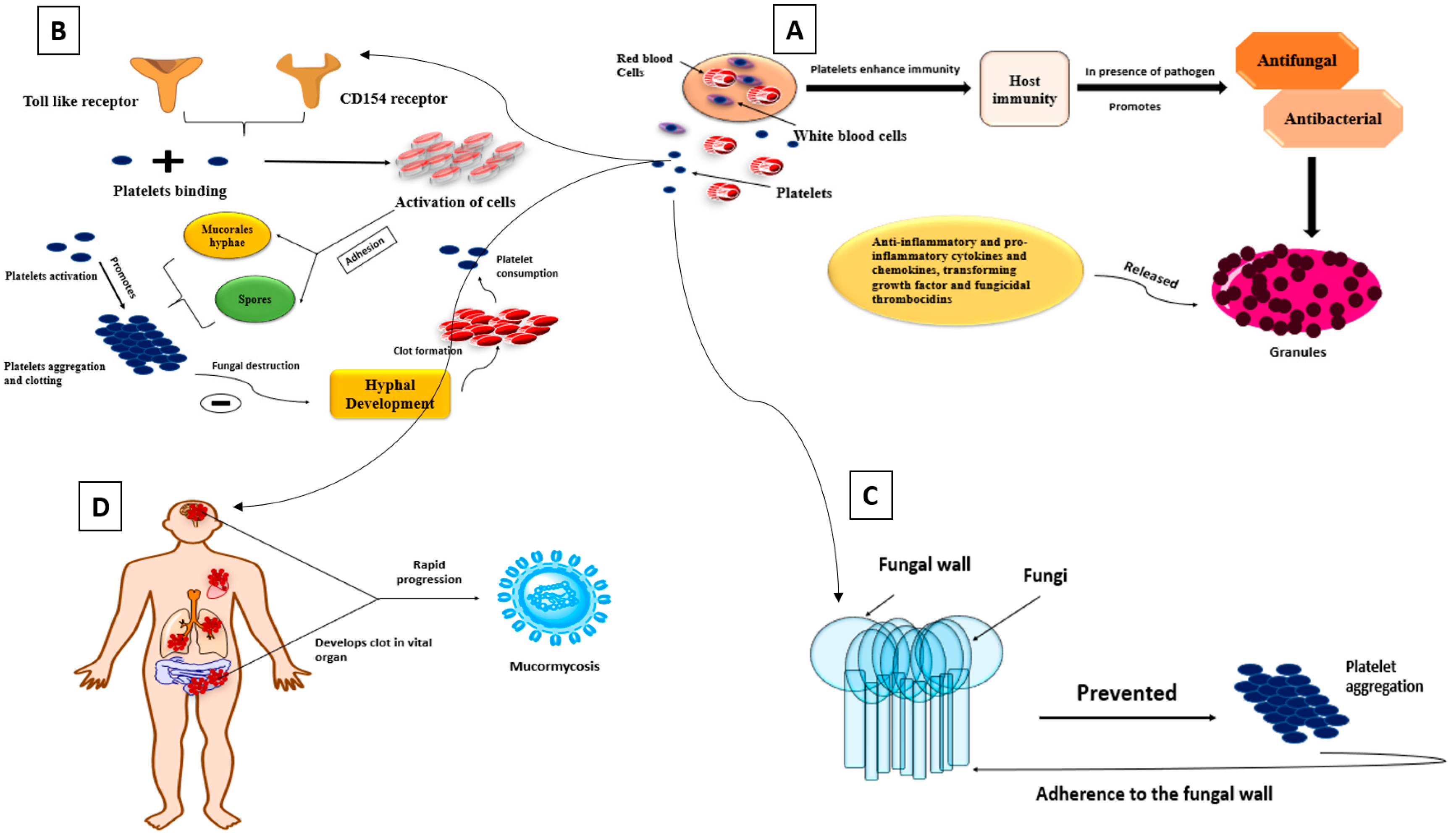
Platelets play an essential function in enhancing host immunity [46] and exhibit antifungal and antibacterial functions after exposure to an invading pathogen: Anti-inflammatory and pro-inflammatory cytokines and chemokines, including transforming growth factor and fungicidal thrombocidins, are released in granules [47]; platelet Toll-like receptors and CD154 are membrane-bound molecules that allow platelet binding and activation of different cells [48,49]; and adhesion to Mucorales hyphae and spores causes platelet activation, promotes platelet aggregation and clotting, as well as fungal destruction, by inhibiting hyphal development, supporting the development of clots and causing platelet consumption (Figure 3) [50,51]. Fungi may also be prevented from spreading hematogenously by platelet aggregation and adherence to the fungal wall [49]. Necrotic areas were discovered in tissues with no obvious fungal development, implying that thrombotic ischemia occurred due to systematic platelet activation [50]. Patients with COVID-19 who have the severe form of the disease frequently develop clots in vital organs, which can lead to further complications. As a result, the progression of mucormycosis is quite rapid.

4.3. Natural Killer Cells

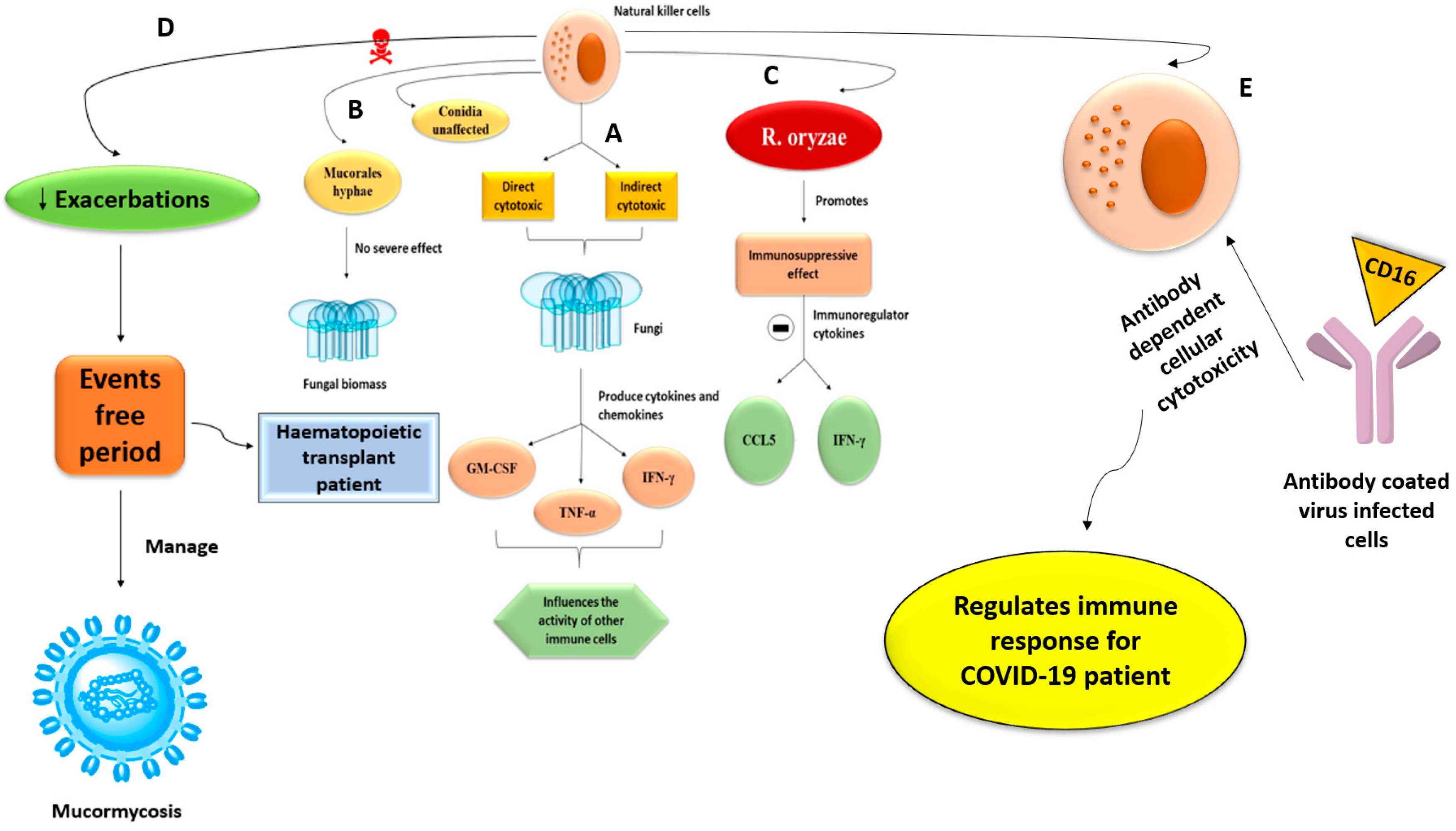
Natural killer cells are a type of innate immune cell that have both direct and indirect cytotoxic effects on the fungi. They also produce cytokines and chemokines such as GM-CSF, TNF-α, and IFN-γ, which influence the activity of other immune cells. Mucorales hyphae can be damaged by natural killer cells, but conidia are unaffected. Moreover, the damage done is inversely proportional to the amount of fungal biomass and has nothing to do with fungal infection. Conversely, in vitro investigations have shown R. oryzae to have an immunosuppressive effect, inhibiting the release of immunoregulatory chemokines RANTES (CCL5) and IFN-γ from natural killer cells [52]. Human natural killer cells are studied for their ability to minimize exacerbations and enhance event-free periods in hematopoietic transplant patients, and their therapeutic effect may also be helpful in managing and providing therapy for invasive mucormycosis (Figure 4) [53,54]. COVID-19 IgG immunity can be severely impaired by high natural killer cell numbers. Antibody-coated virus-infected cells interact with CD16 on natural killer cells, resulting in antibody-dependent cellular cytotoxicity [55]. The innate immune system controls immunological response and acts as a first line of defense against COVID-19.

4.4. Iron Uptake

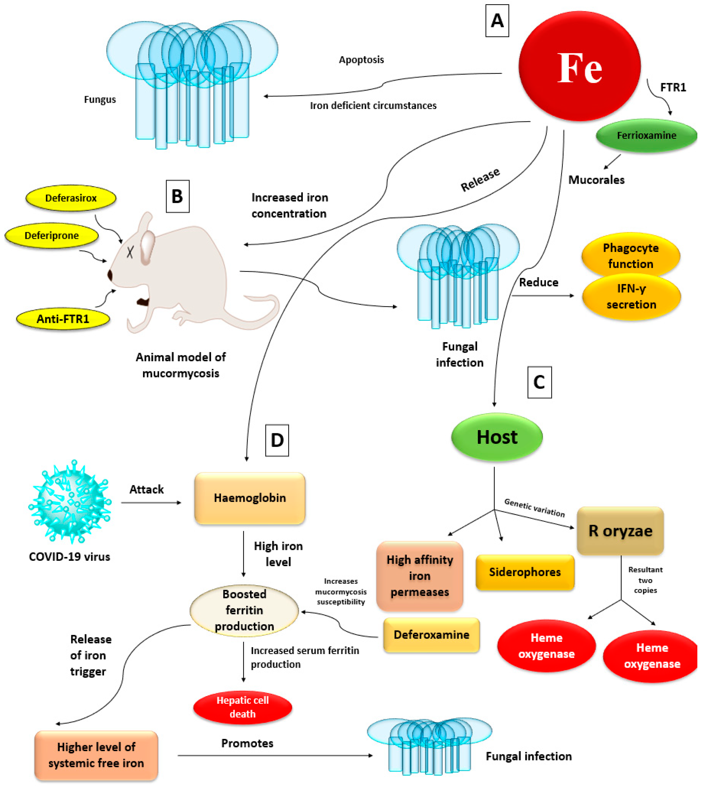
The fungus undergoes apoptosis in iron-deficient circumstances, supporting iron’s necessity for fungal cell growth [54]. In animal models of mucormycosis, increased iron concentrations also aid fungal development by reducing phagocyte function and lowering IFN-γ secretion [56]. Mucorales obtain iron from their hosts via two major mechanisms: high-affinity iron permeases or siderophores [57]. In addition, genetic investigation of R. oryzae revealed the existence of two copies of heme oxygenases, implying a third mechanism of iron acquisition from hemoglobin [58]. The COVID-19 virus may target haemoglobin, causing iron to be released from porphyrins and into the circulation, resulting in iron overload. Ferritin production is increased to compensate for the high iron level. Increased serum ferritin levels can promote hepatic cell death, causing ferritin to release iron, resulting in greater levels of systemic free iron [59]. As a result, it promotes the spread of fungal infection. Deferoxamine, an iron chelator used in those at risk of iron overload, increases mucormycosis susceptibility [60]. Mucorales use ferrioxamine (the iron-rich form of deferoxamine) as a xenosiderophore to collect iron [61]. Various researchers have demonstrated that iron chelation therapy with deferasirox or deferiprone prevents mucormycosis in mice with diabetic ketoacidosis and promotes longevity, whereas adjunctive deferasirox was both effective and tolerated in an open-label study of eight cases of mucormycosis [62,63]. Deferasirox also countered iron’s inhibitory effect on neutrophil chemotaxis [64]. However, recent clinical research in individuals who had mucormycosis that used supplementary deferasirox medication failed to establish a survival effect [65]. FTR1, a high-affinity iron permease that facilitates iron absorption, is upregulated in an iron-depleted state and silenced in iron-rich situations [66]. It has been proposed that FTR1 facilitates the intracellular transfer of iron from ferrioxamine or heme. An experimental investigation demonstrated that anti-FTR1 antibodies protected mice with diabetic ketoacidosis from infection, highlighting its potential as a therapy target (Figure 5) [67].

4.5. Interplay with the Endothelium

Mucorales bind to the surface of the endothelial cells by releasing proteins known as spore coat homologs [68], which are present only in Mucorales and interact with the host endothelium receptor GRP78, causing the fungus to endocytose [41,68]. GRP78, also known as heat shock protein, is found mainly in the endoplasmic reticulum and is presumed to become a specialized host cell receptor [60]. Spore coat homolog protein fungal surface expression and GRP78 endothelium surface expression increase after endothelial cells are confronted with acidosis, high glucose, and high iron levels, as in diabetic ketoacidosis high blood sugar. Thus COVID-19 patients with diabetes comorbidities can increase fungal endocytosis through the endothelium. In one study, GRP78 and spore coat homolog proteins were elevated by acidosis induced by β-hydroxybutyrate (a ketone body) but not affected by higher blood iron levels lactic acidosis. Furthermore, sodium bicarbonate recovered acidosis and protected β-hydroxybutyrate–treated mice from developing mucormycosis, indicating the importance of restoring acidosis as a therapeutic strategy in patients with mucormycosis and diabetic ketoacidosis. Anti-spore coat homolog protein and anti-GRP78 antibodies were used in another investigation to largely prevent R. oryzae endothelium invasion [43].

4.6. Voriconazole Exposure

Clinically relevant epidemiologic data reveal that voriconazole is an antifungal commonly used for prophylaxis in high-risk patients but is ineffective against Mucorales [69]. Voriconazole caused hypervirulent Rhizopus and Mucor strains in mice, increasing lung fungal loads and shortening lifespans [43]. The hypervirulent phenotype was lost when voriconazole treatment stopped, indicating an epigenetic rather than a genetic shift [70]. Voriconazole is currently recommended as first-line therapy for COVID-19 associated pulmonary aspergillosis (CAPA), which can lead to the fast spread of mucormycosis [71]. A recent investigation using rat models found that pre-exposure of fungal spores to voriconazole produces breakthrough infections by R. oryzae, which appear to be less responsive to subsequent antifungal therapy [72]. If voriconazole exposure does indeed select for more virulent strains, then we may be only beginning to understand the processes that contribute to Mucorales pathogenicity.

5. Mucormycosis Outbreak

An outbreak occurs when two or more people are infected by the same source or at the same place or time. The sources of outbreaks may be outdoors or may be in a healthcare setting, such as a hospital [73]. Most published works describe outbreaks of cutaneous mucormycosis, which has been tied to contaminated dressings and is less fatal than other forms of mucormycosis, with a medical literature review revealing 16% mortality versus 67% for rhinocerebral, 83% for pulmonary, and 100% for disseminated and gastrointestinal mucormycosis [74]. Hospital bedding has been identified as a vector for spreading R. delemar to vulnerable patients. In hospital epidemic investigations, DNA-based approaches to fungal species detection have confirmed epidemiological connections. Hospital bedding must be washed, wrapped, distributed, and stored in ways that minimize their exposure to environmental pollutants [75].

6. Effects of Black Fungus on COVID-19 Patients

The outbreak of mucormycosis is yet another unpleasant surprise brought on by the COVID-19 pandemic [76]. It can infect the sinuses and facial bones, infiltrate the brain, and lead to the loss of an eye. If left untreated, mucormycosis kills up to half of patients—and treatment is time-consuming and difficult. The standard treatment approach for severe COVID-19 is a high dose of steroids, antibiotics, and antivirals, which dampen the immune system and make the patient susceptible to infection by bacteria and fungus already present in the body or the environment [77]. Infection with mucormycosis is akin to opening Pandora’s box. Having been infected by SARS-CoV-2, the patient’s body is already ravaged, and infection by a virulent fungus often leads to death.
To save a patient’s life, doctors may surgically remove an organ or tissue from the body, causing emotional anguish for the patient and family members. Moreover, the standard treatment for black fungus is Amphotericin B, which is exorbitantly expensive, to the point that insolvency may arise from treatment [78].

7. Diabetes Patients Are Predisposed to Mucormycosis

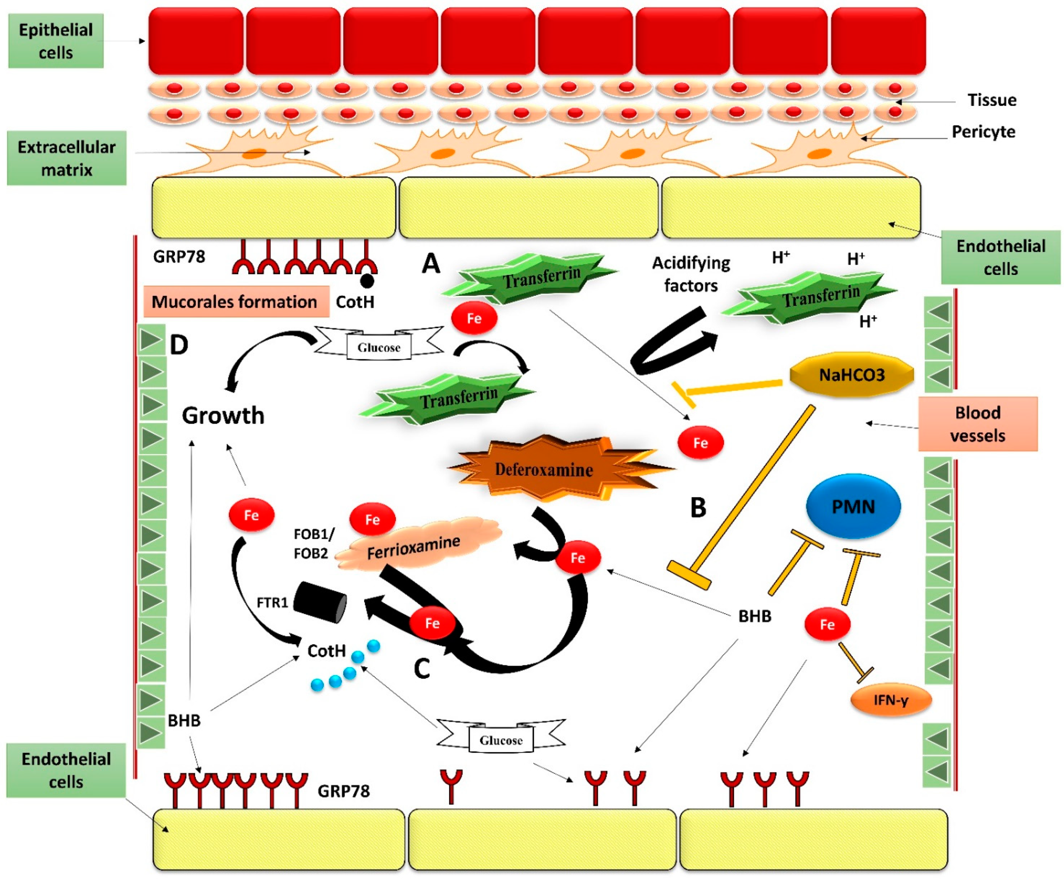
Diabetic ketoacidosis and deferoxamine-treated individuals are prone to mucormycosis in a unique way. Diabetic individuals have a high glucose level in their blood [79]. Excessive glycosylation of proteins like ferritin and transferrin might cause them to lose their iron affinity, which further induces hyperglycemia [80]. Furthermore, when there is an acidic state in the blood vessels owing to the build-up of ketone bodies (e.g., β-hydroxybutyrate), transferrin’s potential to chelate iron is severely impaired [81]. β-hydroxybutyrate, glucose, and iron promote fungal development (Figure 6) [56,82]. They also increase the expression of spore coating protein (CotH) and glucose regulator protein78 (GRP78), which leads to increased fungal invasion and consequent endothelial damage in-vitro [70,82]. β-hydroxybutyrate related acidosis appears to have a direct influence on CotH and GRP78 expression (an effect not observed with lactic acid) as well as an indirect effect on transferrin’s potential to chelate iron, as iron chelation coupled with sodium bicarbonate pH reversal substantially protects endothelial cells against Rhizopus-mediated invasion and damage [82]. Notably, when mice administered with β-hydroxybutyrate or diabetic ketoacidosis mice have shown lower blood pH, higher accessible serum iron, higher GRP78 expression in target organs (e.g., sinuses and lungs), they are more vulnerable to mucormycosis [56,82]. It is also important to highlight that optimum levels of β-hydroxybutyrate, iron, and glucose facilitate fungal growth while suppressing the host immune response via phagocyte-mediated destruction, IL-γ generation, and T-lymphocyte activation (Figure 6) [56,82,83,84,85]. Thus, the particular propensity of diabetic ketoacidosis patients to mucormycosis is explained by the idiosyncratic interactions of CotH and GRP78 proteins, as well as their increased expression under hyperglycemia and ketoacidosis conditions [58]. Therapeutic intervention with either anti-CotH or anti-GRP78 antibodies protects neutropenic and diabetic ketoacidosis mice against mucormycosis, highlighting the significance of CotH/ GRP78 protein interactions in the pathophysiology of mucormycosis [69,70,85].

8. Implement Control and Preventive Measures

Mucormycosis is an aggressively growing infection that follows infection by COVID-19. Its radiographic manifestations are not specific, but diagnosis can be made by microscopic examination of materials collected from necrotic lesions. Treatment requires multidisciplinary expertise, as the fungus enters through the eyes and nose and can even reach the brain [86]. It can be treated or controlled (1) medically, by using antifungal therapies; (2) surgically, by removing all necrotic lesions; or (3) by implementing adjunctive therapies that reverse the risk factors [87].

9. Medical Management of Mucormycosis

9.1. Management by Using Antifungal Drug Therapies

The lipid formulation of amphotericin-B (liposomal Am-B) is the first-line treatment for mucormycosis in COVID-19 patients. Injection of liposomal Am-B, with a starting dosage of 5–7.5 mg/kg/day, diluted in 500 mL of 5% dextrose over 4–5 h for 14–21 days, is commonly used in hospitalized adults and children [88]. Patients who are intolerant of or unresponsive to Am-B can be given alternative agents, such as an oral suspension of posaconazole, 400 mg two times a day or 200 mg four times a day. However, posaconazole alone cannot be recommended as a primary treatment in patients who have mucormycosis [89]. With progressive infection, monotherapy using lipid Am-B, increasing liposomal Am-B dosage (7.5–10 mg/kg per day), addition of posaconazole or an echinocandin, and shifting to posaconazole are viable strategies for treatment [90]. The evidence does not support combination therapy for first-line healing of mucormycosis, but if first-line therapy fails, a salvage therapy combining a polyene and an echinocandin can be used [89].
Liposomal Am-B is considered an efficient treatment for mucormycosis. Amphotericin B binds to ergosterol present in the fungal cell membrane, which causes pores and subsequent ion leakage, followed by the death of the fungal cells. Several studies have demonstrated the in vitro and in vivo binding of liposomes (both amphotericin B loaded liposomes and empty liposomes) to pathogenic fungal cell walls with gold and fluorescent-labeled liposomes [91,92,93]. As long as a liposome does not have Am-B, it binds to the fungus’s cell wall, but both the fungus cell and liposome stay intact. Liposomes containing amphotericin B, however, are capable of killing fungal cells, which implies that the binding causes the liposome to rupture and release amphotericin B. It then binds to ergosterol in the fungus cell membrane, exerting its fungicidal effect [91]. However, the exact mechanism of how amphotericin B crosses the fungal membrane from the liposome is still unclear. As ergosterol is the primary lipid component of the liposome, amphotericin B likely has a higher affinity for it than cholesterol [94]. As a bonus, liposomal Am-B formulations penetrate biofilms far better than conventional Am-B [95,96]. Despite the excellent pharmacokinetic and pharmacodynamic activity of liposomal Am-B, doctors are shifting towards surgery to save the life of patients because of the poor pharmacoeconomic status of liposomal Am-B. High cost and low availability in the market can be the only limitation of liposomal Am-B [90]. Liposomal Am-B will be called the standard gold drug for treating mucormycosis in COVID-19 patients if such limitations are resolved.
Isavuconazole is the first triazole drug approved by the U.S. Food and Drug Administration (FDA) to treat mucormycosis. It inhibits the CP450-dependent 14-lanosterol demethylase in the fungal cell membrane. As a result, cytotoxic sterols accumulate and reduce ergosterol production, which is essential for fungal cell membrane development. It inhibits fungal growth and replication, eventually leading to cell death [97].
The major drawback of this drug is its resistance like other azoles. Resistance occurs after repeated exposure to the drug. Azole resistance mechanisms include overexpression of ABC transporters (ATP binding cassettes), mutation of the gene encoding the target enzyme (ERG11), ERG3 gene mutation which impairs azole-mediated cell membrane disruption [98]. Therefore, it is a reasonable treatment option for mucormycosis patients with other refractory disorders and posaconazole intolerance [99].
Azoles primarily target ergosterol, which ensures membrane fluidity, permeability, and the proper functioning of membrane proteins [100]. It primarily works by inhibiting ergosterol biosynthesis via the fungal cell membrane’s CP450-dependent 14α-lanosterol demethylase, which is responsible for converting lanosterol to ergosterol. Consequently, the integrity of the fungal cell membrane gets altered, affecting its morphology and growth, eventually leading to cell death [101].

9.2. Surgical Management

The high cost and low availability of liposomal Am-B have prompted doctors to conduct surgeries to save the life of patients [90]. Surgical debridement to remove all necrotic lesions remains the hallmark of effective treatment of mucormycosis in COVID-19 patients. Extensive surgery should be conducted as early as possible, with an MRI or CT scan used preoperatively to determine the extent of the tissues in question and the involvement of tissue margins. Repeated surgical removal of necrotic lesions has shown improved outcomes. After successful treatment, the patient may undergo plastic surgery [90]. Surgical recommendations differ by site and by the severity of the condition. Table 1 shows the mode of action, advantages and disadvantages of various medications, as well as surgical management for mucormycosis infection healing.

10. Adjunctive Therapies

Along with antifungal agents and surgeries, adjunctive therapies involving reversal of immunosuppression, correction of metabolic deficits, and strategies for immune augmentation are beneficial for controlling mucormycosis [120]. Popular adjunctive therapies include hyperbaric oxygen, immunomodulation strategies, and iron chelation [89]. Hyperbaric oxygen, which is an effective treatment for diabetic patients who have rhinocerebral or severe cutaneous mucormycosis [121], increases the partial pressure of oxygen and improves neutrophilic function. High oxygen concentrations also improve wound healing by releasing enhanced tissue growth factors [122]. However, such therapy has not been subjected to detailed study and so is not regularly recommended. Accordingly, immune augmentation strategies such as granulocyte colony-stimulating factor (G-CSF) and interferon- γ have been implemented as adjunctive therapies to improve host response [123]. Iron chelators such as deferasirox and deferiprone have also been shown to prevent fungal growth and protect diabetic mice from developing mucormycosis. Case reports indicate that iron chelation therapy is a beneficial adjunctive therapy in diabetic patients, whereas a small double-blinded, placebo-controlled, multi-centered study of 20 patients who had a hematologic disorder showed adverse effects when adding deferasirox to liposomal Am-B. Although the study size was limited, the data did not support a role for deferasirox as an adjunctive therapy [124].

11. Preventive Measures

The Indian Council of Medical Research (ICMR) has released a set of general guidelines for mucormycosis prevention in COVID-19 patients [125]:
  • Good control of sugar level during COVID-19 with or without use of steroids
  • Rational use of steroids in the correct dose, with proper timing, and for a suitable duration
  • Judicious use of antibiotics/antifungals
  • Use of sterile or clean water as humidifiers during oxygen therapy
Additionally, modest preventive actions are indicated for post–COVID-19 recovery patients to avoid mucormycosis:
  • Maintaining personal hygiene by thoroughly bathing and scrubbing the body
  • Wearing face masks and face shields while visiting dirty or polluted environments
  • Wearing concealed shoes, long trousers, long-sleeved shirts, and gloves while handling soil, manure, moss, and the like (especially while gardening)
Because the literature lacks sufficient data on measures for preventing mucormycosis in COVID-19 patients, healthcare professionals follow such general environmental infection control measures to prevent mucormycosis in COVID-19 patients.
Apart from these strategies for managing and preventing mucormycosis in COVID-19 patients, the Ministry of Ayurveda, Unani, Siddha, and Homeopathy (Ayush) in India has suggested different preventive medicines to patients who have recovered from COVID-19 and to those who are on high doses of steroids and are diabetic [126]. The Ministry of Ayush has claimed that ayurvedic formulations can help control black fungus disease among COVID-19 patients, with Unani and homeopathic medicines useful in preventing and treating mucormycosis in COVID-19 patients [127].

12. Nanoparticle Formulation of the Drug/Nanoformulation of the Antifungal Drugs

Mucormycosis occurs worldwide, producing severe morbidity and mortality in COVID-19 and other immunocompromised patients. Many efficient antifungal medications are on the market, but their effectiveness is constrained by resistance and considerations of toxicity. Nanoparticles can overcome such limitations by reducing toxicity and increasing bioavailability. Use of nanotechnology for antifungal therapy began in the 1990s with the launch of a lipid formulation of amphotericin B (Am-B) [128]. Apart from the lipid formulation, alternative formulations such as Am-B colloidal dispersion and liposomal Am-B are available [129] and have been found to be safe and effective compared with conventional Am-B. Traditional Am-B is typically prepared as a colloidal suspension for parenteral delivery with sodium deoxycholate, but renal toxicity and severe infusion-related problems hamper its therapeutic efficacy [130,131]. Nanoformulations significantly reduce the toxic effect of the drug but are highly expensive and are available only in parenteral form, limiting their widespread use even apart from the deleterious effect of COVID-19 on personal finances. At 6000–8000 INR, a vial of liposomal Am-B is out of reach of ordinary people, so development of a cost-effective antifungal nanoformulation is an active area of research. Several nanosystems for oral, topical, vaginal, ocular, and pulmonary delivery of Am-B are currently under development and have shown promising in vitro results [132]. Disappointingly, however, only the nano form of Am-B has completed clinical trials. Research into delivery of antifungal medications should address the challenges to clinical translation of nanoparticle-based formulations so that diseases such as mucormycosis and COVID-19 can be combated [133].

13. Earlier Diagnose Mucormycosis to Overcome the Adverse Effect

Diagnosing mucormycosis appropriately and undergoing the necessary tests is fundamental to the success of the treatment. Patients with malignant hematological conditions whose amphotericin B-based therapy is delayed beyond the first five days of symptoms have double the 12-week mortality rate [132]. Early diagnosis is therefore crucial to ensuring effective treatment in people with mucormycosis [134,135]. Mucormycosis is currently diagnosed mostly through the culture of the organism from generally sterile body locations and/or histopathologic examination of the affected tissue [89,136]. Medium such as Sabouraud-dextrose agar can be used to isolate mucorales, and the fungal invasion can be investigated using methenamine-silver, eosin, and hematoxylin, calcofluor white stain or periodic acid-Schiff [137]. However, these techniques do not provide enough sensitivity, and end up leading to wrong diagnosis. Computed tomography (CT) can be used to detect pulmonary mucormycosis earlier in cancer patients. In the absence of airway-invasive characteristics, a reversed halo sign can aid in distinguishing the condition from invasive pulmonary aspergillosis in patients with a hematologic malignancy or neutropenia [138]. Radiological indicators of disease are frequently suggestive, rather than diagnostic. Recent development in molecular diagnostic technologies, such as the advent of polymerase chain reaction (PCR)-based tests, along with Matrix Assisted Laser Desorption/Ionization-Time of Flight Mass Spectrometry (MALDI-TOF MS), may assist in the earlier diagnosis of mucormycosis as well as the commencement of treatment [139,140].

14. Mucormycosis Diagnosis Limitations in Patients Infected with Microbial Infection

Given the limited treatment choices available, which usually include disfiguring and painful operations, early and accurate diagnosis is, in theory, the most important factor in improving the outcome of mucormycosis. Furthermore, approximately 4 to >90% of suspected mucormycosis cases are not verified until post-mortem investigation [141,142,143]. A combination of variables, the non-specific clinical appearance of mucormycosis, as well as the various limitations of currently available diagnostic techniques, make a definite diagnosis challenging. It is critical to isolate the fungus and identify it to the genus or species level for prognostic, epidemiological, and therapeutic objectives. [142,144,145]. The cultural isolation output ranges from 50–71%, with evidence that it has improved considerably over time [37,141]. Mucorales recovery from clinical microbiology specimens, on the other hand, is difficult. Mucorales hyphae may be difficult to see on wet mounts and require special chitin-binding stains to be seen using a fluorescence microscope, or they may be too few to see. Furthermore, vigorous homogenization or tissue grinding may obliterate the coenocytic hyphae during tissue processing [142,146]. Nonculture techniques, such as detecting biochemical or serological indicators, are currently unavailable to aid in diagnosing invasive mucormycosis. Invasive candidiasis is diagnosed using circulating mannan antigen and (1-3)-β-d-glucan, whereas invasive aspergillosis is diagnosed using galactomannan in bronchoalveolar fluid and serum.

15. Future Perspective

Mucormycosis infection is unpleasant, debilitating, and fatal. It leads to loss of organs and emotional trauma in patients, lowering quality of life, and treatment is prohibitively expensive. Amid the COVID-19 outbreak, prevention is of the utmost importance, as is ensuring the availability of an efficient, cost-effective treatment. Despite significant progress in understanding the microbiological aspects of this infection, the COVID-19 outbreak has underscored the need for more awareness, improved diagnostic tests, a focus on diabetes control, and prudent use of corticosteroids, with patients requiring immediate surgery and antifungal treatment. Further research into using modern tools and approaches to prevent and treat this infection is required, along with exploration of novel ways of delivering antifungal drugs that can increase their therapeutic efficacy.

16. Conclusions

Fungal infections subsequent to COVID-19 have been observed so extensively in many countries that doctors are beginning to design therapeutic strategies to counteract their effects. Fungal spores are everywhere, but human lungs are generally efficient at clearing them out. However, COVID-19 causes lung damage, diminishing the capacity to naturally eliminate spores in patients who are suffering a weakened immune response as a result of steroid treatment. People who have uncontrolled diabetes are more susceptible to black fungus infection, but the early signs of mucormycosis can be challenging to detect. Unlike some other fungal diseases, it cannot be detected through blood testing. Diagnosis requires a biopsy, examination of the sample, and in some cases a CT scan, all of which require specialized personnel and advanced technology—neither of which can be guaranteed in under resourced areas of India’s healthcare system.

Author Contributions

Conceptualization, R.K.S., A.M.A.I. and M.M.S.-B.; methodology, B.B. and R.W.; validation, A.B.; formal analysis, M.A. and Y.A.; writing—original draft preparation, R.K.S., A.S., A.A. and I.E.; writing—review and editing, R.K.S., B.B. and A.B.; supervision, R.K.S. All authors have read and agreed to the published version of the manuscript.

Funding

This research received no external funding.

Acknowledgments

The authors acknowledge the Deanship of Scientific Research, Jazan University, for funding Waed project W41-052.

Conflicts of Interest

The authors declare no conflict of interest.

References

  1. Bassetti, M.; Bouza, E. Invasive mould infections in the ICU setting: Complexities and solutions. J. Antimicrob. Chemother. 2017, 72, i39–i47. [Google Scholar] [CrossRef] [Green Version]
  2. Lin, E.; Moua, T.; Limper, A.H. Pulmonary mucormycosis: Clinical features and outcomes. Infection 2017, 45, 443–448. [Google Scholar] [CrossRef]
  3. Peng, M.; Meng, H.; Sun, Y.; Xiao, Y.; Zhang, H.; Lv, K.; Cai, B. Clinical features of pulmonary mucormycosis in patients with different immune status. J. Thorac. Dis. 2019, 11, 5042. [Google Scholar] [CrossRef]
  4. Perfect, J.R.; Mourad, A. Management of mucormycoses. In Antifungal Therapy; CRC Press: Boca Raton, FL, USA, 2019; pp. 357–362. [Google Scholar]
  5. Mohanty, A.; Gupta, P.; Varshney, S.; Kabi, A.; Angral, S. Breaking the mold: A brief review on the diagnostic and treatment approaches of mucormycosis. Int. J. Otorhinolaryngol. Head Neck Surg. 2021, 7, 1. [Google Scholar] [CrossRef]
  6. Chakrabarti, A. Mucormycosis in Asia. In Clinical Practice of Medical Mycology in Asia; Springer: New York, NY, USA, 2020; pp. 279–292. [Google Scholar]
  7. Juma, F.; Nagaraj, V.; Darwish, A. Placental Mucormycosis of an IVF-Induced Pregnancy in a Diabetic Patient. Bahrain Med. Bull. 2019, 41, 278–280. [Google Scholar]
  8. Shamanna, K.; Fathima, A.; Sowjanya, S. Rhino-Orbito-Cerebral Mucormycosis: Our Experience. Headache 2019, 15, 75. [Google Scholar]
  9. Iqbal, N.; Irfan, M.; Jabeen, K.; Kazmi, M.M.; Tariq, M.U. Chronic pulmonary mucormycosis: An emerging fungal infection in diabetes mellitus. J. Thorac. Dis. 2017, 9, E121. [Google Scholar] [CrossRef] [PubMed] [Green Version]
  10. Prakash, H.; Chakrabarti, A. Global epidemiology of mucormycosis. J. Fungi 2019, 5, 26. [Google Scholar] [CrossRef] [Green Version]
  11. John, T.M.; Jacob, C.N.; Kontoyiannis, D.P. When uncontrolled diabetes mellitus and severe COVID-19 converge: The perfect storm for mucormycosis. J. Fungi 2021, 7, 298. [Google Scholar] [CrossRef] [PubMed]
  12. Jeong, W.; Keighley, C.; Wolfe, R.; Lee, W.L.; Slavin, M.; Kong, D.; Chen, S.A. The epidemiology and clinical manifestations of mucormycosis: A systematic review and meta-analysis of case reports. Clin. Microbiol. Infect. 2019, 25, 26–34. [Google Scholar] [CrossRef] [Green Version]
  13. Ingram, C.W.; Sennesh, J.; Cooper, J.N.; Perfect, J.R. Disseminated Zygomycosis. Report of four cases and review. Rev. Infect. Dis. 1989, 11, 741–754. [Google Scholar] [CrossRef] [PubMed]
  14. Kubin, C.J.; McConville, T.H.; Dietz, D.; Zucker, J.; May, M.; Nelson, B.; Istorico, E.; Bartram, L.; Small-Saunders, J.; Sobieszczyk, M.E. In Characterization of Bacterial and Fungal Infections in Hospitalized Patients with Coronavirus Disease 2019 and Factors Associated with Health Care-Associated Infections. Open Forum Infect. Dis. 2021, 8, 1–10. [Google Scholar] [CrossRef]
  15. Hoang, K.; Abdo, T.; Reinersman, J.M.; Lu, R.; Higuita, N.I.A. A case of invasive pulmonary mucormycosis resulting from short courses of corticosteroids in a well-controlled diabetic patient. Med. Mycol. Case Rep. 2020, 29, 22–24. [Google Scholar]
  16. Nishanth, D.G.; Anitha, D.N.; Babu, D.N.A.; Malathi, D.L. Mucormycosis-A Review. Eur. J. Mol. Clin. Med. 2020, 7, 1786–1791. [Google Scholar]
  17. Kanwar, A.; Jordan, A.; Olewiler, S.; Wehberg, K.; Cortes, M.; Jackson, B.R. A fatal case of Rhizopus azygosporus pneumonia following COVID-19. J. Fungi 2021, 7, 174. [Google Scholar] [CrossRef]
  18. Baldin, C.; Soliman, S.S.; Jeon, H.H.; Alkhazraji, S.; Gebremariam, T.; Gu, Y.; Bruno, V.M.; Cornely, O.A.; Leather, H.L.; Sugrue, M.W. PCR-based approach targeting mucorales-specific gene family for diagnosis of mucormycosis. J. Clin. Microbiol. 2018, 56, e00746-18. [Google Scholar] [CrossRef] [PubMed] [Green Version]
  19. Timesnownews. Available online: https://www.timesnownews.com/india/article/28252-cases-of-mucormycosis-cases-in-the-country-maharashtra-gujarat-lead-the-tally-health-minister/767491 (accessed on 22 August 2021).
  20. Pal, M. Zygomycosis: A highly infectious emerging opportunistic fungal disease of public health concern. Open Access J. Mycol. Mycol. Sci. 2020, 3, 1–4. [Google Scholar] [CrossRef]
  21. Liberatore, C.; Farina, F.; Greco, R.; Giglio, F.; Clerici, D.; Oltolini, C.; Lupo Stanghellini, M.T.; Barzaghi, F.; Vezzulli, P.; Orsenigo, E. Breakthrough Invasive Fungal Infections in Allogeneic Hematopoietic Stem Cell Transplantation. J. Fungi 2021, 7, 347. [Google Scholar] [CrossRef]
  22. Choi, J.K.; Cho, S.Y.; Yoon, S.S.; Moon, J.H.; Kim, S.H.; Lee, J.H.; Kim, J.S.; Cheong, J.W.; Jang, J.H.; Seo, B.J. Epidemiology and risk factors for invasive fungal diseases among allogeneic hematopoietic stem cell transplant recipients in Korea: Results of “RISK” study. Biol. Blood Marrow Transplant. 2017, 23, 1773–1779. [Google Scholar] [CrossRef] [Green Version]
  23. Bavikar, P.; Mehta, V. Rhino-orbital-cerebral mucormycosis: A fatal complication of uncontrolled diabetes mellitus. Cureus 2017, 9, e1841. [Google Scholar] [CrossRef] [Green Version]
  24. Dayal, D.; Jain, P.; Kumar, R.; Bakshi, J.; Menon, P.; Das, A.; Singhi, S.; Singh, M. Clinical spectrum and outcome of invasive filamentous fungal infections in children with Type 1 diabetes: North Indian experience. Clin. Pediatr. Endocrinol. 2015, 24, 51–57. [Google Scholar] [CrossRef] [Green Version]
  25. Manesh, A.; Rupali, P.; Sullivan, M.O.; Mohanraj, P.; Rupa, V.; George, B.; Michael, J.S. Mucormycosis—A clinicoepidemiological review of cases over 10 years. Mycoses 2019, 62, 391–398. [Google Scholar] [CrossRef]
  26. Skiada, A.; Pavleas, I.; Drogari-Apiranthitou, M. Epidemiology and Diagnosis of Mucormycosis: An Update. J. Fungi 2020, 6, 265. [Google Scholar] [CrossRef]
  27. Das, A.; Oberoi, S.; Trehan, A.; Chakrabarti, A.; Bansal, D.; Saxena, A.K.; Sodhi, K.S.; Kakkar, N.; Srinivasan, R. Invasive fungal disease in pediatric acute leukemia in the nontransplant setting: 8 years’ experience from a tertiary care center in North India. J. Pediatr. Hematol. Oncol. 2018, 40, 462–467. [Google Scholar] [CrossRef] [PubMed]
  28. Phoompoung, P.; Henry, B.; Daher-Reyes, G.; Sibai, H.; Husain, S. Invasive Mold Infections in FLT3-Mutated Acute Myeloid Leukemia. Clin. Lymph. Myel. Leuk. 2021, 21, e477–e482. [Google Scholar] [CrossRef]
  29. Patel, M.H.; Patel, R.D.; Vanikar, A.V.; Kanodia, K.V.; Suthar, K.S.; Nigam, L.K.; Patel, H.V.; Patel, A.H.; Kute, V.B.; Trivedi, H.L. Invasive fungal infections in renal transplant patients: A single center study. Ren. Fail. 2017, 39, 294–298. [Google Scholar] [CrossRef] [PubMed] [Green Version]
  30. Godara, S.; Kute, V.; Goplani, K.; Gumber, M.; Gera, D.; Shah, P.; Vanikar, A.; Trivedi, H. Mucormycosis in renal transplant recipients: Predictors and outcome. Saudi J. Kidney Dis. Transplant. 2011, 22, 751. [Google Scholar]
  31. Prakash, H.; Ghosh, A.K.; Rudramurthy, S.M.; Singh, P.; Xess, I.; Savio, J.; Pamidimukkala, U.; Jillwin, J.; Varma, S.; Das, A. A prospective multicenter study on mucormycosis in India: Epidemiology, diagnosis, and treatment. Med. Mycol. 2019, 57, 395–402. [Google Scholar] [CrossRef] [PubMed]
  32. Patel, A.; Kaur, H.; Xess, I.; Michael, J.; Savio, J.; Rudramurthy, S.; Singh, R.; Shastri, P.; Umabala, P.; Sardana, R. A multicentre observational study on the epidemiology, risk factors, management and outcomes of mucormycosis in India. Clin. Microbiol. Infect. 2020, 26, 944.e9–944.e15. [Google Scholar] [CrossRef] [PubMed]
  33. Pandey, M.; Singh, G.; Agarwal, R.; Dabas, Y.; Jyotsna, V.P.; Kumar, R.; Xess, I. Emerging Rhizopus microsporus infections in India. J. Clin. Microbiol. 2018, 56, e00433-18. [Google Scholar] [CrossRef] [Green Version]
  34. Chakrabarti, A.; Kaur, H.; Savio, J.; Rudramurthy, S.M.; Patel, A.; Shastri, P.; Pamidimukkala, U.; Karthik, R.; Bhattacharya, S.; Kindo, A.J. Epidemiology and clinical outcomes of invasive mould infections in Indian intensive care units (FISF study). J. Crit. Care 2019, 51, 64–70. [Google Scholar] [CrossRef]
  35. Balwan, W.K. Epidemiology of Mucormycosis in India: A Notifiable Disease. Saudi J. Pathol. Microbiol. 2021, 6, 187–191. [Google Scholar]
  36. Antinori, S.; Nebuloni, M.; Magni, C.; Fasan, M.; Adorni, F.; Viola, A.; Corbellino, M.; Galli, M.; Vago, G.; Parravicini, C. Trends in the postmortem diagnosis of opportunistic invasive fungal infections in patients with AIDS: A retrospective study of 1,630 autopsies performed between 1984 and 2002. Am. J. Clin. Pathol. 2009, 132, 221–227. [Google Scholar] [CrossRef] [PubMed]
  37. Roden, M.M.; Zaoutis, T.E.; Buchanan, W.L.; Knudsen, T.A.; Sarkisova, T.A.; Schaufele, R.L.; Sein, M.; Sein, T.; Chiou, C.C.; Chu, J.H. Epidemiology and outcome of zygomycosis: A review of 929 reported cases. Clin. Infect. Dis. 2005, 41, 634–653. [Google Scholar] [CrossRef] [PubMed] [Green Version]
  38. Skiada, A.; Lass-Floerl, C.; Klimko, N.; Ibrahim, A.; Roilides, E.; Petrikkos, G. Challenges in the diagnosis and treatment of mucormycosis. Med. Mycol. 2018, 56 (Suppl. 1), S93–S101. [Google Scholar] [CrossRef] [PubMed] [Green Version]
  39. Spellberg, B. Mucormycosis pathogenesis: Beyond rhizopus. Virulence 2017, 8, 1481–1482. [Google Scholar] [CrossRef] [PubMed] [Green Version]
  40. Ibrahim, A.S.; Voelz, K. The mucormycete–host interface. Curr. Opin. Microbiol. 2017, 40, 40–45. [Google Scholar] [CrossRef] [PubMed]
  41. Chamilos, G.; Lewis, R.; Lamaris, G.; Walsh, T.; Kontoyiannis, D. Zygomycetes hyphae trigger an early, robust proinflammatory response in human polymorphonuclear neutrophils through toll-like receptor 2 induction but display relative resistance to oxidative damage. Antimcrob. Agents Chemother. 2008, 52, 722–724. [Google Scholar] [CrossRef] [Green Version]
  42. Petrikkos, G.; Tsioutis, C. Recent advances in the pathogenesis of mucormycoses. Clin. Ther. 2018, 40, 894–902. [Google Scholar] [CrossRef] [Green Version]
  43. Gebremariam, T.; Lin, L.; Liu, M.; Kontoyiannis, D.P.; French, S.; Edwards, J.E.; Filler, S.G.; Ibrahim, A.S. Bicarbonate correction of ketoacidosis alters host-pathogen interactions and alleviates mucormycosis. J. Clin. Investig. 2016, 126, 2280–2294. [Google Scholar] [CrossRef] [PubMed] [Green Version]
  44. Roilides, E.; Kontoyiannis, D.P.; Walsh, T.J. Host defenses against zygomycetes. Clin. Infect. Dis. 2012, 54, S61–S66. [Google Scholar] [CrossRef] [PubMed] [Green Version]
  45. Skiada, A.; Lanternier, F.; Groll, A.H.; Pagano, L.; Zimmerli, S.; Herbrecht, R.; Lortholary, O.; Petrikkos, G.L. Diagnosis and treatment of mucormycosis in patients with hematological malignancies: Guidelines from the 3rd European Conference on Infections in Leukemia(ECIL 3). Haematologica 2013, 98, 492. [Google Scholar] [CrossRef] [Green Version]
  46. Netea, M.G.; Joosten, L.A.; Van Der Meer, J.W.; Kullberg, B.J.; Van De Veerdonk, F.L. Immune defence against Candida fungal infections. Nat. Rev. Immunol. 2015, 15, 630–642. [Google Scholar] [CrossRef]
  47. Putri, I.H.; Tunjungputri, R.N.; De Groot, P.G.; van der Ven, A.J.; de Mast, Q. Thrombocytopenia and Platelet Dysfunction in Acute Tropical Infectious Diseases, Seminars in Thrombosis and Hemostasis; Thieme Medical Publishers: New York, NY, USA, 2018; pp. 683–690. [Google Scholar]
  48. Bongiovanni, D.; Santamaria, G.; Klug, M.; Santovito, D.; Felicetta, A.; Hristov, M.; von Scheidt, M.; Aslani, M.; Cibella, J.; Weber, C. Transcriptome analysis of reticulated platelets reveals a prothrombotic profile. Thromb. Haemost. 2019, 119, 1795–1806. [Google Scholar] [CrossRef]
  49. Schulze, B.; Rambach, G.; Schwartze, V.U.; Voigt, K.; Schubert, K.; Speth, C.; Jacobsen, I.D. Ketoacidosis alone does not predispose to mucormycosis by Lichtheimia in a murine pulmonary infection model. Virulence 2017, 8, 1657–1667. [Google Scholar] [CrossRef] [Green Version]
  50. Perkhofer, S.; Kainzner, B.; Kehrel, B.E.; Dierich, M.P.; Nussbaumer, W.; Lass-Flörl, C. Potential antifungal effects of human platelets against zygomycetes in vitro. J. Infect. Dis. 2009, 200, 1176–1179. [Google Scholar] [CrossRef] [PubMed]
  51. Gill, S.; Olson, J.A.; Negrin, R.S. Natural killer cells in allogeneic transplantation: Effect on engraftment, graft-versus-tumor, and graft-versus-host responses. Biol. Blood Marrow Transplant. 2009, 15, 765–776. [Google Scholar] [CrossRef] [Green Version]
  52. Kuldanek, S.; Pasko, B.; DomBourian, M.; Annen, K. Cellular Therapy in Pediatric Hematologic Malignancies. Clin. Lab. Med. 2021, 41, 121–132. [Google Scholar] [CrossRef]
  53. Bachiller, M.; Battram, A.M.; Perez-Amill, L.; Martín-Antonio, B. Natural Killer Cells in Immunotherapy: Are We Nearly There? Cancers 2020, 12, 3139. [Google Scholar] [CrossRef] [PubMed]
  54. Lax, C.; Pérez-Arques, C.; Navarro-Mendoza, M.I.; Cánovas-Márquez, J.T.; Tahiri, G.; Pérez-Ruiz, J.A.; Osorio-Concepción, M.; Murcia-Flores, L.; Navarro, E.; Garre, V. Genes, pathways, and mechanisms involved in the virulence of mucorales. Genes 2020, 11, 317. [Google Scholar] [CrossRef] [PubMed] [Green Version]
  55. Jegaskanda, S.; Vanderven, H.A.; Tan, H.-X.; Alcantara, S.; Wragg, K.M.; Parsons, M.S.; Chung, A.W.; Juno, J.A.; Kent, S.J. Influenza virus infection enhances antibody-mediated NK cell functions via type I interferon-dependent pathways. J. Virol. 2019, 93, e02090-18. [Google Scholar] [CrossRef] [PubMed] [Green Version]
  56. Guo, D.; Jaber, B.; Lee, S.; Perianayagam, M.; King, A.; Pereira, B.; Balakrishnan, V. Impact of iron dextran on polymorphonuclear cell function among hemodialysis patients. Clin. Nephrol. 2002, 58, 134–142. [Google Scholar] [CrossRef] [PubMed]
  57. Ma, L.J.; Ibrahim, A.S.; Skory, C.; Grabherr, M.G.; Burger, G.; Butler, M.; Elias, M.; Idnurm, A.; Lang, B.F.; Sone, T. Genomic analysis of the basal lineage fungus Rhizopus oryzae reveals a whole-genome duplication. PLoS Genet. 2009, 5, e1000549. [Google Scholar] [CrossRef]
  58. Roilides, E.; Antachopoulos, C.; Simitsopoulou, M. Pathogenesis and host defence against Mucorales: The role of cytokines and interaction with antifungal drugs. Mycoses 2014, 57, 40–47. [Google Scholar] [CrossRef]
  59. Drakesmith, H.; Prentice, A. Viral infection and iron metabolism. Nat. Rev. Microbiol. 2008, 6, 541–552. [Google Scholar]
  60. Liu, M.; Lin, L.; Gebremariam, T.; Luo, G.; Skory, C.D.; French, S.W.; Chou, T.F.; Edwards, J.E., Jr.; Ibrahim, A.S. Fob1 and Fob2 proteins are virulence determinants of Rhizopus oryzae via facilitating iron uptake from ferrioxamine. PLoS Pathog. 2015, 11, e1004842. [Google Scholar] [CrossRef] [Green Version]
  61. Ibrahim, A.S.; Edwards, J.E., Jr.; Fu, Y.; Spellberg, B. Deferiprone iron chelation as a novel therapy for experimental mucormycosis. J. Antimicrob. Chemother. 2006, 58, 1070–1073. [Google Scholar] [CrossRef] [PubMed] [Green Version]
  62. Ibrahim, A.S.; Gebermariam, T.; Fu, Y.; Lin, L.; Husseiny, M.I.; French, S.W.; Schwartz, J.; Skory, C.D.; Edwards, J.E.; Spellberg, B.J. The iron chelator deferasirox protects mice from mucormycosis through iron starvation. J. Clin. Investig. 2007, 117, 2649–2657. [Google Scholar] [CrossRef] [PubMed] [Green Version]
  63. Spellberg, B.; Andes, D.; Perez, M.; Anglim, A.; Bonilla, H.; Mathisen, G.E.; Walsh, T.J.; Ibrahim, A.S. Safety and outcomes of open-label deferasirox iron chelation therapy for mucormycosis. J. Antimicrob. Chemother. 2009, 53, 3122–3125. [Google Scholar] [CrossRef] [Green Version]
  64. Spellberg, B.; Ibrahim, A.S.; Chin-Hong, P.V.; Kontoyiannis, D.P.; Morris, M.I.; Perfect, J.R.; Fredricks, D.; Brass, E.P. The Deferasirox–AmBisome Therapy for Mucormycosis (DEFEAT Mucor) study: A randomized, double-blinded, placebo-controlled trial. J. Antimicrob. Chemother. 2012, 67, 715–722. [Google Scholar] [CrossRef] [Green Version]
  65. Fu, Y.; Lee, H.; Collins, M.; Tsai, H.F.; Spellberg, B.; Edwards, J.E., Jr.; Kwon-Chung, K.J.; Ibrahim, A.S. Cloning and functional characterization of the Rhizopus oryzae high affinity iron permease (rFTR1) gene. FEMS Microbiol. Lett. 2004, 235, 169–176. [Google Scholar] [CrossRef]
  66. Ibrahim, A.S.; Gebremariam, T.; Lin, L.; Luo, G.; Husseiny, M.I.; Skory, C.D.; Fu, Y.; French, S.W.; Edwards, J.; John, E.; et al. The high affinity iron permease is a key virulence factor required for Rhizopus oryzae pathogenesis. Mol. Microbiol. 2010, 77, 587–604. [Google Scholar] [CrossRef] [Green Version]
  67. Gebremariam, T.; Liu, M.; Luo, G.; Bruno, V.; Phan, Q.T.; Waring, A.J.; Edwards, J.E.; Filler, S.G.; Yeaman, M.R.; Ibrahim, A.S. CotH3 mediates fungal invasion of host cells during mucormycosis. J. Clin. Investig. 2014, 124, 237–250. [Google Scholar] [CrossRef] [Green Version]
  68. Liu, M.; Spellberg, B.; Phan, Q.T.; Fu, Y.; Fu, Y.; Lee, A.S.; Edwards, J.E.; Filler, S.G.; Ibrahim, A.S. The endothelial cell receptor GRP78 is required for mucormycosis pathogenesis in diabetic mice. J. Clin. Investig. 2010, 120, 1914–1924. [Google Scholar] [CrossRef] [Green Version]
  69. Pongas, G.; Lewis, R.; Samonis, G.; Kontoyiannis, D. Voriconazole-associated zygomycosis: A significant consequence of evolving antifungal prophylaxis and immunosuppression practices? Clin. Microbiol. Infect. 2009, 15, 93–97. [Google Scholar] [CrossRef] [Green Version]
  70. Lamaris, G.A.; Ben-Ami, R.; Lewis, R.E.; Chamilos, G.; Samonis, G.; Kontoyiannis, D.P. Increased virulence of Zygomycetes organisms following exposure to Voriconazole: A study involving fly and murine models of zygomycosis. J. Infect. Dis. 2009, 199, 1399–1406. [Google Scholar] [CrossRef] [PubMed]
  71. Koehler, P.; Bassetti, M.; Chakrabarti, A.; Chen, S.C.A.; Colombo, A.L.; Hoenigl, M.; Klimko, N.; Lass-Flörl, C.; Oladele, R.O.; Vinh, D.C.; et al. Defining and managing COVID-19-associated pulmonary aspergillosis: The 2020 ECMM/ISHAM consensus criteria for research and clinical guidance. Lancet Infect. Dis. 2021, 21, e149–e162. [Google Scholar] [CrossRef]
  72. Lewis, R.E.; Liao, G.; Wang, W.; Prince, R.A.; Kontoyiannis, D.P. Voriconazole pre-exposure selects for breakthrough mucormycosis in a mixed model of Aspergillus fumigatus-Rhizopus oryzae pulmonary infection. Virulence 2011, 2, 348–355. [Google Scholar] [CrossRef] [PubMed] [Green Version]
  73. Antoniadou, A. Outbreaks of zygomycosis in hospitals Definition of a Hospital Outbreak. Clin. Microbiol. Infect. 2009, 15, 55–59. [Google Scholar] [CrossRef] [Green Version]
  74. Kato, H.; Foster, C.M.; Karri, K. Incidence, predisposing conditions and outcomes of cutaneous mucormycosis: A national database study. Mycoses 2021, 64, 569–572. [Google Scholar] [CrossRef]
  75. Bonifaz, A.; Vázquez-González, D.; Tirado-Sánchez, A.; Ponce-Olivera, R.M. Cutaneous zygomycosis. Clin. Dermatol. 2012, 30, 413–419. [Google Scholar] [CrossRef] [PubMed]
  76. Khan, A.; Ema, N.T.; Rakhi, N.N.; Saha, O.; Rahaman, M.M. Parallel Outbreaks of Deadly Pathogens(SARS-CoV-2, H5N8, EVD, Black fungi) around the World in 2021: Priorities for Achieving Control with Socio-Economic and Public Health Impact. COVID 2021, 1, 203–217. [Google Scholar] [CrossRef]
  77. Rocha, I.C.N.; Hasan, M.M.; Goyal, S.; Patel, T.; Jain, S.; Ghosh, A.; Denise, T.; Cedeño, T.D.D. COVID-19 and mucormycosis syndemic: Double health threat to a collapsing healthcare system in India. Trop. Med. Int. Health 2021, 26, 1016–1018. [Google Scholar] [CrossRef]
  78. Selarka, L.; Sharma, S.; Saini, D.; Sarma, S.; Batra, A.; Waghmare, V.T.; Dileep, P.; Patel, S.; Shah, M.; Parikh, T.; et al. Mucormycosis and COVID-19: An epidemic within a pandemic in India. Mycoses 2021, in press. [CrossRef]
  79. Ibrahim, A.; Edwards, J.; Filler, S. Zygomycosis. In Clinical Mycology; Dismukes, W.E., Pappas, P.G., Sobel, J.D., Eds.; Oxford University Press: New York, NY, USA, 2003; pp. 241–251. [Google Scholar]
  80. Ribes, J.A.; Vanover-Sams, C.L.; Baker, D.J. Zygomycetes in human disease. Clin. Microbiol. Rev. 2020, 13, 236–301. [Google Scholar] [CrossRef] [PubMed]
  81. Shinde, Y.B.; Kore, S. A Review on Mucormycosis with recent pharmacological treatment. J. Drug Deliv. Ther. 2021, 11, 145–149. [Google Scholar] [CrossRef]
  82. Alqarihi, A.; Gebremariam, T.; Gu, Y.; Swidergall, M.; Alkhazraji, S.; Soliman, S.S.; Bruno, V.M.; Edwards, J.E., Jr.; Filler, S.G.; Uppuluri, P. GRP78 and integrins play different roles in host cell invasion during mucormycosis. mBio 2020, 11, e01087-20. [Google Scholar] [CrossRef] [PubMed]
  83. Omara, F.; Blakley, B.; Huang, H.S. Effect of iron status on endotoxin-induced mortality, phagocytosis and interleukin-1 alpha and tumor necrosis factor-alpha production. Vet. Hum. Toxicol. 1994, 36, 423–428. [Google Scholar]
  84. Rauthan, P.; Sharma, D.C. Mucormycosis: Pathogenesis, Diagnosis, and Management. Asian J. Pharm. Res. Dev. 2021, 9, 144–153. [Google Scholar]
  85. Baldin, C.; Ibrahim, A.S. Molecular mechanisms of mucormycosis-the bitter and the sweet. PLoS Pathog. 2017, 13, e1006408. [Google Scholar] [CrossRef] [Green Version]
  86. Deigendesch, N.; Nunez, J.C.; Stenzel, W. Parasitic and fungal infections. Handb. Clin. Neurol. 2018, 145, 245–262. [Google Scholar]
  87. Bonifaz, A.; Tirado-Sánchez, A.; Hernández-Medel, M.L.; Araiza, J.; Kassack, J.J.; del Angel-Arenas, T.; Moisés-Hernández, J.F.; Paredes-Farrera, F.; Gómez-Apo, E.; Treviño-Rangel, R.D.J. Mucormycosis at a tertiary-care center in Mexico. A 35-year retrospective study of 214 cases. Mycoses 2021, 64, 372–380. [Google Scholar] [CrossRef] [PubMed]
  88. Khan, S.; Waqar Elahi, M.; Ullah, W.; Abdullah, H.M.A.; Ahmad, E.; Al Mohajer, M.; Majeed, A. Invasive mucormycosis induced pneumopericardium: A rare cause of pneumopericardium in an immunocompromised patient. Case Rep. Infect. Dis. 2017, 2017, 1424618. [Google Scholar] [CrossRef] [PubMed]
  89. Cornely, O.A.; Alastruey-Izquierdo, A.; Arenz, D.; Chen, S.C.; Dannaoui, E.; Hochhegger, B.; Hoenigl, M.; Jensen, H.E.; Lagrou, K.; Lewis, R.E. Global guideline for the diagnosis and management of mucormycosis: An initiative of the European Confederation of Medical Mycology in cooperation with the Mycoses Study Group Education and Research Consortium. Lancet Infect. Dis. 2019, 19, e405–e421. [Google Scholar] [CrossRef]
  90. Sipsas, N.V.; Gamaletsou, M.N.; Anastasopoulou, A.; Kontoyiannis, D.P. Therapy of mucormycosis. J. Fungi 2018, 4, 90. [Google Scholar] [CrossRef] [Green Version]
  91. Adler-Moore, J. AmBisome targeting to fungal infections. Bone Marrow Transplant. 1994, 14, S3–S7. [Google Scholar]
  92. Lestner, J.M.; Howard, S.J.; Goodwin, J.; Gregson, L.; Majithiya, J.; Walsh, T.J.; Jensen, G.M.; Hope, W.W. Pharmacokinetics and pharmacodynamics of amphotericin B deoxycholate, liposomal amphotericin B, and amphotericin B lipid complex in an in vitro model of invasive pulmonary aspergillosis. Antimicrob. Agents Chemother. 2010, 54, 3432–3441. [Google Scholar] [CrossRef] [Green Version]
  93. Takemoto, K.; Yamamoto, Y.; Ueda, Y. Evaluation of antifungal pharmacodynamic characteristics of AmBisome against Candida albicans. Microbiol. Immunol. 2006, 50, 579–586. [Google Scholar] [CrossRef]
  94. Readio, J.D.; Bittman, R. Equilibrium binding of amphotericin B and its methyl ester and borate complex to sterols. Biochim. Biophys. Acta 1982, 685, 219–224. [Google Scholar] [CrossRef]
  95. Fujimoto, K.; Takemoto, K. Efficacy of liposomal amphotericin B against four species of Candida biofilms in an experimental mouse model of intravascular catheter infection. J. Infect. Chemother. 2018, 24, 958–964. [Google Scholar] [CrossRef]
  96. Kawai, A.; Yamagishi, Y.; Mikamo, H. In vitro efficacy of liposomal amphotericin B, micafungin and fluconazole against non-albicans Candida species biofilms. J. Infect. Chemother. 2015, 21, 647–653. [Google Scholar] [CrossRef] [PubMed]
  97. Natesan, S.K.; Chandrasekar, P.H. Isavuconazole for the treatment of invasive aspergillosis and mucormycosis: Current evidence, safety, efficacy, and clinical recommendations. Infect. Drug Resist. 2016, 9, 291. [Google Scholar] [CrossRef] [PubMed] [Green Version]
  98. Pfaller, M.A. Antifungal drug resistance: Mechanisms, epidemiology, and consequences for treatment. Am. J. Med. 2012, 125, S3–S13. [Google Scholar] [CrossRef] [PubMed]
  99. Ellsworth, M.; Ostrosky-Zeichner, L. Isavuconazole: Mechanism of action, clinical efficacy, and resistance. J. Fungi 2020, 6, 324. [Google Scholar] [CrossRef]
  100. Song, J.; Zhai, P.; Zhang, Y.; Zhang, C.; Sang, H.; Han, G.; Keller, N.P.; Lu, L. The Aspergillus fumigatus damage resistance protein family coordinately regulates ergosterol biosynthesis and azole susceptibility. mBio 2016, 7, e01919-15. [Google Scholar] [CrossRef] [Green Version]
  101. Scorzoni, L.; de Paula e Silva, A.C.; Marcos, C.M.; Assato, P.A.; de Melo, W.C.; de Oliveira, H.C.; Costa-Orlandi, C.B.; Mendes-Giannini, M.J. Fusco-Almeida AM. Antifungal therapy: New advances in the understanding and treatment of mycosis. Front. Microbiol. 2017, 8, 1–23. [Google Scholar] [CrossRef] [PubMed] [Green Version]
  102. Tissot, F.; Agrawal, S.; Pagano, L.; Petrikkos, G.; Groll, A.H.; Skiada, A.; Lass-Flörl, C.; Calandra, T.; Viscoli, C.; Herbrecht, R. ECIL-6 guidelines for the treatment of invasive candidiasis, aspergillosis and mucormycosis in leukemia and hematopoietic stem cell transplant patients. Haematologica 2017, 102, 433. [Google Scholar] [CrossRef] [Green Version]
  103. Shoham, S.; Magill, S.S.; Merz, W.G.; Gonzalez, C.; Seibel, N.; Buchanan, W.L.; Knudsen, T.A.; Sarkisova, T.A.; Walsh, T.J. Primary treatment of zygomycosis with liposomal amphotericin B: Analysis of 28 cases. Med. Mycol. 2010, 48, 511–517. [Google Scholar] [CrossRef] [PubMed] [Green Version]
  104. Rodríguez, M.M.; Serena, C.; Mariné, M.; Pastor, F.J.; Guarro, J. Posaconazole combined with amphotericin B, an effective therapy for a murine disseminated infection caused by Rhizopus oryzae. Antimicrob. Agents Chemother. 2008, 52, 3786–3788. [Google Scholar] [CrossRef] [Green Version]
  105. Pitman, S.K.; Drew, R.H.; Perfect, J.R. Addressing current medical needs in invasive fungal infection prevention and treatment with new antifungal agents, strategies and formulations. Expert Opin. Emerg. Drugs 2011, 16, 559–586. [Google Scholar] [CrossRef]
  106. Groll, A.H.; Desai, A.; Han, D.; Howieson, C.; Kato, K.; Akhtar, S.; Kowalski, D.; Lademacher, C.; Lewis, W.; Pearlman, H. Pharmacokinetic assessment of drug-drug interactions of isavuconazole with the immunosuppressants cyclosporine, mycophenolic acid, prednisolone, sirolimus, and tacrolimus in healthy adults. Clin. Pharmacol. Drug Dev. 2017, 6, 76–85. [Google Scholar] [CrossRef] [PubMed] [Green Version]
  107. Ananda-Rajah, M.R.; Kontoyiannis, D. Isavuconazole: A new extended spectrum triazole for invasive mold diseases. Future Microbiol. 2015, 10, 693–708. [Google Scholar] [CrossRef] [PubMed] [Green Version]
  108. Miceli, M.H.; Kauffman, C.A. Isavuconazole: A new broad-spectrum triazole antifungal agent. Clin. Infect. Dis. 2015, 61, 1558–1565. [Google Scholar] [CrossRef] [Green Version]
  109. Trang, T.P.; Hanretty, A.M.; Langelier, C.; Yang, K. Use of isavuconazole in a patient with voriconazole-induced QT c prolongation. Transpl. Infect. Dis. 2017, 19, e12712. [Google Scholar] [CrossRef] [PubMed]
  110. Seyedmousavi, S.; Rafati, H.; Ilkit, M.; Tolooe, A.; Hedayati, M.T.; Verweij, P. Systemic antifungal agents: Current status and projected future developments. In Human Fungal Pathogen Identification; Springer: New York, NY, USA, 2017; pp. 107–139. [Google Scholar]
  111. Miyazaki, M.; Horii, T.; Hata, K.; Watanabe, N.; Nakamoto, K.; Tanaka, K.; Shirotori, S.; Murai, N.; Inoue, S.; Matsukura, M. In vitro activity of E1210, a novel antifungal, against clinically important yeasts and molds. Antimicrob. Agents Chemother. 2011, 55, 4652–4658. [Google Scholar] [CrossRef] [Green Version]
  112. Gebremariam, T.; Alkhazraji, S.; Alqarihi, A.; Wiederhold, N.P.; Shaw, K.J.; Patterson, T.F.; Filler, S.; Ibrahim, A. In APX001A Protects Immunosuppressed Mice from Rhizopus delemar Infection. Open Forum Infect. Dis. 2017, 4 (Suppl. 1), S475. [Google Scholar] [CrossRef] [Green Version]
  113. Reed, C.; Bryant, R.; Ibrahim, A.S.; Edwards, J., Jr.; Filler, S.G.; Goldberg, R.; Spellberg, B. Combination polyene-caspofungin treatment of rhino-orbital-cerebral mucormycosis. Clin. Infect. Dis. 2008, 47, 364–371. [Google Scholar] [CrossRef] [Green Version]
  114. Spellberg, B.; Fu, Y.; Edwards, J.E., Jr.; Ibrahim, A.S. Combination therapy with amphotericin B lipid complex and caspofungin acetate of disseminated zygomycosis in diabetic ketoacidotic mice. Antimicrob. Agents Chemother. 2005, 49, 830–832. [Google Scholar] [CrossRef] [Green Version]
  115. Ibrahim, A.S.; Gebremariam, T.; Fu, Y.; Edwards, J.E., Jr.; Spellberg, B. Combination echinocandin-polyene treatment of murine mucormycosis. Antimicrob. Agents Chemother. 2008, 52, 1556–1558. [Google Scholar] [CrossRef] [Green Version]
  116. Reed, C.; Ibrahim, A.; Edwards, J.E., Jr.; Walot, I.; Spellberg, B. Deferasirox, an iron-chelating agent, as salvage therapy for rhinocerebral mucormycosis. Antimicrob. Agents Chemother. 2006, 50, 3968–3969. [Google Scholar] [CrossRef] [PubMed] [Green Version]
  117. Pagano, L.; Valentini, C.; Posteraro, B.; Girmenia, C.; Ossi, C.; Pan, A.; Candoni, A.; Nosari, A.; Riva, M.; Cattaneo, C. Zygomycosis in Italy: A survey of FIMUA-ECMM (Federazione Italiana di Micopatologia Umana ed Animale and European Confederation of Medical Mycology). J. Chemother. 2009, 21, 322–329. [Google Scholar] [CrossRef]
  118. Pulle, M.V.; Puri, H.V.; Asaf, B.B.; Bishnoi, S.; Sharma, S.; Kumar, A. Outcomes of early anti-fungal therapy with aggressive surgical resection in pulmonary mucormycosis. Lung India Off. Organ Indian Chest Soc. 2021, 38, 314. [Google Scholar]
  119. Becker, B.; Schuster, F.; Ganster, B.; Seidl, H.; Schmid, I. Cutaneous mucormycosis in an immunocompromised patient. Lancet Infect. Dis. 2006, 6, 536. [Google Scholar] [CrossRef]
  120. Jenks, J.D.; Gangneux, J.P.; Schwartz, I.S.; Alastruey-Izquierdo, A.; Lagrou, K.; Thompson, G.R., III; Lass-Flörl, C.; Hoenigl, M. Diagnosis of breakthrough fungal infections in the clinical mycology laboratory: An ecmm consensus statement. J. Fungi 2020, 6, 216. [Google Scholar] [CrossRef]
  121. Carvalho, B.; Monteiro, P.; Camacho, Ó.; Vaz, R. Hyperbaric oxygen treatment: Results in seven patients with severe bacterial postoperative central nervous system infections and refractory mucormycosis. Diving Hyperb. Med. 2021, 51, 86–93. [Google Scholar]
  122. Bennett, M.; Kaide, C.G.; Matheson, E.; Bari, V. Hyperbaric Oxygen Therapy and Utilization in Infectious Disease. Curr. Emerg. Hosp. Med. Rep. 2018, 6, 101–109. [Google Scholar] [CrossRef]
  123. Ademe, M. Immunomodulation for the Treatment of Fungal Infections: Opportunities and Challenges. Front. Cell. Infect. Microbiol. 2020, 10, 469. [Google Scholar] [CrossRef] [PubMed]
  124. Chakrabarti, A.; Singh, S. Management of Mucormycosis. Curr. Fungal Infect. Rep. 2020, 14, 348–360. [Google Scholar] [CrossRef]
  125. Chatterjee, P.; Nagi, N.; Agarwal, A.; Das, B.; Banerjee, S.; Sarkar, S.; Gupta, N.; Gangakhedkar, R.R. The 2019 novel coronavirus disease (COVID-19) pandemic: A review of the current evidence. Indian J. Med. Res. 2020, 151, 147. [Google Scholar] [CrossRef] [PubMed]
  126. Gandra, S.; Ram, S.; Levitz, S.M. The “Black Fungus” in India: The Emerging Syndemic of COVID-19–Associated Mucormycosis. Ann. Intern. Med. 2021. [Google Scholar] [CrossRef] [PubMed]
  127. Srivastava, A.; Chaurasia, J.; Khan, R.; Dhand, C.; Verma, S. Role of Medicinal plants of Traditional Use in Recuperating Devastating COVID-19 Situation. Med. Aromat. Plants 2020, 9, 359. [Google Scholar]
  128. Voltan, A.R.; Quindós, G.; Alarcón, K.P.M.; Fusco-Almeida, A.M.; Mendes-Giannini, M.J.S.; Chorilli, M. Fungal diseases: Could nanostructured drug delivery systems be a novel paradigm for therapy? Int. J. Nanomed. 2016, 11, 3715. [Google Scholar] [CrossRef] [PubMed] [Green Version]
  129. Mudshinge, S.R.; Deore, A.B.; Patil, S.; Bhalgat, C.M. Nanoparticles: Emerging carriers for drug delivery. Saudi Pharm. J. 2011, 19, 129–141. [Google Scholar] [CrossRef] [Green Version]
  130. Laniado-Laborín, R.; Cabrales-Vargas, M.N. Amphotericin B: Side effects and toxicity. Rev. Iberoam. Micol. 2009, 26, 223–227. [Google Scholar] [CrossRef]
  131. Barratt, G.; Bretagne, S. Optimizing efficacy of Amphotericin B through nanomodification. Int. J. Nanomed. 2007, 2, 301. [Google Scholar]
  132. Faustino, C.; Pinheiro, L. Lipid systems for the delivery of amphotericin b in antifungal therapy. Pharmaceutics 2020, 12, 29. [Google Scholar] [CrossRef] [Green Version]
  133. Soliman, G.M. Nanoparticles as safe and effective delivery systems of antifungal agents: Achievements and challenges. Int. J. Pharm. 2017, 523, 15–32. [Google Scholar] [CrossRef]
  134. Chamilos, G.; Lewis, R.E.; Kontoyiannis, D.P. Delaying amphotericin B–based frontline therapy significantly increases mortality among patients with hematologic malignancy who have zygomycosis. Clin. Infect. Dis. 2008, 47, 503–509. [Google Scholar] [CrossRef] [PubMed]
  135. Spellberg, B.; Kontoyiannis, D.P.; Fredricks, D.; Morris, M.I.; Perfect, J.R.; Chin-Hong, P.V.; Ibrahim, A.S.; Brass, E.P. Risk factors for mortality in patients with mucormycosis. Med. Mycol. 2012, 50, 611–618. [Google Scholar] [CrossRef] [Green Version]
  136. Spellberg, B.; Edwards, J., Jr.; Ibrahim, A. Novel perspectives on mucormycosis: Pathophysiology, presentation, and management. Clin. Microbiol. Rev. 2005, 18, 556–569. [Google Scholar] [CrossRef] [PubMed] [Green Version]
  137. Lass-Flörl, C. Zygomycosis: Conventional laboratory diagnosis. Clin. Microbiol. Infect. 2009, 15, 60–65. [Google Scholar] [CrossRef] [Green Version]
  138. Jung, J.; Kim, M.Y.; Lee, H.J.; Park, Y.S.; Lee, S.O.; Choi, S.H.; Kim, Y.S.; Woo, J.H.; Kim, S.H. Comparison of computed tomographic findings in pulmonary mucormycosis and invasive pulmonary aspergillosis. Clin. Microbiol. Infect. 2015, 21, 684.e11–684.e18. [Google Scholar] [CrossRef] [Green Version]
  139. McCarthy, M.W.; Petraitiene, R.; Walsh, T.J. Nucleic acid amplification methodologies for the detection of pulmonary mold infections. Expert Rev. Mol. Diagn. 2017, 17, 271–279. [Google Scholar] [CrossRef] [PubMed]
  140. Yaman, G.; Akyar, I.; Can, S. Evaluation of the MALDI TOF-MS method for identification of Candida strains isolated from blood cultures. Diagn. Microbiol. Infect. Dis. 2012, 73, 65–67. [Google Scholar] [CrossRef] [PubMed]
  141. Skiada, A.; Pagano, L.; Groll, A.; Zimmerli, S.; Dupont, B.; Lagrou, K.; Lass-Florl, C.; Bouza, E.; Klimko, N.; Gaustad, P.; et al. Zygomycosis in Europe: Analysis of 230 cases accrued by the registry of the European Confederation of Medical Mycology (ECMM) working group on zygomycosis between 2005 and 2007. Clin. Microbiol. Infect. 2011, 17, 1859–1867. [Google Scholar] [CrossRef] [PubMed] [Green Version]
  142. Walsh, T.J.; Gamaletsou, M.N.; McGinnis, M.R.; Hayden, R.T.; Kontoyiannis, D.P. Early clinical and laboratory diagnosis of invasive pulmonary, extrapulmonary, and disseminated mucormycosis (zygomycosis). Clin. Infect. Dis. 2012, 54, S55–S60. [Google Scholar] [CrossRef] [PubMed]
  143. Pagano, L.; Offidani, M.; Fianchi, L.; Nosari, A.; Candoni, A.; Picardi, M.; Corvatta, L.; D’Antonio, D.; Girmenia, C.; Martino, P.; et al. Mucormycosis in hematologic patients. Haematologica 2004, 89, 207–214. [Google Scholar]
  144. Gomes, M.Z.; Lewis, R.E.; Kontoyiannis, D.P. Mucormycosis caused by unusual mucormycetes, non-rhizopus, -mucor, and -lichtheimia species. Clin. Microbiol. Rev. 2011, 24, 411–445. [Google Scholar] [CrossRef] [Green Version]
  145. Petraitis, V.; Petraitiene, R.; Antachopoulos, C.; Hughes, J.E.; Cotton, M.P.; Kasai, M.; Harrington, S.; Gamaletsou, M.N.; Bacher, J.D.; Kontoyiannis, D.P.; et al. Increased virulence of Cunninghamella bertholletiae in experimental pulmonary mucormycosis: Correlation with circulating molecular biomarkers, sporangiospore germination and hyphal metabolism. Med. Mycol. 2013, 51, 72–82. [Google Scholar] [CrossRef] [Green Version]
  146. Monheit, J.E.; Cowan, D.F.; Moore, D.G. Rapid detection of fungi in tissues using calcofluor white and fluorescence microscopy. Arch. Pathol. Lab. Med. 1984, 108, 616–618. [Google Scholar]
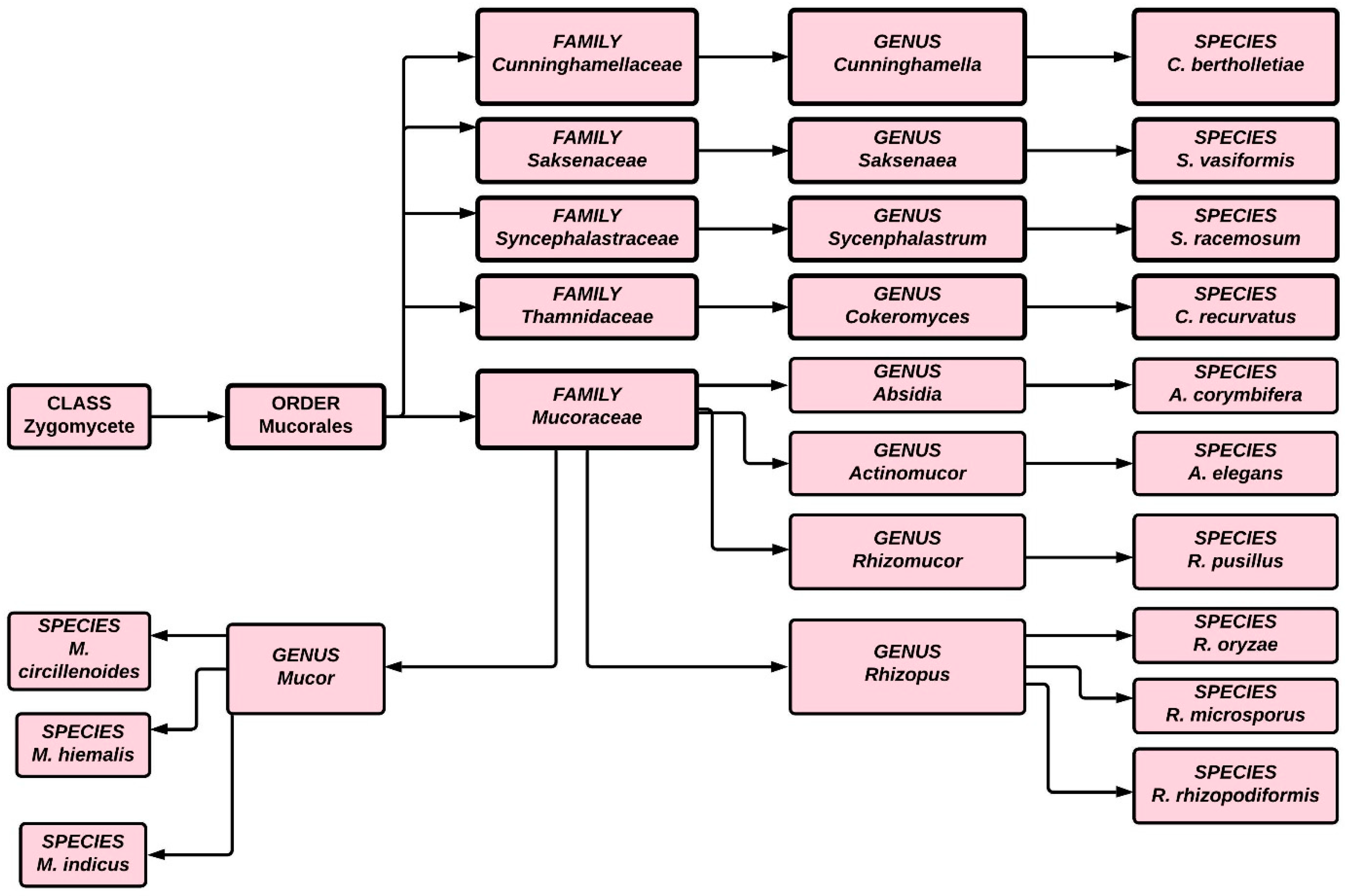
Figure 1. Classification of fungi in the zygomycete order.
Antibiotics 10 01079 g001
Figure 2. Pictorial representation of etiopathogenesis of mucormycosis in COVID-19 patients.
Antibiotics 10 01079 g002
Figure 3. Schematic diagrams of platelets roles; (A) Platelets enhance host immunity, and after invading pathogen/foreign matter it promotes antifungal and antibacterial properties. Thus, various cytokines and chemokines released in granules; (B) The two membrane bound molecules such as TLR and CD154, it allows platelets binding and activates different cells. Thus, adhesion of mucorales hyphae and spores takes place, which causes platelets activation and leads to aggregation and clotting. Furthermore, inhibits hyphal development in presence of fungal destruction, which leads to clot formation and platelets consumption; (C) Prevention of fungi takes place due to the presence of platelet aggregation and therefore, it adheres to fungal wall; (D) COVID-19 patient develops clot in their vital organ and therefore progression of mucormycosis becomes rapid.
Antibiotics 10 01079 g003
Figure 4. Schematic diagrams of natural killer cells roles; (A) Natural killer cells have direct and indirect cytotoxic effects on fungi and produce cytokines and chemokines (includes GM-CSF, TNF-α, IFN-γ. Thus, influences the activity of other immune cells; (B) Damage of mucorales hyphae takes place in presence of natural killer cells, but no effects shown in conidia. Therefore, it leads to no fungal infection; (C) R. oryzae promotes immunosuppressive effects and thus, it inhibits the release of immune regulatory chemokines such as CCL5 and IFN-γ; (D) Human natural killer cells are studied for their ability to minimize exacerbations and enhance event-free periods in hematopoietic transplant patients, and their therapeutic effect may also be helpful in managing and providing therapy for invasive mucormycosis; (E) Natural killer cell counts can be severe to the IgG immunity for COVID-19. The involvement of CD16 on natural killer cells by antibody-coated virus-infected cells results in antibody-dependent cellular cytotoxicity, thus regulates immune response for COVID-19 patient.
Antibiotics 10 01079 g004
Figure 5. Schematic diagrams of Iron uptakes roles; (A) Higher level of Iron (Fe) leads to fungal growth, whereas fungal undergoes apoptosis in iron deficient circumstances; (B) Increase level of iron in animal model cause fungal infection, due to the reduction of phagocyte function and IFN-γ; (C) In the host body the Mucorales obtain iron via three mechanisms: High affinity iron permeases, siderophores and R. oryzae. The R. oryzae promotes the existence of two copies of hemeoxygenases; (D) COVID-19 virus attack haemoglobin and in the presence of higher level of iron it boosted ferritin production, which leads to the increase serum ferritin production and causes hepatic cell death. Therefore, release of iron trigger and leads to higher level of systemic free iron and cause fungal infection. Thus, various drugs show beneficial effects in the management of Mucorales such as deferasirox, deferiprone ferrioxamine and anti-FTR1.
Antibiotics 10 01079 g005
Figure 6. Interaction between Mucorales and endothelial cells during hematogenous proliferation, as well as the influence of host attributes on these relationships and the immune response. (A)—Through glycosylation and protonation, hyperglycemia and ketoacidosis trigger the production of Fe from transferrin.; (B)—Immunological response to disease is harmed by BHB and free Fe, but at the other end, NaHCO3 counteracts this damaging activity by decreasing transferrin iron release and mitigating acidity.; (C)—GRP78 expression on vascular endothelium significantly increased in response to the stressors caused by free Fe, hyperglycemia and ketone substances.; (D)—Free Fe, glucose and BHB increase the production of CotH in fungal cells, resulting in endothelium invasion and increased fungal growth.
Antibiotics 10 01079 g006
Table 1. Demonstrated mode of action, advantages and disadvantages of various medications, as well as surgical management for mucormycosis infection healing.
Treatment ProtocolMode of ActionAdvantagesDisadvantagesRef
Therapeutic Intervention
Lipid formulation of Amphotericin B
(Polyenes derivatives)
The cellular membrane is substantially targeted by Amphotericin B loaded liposomes, which induce fungicidal activity by binding to ergosterol in the fungal cell membrane
Amphotericin B lipid formulations have a higher therapeutic index
Least nephrotoxic, and have better CNS penetration than amphotericin B deoxycholate
Used as first-line treatment for mucormycosis
Hepatotoxicity, infusion-related toxicity
Broad use is limited by the expensive cost and requirement for parenteral administration
[89,102,103]
Posaconazole with or without lipid polyenes
(Triazoles derivative)
Inhibits fungal lanosterol 14 α-demethylase enzyme synthesis
Posaconazole oral dosage is simple and convenient
60–70% success rates were found in a retrospective case study (complete plus partial response)
Posaconazole is an off-label medication for mucormycosis in people who are resistant to amphotericin B
In murine model, the effectiveness of Posaconazole monotherapy was lower than that of polyenes
Posaconazole oral suspension has a reduced absorption rate
[104,105]
Isavuconazole (A new broad-spectrum triazoles derivatives)Cytochrome P450-dependent lanosterol 14-demethylase enzyme, which is required for the production of ergosterol, a component of the fungal membrane, is inhibited by isavuconazole
High oral absorption; less drug-drug interaction; no need for therapeutic drug monitoring; and linear pharmacokinetics
Isavuconazole has a wide range of fungicidal activity and a low risk of side effects
Hepatotoxicity properties limited the use of Isavuconazole
Isavuconazole has a limited number of clinical studies
[106,107,108,109]
VT-1161Potent inhibitor of CYP51 and possess in-vitro activity against Mucorales, including Cunninghamella, Lichtheimia, and Rhizopus oryzae
It prevented Rhizopus delemar infection in immunocompromised mice, and possess modest in-vitro activity against Mucorales
VT-1161 has a lower toxicity potential than existing azoles and polyenes, as well as better pharmacokinetics
VT-1161 increased dose-dependent drug plasma levels as well as increased survival rates.
VT-1161 is expensive
[110]
APX001AThe inositol acyltransferase suppressed by APX001A, which limits the development of Glycosylphosphatidylinositol-anchored proteins and producing antifungal effect
Contribute a significant role in the controlled of infectious illness
APX001A has a limited number of clinical studies
[111,112]
Caspofungin plus lipid polyeneInhibit the enzyme β-1,3-d-glucan synthase
Synergistic in murine disseminated mucormycosis; and favorable toxicity profile
Very limited clinical data of combination therapy
[113,114]
Anidulafungin plus lipid polyeneInhibit the enzyme β-1,3-d-glucan synthase.
Synergistic with liposomal amphotericin B in murine model of disseminated mucormycosis; and favorable toxicity profile
No clinical data
[115]
Deferasirox plus lipid polyenesReducing the available iron load and thus inhibiting the fungal growth and lack siderophore capability
Success in case report and deferasirox oral dosage is simple and convenient
Limited clinical data
[62,116]
Surgical Intervention
Rhino-orbito- cerebral infection; Soft tissue infection; and localized pulmonary lesionA critical component of effective therapy is prompt surgical debridement, which should be repeated if required.
When surgery is required and feasible, it must be robust.
Because the Mucorales hyphae may spread infection swiftly, it is important to remove not just necrotic tissues but also infected healthy-looking tissues in the surrounding area.
Significantly increase the survival and success rate
Expensive
[117,118,119]
Publisher’s Note: MDPI stays neutral with regard to jurisdictional claims in published maps and institutional affiliations.