Article

Antimicrobial and Antibiofilm Activity of the Probiotic Strain Streptococcus salivarius K12 against Oral Potential Pathogens

1
Clinic of Stomatology and Maxillofacial Surgery, Faculty of Medicine, University of Pavol Jozef Šafárik in Košice, 040 01 Košice, Slovakia
2
Department of Microbiology and Immunology, University of Veterinary Medicine and Pharmacy in Košice, 041 81 Košice, Slovakia
*
Author to whom correspondence should be addressed.
Antibiotics 2021, 10(7), 793; https://doi.org/10.3390/antibiotics10070793
Submission received: 31 May 2021 / Revised: 25 June 2021 / Accepted: 25 June 2021 / Published: 29 June 2021
(This article belongs to the Special Issue Oral Microorganisms and Inactivation of Oral Biofilms)

Abstract

:
Oral probiotics are increasingly used in the harmonization of the oral microbiota in the prevention or therapy of various oral diseases. Investigation of the antimicrobial activity of the bacteriocinogenic strain Streptococcus salivarius K12 against oral pathogens shows promising results, not only in suppressing growth, but also in eliminating biofilm formation. Based on these findings, we decided to investigate the antimicrobial and antibiofilm activity of the neutralized cell-free supernatant (nCFS) of S. salivarius K12 at various concentrations against selected potential oral pathogens under in vitro conditions on polystyrene microtiter plates. The nCFS of S. salivarius K12 significantly reduced growth (p < 0.01) in Streptococcus mutans Clarke with increasing concentration from 15 to 60 mg/mL and also in Staphylococcus hominis 41/6 at a concentration of 60 mg/mL (p < 0.001). Biofilm formation significantly decreased (p < 0.001) in Schaalia odontolytica P10 at nCFS concentrations of 60 and 30 mg/mL. Biofilm inhibition (p < 0.001) was also observed in Enterobacter cloacae 4/2 at a concentration of 60 mg/mL. In Schaalia odontolytica P10 and Enterobacter cloacae 4/2, the nCFS had no effect on their growth.

1. Introduction

Bacteria are the predominant microorganisms in the resident oral microbiota. The diversity of bacterial species in the oral cavity is influenced not only by the availability of nutrients but also by the ability to survive in the form of a biofilm, depending on its location (surface of teeth, tongue, and mucous membranes). The resident oral microbiota competes and excludes exogenous pathogens and also contributes to the normal development of tissues and the immune system [1,2]. However, the homeostatic balance between host and microbial communities can be disturbed by many influences, which can lead to the development of oral diseases [3], such as dental caries, gingivitis, periodontitis [4], pharyngotonsillitis, and others [5]. The cause of these diseases is mostly pathogenic bacteria found in the oral cavity. Potential oral pathogenic bacteria are able to form a biofilm, and such bacteria include Streptococcus mutans [6], Schaalia odontolytica [7], Staphylococcus hominis [8,9], and Enterobacter cloacae [10].
Interest in applying new strategies for the treatment of diseases, such as probiotics, is rising significantly due to increasing antibiotic resistance [11]. The living beneficial microorganisms found in probiotic preparations provide a health benefit to the host, either in maintaining or in improving the microbiota [12]. Potentially pathogenic microorganisms enter the body through the mouth or nose, which is why oral probiotics are an excellent first line of defense of the mouth and throat. Oral probiotics have the potential to modify the oral microbiota, thereby helping to prevent or treat oral diseases. They prevent the formation of dental plaque, promote the health of gums and teeth, and prevent halitosis [13,14]. Commonly used oral probiotic bacteria include strains of Lactobacillus reuteri ATCC 55730/ATCC PTA 5289, Lactobacillus rhamnosus GG, and Streptococcus salivarius K12 [15].
The oral cavity of newborns is initially highly selective for bacteria and therefore allows colonization by only a few bacterial species. S. salivarius is one of the first and most important pioneer colonizers of the epithelial surfaces of the oral cavity and upper respiratory tract of humans. It remains there as the predominant member of the oral microbiota throughout life [16,17,18,19]. S. salivarius strains inhibit the formation of a biofilm of S. mutans, which is involved in dental caries, and suppress potentially pathogenic bacteria in the upper respiratory tract, e.g., Streptococcus pneumoniae and Streptococcus pyogenes in vitro [20]. The S. salivarius K12 strain, which has been isolated from the saliva of a healthy child, produces a variety of antimicrobial peptides and is one of the first commercially available oral probiotics [21,22]. The oral probiotic strain S. salivarius K12 has therapeutic potential in pharyngo-tonsillar infections caused by S. pyogenes [23] and in the treatment of halitosis. It has been shown to inhibit Solobacterium moorei, Eubacterium saburreum, Streptococcus anginosus, and Parvimonas micra (formerly Micromonas micros), which are involved in oral malodor [24,25]. It also has a beneficial effect on reducing otitis media [26]. S. salivarius K12 may play a protective role against oral candidiasis. It has not been shown to be directly fungicidal but appears to inhibit Candida adhesion by preferential binding to hyphae [27].
The aim of the present study was to investigate the in vitro antimicrobial and antibiofilm activity of various concentrations of a neutralized cell-free supernatant (nCFS) from the probiotic strain S. salivarius K12 against potential oral pathogens.

2. Results

2.1. Identification of S. salivarius K12

BLASTn analysis within the 16S rRNA variable region of the S. salivarius K12 isolate obtained from Bactoral confirmed its identity with S. salivarius at 99.72%. Genotyping based on specific primers for the gtf gene also confirmed a consensus sequence (543 bp) identical to S. salivarius. The genes encoding salivaricin A (99.67% identity) and B (100% identity) were detected in this isolate. The results show that the isolated strain from the commercially available Bactoral preparation was successfully confirmed as S. salivarius K12.

2.2. Antimicrobial and Antibiofilm Activity of the Neutralized Cell-Free Supernatant of S. salivarius K12 against Potential Oral Pathogens

Potentially oral pathogenic strains, namely S. mutans Clarke, Staphylococcus hominis 41/6, Enterobacter cloacae 4/2, and Schaalia odontolytica P10, were used to test the antimicrobial and antibiofilm activity of various concentrations of the nCFS of S. salivarius K12. Biofilm formation was previously confirmed in the tested strains.
The nCFS of S. salivarius K12 had an inhibitory effect on growth, which subsequently affected the biofilm formation of S. mutans Clarke (Figure 1). Its growth decreased significantly (p < 0.001) compared to the control with increasing concentration from 15 to 60 mg/mL. At a concentration of 7.5 mg/mL, there was no significant inhibitory effect on either growth or biofilm formation. The percentage inhibition of growth was 87.48 ± 1.01, 64.32 ± 3.67, and 27.31 ± 5.23% at a concentration of 60, 30, and 15 mg/mL, respectively.
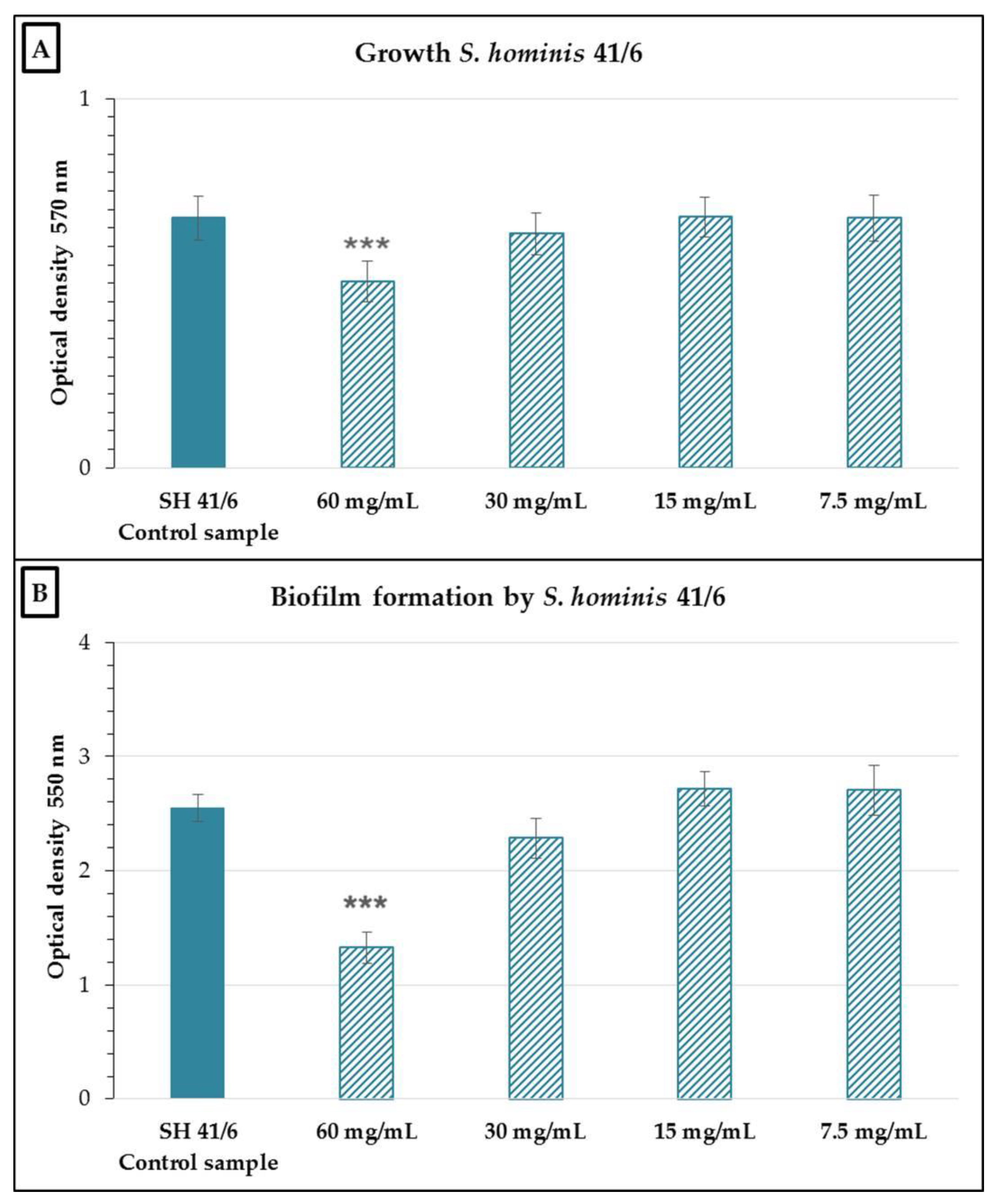
Analysis of growth and biofilm formation in S. hominis 41/6 in the presence of various concentrations of the nCFS showed a significant effect compared to the control (p < 0.001) only at a concentration of 60 mg/mL (Figure 2). In terms of percentage, this represented a 23.16% ± 4.37% growth inhibition and a 42.59% ± 5.07% inhibition of biofilm formation at this concentration.
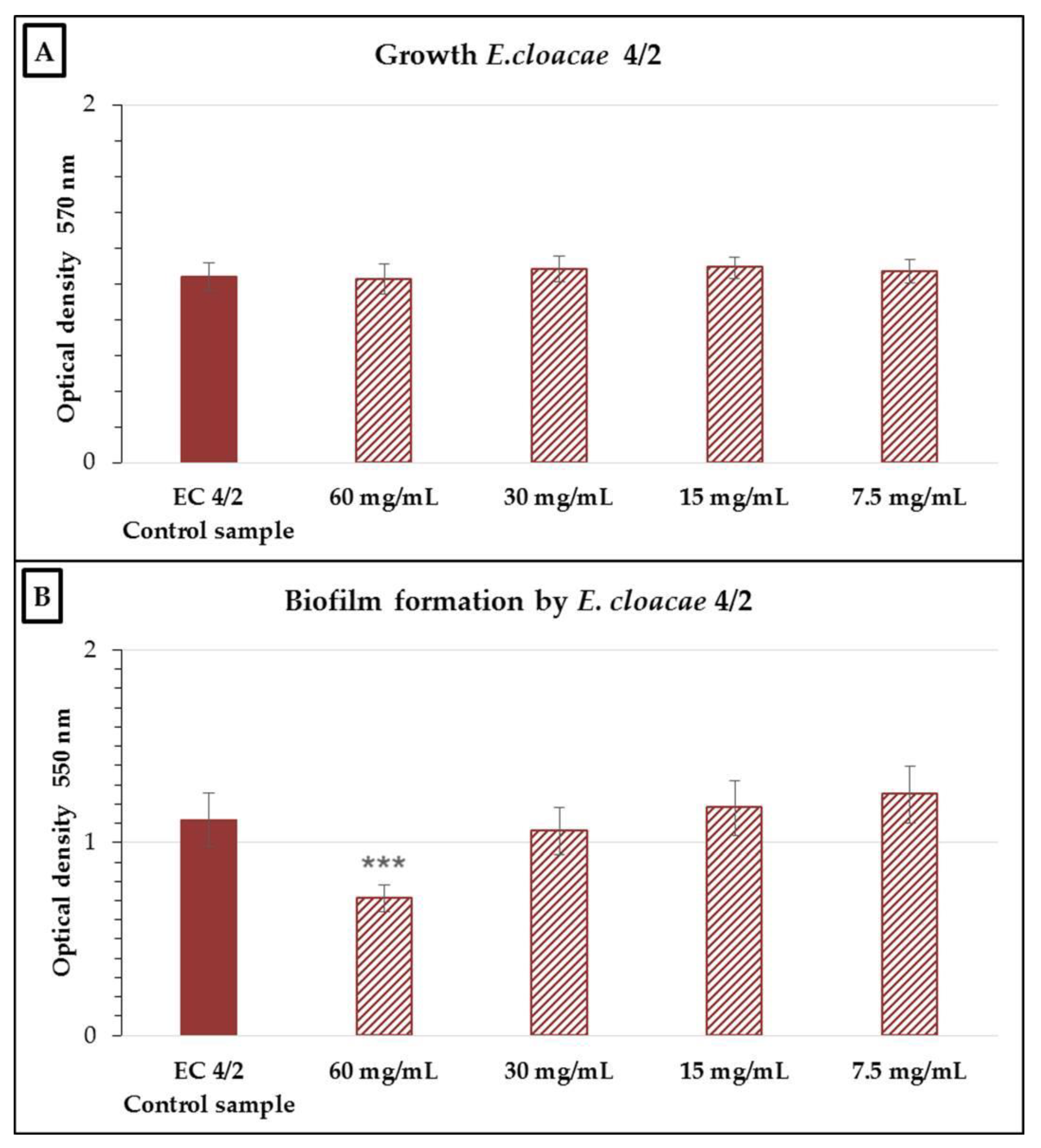
None of the nCFS concentrations tested significantly inhibited the growth of the E. cloacae 4/2 strain (Figure 3). In the evaluation of antibiofilm activity, there was a significant reduction (p < 0.001) in biofilm formation compared to the control at a concentration of 60 mg/mL, which represented a 32.37% ± 3.7% inhibition of biofilm formation.
Similar to E. cloacae 4/2, the nCFS had no significant inhibitory effect on the growth of the Sch. odontolytica P10 strain (Figure 4). Testing of the inhibitory activity of the nCFS against Sch. odontolytica P10 showed a significant effect (p < 0.001) on the reduction in biofilm formation at concentrations of 60 and 30 mg/mL compared to the control. The biofilm inhibition was 91.03% ± 1.09% and 48.56% ± 4.48% at concentrations of 60 and 30 mg/mL, respectively.

3. Discussion

In the present study, the CFS of S. salivarius K12 was investigated against selected oral potential pathogens. Neutralization of the supernatant eliminated the possible effect of acidic products on the inhibition of the tested strains, and its antimicrobial and antibiofilm activity was also demonstrated. The most commonly used method for testing antimicrobial and antibiofilm activity is based on the use of 96-well microtiter plates [28]. Yoo et al. [29] evaluated the antimicrobial activity of the supernatant of oral probiotic strains S. salivarius K12 and M18 in wells of a 96-well polystyrene culture plate against Porphyromonas gingivalis and Treponema denticola. Antimicrobial activity above a certain concentration level has been reported. In this study, a standard assay in microtiter plates was used to examine in vitro antimicrobial and antibiofilm activity, similar to previous studies [29,30]. In addition to the standard methodology, new innovative approaches to in vitro testing of antimicrobial activity with inhibition of biofilm formation are also available, such as the dental implant in vitro model stimulating biofilm formation [31], the in vitro lab catheterization model [32], and biofilm formation in real time using automated microfluidics [28].
S. salivarius strains inhibit biofilm formation by S. mutans and S. pyogenes [33]. In the present study, inhibition of both growth and biofilm formation of strains of S. mutans Clarke and S. hominis 41/6 was noted. S. mutans was more sensitive even at lower concentrations of the nCFS compared with S. hominis. S. mutans is considered the major etiological agent of dental caries and, due to virulence factors, such as glucan production, acid resistance, natural competence, and compact biofilm formation, has a certain advantage over other oral bacteria [34]. Ogawa et al. [30] identified inhibitors produced by S. salivarius HT9R, JCM5707, and ATCC 9759 strains, which prevented biofilm formation in S. mutans GS-5 on the surface of saliva-coated 96-well polystyrene plates and hydroxyapatite supplemented with sucrose. Based on protein analysis, the biofilm-inhibiting agent was identified as the enzyme exo-beta-D-fructosidase (FruA). The results suggest that S. salivarius FruA may modulate sucrose-dependent colonization of S. mutans on oral surfaces. FruA is produced not only by S. salivarius strains but also by other oral streptococci. FruA may play a role in the formation of oral biofilms by bacteria and may regulate microbial pathogenicity in the oral cavity. Hyink et al. [35] identified in S. salivarius K12 a heat-stable inhibitor active against Enterococcus faecalis ATCC 19433 and Actinomyces naeslundii NCTC 10301 and a heat-labile inhibitor active against S. hominis 2203. Frickmann et al. [36] found that the CFS of S. salivarius K12, in addition to reducing and preventing biofilm formation in Staphylococcus epidermidis (ATCC 35984), also affects the dispersion of the already pre-formed biofilm. The CFS of S. salivarius 24SMB and Streptococcus oralis 89a strains isolated from the commercial preparation reduced biofilm formation in vitro and was able to eradicate the pre-formed biofilm of typical pathogenic respiratory bacteria, such as S. pyogenes, S. pneumoniae, Moraxella catarrhalis, Staphylococcus aureus, S. epidermidis, and Propionibacterium acnes [37].
S. salivarius K12 produces the bacteriocins salivaricin A2 and B belonging to the group of lantibiotics [38]. Lantibiotics show two mechanisms of action and mainly have a broad spectrum of activity against Gram-positive bacteria. They inhibit the synthesis of peptidoglycan and are involved in the formation of pores in the cytoplasmic membrane [39]. Salivaricin B differs from salivaricin A by its mechanism of action in interfering with cell wall biosynthesis [40]. Megaplasmid-encoding salivaricin A and salivaricin B carried by the probiotic strain S. salivarius K12 may be transmissible between S. salivarius strains in vitro and in vivo [41]. We hypothesize that salivaricins and their antagonistic effect on strain growth may be involved in the observed inhibitory effect on biofilm formation in S. mutans and S. hominis.
Vacca et al. [31] modeled the interaction between a pathogenic biofilm-forming strain Streptococcus intermedius DSMZ 20573 and a probiotic bacteriocin-producing strain S. salivarius K12. S. intermedius was co-cultured with S. salivarius K12 in vitro in a model simulating biofilm formation in a dental implant composed of a titanium cylindrical surface. Biofilm formation was assessed by quantifying the number of bacteria and the expression level of the luxS gene. The authors recorded an 87% reduction in S. intermedius numbers in the biofilm. In addition, the reduction was accompanied by reduced expression of the luxS gene involved in biofilm formation in S. intermedius.
Sch. odontolytica, also known as Actinomyces odontolyticus, is considered an early colonizer, adhering directly to the salivary pellicle coating the tooth surface [42]. Sch. odontolytica was not generally detected in healthy patients, but it was isolated from persons with advanced dental caries and root canal infections [43]. E. cloacae is not usually found in a healthy oral microbiota but has been most often isolated in adults with advanced periodontitis [44]. In addition, E. cloacae is reported as an important opportunistic and multiresistant bacterial pathogen for humans in hospital wards [45]. In Sch. odontolytica P10 and E. cloacae 4/2 strains, inhibition of biofilm formation was observed without affecting their growth. This may involve influencing the expression of genes involved in the synthesis of adhesins responsible for adherence, i.e., in the initial phase of biofilm formation or in intercellular signaling in quorum sensing [46]. Llena et al. [47] examined the effect of the Streptococcus dentisani 7746 supernatant on the growth of bacteria implicated in root canal infections. Resistance to the antimicrobial effect of the S. dentisani supernatant was detected in the Sch. odontolytica AM98a strain. The antimicrobial and antibiofilm activity of S. salivarius K12, either of the strain itself or of the supernatant, has not been previously studied in Sch. odontolytica and E. cloacae.

4. Materials and Methods

4.1. Bacterial Strains and Culture Conditions

The bacterial strain of S. mutans Clarke (ATCC® 35668™) was acquired from the Faculty of Natural Sciences of Comenius University in Bratislava. Potential pathogenic bacteria, including S. hominis 41/6, E. cloacae 4/2, and Sch. odontolytica P10, were isolated from the dental biofilm and teeth extracted from humans with periodontitis. The test strains were grown on blood agar under aerobic conditions, except Sch. odontolytica P10, which requires anaerobic conditions (BBL GasPakTM Plus, Becton, Dickinson and Co., Sparks, MD, USA). The blood agar was prepared as Tryptone Soya Agar (HiMedia, Mumbai, India) supplemented with 5% sterile horse blood. The inoculated agar plates with S. mutans Clarke were incubated for 48 h at 37 °C. The culture conditions of S. hominis 41/6 and E. cloacae 4/2 were 24 h at 37 °C, while those of Sch. odontolytica P10 were 72 h at 37 °C.
S. salivarius K12 was obtained from the commercially available oral probiotic preparation Bactoral (Pharmaceutical Biotechnology, Czech Republic). One tablet was dissolved in 50 mL of brain heart infusion (BHI) broth (pH 7.4; HiMedia Laboratories, Mumbai, India) in a Falcon conical centrifuge tube and incubated for 24 h at 37 °C. Then, 100 μL of the broth was inoculated in blood agar, followed by further incubation for 24 h at 37 °C aerobically.
S. hominis 41/6, E. cloacae 4/2, Sch. odontolytica P10, and S. salivarius were identified by PCR for the variable region of the 16S rRNA gene with universal primers [48]. Based on studies to identify S. salivarius, PCR analysis was used for the gtf gene encoding glucosyltransferase production [49] and the genes encoding salivaricin A [50] and B [51] with specific primers. DNA from S. salivarius K12 was isolated using DNAzol® Direct (Molecular Research Center Inc., Cincinnati, OH, USA) according to the manufacturer’s instructions. The amplification products were sent for purification and sequencing (Microsynth, Wien, Austria).

4.2. Preparation of the nCFS of S. salivarius K12

The preparation of the CFS of S. salivarius K12 was carried out according to the method described by Lin et al. [52]. Briefly, the strain was inoculated on BHI agar (pH 7.4; HiMedia Laboratories, Mumbai, India) supplemented with 1% glucose. The inoculated agar plates were incubated under aerobic conditions for 24 h at 37 °C. A standardized suspension of S. salivarius K12 was prepared by resuspending solitary colonies in 5 mL of sterile 0.9% NaCl solution and then adjusting to 1 McFarland turbidity (McF). BHI broth (pH 7.4; HiMedia Laboratories, Mumbai, India) supplemented with 1% glucose was inoculated with a strain suspension (2% inoculum) and incubated for 18 h at 37 °C. The bacterial cells were centrifuged at 4 °C and 4500 RPM for 60 min. The obtained CFS was neutralized with 10 M NaOH to pH 7 (elimination of the effect of low pH) and filtered through a syringe filter with a 0.22 μm pore size (Minisart® Syringe Filter; Sartorius Stedim Biotech, Göttingen, Germany). Subsequently, the nCFS was lyophilized and stored at −70 °C.

4.3. In Vitro Assay for Antibiofilm and Antimicrobial Activity of the nCFS

The lyophilized nCFS of S. salivarius K12 was resuspended in BHI broth to a concentration of 120 mg/mL. The nCFS, a volume of 180 μL, was added to the first wells of a polystyrene microtiter plate (Greiner ELISA 8 Well Strips, 350 μL, flat bottom, medium binding; Cruinn Diagnostics Ltd., Dublin, Ireland) containing 180 μL of BHI broth and serially diluted, each time by half, using a multipipette, to prepare concentrations of 60 to 7.5 mg/mL. A standardized inoculum of selected potentially pathogenic strains was then inoculated (1 McF, 20 μL). These strains were selected for the study based on biofilm formation. BHI broth with sterile 0.9% NaCl solution and BHI broth with various concentrations of the nCFS were used as negative controls. BHI broth with potentially pathogenic strains without the nCFS was used as a positive control. Microtiter plates with E. cloacae 4/2 and S. hominis 41/6 were incubated for 24 h at 37 ° C under aerobic conditions and with S. mutans Clarke for 48 h at 37 ° C aerobically. Microtiter plates with Sch. odontolytica P10 were incubated for 72 h at 37 ° C under anaerobic conditions (BBL GasPakTM Plus; Becton, Dickinson and Co., Maryland, USA). To evaluate the growth of the strains in the presence of various concentrations of the nCFS, the optical density at 570 nm was measured after incubation (SynergyTM 4 Multi-Mode Microplate Reader; BioTek, USA).
The contents of the wells were aspirated, and the biofilm formed was quantified using the crystal violet assay, as described previously [53]. The wells were washed with deionized water and, after drying, stained with 200 μL of 0.1% crystal violet solution for 30 min at room temperature. This was followed by washing again with deionized water and drying, and the biofilm-bound dye was extracted in 200 µL of 10% glacial acetic acid. The optical density was measured at 550 nm (SynergyTM 4 Multi-Mode Microplate Reader; BioTek, USA). Strains were tested in at least three independent experiments, each with 8 replicates. The results were interpreted as the arithmetic mean of the measured values ± standard deviation. The percentage inhibition of growth or biofilm formation was calculated according to the formula described in a study by Jadhav et al. [54]. ACFS represents the absorbance of the well with the test strain and the nCFS and Ao the absorbance of the well with the test strain without the nCFS.
Percentage inhibition = [1 − (ACFS/Ao)] × 100

4.4. Statistical Analysis

The results of antimicrobial and antibiofilm activity were evaluated using one-way analysis of variance (ANOVA) with an additional Dunnett’s test in the statistical software GraphPad Prism 6.01 (GraphPad Inc., San Diego, CA, USA).

5. Conclusions

The nCFS of S. salivarius K12 significantly reduced growth in Streptococcus mutans Clarke and in Staphylococcus hominis 41/6. Biofilm formation decreased in Schaalia odontolytica and in Enterobacter cloacae 4/2. In Schaalia odontolytica P10 and Enterobacter cloacae 4/2, the nCFS had no effect on their growth. Further studies will require elucidation of the mechanism of the antibiofilm effect at the molecular level as well, focusing on the evaluation of changes in the expression of genes responsible for the synthesis of bacterial factors involved in the biofilm formation process.

Author Contributions

Conceptualization, R.N. and M.M.; methodology, R.N.; software, R.N.; validation, R.N. and A.S.; formal analysis, M.S.; investigation, A.S., J.K. and M.S.; resources, R.N. and M.M.; data curation, R.N. and A.S.; writing—original draft preparation, A.S. and M.S.; writing—review and editing, M.M., J.K. and R.N.; visualization, A.S. and M.S.; supervision, R.N.; project administration, R.N. and M.M.; funding acquisition, R.N. and M.M. All authors have read and agreed to the published version of the manuscript.

Funding

This research was funded by the Scientific Grand Agency of the Ministry of Education of the Slovak Republic (grant no. VEGA 1/0788/19) and the Slovak Research and Development Agency (grant no. APVV-15-0377).

Institutional Review Board Statement

Not applicable.

Informed Consent Statement

Not applicable.

Conflicts of Interest

The authors declare no conflict of interest.

References

  1. Allaker, R.P.; Ian Douglas, C.W. Non-conventional therapeutics for oral infections. Virulence 2015, 6, 196–207. [Google Scholar] [CrossRef] [PubMed]
  2. Samaranayake, L.; Bandara, N.; Pesee, S. Oral Biofilms: What Are They? In Oral Biofilms and Modern Dental Materials, 1st ed.; Ionescu, A.C., Hahnel, S., Eds.; Springer: Cham, Switzerland, 2021; pp. 1–7. [Google Scholar]
  3. Lamont, R.J.; Koo, H.; Hajishengallis, G. The oral microbiota: Dynamic communities and host interactions. Nat. Rev. Microbiol. 2018, 16, 745–759. [Google Scholar] [CrossRef] [PubMed]
  4. Nyvad, B.; Takahashi, N. Integrated hypothesis of dental caries and periodontal diseases. J. Oral Microbiol. 2020, 12, 1710953. [Google Scholar] [CrossRef] [PubMed] [Green Version]
  5. Di Pierro, F.; Colombo, M.; Zanvit, A.; Rottoli, A.S. Positive clinical outcomes derived from using Streptococcus salivarius K12 to prevent streptococcal pharyngotonsillitis in children: A pilot investigation. Drug Healthc. Patient Saf. 2016, 8, 77–81. [Google Scholar] [CrossRef] [Green Version]
  6. Zhang, G.; Lu, M.; Liu, R.; Tian, Y.; Vu, V.H.; Li, Y.; Liu, B.; Kushmaro, A.; Li, Y.; Sun, Q. Inhibition of Streptococcus mutans Biofilm Formation and Virulence by Lactobacillus plantarum K41 Isolated from Traditional Sichuan Pickles. Front. Microbiol. 2020, 11, 774. [Google Scholar] [CrossRef] [PubMed]
  7. Könönen, E.; Wade, W.G. Actinomyces and related organisms in human infections. Clin. Microbiol. Rev. 2015, 28, 419–442. [Google Scholar] [CrossRef] [Green Version]
  8. Szczuka, E.; Telega, K.; Kaznowski, A. Biofilm formation by Staphylococcus hominis strains isolated from human clinical specimens. Folia Microbiol. 2015, 60, 1–5. [Google Scholar] [CrossRef]
  9. Wischer, D.; Schneider, D.; Poehlein, A.; Herrmann, F.; Oruc, H.; Meinhardt, J.; Wagner, O.; Ahmed, R.; Kharin, S.; Novikova, N.; et al. Novel Antimicrobial Cellulose Fleece Inhibits Growth of Human-Derived Biofilm-Forming Staphylococci During the SIRIUS19 Simulated Space Mission. Front. Microbiol. 2020, 11, 1626. [Google Scholar] [CrossRef]
  10. Zurob, E.; Dennett, G.; Gentil, D.; Montero-Silva, F.; Gerber, U.; Naulín, P.; Gómez, A.; Fuentes, R.; Lascano, S.; Rodrigues da Cunha, T.H.; et al. Inhibition of Wild Enterobacter cloacae Biofilm Formation by Nanostructured Graphene- and Hexagonal Boron Nitride-Coated Surfaces. Nanomaterials 2019, 9, 49. [Google Scholar] [CrossRef] [Green Version]
  11. Sweileh, W.M.; Shraim, N.Y.; Al-Jabi, S.W.; Sawalha, A.F.; Rahhal, B.; Khayyat, R.A.; Zyoud, S.H. Assessing worldwide research activity on probiotics in pediatrics using Scopus database: 1994–2014. World Allergy Organ. J. 2016, 9, 1–12. [Google Scholar] [CrossRef] [Green Version]
  12. Zommiti, M.; Feuilloley, M.G.J.; Connil, N. Update of Probiotics in Human World: A Nonstop Source of Benefactions till the End of Time. Microorganisms 2020, 8, 1907. [Google Scholar] [CrossRef]
  13. Mahasneh, S.A.; Mahasneh, A.M. Probiotics: A Promising Role in Dental Health. Dent. J. 2017, 5, 26. [Google Scholar] [CrossRef] [PubMed] [Green Version]
  14. Allaker, R.P.; Stephen, A.S. Use of Probiotics and Oral Health. Curr. Oral Health Rep. 2017, 4, 309–318. [Google Scholar] [CrossRef] [Green Version]
  15. Dodoo, C.C.; Stapleton, P.; Basit, A.W.; Gaisford, S. The potential of Streptococcus salivarius oral films in the management of dental caries: An inkjet printing approach. Int. J. Pharm. 2020, 591, 119962. [Google Scholar] [CrossRef] [PubMed]
  16. Couvigny, B.; Lapaque, N.; Rigottier-Gois, L.; Guillot, A.; Chat, S.; Meylheuc, T.; Kulakauskas, S.; Rohde, M.; Mistou, M.Y.; Renault, P.; et al. Three glycosylated serine-rich repeat proteins play a pivotal role in adhesion and colonization of the pioneer commensal bacterium, Streptococcus salivarius. Environ. Microbiol. 2017, 19, 3579–3594. [Google Scholar] [CrossRef] [Green Version]
  17. Gong, S.G.; Chan, Y.; Lévesque, C.M. Complete Genome Sequence of Megaplasmid-Bearing Streptococcus salivarius Strain LAB813, Isolated from the Dental Plaque of a Caries-Free Child. Microbiol. Resour. Announc. 2019, 8, e01092-19. [Google Scholar] [CrossRef] [Green Version]
  18. Hols, P.; Ledesma-García, L.; Gabant, P.; Mignolet, J. Mobilization of Microbiota Commensals and Their Bacteriocins for Therapeutics. Trends Microbiol. 2019, 27, 690–702. [Google Scholar] [CrossRef] [PubMed] [Green Version]
  19. Fantinato, V.; Camargo, H.R.; Sousa, A.L.O.P. Probiotics study with Streptococcus salivarius and its ability to produce bacteriocins and adherence to KB cells. Rev. Odontol. UNESP 2019, 48, e20190029. [Google Scholar] [CrossRef]
  20. Hu, L.; Mao, Q.; Zhou, P.; Lv, X.; Hua, H.; Yan, Z. Effects of Streptococcus salivarius K12 with nystatin on oral candidiasis-RCT. Oral Dis. 2019, 25, 1573–1580. [Google Scholar] [CrossRef]
  21. Zupancic, K.; Kriksic, V.; Kovacevic, I.; Kovacevic, D. Influence of Oral Probiotic Streptococcus salivarius K12 on Ear and Oral Cavity Health in Humans: Systematic Review. Probiotics Antimicrob. Proteins 2017, 9, 102–110. [Google Scholar] [CrossRef]
  22. Reid, P.; Heng, N.C.K.; Hale, J.D.; Krishnan, D.; Crane, J.; Tagg, J.R.; Milne, T.J. A TaqMan™-based quantitative PCR screening assay for the probiotic Streptococcus salivarius K12 based on the specific detection of its megaplasmid-associated salivaricin B locus. J. Microbiol. Methods 2020, 170, 105837. [Google Scholar] [CrossRef] [PubMed]
  23. Gregori, G.; Righi, O.; Risso, P.; Boiardi, G.; Demuru, G.; Ferzetti, A.; Galli, A.; Ghisoni, M.; Lenzini, S.; Marenghi, C.; et al. Reduction of group A beta-hemolytic streptococcus pharyngo-tonsillar infections associated with use of the oral probiotic Streptococcus salivarius K12: A retrospective observational study. Ther. Clin. Risk Manag. 2016, 12, 87–92. [Google Scholar] [CrossRef] [Green Version]
  24. Masdea, L.; Kulik, E.M.; Hauser-Gerspach, I.; Ramseier, A.M.; Filippi, A.; Waltimo, T. Antimicrobial activity of Streptococcus salivarius K12 on bacteria involved in oral malodour. Arch. Oral Biol. 2012, 57, 1041–1047. [Google Scholar] [CrossRef]
  25. Di Pierro, F.; Colombo, M.; Zanvit, A.; Risso, P.; Rottoli, A.S. Use of Streptococcus salivarius K12 in the prevention of streptococcal and viral pharyngotonsillitis in children. Drug Healthc. Patient Saf. 2014, 6, 15–20. [Google Scholar] [CrossRef] [PubMed] [Green Version]
  26. Chen, T.Y.; Hale, J.D.F.; Tagg, J.R.; Jain, R.; Voss, A.L.; Mills, N.; Best, E.J.; Stevenson, D.S.; Bird, P.A.; Walls, T. In vitro Inhibition of Clinical Isolates of Otitis Media Pathogens by the Probiotic Streptococcus salivarius BLIS K12. Probiotics Antimicrob. Proteins 2021, 13, 734–738. [Google Scholar] [CrossRef] [PubMed]
  27. Ishijima, S.A.; Hayama, K.; Burton, J.P.; Reid, G.; Okada, M.; Matsushita, Y.; Abe, S. Effect of Streptococcus salivarius K12 on the in vitro growth of Candida albicans and its protective effect in an oral candidiasis model. Appl. Environ. Microbiol. 2012, 78, 2190–2199. [Google Scholar] [CrossRef] [PubMed] [Green Version]
  28. Van Dijck, P.; Sjollema, J.; Cammue, B.P.; Lagrou, K.; Berman, J.; d’Enfert, C.; Andes, D.R.; Arendrup, M.C.; Brakhage, A.A.; Calderone, R.; et al. Methodologies for in vitro and in vivo evaluation of efficacy of antifungal and antibiofilm agents and surface coatings against fungal biofilms. Microb. Cell 2018, 5, 300–326. [Google Scholar] [CrossRef]
  29. Yoo, H.J.; Jwa, S.K.; Kim, D.H.; Ji, Y.J. Inhibitory effect of Streptococcus salivarius K12 and M18 on halitosis in vitro. Clin. Exp. Dent. Res. 2020, 6, 207–214. [Google Scholar] [CrossRef] [Green Version]
  30. Ogawa, A.; Furukawa, S.; Fujita, S.; Mitobe, J.; Kawarai, T.; Narisawa, N.; Sekizuka, T.; Kuroda, M.; Ochiai, K.; Ogihara, H.; et al. Inhibition of Streptococcus mutans biofilm formation by Streptococcus salivarius FruA. Appl. Environ. Microbiol. 2011, 77, 1572–1580. [Google Scholar] [CrossRef] [Green Version]
  31. Vacca, C.; Contu, M.P.; Rossi, C.; Ferrando, M.L.; Blus, C.; Szmukler-Moncler, S.; Scano, A.; Orrù, G. In vitro Interactions between Streptococcus intermedius and Streptococcus salivarius K12 on a Titanium Cylindrical Surface. Pathogens 2020, 9, 1069. [Google Scholar] [CrossRef] [PubMed]
  32. Vamanu, E.; Dinu, L.D.; Luntraru, C.M.; Suciu, A. In Vitro Coliform Resistance to Bioactive Compounds in Urinary Infection, Assessed in a Lab Catheterization Model. Appl. Sci. 2021, 11, 4315. [Google Scholar] [CrossRef]
  33. Chaffanel, F.; Charron-Bourgoin, F.; Libante, V.; Leblond-Bourget, N.; Payot, S. Resistance Genes and Genetic Elements Associated with Antibiotic Resistance in Clinical and Commensal Isolates of Streptococcus salivarius. Appl. Environ. Microbiol. 2015, 81, 4155–4163. [Google Scholar] [CrossRef] [Green Version]
  34. Napimoga, M.H.; Höfling, J.F.; Klein, M.I.; Kamiya, R.U.; Gonçalves, R.B. Tansmission, diversity and virulence factors of Sreptococcus mutans genotypes. J. Oral Sci. 2005, 47, 59–64. [Google Scholar] [CrossRef] [Green Version]
  35. Hyink, O.; Wescombe, P.A.; Upton, M.; Ragland, N.; Burton, J.P.; Tagg, J.R. Salivaricin A2 and the novel lantibiotic salivaricin B are encoded at adjacent loci on a 190-kilobase transmissible megaplasmid in the oral probiotic strain Streptococcus salivarius K12. Appl. Environ. Microbiol. 2007, 73, 1107–1113. [Google Scholar] [CrossRef] [Green Version]
  36. Frickmann, H.; Klenk, C.; Warnke, P.; Redanz, S.; Podbielski, A. Influence of Probiotic Culture Supernatants on In Vitro Biofilm Formation of Staphylococci. Eur. J. Microbiol. Immunol. 2018, 8, 119–127. [Google Scholar] [CrossRef] [PubMed]
  37. Bidossi, A.; De Grandi, R.; Toscano, M.; Bottagisio, M.; De Vecchi, E.; Gelardi, M.; Drago, L. Probiotics Streptococcus salivarius 24SMB and Streptococcus oralis 89a interfere with biofilm formation of pathogens of the upper respiratory tract. BMC Infect. Dis. 2018, 18, 653. [Google Scholar] [CrossRef]
  38. Barbour, A.; Wescombe, P.; Smith, L. Evolution of Lantibiotic Salivaricins: New Weapons to Fight Infectious Diseases. Trends Microbiol. 2020, 28, 578–593. [Google Scholar] [CrossRef] [PubMed]
  39. Simons, A.; Alhanout, K.; Duval, R.E. Bacteriocins, Antimicrobial Peptides from Bacterial Origin: Overview of Their Biology and Their Impact against Multidrug-Resistant Bacteria. Microorganisms 2020, 8, 639. [Google Scholar] [CrossRef] [PubMed]
  40. Barbour, A.; Tagg, J.; Abou-Zied, O.K.; Philip, K. New insights into the mode of action of the lantibiotic salivaricin B. Sci. Rep. 2016, 6, 31749. [Google Scholar] [CrossRef] [PubMed] [Green Version]
  41. Wescombe, P.A.; Heng, N.C.; Burton, J.P.; Tagg, J.R. Something Old and Something New: An Update on the Amazing Repertoire of Bacteriocins Produced by Streptococcus salivarius. Probiotics Antimicrob. Proteins 2010, 2, 37–45. [Google Scholar] [CrossRef]
  42. Jalali, F.; Ellett, F.; Balani, P.; Duncan, M.J.; Dewhirst, F.E.; Borisy, G.G.; Irimia, D. No man’s land: Species-specific formation of exclusion zones bordering Actinomyces graevenitzii microcolonies in nanoliter cultures. Microbiologyopen 2021, 10, e1137. [Google Scholar] [CrossRef]
  43. Guo, H.; Rivailler, P.; Wang, J.; Wang, H.; Xu, W.; Xu, S.; Xu, H.; Hu, R. Metagenomic Analysis of a Throat Swab Sample Collected in China on A Patient Infected with Varicella Zoster Virus. Res. Sq. 2021, in press. [Google Scholar] [CrossRef]
  44. Davis, I.J.; Richards, H.; Mullany, P. Isolation of silver- and antibiotic-resistant Enterobacter cloacae from teeth. Oral Microbiol. Immunol. 2005, 20, 191–194. [Google Scholar] [CrossRef] [PubMed]
  45. Davin-Regli, A.; Pagès, J.M. Enterobacter aerogenes and Enterobacter cloacae; versatile bacterial pathogens confronting antibiotic treatment. Front. Microbiol. 2015, 6, 392. [Google Scholar] [CrossRef] [PubMed] [Green Version]
  46. Davey, M.E.; O’toole, G.A. Microbial biofilms: From ecology to molecular genetics. Microbiol. Mol. Biol. Rev. 2000, 64, 847–867. [Google Scholar] [CrossRef] [PubMed] [Green Version]
  47. Llena, C.; Almarche, A.; Mira, A.; López, M.A. Antimicrobial efficacy of the supernatant of Streptococcus dentisani against microorganisms implicated in root canal infections. J. Oral Sci. 2019, 61, 184–194. [Google Scholar] [CrossRef] [Green Version]
  48. Marchesi, J.R.; Sato, T.; Weightman, A.J.; Martin, T.A.; Fry, J.C.; Hiom, S.J.; Dymock, D.; Wade, W.G. Design and evaluation of useful bacterium-specific PCR primers that amplify genes coding for bacterial 16S rRNA. Appl. Environ. Microbiol. 1998, 64, 795–799. [Google Scholar] [CrossRef] [Green Version]
  49. Hoshino, T.; Kawaguchi, M.; Shimizu, N.; Hoshino, N.; Ooshima, T.; Fujiwara, T. PCR detection and identification of oral streptococci in saliva samples using gtf genes. Diagn. Microbiol. Infect. Dis. 2004, 48, 195–199. [Google Scholar] [CrossRef]
  50. O’Shea, E.F.; Gardiner, G.E.; O’Connor, P.M.; Mills, S.; Ross, R.P.; Hill, C. Characterization of enterocin- and salivaricin-producing lactic acid bacteria from the mammalian gastrointestinal tract. FEMS Microbiol. Lett. 2009, 291, 24–34. [Google Scholar] [CrossRef] [Green Version]
  51. Wescombe, P.A.; Burton, J.P.; Cadieux, P.A.; Klesse, N.A.; Hyink, O.; Heng, N.C.; Chilcott, C.N.; Reid, G.; Tagg, J.R. Megaplasmids encode differing combinations of lantibiotics in Streptococcus salivarius. Antonie Van Leeuwenhoek 2006, 90, 269–280. [Google Scholar] [CrossRef]
  52. Lin, X.; Chen, X.; Chen, Y.; Jiang, W.; Chen, H. The effect of five probiotic lactobacilli strains on the growth and biofilm formation of Streptococcus mutans. Oral Dis. 2015, 21, e128–e134. [Google Scholar] [CrossRef] [PubMed]
  53. O’Toole, G.A.; Pratt, L.A.; Watnick, P.I.; Newman, D.K.; Weaver, V.B.; Kolter, R. Genetic approaches to study of biofilms. Methods Enzymol. 1999, 310, 91–109. [Google Scholar] [CrossRef] [PubMed]
  54. Jadhav, S.; Shah, R.; Bhave, M.; Palombo, E.A. Inhibitory activity of yarrow essential oil on Listeria planktonic cells and biofilms. Food Control 2013, 29, 125–130. [Google Scholar] [CrossRef]
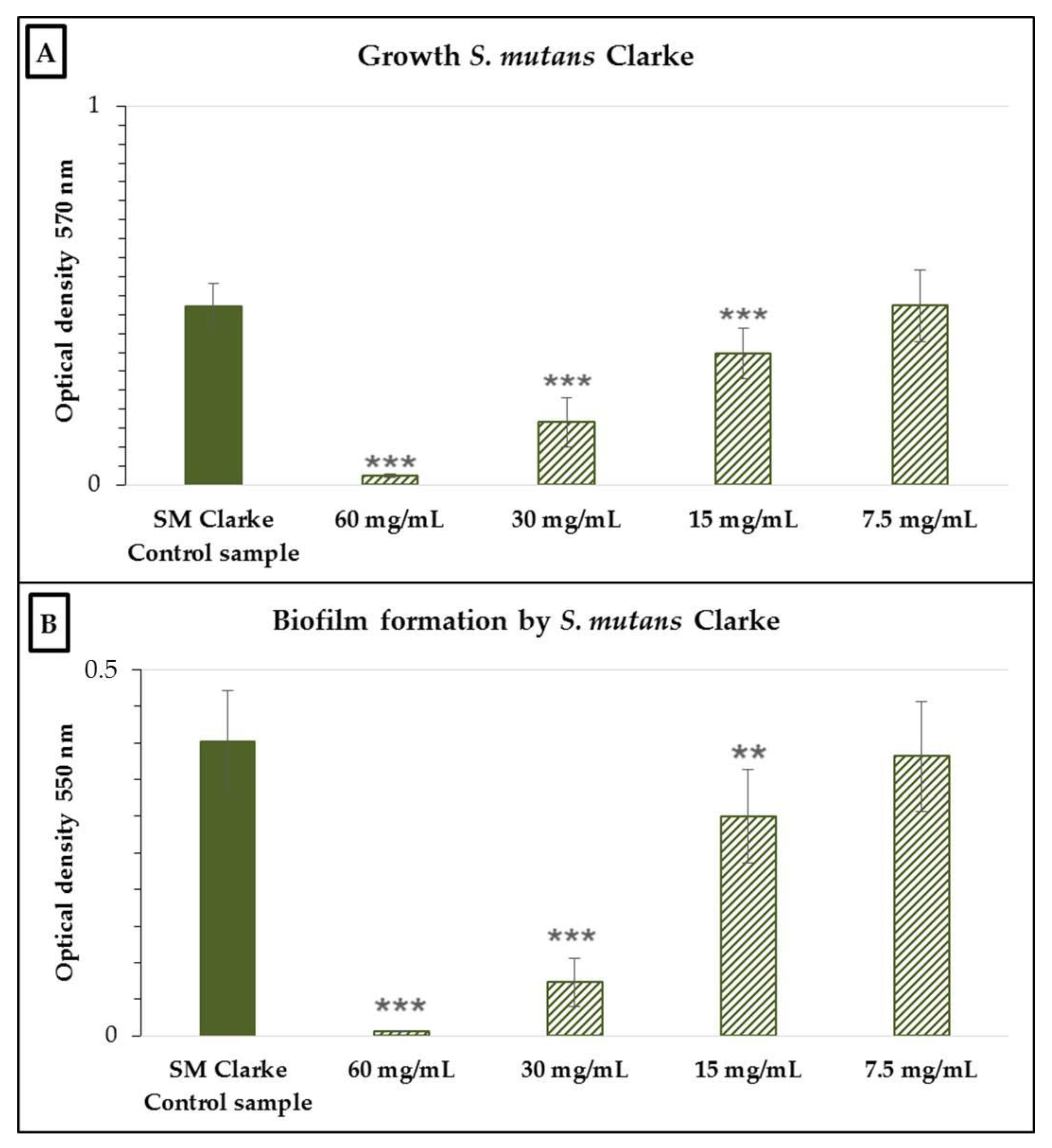
Figure 1. Growth (A) and biofilm formation (B) of Streptococcus mutans Clarke in the presence of various concentrations of the neutralized cell-free supernatant of Streptococcus salivarius K12. SM Clarke control sample: S. mutans Clarke in BHI without the nCFS of S. salivarius K12. Data are expressed as the arithmetic mean ± standard deviation; *** p < 0.001 and ** p < 0.01 compared to the control.
Antibiotics 10 00793 g001
Figure 2. Growth (A) and biofilm formation (B) of Staphylococcus hominis 41/6 in the presence of various concentrations of the nCFS of S. salivarius K12. SH 41/6 control sample: S. hominis 41/6 in BHI without the nCFS of S. salivarius K12. Data are expressed as the arithmetic mean ± standard deviation; *** p < 0.001 compared to the control.
Antibiotics 10 00793 g002
Figure 3. Growth (A) and biofilm formation (B) of Enterobacter cloacae 4/2 in the presence of various concentrations of the nCFS of S. salivarius K12. EC 4/2 control sample: E. cloacae 4/2 in BHI without the nCFS of S. salivarius K12. Data are expressed as the arithmetic mean ± standard deviation; *** p < 0.001 compared to the control.
Antibiotics 10 00793 g003
Figure 4. Growth (A) and biofilm formation (B) of Schaalia odontolytica P10 in the presence of various concentrations of the nCFS of S. salivarius K12. SchO P10 control sample: Sch. odontolytica P10 in BHI without the nCFS of S. salivarius K12. Data are expressed as the arithmetic mean ± standard deviation; *** p < 0.001 compared to the control.
Antibiotics 10 00793 g004
Publisher’s Note: MDPI stays neutral with regard to jurisdictional claims in published maps and institutional affiliations.