Article

Antimicrobial Resistance Determinants in Genomes and Plasmids from Acinetobacter baumannii Clinical Isolates

by
Itziar Chapartegui-González
1,*,
María Lázaro-Díez
2,
Santiago Redondo-Salvo
3,
Jesús Navas
4,5 and
José Ramos-Vivas
6,7
1
Department of Microbiology and Immunology, UTMB, Galveston, TX 77550, USA
2
Instituto de Agrobiotecnología, CSIC-Gobierno de Navarra, 31192 Mutilva, Spain
3
Instituto de Biomedicina y Biotecnología de Cantabria (IBBTEC), Universidad de Cantabria-CSIC, 39011 Santander, Spain
4
Grupo BIOMEDAGE, Facultad de Medicina, Universidad de Cantabria, 39011 Santander, Spain
5
Instituto de Investigación Valdecilla (IDIVAL), 39011 Santander, Spain
6
Research Group on Foods, Nutritional Biochemistry and Health, Universidad Europea del Atlántico, 39011 Santander, Spain
7
Universidad Internacional Iberoamericana, Campeche 24560, Mexico
*
Author to whom correspondence should be addressed.
Antibiotics 2021, 10(7), 753; https://doi.org/10.3390/antibiotics10070753
Submission received: 1 June 2021 / Accepted: 13 June 2021 / Published: 22 June 2021

Abstract

:
Acinetobacter baumannii is a Gram-negative coccoid rod species, clinically relevant as a human pathogen, included in the ESKAPE group. Carbapenem-resistant A. baumannii (CRAB) are considered by the World Health Organization (WHO) as a critical priority pathogen for the research and development of new antibiotics. Some of the most relevant features of this pathogen are its intrinsic multidrug resistance and its ability to acquire rapid and effective new resistant determinants against last-resort clinical antibiotics, mostly from other ESKAPE species. The presence of plasmids and mobile genetic elements in their genomes contributes to the acquisition of new antimicrobial resistance determinants. However, although A. baumannii has arisen as an important human pathogen, information about these elements is still not well understood. Current genomic analysis availability has increased our ability to understand the microevolution of bacterial pathogens, including point mutations, genetic dissemination, genomic stability, and pan- and core-genome compositions. In this work, we deeply studied the genomes of four clinical strains from our hospital, and the reference strain ATCC®19606TM, which have shown a remarkable ability to survive and maintain their effective capacity when subjected to long-term stress conditions. With that, our aim was presenting a detailed analysis of their genomes, including antibiotic resistance determinants and plasmid composition.

1. Introduction

The discovery of penicillin in 1928 marked the beginning of the antibiotic era, which meant a revolution in public health. However, their wrong utilization in the last decades has significantly increased the number of antimicrobial-resistant strains [1]. Despite the fact that multidrug-resistant bacteria pose a threat to both global morbidity and mortality, pharmaceutics research and development has not been able to meet the clinical need for new antibiotics. As a consequence, the World Health Organization (WHO) has created “a priority list of antibiotic-resistant bacteria”, in which the Acinetobacter genus is included [2]. Acinetobacter spp. are Gram-negative coccoid rods, mostly of an environmental origin, but with some species being clinically relevant as human pathogens, especially A. baumannii, a human pathogenic one included in the ESKAPE group [3]. Importantly, carbapenem-resistant A. baumannii (CRAB) strains are considered by the WHO as critical priority pathogens for the research and development of new antibiotics due to their limiting therapeutic options [2]. A. baumannii is usually described as a low virulence pathogen, with its lethality being associated with the activation of the immune system due to its highly toxic lipopolysaccharide (LPS) [4,5]. As a human opportunistic pathogen, it is usually associated with the most critical patients in Intensive Care Units (ICUs) in whom they could cause bacteremia, pneumonia, and different wound infections, among other things [6,7].
Some of the most important features of this pathogen are being naturally antimicrobial-resistant, and being able to acquire rapid and effective new resistant determinants against last-resort clinical antibiotics [4], mostly from Pseudomonas, Salmonella, or Escherichia strains [6]. Therefore, antibiotic resistance genes should be closely surveilled in Acinetobacter strains isolated at the clinical setting. For a long time, their natural antibiotic resistance was considered a consequence of the low wall permeability, but now it is known that antimicrobial efflux pumps are crucial for this phenotype [8,9]. As the definition of multidrug-resistant (MDR) in practice depends on the genus, for Acinetobacter, the following terms have been suggested [10]: (i) MDR, resistant to three different antimicrobial classes; (ii) XDR (extensively drug-resistant), resistant to more than one compound in all except two antimicrobial classes (MDR resistant to carbapenem); (iii) PDR (pandrug-resistant), resistant to all compounds in every antimicrobial class (XDR resistant to polymyxin and tigecycline). Notably, PDR strains from diverse sources have already been described [11,12,13]. A. baumannii strains display the main antimicrobial-resistant mechanisms described for other Gram-negative bacteria. Beta-lactam resistance in Acinetobacter species is associated with a broad repertoire of β-lactamases, which confer resistance against penicillin, cephalosporins, and carbapenems. Within this group, the most common in A. baumannii are oxacillinases (OXAs), with more than 400 different enzymes clustered in six homology groups with different targets [5]: OXA-51, chromosomic and intrinsic, the biggest subgroup; OXA-23, chromosomic or plasmid; OXA-40/24, acquired carbapenemases; and OXA-58, OXA-143, and OXA-48, more recently described as having low hydrolytic activity. The presence of plasmids and other mobile genetic elements in A. baumannii genomes contributes to its ability to acquire new resistance mechanisms rapidly [14]. Moreover, a replicon-based typing method for A. baumannii resistance plasmids has been recently proposed [15]. However, although A. baumannii has arisen as an important human pathogen, information about these genetic elements is still not well understood.
Nowadays, genomic analysis from whole-genome sequencing (WGS) has increased our ability to evaluate bacterial pathogens’ microevolution, which includes point mutations, antimicrobial-resistant determinant dissemination, genomic stability across the world, and both pan- and core-genome compositions [16]. These new tools, as well as genetic manipulation, have allowed us to gain a better comprehension of some of these pathogens’ features, such as host–pathogen interaction mechanisms, their virulence factors, and their environmental survival ability [17,18,19]. The presence or absence of classic virulence factors, evidenced by robust animal or cellular models, is of special interest due to the link to its pathogenicity with the activation of the host’s immune system [20,21]. In this species, the presence of iron uptake mechanisms is remarkable and is related to its survival ability, acinetobactin being the most common and studied system whose presence is strain-specific [19].
Four A. baumannii clinical strains isolated in our hospital and included in this study showed a remarkable ability to survive and maintain their effective capacity when subjected to stress conditions, such as those found by nosocomial bacteria in the hospital conditions, despite their lack of virulence in cell models, which is the key point for their transmission [22,23]. Considering the above, the aim of this work was to present a detailed analysis of plasmids and antibiotic resistance determinants.

2. Results

2.1. Genomic Annotations and Subsystem Categories

Genome sizes and % GC, subsystem and predicted protein-coding sequences (CDS) by RAST and Prokka annotations, and the sequence typing are shown in Table 1. Genome sizes range from 3.81 Mb to 4.06 Mb, all of them with a GC content of around 39% (±0.1). The RAST annotation yielded between 3556 and 3882 protein-coding sequences, belonging to between 451 and 462 subsystems, while Prokka annotation predicted between 3542 and 3886 CDS.
The average of the CDS included in each subsystem is shown in Figure 1. Most belonged to metabolism and molecular compounds such as amino acids and carbohydrates, among other things. Between 75 and 96 CDS fit into the “Virulence and defense” subsystem category (mean = 84.6 ± 11.47 standard deviation -SD-), where antimicrobial resistance-related genes are included.

2.2. Strain Comparison and Pan-Genome Analysis

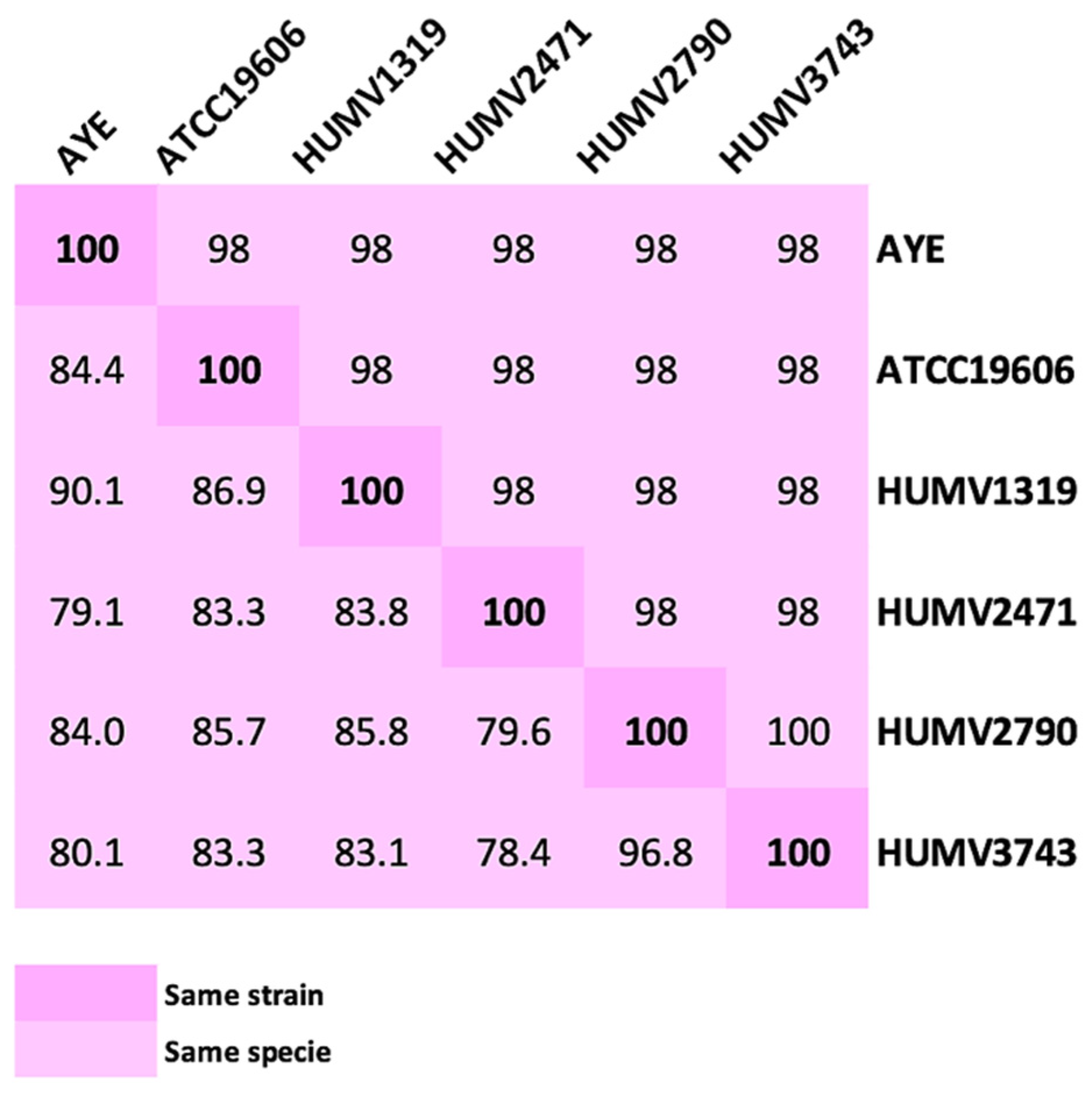
A matrix created using Average Nucleotide Identity (ANI) and DNA-DNA hybridation (DDH) values shows the relationship and similarities among our A. baumannii and reference strains (ATCC®19606TM and AYE) (Figure 2). Both methods indicated that HUMV2790 and HUMV3743 strains were the most similar (100 and 96.8% as ANI and DDH values, respectively), while HUMV1319 and HUMV3743 were the most far among them according to DDH value (83.1%).
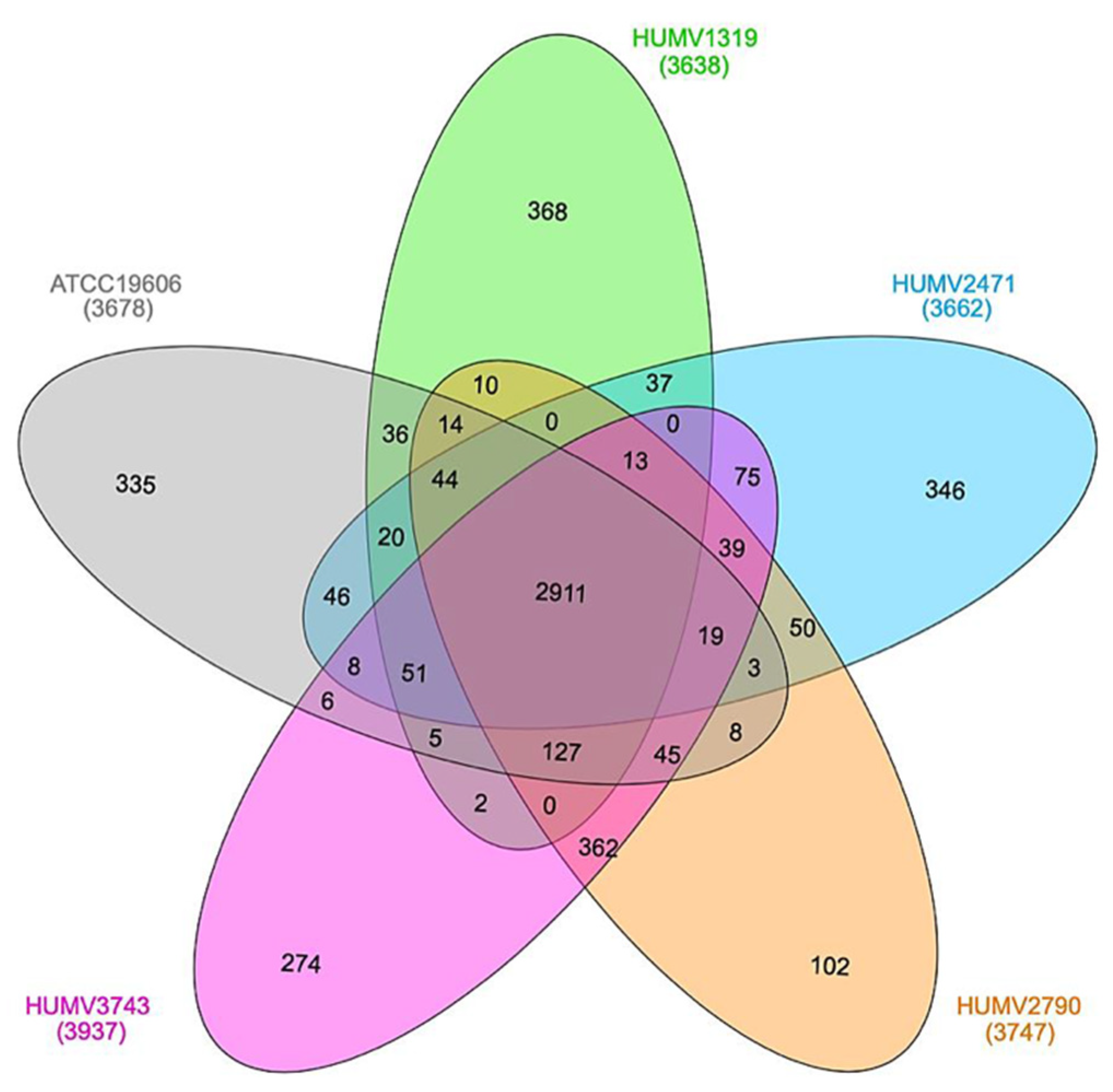
The five strains’ pan-genome was composed of 5244 genes, while the core-genome was composed of 2911 genes, the former being the total number of different CDS and the latter being the number of them shared among all the group. Figure 3 shows the relationship among the isolates, with the numbers signifying the common CDS for each couple or group. Again, the strains HUMV2790 and HUMV3743 are the couple that shares the largest number of genes (362), with the lowest being between HUMV1319 and HUMV3743 (2).

2.3. Susceptibility of A. baumannii to Antimicrobial Agents

The MICs for the seven compounds tested are shown in Table 2. Strains HUMV2790 and HUMV3743, isolated from skin ulcer and wound exudate, respectively, were resistant to the all the tested compounds. The five strains were resistant to ampicillin, meropenem, and erythromycin.

2.4. Antimicrobial Resistance Determinants

In Table 3, the antimicrobial resistance genes detected for each strain are shown and related to their resistance to aminoglycosides, β-lactams, and sulfonamides.
All strains carried at least one aminoglycoside modifying enzyme (AME), the type and number of them among the isolates being variable. The reference strain was the only one susceptible to gentamicin, which only harbors one AME encoding gene (ant(3’’)-IIa). The strains HUMV1319 and HUMV3743 showed the biggest repertoire of AME genes, with representation of the three enzymatic classes: acetyltransferases (AAC), nucleotidyltransferases (ANT), and phosphotransferases (APH). Related to β-lactam compound resistance, all the strains carried two encoding genes for the chromosomic endogenous class D β-lactamase OXA-51, with different allotypes and an Acinetobacter-derived cephalosporinase (ADC). The strains HUMV1319 and HUMV2471 also presented a commonly plasmid-harbored blaOXA-24/40 gene. Additionally, three out the five strains carried the gene sul, related to sulfonamide resistance.
All the clinical samples were resistant to ciprofloxacin (Table 2). In all of them, point mutations in quinolone resistance-determining regions (QRDR) of gyrA and parC genes were detected. In the gyrA gene, the amino acid substitution Ser-81→Leu-81 was common for all the strains. However, in the parC product, the Ser-84→Leu-84 mutation was present in all strains except HUMV2471, which showed a replacement in Glu-88→Lys-88. A. baumannii numbering was used in both cases.
Related to colistin resistance, mutations in lpxACD and pmrAB were mapped. The LpxA subunit showed no variations among the strains; LpxC showed a substitution Asp-287 → Asn-287 in ATCC®19606TM, while in LpxD, different strains harbored different mutations: in HUMV2471, Val-63 → Ile-63; in HUMV1319, Glu-73 → Asp-73; and in HUMV2790 and HUMV3743 (both colistin-resistant), Glu-117 → Lys-117. In regard to the pmrAB operon, in position 119 of PmrA, the HUMV1319 presented a Thr while the others had a Ser. The 4 clinical strains harbored His-440 in PmrB instead of the Asn-400 of the reference strain ATCC®19606TM. The plasmid-borne gene mcr-1, also related to colistin resistance, was not detected in any strain.
All the strains harbored a broad repertoire of efflux pumps genes, including resistance-nodulation-cell division (RND) multidrug efflux pumps (AdeABC, AdeFGH, and AdeIJK), small multidrug resistance (SMR) (AbeS), and multidrug and toxic extrusion (MATE) (AbeM) family efflux pumps. Regulatory genes adeL and adeN were also identified in all the strains, related to adeFGH and adeIJK overexpression respectively, as well as the adeRS operon (adeABC), except HUMV2471, which only presented the adeR subunit.

2.5. Plasmid Prediction and Analysis

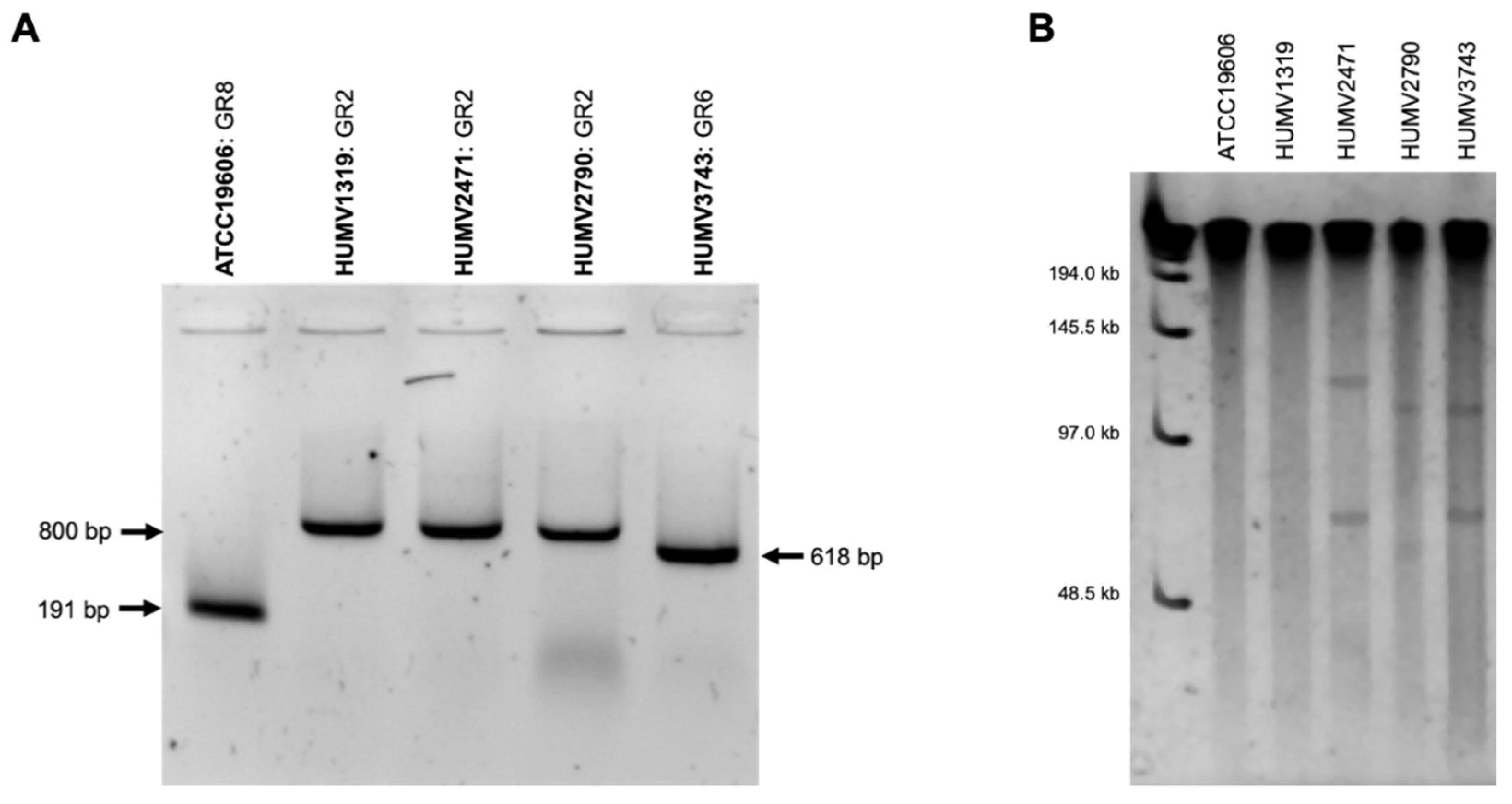
Using Bertini et al. [15] specific resistance plasmid primers, positive results (Figure 4A) were obtained for all the strains in three different typing groups (GR): ATCC®19606TM GR8; HUMV1319, HUMV2471, and HUMV2790 GR2; and HUMV3743 GR6.
Through a PFGE electrophoresis after an S1 digestion plasmid, the presence of plasmids was obtained for HUMV2471, HUMV2790, and HUMV3743 strains, with sizes ranging from 48.5 to 145.5 kb (ladder) (Figure 4B).
The PLACNETw server predicted putative plasmids in all A. baumannii Illumina draft genomes analyzed (Figure S1): in ATCC®19606TM, two putative small plasmids of 11 and 15 kb; in HUMV1319, a small 8.90 kb plasmid and three smaller accessory structures from 1 to 7 kb with not enough identity to be considered as plasmids; in HUMV2471, two big plasmids of 118.39 and 69.2 kb and two small ones of 6 and 8.95 kb; in HUMV2790, a 105.1 kb big plasmid and an 11.55 kb small one; and in HUMV3743, two big plasmids of over 107 kb and 78.73 kb.
The 15 kb plasmid of strain ATCC®19606TM harbored a MOBQ class relaxase (detected by PLACNETw). This is a mobilizable plasmid and no genes coding for a Type IV Secretion System have been detected. Plasmid pMAC (NCBI: NC_006877) is the most similar plasmid with a 99.97% ANI.
The 8.90 kb plasmid of strain HUMV1319, the 8.95 kb plasmid of HUMV2471, and the 11.55 kb plasmid of HUMV2790 are a group of similar plasmids to plasmid pMMCU3 (NCBI: NC_019199). The ANI is 100%, 99.87% and 99.95% respectively.
PLACNETw detected a MOBF class relaxase in the 69.2 kb plasmid of strain HUMV2471 and in the 78.73 kb plasmid from HUMV3743. Moreover, both plasmids are putative conjugative plasmids as, in addition to the relaxase, they had genes coding for a Type IV Secretion System (traA, traL, traE, traK, traB, traV, traC, traW, traU, traN, traN, traH, traG, trwB, and trwC). Plasmid pD72-2 (NCBI: NC_025111) is one of the most similar plasmids in NCBI with an ANI of 99.9% for both plasmids.
According to PLACNETw, plasmid pABUH5-114 (NCBI: NZ_AYOI01000002) is a similar structure to the 107 kb plasmid of strain HUMV3743 and the 105.1 kb plasmid of HUMV2790. However, even with both being large plasmids, no known relaxase nor replicon was found in either plasmid. The case is similar for the 118.39 kb plasmid of strain HUMV2471, but the reference plasmid is pABTJ2 (NCBI: NC_020524) with a 99.52% ANI. For the 6 kb plasmid of strain HUMV2471, the reference plasmid is pRAY (NCBI: NC_025068) with a 100% ANI.

2.6. Virulence Factors

Gene clusters related to siderophores (acinetobactin, actinoferrin) and aryl polyene (berninamycin) detected using antiSMASH were common in all strains and others, as K53 capsular polysaccharide (HUMV2790 and HUMV3743) was only present in some isolates. Analysis of virulence genes in the strains used in this study revealed the existence of a few classical virulence genes in all of them. The virulence of the strains would be related only to genes responsible for the synthesis of lipopolysaccharide and structural genes for the formation of the capsule. These virulence genes are listed in Table S1.

3. Discussion

We performed a bioinformatic analysis of five draft genomes from four A. baumannii clinical isolates from a Spanish hospital, and a reference strain showing a high level of resistance to most antimicrobials tested. These strains have been previously studied and do not show virulence in vitro [22]. The data from the genomic analysis agree with these results, revealing only a few genes classically implicated in the virulence of other Gram-negative pathogens (Table S1). On the other hand, some of them have a marked resistance to environmental conditions, which makes them very persistent in hospital settings [23]. This is consistent with the presence of capsular polysaccharides, which may play a relevant role in such survival under adverse environmental conditions, protecting them from external factors, including desiccation. The capsule also protects bacteria from phagocytosis, although these strains were easily captured by human neutrophils in vitro [20].
We evaluated their susceptibility to seven antibiotics. All the strains showed resistance to meropenem, ampicillin, and erythromycin. Strains also presented resistance to gentamicin, ciprofloxacin, and tetracycline. Only two strains showed resistance to the last-resort antibiotic colistin. These strains could only be considered as MDR because only one compound for each class was tested [10]. These results are in concordance with previous studies that highlight the high prevalence of antimicrobial resistance among A. baumannii strains [12,26].
The increased accessibility to WGS platforms has led to a growing number of sequenced bacterial genomes, which allows performing comparative analysis to be more comprehensive and complete. Genomic sequencing allows, among other things, identification of putative virulence factors, possible targets for diagnosis, treatment, or vaccine development, and/or identification of colonized patients in hospital outbreaks [27,28]. Comparative genomic analysis can also help in the identification of regions for the acquisition and/or spread of antimicrobial resistance, as well as the degree of variability among genome estimation [28,29].
The Acinetobacter genus, in particular A. baumannii, has aroused a lot of interest for its speed and apparent ease to acquire new antimicrobial resistance mechanisms. In this work, different databases (ResFinder and CARD) were used to identify antimicrobial resistance determinants, efflux pump encoding genes (only detected by CARD), and point mutations in target antimicrobial related genes (MEGA7, PointFinder) [30,31,32]. PointFinder was unable to determine the specific modifications related to the resistance phenotype, so they were manually mapped.
Related to aminoglycoside resistance, eight genes encoding AMEs were identified (two AAC, two ANT, and four APH), and were previously described for this bacterium [26,33,34,35]. The most prevalent gene was ant(3”)-IIa, common to all strains, including ATCC®19606TM, which was the only strain with gentamicin susceptibility, so these genes should be related to resistance to other aminoglycosides (Table 3).
Regarding β-lactams resistance, all strains harbored at least one cephalosporinase encoding gene (ampC), with a wide variety of allotypes (four enzymes in five strains) (Table 3). The most prevalent isoform was ADC-25, encoding for the closest strains HUMV2790 and HUMV3743 previously identified [36,37]. As for oxacillinase genes, all strains carried many types, the endogenous blaOXA-51 being the most common with two different isoforms in each strain (six different isoforms in total). Surprisingly, ATCC®19606TM and HUMV2471 strains harbored the same varieties, as well as HUMV2790 and HUMV3743 between them. Strains HUMV1319 and HUMV2471 carried an additional blaOXA-24/40 gene, commonly plasmid-located (such as pMMCU3) [38]. The gene blaOXA-23 was not identified in our strains, although it was the most prevalent in other studies [39,40].
Other mechanisms to promote an MDR phenotype are point mutations in target antimicrobial proteins, such as for quinolones and colistin. Quinolone targets are DNA gyrase subunit A (gyrA) and topoisomerase IV (parC), whose mutations are typically concentrated in QRDR [6,33]. Among our clinical isolates, all showed a Ser81Leu (A. baumannii numbering) substitution, the ATCC®19606TM strain being the only one susceptible to ciprofloxacin, which harbored the serine residue at position 81. For parC, HUMV2471 had a Glu88Lys substitution, while the other three strains presented a Ser84Leu replacement. While gyrA and parC mutations in A. baumannii were described in the 1990s [41,42], a substitution in position 88 of parC has been more recently described [43]. Although these alterations have been widely studied, terminological differences are usually found due to the use of the E. coli gene sequences as the reference in protein alignments (particularly in older publications), which displaces the mutation position [26,43,44,45]. On the other hand, resistance to the last-resort antibiotic colistin has been associated with mutations in lpxACD operon and in pmrAB two components, both related to LPS biosynthesis, the pharmacological target of polymyxins [26,46]. In this study, point mutations have been found in many genes, but only one was common and exclusive for the colistin-resistant strains: Glu117Lys in lpxD, not previously described as far as we know. No strain harbored the mcr-1 plasmid gene after it was manually searched for with the tblastn tool, traditionally linked with polymyxin resistance [47]. Moreover, all strains owned RND multidrug efflux pumps operons adeABC, adeFGH, and adeIJK, each one with a group of substrates, among which are β-lactams, macrolides, tetracyclines, and fluoroquinolones [33,48]. The regulatory genes adeL and adeN were also identified, which are responsible for the overexpression of adeFGH and adeIJK, respectively, and the adeRS operon that regulates adeABC expression [48,49]. Likewise, all strains were positive to other efflux pumps, the SMR abeS which is able to eliminate macrolides, and the MATE abeM, whose substrates are fluoroquinolones [33,49].
Constant and updated genomic information enables increasing knowledge about the bacterial pathogens. For these five A. baumannii clinical isolates, RAST software was used to classify the identified genes of the sequenced and annotated genomes in subsystems based on their function [50]. RAST was not used for the annotation of antimicrobial determinants because it usually appoints genes with enterobacterial names, although there was specific terminology for another species. The largest number of genes with identified functions were included in features associated with “Metabolism” (protein, nucleic, phosphorus, sulfur, nitrogenous) (Figure 1). The following, based on the number of genes, were the features related to cellular components (carbohydrates, amino acids, cofactors, lipids). Another relevant group for both pathogenic relevance and genomic representation was the “Virulence and defense” group, in which antimicrobial resistance genes are included, with an average value of 84.6, which represents around 2.28% of the total. This is the largest number in comparison with reference strains from the ESKAPE pathogen groups: Enterococcus faecium DO 1.40% (44 “Virulence and defense” related genes in 3124 CDS), Staphylococcus aureus NCTC8325 2.15% (58 in 2687), Klebsiella pneumoniae HS11286 1.18% (68 in 5731), Pseudomonas aeruginosa PAO-1 1.17% (69 in 5858), and Enterobacter sp. EA-1 0.97% (126 in 12,965). It should also be noted that this bacterium has a small genome compare to the others Gram-negative pathogens from this group, and hence fewer CDS.
Other studies focusing on differentiating between MDR and susceptible strains, as well as pathogenic and environmental ones, showed greater differences in the number of elements classified for metabolic functions (including catabolism and cellular processes) [17,51]. However, all strains tested here were pathogens with a multi-resistant phenotype, so these differences could not be established.
In the last decade, comparative analysis of A. baumannii genomes has been performed, mainly to determine pan- and core-genome sizes, which enables understanding of both the relationship and variability among strains [19,51,52,53,54]. In this work, PanOCT and InteractiVenn softwares were used to visualize not only the sizes (Figure 3), but also the relationship and number of shared genes among the five Illumina sequenced A. baumannii strains [55,56]. Taxonomic relationships and evolutionary distances among strains were determined using ANI [57] and DDH [58] scores including the well-known and publicly accessible genome of the multi-resistant AYE strain (#GCA_000069245.1) in order to confirm the species. Their relationship values ranged from 100 and 96.8% (ANI and DDH, respectively) for the closest strains, to 78.4% (DDH score) for the most distant ones (Figure 2). As ANI distances were determined for less restrictive values than DDH ones, they may lead to the mistake of considering two similar strains as one, so for close genomes, DDH scores are more recommended. Through the orthologs analysis, 5244 CDS for pan-genome and 2911 for core-genome were established. Accessory genes were between 102 (HUMV2790) and 368 (HUMV1319). Comparing PanOCT with ANI/DDH results, similar relationships were obtained, with HUMV2790 and HUMV3743 sharing more genes between each other (362) than among the rest. The second largest group of shared genes was 127, including all strains except HUMV2471, which also showed the lowest values in general in ANI/DDH matrix. Pan-genome and core-genome sizes in other studies vary depending on the strain set and isolate origin, being also affected by sample size, with pan-genome values between 3000 and 6500, similar to our results [52,53,54].
Acinetobacter baumannii human pathogenic strains are clinically relevant for their high resistance rate. Plasmids, as mobile elements, may disseminate genetic resistance determinants [59,60]. Despite this pathogen relevance, information about A. baumannii plasmids is scarce, with structures ranging from 2 kb to more than 100 kb [13,61]. In this study, different approaches have been applied to identify and characterize A. baumannii sequenced strains: PFGE with an S1 endonuclease digestion, PCR detection with specific A. baumannii plasmids primers, and PLACNETw software as bioinformatic analysis.
Regarding the PFGE profile, as far as we know, this was never used before for Acinetobacter plasmid detection, but for patterns of chromosomic DNA [28]. For PCR detection, specific primers for A. baumannii plasmids designed by Bertini et al. were used, in which the authors grouped the resistance plasmid by replicase homology (19 groups). However, based on subsequent results, these authors’ PBRT scheme might be reviewed and new homology groups added, due to the increasing number of this bacterium plasmid sequence [13,53]. PLACNETw was used not only to detect, but also to identify the plasmid type, which is limited by the annotated and deposited genomes [62].
Taking the results of the three approaches together, we obtained common predictions in most strains, highlighting the importance of using different validating methods. In the ATCC®19606TM strain, plasmid prediction was positive through two methods and the PLACNETw putative plasmid had enough identity to be considered as a pMAC [63], which is classified in Bertini GR8. Small-sized plasmids could be the reason for not obtaining a PFGE prediction (Figure 4B). In the HUMV1319 strain, the PLACNETw server identified a putative pMMCU3 plasmid of 8.9 kb, which is included in the Bertini classification as GR2. The same results were obtained for one small HUMV2471 plasmid (8.9 kb), and a high identity of the 6 kb plasmid with the pRAY structure [13]. The pMMCU3 sequence similarities were confirmed through a Clustal W [64] alignment, being the strains that harbored the blaOXA-24/40 gene typically carried by this mobile structure [38]. In HUMV2471, two plasmids of 118.39 and 69.2 kb were also identified by PLACNETw and positively identified with similar sizes in the PFGE gel, but not enough identity was obtained in PLACNETw to classify them correctly, while the biggest one presented some similarity with pABUH4-111 and pABTJ2 plasmids [59]. In HUMV2790, a band between 97 and 145.5 kb with a PFGE analysis showed the presence of a big plasmid, which was also detected with PLACNETw as a 105.1 kb structure identified as pABUH5-114, recently described for the first time as a large A. baumannii plasmid that carries phage-related genes [53]. The 11.55 kb small one predicted with the bioinformatic tools as a putative pMMCU3 plasmid could correlate with the Bertini GR2 positive classification. Furthermore, in HUMV3743 through PFGE identification, two plasmids of 97–145.5 kb and 48.5–97 kb were detected in concordance with the PLACNETw putative plasmid sizes predicted (107 and 78 kb, respectively), and identified as pABUH5-114 and pABUH1-74, respectively, included in pACICU2-like plasmids which belong to Bertini GR6 [15,53].
Therefore, putting this all together, both traditional microbiology and genomic approaches could be useful to decipher the nosocomial pathogens’ behaviors when more routine methods are not enough. In addition, using genomic tools for non-enterobacteria would improve our knowledge of human pathogen bacteria even it is usually a bigger challenge due to the lack of databases for them.

4. Materials and Methods

4.1. Bacterial Strains

Four A. baumannii clinical isolates were included in this work. All isolates were obtained from different patients (standard service of routine) at the Hospital Universitario Marqués de Valdecilla (HUMV) from Santander (Spain), whose phenotypes were previously described [22]. HUMV1319 and HUMV3743 were isolated from wound exudate, HUMV2471 from sputum, and HUMV2790 from a skin ulcer. Reference strain A. baumannii ATCC®19606TM [19] was also included. A. baumannii strains were routinely cultured on blood agar (BA) plates, or Luria broth (LB) at 37 °C, and stock cultures were frozen at −80 °C with 20% (vol/vol) glycerol.

4.2. Antibiotic Susceptibility Assays

Antibiotic susceptibility was tested by microdilution following CLSI guidelines [25]. Seven compounds belonging to 6 of the main antibiotic families were tested: ampicillin, meropenem, colistin, ciprofloxacin, erythromycin, gentamicin, and tetracycline. EUCAST 2019 breakpoints for Acinetobacter spp. [24] were used to classify the isolates as resistant or susceptible. Briefly, bacterial suspension was adjusted to OD620 = 0.05, which equals 0.5 McFarland units. Antimicrobials were tested at the following ranges: ampicillin 1–64 mg/L, meropenem 0.25–6 mg/L, colistin 0.125–8 mg/L, ciprofloxacin 0.06–4 mg/L, erythromycin 2–128 mg/L, gentamicin 0.5–32 mg/L, and tetracycline 0.5–32 mg/L.

4.3. DNA Isolation and Sequencing

The total genomic sample of A. baumannii clinical strains and the ATCC®19606TM reference one was extracted and purified using the GeneJET genomic DNA (gDNA) isolation kit (Thermo Scientific, Carlsbad, CA, USA) after growth on LB for 24 h at 37 °C. The gDNA was submitted to Fisabio (Valencia, Spain) for Illumina (MiSeq 2 × 300 bp) sequencing. DNA libraries were generated following the Nextera XT Illumina protocol (Nextera XT library prep kit [catalog number FC-131-1024]). Next, 0.2 ng/μL of purified gDNA was used to initiate the protocol. The multiplexing step was performed using the Nextera XT index kit (catalog number FC-131-1096). The libraries were sequenced using a 2 × 300 bp paired-end run (MiSeq v3 reagent kit [catalog number MS-102-3003] on a MiSeq sequencer according to the manufacturer’s instructions [Illumina, Valencia, Spain]).

4.4. Genomic Assembly and Annotation

The whole-genome sequence was assembled using Unicycler v0.3.0.b with both paired and unpaired reads [65].
Annotation of the genome sequence was performed with the Rapid Annotations using Subsystems Technology (RAST) server, as well as Prokka 1.13 to identify the number of coding sequences (CDS), subsystem feature counts, and RNA identification [32,50]. The genotyping was performed using the PubMLST database [66].
Gene clusters related to the secondary metabolite biosynthetic were detected using antiSMASH (bacterial version) [67]. For antimicrobial resistance genes, a preliminary analysis using Prokka 1.13 [32] and ABRicate 0.8 software (https://www.github.com/tseemann/abricate, accessed on: 1 June 2018) (Resfinder 3.2 database) [68], as well as CARD database [30] for efflux pumps encoding genes were used. BLAST alignment with the tblastn option (https://blast.ncbi.nlm.nih.gov/, accessed on: 1 June 2018) was used to identify specific genes. The software MEGA7 was used for point mutation identification [31], as well as the PointFinder database from ABRicate software.

4.5. Plasmid Prediction and Identifications

The PLACNETw server was used to predict feasible plasmids in Illumina sequenced genomes [62]. Identification using the Bertini et al. [15] approach for PCR-based replicon typing (PBRT) with specific A. baumannii resistance plasmid primers was also used to detect and characterize the positive harboring strains. Plasmid presence identification by Pulsed-field Gel Electrophoresis (PFGE) digestion was performed with a CHEF-DR® III system (Bio-Rad, Hercules, CA, USA) as following. Bacteria were grown in LB overnight (O/N) with shaking (175 rpm), and 500 µL of bacterial suspension was mixed with the same volume of 2% LM agarose in TE buffer (10 mM Tris, 1 mM EDTA) for making plugs. Then, 1 mL of lysis solution with 0.5 mg/mL of lysozyme was added and incubated O/N at 37 °C with shaking. After, an equal volume of 0.5 mg/mL of proteinase K in ES (EDTA 0.5 M, sarcosyl 1%) buffer was added and incubated during 16–20 h at 56 °C in static. The plugs were washed six times with pre-warmed TE buffer and then digested with 20 U of S1 restriction enzyme for 30 min at 37 °C. Electrophoresis was performed in a 1% agarose gel at 6 V/cm and 14 °C with 0.5× TBE buffer. Pulse times ramped from 1 to 15 s for 6 h and 15 to 35 s for 16 h. Low range PFGE marker (New England Biolabs, Ipswich, MA, USA) was used as the molecular size marker.

4.6. Strain Comparison and Pan-Genome Analysis

ANI [57] and DDH [58] values were calculated for each strain to determine evolutionary distances and to identify the species, using AYE [17] as a reference strain.
For pan-genome comparison, PanOCT software [55] was used to cluster orthologous proteins, and the EggNOG v4.0 database [69,70] to annotate their functional categories. InteractiVenn was used for diagram illustration [56].

Supplementary Materials

The following are available online at https://www.mdpi.com/article/10.3390/antibiotics10070753/s1, Figure S1: Plasmid prediction with PLACNET of 5 A. baumannii strains; Table S1: List of virulence genes of 5 A. baumannii strains.

Author Contributions

Conceptualization, I.C.-G., M.L.-D., J.N. and J.R.-V.; designed research, I.C.-G. and J.R.-V.; bioinformatic analysis, I.C.-G., M.L.-D. and S.R.-S.; data analysis, I.C.-G., M.L.-D. and S.R.-S.; writing-original draft preparation, I.C.-G., J.N. and J.R.-V.; and funding acquisition, J.R.-V. All authors have read and agreed to the published version of the manuscript.

Funding

This research was funded by ‘Plan Nacional de I+D+i and Instituto de Salud Carlos III (Fondo de Investigaciones Sanitarias PI16/01103 to J.R.-V.), Subdirección General de Redes y Centros de Investigación Cooperativa, Spanish Ministry of Economy and Competitiveness, Spanish Network for Research in Infectious Diseases (REIPI RD12/0015) and (REIPI RD16/0016) co-financed by the European Development Regional Fund “A way to achieve Europe” ERDF and Ministerio de Ciencia e Innovación, Acciones de dinamización «Redes de Investigación» RED2018-102469-T.

Data Availability Statement

HUMV1319, HUMV2471, HUMV2790, and HUMV3743 whole-genome shotgun projects have been deposited at DDBJ/ENA/GenBank under the accession numbers SRR13361577, SRR13361576, SRR13361575, and SRR8264720, respectively; included in the BioProject PRJNA507520.

Acknowledgments

The authors want to thank Laura Álvarez (HUMV) for her help with the PFGE assay.

Conflicts of Interest

The authors declare no conflict of interest.

References

  1. Khan, S.N.; Khan, A.U. Breaking the Spell: Combating Multidrug Resistant ‘Superbugs’. Front. Microbiol. 2016, 7, 174. [Google Scholar] [CrossRef] [PubMed]
  2. Tacconelli, E.; Carrara, E.; Savoldi, A.; Harbarth, S.; Mendelson, M.; Monnet, D.L.; Pulcini, C.; Kahlmeter, G.; Kluytmans, J.; Carmeli, Y.; et al. Discovery, research, and development of new antibiotics: The WHO priority list of antibiotic-resistant bacteria and tuberculosis. Lancet Infect. Dis. 2018, 18, 318–327. [Google Scholar] [CrossRef]
  3. Bergogne-Bérézin, E.; Towner, K. Acinetobacter spp. as Nosocomial Pathogens: Microbiological, Clinical, and Epidemiological Features. Clin. Microbiol. Rev. 1996, 9, 148–165. [Google Scholar] [CrossRef]
  4. Dijkshoorn, L.; Nemec, A.; Seifert, H. An increasing threat in hospitals: Multidrug-resistant Acinetobacter baumannii. Nat. Rev. Microbiol. 2007, 5, 939–951. [Google Scholar] [CrossRef] [PubMed]
  5. Nowak, P.; Paluchowska, P. Acinetobacter baumannii: Biology and drug resistance—Role of carbapenemases. Folia Histochem. Cytobiol. 2016, 54, 61–74. [Google Scholar] [CrossRef] [PubMed] [Green Version]
  6. Peleg, A.Y.; Seifert, H.; Paterson, D.L. Acinetobacter baumannii: Emergence of a Successful Pathogen. Clin. Microbiol. Rev. 2008, 21, 538–582. [Google Scholar] [CrossRef] [Green Version]
  7. Perez, F.; Ponce-Terashima, R.; Adams, M.D.; Bonomo, R.A. Are we closing in on an “elusive enemy”? The current status of our battle with Acinetobacter baumannii. Virulence 2011, 2, 86–90. [Google Scholar] [CrossRef] [Green Version]
  8. Li, X.Z.; Livermore, D.M.; Nikaido, H. Role of efflux pump (s) in intrinsic resistance of Pseudomonas aeruginosa: Active efflux as a contributing factor to b-lactam resistance. Antimicrob. Agent Chemother. 1994, 38, 1732–1741. [Google Scholar] [CrossRef] [Green Version]
  9. Wong, D.; Nielsen, T.B.; Bonomo, R.A.; Pantapalangkoor, P.; Luna, B.; Spellberg, B. Clinical and Pathophysiological Overview of Acinetobacter Infections: A Century of Challenges. Clin. Microbiol. Rev. 2017, 30, 409–447. [Google Scholar] [CrossRef] [PubMed] [Green Version]
  10. Magiorakos, A.-P.; Srinivasan, A.; Carey, R.B.; Carmeli, Y.; Falagas, M.E.; Giske, C.G.; Harbarth, S.; Hindler, J.F.; Kahlmeter, G.; Olsson-Liljequist, B.; et al. Multidrug-resistant, extensively drug-resistant and pandrug-resistant bacteria: An international expert proposal for interim standard definitions for acquired resistance. Clin. Microbiol. Infect. 2012, 18, 268–281. [Google Scholar] [CrossRef] [PubMed] [Green Version]
  11. Valencia, R.; Arroyo, L.A.; Conde, M.; Aldana, J.M.; Torres, M.-J.; Fernández-Cuenca, F.; Garnacho-Montero, J.; Cisneros, J.M.; Ortiz, C.; Pachón, J.; et al. Nosocomial Outbreak of Infection With Pan-Drug-Resistant Acinetobacter baumannii in a Tertiary Care University Hospital. Infect. Control Hosp. Epidemiol. 2009, 30, 257. [Google Scholar] [CrossRef] [PubMed]
  12. Goic-Barisic, I.; Music, M.S.; Kovacic, A.; Tonkic, M.; Hrenovic, J. Pan Drug-Resistant Environmental Isolate of Acinetobacter baumannii from Croatia. Microb. Drug Resist. 2017, 23, 494–496. [Google Scholar] [CrossRef] [PubMed] [Green Version]
  13. Lean, S.S.; Yeo, C.C. Small, Enigmatic Plasmids of the Nosocomial Pathogen, Acinetobacter baumannii: Good, Bad, Who Knows? Front. Microbiol. 2017, 8, 1547. [Google Scholar] [CrossRef] [PubMed] [Green Version]
  14. Partridge, S.R.; Kwong, S.M.; Firth, N.; Jensen, S.O. Mobile Genetic Elements Associated with Antimicrobial Resistance. Clin. Microbiol. Rev. 2018, 31, e00088-17. [Google Scholar] [CrossRef] [PubMed] [Green Version]
  15. Bertini, A.; Poirel, L.; Mugnier, P.D.; Villa, L.; Nordmann, P.; Carattoli, A. Characterization and PCR-Based Replicon Typing of Resistance Plasmids in Acinetobacter baumannii. Antimicrob. Agents Chemother. 2010, 54, 4168–4177. [Google Scholar] [CrossRef] [PubMed] [Green Version]
  16. Adams, M.D.; Chan, E.R.; Molyneaux, N.D.; Bonomo, R.A. Genomewide Analysis of Divergence of Antibiotic Resistance Determinants in Closely Related Isolates of Acinetobacter baumannii. Antimicrob. Agents Chemother. 2010, 54, 3569–3577. [Google Scholar] [CrossRef] [Green Version]
  17. Vallenet, D.; Nordmann, P.; Barbe, V.; Poirel, L.; Mangenot, S.; Bataille, E.; Dossat, C.; Gas, S.; Kreimeyer, A.; Lenoble, P.; et al. Comparative Analysis of Acinetobacters: Three Genomes for Three Lifestyles. PLoS ONE 2008, 3, e1805. [Google Scholar] [CrossRef]
  18. Subashchandrabose, S.; Smith, S.; DeOrnellas, V.; Crepin, S.; Kole, M.; Zahdeh, C.; Mobley, H.L.T. Acinetobacter baumannii Genes Required for Bacterial Survival during Bloodstream Infection. mSphere 2016, 1, e00013–15. [Google Scholar] [CrossRef] [Green Version]
  19. Peleg, A.Y.; de Breij, A.; Adams, M.D.; Cerqueira, G.M.; Mocali, S.; Galardini, M.; Nibbering, P.H.; Earl, A.; Ward, D.V.; Paterson, D.L.; et al. The Success of Acinetobacter Species; Genetic, Metabolic and Virulence Attributes. PLoS ONE 2012, 7, e46984. [Google Scholar] [CrossRef]
  20. Lázaro-Díez, M.; Chapartegui-González, I.; Redondo-Salvo, S.; Leigh, C.; Merino, D.; Segundo, D.S.; Fernández, A.; Navas, J.; Icardo, J.M.; Acosta, F.; et al. Human neutrophils phagocytose and kill Acinetobacter baumannii and A. pittii. Sci. Rep. 2017, 7, 1–11. [Google Scholar] [CrossRef] [Green Version]
  21. Lázaro-Díez, M.; Chapartegui-González, I.; Suberbiola, B.; Ocejo-Vinyals, J.G.; López-Hoyos, M.; Ramos-Vivas, J. Gene expression profiling in human neutrophils after infection with Acinetobacter baumannii in vitro. PLoS ONE 2020, 15, e0242674. [Google Scholar] [CrossRef]
  22. Lázaro-Díez, M.; Navascués-Lejarza, T.; Remuzgo-Martínez, S.; Navas, J.; Icardo, J.M.; Acosta, F.; Martìnez-Martìnez, L.; Ramos-Vivas, J. Acinetobacter baumannii and A. pittii clinical isolates lack adherence and cytotoxicity to lung epithelial cells in vitro. Microbes Infect. 2016, 18, 559–564. [Google Scholar] [CrossRef]
  23. Chapartegui-González, I.; Lázaro-Díez, M.; Bravo, Z.; Navas, J.; Icardo, J.M.; Ramos-Vivas, J. Acinetobacter baumannii maintains its virulence after long-time starvation. PLoS ONE 2018, 13, e0201961. [Google Scholar] [CrossRef]
  24. EUCAST European Committee on Antimicrobial Susceptibility Testing. Breakpoint Tables for Interpretation of MICs and Zone Diameters. Version 8.1. Available online: http://www.eucast.org (accessed on 1 June 2018).
  25. CLSI. Performance Standards for Antimicrobial Susceptibility Testing, 28th ed.; Clinical and Laboratory Standars Institute: Wayne, PA, USA, 2018; ISBN 1-56238-838. [Google Scholar]
  26. Lean, S.-S.; Yeo, C.C.; Suhaili, Z.; Thong, K.-L. Comparative Genomics of Two ST 195 Carbapenem-Resistant Acinetobacter baumannii with Different Susceptibility to Polymyxin Revealed Underlying Resistance Mechanism. Front. Microbiol. 2016, 6, 1445. [Google Scholar] [CrossRef] [Green Version]
  27. Sahl, J.W.; Gillece, J.D.; Schupp, J.M.; Waddell, V.G.; Driebe, E.M.; Engelthaler, D.M.; Keim, P. Evolution of a Pathogen: A Comparative Genomics Analysis Identifies a Genetic Pathway to Pathogenesis in Acinetobacter. PLoS ONE 2013, 8, e54287. [Google Scholar] [CrossRef] [Green Version]
  28. Mancilla-Rojano, J.; Castro-Jaimes, S.; Ochoa, S.A.; Bobadilla del Valle, M.; Luna-Pineda, V.M.; Bustos, P.; Laris-González, A.; Arellano-Galindo, J.; Parra-Ortega, I.; Hernández-Castro, R.; et al. Whole-Genome Sequences of Five Acinetobacter baumannii Strains From a Child With Leukemia M2. Front. Microbiol. 2019, 10, 132. [Google Scholar] [CrossRef] [PubMed]
  29. Lin, M.-F.; Lan, C.-Y. Antimicrobial resistance inAcinetobacter baumannii: From bench to bedside. World J. Clin. Cases 2014, 2, 787. [Google Scholar] [CrossRef]
  30. McArthur, A.G.; Waglechner, N.; Nizam, F.; Yan, A.; Azad, M.A.; Baylay, A.J.; Bhullar, K.; Canova, M.J.; De Pascale, G.; Ejim, L.; et al. The Comprehensive Antibiotic Resistance Database. Antimicrob. Agents Chemother. 2013, 57, 3348–3357. [Google Scholar] [CrossRef] [PubMed] [Green Version]
  31. Kumar, S.; Stecher, G.; Tamura, K. MEGA7: Molecular Evolutionary Genetics Analysis Version 7.0 for Bigger Datasets. Mol. Biol. Evol. 2016, 33, 1870–1874. [Google Scholar] [CrossRef] [PubMed] [Green Version]
  32. Seemann, T. Prokka: Rapid Prokaryotic Genome Annotation. Bioinformatics 2014, 30, 2068–2069. [Google Scholar] [CrossRef]
  33. Gordon, N.C.; Wareham, D.W. Multidrug-resistant Acinetobacter baumannii: Mechanisms of virulence and resistance. Int. J. Antimicrob. Agents 2010, 35, 219–226. [Google Scholar] [CrossRef]
  34. Roca, I.; Espinal, P.; Vila-Farrés, X.; Vila Estapé, J. The Acinetobacter baumannii Oxymoron: Commensal Hospital Dweller Turned Pan-Drug-Resistant Menace. Front. Microbiol. 2012, 3, 148. [Google Scholar] [CrossRef] [Green Version]
  35. Costello, S.E.; Deshpande, L.M.; Davis, A.P.; Mendes, R.E.; Castanheira, M. Aminoglycoside-modifying enzyme and 16S ribosomal RNA methyltransferase genes among a global collection of Gram-negative isolates. J. Glob. Antimicrob. Resist. 2019, 16, 278–285. [Google Scholar] [CrossRef] [PubMed]
  36. Boo, T.W.; Walsh, F.; Crowley, B. Molecular characterization of carbapenem-resistant Acinetobacter species in an Irish university hospital: Predominance of Acinetobacter genomic species 3. J. Med Microbiol. 2009, 58, 209–216. [Google Scholar] [CrossRef] [PubMed]
  37. Lv, W.; Zhang, X.; Hou, M.; Han, D.; Li, Y.; Xiong, W. Draft genome sequence of an OXA-23, OXA-66, ADC-25 and TEM-1D co-producing Acinetobacter baumannii ST195 isolated from a patient with neonatal pneumonia in China. J. Glob. Antimicrob. Resist. 2019, 16, 1–3. [Google Scholar] [CrossRef] [PubMed]
  38. Rumbo, C.; Fernández-Moreira, E.; Merino, M.; Poza, M.; Mendez, J.A.; Soares, N.C.; Mosquera, A.; Chaves, F.; Bou, G. Horizontal Transfer of the OXA-24 Carbapenemase Gene via Outer Membrane Vesicles: A New Mechanism of Dissemination of Carbapenem Resistance Genes in Acinetobacter baumannii. Antimicrob. Agents Chemother. 2011, 55, 3084–3090. [Google Scholar] [CrossRef] [Green Version]
  39. Doi, Y.; Murray, G.L.; Peleg, A.Y. Acinetobacter baumannii: Evolution of Antimicrobial Resistance—Treatment Options. Semin. Respir. Crit. Care Med. 2015, 36, 85. [Google Scholar] [CrossRef] [PubMed] [Green Version]
  40. Shamsizadeh, Z.; Nikaeen, M.; Esfahani, B.N.; Mirhoseini, S.H.; Hatamzadeh, M.; Hassanzadeh, A. Detection of antibiotic resistant Acinetobacter baumannii in various hospital environments: Potential sources for transmission of Acinetobacter infections. Environ. Heal. Prev. Med. 2017, 22, 1–7. [Google Scholar] [CrossRef] [PubMed]
  41. Vila, J.; Ruiz, J.; Goni, P.; Marcos, A.; Jimenez de Anta, T. Mutation in the gyrA gene of quinolone-resistant clinical isolates of Acinetobacter baumannii. Antimicrob. Agents Chemother. 1995, 39, 1201–1203. [Google Scholar] [CrossRef] [Green Version]
  42. Vila, J.; Ruiz, J.; Goni, P.; De Anta, T.J. Quinolone-resistance mutations in the topoisomerase IV parC gene of Acinetobacter baumannii. J. Antimicrob. Chemother. 1997, 39, 757–762. [Google Scholar] [CrossRef]
  43. Chan, A.P.; Sutton, G.; DePew, J.; Krishnakumar, R.; Choi, Y.; Huang, X.-Z.; Beck, E.; Harkins, D.M.; Kim, M.; Lesho, E.P.; et al. A novel method of consensus pan-chromosome assembly and large-scale comparative analysis reveal the highly flexible pan-genome of Acinetobacter baumannii. Genome Biol. 2015, 16, 1–28. [Google Scholar] [CrossRef] [PubMed] [Green Version]
  44. Holt, K.; Kenyon, J.J.; Hamidian, M.; Schultz, M.B.; Pickard, D.J.; Dougan, G.; Hall, R. Five decades of genome evolution in the globally distributed, extensively antibiotic-resistant Acinetobacter baumannii global clone 1. Microb. Genom. 2016, 2. [Google Scholar] [CrossRef]
  45. Ostrer, L.; Khodursky, R.F.; Johnson, J.R.; Hiasa, H.; Khodursky, A. Analysis of mutational patterns in quinolone resistance-determining regions of GyrA and ParC of clinical isolates. Int. J. Antimicrob. Agents 2019, 53, 318–324. [Google Scholar] [CrossRef]
  46. Nhu, N.T.K.; Riordan, D.W.; Nhu, T.D.H.; Thanh, D.P.; Thwaites, G.; Lan, N.P.H.; Wren, B.W.; Baker, S.; Stabler, R.A. The induction and identification of novel Colistin resistance mutations in Acinetobacter baumannii and their implications. Sci. Rep. 2016, 6, 1–8. [Google Scholar] [CrossRef]
  47. Liu, Y.-Y.; Wang, Y.; Walsh, T.R.; Yi, L.-X.; Zhang, R.; Spencer, J.; Doi, Y.; Tian, G.; Dong, B.; Huang, X.; et al. Emergence of plasmid-mediated colistin resistance mechanism MCR-1 in animals and human beings in China: A microbiological and molecular biological study. Lancet Infect. Dis. 2016, 16, 161–168. [Google Scholar] [CrossRef]
  48. Coyne, S.; Courvalin, P.; Périchon, B. Efflux-Mediated Antibiotic Resistance in Acinetobacter spp. Antimicrob. Agents Chemother. 2011, 55, 947–953. [Google Scholar] [CrossRef] [Green Version]
  49. Du, D.; Wang-Kan, X.; Neuberger, A.; van Veen, H.W.; Pos, K.M.; Piddock, L.J.V.; Luisi, B.F. Multidrug efflux pumps: Structure, function and regulation. Nat. Rev. Microbiol. 2018, 16, 523–539. [Google Scholar] [CrossRef] [PubMed]
  50. Aziz, R.K.; Bartels, D.; Best, A.A.; DeJongh, M.; Disz, T.; Edwards, R.A.; Formsma, K.; Gerdes, S.; Glass, E.M.; Kubal, M.; et al. The RAST Server: Rapid annotations using subsystems technology. BMC Genom. 2008, 9, 1–15. [Google Scholar] [CrossRef] [Green Version]
  51. Adams, M.D.; Goglin, K.; Molyneaux, N.; Hujer, K.M.; Lavender, H.; Jamison, J.J.; MacDonald, I.J.; Martin, K.M.; Russo, T.; Campagnari, A.A.; et al. Comparative Genome Sequence Analysis of Multidrug-Resistant Acinetobacter baumannii. J. Bacteriol. 2008, 190, 8053–8064. [Google Scholar] [CrossRef] [PubMed] [Green Version]
  52. Sahl, J.W.; Johnson, J.K.; Harris, A.D.; Phillippy, A.M.; Hsiao, W.W.; Thom, K.A.; Rasko, D.A. Genomic comparison of multi-drug resistant invasive and colonizing Acinetobacter baumannii isolated from diverse human body sites reveals genomic plasticity. BMC Genom. 2011, 12, 1–12. [Google Scholar] [CrossRef] [Green Version]
  53. Wright, M.S.; Haft, D.H.; Harkins, D.M.; Perez, F.; Hujer, K.M.; Bajaksouzian, S.; Benard, M.F.; Jacobs, M.R.; Bonomo, R.A.; Adams, M.D. New Insights into Dissemination and Variation of the Health Care-Associated Pathogen Acinetobacter baumannii from Genomic Analysis. mBio 2014, 5, e00963-13. [Google Scholar] [CrossRef] [PubMed] [Green Version]
  54. Di Nocera, P.P.; Rocco, F.; Giannouli, M.; Triassi, M.; Zarrilli, R. Genome organization of epidemic Acinetobacter baumannii strains. BMC Microbiol. 2011, 11, 1–17. [Google Scholar] [CrossRef] [Green Version]
  55. Fouts, D.E.; Brinkac, L.; Beck, E.; Inman, J.; Sutton, G. PanOCT: Automated clustering of orthologs using conserved gene neighborhood for pan-genomic analysis of bacterial strains and closely related species. Nucleic Acids Res. 2012, 40, e172. [Google Scholar] [CrossRef] [PubMed]
  56. Heberle, H.; Meirelles, G.V.; da Silva, F.R.; Telles, G.P.; Minghim, R. InteractiVenn: A web-based tool for the analysis of sets through Venn diagrams. BMC Bioinform. 2015, 16, 1–7. [Google Scholar] [CrossRef]
  57. Rodriguez-R, L.M.; Konstantinidis, K.T. The enveomics collection: A toolbox for specialized analyses of microbial genomes and metagenomes. Peer J. Prepr. 2016. [Google Scholar] [CrossRef]
  58. Meier-Kolthoff, J.P.; Auch, A.F.; Klenk, H.-P.; Göker, M. Genome sequence-based species delimitation with confidence intervals and improved distance functions. BMC Bioinform. 2013, 14, 1–14. [Google Scholar] [CrossRef] [PubMed] [Green Version]
  59. Huang, H.; Dong, Y.; Yang, Z.-L.; Luo, H.; Zhang, X.; Gao, F. Complete Sequence of pABTJ2, A Plasmid from Acinetobacter baumannii MDR-TJ, Carrying Many Phage-like Elements. Genom. Proteom. Bioinform. 2014, 12, 172–177. [Google Scholar] [CrossRef] [Green Version]
  60. San Millan, A.; MacLean, R.C. Fitness Costs of Plasmids: A Limit to Plasmid Transmission. Microbiol. Spectr. 2017, 5, 5. [Google Scholar] [CrossRef] [Green Version]
  61. Touchon, M.; Cury, J.; Yoon, E.-J.; Krizova, L.; Cerqueira, G.C.; Murphy, C.; Feldgarden, M.; Wortman, J.; Clermont, D.; Lambert, T.; et al. The Genomic Diversification of the Whole Acinetobacter Genus: Origins, Mechanisms, and Consequences. Genome Biol. Evol. 2014, 6, 2866–2882. [Google Scholar] [CrossRef] [Green Version]
  62. Vielva, L.; de Toro, M.; Lanza, V.F.; de la Cruz, F. PLACNETw: A web-based tool for plasmid reconstruction from bacterial genomes. Bioinformatics 2017, 33, 3796–3798. [Google Scholar] [CrossRef]
  63. Dorsey, C.W.; Tomaras, A.P.; Actis, L.A. Sequence and organization of pMAC, an Acinetobacter baumannii plasmid harboring genes involved in organic peroxide resistance. Plasmid 2006, 56, 112–123. [Google Scholar] [CrossRef] [PubMed]
  64. Larkin, M.A.; Blackshields, G.; Brown, N.P.; Chenna, R.; Mcgettigan, P.A.; McWilliam, H.; Valentin, F.; Wallace, I.M.; Wilm, A.; Lopez, R.; et al. Clustal W and Clustal X version 2.0. Bioinformatics 2007, 23, 2947–2948. [Google Scholar] [CrossRef] [Green Version]
  65. Wick, R.R.; Judd, L.M.; Gorrie, C.L.; Holt, K.E. Unicycler: Resolving bacterial genome assemblies from short and long sequencing reads. PLoS Comput. Biol. 2017, 13, e1005595. [Google Scholar] [CrossRef] [Green Version]
  66. Jolley, K.A.; Bray, J.E.; Maiden, M.C.J. Open-access bacterial population genomics: BIGSdb software, the PubMLST.org website and their applications. Wellcome Open Res. 2018, 3, 124. [Google Scholar] [CrossRef]
  67. Weber, T.; Blin, K.; Duddela, S.; Krug, D.; Kim, H.U.; Bruccoleri, R.; Lee, S.Y.; Fischbach, M.A.; Müller, R.; Wohlleben, W.; et al. AntiSMASH 3.0—A comprehensive resource for the genome mining of biosynthetic gene clusters. Nucleic Acids Res. 2015, 43, W237–W243. [Google Scholar] [CrossRef] [PubMed] [Green Version]
  68. Zankari, E.; Hasman, H.; Cosentino, S.; Vestergaard, M.; Rasmussen, S.; Lund, O.; Aarestrup, F.M.; Larsen, M.V. Identification of acquired antimicrobial resistance genes. J. Antimicrob. Chemother. 2012, 67, 2640–2644. [Google Scholar] [CrossRef] [PubMed]
  69. Jensen, L.J.; Julien, P.; Kuhn, M.; von Mering, C.; Muller, J.; Doerks, T.; Bork, P. eggNOG: Automated construction and annotation of orthologous groups of genes. Nucleic Acids Res. 2007, 36, D250–D254. [Google Scholar] [CrossRef]
  70. Powell, S.; Forslund, K.; Szklarczyk, D.; Trachana, K.; Roth, A.; Huerta-Cepas, J.; Gabaldón, T.; Rattei, T.; Creevey, C.; Kuhn, M.; et al. eggNOG v4.0: Nested orthology inference across 3686 organisms. Nucleic Acids Res. 2014, 42, D231–D239. [Google Scholar] [CrossRef] [PubMed]
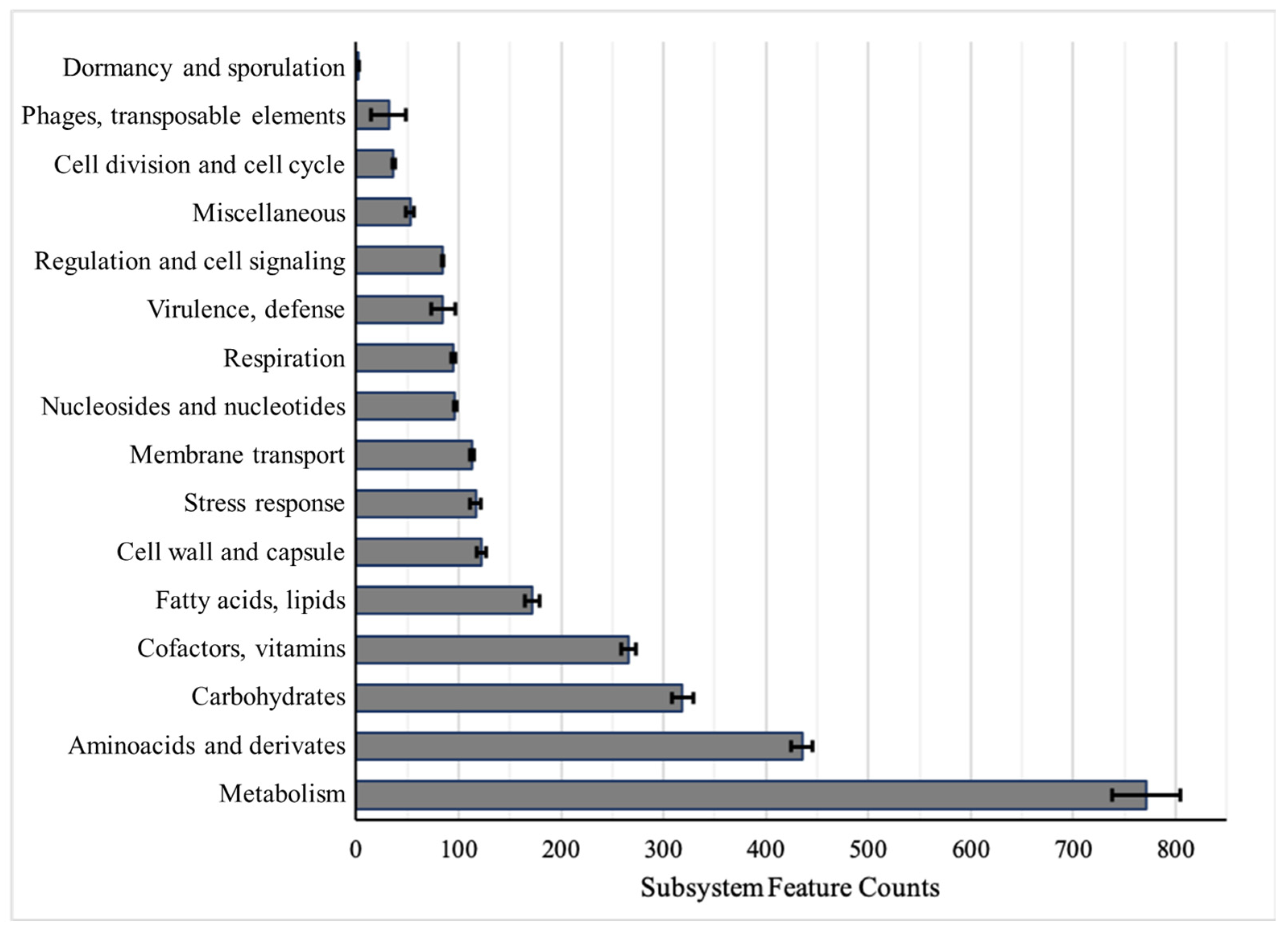
Figure 1. Protein-coding sequences included in each subsystem category. Each bar shows the average of five strains ± SE (standard error).
Antibiotics 10 00753 g001
Figure 2. Matrix distance values among A. baumannii strains. The upper part shows ANI values; the lower part, DDH values. An AYE reference strain was also included to confirm the bacterial species.
Antibiotics 10 00753 g002
Figure 3. Venn diagram with pan-genome composition of five A. baumannii strains. In the middle, core-genome size. Each value corresponds with the number of CDS shared for two or more strains, and the single ones in the end of each ellipse.
Antibiotics 10 00753 g003
Figure 4. Molecular detection of plasmids in A. baumannii. (A) PCR-based replicon type (PBRT) and (B) PFGE gel for plasmids detection in A. baumannii strains. In (B), a molecular size ladder (Lambda ladder PFGE marker, NEB Biolabs) was used in first lane.
Antibiotics 10 00753 g004
Table 1. Draft A. baumannii genomes features according to RAST and Prokka annotation. In ATCC19606 and HUMV2471, one repeat region was also identified in each strain. CDS, coding sequences.
Strains
ATCC19606HUMV1319HUMV2471HUMV2790HUMV3743
Contigs96110108118176
Size (bp)3,914,2943,916,8883,810,8723,935,8044,006,761
% GC39.13938.93939
Subsystems462451453453451
CDSRAST36893703355637063882
Prokka36673680354236893886
tRNA6463646263
mRNA11111
rRNA33332
MLSTOxfordST921ST106 ST350ST218
PasteurST52ST3ST49ST2ST2
Table 2. Susceptibility of A. baumannii strains against seven antimicrobial agents. Isolates were classified as resistant according to EUCAST breakpoints [24]. For the resistance profile: COL (C) is colistin; MER (M) is meropenem; AMP (A) is ampicillin; GEN (G) is gentamicin; CIP (P) is ciprofloxacin; TET (T) is tetracycline; and ERY (E) is erythromycin. MIC is minimum inhibitory concentration; MIC50 is MIC that inhibits 50% of the isolates; MIC90 is MIC that inhibits 90% of the isolates. * No specific value for Acinetobacter, used general no species-specific breakpoints. ** CLSI breakpoints used [25].
COLMERAMP *GENCIPTET **ERYResistance Profile
MICATCC1960624>644>0.251>128MAE
HUMV13192>16>64>128>48>128MAGPTE
HUMV24712>16>64>128>41632MAGPTE
HUMV279084>648>4>32>128CMAGPTE
HUMV37434>16>64>128>4>3264CMAGPTE
MIC502>16>64>32>416>128
MIC90>8>16>64>32>4>32>128
Range (mg/L)0.125–80.25–161–640.5–320.06–40.5–322–128
Table 3. Antimicrobial resistance genes identified with Prokka and CARD databases.
STRAINSANTIMICROBIAL CLASSES RESISTANCE GENES
AMINOGLYCOSIDESβ-LACTAMSULFONAMIDES
ampCOXA
ATCC19606ant(3′’)-IIaADC-2OXA-51 (98)sul
OXA-51 (386)
HUMV1319aac(6’)-Ib’ADC-7OXA-24
OXA-51 (71)
OXA-51 (385)
sul
ant(2’’)-Ia
ant(3’’)-IIa
aph(3’)-VI
aph(3’)-VIa
HUMV2471ant(2’’)-Ia
ant(3’’)-IIa
ADC-39OXA-24
OXA-51 (98)
OXA-51 (386)
HUMV2790ant(3’’)-IIa
aph(3’’)-Ib
aph(6)-Id
ADC-25OXA-51 (66)
OXA-51 (109)
sul
HUMV3743aac(3)-IIa
ant(3’’)-IIa
aph(3’’)-Ib
aph(3’)-VIa
aph(6)-Id
ADC-25OXA-51 (66)
OXA-51 (109)
Publisher’s Note: MDPI stays neutral with regard to jurisdictional claims in published maps and institutional affiliations.