Article

The Glucocorticoid PYED-1 Disrupts Mature Biofilms of Candida spp. and Inhibits Hyphal Development in Candida albicans

1
Department of Chemical, Materials and Production Engineering, University of Naples Federico II, 80126 Naples, Italy
2
Department of Public Health, University of Naples Federico II, 80131 Naples, Italy
3
Complex Operative Unit of Clinical Pathology, Ospedale del Mare-ASL NA1 Centro, 80145 Naples, Italy
4
Department of Molecular Medicine and Medical Biotechnology, University of Naples Federico II, 80131 Naples, Italy
*
Author to whom correspondence should be addressed.
Antibiotics 2021, 10(11), 1396; https://doi.org/10.3390/antibiotics10111396
Submission received: 14 October 2021 / Revised: 5 November 2021 / Accepted: 10 November 2021 / Published: 13 November 2021

Abstract

:
Invasive Candida infections have become a global public health problem due to the increase of Candida species resistant against antifungal therapeutics. The glucocorticoid PYED-1 (pregnadiene-11-hydroxy-16α,17α-epoxy-3,20-dione-1) has antimicrobial activity against various bacterial taxa. Consequently, it might be considered for the treatment of Candida infections. The antifungal activity of PYED-1 was evaluated against several fungal strains that were representative of the five species that causes the majority of Candida infections—namely, Candida albicans, Candida glabrata, Candida tropicalis, Candida parapsilosis and Candida krusei. PYED-1 exhibited a weak antifungal activity and a fungistatic effect on all five Candida species. On the other hand, PYED-1 exhibited a good anti-biofilm activity, and was able to eradicate the preformed biofilms of all Candida species analyzed. Moreover, PYED-1 inhibited germ tube and hyphae formation of C. albicans and reduced adhesion of C. albicans to abiotic surfaces by up to 30%.

1. Introduction

Candidemia and invasive candidiasis are severe healthcare-associated infections with a high mortality rate and limited therapeutic options [1,2]. Candida albicans, Candida glabrata, Candida tropicalis, Candida parapsilosis and Candida krusei are the five species that cause approximately 90% of all infections [3]. C. albicans is the most prevalent cause of invasive fungal infections. The therapeutic failures of C. albicans infections are quite high [4], although the levels of antifungal resistance reported for this organism are very low [5]. Recent studies reported a growing proportion of non-albicans Candida infections [6]. Patients with a non-albicans Candida infection have a less effective response to antifungal therapies and, as a consequence, a high degree of mortality [4]. Candida spp. can form biofilm on biotic and abiotic surfaces, becoming less susceptible to antifungal drugs. Biofilm formation is highly associated with persistent candidemia [7].
The increasing resistance of fungal species to antifungal drugs has led to a need for the development of novel therapeutic agents with a broad spectrum of activity, along with low toxicity and high efficacy against all Candida species.
As part of our interest in the synthesis of compounds endowed with pharmacological activity [8,9,10,11,12], we recently developed a novel synthetic route aimed at the preparation of the corticosteroid drug deflazacort (DFZ, Figure 1) in order to explore its antimicrobial activity.
In this context, while DFZ showed no activity, one of its synthetic precursors, the heterocyclic corticosteroid PYED-1 (pregnadiene-11-hydroxy-16α,17α-epoxy-3,20-dione-1), showed significant antimicrobial, antibiofilm and anti-virulence activities against Gram-positive and Gram-negative bacteria without showing cytotoxicity at concentrations up to 128 µg/mL [13,14,15,16]; nevertheless, its ability to interfere with fungal pathogens has not yet been investigated. Herein we describe the evaluation of the corticosteroid PYED-1 as an inhibitor of the growth and biofilm formation in five species of Candida, namely, C. albicans, C. glabrata, C. tropicalis, C. parapsilosis and C. krusei.

2. Results and Discussion

2.1. Antifungal Susceptibility to Glucocorticoid PYED-1 of Candida Species

The minimal inhibitory concentration (MIC) and minimal lethal concentration (MLC) of PYED-1 were determined for C. albicans, C. glabrata, C. parapsilosis, C. tropicalis and C. krusei clinical isolates. The drug fluconazole was used as a positive control. Results in Table 1 show that PYED-1 had anticandidal activity on all clinical isolates of all Candida spp. assayed.
PYED-1 exhibited weak inhibitory activity against all Candida species with MIC values ranging from 16 μg/mL to 128 μg/mL, and the effect was fungistatic for the MIC values.
To test whether PYED-1 can synergize with available antifungal drugs, checkerboard assays were performed. Fluconazole or posaconazole had neither synergistic nor antagonistic interactions with PYED-1 (data not shown), indicating that PYED-1 co-treatment cannot impair or attenuate the efficacy of these antifungal drugs.

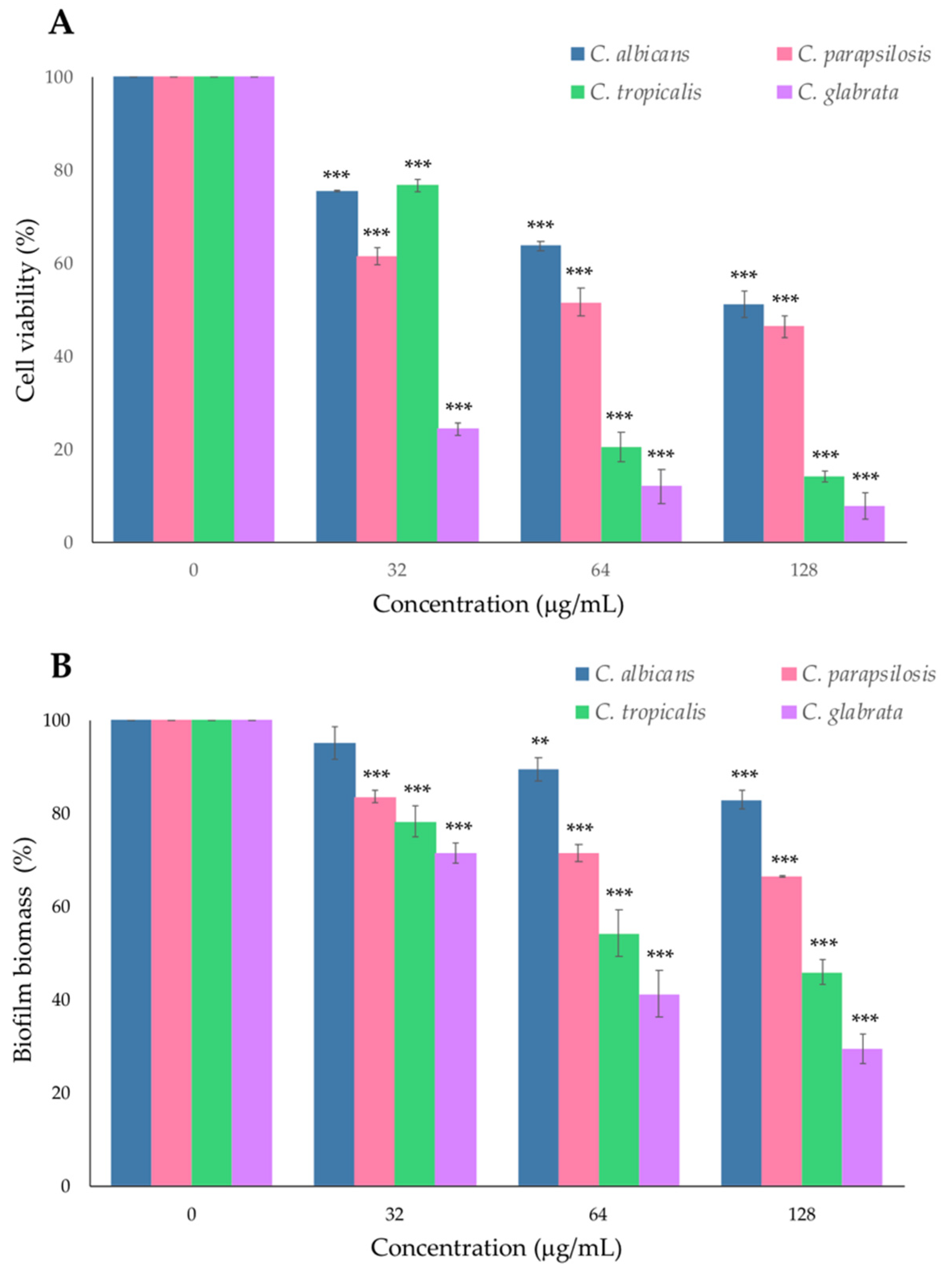
2.2. Effect of PYED-1 on Preformed Candida spp. Biofilm

The formation of fungal biofilm on biotic and abiotic surfaces is a major virulence factor in Candida spp. It causes a reduction in the penetration and, therefore, in the efficacy of antifungal drugs. It contributes to the appearance of drug resistance and recurrent fungal infections in the clinic [17]. Therefore, it is of interest to identify novel molecules with antibiofilm activity. The antibiofilm activity of PYED-1 was evaluated against preformed biofilms of C. albicans, C. glabrata, C. parapsilosis and C. tropicalis strains by XTT (2,3-bis (2-methyloxy-4-nitro-5-sulfophenyl)-2H-tetrazolium-5-carboxanilide) reduction and crystal violet (CV) assays. As shown in Figure 2, results varied according to the Candida species.
In the eradication biofilm assay, exposure of preformed C. glabrata biofilms to 128, 64 and 32 μg/mL of PYED-1 reduced the cell viability by 75–92% (Figure 2A) and the biofilm biomass by 30–70% (Figure 2B), as compared to untreated controls. PYED-1 at 64 μg/mL was able to reduce the number of viable C. tropicalis to over 80% (Figure 2A), while biofilm biomass was diminished by 50% (Figure 2B). In contrast, PYED-1 displayed a significant reduction in the number of viable cells of preformed biofilms of C. albicans and C. parapsilosis when compared to untreated controls, but only marginally reduced the biofilm biomass (Figure 2). Candida biofilms are widely considered to be a key determinant of the high mortality rate attributed to candidiasis [7]. Candida can form biofilms on biomedical implants that are difficult to eradicate by clinical drugs such as amphotericin B, fluconazole, flucytosine and itraconazole. They are also responsible for the high resistance to antifungal drugs [18]. Antifungal resistance due to biofilm formation is frequently reported in clinical settings, and it counteracts the achievement of effective treatment strategies [19]. Antibiofilm agents represent a novel approach to reducing antifungal resistance, destroying preformed biofilm and making the persistent cells susceptible to antifungal drugs [20]. Thus, the ability to disrupt preformed biofilms of all four Candida species analyzed by the death of fungal cells wrapped by the extracellular matrix suggests a potential therapeutic role for PYED-1.

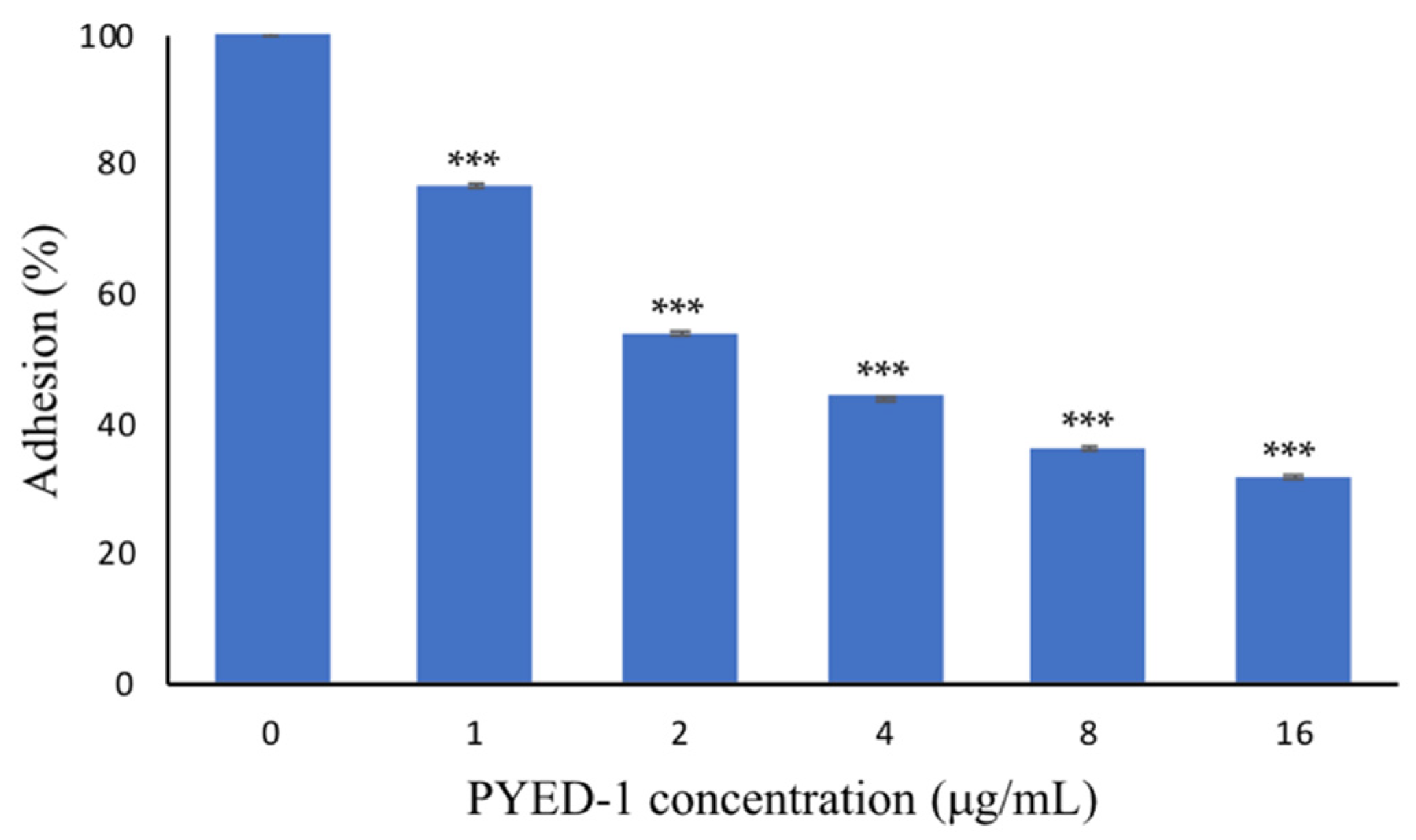
2.3. PYED-1 Inhibited the Filamentation and Adhesion of C. albicans

Mature Candida biofilms are formed by a mixture of yeast cells, pseudohyphae and hyphae [21], and the dimorphic switch of yeast cells to hyphal cells is required for the maturation of structured biofilms by C. albicans [22]. The transition of yeast cells to hyphae is considered an important virulence factor of C. albicans: it can promote fungal invasion of human mucosal tissues, cause invasive fungal infections and aid in escape from immune cells [23]. Hyphal formation is associated with virulence, as genes encoding virulence factors are coregulated with hypha-specific genes [23]. The ability of PYED-1 to inhibit C. albicans filamentation at different concentrations after 5 h of incubation at 37 °C was investigated by microscopy (Figure 3).
The filamentation of C. albicans induced by serum was effectively suppressed in the presence of PYED-1 in RPMI-FBS (fetal bovine serum) medium in a concentration-dependent manner. After 5 h of drug exposure, PYED-1 fully inhibited hyphae formation of C. albicans ATCC 10231 strain at 32 μg/mL, as compared to the untreated control. Microscopic images of C. albicans untreated or treated with 2 μg/mL and 4 μg/mL of PYED-1 formed long hyphae (Figure 3A–C). In cells treated with 8 μg/mL and 16 μg/mL of the drug, fewer filaments and more yeasts per field were observed (Figure 3D,E). Microscopic images of C. albicans cells treated with 32 μg/mL of PYED-1 showed yeast cells but no filaments (Figure 3F). Many molecules induce changes in cell wall components associated with the transition from yeast to hyphal growth and/or reduce the expression of various virulence factors [24]. Molecules with antimicrobial activity, which exhibit a synergistic effect with conventional drugs or reduce the expression of pathogen virulence factors, can represent new treatment options that are effective in combating fungal infections [24,25]. The prevention of hyphae growth by PYED-1 is a significant result since yeast-to-hypha transition is a critical virulence feature which mediates C. albicans invasion into infected tissue.
A hyperfilamentous strain of C. albicans is characterized by a high adhesion ability [26]. Hypha-specific genes encode proteins that are involved in the adhesion to host cells [23]. Strong adherence to cell/tissue or the surfaces of medical devices is the earliest stage of a Candida infection. Since PYED-1 influenced the yeast-to-hyphal morphological transition, we thought it interesting to check whether this compound could affect cell adhesion to abiotic surfaces. As shown in Figure 4, PYED-1 significantly reduced the adhesion of C. albicans ATCC 10,231 to polystyrene surface by up to 30% compared with the negative control.
PYED-1 exhibited a 46% and 55% reduction in the adhesion of C. albicans at sub-MIC concentrations of 2 µg/mL and 4 µg/mL, respectively. At the highest concentration used (16 µg/mL), PYED-1 decreased about 70% of the adhesion, as compared to the untreated controls. PYED-1 can inhibit the adherence of C. albicans and the formation of hyphae. This suggests that PYED-1 may prevent the colonization and tissue penetration of C. albicans. Several studies have shown that adherence and hyphae formation are closely connected. The ALS gene family, encoding a set of cell glycoproteins that promote fungal adherence and biofilm formation, is expressed specifically during hyphal development [27]. Hwp1, a C. albicans cell-surface protein expressed only on hyphae, plays an important role in hyphal development, but is also required for fungal adherence and biofilm formation [27,28].
Blastospores, pseudohyphae and hyphae play a critical role in the process of C. albicans biofilm development and maturation, providing stability to the biofilm structure [23]. Therefore, inhibition of the adhesion of yeast cells and their differentiation in filamentous form can represent an effective therapeutic alternative [29]. In light of this, the glucocorticoid PYED-1 with a broad antimicrobial activity has the potential to serve as a preventive tool. It functions by affecting the adhesion of yeast cells and their differentiation in filamentous form, and by inhibiting existing or forming C. albicans biofilms. However, the specific antibiofilm mechanism of PYED-1 has not yet been described. Future studies will be necessary to understand the mode of action of PYED-1 and to evaluate its therapeutic use during Candida spp. infections.

3. Materials and Methods

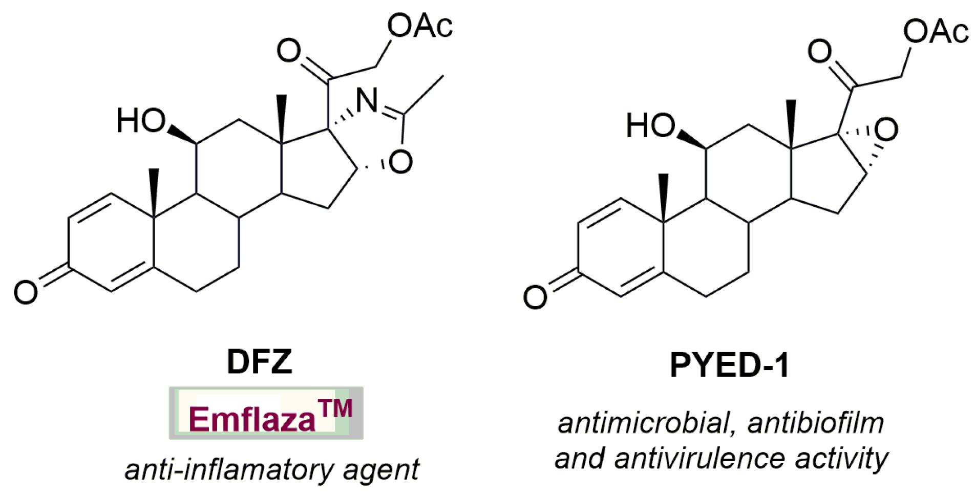
3.1. Fungal Strain, Culture Condition and Compound Synthesis

Candida strains evaluated in this study included the C. albicans ATCC 10231 reference strain and 6–8 clinical isolates of each Candida species belonging to a bacterial collection that was previously established. No ethical approval was required for this study as there was no access to patients’ data. Strains were routinely grown on Sabouraud dextrose agar (SDA) and in RPMI-1640 broth buffered with MOPS (pH 7.0) containing glucose 2% (RPMI-1640 2% G) at 37 °C. The chemical synthesis and structural characterization of PYED-1 were accomplished as previously reported [13]. Stock solutions of the compounds at the concentration of 50 mg/mL were prepared in dimethyl sulfoxide (DMSO) and kept at −20 °C and diluted in RPMI-1640 2% G broth just prior to the assays.

3.2. Determination of the Minimum Inhibitory Concentration (MIC) and the Minimum Fungicide Concentration (MFC)

PYED-1 was evaluated for its in vitro antifungal activities according to EUCAST guidelines using the two-fold serial dilution technique in 96-well microtiter plates [30]. Briefly, yeasts from overnight cultures on SDA were suspended in RPMI-1640 2% G broth to get 1–5 × 10⁵ colony-forming units (cfu)/mL. Two-fold serial dilutions of PYED-1 were prepared in RPMI-1640 2% G medium, starting from 512 to 1 µg/mL, in order to obtain a 2× final concentration of the antifungal agent. Each well of plate was inoculated with 100 µL of 1–5 × 10⁵ cfu/mL yeast suspension and 100 µL of twice the final antifungal agent PYED-1 or 100 µL of sterile drug-free medium (growth control). A sterility control containing 200 mL RPMI-1640 medium was included in each plate. The plate was incubated without agitation at 37 °C for 24–28 h. The optical density at 450 nm was measured using a microplate reader (Bio-Rad Laboratories S.r.l., Segrate (MI) - Italy). MIC was the lowest concentration inhibiting ≥90% growth when compared to growth control. The effect of different concentrations of DMSO (ranging from 0.1% to 0.5%) on fungal growth was separately tested. In order to evaluate the minimum lethal concentration (MLC, i.e., the lowest concentration showing a growth inhibition of 100%), 20 µL of yeast suspensions from wells without visible growth were transferred to SDA plates. These plates were incubated at 37 °C and checked for growth after 24 h. All experiments were performed in triplicate and were repeated independently at least three times.

3.3. Checkboard Microdilution Assay

The checkerboard method was used to evaluate the MICs for PYED-1 alone and in combination with the antifungal agents fluconazole and posaconazole for C. parapsilosis 84,609 and 84,614, C. glabrata 61,115 and C. albicans 61,446 and 61,691 strains, as described previously [31]. The plates were incubated at 37 °C for 24–28 h. The fractional inhibitory concentration index was calculated for the compound combinations, with a FICI < 0.5 indicating “synergism”, a FICI > 4 indicating “antagonism”, and a FICI between 0.5 and 4 defined as “without interaction” [32]. The results were obtained from tests that were repeated three times.

3.4. Germ Tube Inhibition Assay

To determine serum-induced cell filamentation, C. albicans ATCC 10,231 cells were inoculated in 2 mL of RPMI-1640 medium containing 10% (v/v) FBS at a density of 106 cfu/mL in a glass tube without (control) or with the addition of PYED-1 solutions at 2, 4, 8, 16 and 32 µg/mL. After a 5 h incubation at 37 °C, 30 µL cells from treated and untreated samples were smeared, fixed, stained with 0.3% crystal violet and observed under a light microscope.

3.5. Adhesion Assay

The adhesion prevention efficacy of PYED-1 was assayed using the crystal violet biofilm staining method [33]. Cultures of C. albicans ATCC 10,231 were grown in RPMI-1640 2% G and incubated overnight at 30 °C with agitation (100 rpm). Cells were harvested and washed with PBS and diluted to a density of 1 × 10⁶ cfu/mL in RPMI-1640 2% G. The wells of polystyrene flat-bottom 96-well microtiter plates were conditioned with 50% FBS in PBS for at least 30 min at room temperature. Then, the FBS was aspirated and wells were rinsed once with PBS. One hundred microliters of the yeast suspension was added to each well containing 100 µL of twice the final concentration of antifungal agent PYED-1 (2, 4, 8, 16 and 32 µg/mL) or 100 µL of sterile drug-free medium (growth control). The plate was incubated at 37 °C for 90 min. Then, the medium was aspirated, wells were washed with PBS twice and 100 µL of 0.1% crystal violet was added to each well. After 15 min, the plate was washed two times and CV was dissolved with 200 µL of 100% ethanol. Absorbance was measured at 595 nm using a microplate reader (Bio-Rad Laboratories S.r.l.). The percentage of biofilm mass reduction was calculated as [(Ac−At)/Ac] × 100, where Ac is the OD595 for the control well and At is the OD595 for the biofilm in the presence of PYED-1. All data points are expressed as means + SDs of three separate experiments performed in triplicate.

3.6. Anti-Preformed-Biofilm Assay

Biofilms were allowed to form in each well as described in the adhesion assay and were treated following 24 h of incubation. After the adhesion step, nonadherent cells were removed and wells were washed with PBS. Two hundred microliters of RPMI-1640 2% G was added to each well and subsequently incubated at 37 °C for 18–24 h in an orbital shaker (100 rpm). After incubation, wells were washed with PBS and 200 µL of twice the final antifungal agent PYED-1 (64, 128 and 256 µg/mL) or 100 µL of sterile drug-free medium (growth control) was added. Biofilm was incubated statically at 37 °C for an additional 18–24 h. After PYED-1 treatment, the metabolic activities of biofilm cells and biofilm biomass were measured by XTT reduction assay and CV staining [34], respectively. Briefly, 200 μL of a solution of XTT (0.5 mg/mL in PBS; Sigma, Milan, Italy) menadione (1 µM in acetone; Sigma) was added to each prewashed well. Following incubation in the dark for up to 2 h at 37 °C, the biofilm metabolic activity was determined through the measurement of the absorbance value at 490 nm using a microplate reader (Bio-Rad Laboratories S.r.l.). Viability values were compared with respect to control samples treated with scalar doses of DMSO (concentrations ranging from 0.064% to 0.256%). All data points are expressed as means + SDs of three separate experiments performed in triplicate.

3.7. Statistical Analysis

All statistical analyses were performed with GraphPad Prism 8 software (GraphPad, San Diego, CA, USA). All results are presented as arithmetic means ± standard deviations. Statistical differences between PYED-1 treated and untreated samples were analyzed by one-way analysis of variance (ANOVA) followed by Student’s t-test. Statistical significance was considered at p-value of <0.05.

Author Contributions

Conceptualization, E.D.G.; methodology, A.E., A.M. and V.D.I.; formal analysis, E.D.G.; investigation, E.D.G., A.E. and A.M.; data curation, A.G. and E.D.G.; writing—original draft preparation, A.G., R.Z. and E.D.G.; funding acquisition, R.Z. All authors have read and agreed to the published version of the manuscript.

Funding

This research was funded in part by a grant from the Italian Ministry of Education, University and Research (MIUR): PRIN2017 (Grant 2017SFBFER to RZ).

Acknowledgments

We gratefully acknowledge Pierpaolo Di Nocera for his comments and critical reading of the manuscript.

Conflicts of Interest

The authors declare no conflict of interest.

References

  1. Pappas, P.G.; Lionakis, M.S.; Arendrup, M.C.; Ostrosky-Zeichner, L.; Kullberg, B.J. Invasive candidiasis. Nat. Rev. Dis. Primers 2018, 4, 18026. [Google Scholar] [CrossRef]
  2. Martin-Loeches, I.; Antonelli, M.; Cuenca-Estrella, M.; Dimopoulos, G.; Einav, S.; De Waele, J.J.; Garnacho-Montero, J.; Kanj, S.S.; Machado, F.R.; Montravers, P.; et al. ESICM/ESCMID task force on practical management of invasive candidiasis in critically ill patients. Intensive Care Med. 2019, 45, 789–805. [Google Scholar] [CrossRef]
  3. Turner, S.A.; Butler, G. The Candida pathogenic species complex. Cold Spring Harb. Perspect. Med. 2014, 4, a019778. [Google Scholar] [CrossRef] [PubMed] [Green Version]
  4. Sanguinetti, M.; Posteraro, B.; Lass-Flörl, C. Antifungal drug resistance among Candida species: Mechanisms and clinical impact. Mycoses 2015, 58, 2–13. [Google Scholar] [CrossRef]
  5. Pfaller, M.A.; Diekema, D.J.; Turnidge, J.D.; Castanheira, M.; Jones, R.N. Twenty years of the SENTRY antifungal surveillance program: Results for Candida species from 1997-2016. Open Forum Infect. Dis. 2019, 6, S79–S94. [Google Scholar] [CrossRef] [PubMed] [Green Version]
  6. Koehler, P.; Stecher, M.; Cornely, O.A.; Koehler, D.; Vehreschild, M.J.G.T.; Bohlius, J.; Wisplinghoff, H.; Vehreschild, J.J. Morbidity and mortality of candidaemia in Europe: An epidemiologic meta-analysis. Clin. Microbiol. Infect. 2019, 25, 1200–1212. [Google Scholar] [CrossRef] [PubMed]
  7. Li, W.S.; Chen, Y.C.; Kuo, S.F.; Chen, F.J.; Lee, C.H. The impact of biofilm formation on the persistence of candidemia. Front. Microbiol. 2018, 9, 1196. [Google Scholar] [CrossRef] [Green Version]
  8. De Fenza, M.; D’Alonzo, D.; Esposito, A.; Munari, S.; Loberto, N.; Santangelo, A.; Lampronti, I.; Tamanini, A.; Rossi, A.; Ranucci, S.; et al. Exploring the effect of chirality on the therapeutic potential of N-alkyl-deoxyiminosugars: Anti-inflammatory response to Pseudomonas aeruginosa infections for application in CF lung disease. Eur. J. Med. Chem. 2019, 175, 63–71. [Google Scholar] [CrossRef]
  9. De Gregorio, E.; Esposito, A.; Vollaro, A.; De Fenza, M.; D’Alonzo, D.; Migliaccio, A.; Iula, V.D.; Zarrilli, R.; Guaragna, A. N-Nonyloxypentyl-l-Deoxynojirimycin Inhibits Growth, Biofilm Formation and Virulence Factors Expression of Staphylococcus aureus. Antibiotics 2020, 9, 362. [Google Scholar] [CrossRef]
  10. Esposito, A.; D’Alonzo, D.; De Fenza, M.; Gregorio, E.; Tamanini, A.; Lippi, G.; Dechecchi, M.C.; Guaragna, A. Synthesis and Therapeutic Applications of Iminosugars in Cystic Fibrosis. Int. J. Mol. Sci. 2020, 21, 3353. [Google Scholar] [CrossRef]
  11. Esposito, A.; D’Alonzo, D.; D’Errico, S.; De Gregorio, E.; Guaragna, A. Toward the Identification of Novel Antimicrobial Agents: One-Pot Synthesis of Lipophilic Conjugates of N-Alkyl d- and l-Iminosugars. Mar. Drugs 2020, 18, 572. [Google Scholar] [CrossRef]
  12. Esposito, A.; di Giovanni, C.; De Fenza, M.; Talarico, G.; Chino, M.; Palumbo, G.; Guaragna, A.; D’Alonzo, D. A Stereoconvergent Tsuji-Trost Reaction in the Synthesis of Cyclohexenyl Nucleosides. Chemistry 2020, 26, 2597–2601. [Google Scholar] [CrossRef]
  13. Esposito, A.; De Gregorio, E.; De Fenza, M.; D’Alonzo, D.; Satawani, A.; Guaragna, A. Expeditious synthesis and preliminary antimicrobial activity of deflazacort and its precursors. RSC Adv. 2019, 9, 21519–21524. [Google Scholar] [CrossRef] [Green Version]
  14. Esposito, A.; Vollaro, A.; Esposito, E.P.; D’Alonzo, D.; Guaragna, A.; Zarrilli, R.; De Gregorio, E. Antibacterial and antivirulence activity of glucocorticoid PYED-1 against Stenotrophomonas maltophilia. Antibiotics 2020, 9, 105. [Google Scholar] [CrossRef] [PubMed] [Green Version]
  15. Vollaro, A.; Esposito, A.; Antonaki, E.; Iula, V.D.; D’Alonzo, D.; Guaragna, A.; De Gregorio, E. Steroid derivatives as potential antimicrobial agents against Staphylococcus aureus planktonic cells. Microorganisms 2020, 8, 468. [Google Scholar] [CrossRef] [PubMed] [Green Version]
  16. Vollaro, A.; Esposito, A.; Esposito, E.P.; Zarrilli, R.; Guaragna, A.; De Gregorio, E. PYED-1 inhibits biofilm formation and disrupts the preformed biofilm of Staphylococcus aureus. Antibiotics 2020, 9, 240. [Google Scholar] [CrossRef] [PubMed]
  17. Mayer, F.L.; Wilson, D.; Hube, B. Candida albicans pathogenicity mechanisms. Virulence 2013, 4, 119–128. [Google Scholar] [CrossRef] [Green Version]
  18. Chandra, J.; Mukherjee, P.K.; Ghannoum, M.A. Candida biofilms associated with CVC and medical devices. Mycoses 2012, 55, 46–57. [Google Scholar] [CrossRef]
  19. Mah, T.F. Biofilm-specific antibiotic resistance. Future Microbiol. 2012, 7, 1061–1072. [Google Scholar] [CrossRef] [Green Version]
  20. Ribeiro, S.M.; Felício, M.R.; Boas, E.V.; Gonçalves, S.; Costa, F.F.; Samy, R.P.; Santos, N.C.; Franco, O.L. New frontiers for anti-biofilm drug development. Pharmacol. Ther. 2016, 160, 133–144. [Google Scholar] [CrossRef]
  21. Nobile, C.J.; Mitchell, A.P. Regulation of cell-surface genes and biofilm formation by the C. albicans transcription factor Bcr1p. Curr. Biol. 2005, 15, 1150–1155. [Google Scholar] [CrossRef] [PubMed] [Green Version]
  22. Chandra, J.; Kuhn, D.M.; Mukherjee, P.K.; Hoyer, L.L.; McCormick, T.; Ghannoum, M.A. Biofilm formation by the fungal pathogen Candida albicans: Development, architecture, and drug resistance. J. Bacteriol. 2001, 183, 5385–5394. [Google Scholar] [CrossRef] [PubMed] [Green Version]
  23. Lu, Y.; Su, C.; Liu, H. Candida albicans hyphal initiation and elongation. Trends Microbiol. 2014, 22, 707–714. [Google Scholar] [CrossRef] [Green Version]
  24. Sardi, J.C.; Scorzoni, L.; Bernardi, T.; Fusco-Almeida, A.M.; Mendes Giannini, M.J. Candida species: Current epidemiology, pathogenicity, biofilm formation, natural antifungal products and new therapeutic options. J. Med. Microbiol. 2013, 62, 10–24. [Google Scholar] [CrossRef] [PubMed]
  25. Ramage, G.; Robertson, S.N.; Williams, C. Strength in numbers: Antifungal strategies against fungal biofilms. Int. J. Antimicrob. Agents 2013, 43, 114e120. [Google Scholar] [CrossRef] [PubMed]
  26. McCall, A.D.; Pathirana, R.U.; Prabhakar, A. Candida albicans biofilm development is governed by cooperative attachment and adhesion maintenance proteins. NPJ Biofilms Microbiomes 2019, 21, 94–95. [Google Scholar] [CrossRef] [Green Version]
  27. de Groot, P.W.J.; Bader, O.; de Boer, A.D.; Weig, M.; Chauhan, N. Adhesins in human fungal pathogens: Glue with plenty of stick. Eukaryot. Cell 2013, 12, 470–481. [Google Scholar] [CrossRef] [Green Version]
  28. Nobile, C.J.; Nett, J.E.; Andes, D.R.; Mitchell, A.P. Function of Candida albicans adhesin Hwp1 in biofilm formation. Eukaryot. Cell 2006, 5, 1604–1610. [Google Scholar] [CrossRef] [Green Version]
  29. Gauwerky, K.; Borelli, C.; Korting, H.C. Targeting virulence: A new paradigm for antifungals. Drug Discov. Today 2009, 14, 214–222. [Google Scholar] [CrossRef]
  30. EUCAST Definitive Document EDef 7.1: Method for the determination of broth dilution MICs of antifungal agents for fermentative yeasts. Clin. Microbiol. Infect. 2008, 14, 398–405. [CrossRef]
  31. Dantas-Medeiros, R.; Zanatta, A.C.; de Souza, L.B.F.C.; Fernandes, J.M.; Amorim-Carmo, B.; Torres-Rêgo, M.; Fernandes-Pedrosa, M.F.; Vilegas, W.; Araújo, T.A.S.; Michel, S.; et al. Antifungal and Antibiofilm Activities of B-Type Oligomeric Procyanidins From Commiphora leptophloeos Used Alone or in Combination with Fluconazole Against Candida spp. Front Microbiol. 2021, 12, 613155. [Google Scholar] [CrossRef] [PubMed]
  32. Mukherjee, P.K.; Sheehan, D.J.; Hitchcock, C.A.; Ghannoum, M.A. Combination treatment of invasive fungal infections. Clin. Microbiol. Rev. 2005, 18, 163–194. [Google Scholar] [CrossRef] [PubMed] [Green Version]
  33. Krom, B.P.; Cohen, J.B.; McElhaney-Feser, G.; Busscher, H.J.; van der Mei, H.C.; Cihlar, R.L. Conditions for optimal Candida biofilm development in microtiter plates. Methods Mol. Biol. 2009, 499, 55–62. [Google Scholar]
  34. De Gregorio, E.; Esposito, E.P.; Zarrilli, R.; Di Nocera, P.P. Contact-dependent growth inhibition proteins in Acinetobacter baylyi ADP1. Curr. Microbiol. 2018, 75, 1434–1440. [Google Scholar] [CrossRef] [PubMed] [Green Version]
Figure 1. The corticosteroid drug deflazacort (DFZ) and its synthetic precursor PYED-1.
Antibiotics 10 01396 g001
Figure 2. Eradication biofilm assay. The effect of treatment with 128, 64 and 32 μg/mL of PYED-1 on preformed biofilm of Candida species. The metabolic activities of biofilm cells (A) and biofilm biomass (B) were measured by XTT reduction assay and CV staining, respectively. All experiments were performed in triplicate. Asterisks indicate a statistically significant difference (** p-value ≤ 0.01 and *** p-value ≤ 0.001 from the nontreated controls without PYED-1).
Antibiotics 10 01396 g002
Figure 3. Light-microscopy images taken at 100× magnification of C. albicans ATCC 10231 stained with 0.3% crystal violet, without (A) or with the addition of PYED-1 solutions at 2 µg/mL (B), 4 µg/mL (C), 8 µg/mL (D), 16 µg/mL (E) and 32 µg/mL (F).
Antibiotics 10 01396 g003
Figure 4. The adhesion of C. albicans with the addition of different concentrations of PYED-1. The adhesion was measured after 90 min of addition of PYED-1 by CV staining. All experiments were performed in triplicate. Asterisks indicate a statistically significant difference (*** p-value ≤ 0.001 from the nontreated controls without PYED-1).
Antibiotics 10 01396 g004
Table 1. Minimal inhibitory concentration (MIC) and minimal lethal concentration (MLC) of PYED-1 against Candida species.
SpeciesStrainPYED-1Fluconazole
MIC (µg/mL)MLC (µg/mL)MIC (g/mL)
C. albicansATCC 1023116641
6114216640.5
6128016640.5
614463264128
6154016640.5
61558321281
61691326464
6202016640.5
6203316641
C. glabrata6093232640.5
6094032640.5
61112161281
61115321284
6582164>1281
6767664>1281
8126332640.5
C. parapsilosis60568321280.5
6144664>1282
66627128>1280.5
80149128>1281
8120864>1280.5
81879128>1280.5
84609128>12832
84614128>12832
C. tropicalis6098116320.5
6122064>1281
8122232640.5
8125232640.5
8141964>1280.5
82193321281
C. krusei6115932>12832
6705332>12864
6978832>12832
7145632>12832
812883212816
8166732>12832
Publisher’s Note: MDPI stays neutral with regard to jurisdictional claims in published maps and institutional affiliations.

