Nanotechnology-Based Detection of Sickle Cell Disease and Thalassemia: A Systematic Review

 , , , and
Abstract
1. Introduction
2. Materials and Methods
2.1. Focus Question
2.2. Inclusion Criteria
- Study Design: Original research articles, including experimental, observational, and diagnostic accuracy studies.
- Population: Studies focusing on human subjects with SCD and thalassemia.
- Intervention/Exposure: Studies evaluating the use of nanotechnology for the detection or quantification of SCD and thalassemia.
- Outcomes: Studies reporting on analytical sensitivity, analytical specificity, limit of detection and limit of quantitation.
- Language: Studies published in English.
- Publication Status: Peer-reviewed articles published in scientific journals.
- Timeframe: Studies published from inception to the present.
2.3. Exclusion Criteria
- Editorials, or conference proceedings
- Studies focusing on non-human subjects
- Studies not using nanotechnology
- Abstracts or unpublished data
- Review or systematic review articles.
- Studies published in languages other than English.
2.4. Search Strategy and Information Sources
- Preliminary search: A limited search of PubMed to identify relevant articles.
- Keyword extraction: Relevant words from article titles, abstracts, and index terms were extracted to develop a full search strategy.
- Database searching: Multiple databases/sources (listed in Table 2) were searched using the adapted search strategy.
- Vocabulary control: Controlled vocabularies were used to identify synonyms and index terms.
2.5. Study Selection and Screening Process
2.6. Data Extraction
2.7. Data Synthesis
3. Results and Discussion
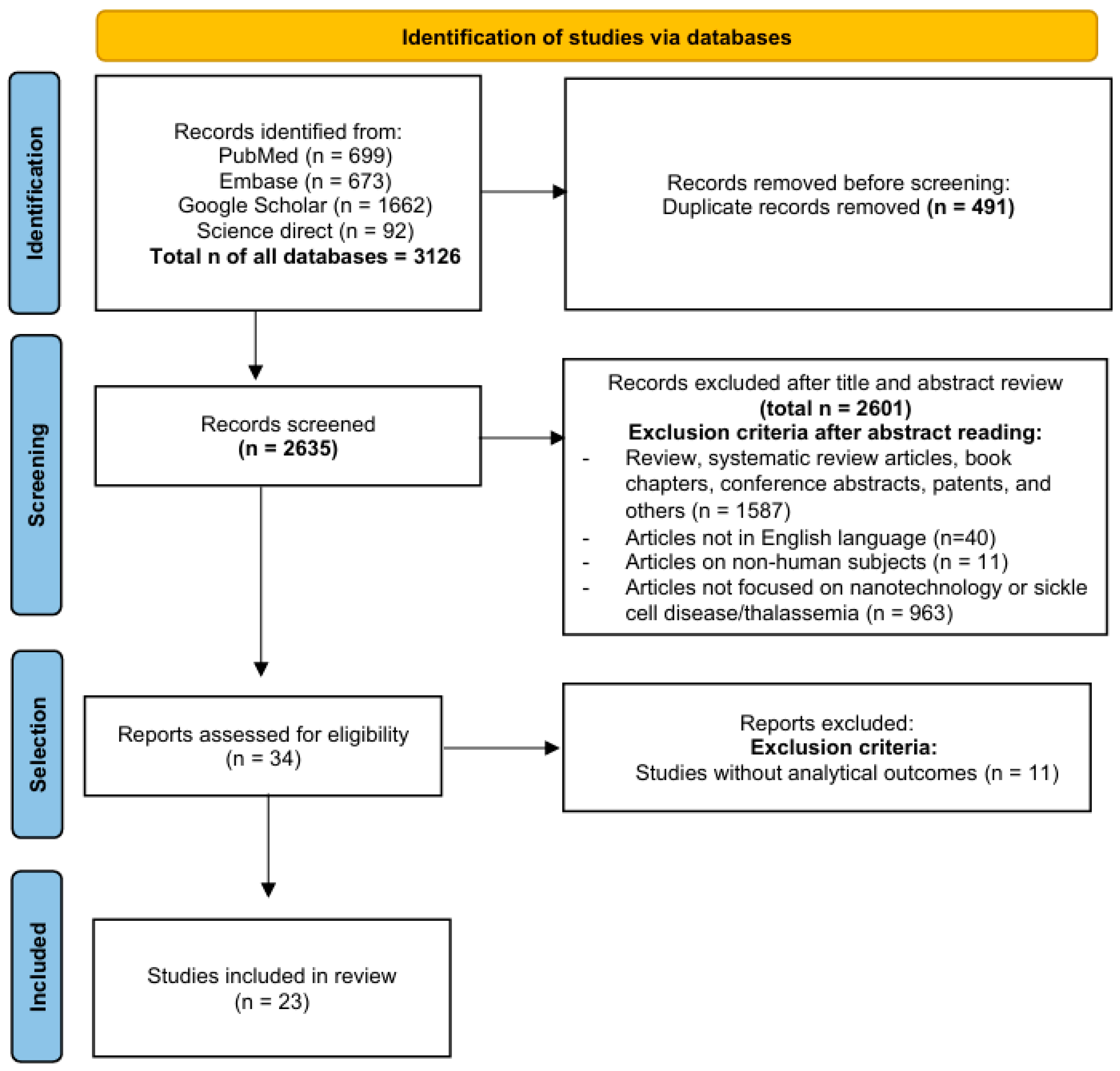
3.1. Study Selection
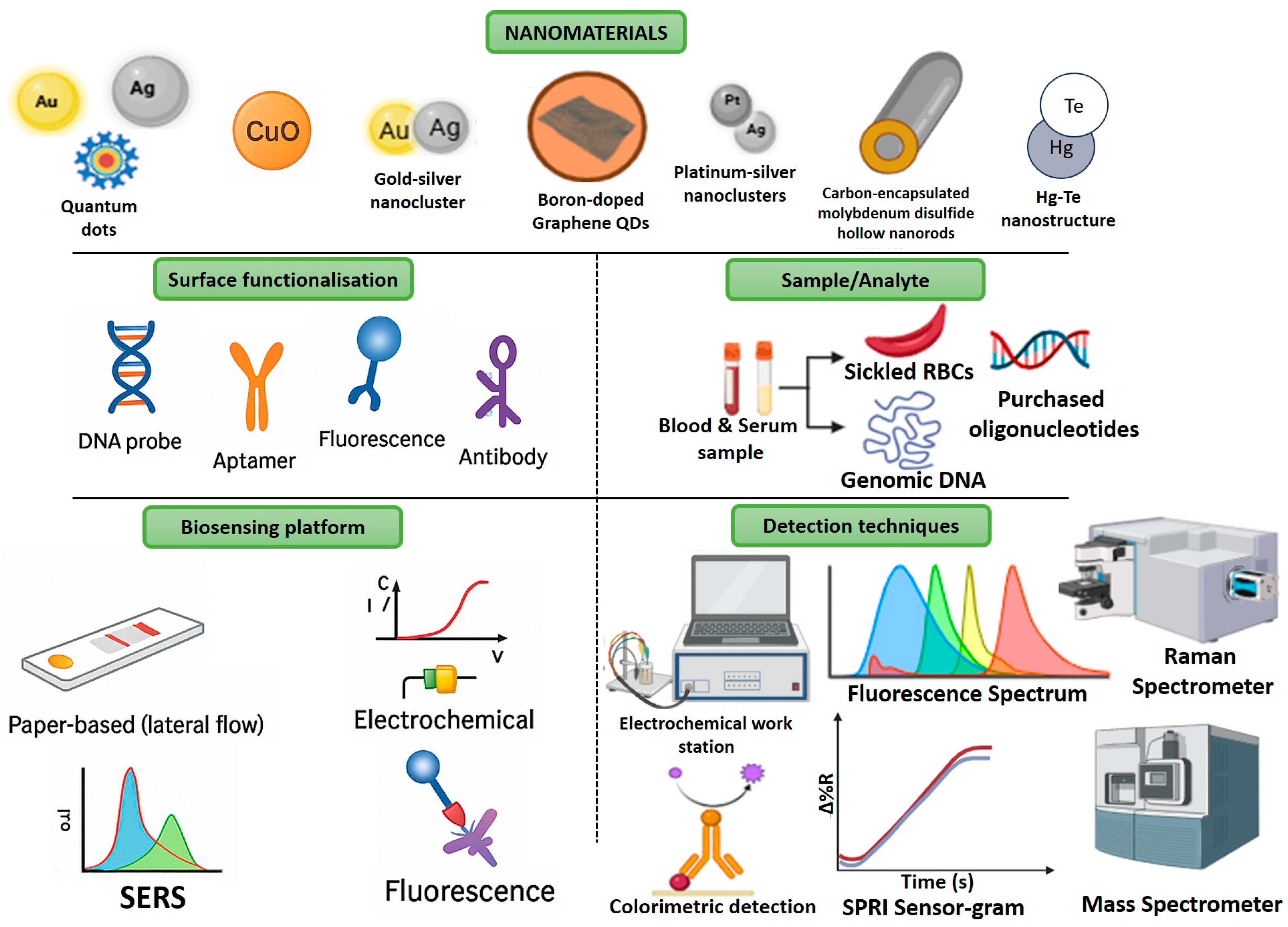
3.2. Analytical Performance of Nanotechnology-Based Approaches
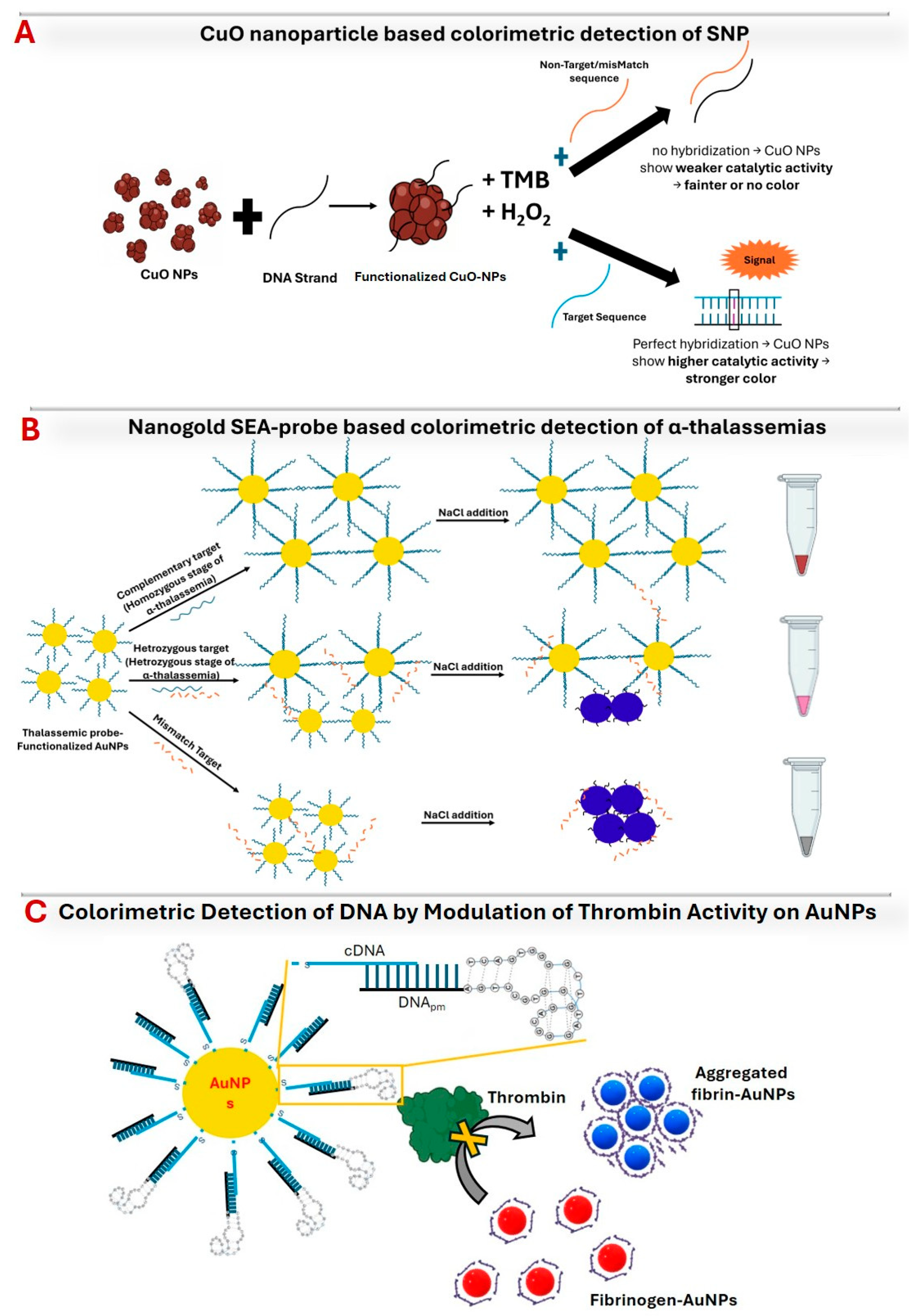
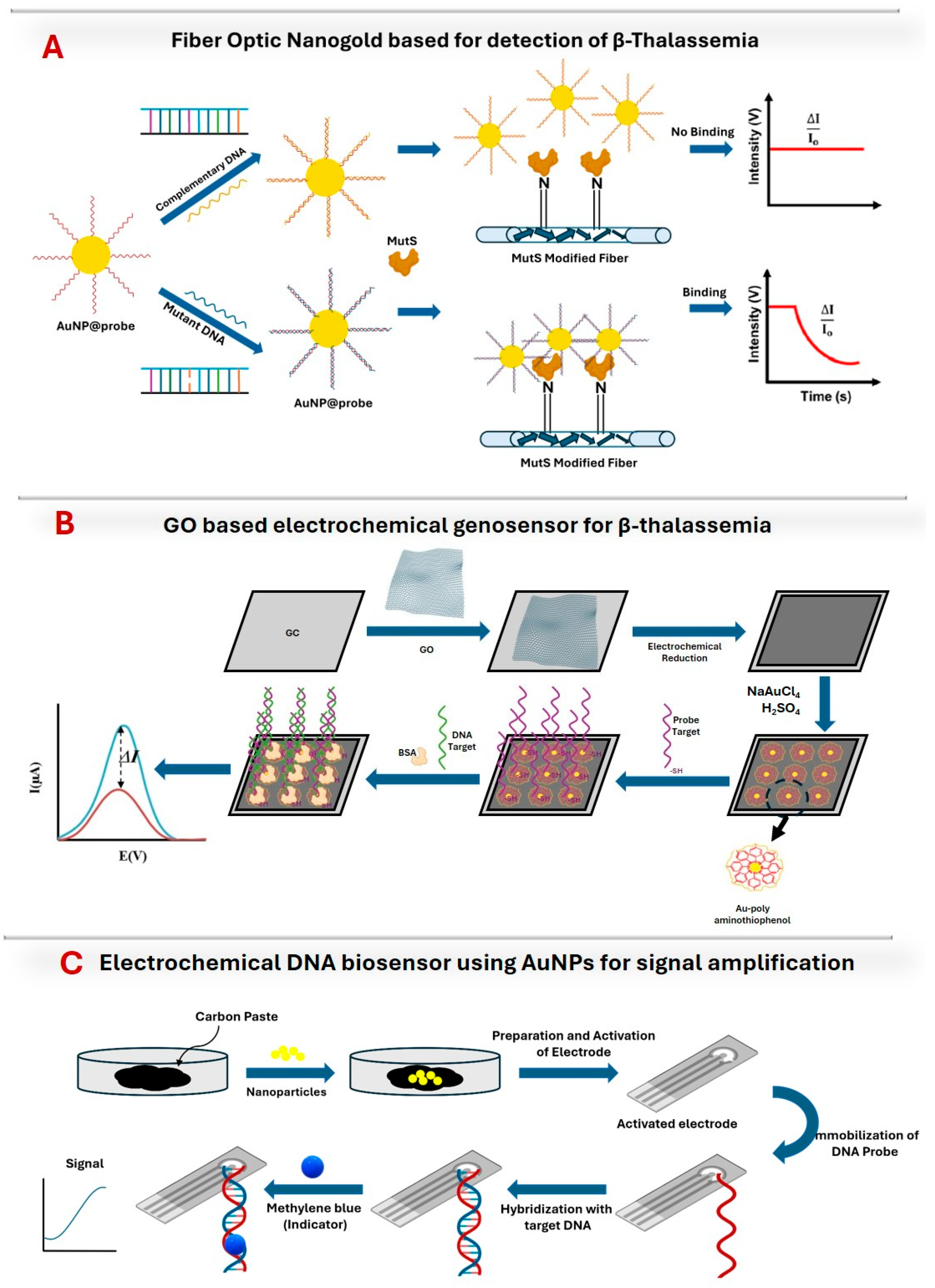
3.2.1. Gold Nanoparticles
3.2.2. Quantum Dots
3.2.3. Silver Nanoparticles
3.2.4. Other Nanoparticles
3.2.5. Sample/Analyte Requirements
3.2.6. Nanoparticle Modifications and Functionalization
3.2.7. Detection Techniques
3.2.8. Clinical Validation
3.3. Practical Implications: Cost, Sample Requirements, and Scalability
3.4. Limitations
4. Conclusions
Author Contributions
Funding
Institutional Review Board Statement
Informed Consent Statement
Data Availability Statement
Acknowledgments
Conflicts of Interest
Abbreviations
| 4-ATP | 4-Aminothiophenol |
| 6-AFM | 6-Amino-Fluorescein |
| AgNPs | Silver Nanoparticles |
| Au@Ag NPs | Gold–Silver Bimetallic Nanoparticles |
| AuNPs | Gold Nanoparticles |
| BSA | Bovine Serum Albumin |
| CNBr | Cyanogen Bromide |
| CuO | Copper Oxide |
| CV | Cyclic Voltammetry |
| DPV | Differential Pulse Voltammetry |
| EIS | Electrochemical Impedance Spectroscopy |
| EOCV | Electrochemical Open Circuit Voltage |
| EW-CRDS | Enhanced Waveguide-Cavity Ring-Down Spectroscopy |
| FONLISA | Fiber Optic Nanogold-Linked Sorbent Assay |
| FOPPR | Fiber Optic Particle Plasmon Resonance |
| GC | Glassy Carbon |
| GDY | Graphdiyne |
| GOD | Glucose Oxidase |
| H2O2 | Hydrogen Peroxide |
| H2SO4 | Sulfuric Acid |
| HPLC | High-Performance Liquid Chromatography |
| HgTe | Mercury Telluride |
| LNA | Locked Nucleic Acid |
| LOD | Limit of Detection |
| LSV | Linear Sweep Voltammetry |
| MCPE | Modified Carbon Paste Electrode |
| ME | Magnetoelastic |
| MHDA | 16-Mercaptohexadecanoic Acid |
| MoS2 | Molybdenum Disulfide |
| MoS2@C | Carbon-Encapsulated Molybdenum Disulfide Hollow Nanorods |
| MNPs | Magnetic Nanoparticles |
| MS | Mass Spectrometry |
| NaAuCl4 | Sodium Tetrachloroaurate |
| NaCl | Sodium Chloride |
| NMs | Nanomaterials |
| NPs | Nanoparticles |
| OCP | Open Circuit Potential |
| PAT | Poly(4-Aminothiophenol) |
| PDDA | Poly(Diallyldimethylammonium Chloride) |
| PRISMA | Preferred Reporting Items for Systematic Reviews and Meta-Analyses |
| Pt | Platinum |
| QCM | Quartz Crystal Microbalance |
| QDs | Quantum Dots |
| rGO | Reduced Graphene Oxide |
| RT-qPCR | Reverse Transcription Quantitative Polymerase Chain Reaction |
| SALDI-MS | Surface-Assisted Laser Desorption/Ionization Mass Spectrometry |
| SAM | Self-Assembled Monolayer |
| SB | Sulfobetaine |
| SCA | Sickle Cell Anemia |
| SCD | Sickle Cell Disease |
| SEA | Southeast Asian |
| SEA-probe | Thiol-Modified DNA Probes |
| SERS | Surface-Enhanced Raman Scattering |
| SNP | Single Nucleotide Polymorphism |
| SNVs | Single-Nucleotide Variants |
| SPRI | Surface Plasmon Resonance Imaging |
| SWV | Square Wave Voltammetry |
| TBA29 | Thrombin-Binding Aptamer (29-mer) |
| TMB | Tetramethylbenzidine |
| tDNA | Target DNA |
| tri-plex-Ag/PtNCs | Triplex DNA-Templated Ag/Pt Bimetallic Nanoclusters |
References
- Piel, F.B.; Patil, A.P.; Howes, R.E.; Nyangiri, O.A.; Gething, P.W.; Dewi, M.; Temperley, W.H.; Williams, T.N.; Weatherall, D.J.; Hay, S.I. Global epidemiology of sickle haemoglobin in neonates: A contemporary geostatistical model-based map and population estimates. Lancet 2013, 381, 142–151. [Google Scholar] [CrossRef]
- Brousseau, D.C.; Panepinto, J.A.; Nimmer, M.; Hoffmann, R.G. The number of people with sickle-cell disease in the United States: National and state estimates. Am. J. Hematol. 2010, 85, 77–78. [Google Scholar]
- CDC. Data and Statistics on Sickle Cell Disease; US Department of Health and Human Services, CDC: Atlanta, GA, USA, 2021. Available online: https://www.cdc.gov/ncbddd/sicklecell/data.html (accessed on 21 November 2024).
- Payne, A.B.; Mehal, J.M.; Chapman, C.; Haberling, D.L.; Richardson, L.C.; Bean, C.J.; Hooper, W.C. Trends in sickle cell disease-related mortality in the United States,1979 to 2017. Ann. Emerg. Med. 2020, 76, S28–S36. [Google Scholar] [CrossRef]
- Brousseau, D.C.; Owens, P.L.; Mosso, A.L.; Panepinto, J.A.; Steiner, C.A. Acute care utilization and rehospitalizations for sickle cell disease. JAMA 2010, 303, 1288–1294. [Google Scholar] [CrossRef] [PubMed]
- Sil, S.; Cohen, L.L.; Dampier, C. Psychosocial and functional outcomes in youth with chronic sickle cell pain. Clin. J. Pain 2016, 32, 527–533. [Google Scholar] [CrossRef] [PubMed]
- Brancaleoni, V.; Di Pierro, E.; Motta, I.; Cappellini, M. Laboratory diagnosis of thalassemia. Int. J. Lab. Hematol. 2016, 38, 32–40. [Google Scholar]
- Aydinok, Y. Thalassemia. Hematology 2012, 17 (Suppl. S1), S28–S31. [Google Scholar] [CrossRef]
- Munkongdee, T.; Chen, P.; Winichagoon, P.; Fucharoen, S.; Paiboonsukwong, K. Update in laboratory diagnosis of thalassemia. Front. Mol. Biosci. 2020, 7, 74. [Google Scholar] [CrossRef]
- Origa, R. β-Thalassemia. Genet. Med. 2017, 19, 609–619. [Google Scholar] [CrossRef]
- Brandow, A.M.; Liem, R.I. Advances in the diagnosis and treatment of sickle cell disease. J. Hematol. Oncol. 2022, 15, 20. [Google Scholar] [CrossRef]
- El-Beshlawy, A.; El-Ghamrawy, M. Recent trends in treatment of thalassemia. Blood Cells Mol. Dis. 2019, 76, 53–58. [Google Scholar] [CrossRef] [PubMed]
- Tari, K.; Valizadeh Ardalan, P.; Abbaszadehdibavar, M.; Atashi, A.; Jalili, A.; Gheidishahran, M. Thalassemia an update: Molecular basis, clinical features and treatment. Int. J. Biomed. Public Health 2018, 1, 48–58. [Google Scholar] [CrossRef]
- Needs, T.; Gonzalez-Mosquera, L.F.; Lynch, D.T. Beta Thalassemia; StatPearls Publishing: Treasure Island, FL, USA, 2018. [Google Scholar]
- Mettananda, S.; Higgs, D.R. Molecular basis and genetic modifiers of thalassemia. Hematol. Oncol. Clin. 2018, 32, 177–191. [Google Scholar] [CrossRef] [PubMed]
- Lee, J.S.; Cho, S.I.; Park, S.S.; Seong, M.-W. Molecular basis and diagnosis of thalassemia. Blood Res. 2021, 56, 39–43. [Google Scholar] [CrossRef]
- De Simone, G.; Quattrocchi, A.; Mancini, B.; di Masi, A.; Nervi, C.; Ascenzi, P. Thalassemias: From gene to therapy. Mol. Asp. Med. 2022, 84, 101028. [Google Scholar] [CrossRef]
- Hardison, R.C. Evolution of hemoglobin and its genes, Cold Spring Harb. Perspect. Med. 2012, 2, a011627. [Google Scholar]
- Arishi, W.A.; Alhadrami, H.A.; Zourob, M. Techniques for the detection of sickle cell disease: A review. Micromachines 2021, 12, 519. [Google Scholar] [CrossRef]
- Khosa, S.M.; Usman, M.; Moinuddin, M.; Mehmood, H.O.; Qamar, K. Comparative analysis of cellulose acetate hemoglobin electrophoresis and high-performance liquid chromatography for quantitative determination of hemoglobin A2. Blood Res. 2015, 50, 46–50. [Google Scholar] [CrossRef]
- Ashley-Koch, A.; Yang, Q.; Olney, R.S. Sickle hemoglobin (Hb S) allele and sickle cell disease: A HuGE review. Am. J. Epidemiol. 2000, 151, 839–845. [Google Scholar] [CrossRef]
- Farid, Y.; Bowman, N.S.; Lecat, P. Biochemistry, Hemoglobin Synthesis; StatPearls Publishing: Treasure Island, FL, USA, 2019. [Google Scholar]
- Yatsunyk, L.A.; Mendoza, O.; Mergny, J.L. “Nano-oddities”: Unusual nucleic acid assemblies for DNA-based nanostructures and nanodevices. Acc. Chem. Res. 2014, 47, 1836–1844. [Google Scholar] [CrossRef]
- Higashi, S.L.; Shibata, A.; Kitamura, Y.; Hirosawa, K.M.; Suzuki, K.G.N.; Matsuura, K.; Ikeda, M. Hybrid soft nanomaterials composed of DNA microspheres and supramolecular nanostructures of semi-artificial glycopeptides. Chem. Eur. J. 2019, 25, 11955–11962. [Google Scholar] [CrossRef] [PubMed]
- Gomes, L.P.; Andrade, C.T.; Aguila, E.M.; Del Alexander, C.; Paschoalin, V.M.F. Assessing the antimicrobial activity of chitosan nanoparticles by fluorescence-labeling. World Acad. Sci. Eng. Technol. Int. J. Biotechnol. Bioeng. 2018, 12, 112–117. [Google Scholar]
- Melegari, S.P.; Fuzinatto, C.F.; Gonçalves, R.A.; Oscar, B.V.; Vicentini, D.S.; Matias, W.G. Can the surface modification and/or morphology affect the ecotoxicity of zinc oxide nanomaterials? Chemosphere 2019, 224, 237–246. [Google Scholar] [CrossRef] [PubMed]
- Wang, J.; Zhuang, Z.; Peng, L.; Li, Y. Nanotechnology for early cancer detection and precision medicine. Nat. Biomed. Eng. 2020, 4, 623–644. [Google Scholar]
- Li, Y.; Wu, X.; Liu, R.; Liu, R.; Wang, Z. Advances in nanomaterial-based electrochemical biosensors for cancer detection. J. Mater. Chem. B 2020, 8, 1005–1024. [Google Scholar]
- Meenambiga, S.S.; Sakthiselvan, P.; Hari, S.; Umai, D. Nanotechnology for blood test to predict the blood diseases/blood disorders. In Nanotechnology for Hematology, Blood Transfusion, and Artificial Blood; Denizli, A., Nguyen, T.A., Rajan, M., Alam, M.F., Rahman, K., Eds.; Elsevier: Amsterdam, The Netherlands, 2022; Chapter 13; pp. 285–311. [Google Scholar]
- Mukhtar, M.; Sargazi, S.; Barani, M.; Madry, H.; Rahdar, A.; Cucchiarini, M. Application of nanotechnology for sensitive detection of low-abundance single-nucleotide variations in genomic DNA: A review. Nanomaterials 2021, 11, 1384. [Google Scholar] [CrossRef]
- Shahrashoob, M.; Dehshiri, M.; Yousefi, V.; Moassesfar, M.; Saberi, H.; Molaabasi, F.; Zare, Y.; Rhee, K.Y. Optical and electrochemical biosensors for detection of pathogens using metal nanoclusters: A systematic review. Biosensors 2025, 15, 460. [Google Scholar] [CrossRef]
- Shoaib, A.; Darraj, A.; Khan, M.E.; Azmi, L.; Alalwan, A.; Alamri, O.; Tabish, M.; Khan, A.U. A nanotechnology-based approach to biosensor application in current diabetes management practices. Nanomaterials 2023, 13, 867. [Google Scholar] [CrossRef]
- Jiang, P.; Gao, N.; Chang, G.; Wu, Y. Biosensors for early detection of Parkinson’s disease: Principles, applications, and future prospects. Biosensors 2025, 15, 280. [Google Scholar] [CrossRef]
- Raveendran, R.; Prabakaran, L.; Senthil, R.; Yesudhason, B.V.; Dharmalingam, S.; Sathyaraj, W.V.; Atchudan, R. Current innovations in intraocular pressure monitoring biosensors for diagnosis and treatment of glaucoma—Novel strategies and future perspectives. Biosensors 2023, 13, 663. [Google Scholar] [CrossRef]
- Ramesh, M.; Janani, R.; Deepa, C.; Rajeshkumar, L. Nanotechnology-enabled biosensors: A review of fundamentals, design principles, materials, and applications. Biosensors 2022, 13, 40. [Google Scholar] [CrossRef] [PubMed]
- Tariq, Z.; Qadeer, M.I.; Anjum, I.; Hano, C.; Anjum, S. Thalassemia and nanotheragnostics: Advanced approaches for diagnosis and treatment. Biosensors 2023, 13, 450. [Google Scholar] [CrossRef] [PubMed]
- Gholivand, M.B.; Akbari, A. A sensitive electrochemical genosensor for highly specific detection of thalassemia gene. Biosens. Bioelectron. 2019, 129, 182–188. [Google Scholar] [CrossRef] [PubMed]
- Jian, J.W.; Huang, C.C. Colorimetric detection of DNA by modulation of thrombin activity on gold nanoparticles. Chem. Eur. J. 2011, 17, 2374–2380. [Google Scholar]
- D’Agata, R.; Breveglieri, G.; Zanoli, L.M.; Borgatti, M.; Spoto, G.; Gambari, R. Direct detection of point mutations in nonamplified human genomic DNA. Anal. Chem. 2011, 83, 8711–8717. [Google Scholar] [CrossRef]
- Ngo, L.T.; Chaudhari, P.; Wang, W.K.; Tseng, Y.T.; Kuo, P.L.; Huang, C.J.; Chiang, C.Y.; Chau, L.K.; Huang, T.T. Noninvasive prenatal genetic screening of cell-free fetal DNA for early prediction of β-thalassemia using fiber optic nanogold-linked sorbent assay. ACS Sens. 2024, 9, 4207–4215. [Google Scholar] [CrossRef]
- Yao, Y.J.; Lin, K.C. DNA interaction probed by evanescent wave cavity ring-down absorption spectroscopy via functionalized gold nanoparticles. Anal. Chim. Acta 2014, 820, 1–8. [Google Scholar] [CrossRef]
- Pang, L.; Li, J.; Jiang, J.; Shen, G.; Yu, R. DNA point mutation detection based on DNA ligase reaction and nano-Au amplification: A piezoelectric approach. Anal. Biochem. 2006, 358, 99–103. [Google Scholar] [CrossRef]
- Chomean, S.; Wangmaung, N.; Sritongkham, P.; Promptmas, C.; Mas-Oodi, S.; Tanyong, D.; Ittarat, W. Molecular diagnosis of α-thalassemias by the colorimetric nanogold. Analyst 2014, 139, 813–822. [Google Scholar] [CrossRef]
- Li, J.; Deng, T.; Chu, X.; Yang, R.; Jiang, J.; Shen, G.; Yu, R. Rolling circle amplification combined with gold nanoparticle aggregates for highly sensitive identification of single-nucleotide polymorphisms. Anal. Chem. 2010, 82, 2811–2816. [Google Scholar] [CrossRef]
- Guo, X.; Wang, J.; Zhao, Y.; Liu, R.; Zhang, Q.; Yuan, Z.; Sang, S. A wireless magnetoelastic DNA-biosensor amplified by AuNPs for the detection of a common mutated DNA causing β-thalassaemia. Biochem. Eng. J. 2020, 156, 107498. [Google Scholar] [CrossRef]
- Thai, D.A.; Park, S.K.; Lee, N.Y. A paper-embedded thermoplastic microdevice integrating additive-enhanced allele-specific amplification and silver nanoparticle-based colorimetric detection for point-of-care testing. Lab Chip 2023, 23, 5081–5091. [Google Scholar] [CrossRef] [PubMed]
- Liu, P.; Na, N.; Liu, T.; Huang, L.; He, D.; Hua, W.; Ouyang, J. Ultrasensitive detection of ferritin in human serum by Western blotting based on quantum dots-labeled avidin-biotin system. Proteomics 2011, 11, 3510–3517. [Google Scholar] [CrossRef] [PubMed]
- Kasiri, M.; Rahaie, M. A visible and colorimetric nanobiosensor based on DNA-CuO nanoparticle for detection of single nucleotide polymorphism involved in sickle cell anemia disease. Mater. Today Commun. 2021, 27, 102423. [Google Scholar] [CrossRef]
- Shi, J.; Tang, D.; Lin, Y.; Wu, Y.; Luo, H.; Yan, J.; Huang, K.J.; Tan, X. A highly sensitive self-powered sensing method designed on DNA circuit strategy and MoS2 hollow nanorods for detection of thalassemia. Anal. Chim. Acta 2023, 1278, 341713. [Google Scholar] [CrossRef]
- Chen, Q.; Chen, H.; Kong, H.; Chen, R.; Gao, S.; Wang, Y.; Zhou, P.; Huang, W.; Cheng, H.; Li, L.; et al. Enzyme-free sensitive SERS biosensor for the detection of thalassemia-associated microRNA-210 using a cascade dual-signal amplification strategy. Anal. Chim. Acta 2024, 1292, 342255. [Google Scholar] [CrossRef]
- Li, P.; Wei, Y.; Shi, J.; Wu, J.; Wu, Y.; Yan, J.; Liu, S.; Tan, X.; Huang, K.J. CRISPR/Cas12a-triggered ordered concatemeric DNA probes signal-on/off multifunctional analytical sensing system for ultrasensitive detection of thalassemia. Int. J. Biol. Macromol. 2024, 276, 133884. [Google Scholar] [CrossRef] [PubMed]
- Ertuğrul Uygun, H.D. Impedimetric CRISPR-dCas9 based biosensor system for sickle cell anemia mutation. J. Turk. Chem. Soc. Sect. A Chem. 2022, 9, 631–638. [Google Scholar] [CrossRef]
- Hamidi-Asl, E.; Raoof, J.B.; Naghizadeh, N.; Akhavan-Niaki, H.; Ojani, R.; Banihashemi, A. A bimetallic nanocomposite modified genosensor for recognition and determination of thalassemia gene. Int. J. Biol. Macromol. 2016, 91, 400–408. [Google Scholar] [CrossRef]
- Chen, W.T.; Huang, M.F.; Chang, H.T. Using surface-assisted laser desorption/ionization mass spectrometry to detect ss- and ds-oligodeoxynucleotides. J. Am. Soc. Mass Spectrom. 2013, 24, 877–883. [Google Scholar] [CrossRef]
- Wu, F.; Lin, Q.; Wang, L.; Zou, Y.; Chen, M.; Xia, Y.; Lan, J.; Chen, J. A DNA electrochemical biosensor based on triplex DNA-templated Ag/Pt nanoclusters for the detection of single-nucleotide variant. Talanta 2020, 207, 120257. [Google Scholar] [CrossRef]
- Kumar, M.; Guo, Y.; Zhang, P. Highly sensitive and selective oligonucleotide sensor for sickle cell disease gene using photon upconverting nanoparticles. Biosens. Bioelectron. 2009, 24, 1522–1526. [Google Scholar] [CrossRef]
- Tang, D.; Shi, J.; Wu, Y.; Luo, H.; Yan, J.; Huang, K.J.; Tan, X. Flexible self-powered sensing system based on novel DNA circuit strategy and graphdiyne for thalassemia gene by rapid naked-eye tracking and open-circuit voltage. Anal. Chem. 2023, 95, 16374–16382. [Google Scholar] [CrossRef] [PubMed]
- Bansal, S.S.; Abbate, V.; Bomford, A.; Halket, J.M.; Macdougall, I.C.; Thein, S.L.; Hider, R.C. Quantitation of hepcidin in serum using ultra-high-pressure liquid chromatography and a linear ion trap mass spectrometer. Rapid Commun. Mass Spectrom. 2010, 24, 1251–1259. [Google Scholar] [CrossRef] [PubMed]
- Ji, L.; Chen, L.; Wu, P.; Gervasio, D.F.; Cai, C. Highly selective fluorescence determination of the hematin level in human erythrocytes with no need for separation from bulk hemoglobin. Anal. Chem. 2016, 88, 3935–3944. [Google Scholar] [CrossRef] [PubMed]



| Population | Human Suffering from Hemoglobinopathies Like SCD and Thalassemia. |
|---|---|
| Intervention | Use of nanotechnology for detection or quantification of hemoglobinopathy-SCD and thalassemia |
| Comparator | - |
| Outcome | Sensitivity, specificity, limit of detection, limit of quantitation (any of these) |
| Type of Database/Literature | Sources |
|---|---|
| Academic literature | PubMed, Science Direct, Embase |
| Non-academic search engines | Google Scholar |
| Nanoparticles | Modifications | Analytes | Techniques | Application | LOD/LOQ | Linear Ranges | Validation | Ref. |
|---|---|---|---|---|---|---|---|---|
| Gold | - AuNPs integrated with conductive polymer PAT-Thiolated probe DNA (via self-assembly) - Composite of AuNPs with rGO and PAT | DNA sequences (Human sample) | DPV; EIS | β-thalassemia gene detection in human serum | LOD: 0.035 pM | 0.5 pM to 400.0 pM | - | [37] |
| Gold | - Fibrinogen conjugation - cDNA functionalization - TBA29 assembly | DNA (SNP) related to SCA (Oligonucleotide) | Colorimetry; UV/Vis spectroscopy | Specific SNP detection for SCA | LOD: 12 pM | 20–500 pM | - | [38] |
| Gold | - AuNPs conjugated to biotinylated DNA - Streptavidin attached to the AuNPs | Genomic DNA (Human sample) | SPRI | Diagnosis of β-thalassemia | LOD: 2.6 aM (5 pg/μL) | NA | 32 (18 controls, 7 homozygous, 7 heterozygous) | [39] |
| Gold | DNA probe was physiosorbed to AuNP surface. Surface functionalization with SB thiol and MHDA | Cell-free genomic DNA (Human sample) | FONLISA using FOPPR biosensor | Non-invasive way to diagnose β-thalassemia in fetuses | LOD: 5.2 × 10⁻¹⁶ M | NA | - | [40] |
| Gold | AuNPs are modified with thiol groups, then conjugated with DNA | DNA (gene associated with SCA) (Oligonucleotides) | EW-CRDS | Early disease diagnosis-SCD | LOD: 1.2 pM | NA | - | [41] |
| Gold | The AuNPs were functionalized with a 30-terminal sulfur of the oligonucleotide. Nano-Au label: The AuNPs were used as a label | Oligonucleotides (point mutation in the β-thalassemia gene) | Piezoelectric method based on the DNA ligase reaction and nano-Au-amplified DNA probes, utilizing QCM for detection | Diagnosis of β-thalassemia | LOD: 2.6 × 10⁻⁹ mol/L | NA | - | [42] |
| Gold | Conjugation of AuNPs with SEA-probe to create nanogold SEA-probe | DNA (α-globin gene associated with α-thalassemia 1, SEA deletion) (Human sample) | NP-based colorimetric detection and DNA hybridization techniques | Colorimetric detection of alpha thalassemia | LOD: 200 ug/mL of target DNA by the naked eye | NA | 45 | [43] |
| Gold | AuNPs were modified with 3′- or 5′-(alkanethiol) oligonucleotides | DNA (SNP in the β-thalassemia gene) (Oligonucleotides) | Colorimetric analysis | Identification of point mutations in β-thalassemia gene | LOD: 70 fM | 0.3 pM to 80 pM | - | [44] |
| Gold | - Thiolated sDNA modification allowed the AuNPs to hybridize with the tDNA sequence - sDNA-AuNPs were labeled with 6-AFM for fluorescence microscopy imaging | DNA sequence (4 bp deletion in codon 41/42) (Oligonucleotides) | Wireless resonant frequency measurement using a ME DNA-biosensor | Diagnosis of β-thalassemia | LOD: 0.571 pM. | 1.0 × 10⁻⁸ M to 1.0 × 10⁻¹² M | - | [45] |
| Nanoparticles | Modifications | Analytes | Techniques | Application | LOD/LOQ | Linear Range | Validation | Ref. |
|---|---|---|---|---|---|---|---|---|
| AgNPs | - | SNPs related to SCA (Oligonucleotides) | Digital imaging (smartphone used to capture images of colorimetric detection and analysis using ImageJ) | SCA Detection: Detection of HbS mutation in the Hbb gene for identifying SCA | LOD: 103 copies per μL of target DNA (HbS) in serum samples | NA | - | [46] |
| QDs | QDs were modified with avidin, specifically QDs-labeled avidin | Human serum ferritin (Human sample) | - QDs were used as fluorescent labels to detect the binding of the secondary antibody to the primary antibody, allowing for the visualization of ferritin | Thalassemia diagnosis | LOD: 0.27 ng Linear range: 0.27 to 1.1 ng | 0.27 to 1.1 ng | 16 (4 thalassemia, 10 hepatoma, 2 controls) | [47] |
| CuO NPs | Interaction of CuO NPs with DNA | Cell-free fetal DNA (Human sample) | Colorimetric nanobiosensor | For prenatal diagnosis of SCA disease | LOD: 0.64 nM Linear range: 2 nM to 12 nM | 2 nM to 12 nM | - | [48] |
| Nanoparticles | Modifications | Analytes | Techniques | Application | LOD/LOQ | Linear Range | Validation | Ref. |
|---|---|---|---|---|---|---|---|---|
| AuNPs and MoS2@C | -MoS2 NPs encapsulated in carbon to form MoS2@C NPs MoS2@C NPs modified with PDDA AuNPs conjugated to PDDA-modified MoS2@C NPs to form AuNPs/MoS2@C NPs | CD122 gene (Human sample) | - EOCV - Colorimetric detection using a smartphone camera | Thalassemia gene detection | - EOCV: LOD: 78.7 aM - Colorimetric: LOD: 58.5 aM. | - EOCV: 0.0001–100 pM - Colorimetric: 0.0001–10,000 Pm | - | [49] |
| Au@Ag NPs | Sulphhydryl-modified capture probe bound to Au@Ag NPs via Ag-S bonding | MicroRNA-210 (miR-210) (Human sample) | - SERS - RT-qPCR | Early and rapid screening of β-thalassemia | LOD: 5.13 fM | 10 fM–1.0 nM | 8 (4 β-thalassemia, 4 controls) | [50] |
| AuNPs/GDY nanocomposite | - AuNPs GDY nanosheets - AuNPs/GDY nanocomposite functionalized with a substrate material to create a bioelectrode - Bioelectrode was modified with glucose oxidase and a CRISPR/Cas12a-ordered concatemeric DNA probe to create a self-powered biosensing system | CD142 gene (Human sample) | - OCP - Visual signal detection (using colorimetric assay with TMB and HRP) - CRISPR/Cas12a-based biosensing | Detection of thalassemia in human serum samples. | LOD: 48.1 aM of CD142 DNA sequence. | 0.0001–100 pM | - | [51] |
| AuNPs and graphene oxide NPs | - AuNPs - 4-Aminothiophenol (4-ATP) to form a self-assembled monolayer (SAM) - Glutaraldehyde - Graphene oxide NPs—ammonium functionalization | DNA (SCA mutation) (Oligonucleotide) | EIS | Diagnosing and screening SCA | LOD: 40 pM | 40–1000 pM | - | [52] |
| Platinum (Pt) and AgNP | Modified with carbon paste to form MCPE. Additionally, the AgNPs were also modified with Pt to form bimetallic nanocomposite electrode | PCR-amplified β-globin gene sequence (Human sample) | EIS and LSV | Detection of β-thalassemia | LOD: 470.0 pg/μL | NA | - | [53] |
| HgTe nanostructures Gold | HgTe nanostructures were used as the matrix for SALDI-MS analysis. AuNPs were functionalized with thiol-modified capture ODNs | Single- and double-stranded oligodeoxynucleotides (related to SCD) (Oligonucleotide) | SALDI-MS Analysis | Analyzing a SNP that determines the fate of the valine residue in the β-globin chain of sickle cell megaloblasts. | LOD: 0.05 μM | 0.1–1.0 μM | - | [54] |
| Ag/Pt bimetallic nanoclusters | Tri-plex-Ag/PtNCs and LNA modified X-shaped DNA probe | DNA (single-nucleotide variant related to β-thalassemia) (Oligonucleotide) | EIS, CV, and SWV | Detection of single-nucleotide variants (SNVs) related to β-thalassemia. | LOD: 0.8 fM | NA | - | [55] |
| NaYF4:Yb3+, Er3+ NPs | Silica coating: A thin layer of SiO2 was deposited on the NPs. DNA was conjugated to the silica-coated NPs using a CNBr activation method | DNA (Oligonucleotide) | - Photoluminescence measurements - Spectro fluorometry | Detection of point mutation associated with SCD | LOD: 120 fM | NA | - | [56] |
| AuNPs/GDY nanocomposite | AuNPs were functionalized with graphdiyne (GDY) to form AuNPs/GDY nanocomposites. These were then used to modify the surface of a flexible carbon cloth (CP) electrode | CD122 gene (Human sample) | - EIS, CV, and LSV to detect the target CD122 gene - Colorimetric assay to detect the target CD122 gene | Detection of the CD122 gene associated with thalassemia in human serum samples | LOD: 36.3 aM (electrochemical mode) and 12.1 aM (colorimetric mode) | 0.0001–10,000 pM | - | [57] |
| Technique | Advantages | Limitations | Sensitivity | Ref. |
|---|---|---|---|---|
| EIS | High sensitivity, label-free detection | Requires specialized equipment, limited multiplexing capabilities | LOD: 0.035 pM Linear range: 0.5 pM to 400.0 pM | [37] |
| SPRI | Real-time detection | Requires specialized equipment, limited multiplexing capabilities | LOD: 2.6 aM (5 pg/μL) Linear range: NA | [39] |
| Fluorescence spectroscopy | Multiplexing capabilities | Requires fluorescent labels, photobleaching concerns | LOD: 120 fM Linear range: NA | [56] |
| Colorimetric detection | Low cost, easy to perform, label-free detection | Limited sensitivity, subjective interpretation. | LOD: 0.64 nM Linear range: 2 nM to 12 nM | [48] |
| SERS | High sensitivity, multiplexing capabilities | Requires specialized equipment, limited reproducibility | LOQ: 5.13 fM Linear range: 10 fM–1.0 nM | [50] |
| QCM | Real-time detection | Requires specialized equipment, limited multiplexing capabilities | LOD: 1.2 pM Linear range: NA | [41] |
| LSV and CV | Provides information on redox reactions and simple equipment required | Time-consuming and require specialized equipment | LOD: 470.0 pg/μL Linear range: NA | [53] |
| MS | Sensitivity and specificity, multiplexing capabilities, and real-time detection | Requires specialized equipment and Time consuming | LOQ 0.4 nmol/L Linear range: NA | [58] |
| Nanoplatform/Technique | Target (Gene/Marker) | Sample Type | Validation Context | Sample Size | Ref. |
|---|---|---|---|---|---|
| AuNPs-SPRI | β-thalassemia point mutations | Genomic DNA | Patient-derived samples | 32 (18 controls, 7 homozygous, 7 heterozygous) | [39] |
| Au@Ag NPs + SERS | miR-210 | Erythrocytes | Patient-derived samples | 8 (4 β-thalassemia, 4 controls) | [50] |
| AuNP nanogold–colorimetric assay | α-thalassemia (SEA deletion) | DNA | Patient-derived samples | 45 | [43] |
| Quantum dots | Serum ferritin | Human serum | Patient-derived samples | 16 (4 thalassemia, 10 hepatoma, 2 controls) | [47] |
| Boron-doped graphene QDs | Hematin | Erythrocytes | Patient-derived samples | 7 (5 controls, 2 SCD) | [59] |
Disclaimer/Publisher’s Note: The statements, opinions and data contained in all publications are solely those of the individual author(s) and contributor(s) and not of MDPI and/or the editor(s). MDPI and/or the editor(s) disclaim responsibility for any injury to people or property resulting from any ideas, methods, instructions or products referred to in the content. |
© 2025 by the authors. Licensee MDPI, Basel, Switzerland. This article is an open access article distributed under the terms and conditions of the Creative Commons Attribution (CC BY) license.
Share and Cite
Kaur, M.; Gautam, J.K.; Sharma, A.R.; Singh, V.; Chouhan, D.; Baghel, A.; Babu, B.V.; Mohanty, S.S. Nanotechnology-Based Detection of Sickle Cell Disease and Thalassemia: A Systematic Review. Biosensors 2026, 16, 373. https://doi.org/10.3390/bios16070373
Kaur M, Gautam JK, Sharma AR, Singh V, Chouhan D, Baghel A, Babu BV, Mohanty SS. Nanotechnology-Based Detection of Sickle Cell Disease and Thalassemia: A Systematic Review. Biosensors. 2026; 16(7):373. https://doi.org/10.3390/bios16070373
Chicago/Turabian StyleKaur, Manjyot, Janesh Kumar Gautam, Aishwarya Rajendra Sharma, Vishal Singh, Disha Chouhan, Akash Baghel, Bontha V. Babu, and Suman Sundar Mohanty. 2026. "Nanotechnology-Based Detection of Sickle Cell Disease and Thalassemia: A Systematic Review" Biosensors 16, no. 7: 373. https://doi.org/10.3390/bios16070373
APA StyleKaur, M., Gautam, J. K., Sharma, A. R., Singh, V., Chouhan, D., Baghel, A., Babu, B. V., & Mohanty, S. S. (2026). Nanotechnology-Based Detection of Sickle Cell Disease and Thalassemia: A Systematic Review. Biosensors, 16(7), 373. https://doi.org/10.3390/bios16070373