E50A Mutation Increases the Bioluminescence Activity of picALuc


Abstract
1. Introduction
2. Methods
2.1. Structural Modeling of WT and E50A Mutant picALuc
2.2. MD Simulations
2.3. mGreenLantern (mGL)-picALuc Plasmid Construct Generation
2.4. Cell Culture and Transfection
2.5. Live Cell Assays
2.6. In Vitro Assays
2.7. Data Analysis and Figure Preparation
3. Results and Discussion
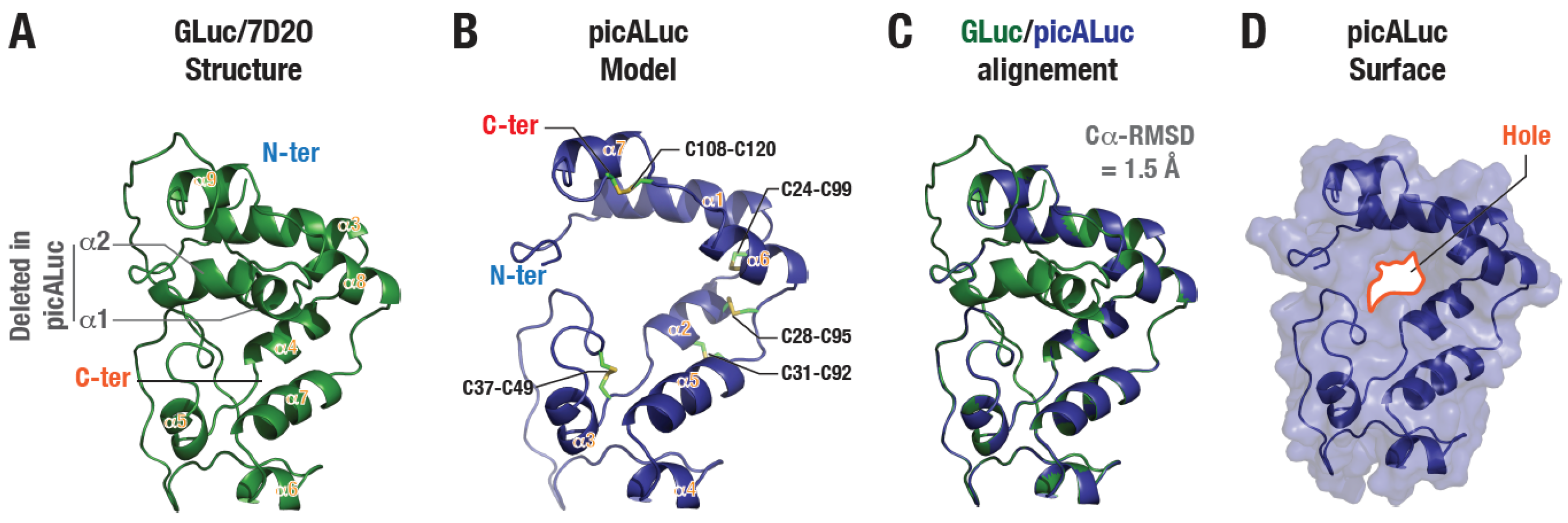
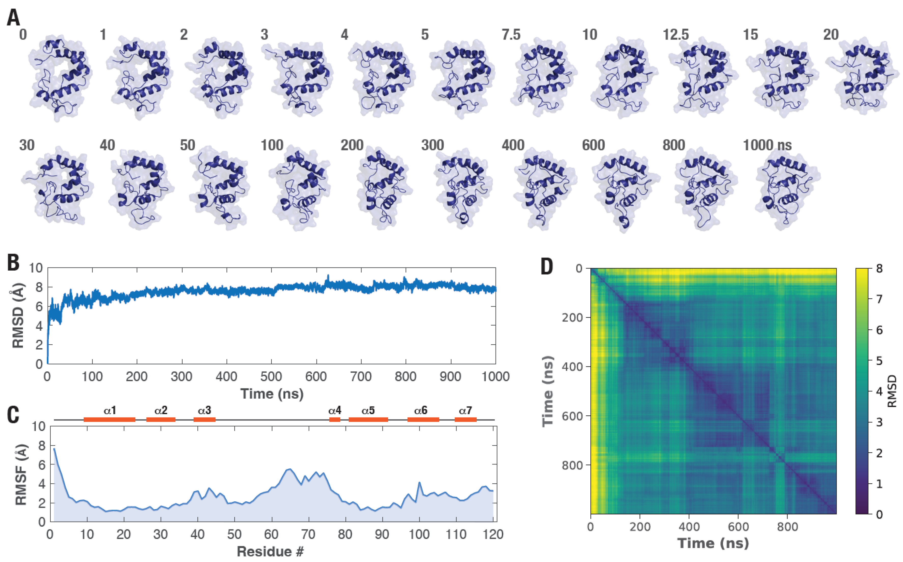
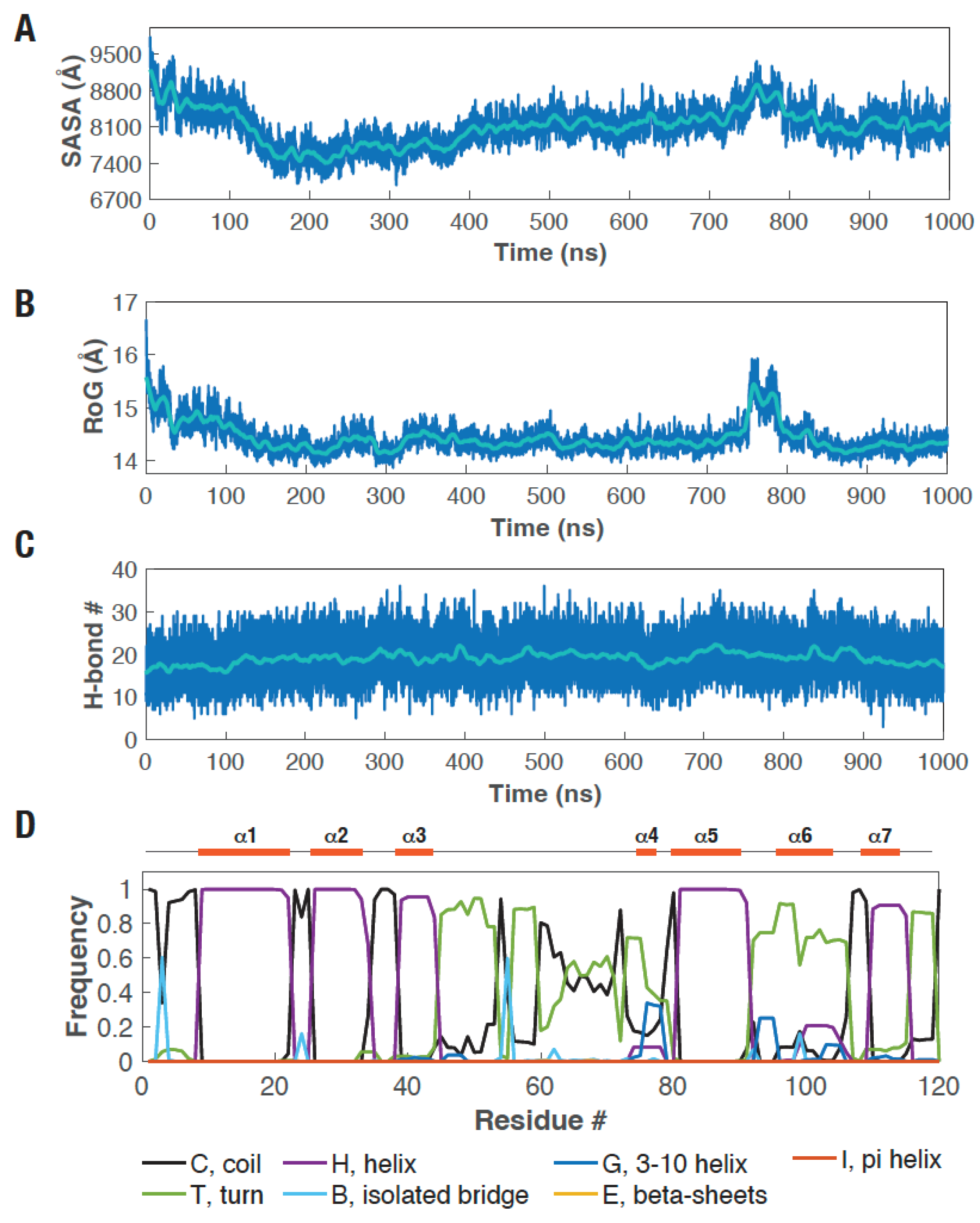
3.1. Molecular Dynamics Simulation Reveals picALuc Structural Features and Reorganization
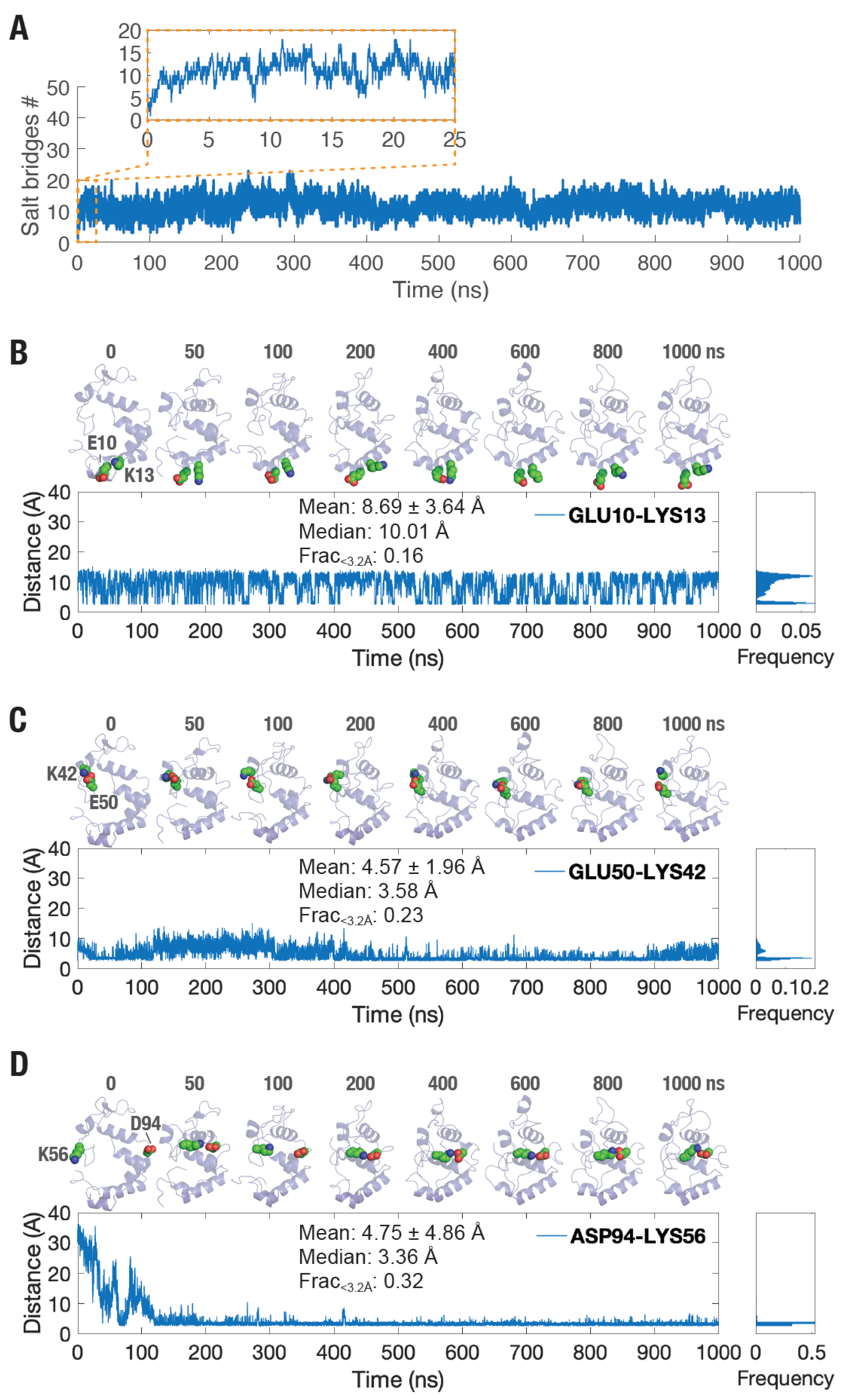
3.2. Increased Number of Salt Bridge Interactions Formed in picALuc
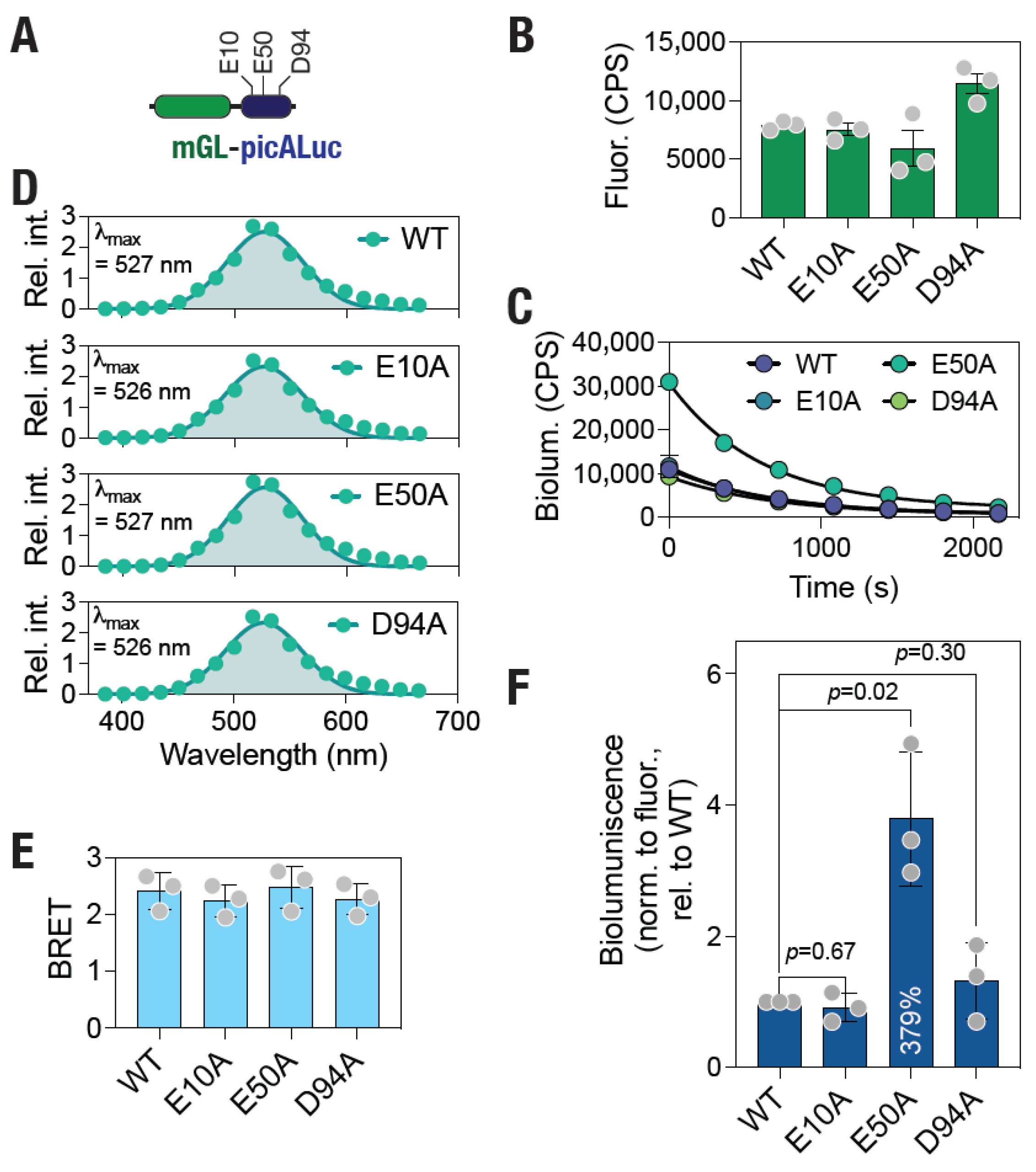
3.3. Mutational Analysis Reveals Increased Bioluminescence of picALuc
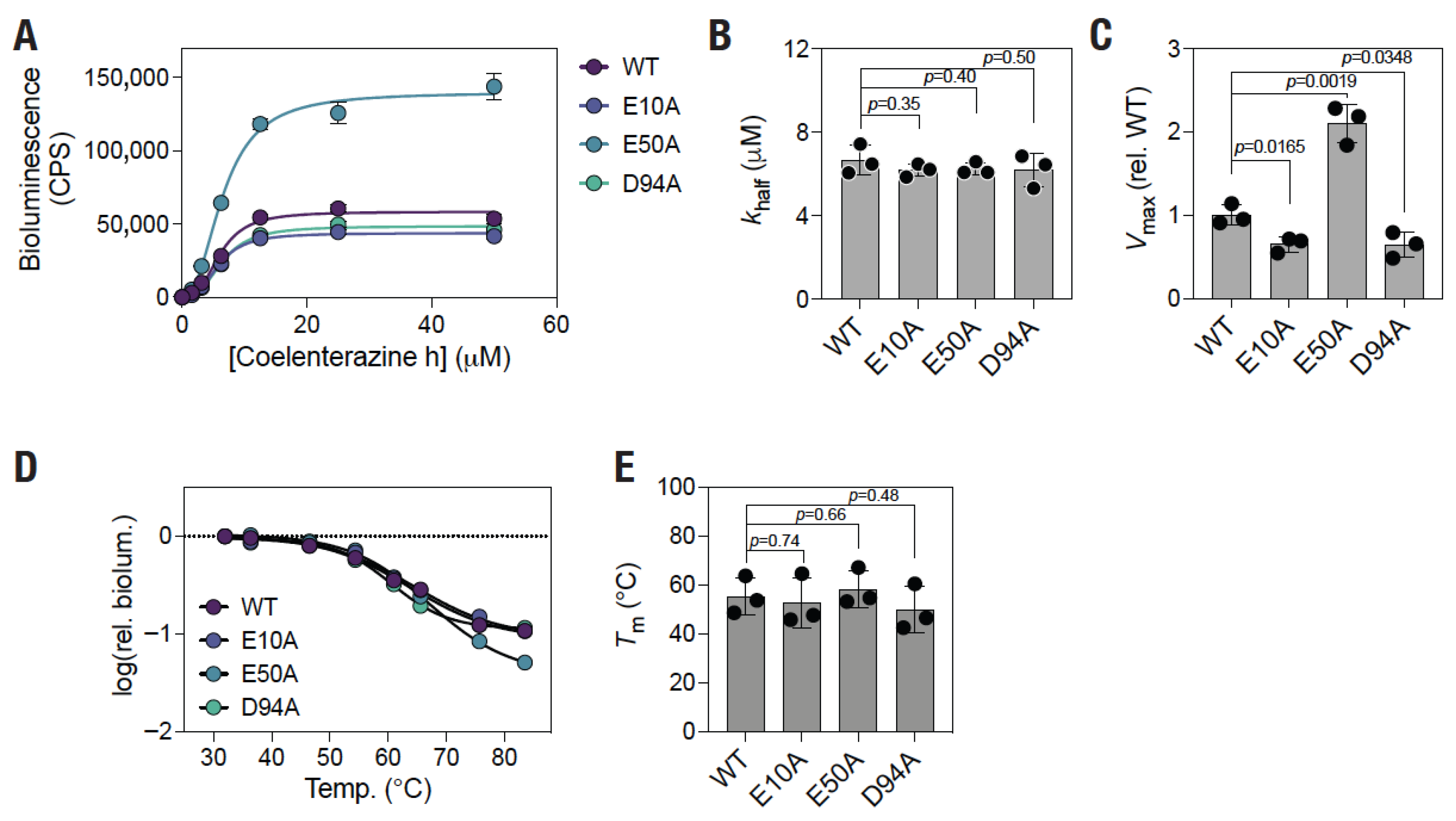
3.4. E50A Mutation Results in Increased Enzymatic Activity Without Altering Thermal Stability of the Protein
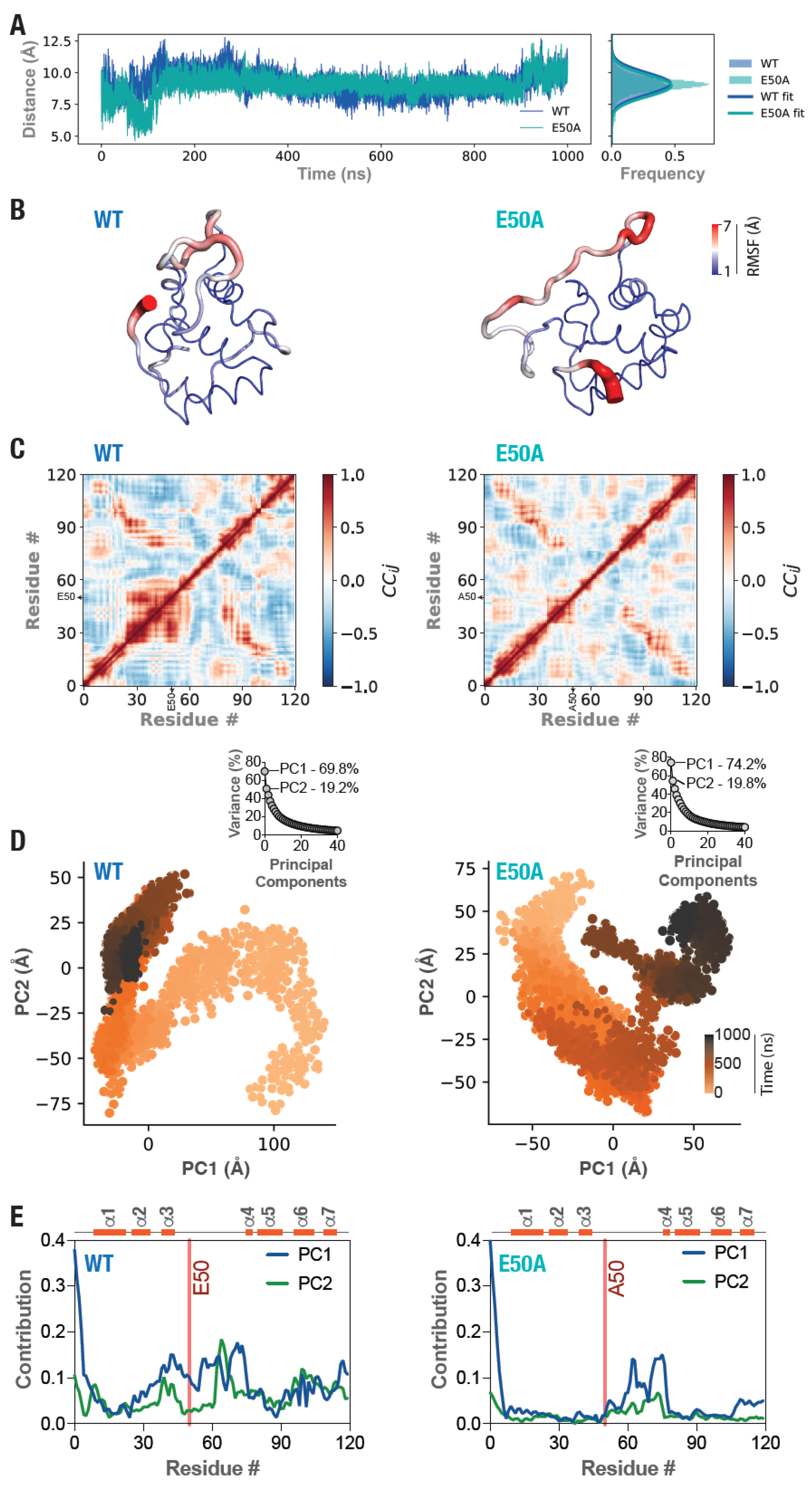
3.5. Altered Structural Dynamics in the E50A Mutant picALuc
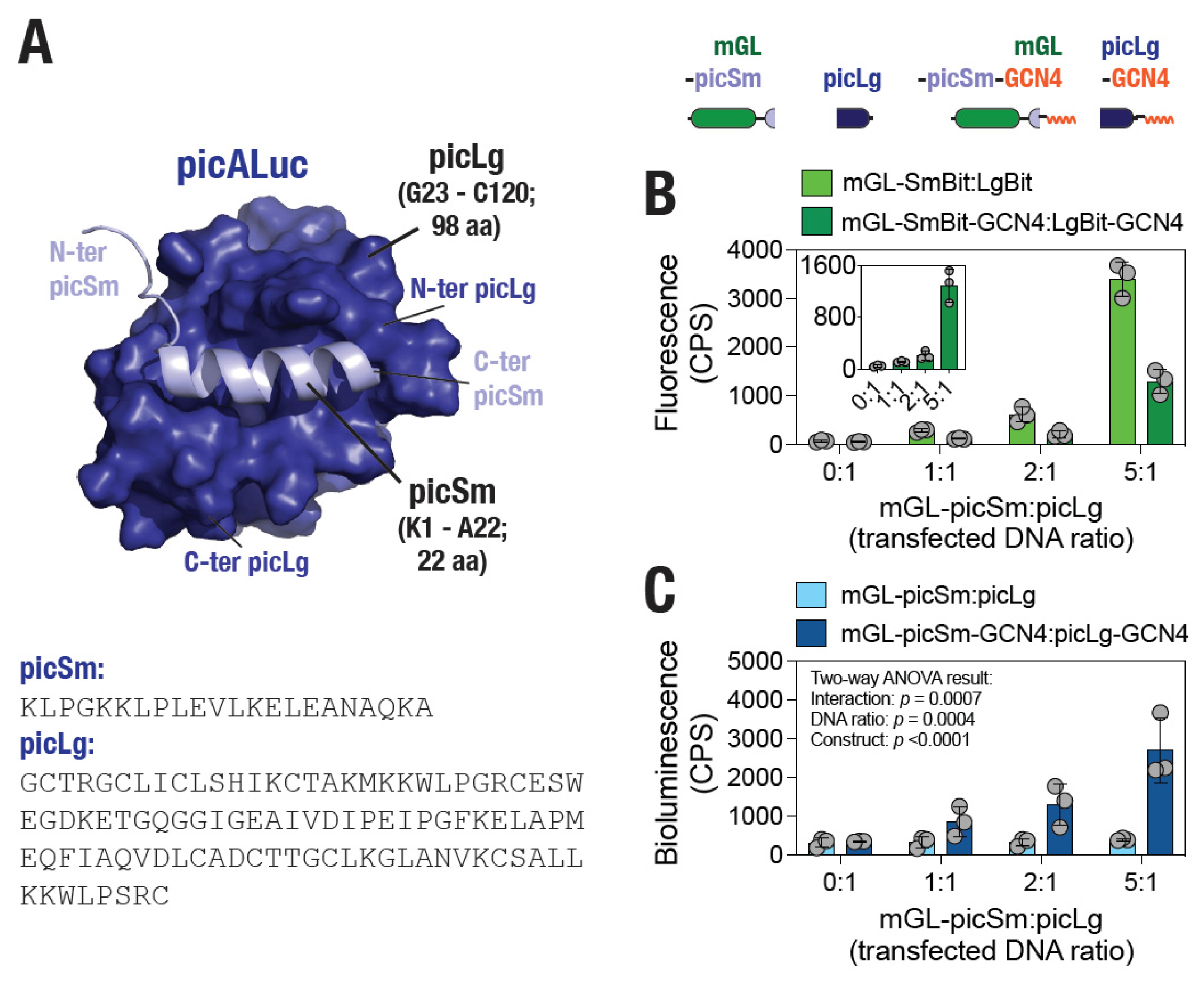
3.6. Design of a picALuc-Based Split Protein for Monitoring PPI in Living Cells
4. Conclusions
Supplementary Materials
Funding
Institutional Review Board Statement
Informed Consent Statement
Data Availability Statement
Acknowledgments
Conflicts of Interest
Correction Statement
References
- Syed, A.J.; Anderson, J.C. Applications of bioluminescence in biotechnology and beyond. Chem. Soc. Rev. 2021, 50, 5668–5705. [Google Scholar] [CrossRef] [Scilit] [PubMed]
- Zambito, G.; Chawda, C.; Mezzanotte, L. Emerging tools for bioluminescence imaging. Curr. Opin. Chem. Biol. 2021, 63, 86–94. [Google Scholar] [CrossRef] [Scilit] [PubMed]
- Frank, L.A.; Krasitskaya, V.V. Application of enzyme bioluminescence for medical diagnostics. Adv. Biochem. Eng. Biotechnol. 2014, 144, 175–197. [Google Scholar] [PubMed]
- Kelkar, M.; De, A. Bioluminescence based in vivo screening technologies. Curr. Opin. Pharmacol. 2012, 12, 592–600. [Google Scholar] [CrossRef] [Scilit]
- Titushin, M.S.; Feng, Y.; Lee, J.; Vysotski, E.S.; Liu, Z.J. Protein-protein complexation in bioluminescence. Protein Cell 2011, 2, 957–972. [Google Scholar] [CrossRef] [Scilit]
- Wilson, T.; Hastings, J.W. Bioluminescence. Annu. Rev. Cell Dev. Biol. 1998, 14, 197–230. [Google Scholar] [CrossRef] [Scilit]
- Schobesberger, S.; Thumfart, H.; Selinger, F.; Spitz, S.; Gonzalez, C.; Pei, L.; Plglitsch, M.; Ertl, P. Microfluidic immunoassay for the detection of SARS-CoV-2 neutralizing antibodies in ultralow-volume human plasma samples. Sens. Actuators Rep. 2025, 9, 100336. [Google Scholar] [CrossRef] [Scilit]
- Krishnan, S.; Syed, Z.u.Q. Colorimetric Visual Sensors for Point-of-needs Testing. Sens. Actuators Rep. 2022, 4, 100078. [Google Scholar] [CrossRef] [Scilit]
- Giannetti, A.; Tombelli, S. Aptamer optical switches: From biosensing to intracellular sensing. Sens. Actuators Rep. 2021, 3, 100030. [Google Scholar] [CrossRef] [Scilit]
- Loening, A.M.; Wu, A.M.; Gambhir, S.S. Red-shifted Renilla reniformis luciferase variants for imaging in living subjects. Nat. Methods 2007, 4, 641–643. [Google Scholar] [CrossRef] [Scilit]
- Tamaki, S.; Kitada, N.; Kiyama, M.; Fujii, R.; Hirano, T.; Kim, S.B.; Maki, S. Color-tunable bioluminescence imaging portfolio for cell imaging. Sci. Rep. 2021, 11, 2219. [Google Scholar] [CrossRef] [Scilit]
- Takai, A.; Nakano, M.; Saito, K.; Haruno, R.; Watanabe, T.M.; Ohyanagi, T.; Jin, T.; Okada, Y.; Nagai, T. Expanded palette of Nano-lanterns for real-time multicolor luminescence imaging. Proc. Natl. Acad. Sci. USA 2015, 112, 4352–4356. [Google Scholar] [CrossRef] [Scilit] [PubMed]
- Viviani, V.R.; Pelentir, G.F.; Bevilaqua, V.R. Bioluminescence Color-Tuning Firefly Luciferases: Engineering and Prospects for Real-Time Intracellular pH Imaging and Heavy Metal Biosensing. Biosensors 2022, 12, 400. [Google Scholar] [CrossRef] [Scilit] [PubMed]
- Al-Handawi, M.B.; Polavaram, S.; Kurlevskaya, A.; Commins, P.; Schramm, S.; Carrasco-Lopez, C.; Lui, N.M.; Solntsev, K.M.; Laptenok, S.P.; Navizet, I.; et al. Spectrochemistry of Firefly Bioluminescence. Chem. Rev. 2022, 122, 13207–13234. [Google Scholar] [CrossRef] [Scilit] [PubMed]
- Loening, A.M.; Fenn, T.D.; Wu, A.M.; Gambhir, S.S. Consensus guided mutagenesis of Renilla luciferase yields enhanced stability and light output. Protein Eng. Des. Sel. 2006, 19, 391–400. [Google Scholar] [CrossRef] [Scilit]
- Rahnama, S.; Saffar, B.; Kahrani, Z.F.; Nazari, M.; Emamzadeh, R. Super RLuc8: A novel engineered Renilla luciferase with a red-shifted spectrum and stable light emission. Enzym. Microb. Technol. 2017, 96, 60–66. [Google Scholar] [CrossRef] [Scilit]
- Hall, M.P.; Unch, J.; Binkowski, B.F.; Valley, M.P.; Butler, B.L.; Wood, M.G.; Otto, P.; Zimmerman, K.; Vidugiris, G.; Machleidt, T. Engineered Luciferase Reporter from a Deep Sea Shrimp Utilizing a Novel Imidazopyrazinone Substrate. ACS Chem. Biol. 2012, 7, 1848. [Google Scholar] [CrossRef] [Scilit]
- Biewenga, L.; Rosier, B.; Merkx, M. Engineering with NanoLuc: A playground for the development of bioluminescent protein switches and sensors. Biochem. Soc. Trans. 2020, 48, 2643–2655. [Google Scholar] [CrossRef] [Scilit]
- England, C.G.; Ehlerding, E.B.; Cai, W. NanoLuc: A Small Luciferase Is Brightening Up the Field of Bioluminescence. Bioconjug. Chem. 2016, 27, 1175–1187. [Google Scholar] [CrossRef] [Scilit]
- Boute, N.; Lowe, P.; Berger, S.; Malissard, M.; Robert, A.; Tesar, M. NanoLuc Luciferase—A Multifunctional Tool for High Throughput Antibody Screening. Front. Pharmacol. 2016, 7, 27. [Google Scholar] [CrossRef] [Scilit]
- Kim, S.B.; Torimura, M.; Tao, H. Creation of artificial luciferases for bioassays. Bioconjug. Chem. 2013, 24, 2067–2075. [Google Scholar] [CrossRef] [Scilit]
- Kim, S.B.; Nishihara, R.; Citterio, D.; Suzuki, K. Fabrication of a New Lineage of Artificial Luciferases from Natural Luciferase Pools. ACS Comb. Sci. 2017, 19, 594–599. [Google Scholar] [CrossRef] [Scilit] [PubMed]
- Ohmuro-Matsuyama, Y.; Furuta, T.; Matsui, H.; Kanai, M.; Ueda, H. Miniaturization of Bright Light-Emitting Luciferase ALuc: picALuc. ACS Chem. Biol. 2022, 17, 864–872. [Google Scholar] [CrossRef] [Scilit] [PubMed]
- Geethakumari, A.M.; Ahmed, W.S.; Rasool, S.; Fatima, A.; Nasir Uddin, S.M.; Aouida, M.; Biswas, K.H. A genetically encoded BRET-based SARS-CoV-2 M(pro) protease activity sensor. Commun. Chem. 2022, 5, 117. [Google Scholar] [CrossRef] [Scilit] [PubMed]
- Altamash, T.; Ahmed, W.; Rasool, S.; Biswas, K.H. Intracellular Ionic Strength Sensing Using NanoLuc. Int. J. Mol. Sci. 2021, 22, 677. [Google Scholar] [CrossRef] [Scilit]
- Biswas, K.H.; Visweswariah, S.S. Distinct allostery induced in the cyclic GMP-binding, cyclic GMP-specific phosphodiesterase (PDE5) by cyclic GMP, sildenafil, and metal ions. J. Biol. Chem. 2011, 286, 8545–8554. [Google Scholar] [CrossRef] [Scilit]
- Biswas, K.H.; Sopory, S.; Visweswariah, S.S. The GAF domain of the cGMP-binding, cGMP-specific phosphodiesterase (PDE5) is a sensor and a sink for cGMP. Biochemistry 2008, 47, 3534–3543. [Google Scholar] [CrossRef] [Scilit]
- Kim, N.; Shin, S.; Bae, S.W. cAMP Biosensors Based on Genetically Encoded Fluorescent/Luminescent Proteins. Biosensors 2021, 11, 39. [Google Scholar] [CrossRef] [Scilit]
- Wu, N.; Kobayashi, N.; Tsuda, K.; Unzai, S.; Saotome, T.; Kuroda, Y.; Yamazaki, T. Solution structure of Gaussia Luciferase with five disulfide bonds and identification of a putative coelenterazine binding cavity by heteronuclear NMR. Sci. Rep. 2020, 10, 20069. [Google Scholar] [CrossRef] [Scilit]
- Waterhouse, A.; Bertoni, M.; Bienert, S.; Studer, G.; Tauriello, G.; Gumienny, R.; Heer, F.T.; de Beer, T.A.P.; Rempfer, C.; Bordoli, L.; et al. SWISS-MODEL: Homology modelling of protein structures and complexes. Nucleic Acids Res. 2018, 46, W296–W303. [Google Scholar] [CrossRef] [Scilit]
- Biswas, K.H.; Badireddy, S.; Rajendran, A.; Anand, G.S.; Visweswariah, S.S. Cyclic nucleotide binding and structural changes in the isolated GAF domain of Anabaena adenylyl cyclase, CyaB2. PeerJ 2015, 3, e882. [Google Scholar] [CrossRef] [Scilit]
- Ahmed, W.S.; Philip, A.M.; Biswas, K.H. Decreased Interfacial Dynamics Caused by the N501Y Mutation in the SARS-CoV-2 S1 Spike:ACE2 Complex. Front. Mol. Biosci. 2022, 9, 846996. [Google Scholar]
- Jumper, J.; Evans, R.; Pritzel, A.; Green, T.; Figurnov, M.; Ronneberger, O.; Tunyasuvunakool, K.; Bates, R.; Zidek, A.; Potapenko, A.; et al. Highly accurate protein structure prediction with AlphaFold. Nature 2021, 596, 583–589. [Google Scholar] [CrossRef] [Scilit] [PubMed]
- Chen, V.B.; Arendall, W.B., 3rd; Headd, J.J.; Keedy, D.A.; Immormino, R.M.; Kapral, G.J.; Murray, L.W.; Richardson, J.S.; Richardson, D.C. MolProbity: All-atom structure validation for macromolecular crystallography. Biol. Crystallogr. 2010, 66, 12–21. [Google Scholar] [CrossRef] [Scilit] [PubMed]
- Ribeiro, J.V.; Bernardi, R.C.; Rudack, T.; Stone, J.E.; Phillips, J.C.; Freddolino, P.L.; Schulten, K. QwikMD—Integrative Molecular Dynamics Toolkit for Novices and Experts. Sci. Rep. 2016, 6, 26536. [Google Scholar] [CrossRef] [Scilit] [PubMed]
- Humphrey, W.; Dalke, A.; Schulten, K. VMD: Visual molecular dynamics. J. Mol. Graph. 1996, 14, 33. [Google Scholar] [CrossRef] [Scilit]
- Phillips, J.C.; Braun, R.; Wang, W.; Gumbart, J.; Tajkhorshid, E.; Villa, E.; Chipot, C.; Skeel, R.D.; Kalé, L.; Schulten, K. Scalable Molecular Dynamics with NAMD. J. Comput. Chem. 2005, 26, 1781. [Google Scholar] [CrossRef] [Scilit]
- Best, R.B.; Zhu, X.; Shim, J.; Lopes, P.E.; Mittal, J.; Feig, M.; MacKerell, A.D., Jr. Optimization of the additive CHARMM all-atom protein force field targeting improved sampling of the backbone φ, ψ and side-chain χ1 and χ2 dihedral angles. J. Chem. Theory Comput. 2012, 8, 3257. [Google Scholar] [CrossRef] [Scilit]
- Jorgensen, W.L.; Chandrasekhar, J.; Madura, J.D.; Impey, R.W.; Klein, M.L. Comparison of simple potential functions for simulating liquid water. J. Chem. Phys. 1983, 79, 926–935. [Google Scholar] [CrossRef] [Scilit]
- Feller, S.E.; Zhang, Y.; Pastor, R.W.; Brooks, B.R. Constant pressure molecular dynamics simulation: The Langevin piston method. J. Chem. Phys. 1995, 103, 4613–4621. [Google Scholar] [CrossRef] [Scilit]
- Pang, Y.T.; Miao, Y.; Wang, Y.; McCammon, J.A. Gaussian Accelerated Molecular Dynamics in NAMD. J. Chem. Theory Comput. 2017, 13, 9. [Google Scholar] [CrossRef] [Scilit]
- Miao, Y.; Feixas, F.; Eun, C.; McCammon, J.A. Accelerated Molecular Dynamics Simulations of Protein Folding. J. Comput. Chem. 2015, 36, 1536. [Google Scholar] [CrossRef] [Scilit] [PubMed]
- Miao, Y.; Feher, V.A.; McCammon, J.A. Gaussian Accelerated Molecular Dynamics: Unconstrained Enhanced Sampling and Free Energy Calculation. J. Chem. Theory Comput. 2015, 11, 3584–3595. [Google Scholar] [CrossRef] [Scilit] [PubMed]
- Zhang, Q.; Tan, S.; Xiao, T.; Liu, H.; Shah, S.J.A.; Liu, H. Probing the Molecular Mechanism of Rifampin Resistance Caused by the Point Mutations S456L and D441V on Mycobacterium Tuberculosis RNA Polymerase Through Gaussian Accelerated Molecular Dynamics Simulation. Antimicrob. Agents Chemother. 2020, 64, e02476-19. [Google Scholar] [CrossRef] [Scilit] [PubMed]
- Miao, Y.; McCammon, J.A. Gaussian Accelerated Molecular Dynamics: Theory, Implementation, and Applications. Annu. Rep. Comput. Chem. 2017, 13, 231. [Google Scholar]
- Michaud-Agrawal, N.; Denning, E.J.; Woolf, T.B.; Beckstein, O. MDAnalysis: A toolkit for the analysis of molecular dynamics simulations. J. Comput. Chem. 2011, 32, 2319–2327. [Google Scholar] [CrossRef] [Scilit]
- Gowers, R.J.; Linke, M.; Barnoud, J.; Reddy, T.J.E.; Melo, M.N.; Seyler, S.L.; Domanski, J.; Doston, D.L.; Buchoux, S.; Kenney, L.M.; et al. MDAnalysis: A Python package for the rapid analysis of molecular dynamics simulations. In Proceedings of the 15th Python in Science Conference; Benthall, S., Rostrup, S., Eds.; SciPy: Austin, TX, USA, 2016; pp. 98–105. [Google Scholar]
- Brielle, E.S.; Arkin, I.T. Quantitative Analysis of Multiplex H-Bonds. J. Am. Chem. Soc. 2020, 142, 14150. [Google Scholar] [CrossRef] [Scilit]
- Mallik, S.; Prasad, R.; Das, K.; Sen, P. Alcohol functionality in the fatty acid backbone of sphingomyelin guides the inhibition of blood coagulation. RSC Adv. 2021, 11, 3390–3398. [Google Scholar] [CrossRef] [Scilit]
- Brown, D.K.; Penkler, D.L.; Sheik Amamuddy, O.; Ross, C.; Atilgan, A.R.; Atilgan, C.; Tastan Bishop, O. MD-TASK: A software suite for analyzing molecular dynamics trajectories. Bioinformatics 2017, 33, 2768–2771. [Google Scholar] [CrossRef] [Scilit]
- Schindelin, J.; Arganda-Carreras, I.; Frise, E.; Kaynig, V.; Longair, M.; Pietzsch, T.; Preibisch, S.; Rueden, C.; Saalfeld, S.; Schmid, B. Fiji: An open-source platform for biological-image analysis. Nat. Methods 2012, 9, 676–682. [Google Scholar] [CrossRef] [Scilit]
- Jin, Z.; Du, X.; Xu, Y.; Deng, Y.; Liu, M.; Zhao, Y.; Zhang, B.; Li, X.; Zhang, L.; Peng, C.; et al. Structure of M(pro) from SARS-CoV-2 and discovery of its inhibitors. Nature 2020, 582, 289–293. [Google Scholar] [CrossRef] [Scilit]
- Gordon, D.E.; Jang, G.M.; Bouhaddou, M.; Xu, J.; Obernier, K.; White, K.M.; O’Meara, M.J.; Rezelj, V.V.; Guo, J.Z.; Swaney, D.L.; et al. A SARS-CoV-2 protein interaction map reveals targets for drug repurposing. Nature 2020, 583, 459–468. [Google Scholar] [CrossRef] [Scilit] [PubMed]
- Geethakumari, A.M.; Sultana, A.; Fatima, A.; Uddin, S.M.N.; Abdulhakim, S.; Mohamed, A.; Rahman, S.; Al-Buainain, K.; Yassine, H.M.; Khatib, H.A.A.; et al. A BRET-based Mpro biosensor containing a nanobody and tandem cleavage sites shows an increased cleavage rate. Sens. Actuators Rep. 2025, 9, 100315. [Google Scholar] [CrossRef] [Scilit]
- Ahmed, W.S.; Geethakumari, A.M.; Sultana, A.; Tiwari, A.; Altamash, T.; Arshad, N.; Visweswariah, S.S.; Biswas, K.H. Coevolving residues distant from the ligand binding site are involved in GAF domain function. Commun. Chem. 2025, 8, 107. [Google Scholar] [CrossRef] [Scilit] [PubMed]
- Uddin, S.M.N.; Rasool, S.; Geethakumari, A.M.; Ahmed, W.S.; Biswas, K.H. Engineering β-catenin-derived peptides for α-catenin binding. Emergent Mater. 2024, 8, 1183–1197. [Google Scholar] [CrossRef] [Scilit]
- Sultana, A.; Geethakumari, A.M.; Islam, Z.; Kolatkar, P.R.; Biswas, K.H. BRET-based biosensors for SARS-CoV-2 oligonucleotide detection. Front. Bioeng. Biotechnol. 2024, 12, 1353479. [Google Scholar] [CrossRef] [Scilit]
- Fatima, A.; Geethakumari, A.M.; Ahmed, W.S.; Biswas, K.H. A potential allosteric inhibitor of SARS-CoV-2 main protease (M(pro)) identified through metastable state analysis. Front. Mol. Biosci. 2024, 11, 1451280. [Google Scholar] [CrossRef] [Scilit]
- Ahmed, W.S.; Geethakumari, A.M.; Sultana, A.; Fatima, A.; Philip, A.M.; Uddin, S.M.N.; Biswas, K.H. A slow but steady nanoLuc: R162A mutation results in a decreased, but stable, nanoLuc activity. Int. J. Biol. Macromol. 2024, 269, 131864. [Google Scholar] [CrossRef] [Scilit]
- Jan, Z.; Geethakumari, A.M.; Biswas, K.H.; Jithesh, P.V. Protegrin-2, a potential inhibitor for targeting SARS-CoV-2 main protease M(pro). Comput. Struct. Biotechnol. J. 2023, 21, 3665–3671. [Google Scholar] [CrossRef] [Scilit]
- Moovarkumudalvan, B.; Geethakumari, A.M.; Ramadoss, R.; Biswas, K.H.; Mifsud, B. Structure-Based Virtual Screening and Functional Validation of Potential Hit Molecules Targeting the SARS-CoV-2 Main Protease. Biomolecules 2022, 12, 1754. [Google Scholar] [CrossRef] [Scilit]
- Yang, X.; Zhang, R.; Han, W.; Han, L. Molecular Dynamics Simulation Combined with Neural Relationship Inference and Markov Model to Reveal the Relationship between Conformational Regulation and Bioluminescence Properties of Gaussia Luciferase. Molecules 2024, 29, 4029. [Google Scholar] [CrossRef] [Scilit]
- Nemergut, M.; Pluskal, D.; Horackova, J.; Sustrova, T.; Tulis, J.; Barta, T.; Baatallah, R.; Gagnot, G.; Novakova, V.; Majerova, M.; et al. Illuminating the mechanism and allosteric behavior of NanoLuc luciferase. Nat. Commun. 2023, 14, 7864. [Google Scholar] [CrossRef] [Scilit]
- Yeh, A.H.; Norn, C.; Kipnis, Y.; Tischer, D.; Pellock, S.J.; Evans, D.; Ma, P.; Lee, G.R.; Zhang, J.Z.; Anishchenko, I.; et al. De novo design of luciferases using deep learning. Nature 2023, 614, 774–780. [Google Scholar] [CrossRef] [Scilit]
- Wu, N.; Xu, Z.-C.; Du, K.-D.; Huang, S.; Kobayashi, N.; Kuroda, Y.; Bai, Y.-H. A Structural Model of Truncated Gaussia princeps Luciferase Elucidating the Crucial Catalytic Function of No.76 Arginine towards Coelenterazine Oxidation. PLoS Comput. Biol. 2025, 21, e1012722. [Google Scholar] [CrossRef] [Scilit]
- Gedi, V.; Kim, E.H.; Oh, B.; Kim, Y.-P. Advanced Bioluminescence Reporter with Engineered Gaussia Luciferase via Sequence-Guided Mutagenesis. Biosensors 2024, 14, 528. [Google Scholar] [CrossRef] [Scilit] [PubMed]
- Calabretta, M.M.; Gregucci, D.; Martínez-Pérez-Cejuela, H.; Michelini, E. A Luciferase Mutant with Improved Brightness and Stability for Whole-Cell Bioluminescent Biosensors and In Vitro Biosensing. Biosensors 2022, 12, 742. [Google Scholar] [CrossRef] [Scilit] [PubMed]
- Gardiner, S.; Talley, J.; Green, T.; Haynie, C.; Kubalek, C.; Argyle, M.; Heaps, W.; Ebbert, J.; Allen, D.; Chipman, D.; et al. Advancing NanoLuc Luciferase Stability beyond Directed Evolution and Rational Design through Expert-Guided Deep Learning. ACS Catal. 2026, 16, 2849–2860. [Google Scholar] [CrossRef] [Scilit] [PubMed]
- Campbell, B.C.; Nabel, E.M.; Murdock, M.H.; Lao-Peregrin, C.; Tsoulfas, P.; Blackmore, M.G.; Lee, F.S.; Liston, C.; Morishita, H.; Petsko, G.A. mGreenLantern: A bright monomeric fluorescent protein with rapid expression and cell filling properties for neuronal imaging. Proc. Natl. Acad. Sci. USA 2020, 117, 30710–30721. [Google Scholar] [CrossRef] [Scilit]
- Liu, Z.; Chen, O.; Wall, J.B.J.; Zheng, M.; Zhou, Y.; Wang, L.; Vaseghi, H.R.; Qian, L.; Liu, J. Systematic comparison of 2A peptides for cloning multi-genes in a polycistronic vector. Sci. Rep. 2017, 7, 2193. [Google Scholar] [CrossRef] [Scilit]
- Kim, J.H.; Lee, S.R.; Li, L.H.; Park, H.J.; Park, J.H.; Lee, K.Y.; Kim, M.K.; Shin, B.A.; Choi, S.Y. High cleavage efficiency of a 2A peptide derived from porcine teschovirus-1 in human cell lines, zebrafish and mice. PLoS ONE 2011, 6, e18556. [Google Scholar] [CrossRef] [Scilit]
- Biswas, K.H. A Brighter picALuc Generated Through the Loss of a Salt Bridge Interaction. bioRxiv 2023. [Google Scholar] [CrossRef] [Scilit]
- Ohmuro-Matsuyama, Y.; Matsui, H.; Kanai, M.; Furuta, T. Glow-type conversion and characterization of a minimal luciferase via mutational analyses. FEBS J. 2023, 290, 5554–5565. [Google Scholar] [CrossRef] [Scilit] [PubMed]
- Kitao, A. Principal Component Analysis and Related Methods for Investigating the Dynamics of Biological Macromolecules. J 2022, 5, 298–317. [Google Scholar] [CrossRef] [Scilit]
- O’Shea, E.K.; Klemm, J.D.; Kim, P.S.; Alber, T. X-ray structure of the GCN4 leucine zipper, a two-stranded, parallel coiled coil. Science 1991, 254, 539–544. [Google Scholar] [CrossRef] [Scilit] [PubMed]
- Tanenbaum, M.E.; Gilbert, L.A.; Qi, L.S.; Weissman, J.S.; Vale, R.D. A protein-tagging system for signal amplification in gene expression and fluorescence imaging. Cell 2014, 159, 635–646. [Google Scholar] [CrossRef] [Scilit]
- Sekhon, H.; Loh, S.N. Engineering protein activity into off-the-shelf DNA devices. Cell Rep. Methods 2022, 2, 100202. [Google Scholar] [CrossRef] [Scilit]
- Dixon, A.S.; Schwinn, M.K.; Hall, M.P.; Zimmerman, K.; Otto, P.; Lubben, T.H.; Butler, B.L.; Binkowski, B.F.; Machleidt, T.; Kirkland, T.A.; et al. NanoLuc Complementation Reporter Optimized for Accurate Measurement of Protein Interactions in Cells. ACS Chem. Biol. 2016, 11, 400–408. [Google Scholar] [CrossRef] [Scilit]
- Bhuckory, S.; Kays, J.C.; Dennis, A.M. In Vivo Biosensing Using Resonance Energy Transfer. Biosensors 2019, 9, 76. [Google Scholar] [CrossRef] [Scilit]
- Kim, S.-B.; Furuta, T.; Thangudu, S.; Natarajan, A.; Paulmurugan, R. Molecular Association Assay Systems for Imaging Protein–Protein Interactions in Mammalian Cells. Biosensors 2025, 15, 299. [Google Scholar] [CrossRef] [Scilit]
- Sureda-Vives, M.; Sarkisyan, K.S. Bioluminescence-Driven Optogenetics. Life 2020, 10, 318. [Google Scholar] [CrossRef] [Scilit]
- Crespo, E.L.; Prakash, M.; Bjorefeldt, A.; Medendorp, W.E.; Shaner, N.C.; Lipscombe, D.; Moore, C.I.; Hochgeschwender, U. Bioluminescent optogenetic (BL-OG) activation of neurons during mouse postnatal brain development. STAR Protoc. 2021, 2, 100667. [Google Scholar] [CrossRef] [Scilit]
- Adir, O.; Albalak, M.R.; Abel, R.; Weiss, L.E.; Chen, G.; Gruber, A.; Staufer, O.; Kurman, Y.; Kaminer, I.; Shklover, J.; et al. Synthetic cells with self-activating optogenetic proteins communicate with natural cells. Nat. Commun. 2022, 13, 2328. [Google Scholar] [CrossRef] [Scilit]
- Kim, C.K.; Cho, K.F.; Kim, M.W.; Ting, A.Y. Luciferase-LOV BRET enables versatile and specific transcriptional readout of cellular protein-protein interactions. Elife 2019, 8, e43826. [Google Scholar] [CrossRef] [Scilit]




Disclaimer/Publisher’s Note: The statements, opinions and data contained in all publications are solely those of the individual author(s) and contributor(s) and not of MDPI and/or the editor(s). MDPI and/or the editor(s) disclaim responsibility for any injury to people or property resulting from any ideas, methods, instructions or products referred to in the content. |
© 2026 by the author. Licensee MDPI, Basel, Switzerland. This article is an open access article distributed under the terms and conditions of the Creative Commons Attribution (CC BY) license.
Share and Cite
Biswas, K.H. E50A Mutation Increases the Bioluminescence Activity of picALuc. Biosensors 2026, 16, 167. https://doi.org/10.3390/bios16030167
Biswas KH. E50A Mutation Increases the Bioluminescence Activity of picALuc. Biosensors. 2026; 16(3):167. https://doi.org/10.3390/bios16030167
Chicago/Turabian StyleBiswas, Kabir H. 2026. "E50A Mutation Increases the Bioluminescence Activity of picALuc" Biosensors 16, no. 3: 167. https://doi.org/10.3390/bios16030167
APA StyleBiswas, K. H. (2026). E50A Mutation Increases the Bioluminescence Activity of picALuc. Biosensors, 16(3), 167. https://doi.org/10.3390/bios16030167