Studies on the Aptasensor Miniaturization for Electrochemical Detection of Lead Ions


Abstract
1. Introduction
2. Materials and Methods
2.1. Apparatus
2.2. Reagents
2.3. Solutions
2.4. Gold Electrode Cleaning and Modification
3. Results and Discussion
4. Conclusions
Author Contributions
Funding
Institutional Review Board Statement
Informed Consent Statement
Data Availability Statement
Conflicts of Interest
References
- Meima, J.A.; Comans, R.N.J. The leaching of trace elements from municipal solid waste incinerator bottom ash at different stages of weathering. Appl. Geochem. 1999, 14, 159–171. [Google Scholar] [CrossRef]
- Senanayake, J.; Rahman, R.H.; Safwat, F.; Riar, S.; Ampalloor, G. Asymptomatic Lead Poisoning in a Pediatric Patient. Cureus 2023, 15, e34940. [Google Scholar] [CrossRef]
- Wani, A.L.; Ara, A.; Usmani, J.A. Lead toxicity: A review. Interdiscip. Toxicol. 2015, 8, 55–64. [Google Scholar] [CrossRef]
- Rouzi, L.; Elhamri, H.; Kalouch, S.; Salam, S.; Moutawakil, B.E.; Chaoui, H.; Badrane, N.; Fekhaoui, M.; Jouhadi, Z. Lead poisoning with encephalic and neuropathic involvement in a child: Case report. Pan Afr. Med. J. 2022, 42, 276. [Google Scholar] [CrossRef]
- Ma, R.; Van Mol, W.; Adams, F. Determination of cadmium, copper and lead in environmental samples. An evaluation of flow injection on-line sorbent extraction for flame atomic absorption spectrometry. Anal. Chim. Acta 1994, 285, 33–43. [Google Scholar] [CrossRef]
- Dasbas, T.; Saçmacı, S.; Ülgen, A.; Kartal, S. A solid phase extraction procedure for the determination of Cd(II) and Pb(II) ions in food and water samples by flame atomic absorption spectrometry. Food Chem. 2015, 174, 591–596. [Google Scholar] [CrossRef] [PubMed]
- Ashoka, S.; Peake, B.M.; Bremner, G.; Hageman, K.J.; Reid, M.R. Comparison of digestion methods for ICP-MS determination of trace elements in fish tissues. Anal. Chim. Acta 2009, 653, 191–199. [Google Scholar] [CrossRef] [PubMed]
- Sheen, S.-R.; Shih, J.-S. Lead(ii) Ion-selective Electrodes Based on Crown Ethers. Analyst 1992, 117, 1691–1695. [Google Scholar] [CrossRef]
- Guzinski, M.; Lisak, G.; Kupis, J.; Jasinski, A.; Bochenska, M. Lead(II)-selective ionophores for ion-selective electrodes: A review. Anal. Chim. Acta 2013, 791, 1–12. [Google Scholar] [CrossRef] [PubMed]
- Serrano, N.; González-Calabuig, A.; Valle, M. Crown ether-modified electrodes for the simultaneous stripping Voltametric determination of Cd(II), Pb(II) and Cu(II). Talanta 2015, 138, 130–137. [Google Scholar] [CrossRef] [PubMed]
- Xiong, W.; Zhou, L.; Liu, S. Development of gold-doped carbon foams as a sensitive electrochemical sensor for simultaneous determination of Pb (II) and Cu (II). Chem. Eng. J. 2016, 284, 650–656. [Google Scholar] [CrossRef]
- Chen, Y.; Xu, S.; Liu, G.; Li, W.; Liu, L.; Wang, Z.; Dai, X.; Jiang, X. An electrochemical sensor based on PEI/CS/GN composite–modified glassy carbon electrode for determination of Pb(II). Ionics 2023, 29, 2031–2041. [Google Scholar] [CrossRef]
- Buchanan, E.B., Jr.; Schroeder, T.D.; Novosel, B. Square-Wave Polarographic Determination of Lead as a Pollutant in River Water. Anal. Chem. 1970, 42, 370–372. [Google Scholar] [CrossRef]
- Taher, M.A.; Puri, B.K.; Bansal, R.K. Simultaneous Determination of Cadmium and Lead in Real and Environmental Samples by Differential Pulse Polarography after Adsorption of Their 2-Nitroso-1-naphthol-4-sulfonic acid–Tetradecyldimethylbenzylammonium Ion-Associated Complex on Microcrystalline Naphthalene. Microchem. J. 1998, 58, 21–30. [Google Scholar] [CrossRef]
- Xiao, A.L.; Xu, H.; Zhou, S.; Song, T.; Wang, H.; Li, S.; Gan, W.; Yuan, Q. Simultaneous detection of Cd(II) and Pb(II) by differential pulse anodic stripping voltammetry at a nitrogen-doped microporous carbon/Nafion/bismuth-film electrode. Electrochim. Acta 2014, 143, 143–151. [Google Scholar] [CrossRef]
- Zhu, L.; Xu, L.; Huang, B.; Jia, N.; Tan, L.; Yao, S. Simultaneous determination of Cd(II) and Pb(II) using square wave anodic stripping voltammetry at a gold nanoparticle-graphene-cysteine composite modified bismuth film electrode. Electrochim. Acta 2014, 115, 471–477. [Google Scholar] [CrossRef]
- Hocevar, S.B.; Wang, J.; Deo, R.P.; Ogorevc, B. Potentiometric Stripping Analysis at Bismuth-Film Electrode. Electroanalysis 2002, 14, 112–115. [Google Scholar] [CrossRef]
- Tarley, C.R.T.; Santos, V.S.; Baêta, B.E.L.; Pereira, A.C.; Kubota, L.T. Simultaneous determination of zinc, cadmium and lead in environmental water samples by potentiometric stripping analysis (PSA) using multiwalled carbon nanotube electrode. J. Hazard. Mater. 2009, 169, 256–262. [Google Scholar] [CrossRef]
- Tonle, I.K.; Letaief, S.; Ngameni, E.; Walcarius, A.; Detellier, C. Square Wave Voltammetric Determination of Lead(II) Ions Usinga Carbon Paste Electrode Modified by a Thiol-Functionalized Kaolinite. Electroanalysis 2011, 23, 245–252. [Google Scholar] [CrossRef]
- Lee, S.; Park, S.-K.; Choi, E.; Piao, Y. Voltammetric determination of trace heavy metals using an electrochemically deposited graphene/bismuth nanocomposite film-modified glassy carbon electrode. J. Electroanal. Chem. 2016, 766, 120–127. [Google Scholar] [CrossRef]
- Knihnicki, P.; Skrzypek, A.; Jakubowska, M.; Porada, R.; Rokicinska, A.; Kustrowski, P.; Koscielniak, P.; Kochana, J. Electrochemical Sensing of Pb2+ and Cd2+ Ions with the Use of Electrode Modified with Carbon-Covered Halloysite and Carbon Nanotubes. Molecules 2022, 27, 4608. [Google Scholar] [CrossRef] [PubMed]
- Brown, A.K.; Li, J.; Pavot, C.M.B.; Lu, Y. A lead-dependent DNAzyme with a two-step mechanism. Biochemistry 2003, 42, 7152–7161. [Google Scholar] [CrossRef]
- Zhang, L.; Han, B.; Liab, T.; Wang, E. Label-free DNAzyme-based fluorescing molecular switch for sensitive and selective detection of lead ions. Chem. Commun. 2011, 47, 3099–3101. [Google Scholar] [CrossRef]
- Guo, L.; Nie, D.; Qiu, C.; Zheng, Q.; Wu, H.; Ye, P.; Hao, Y.; Fu Fu, F.; Chen, G. G-quadruplex based label-free fluorescent biosensor for lead ion. Biosen. Bioelectron. 2012, 35, 123–127. [Google Scholar] [CrossRef]
- Liu, C.-W.; Huang, C.-C.; Chang, H.-T. Highly Selective DNA-Based Sensor for Lead(II) and Mercury(II) Ions. Anal. Chem. 2009, 81, 2383–2387. [Google Scholar] [CrossRef]
- Niemeyer, C.M. Nanoparticles, proteins, and nucleic acids: Biotechnology meets materials science. Angew. Chem. Int. Ed. 2001, 40, 4128–4158. [Google Scholar] [CrossRef]
- Xiao, Y.; Rowe, A.A.; Plaxco, K.W. Electrochemical detection of parts-per-billion lead via an electrode-bound DNAzyme assembly. J. Am. Chem. Soc. 2007, 129, 262–263. [Google Scholar] [CrossRef]
- Liang, R.; Dong, J.; Li, J.; Jin, H.; Wei, M.; Bai, T.; Ren, W.; Xu, Y.; He, B.; Suo, Z. DNAzyme-driven bipedal DNA walker and catalytic hairpin assembly multistage signal amplified electrochemical biosensor based on porous AuNPs@Zr-MOF for detection of Pb2+. Food Chem. 2024, 435, 137503. [Google Scholar] [CrossRef]
- Tang, S.; Tong, P.; Li, H.; Tang, J.; Zhang, L. Ultrasensitive electrochemical detection of Pb 2þ based on rolling circle amplification and quantum dots tagging. Biosen. Bioelectron. 2013, 42, 608–611. [Google Scholar] [CrossRef] [PubMed]
- Lin, Z.; Chen, Y.; Li, X.; Fang, W. Pb2+ induced DNA conformational switch from hairpin to G-quadruplex: Electrochemical detection of Pb2+. Analyst 2011, 136, 2367. [Google Scholar] [CrossRef]
- Lin, Z.; Li, X.; Kraatz, H.-B. Impedimetric Immobilized DNA-Based Sensor for Simultaneous Detection of Pb2+, Ag+, and Hg2+. Anal. Chem. 2011, 83, 6896–6901. [Google Scholar] [CrossRef]
- Liu, J.; Cao, Z.; Lu, Y. Functional Nucleic Acid Sensors. Chem. Rev. 2009, 109, 1948–1998. [Google Scholar] [CrossRef]
- Stoltenburg, R.; Reinemann, C.; Strehlitz, B. SELEX—A (r)evolutionary method to generate high-affinity nucleic acid ligands. Biomol. Eng. 2007, 24, 381–403. [Google Scholar] [CrossRef]
- Wang, L.; Liu, X.; Hu, X.; Song, S.; Fan, C. Unmodified gold nanoparticles as a colorimetric probe for potassium DNA aptamers. Chem. Commun. 2006, 36, 3780–3782. [Google Scholar] [CrossRef] [PubMed]
- Wu, Y.; Zhan, S.; Wang, L.; Zhou, P. Selection of a DNA aptamer for cadmium detection based on cationic polymer mediated aggregation of gold nanoparticles. Analyst 2014, 139, 1550. [Google Scholar] [CrossRef] [PubMed]
- Mou, Y.; Zhang, Y.; Lin, X.; Chen, M.; Xia, Y.; Zhu, S.; Wei, C.; Luo, X. Construction of a novel fluorescent DNA aptasensor for the fast-response and sensitive detection of copper ions in industrial sewage. Anal. Methods 2023, 15, 3466. [Google Scholar] [CrossRef]
- Samimi, H.A.; Saberi, Z. A novel fluorescent aptasensing assay using carbon dots and Ag-Au nanoparticles for the detection of Pb2+ ions. Polyhedron 2023, 245, 116636. [Google Scholar] [CrossRef]
- Radi, A.-E.; O’Sullivan, C.K. Aptamer conformational switch as sensitive electrochemical biosensor for potassium ion recognition. Chem. Commun. 2006, 32, 3432–3434. [Google Scholar] [CrossRef]
- Ferapontova, E.E.; Olsen, E.M.; Gothelf, K.V. An RNA Aptamer-Based Electrochemical Biosensor for Detection of Theophylline in Serum. J. Am. Chem. Soc. 2008, 130, 4256–4258. [Google Scholar] [CrossRef]
- Lai, R.Y.; Plaxco, K.W.; Heeger, A.J. Aptamer-Based Electrochemical Detection of Picomolar Platelet-Derived Growth Factor Directly in Blood Serum. Anal. Chem. 2007, 79, 229–233. [Google Scholar] [CrossRef]
- Zhao, S.; Yang, W.; Lai, R.Y. A folding-based electrochemical aptasensor for detection of vascular endothelial growth factor in human whole blood. Biosen. Bioelectron. 2011, 26, 2442–2447. [Google Scholar] [CrossRef]
- Jarczewska, M.; Kierzkowska, E.; Ziółkowski, R.; Górski, Ł.; Malinowska, E. Electrochemical oligonucleotide-based biosensor for the determination of lead ion. Bioelectrochemistry 2015, 101, 35–41. [Google Scholar] [CrossRef]
- Lead at Superfund Sites: Human Health. Available online: https://www.epa.gov/superfund/lead-superfund-sites-human-health (accessed on 16 October 2023).
- Feng, B.; Zhu, R.; Xu, S.; Chen, Y.; DI, J. A sensitive LSPR sensor based on glutathione functionalized gold nanoparticles on a substrate for the detection of Pb2+ ions. RSC Adv. 2018, 8, 4049. [Google Scholar] [CrossRef]
- Ferapontova, E.E. Electrochemical Indicators for DNA Electroanalysis. Curr. Anal. Chem. 2011, 7, 51–62. [Google Scholar] [CrossRef]
- Yang, W.; Ozsoz, M.; Hibbert, D.B.; Gooding, J.J. Evidence for the Direct Interaction Between Methylene Blue and Guanine Bases Using DNA-Modified Carbon Paste Electrodes. Electroanalysis 2002, 14, 1299–1302. [Google Scholar] [CrossRef]
- Chowdhurdy, S.; Bansal, M. G-Quadruplex structure can be stable with only some coordination sites being occupied by cations: A six nanosecond molecular dynamics simulation. J. Phys. Chem. B 2001, 31, 7572–7578. [Google Scholar] [CrossRef]
- Paramasivan, S.; Rujan, I.; Bolton, P.H. Circular dichroism of quadruplex DNAs: Applications to structure, cation effects and ligand binding. Methods 2007, 43, 324–331. [Google Scholar] [CrossRef]
- Smirnov, I.; Shafer, R.H. Lead is Unusually Effective in Sequence-specific Folding of DNA. J. Mol. Biol. 2000, 296, 1–5. [Google Scholar] [CrossRef]
- Bhattacharyya, D.; Mirihana Arachchilage, G.; Basu, S. Metal Cations in G-Quadruplex Folding and Stability. Front. Chem. 2016, 4, 38. [Google Scholar] [CrossRef]
- Hardin, C.C.; Watson, T.; Corregan, M.; Bailey, C. Cation-dependent transition between the quadruplex and Watson-Crick hairpin forms of d(CGCG3GCG). Biochemistry 1992, 31, 833–841. [Google Scholar] [CrossRef]
- Venczel, E.A.; Sen, D. Parallel and antiparallel G-DNA structures from a complex telomeric sequence. Biochemistry 1993, 32, 6220–6228. [Google Scholar] [CrossRef]
- Sravani, M.; Nagaveni, V.; Prabhakar, S.; Vairamani, M. G-Quadruplex formation of deoxyguanosine in the presence of alkaline earth metal ions studied by electrospray ionization mass spectrometry. Rapid Commun. Mass Spectrom. 2011, 25, 2095–2098. [Google Scholar] [CrossRef] [PubMed]
- Blume, S.W.; Guarcello, V.; Zacharias, W.; Miller, D.M. Divalent transition metal cations counteract potassium-induced quadruplex assembly of oligo (dG) sequences. Nucleic Acids Res. 1997, 25, 617–625. [Google Scholar] [CrossRef]
- Laughlan, G.; Murchie, A.I.H.; Norman, D.G.; Moore, M.H.; Moody, P.C.E.; Lilley, D.M.J.; Luisi, B. The high-resolution crystal structure of a parallel-stranded guanine tetraplex. Science 1994, 265, 520–524. [Google Scholar] [CrossRef] [PubMed]
- Lee, J.S. The stability of polypurine tetraplexes in the presence of mono- and divalent cations. Nucleic Acids Res. 1990, 18, 6057–6060. [Google Scholar] [CrossRef] [PubMed]
- Nagesh, N.; Chatterji, D. Ammonium ion at low concentration stabilizes the G-quadruplex formation by telomeric sequence. J. Biochem. Biophys. Methods 1995, 30, 1–8. [Google Scholar] [CrossRef] [PubMed]
- Miyoshi, D.; Nakao, A.; Toda, T.; Sugimoto, N. Effect of divalent cations on antiparallel G-quartet structure of d(G4T4G4). FEBS Lett. 2001, 496, 128–133. [Google Scholar] [CrossRef] [PubMed]
- Liu, W.; Zhu, H.; Bin Zheng, B.; Cheng, S.; Fu, Y.; Lau, T.-C.; Liang, H. Kinetics and mechanism of G-quadruplex formation and conformational switch in a G-quadruplex of PS2.M induced by Pb2+. Nucleic Acids Res. 2012, 40, 4229–4236. [Google Scholar] [CrossRef]
- Li, T.; Dong, S.; Wang, E. A Lead(II)-Driven DNA Molecular Device for Turn-On Fluorescence Detection of Lead(II) Ion with High Selectivity and Sensitivity. J. Am. Chem. Soc. 2010, 132, 13156–13157. [Google Scholar] [CrossRef]
- Zang, Y.; Lei, J.; Hao, Q.; Ju, H. “Signal-On” Photoelectrochemical Sensing Strategy Based on Target-Dependent Aptamer Conformational Conversion for Selective Detection of Lead(II) Ion. ACS Appl. Mater. Interfaces 2014, 6, 15991–15997. [Google Scholar] [CrossRef]
- Zhang, D.; Yin, L.; Meng, Z.; Yu, A.; Guo, L.; Wang, H. A sensitive fluorescence anisotropy method for detection of lead (II) ion by a G-quadruplex-inducible DNA aptamer. Anal. Chim. Acta 2014, 812, 161–167. [Google Scholar] [CrossRef] [PubMed]
- Liu, M.; Zhao, G.; Tang, Y.; Yu, Z.; Lei, Y.; Li, M.; Zhang, Y.; Li, A. A simple, stable and picomole level lead sensor fabricated on DNA-based carbon hybridized TiO2 nanotube arrays. Environ. Sci. Technol. 2010, 44, 4241–4246. [Google Scholar] [CrossRef] [PubMed]
- Li, Y.; Liu, X.-R.; Ning, X.-H.; Huang, C.-C.; Zheng, J.-B.; Zhang, J.-C. An ionic liquid supported CeO2 nanoparticles–carbon nanotubes composite-enhanced electrochemical DNA-based sensor for the detection of Pb2+. J. Pharm. Anal. 2011, 1, 258–263. [Google Scholar] [CrossRef] [PubMed]
- Kotch, F.W.; Fettinger, J.C.; Davis, J.T. A Lead-Filled G-Quadruplex: Insight into the G-Quartet’s Selectivity for Pb2+ over K+. Org. Lett. 2000, 2, 3277–3280. [Google Scholar] [CrossRef] [PubMed]
- Smirnov, I.V.; Kotch, F.W.; Pickering, I.J.; Davis, J.T.; Shafer, R.H. Pb EXAFS Studies on DNA Quadruplexes: Identification of Metal Ion Binding Site. Biochemistry 2002, 41, 12133–12139. [Google Scholar] [CrossRef] [PubMed]
- Liu, W.; Fu, Y.; Zheng, B.; Cheng, S.; Li, W.; Lau, T.-C.; Liang, H. Kinetics and Mechanism of Conformational Changes in a G-Quadruplex of Thrombin-Binding Aptamer Induced by Pb2+. J. Phys. Chem. B 2011, 115, 13051–13056. [Google Scholar] [CrossRef]
- Faridan, A.; Bahmaei, M.; Sharif, A.M. Simultaneous Determination of Hg(II), Cd(II), Pb(II) and Zn(II) by Anodic Stripping Voltammetry using Modified Carbon Paste Ionic Liquid Electrode. Anal. Bioanal. Electrochem. 2020, 12, 810–827. [Google Scholar]
- Anagnostou, D.-D.; Fiamegosand, Y.C.; Stalikas, C.D. Determination of cadmium and lead in aqueous environmentalsamples after complexation and microextraction onto thesurface of an empty solvent-impregnated polypropylenefibre coupled with electrothermal AAS. Intern. J. Environ. Anal. Chem. 2012, 92, 1227–1238. [Google Scholar] [CrossRef]
- Tian, W.; Wang, S.; Li, X.; Zhou, M.; Wu, Y.; Zhang, J.; Chen, X. An automatic and smart platform for rapid detection of cadmium and lead simultaneously in rice using triple-amplified chemiluminescence immunoassay. Food Chem. 2024, 437, 137900. [Google Scholar] [CrossRef]
- Yuan, M.; Song, Z.; Fei, J.; Wang, X.; Xu, F.; Cao, H.; Yu, J. Aptasensor for lead(II) based on the use of a quartz crystal microbalance modified with gold nanoparticles. Microchim. Acta 2017, 184, 1397–1403. [Google Scholar] [CrossRef]


| Pb2+ Ions Concentration | Biosensor Response (Mean Value ± SD) | RSD |
|---|---|---|
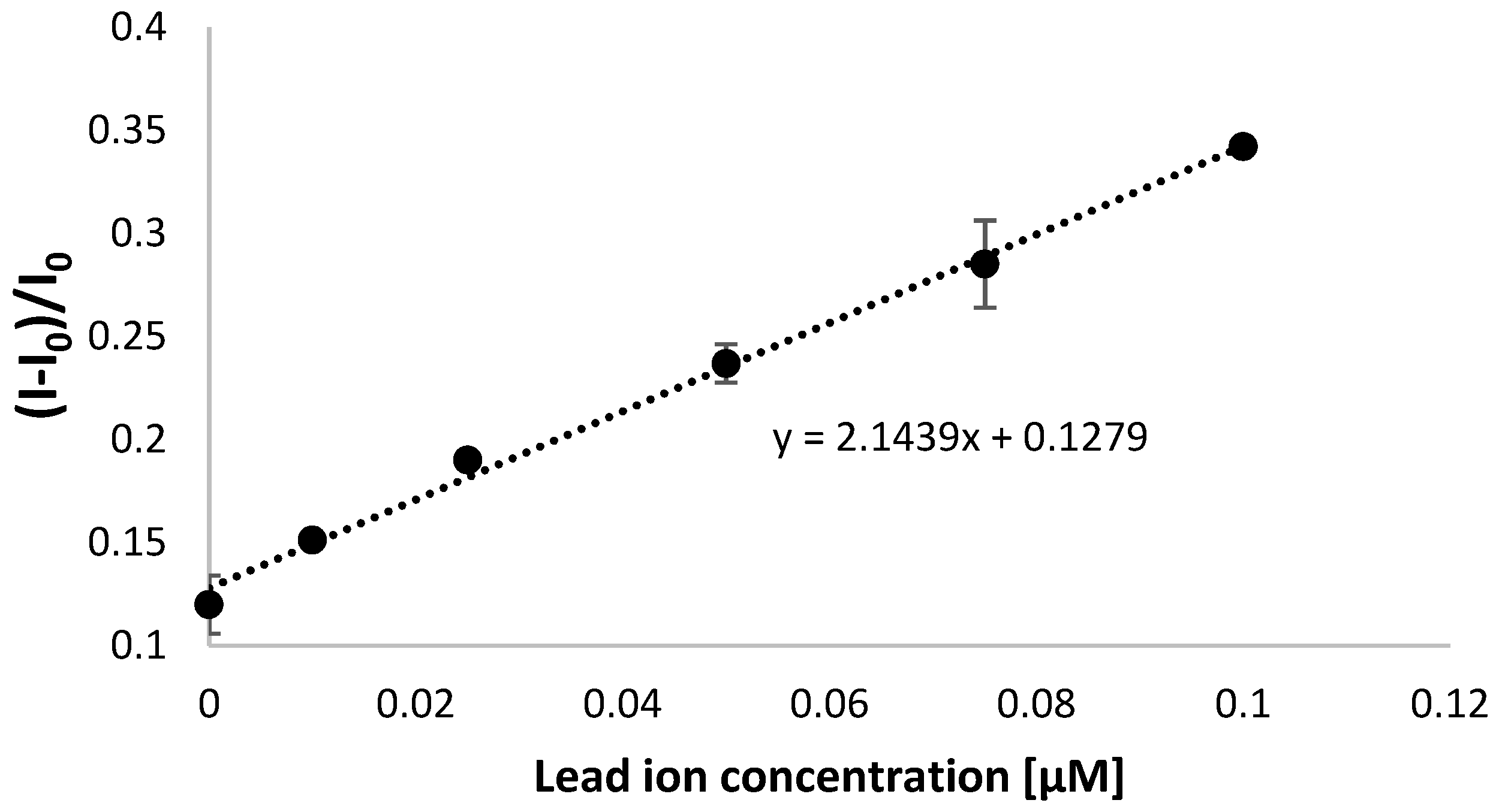
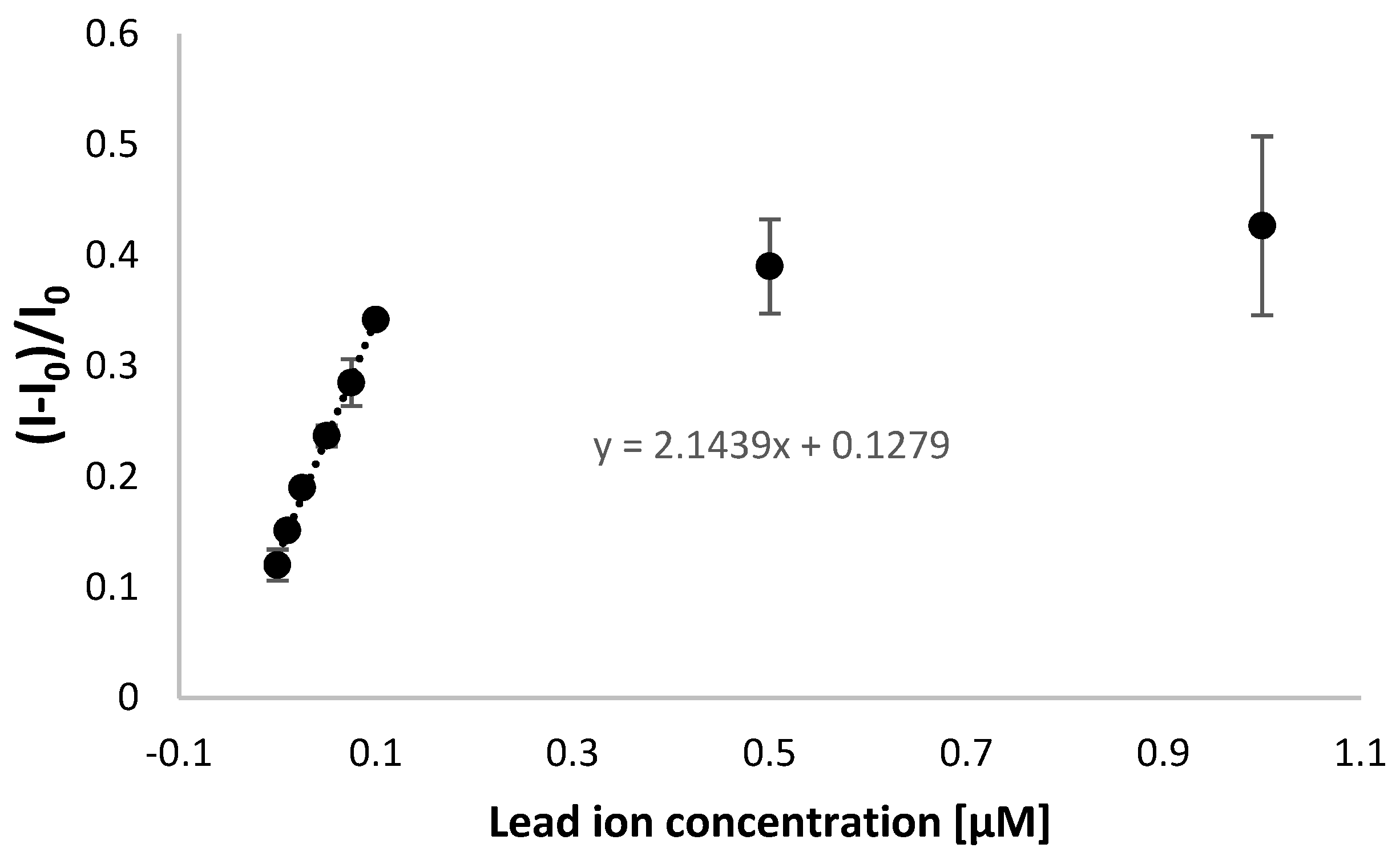
| 50 nM | 0.24 ± 0.009 | 3.75% |
| 100 nM | 0.35 ± 0.01 | 2.87% |
| Nature of Pb Detection | Detection Technique | Linear Range of Response | Lower Limit of Detection | Response Time | Reference |
|---|---|---|---|---|---|
| L/CuO-CoO-MnO/SiO2/IL/CP electrode | SWASV | 0.9 nM–230 nM 230 nM–27 µM | 0.24 nM | 3 min | [68] |
| Polypropylene hollow-fiber preconcentration | ETAAS | 173 pM–2.41 nM | 57 pM | 40 min | [69] |
| Immunoassay using the secondary antibody labeled ALP, SA-coated MMPs, and biotin-labeled Cd/Pb-BSA | chemiluminescence | 2.4–38 nM | 4.8 nM | 30 min | [70] |
| CSAP-AuNPs/SAP/MCH/Au electrode | QCM | 5–200 nM | 4 nM | 45 min | [71] |
| Disulfide-modified TBA aptamer/MCH/SP Au | SWV | 10–100 nM | 10 nM | 11 min | This work |
Disclaimer/Publisher’s Note: The statements, opinions and data contained in all publications are solely those of the individual author(s) and contributor(s) and not of MDPI and/or the editor(s). MDPI and/or the editor(s) disclaim responsibility for any injury to people or property resulting from any ideas, methods, instructions or products referred to in the content. |
© 2024 by the authors. Licensee MDPI, Basel, Switzerland. This article is an open access article distributed under the terms and conditions of the Creative Commons Attribution (CC BY) license (https://creativecommons.org/licenses/by/4.0/).
Share and Cite
Jarczewska, M.; Sokal, M.; Olszewski, M.; Malinowska, E. Studies on the Aptasensor Miniaturization for Electrochemical Detection of Lead Ions. Biosensors 2024, 14, 110. https://doi.org/10.3390/bios14020110
Jarczewska M, Sokal M, Olszewski M, Malinowska E. Studies on the Aptasensor Miniaturization for Electrochemical Detection of Lead Ions. Biosensors. 2024; 14(2):110. https://doi.org/10.3390/bios14020110
Chicago/Turabian StyleJarczewska, Marta, Marta Sokal, Marcin Olszewski, and Elzbieta Malinowska. 2024. "Studies on the Aptasensor Miniaturization for Electrochemical Detection of Lead Ions" Biosensors 14, no. 2: 110. https://doi.org/10.3390/bios14020110
APA StyleJarczewska, M., Sokal, M., Olszewski, M., & Malinowska, E. (2024). Studies on the Aptasensor Miniaturization for Electrochemical Detection of Lead Ions. Biosensors, 14(2), 110. https://doi.org/10.3390/bios14020110