Review

Point-of-Care Diagnostic Devices for Detection of Escherichia coli O157:H7 Using Microfluidic Systems: A Focused Review

1
Department of Mechanical Engineering, Sejong University, Gwangjin-gu, Seoul 05006, Republic of Korea
2
Laboratory of Stem Cell & Neurobiology, Department of Oral Anatomy & Dental Research Institute, Seoul National University School of Dentistry, Jongno-gu, Seoul 03080, Republic of Korea
3
Interdisciplinary Program in Neuroscience, Seoul National University College of Natural Sciences, Gwanak-gu, Seoul 08826, Republic of Korea
4
Sensor System Research Center, Advanced Materials Research Division, Korea Institute of Science and Technology (KIST), Seongbuk-gu, Seoul 02792, Republic of Korea
5
Biomedical Engineering Division, KIST School, University of Science and Technology, Seoul 02792, Republic of Korea
*
Authors to whom correspondence should be addressed.
Biosensors 2023, 13(7), 741; https://doi.org/10.3390/bios13070741
Submission received: 23 June 2023 / Revised: 14 July 2023 / Accepted: 15 July 2023 / Published: 17 July 2023
(This article belongs to the Special Issue Biosensors Based on Microfluidic Devices)

Abstract

:
Bacterial infections represent a serious and global threat in modern medicine; thus, it is very important to rapidly detect pathogenic bacteria, such as Escherichia coli (E. coli) O157:H7. Once treatments are delayed after the commencement of symptoms, the patient’s health quickly deteriorates. Hence, real-time detection and monitoring of infectious agents are highly critical in early diagnosis for correct treatment and safeguarding public health. To detect these pathogenic bacteria, many approaches have been applied by the biosensors community, for example, widely-used polymerase chain reaction (PCR), enzyme-linked immunosorbent assay (ELISA), culture-based method, and adenosine triphosphate (ATP) bioluminescence. However, these approaches have drawbacks, such as time-consumption, expensive equipment, and being labor-intensive, making it critical to develop ultra-sensitive and highly selective detection. The microfluidic platform based on surface plasmon resonance (SPR), electrochemical sensing, and rolling circle amplification (RCA) offers proper alternatives capable of supplementing the technological gap for pathogen detection. Note that the microfluidic biochip allows to develop rapid, sensitive, portable, and point-of-care (POC) diagnostic tools. This review focuses on recent studies regarding accurate and rapid detection of E. coli O157:H7, with an emphasis on POC methods and devices that complement microfluidic systems. We also examine the efficient whole-body detection by employing antimicrobial peptides (AMPs), which has attracted growing attention in many applications.

1. Introduction

Ingesting contaminated food such as chicken, beef, milk, juice, vegetables, and tap water not only endangers human health but also results in significant financial losses, posing a serious threat to public health [1]. Among the pathogenic bacteria, Escherichia coli (E. coli) O157:H7 is a quite important foodborne or waterborne infectious pathogen of global concern, as well as the most common and lethal serotype of enterohemorrhagic E. coli [2]. In addition, contamination of water sources with this pathogenic bacteria is a big problem in both developing and developed countries [3]. Infants, children, immunocompromised individuals, and the elderly are the most vulnerable to waterborne infections. Ingestion of as few as 10 organisms can result in life-threatening symptoms, especially in young and immunocompromised people [4,5,6]. Majowicz et al. [7] published a systematic review of outbreak surveillance data on the global prevalence of Shiga-toxin generating E. coli infections and calculated that over 2.8 million people are affected each year. Between 1998 and 2017, there were approximately 350 occurrences of E. coli O157:H7 in more than 45 US states, Canada, the United Kingdom, Japan, and Ireland. More than half of all outbreaks (54%) were caused by contaminated water and food. In addition to the safety issues related to food, it has also become a significant medical and global public health problem. Therefore, establishing the effective and rapid detection of E. coli O157:H7 is really critical for diagnostic platform. Developing a microfluidic device that is ultra-sensitive, fast, highly selective (specific), easy to use, and portable as valuable proof-of-concept should be a challenge for public health [8].
Fortunately, several advanced technologies have been developed for the efficient detection of E. coli O157:H7. So far, its clinical detection has relied on standard approaches, such as real-time PCR [9,10,11], antibody-based detection, and the well-known ELISA method [12,13]. Antibody-based detection (immunoassay) is limited by low sensitivity and dependence on specific antibodies. PCR, which amplifies specific DNA sequences unique to E. coli O157:H7, is a commonly used method. This technique enables rapid and sensitive detection of bacteria in food samples or clinical specimens. PCR-based assays can provide results within hours, making them invaluable for timely identification and response to potential outbreaks. PCR has been widely used in laboratory settings for several decades, significantly contributing to advancements in various fields, including molecular biology, medical diagnostics, and forensic sciences. Its established methodology and robustness have made it the gold standard for many applications. However, when comparing conventional PCR to point-of-care (POC) devices, multiple parameters were considered including time and speed, portability, accessibility, sensitivity and specificity, and cost and affordability. Conventional PCR has a proven track record of high sensitivity and specificity, allowing for accurate detection and quantification of nucleic acids.
While POC devices may not always match the sensitivity of conventional PCR, recent advancements have significantly improved their performance. It is worth noting that point-of-care devices are often designed to target specific pathogens or genetic markers, optimizing their sensitivity and specificity for the intended diagnostic purpose. Similarly, POC devices are typically portable, compact, and easy to use, making them suitable for deployment in resource-limited settings, remote areas, or during emergencies. In contrast, conventional PCR machines are larger, stationary instruments that require specialized laboratory infrastructure and trained personnel. The accessibility and convenience of POC devices broaden their potential applications and impact.
Another innovative technology for E. coli O157:H7 detection is immunological assays, such as ELISA and lateral flow immunoassays (LFIA). These tests utilize antibodies that specifically bind to E. coli O157:H7 antigens, allowing for the detection of the bacteria in various samples. Immunoassays are relatively quick, cost-effective, and can be performed in the field, making them suitable for rapid screening purposes. In addition, biosensors have emerged as promising tools for the detection of E. coli O157:H7. Biosensors employ biological components, such as antibodies or DNA probes, coupled with transducers to convert the presence of bacteria into measurable signals. These devices offer real-time monitoring, high sensitivity, and portability, facilitating on-site testing and enabling early identification of contaminated sources. Lastly, advancements in genomic sequencing technologies have revolutionized the detection of E. coli O157:H7. Whole-genome sequencing (WGS) allows for comprehensive analysis of the bacteria’s genetic material, enabling the identification of specific virulence factors and tracing outbreaks back to their sources. WGS provides valuable insights into the epidemiology and evolution of E. coli O157:H7 strains, aiding in the development of targeted prevention and control strategies. WGS offers in-depth genetic analysis and is primarily used for long-term surveillance and research purposes. On the other hand, POC detection provides rapid, on-site testing, enabling quick decision-making and response to E. coli O157:H7 contamination events. Furthermore, it is necessary to design and fabricate a highly precise and cost-effective pathogenic bacteria diagnostic test that can be easily utilized in underdeveloped nations to combat infectious bacterial/viral infections without causing financial burden. POC devices aimed at clinical diagnostics are a good example of an interface where biomedical and engineering disciplines meet [14,15,16].
The incorporation of leading-edge technology into the microfluidic platform may provide very useful solutions for the relevant issues of diagnostic devices. A microfluid-based system offers advantages of relatively simple operation, low cost, multiple-target testing, ease of automation, portability, and compactness. As a consequence, surface plasmon resonance (SPR), electrochemical, and rolling circle amplification (RCA) have been revitalized due to rapid analysis, highly sensitive and selective performance, and consistent detection limit. Furthermore, these methods incorporated with a microfluidic system can be used in any location or POC, even if there is no electricity accessible, due to the ubiquitous concept.
This review focuses on the detection of E. coli O157:H7 using microfluidic systems, such as POC diagnostic devices including SPR, electrochemical sensing, and RCA. The most important performances for these methods are the limit of detection (LOD) and the detection time. The LOD and detection time were compared with each other from recent literature information, and then the best results of LOD and detection time were discussed for the detection of pathogenic bacteria. In addition, this review also presents the actually advanced E. coli detection by employing antimicrobial peptides (AMPs), which were introduced as emerging alternatives to antibodies. Among the different types of POC, AMP-based microfluidic platforms for pathogen sensing have gained much attention in view of whole-body detection with selectively binding affinity.

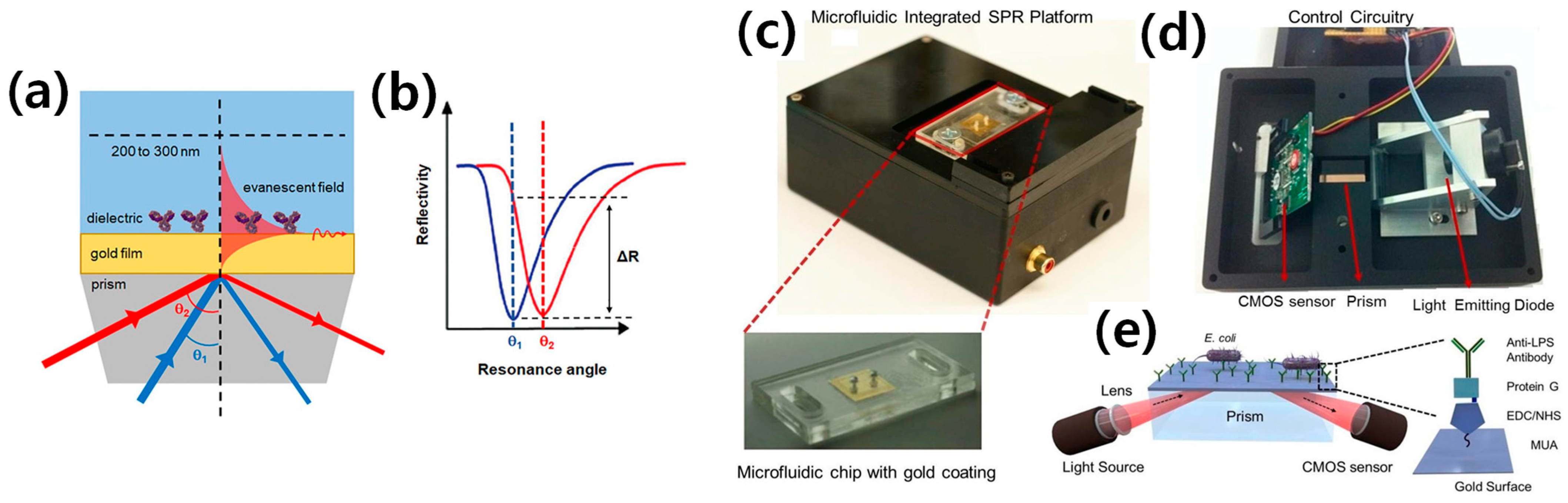
2. SPR-Based E. coli O157:H7 Detection

SPR-based biosensors are rapidly emerging as a direct label-free technology for the rapid and highly sensitive detection of chemical and biological analytes in critical fields, such as medical diagnostics, food safety, and environmental sensing, due to the unique optical properties they possess [17,18,19]. SPR utilizes the simple phenomenon of refractive index changes in conjunction with the binding of target biomolecules (either antigens or protein samples) to identify these changes. The SPR phenomena occur due to the excitement and collective vibration of free electrons in metal and dielectric junctions. This excitation is caused by the interaction of electromagnetic waves in the visible area with the plasmon of gold and silver nanoparticles [20,21,22]. When combined with well-developed microfluidic technology, SPR offers numerous advantages, which are explained and summarized in the concluding section of this review article. The complete binding mechanism is illustrated in Figure 1a,b, where the binding event is indicated by a change in reflectivity ΔR at a fixed observed angle. Before antibody binding, θ1 (blue) is shown in both panels (panel A and panel B), while after the binding of antibody, θ2 (red) is observed. The change in reflectance ΔR represents the binding event or sensitivity at the fixed observed angle. In the 1990s, the United States Department of Agriculture (USDA) introduced a commercial SPR apparatus for the detection of E. coli O157:H7, where the antibodies were sufficiently immobilized on the surface of a substrate. Since then, SPR has become a popular label-free tool in the biosensors society for detecting a variety of chemical and biological samples [23], serving as a POC technique in microfluidics.
Numerous researchers have worked in the SPR domain as a POC diagnostic device for the accurate, rapid, and highly sensitive detection of E. coli O157:H7 with no cross-reaction. To achieve this goal, Tokel et al. [24] developed a microfluidic SPR biochip capable of detecting and quantifying E. coli and Staphylococcus aureus. A microfluidic channel with a volume of 4 µL was created by laser cutting a microchannel form in a double-sided adhesive (DSA) layer with a thickness of 50 μm. The DSA layer held together the polymethyl methacrylate (PMMA) layer and the gold-coated glass substrate. The sample was injected into the channel and adhered to the gold surface after opening a single inlet and outflow port on the PMMA layer. For SPR measurement, the prism was placed on the PMMA-DSA-glass setup. Figure 1c,d depicts the custom-made SPR platform for microfluidic integration based on a complementary metal–oxide–semiconductor (CMOS) sensor. The lower part of Figure 1c shows an enlarged view of the microfluidic chip with a gold coating. Figure 1e illustrates the schematic of the microfluidic integrated SPR system, in which the gold surfaces were modified with different activators, including 11-mercaptoundeconoic acid (MUA), N-(3-dimethylaminopropyl)-N′-ethylcarbodiimide hydrochloride (EDC), N-hyroxysuccinimide (NHS), and anti-liposaccharide (LPS) antibodies, to capture E. coli. The antibodies in the microchannel capture bacteria, resulting in a change in the local refractive index. This change is detected by the reflected light, collected by the sensor, and delivered to a computer for analysis. The microfluidic SPR sensor was used to investigate the capture of E. coli with a LOD of 106 CFU/mL. A detection time of 20 min was observed using this POC microfluidic system, which is considered a good reaction/detection time in biosensor design and fabrication. Previous studies have reported a lower LOD of 94 CFU/mL with SPR, which is approximately 104 times better than the previous system [24,25]. Table 1 summarizes the most recent studies, including various parameters such as material type, method, LOD, detection time, detection range for linear analysis (to calculate R2 value using regression analysis), sample type, and sensor response.
Figure 1. The principle of SPR as a label-free biosensing technology and SPR-based microfluidic platform for E. coli detection as a POC. (a) The incident light travels along the dielectric interface and excites plasmon at this interface. (b) Accurate binding of a biomolecule (antibody) on the surface of a gold film results in a shift in SPR resonance angle with before antibody binding θ1 (blue) and after binding θ2 (red). The occurrence of binding is denoted as the reflectivity angle (ΔR) with the measured resonance angle fixed. Reprinted with permission from [26]. (c) The disposable microfluidic chips, which are surface activated, are placed on the top of the device. (d) The electronic configuration of the device is depicted from bottom to top. A light-emitting diode (LED) illuminates a cylindrical lens, which focuses the light onto a rectangular prism. The reflected light is captured by a CMOS sensor, and the image is transmitted to a portable computer via control circuitry. (e) The schematic shows a microfluidic integrated SPR platform. Reprinted with permission from [24].
Biosensors 13 00741 g001
In recent years, numerous studies have aimed at developing SPR biosensors for the fast and accurate detection of E. coli O157:H7. However, these studies often demonstrate the ability to identify bacterial pathogens only at higher concentrations and longer incubation times using expensive equipment, which is inadequate for most hazardous infections.
According to the literature, several assays have been employed for the detection of E. coli using the SPR method, including monoclonal antibody, polyclonal antibody, T4 bacteriophage, and AMP with magainin 1-C-based SPR detection. Each method possesses its own advantages and disadvantages, depending on factors such as the LOD, detection time, cost, and commercialization feasibility. The minimum detection time achieved was 10 min using a polyclonal antibody (rabbit anti-goat IgG), while a detection time of 120 min was observed when utilizing a chicken antibody for E. coli detection. Furthermore, the best LOD achieved using the SPR method was 10 CFU/mL. This notable achievement was made possible by detecting E. coli using murine anti-E. coli O157:H7 monoclonal antibodies, utilizing magnetic nanoparticles coated with an Au shell. In conclusion, SPR-based detection of E. coli O157:H7 offers a sensitive and real-time approach for the identification and quantification of the bacteria. This advancement contributes to enhanced food safety measures and improved public health.
The specificity of E. coli detection plays a crucial role in SPR-based biosensors. When specific antibodies or aptamers designed to recognize and bind to E. coli antigens are employed as capture molecules in an SPR assay, the specificity can be significantly high. These capture molecules are carefully selected to have minimal cross-reactivity with other bacterial species or contaminants, ensuring their specific interaction with the target E. coli bacteria. Numerous researchers have investigated the specificity of E. coli O157:H7 using the SPR method. For instance, Li et al. [27] determined the specificity of E. coli O157:H7 against E. coli, S. typhimurium, S. aureus, and V. parahemolyticus. They evaluated the signal intensities and found that the intensities associated with E. coli O157:H7 were almost five-fold stronger than those of the non-target pathogens. The most recent studies that focused on accurate detection using the SPR method are summarized in Table 2.

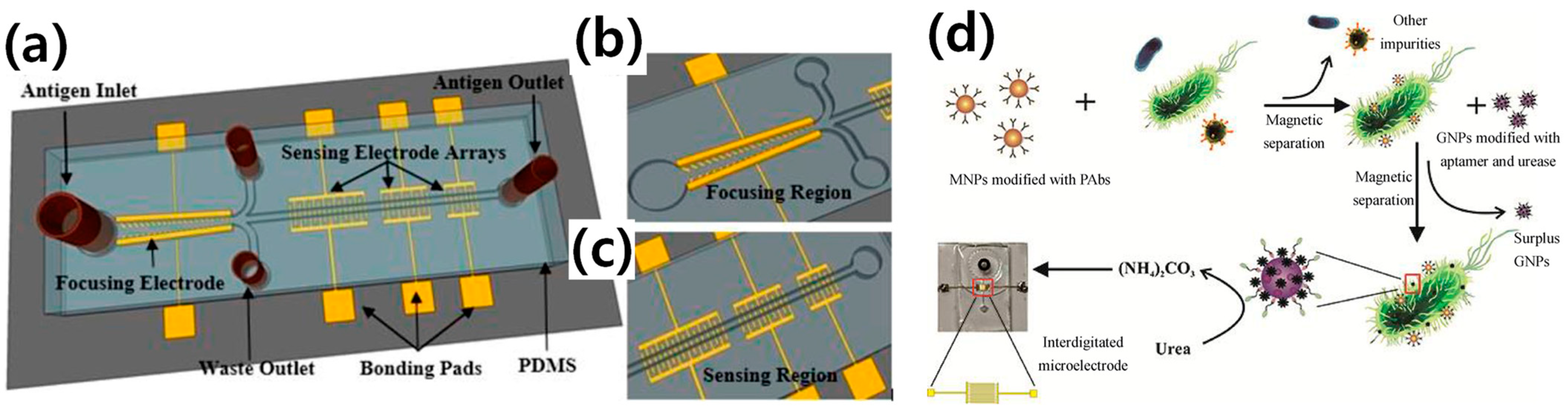
3. Electrochemical-Based E. coli O157:H7 Detection

Electrochemical biosensors are commonly produced and widely employed for the detection of food-borne and water-borne pathogens due to the possibilities of miniaturization and the ability to build disposable, flexible, and affordable sensing systems. Over the last decade, various methodologies have been developed for the rapid quantification of E. coli O157:H7. According to the International Union of Pure and Applied Chemistry (IUPAC), an electrochemical biosensor is a self-contained device that utilizes a bioreceptor in conjunction with an electrochemical transduction component to provide quantitative or semi-quantitative analytical data [36]. Many researchers have made efforts to detect E. coli O157:H7 as a POC device using electrochemical methods. For this purpose, Dastider et al. [37] developed a microfluidic platform for accurate and sensitive detection, known as the MEMS sensor, which operates on the principle of dielectrophoresis force. The key components of this MEMS device are the design of the focusing region and the sensing region. The focusing region generates a positive dielectrophoresis force that moves the cells to the edges of the tilted thin-film electrode fingers in the center of the microchannel. The fluidic drag force then transports the focused cells to the sensing region, which contains multiple pairs of three interdigitated electrode arrays (IDEAs) embedded inside the microchannel. This technique allows for highly concentrated samples in the sensing region. For specific sensing of E. coli 0157:H7, the sensing IDEAs are functionalized with an anti-E. coli antibody. When E. coli binds to the antibody, there is an impedance change. The complete design of this MEMS microfluidic biosensor is shown in Figure 2. The detection time is 120 min and the LOD is 39 CFU/mL. Despite the good LOD, the long detection time makes this device less efficient for a POC application.
In addition, to further improve the LOD and detection time for accurate detection, Yao et al. [38] designed and fabricated a microfluidic biosensor based on electrochemical impedance spectroscopy (EIS) combined with magnetic nanoparticles (MNPs). MNP–bacteria–GNP–urease complexes were created by combining MNP–bacteria with gold nanoparticles (GNPs) modified with urease and aptamers against E. coli O157:H7. The complete development is shown in Figure 2d. A satisfactory linear relationship between the relative rate of impedance change of the catalysate and the concentration of the bacteria was established with a low detection limit of 12 CFU/mL and a detection period of 15 min. This represents an excellent LOD and detection time for POC application. The most recent studies on the detection of E. coli O157:H7 are summarized in Table 3, including the LOD, detection time, material type, methodology, assay structure, sample type, and detection range for linearity checking (R2 using regression analysis). In addition, the most recent studies that have focused on the accurate detection (specificity) using electrochemical methods are summarized in Table 4.

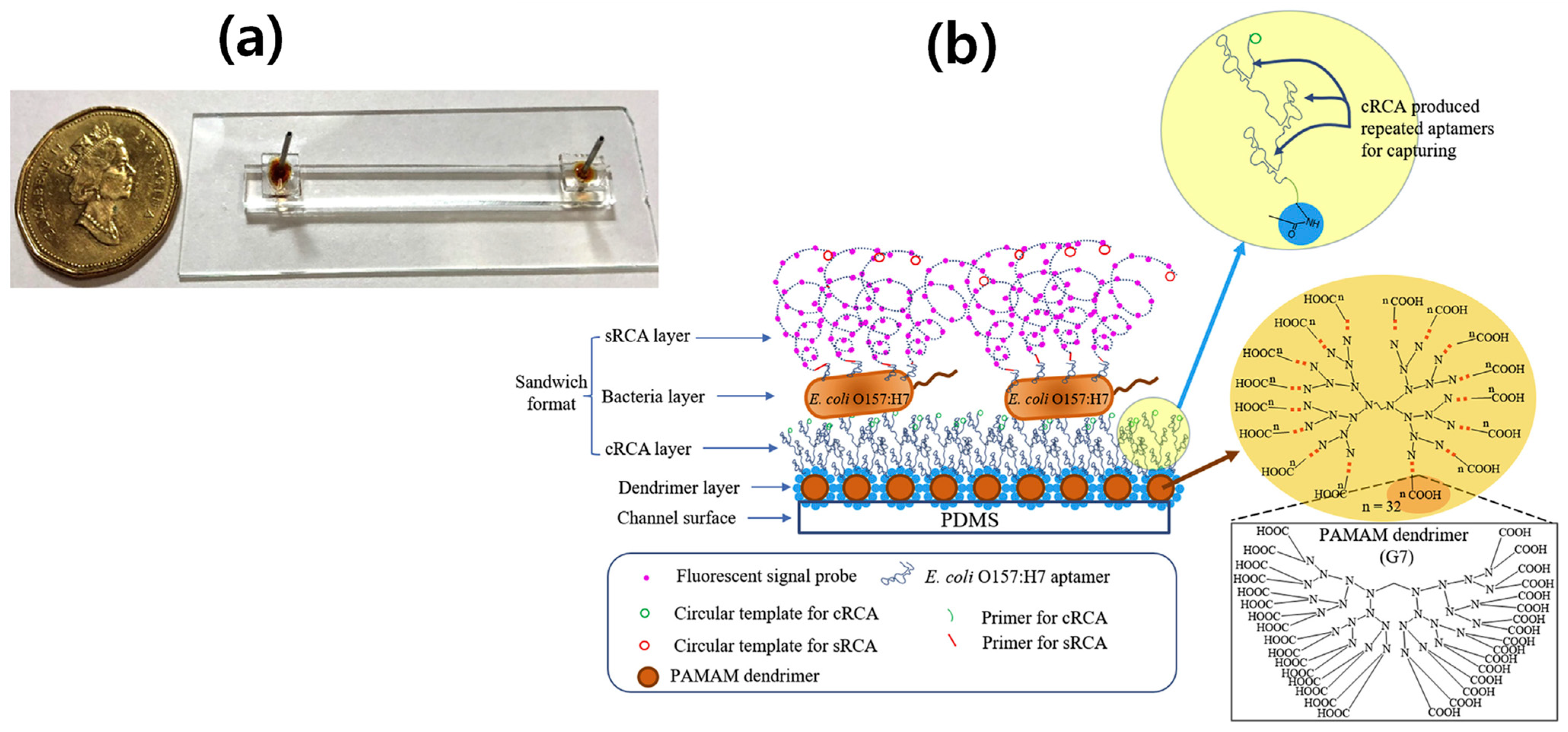
4. RCA-Based E. coli O157:H7 Detection

Rolling circle amplification (RCA) driven by DNA polymerase is an effective enzymatic isothermal process. It involves the synthesis of long single-stranded (ss) DNA molecules on a short circular ssDNA template using a single DNA or RNA primer. In addition, RCA can replicate circularized oligonucleotide probes with either linear or geometric kinetics. By modifying the template, introducing functional sequences during the reaction, and hybridizing the RCA products to oligonucleotides with specific functions, RCA can serve multiple purposes. Due to these innovative features, RCA-based nanotechnology has found wide applications in biological detection, medication delivery, and other fields [52]. A brief description of the RCA is as follows: First, a primer is conjugated to the necessary surface by employing surface activation and functionalization. The primer is then hybridized with a padlock probe, which is carefully designed to include a primer-binding site, a pathogen-binding site, and a self-assembly area that generates a molecular dumbbell shape. By regulating the length of each binding area, the padlock probe is designed to possess thermal stability at room temperature and specificity for primer and pathogen hybridization. After annealing, the padlock probe takes an asymmetric dumbbell shape. Subsequently, the probe is hybridized with the primer immobilized on the required surface. Hybridization occurs when the padlock probe comes into contact with a target pathogen. The opening padlock probe is ligated with a ligase to create a closed-loop template that can be utilized in the RCA procedure. During the RCA process, complementary single-stranded DNA gradually extends in a dumbbell form over time by DNA polymerase. The amplification of long DNAs, owing to the dumbbell-shaped template, leads to their easy entanglement with each other, resulting in aggregation with surrounding DNAs and the formation of a DNA gel. This DNA hydrogel has been developed for the visualized, simple, and rapid detection of E. coli O157:H7.
To achieve this goal, many researchers have made efforts to design and develop microfluidics RCA sensors for the rapid and accurate detection of E. coli O157:H7. Recently, Jiang et al. [53] developed an aptamer-based microfluidic platform for the detection of whole cells by applying the dual RCA technique. The complete design of dual-RCA microfluidic biosensor design is illustrated in Figure 3. The detection time and LOD are 120 min and 39 CFU/mL, respectively, which are better results than those with an incubation time of 120 min and LOD of 4.0 × 10² CFU/mL [54]. The most recent studies on E. coli detection, including LOD and detection time, material type, methodology, assay structure, and sample type, are summarized in Table 5. Further exploration is still needed for the identification/detection of E. coli in order to improve the detection time and LOD.
PCR, the gold standard method, amplifies specific DNA sequences unique to E. coli O157:H7, enabling rapid and sensitive detection of the bacteria in food samples or clinical specimens. PCR-based assays provide results within hours, making them invaluable for timely identification and response to potential outbreaks. PCR has been extensively employed in laboratory settings for several decades, leading to significant advancements across various fields. When comparing RCA and PCR, both techniques serve as powerful molecular methods for nucleic acid amplification. While PCR is widely established and extensively used, RCA offers distinct advantages in specific applications. The following section outlines a comparative analysis of RCA and PCR, highlighting the advantages of RCA:
Amplification Mechanism: The fundamental amplification mechanisms of RCA and PCR differ. PCR utilizes a thermal cycling process to amplify a specific DNA segment, employing heat-stable DNA polymerases. On the other hand, RCA employs a rolling circle mechanism, where a circular template is exponentially amplified by a DNA polymerase with strand-displacement activity. This mechanism enables RCA to generate long, single-stranded DNA products, which can be advantageous for various downstream applications, such as DNA sequencing or in situ hybridization techniques.
Sensitivity: In certain scenarios, RCA has demonstrated higher sensitivity compared to PCR. Due to its isothermal nature, RCA can produce a larger number of amplification products, resulting in increased sensitivity for detecting low-abundance targets. This sensitivity advantage has proven particularly useful in applications such as detecting rare genetic mutations, single-cell analysis, or amplifying targets with low copy numbers.
Simplified Workflow: RCA offers a simplified workflow compared to PCR. RCA reactions can be performed under isothermal conditions, eliminating the need for sophisticated thermal cycling equipment. This simplification can reduce overall costs and technical complexity associated with amplification procedures, making RCA an appealing option for resource-limited settings or POC applications.
Product Length: As mentioned earlier, RCA can generate long, single-stranded DNA products. This feature proves advantageous in applications where longer DNA fragments are desired, such as generating templates for DNA sequencing or studying DNA–protein interactions. In contrast, PCR typically produces shorter amplicons due to limitations inherent in the polymerase enzyme and primer design considerations.
Enzyme Selection: RCA can be conducted using various DNA polymerases, including both strand-displacing and nick-translating enzymes. This flexibility allows researchers to choose an appropriate enzyme based on their specific requirements, such as amplification efficiency, fidelity, or compatibility with specific detection methods. In contrast, PCR primarily relies on thermostable DNA polymerases, which may have limitations in certain applications, such as amplifying challenging templates or incorporating modified nucleotides.
While RCA offers several advantages over PCR in certain applications, it is important to note that PCR still maintains its strengths and remains the method of choice for many diagnostic and research purposes. PCR benefits from a well-established protocol, extensive commercial availability of reagents, and a vast body of literature supporting its applications. The selection between RCA and PCR should be based on the specific requirements of the experiment, the desired output, and the available resources. Additionally, the most recent studies focused on this accurate detection (specificity) using RCA method are summarized in Table 6.

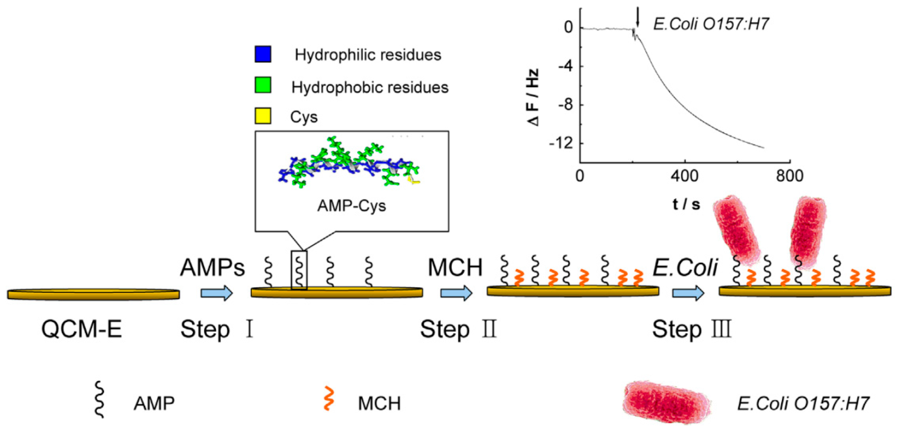
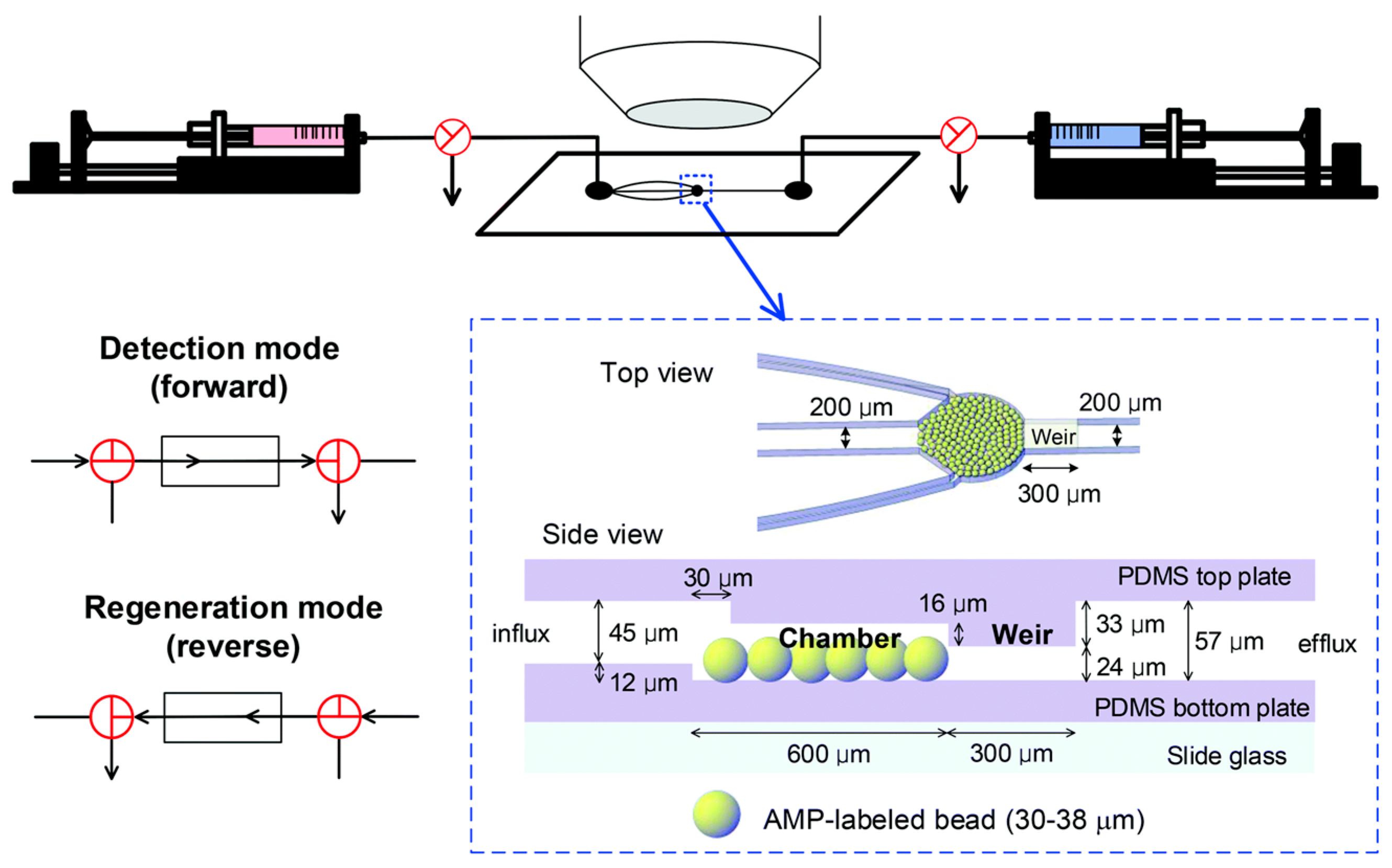
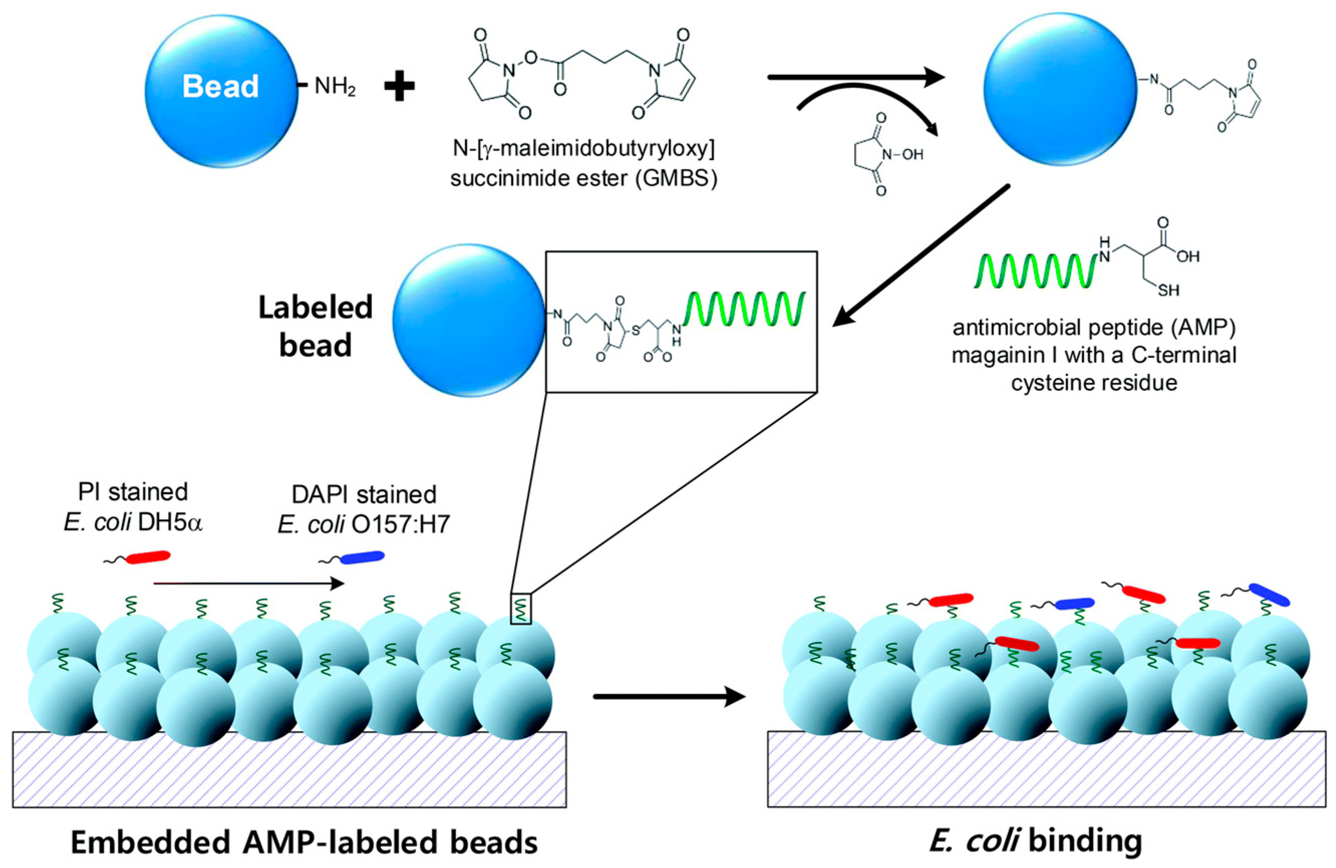
5. Attraction of Antimicrobial Peptide (AMP) for E. coli O157:H7 Detection

The novel antimicrobial compounds have great potential for combating antibiotic resistance in bacteria. As part of the innate immune system, there is a growing demand for these compounds. To address this need, researchers have employed several alternative techniques, including the use of metals, metal oxides, polymer materials, and AMPs [60]. Among these techniques, AMPs show impressive potential for direct detection of both pathogenic and nonpathogenic bacteria. They are also considered strong candidates for whole bacteria detection. AMPs can be found in various natural niches and act as the organism’s first line of defense against bacterial attacks, forming an important part of the unique immune system that protects hosts from pathogens [61]. Most AMPs consist of 6 to 50 positively charged amino acid residues and a large number of hydrophobic residues [62]. Due to their excellent stability and low cost, AMPs have been extensively explored as alternative recognition elements in biosensors for bacterial detection. Since the affinity of AMPs is important in detection, the elements that influence the affinity of AMPs will be discussed before addressing various AMP-based approaches. Microfluidics have also been combined with AMP-based approaches to identify foodborne infections, making it a promising new tool. Finally, the future opportunities and challenges in constructing reliable and sensitive AMP-based platforms will be highlighted.
Although the mechanisms of action of AMPs have not been fully elucidated, there is a widely accepted model that explains their mode of action. According to this hypothesis, cationic AMPs bind to bacterial surfaces through electrostatic and hydrophobic interactions, leading to various modifications in membrane structures and disruption of the integrity of the bacterial cytoplasmic membrane, ultimately killing microorganisms. Based on their secondary structures, AMPs’ bioactivity toward microbial cells can be categorized into many groups [63]. Many AMPs adopt amphipathic conformations that allow them to target the negatively charged head groups of lipids on the bacterial membrane, utilizing spatial hydrophobic clustering from cationic amino acids. Plant and animal membranes, on the other hand, isolate negative charges from the interior leaflet and contain cholesterols that inhibit AMP activation [64]. AMPs, as antibiotics, have exhibited remarkable resistance to acquired resistance by targeting the fundamental structure of the bacterial cell membrane, making them difficult for proteases to identify. Linear cationic peptides such as magainin, with their small molecular size and inherent stability, are particularly attractive for microbial sensing applications [65]. As a precursor to bactericidal activity, the positively charged AMP magainin I (GIGKFLHSAGKFGKAFVGEIMKS) exhibits selective binding to the pathogenic bacteria E. coli O157:H7 [66,67]. Magainin I also demonstrates broad-spectrum efficacy against other Gram-negative bacteria responsible for a majority of human pathogenic infections.
In AMP-based biosensors, AMPs are often immobilized on substrate surfaces. The immobilized AMPs are believed to interact with the outer membrane through electrostatic contacts, triggering various interactions with the cytoplasmic membrane such as amphipathic conformations and insertion into the membrane, leading to disturbances in the lipid bilayer. Immobilizing peptides may restrict their mobility and ability to interact with cellular membranes compared to their soluble state. However, due to limited research on soluble and immobilized AMPs, it is challenging to determine the similarities and differences in their activities. The strong affinity of AMPs for bacterial surfaces has garnered more attention in biosensing applications than their antibacterial activity because AMP surfaces serve as recognition elements. The AMPs adsorbed on the surface of biosensors are believed to facilitate bacterial attachment through electrostatic and hydrophobic interactions. However, few studies have focused on the interactions of immobilized AMPs with the bacterial surface in biosensors. Despite the unknown recognition processes, AMPs have gained significant attention in biosensing due to their strong affinity for bacteria. The AMP database currently contains over 3000 natural AMPs, with their sequences and structures extensively studied by the research community.
One major advantage of AMPs as recognition elements is their ability to engage with multiple pathogens, thanks to their selective binding nature to target cells. By replacing existing antibody-based probes with more durable and stable antimicrobial peptides in biological sensors, the shelf life of current diagnostic systems can be extended. Due to these advantages, AMPs have been utilized to construct various bacterial biosensors. Numerous researchers have worked on developing microfluidic-based AMPs with specific identification elements for rapid and accurate detection of E. coli O157:H7 [68,69].
Various recognition elements, including magainin I, leucocyte A, clavA, colicin V, and cecropin P1, have been employed for bacterial detection. Among them, magainin I has shown the most promising binding results for reliable detection. Until recently, the majority of AMP research had focused on magainin II, but magainin I has proven to be a suitable bacterial recognition factor. Manoor et al. [69] demonstrated that immobilizing AMP magainin I on a gold microelectrode array via a C-terminal cysteine residue enabled the capture and measurement of the binding effect on pathogenic E. coli. They developed a robust and portable biosensor for the detection of E. coli O157:H7 using impedance spectroscopy for evaluation. The biosensor achieved a detection time of 30 min and a LOD of 1000 CFU/mL in water samples, with a detection range of 101 to 105. While this detection time is favorable, the LOD, which is a key factor for POC devices, needs improvement. To further improve the LOD and detection time, Dong and Zhao [70] fabricated the microfluidic E. coli biosensor with an excellent detection time and a lower LOD of 400 CFU/mL, which is 2.5 times better than the previous study [69]. They demonstrated that an AMP specific to E. coli O157:H7, tagged with a cysteine residue at the C-terminal for immobilization on a gold quartz crystal microbalance (QCM) electrode surface, could be used to capture and measure the binding effect. The complete detection mechanism is shown in Figure 4. While these approaches have achieved low bacterial cell detection limits, many of them require extensive sample preparation and, in some cases, multiple reagent and assay steps before detection. Therefore, developing an integrated detection system with high mobility, robustness, sensitivity, and selectivity for harmful organisms remains challenging. Further exploration is needed to improve the detection time and LOD for the identification/detection of E. coli O157:H7.
To further enhance the LOD and decrease detection time, our research group [68] investigated the detection of pathogenic E. coli O157:H7 using a microfluid-based biosensing device embedded with AMP-labeled microbeads. Through effective immobilization of AMP magainin I on the surface of glass microbeads, the LOD as well as the detection time were significantly improved. We achieved an outstanding LOD of 10 CFU/mL with a detection time of less than 20 min. In addition, we successfully detected the entire bacteria, which is crucial for sensing pathogenic bacteria. Furthermore, we explored this microfluidic platform for effective regeneration capabilities. The strategic fabrication of our system is illustrated in Figure 5, highlighting the importance of the weir structure in the microfluidic channel. Figure 6 provides an explanation of the binding mechanism between the entire E. coli O157:H7 and the AMP-immobilized glass microbeads.
Most recent studies for the E. coli detection based on AMPs structure are summarized in Table 7 with the LOD and detection time. Further explorations are still needed for the identification/detection of E. coli O157:H7 to improve the detection time and LOD.
The accurate detection of pathogenic bacteria is a critical issue for POC devices to ensure better therapy for the corresponding disease and early-stage control. Currently, significant efforts are being directed towards achieving accurate and highly specific detection. To address this, Bai et al. [71] developed a sensor for the precise detection of E. coli O157:H7 using copper phosphate nanocomposites embedded with AMP magainin I and cecropin P1. The highly specific detection of E. coli O157:H7 was evaluated against multiple bacterial samples, with a bacterial density of 105 CFU/mL (n = 3). In addition, Qi et al. proposed a novel microfluidic biosensor for the colorimetric detection of foodborne pathogens [80], and Zhang et al. introduced an integrated colorimetric and photothermal lateral flow immunoassay based on bimetallic Ag–Au urchin-like hollow structures for the sensitive detection of E. coli O157:H7 [80,81]. The most recent studies focused on this accurate detection are summarized in Table 8.

6. Conclusions and Future Challenges

This review provides an overview of recent advancements in the design and development of POC microfluidic-based biosensors for the detection of pathogenic bacteria, especially E. coli O157:H7. These biosensors offer a valuable means of delivering clinically relevant information in a simple, rapid, and cost-effective manner, catering to the demands of POC testing. The LOD and detection time are crucial parameters for detecting these pathogenic bacteria at an early stage of infection. While many of the proposed systems may not match the accuracy of traditional analytical methods, they can still provide sufficient information for routine testing and screening of food and water samples. This review summarizes the detection of E. coli O157:H7 using popular POC methods, such as SPR, RCA, and electrochemical methods. In addition, the impressive role of AMPs in terms of LOD and detection time is evaluated. Developing a simple, fast, and cost-effective method for E. coli detection not only ensures the safety of water and food supplies for consumers but also prevents costly recalls.
AMPs have emerged as a promising alternative to antibodies, and in particular, AMP-based microfluidic platforms for bacteria detection have garnered significant attention due to their simplicity, sensitivity, and high selectivity. In summary, while POC testing still has room for improvement, microfluidic biosensors hold great promise as practical tools for real-world applications. However, there are limitations in terms of LOD, detection time, and specific detection.
Improving the LOD, detection time, and accuracy of microfluidic chips is crucial. Additionally, it is equally important to detect the entire bacteria rather than solely focusing on its DNA or antibodies in order to save time and costs. AMPs present a viable option as they enable rapid and accurate detection of the entire bacteria. However, AMP-based detection faces challenges in terms of repeatability. Therefore, it is necessary to improve repeatability, along with the LOD, detection time, and selectivity, in order to develop efficient and useful biosensors for detecting pathogenic bacteria. Finally, in Table 9, a comparison is provided for widely applied methods used as POC devices for the detection of pathogenic E. coli O157:H7. Both the advantages and disadvantages of each method are summarized in this table.

Author Contributions

Writing—original draft, investigation, methodology, data curation, and formal analysis, N.A.; investigation, resources, and validation, S.S.; writing—revise and editing, validation, and literature survey, M.-S.C. (Mi-Sook Chang); draft editing, conceptualization, project administration, and funding acquisition, M.-S.C. (Myung-Suk Chun). All authors have read and agreed to the published version of the manuscript.

Funding

This work was supported by the KIST K-DARPA Program (project no. 2V09008), including the postdoctoral research of N.A. performed at the KIST. We are also thankful for the support from the KIST Institutional Program (project no. 2E32452).

Institutional Review Board Statement

Not applicable.

Informed Consent Statement

Not applicable.

Data Availability Statement

Not applicable.

Conflicts of Interest

The authors declare no conflict of interest.

References

  1. Sharma, V.K.; Akavaram, S.; Schaut, R.G.; Bayles, D.O. Comparative genomics reveals structural and functional features specific to the genome of a foodborne Escherichia coli O157: H7. BMC Genom. 2019, 20, 196. [Google Scholar] [CrossRef] [Green Version]
  2. Zou, Y.; Duan, N.; Wu, S.; Shen, M.; Wang, Z. Selection, identification, and binding mechanism studies of an ssDNA aptamer targeted to different stages of E. coli O157: H7. J. Agric. Food Chem. 2018, 66, 5677–5682. [Google Scholar] [CrossRef] [PubMed]
  3. Florentin, A.; Lizon, J.; Asensio, E.; Forin, J.; Rivier, A. Water and surface microbiologic quality of point-of-use water filters: A comparative study. Am. J. Infect. Control 2016, 44, 1061–1062. [Google Scholar] [CrossRef] [PubMed]
  4. Lee, J.-I.; Kim, S.-S.; Kang, D.-H. Susceptibility of Escherichia coli O157: H7 grown at low temperatures to the krypton-chlorine excilamp. Sci. Rep. 2019, 9, 563. [Google Scholar] [CrossRef] [PubMed] [Green Version]
  5. Oloketuyi, S.F.; Khan, F. Strategies for biofilm inhibition and virulence attenuation of foodborne pathogen-Escherichia coli O157: H7. Curr. Microbiol. 2017, 74, 1477–1489. [Google Scholar] [CrossRef] [PubMed]
  6. Lei, X.-Y.; Xiao, L.-M.; Liu, Y.-N.; Li, Y.-M. Prevalence of depression among Chinese University students: A meta-analysis. PLoS ONE 2016, 11, e0153454. [Google Scholar] [CrossRef] [PubMed] [Green Version]
  7. Majowicz, S.E.; Scallan, E.; Jones-Bitton, A.; Sargeant, J.M.; Stapleton, J.; Angulo, F.J.; Yeung, D.H.; Kirk, M.D. Global incidence of human Shiga toxin–producing Escherichia coli infections and deaths: A systematic review and knowledge synthesis. Foodborne Pathog. Dis. 2014, 11, 447–455. [Google Scholar] [CrossRef] [Green Version]
  8. Quintela, I.A.; Vasse, T.; Lin, C.-S.; Wu, V.C.H. Advances, applications, and limitations of portable and rapid detection technologies for routinely encountered foodborne pathogens. Front. Microbiol. 2022, 13, 1054782. [Google Scholar] [CrossRef]
  9. Kim, J.-H.; Oh, S.-W. Rapid detection of E. coli O157: H7 by a novel access with combination of improved sample preparation and real-time PCR. Food Sci. Biotechnol. 2020, 29, 1149–1157. [Google Scholar] [CrossRef]
  10. Du, J.; Wu, S.; Niu, L.; Li, J.; Zhao, D.; Bai, Y. A gold nanoparticles-assisted multiplex PCR assay for simultaneous detection of Salmonella typhimurium, Listeria monocytogenes and Escherichia coli O157: H7. Anal. Methods 2020, 12, 212–217. [Google Scholar] [CrossRef]
  11. Kim, J.-H.; Oh, S.-W. Rapid and sensitive detection of E. coli O157: H7 and S. Typhimurium in iceberg lettuce and cabbage using filtration, DNA concentration, and qPCR without enrichment. Food Chem. 2020, 327, 127036. [Google Scholar] [CrossRef]
  12. Wang, C.; Xing, K.; Zhang, G.; Yuan, M.; Xu, S.; Liu, D.; Chen, W.; Peng, J.; Hu, S.; Lai, W.-H. Novel ELISA based on fluorescent quenching of DNA-stabilized silver nanoclusters for detecting E. coli O157: H7. Food Chem. 2019, 281, 91–96. [Google Scholar] [CrossRef]
  13. Pang, B.; Zhao, C.; Li, L.; Song, X.; Xu, K.; Wang, J.; Liu, Y.; Fu, K.; Bao, H.; Song, D.J.; et al. Development of a low-cost paper-based ELISA method for rapid Escherichia coli O157: H7 detection. Anal. Biochem. 2018, 542, 58–62. [Google Scholar] [CrossRef] [PubMed]
  14. Safavieh, M.; Ahmed, M.U.; Sokullu, E.; Ng, A.; Braescu, L.; Zourob, M. A simple cassette as point-of-care diagnostic device for naked-eye colorimetric bacteria detection. Analyst 2014, 139, 482–487. [Google Scholar] [CrossRef]
  15. Amin, N.; Torralba, A.S.; Álvarez-Diduk, R.; Afkhami, A.; Merkoçi, A. Lab in a tube: Point-of-care detection of Escherichia coli. Anal. Chem. 2020, 92, 4209–4216. [Google Scholar] [CrossRef] [PubMed]
  16. Nguyen, T.; Chidambara, V.A.; Andreasen, S.Z.; Golabi, M.; Linh, Q.T.; Bang, D.D.; Wolff, A. Point-of-care devices for pathogen detections: The three most important factors to realize towards commercialization. TrAC-Trends Anal. Chem. 2020, 131, 116004. [Google Scholar] [CrossRef]
  17. Badshah, M.A.; Kim, J.; Yeom, J.; Abbas, N.; Haq, M.R.; Kim, Y.; Lu, X.; Kim, S.-M. Glass nanoimprinted plasmonic nanostructure for high power laser stable surface-enhanced Raman spectroscopy substrate. Appl. Surf. Sci. 2021, 542, 148587. [Google Scholar] [CrossRef]
  18. Badshah, M.A.; Michel, D.; Alam, N.E.; Madni, I.; Abbas, N.; Alameh, K.; Kim, S.-M. Enhancing the sensitivity of a surface plasmon resonance sensor with glancing angle deposited nanostructures. Plasmonics 2020, 15, 2161–2168. [Google Scholar] [CrossRef]
  19. Badshah, M.A.; Koh, N.Y.; Zia, A.W.; Abbas, N.; Zahra, Z.; Saleem, M.W. Recent developments in plasmonic nanostructures for metal enhanced fluorescence-based biosensing. Nanomaterials 2020, 10, 1749. [Google Scholar] [CrossRef]
  20. Abbas, N.; Lu, X.; Badshah, M.A.; In, J.B.; Heo, W.I.; Park, K.Y.; Lee, M.-K.; Kim, C.H.; Kang, P.; Chang, W.-J.; et al. Development of a protein microarray chip with enhanced fluorescence for identification of semen and vaginal fluid. Sensors 2018, 18, 3874. [Google Scholar] [CrossRef] [Green Version]
  21. Lu, X.; Lee, S.; Kim, J.; Abbas, N.; Badshah, M.A.; Kim, S.-M. Fabrication of Ag nanorods on micropost array for a metal-enhanced fluorescence substrate with a high signal-to-background ratio. Biosens. Bioelectron. 2021, 175, 112881. [Google Scholar] [CrossRef] [PubMed]
  22. Kim, J.; Abbas, N.; Lee, S.; Yeom, J.; Asgar, M.A.; Badshah, M.A.; Lu, X.; Kim, Y.K.; Kim, S.-M. Fabrication of a Plasmonic Nanoantenna Array Using Metal Deposition on Polymer Nanoimprinted Nanodots for an Enhanced Fluorescence Substrate. Polymers 2021, 13, 48. [Google Scholar] [CrossRef] [PubMed]
  23. Fratamico, P.M.; Strobaugh, T.P.; Medina, M.B.; Gehring, A.G. Detection of Escherichia coli 0157: H7 using a surface plasmon resonance biosensor. Biotechnol. Tech. 1998, 12, 571–576. [Google Scholar] [CrossRef]
  24. Tokel, O.; Yildiz, U.H.; Inci, F.; Durmus, N.G.; Ekiz, O.O.; Turker, B.; Cetin, C.; Rao, S.; Sridhar, K.; Natarajan, N.; et al. Portable microfluidic integrated plasmonic platform for pathogen detection. Sci. Rep. 2015, 5, 9152. [Google Scholar] [CrossRef] [PubMed] [Green Version]
  25. Kaushik, S.; Tiwari, U.K.; Pal, S.S.; Sinha, R.K. Rapid detection of Escherichia coli using fiber optic surface plasmon resonance immunosensor based on biofunctionalized Molybdenum disulfide (MoS2) nanosheets. Biosens. Bioelectron. 2019, 126, 501–509. [Google Scholar] [CrossRef] [PubMed]
  26. Wang, B.; Park, B. Immunoassay biosensing of foodborne pathogens with surface plasmon resonance imaging: A review. J. Agric. Food Chem. 2020, 68, 12927–12939. [Google Scholar] [CrossRef]
  27. Li, Y.; Gao, F.; Lu, C.; Fauconnier, M.-L.; Zheng, J. Bio-specific Au/Fe3+ porous spongy nanoclusters for sensitive SERS detection of Escherichia coli O157: H7. Biosensors 2021, 11, 354. [Google Scholar] [CrossRef]
  28. Yaghubi, F.; Zeinoddini, M.; Saeedinia, A.R.; Azizi, A.; Nemati, A.S. Design of localized surface plasmon resonance (LSPR) biosensor for immunodiagnostic of E. coli O157: H7 using gold nanoparticles conjugated to the chicken antibody. Plasmonics 2020, 15, 1481–1487. [Google Scholar] [CrossRef]
  29. Zheng, L.; Cai, G.; Wang, S.; Liao, M.; Li, Y.; Lin, J. A microfluidic colorimetric biosensor for rapid detection of Escherichia coli O157: H7 using gold nanoparticle aggregation and smart phone imaging. Biosens. Bioelectron. 2019, 124–125, 143–149. [Google Scholar] [CrossRef]
  30. Lee, N.; Choi, S.-W.; Chang, H.-J.; Chun, H.S. Rapid Detection of Escherichia coli O157: H7 in fresh lettuce based on localized surface plasmon resonance combined with immunomagnetic separation. J. Food Prot. 2018, 81, 713–718. [Google Scholar] [CrossRef]
  31. Zhou, C.; Zou, H.; Li, M.; Sun, C.; Ren, D.; Li, Y. Fiber optic surface plasmon resonance sensor for detection of E. coli O157: H7 based on antimicrobial peptides and AgNPs-rGO. Biosens. Bioelectron. 2018, 117, 347–353. [Google Scholar] [CrossRef]
  32. Song, L.; Zhang, L.; Huang, Y.; Chen, L.; Zhang, G.; Shen, Z.; Zhang, J.; Xiao, Z.; Chen, T. Amplifying the signal of localized surface plasmon resonance sensing for the sensitive detection of Escherichia coli O157: H7. Sci. Rep. 2017, 7, 3288. [Google Scholar] [CrossRef]
  33. Vaisocherová-Lísalová, H.; Víšová, I.; Ermini, M.L.; Špringer, T.; Song, X.C.; Mrázek, J.; Lamačová, J.; Lynn, N.S., Jr.; Šedivák, P.; Homola, J. Low-fouling surface plasmon resonance biosensor for multi-step detection of foodborne bacterial pathogens in complex food samples. Biosens. Bioelectron. 2016, 80, 84–90. [Google Scholar] [CrossRef] [PubMed]
  34. Tawil, N.; Sacher, E.; Mandeville, R.; Meunier, M. Surface plasmon resonance detection of E. coli and methicillin-resistant S. aureus using bacteriophages. Biosens. Bioelectron. 2012, 37, 24–29. [Google Scholar] [CrossRef] [PubMed]
  35. Wang, Y.; Ye, Z.; Si, C.; Ying, Y. Subtractive inhibition assay for the detection of E. coli O157: H7 using surface plasmon resonance. Sensors 2011, 11, 2728–2739. [Google Scholar] [CrossRef] [Green Version]
  36. Thévenot, D.R.; Toth, K.; Durst, R.A.; Wilson, G.S. Electrochemical biosensors: Recommended definitions and classification. Biosens. Bioelectron. 2001, 16, 121–131. [Google Scholar] [CrossRef]
  37. Dastider, S.G.; Abdullah, A.; Jasim, I.; Yuksek, N.S.; Dweik, M.; Almasri, M. Low concentration E. coli O157: H7 bacteria sensing using microfluidic MEMS biosensor. Rev. Sci. Instrum. 2018, 89, 125009. [Google Scholar] [CrossRef]
  38. Yao, L.; Wang, L.; Huang, F.; Cai, G.; Xi, X.; Lin, J. A microfluidic impedance biosensor based on immunomagnetic separation and urease catalysis for continuous-flow detection of E. coli O157: H7. Sens. Actuators B 2018, 259, 1013–1021. [Google Scholar] [CrossRef]
  39. Qaanei, M.; Taheri, R.A.; Eskandari, K. Electrochemical aptasensor for Escherichia coli O157: H7 bacteria detection using a nanocomposite of reduced graphene oxide, gold nanoparticles and polyvinyl alcohol. Anal. Methods 2021, 13, 3101–3109. [Google Scholar] [CrossRef]
  40. Zheng, T.; Jiang, X.; Li, N.; Jiang, X.; Liu, C.; Xu, J.-J.; Wu, P. A portable, battery-powered photoelectrochemical aptasesor for field environment monitoring of E. coli O157: H7. Sens. Actuators B 2021, 346, 130520. [Google Scholar] [CrossRef]
  41. Ropero-Vega, J.L.; Redondo-Ortega, J.F.; Galvis-Curubo, Y.J.; Rondón-Villarreal, P.; Flórez-Castillo, J.M. A Bioinspired Peptide in TIR Protein as Recognition Molecule on Electrochemical Biosensors for the Detection of E. coli O157: H7 in an Aqueous Matrix. Molecules 2021, 26, 2559. [Google Scholar] [CrossRef] [PubMed]
  42. Li, T.; Ou, G.; Chen, X.; Li, Z.; Hu, R.; Li, Y.; Yang, Y.; Liu, M. Naked-eye based point-of-care detection of E. coli O157: H7 by a signal-amplified microfluidic aptasensor. Anal. Chim. Acta 2020, 1130, 20–28. [Google Scholar] [CrossRef] [PubMed]
  43. Park, Y.M.; Park, J.; Lim, S.Y.; Kwon, Y.; Bae, N.H.; Park, J.-K.; Lee, S.J. Integrated pumpless microfluidic chip for the detection of foodborne pathogens by polymerase chain reaction and electrochemical analysis. Sens. Actuators B 2021, 329, 129130. [Google Scholar] [CrossRef]
  44. Dhull, N.; Kaur, G.; Jain, P.; Mishra, P.; Singh, D.; Ganju, L.; Gupta, V.; Tomar, M. Label-free amperometric biosensor for Escherichia coli O157: H7 detection. Appl. Surf. Sci. 2019, 495, 143548. [Google Scholar] [CrossRef]
  45. Chen, Q.; Wang, D.; Cai, G.; Xiong, Y.; Li, Y.; Wang, M.; Huo, H.; Lin, J. Fast and sensitive detection of foodborne pathogen using electrochemical impedance analysis, urease catalysis and microfluidics. Biosens. Bioelectron. 2016, 86, 770–776. [Google Scholar] [CrossRef]
  46. Wang, Y.; Ping, J.; Ye, Z.; Wu, J.; Ying, Y. Impedimetric immunosensor based on gold nanoparticles modified graphene paper for label-free detection of Escherichia coli O157: H7. Biosens. Bioelectron. 2013, 49, 492–498. [Google Scholar] [CrossRef]
  47. Altintas, Z.; Akgun, M.; Kokturk, G.; Uludag, Y. A fully automated microfluidic-based electrochemical sensor for real-time bacteria detection. Biosens. Bioelectron. 2018, 100, 541–548. [Google Scholar] [CrossRef]
  48. Yang, Z.; Liu, Y.; Lei, C.; Sun, X.-C.; Zhou, Y. Ultrasensitive detection and quantification of E. coli O157: H7 using a giant magnetoimpedance sensor in an open-surface microfluidic cavity covered with an antibody-modified gold surface. Microchim. Acta 2016, 183, 1831–1837. [Google Scholar] [CrossRef]
  49. Bai, H.; Bu, S.; Liu, W.; Wang, C.; Li, Z.; Hao, Z.; Wan, J.; Han, Y. An electrochemical aptasensor based on cocoon-like DNA nanostructure signal amplification for the detection of Escherichia coli O157: H7. Analyst 2020, 145, 7340–7348. [Google Scholar] [CrossRef]
  50. Li, Y.; Liu, H.; Huang, H.; Deng, J.; Fang, L.; Luo, J.; Zhang, S.; Huang, J.; Liang, W.; Zheng, J. A sensitive electrochemical strategy via multiple amplification reactions for the detection of E. coli O157: H7. Biosens. Bioelectron. 2020, 147, 111752. [Google Scholar] [CrossRef]
  51. Li, T.; Zhu, F.; Guo, W.; Gu, H.; Zhao, J.; Yan, M.; Liu, S. Selective capture and rapid identification of E. coli O157: H7 by carbon nanotube multilayer biosensors and microfluidic chip-based LAMP. RSC Adv. 2017, 7, 30446–30452. [Google Scholar] [CrossRef] [Green Version]
  52. Xu, L.; Duan, J.; Chen, J.; Ding, S.; Cheng, W. Recent advances in rolling circle amplification-based biosensing strategies: A review. Anal. Chim. Acta 2021, 1148, 238187. [Google Scholar] [CrossRef]
  53. Jiang, Y.; Qiu, Z.; Le, T.; Zou, S.; Cao, X. Developing a dual-RCA microfluidic platform for sensitive E. coli O157: H7 whole-cell detections. Anal. Chim. Acta 2020, 1127, 79–88. [Google Scholar] [CrossRef]
  54. Sun, X.; Wang, Y.; Zhang, L.; Liu, S.; Zhang, M.; Wang, J.; Ning, B.; Peng, Y.; He, J.; Hu, Y.; et al. CRISPR-Cas9 triggered two-step isothermal amplification method for E. coli O157: H7 detection based on a metal–organic framework platform. Anal. Chem. 2020, 92, 3032–3041. [Google Scholar] [CrossRef] [PubMed]
  55. Li, S.; Jiang, Y.; Yang, X.; Lin, M.; Dan, H.; Zou, S.; Cao, X. In situ rolling circle amplification surface modifications to improve E. coli O157: H7 capturing performances for rapid and sensitive microfluidic detection applications. Anal. Chim. Acta 2021, 1150, 338229. [Google Scholar] [CrossRef] [PubMed]
  56. Jiang, Y.; Zou, S.; Cao, X. A simple dendrimer-aptamer based microfluidic platform for E. coli O157: H7 detection and signal intensification by rolling circle amplification. Sens. Actuators B 2017, 251, 976–984. [Google Scholar] [CrossRef]
  57. Luo, F.; Li, Z.; Dai, G.; Lu, Y.; He, P.; Wang, Q. Ultrasensitive biosensing pathogenic bacteria by combining aptamer-induced catalysed hairpin assembly circle amplification with microchip electrophoresis. Sens. Actuators B 2020, 306, 127577. [Google Scholar] [CrossRef]
  58. Zhang, T.; Tao, Q.; Bian, X.-J.; Chen, Q.; Yan, J. Rapid Visualized Detection of Escherichia coli O157: H7 by DNA Hydrogel Based on Rolling Circle Amplification. Chin. J. Anal. Chem. 2021, 49, 377–386. [Google Scholar] [CrossRef]
  59. Guo, Y.; Wang, Y.; Liu, S.; Yu, J.; Pei, Q.; Leng, X.; Huang, J. A functional oligonucleotide probe from an encapsulated silver nanocluster assembled by rolling circle amplification and its application in label-free sensors. RSC Adv. 2016, 6, 88967–88973. [Google Scholar] [CrossRef]
  60. Makhlynets, O.V.; Caputo, G.A. Characteristics and therapeutic applications of antimicrobial peptides. Biophys. Rev. 2021, 2, 011301. [Google Scholar] [CrossRef]
  61. Zhan, J.; Wang, L.; Zhu, Y.; Gao, H.; Chen, Y.; Chen, J.; Jia, Y.; He, J.; Fang, Z.; Zhu, Y.; et al. Temperature-controlled reversible exposure and hiding of antimicrobial peptides on an implant for killing bacteria at room temperature and improving biocompatibility in vivo. ACS Appl. Mater. Interfaces 2018, 10, 35830–35837. [Google Scholar] [CrossRef] [PubMed]
  62. Lai, Z.; Tan, P.; Zhu, Y.; Shao, C.; Shan, A.; Li, L. Highly stabilized α-helical coiled coils kill gram-negative bacteria by multicomplementary mechanisms under acidic condition. ACS Appl. Mater. Interfaces 2019, 11, 22113–22128. [Google Scholar] [CrossRef] [PubMed]
  63. Boman, H.G. Peptide antibiotics and their role in innate immunity. Annu. Rev. Immunol. 1995, 13, 61–92. [Google Scholar] [CrossRef] [PubMed]
  64. Zasloff, M. Antimicrobial peptides of multicellular organisms. Nature 2002, 415, 389–395. [Google Scholar] [CrossRef] [PubMed]
  65. Duclohier, H.; Molle, G.; Spach, G. Antimicrobial peptide magainin I from Xenopus skin forms anion-permeable channels in planar lipid bilayers. Biophys. J. 1989, 56, 1017–1021. [Google Scholar] [CrossRef] [PubMed] [Green Version]
  66. Matsuzaki, K.; Sugishita, K.-i.; Harada, M.; Fujii, N.; Miyajima, K. Interactions of an antimicrobial peptide, magainin 2, with outer and inner membranes of Gram-negative bacteria. Biochim. Biophys. Acta 1997, 1327, 119–130. [Google Scholar] [CrossRef] [Green Version]
  67. Kulagina, N.V.; Shaffer, K.M.; Anderson, G.P.; Ligler, F.S.; Taitt, C.R. Antimicrobial peptide-based array for Escherichia coli and Salmonella screening. Anal. Chim. Acta 2006, 575, 9–15. [Google Scholar] [CrossRef]
  68. Chang, M.-S.; Yoo, J.H.; Woo, D.H.; Chun, M.-S. Efficient detection of Escherichia coli O157: H7 using a reusable microfluidic chip embedded with antimicrobial peptide-labeled beads. Analyst 2015, 140, 7997–8006. [Google Scholar] [CrossRef]
  69. Mannoor, M.S.; Zhang, S.; Link, A.J.; McAlpine, M.C. Electrical detection of pathogenic bacteria via immobilized antimicrobial peptides. Proc. Natl. Acad. Sci. USA 2010, 107, 19207–19212. [Google Scholar] [CrossRef]
  70. Dong, Z.-M.; Zhao, G.-C. Label-free detection of pathogenic bacteria via immobilized antimicrobial peptides. Talanta 2015, 137, 55–61. [Google Scholar] [CrossRef]
  71. Bai, H.; Bu, S.; Wang, C.; Ma, C.; Li, Z.; Hao, Z.; Wan, J.; Han, Y. Sandwich immunoassay based on antimicrobial peptide-mediated nanocomposite pair for determination of Escherichia coli O157: H7 using personal glucose meter as readout. Microchim. Acta 2020, 187, 220. [Google Scholar] [CrossRef]
  72. Ding, S.-Y.; Faraj, Y.; Wei, J.; Wang, W.; Xie, R.; Liu, Z.; Ju, X.-J.; Chu, L.-Y. Antimicrobial peptide-functionalized magnetic nanoparticles for rapid capture and removal of pathogenic bacteria. Microchem. J. 2020, 159, 105493. [Google Scholar] [CrossRef]
  73. Yang, G.; Wang, H.; Dong, Y.; Li, Z.; Wang, G.-L. High-throughput photoelectrochemical determination of E. coli O157: H7 by modulation of the anodic photoelectrochemistry of CdS quantum dots via reversible deposition of MnO2. Microchim. Acta 2020, 187, 16. [Google Scholar] [CrossRef] [PubMed]
  74. Qiao, Z.; Lei, C.; Fu, Y.; Li, Y. An antimicrobial peptide-based colorimetric bioassay for rapid and sensitive detection of E. coli O157: H7. RSC Adv. 2017, 7, 15769–15775. [Google Scholar] [CrossRef]
  75. Qiao, Z.; Lei, C.; Fu, Y.; Li, Y. Rapid and sensitive detection of E. coli O157: H7 based on antimicrobial peptide functionalized magnetic nanoparticles and urease-catalyzed signal amplification. Anal. Methods 2017, 9, 5204–5210. [Google Scholar] [CrossRef]
  76. Jiang, K.; Etayash, H.; Azmi, S.; Naicker, S.; Hassanpourfard, M.; Shaibani, P.M.; Thakur, G.; Kaur, K.; Thundat, T. Rapid label-free detection of E. coli using antimicrobial peptide assisted impedance spectroscopy. Anal. Methods 2015, 7, 9744–9748. [Google Scholar] [CrossRef] [Green Version]
  77. Yoo, J.H.; Woo, D.H.; Chang, M.-S.; Chun, M.-S. Microfluidic based biosensing for Escherichia coli detection by embedding antimicrobial peptide-labeled beads. Sens. Actuators B 2014, 191, 211–218. [Google Scholar] [CrossRef]
  78. Li, Y.; Afrasiabi, R.; Fathi, F.; Wang, N.; Xiang, C.; Love, R.; She, Z.; Kraatz, H.-B. Impedance based detection of pathogenic E. coli O157: H7 using a ferrocene-antimicrobial peptide modified biosensor. Biosens. Bioelectron. 2014, 58, 193–199. [Google Scholar] [CrossRef]
  79. Schwartz, O.; Bercovici, M. Microfluidic assay for continuous bacteria detection using antimicrobial peptides and isotachophoresis. Anal. Chem. 2014, 86, 10106–10113. [Google Scholar] [CrossRef]
  80. Qi, W.; Zheng, L.; Hou, Y.; Duan, H.; Wang, L.; Wang, S.; Liu, Y.; Li, Y.; Liao, M.; Lin, J. A finger-actuated microfluidic biosensor for colorimetric detection of foodborne pathogens. Food Chem. 2022, 381, 131801. [Google Scholar] [CrossRef]
  81. Gan, Z.; Hu, H.; Deng, S.; Xiao, X.; Xiong, Y.; Peng, J.; Lai, W. An integrated colorimetric and photothermal lateral flow immunoassay based on bimetallic Ag–Au urchin-like hollow structures for the sensitive detection of E. coli O157: H7. Biosens. Bioelectron. 2023, 225, 115090. [Google Scholar]
Figure 2. The schematic of (a) the impedance-based microfluidic biosensor for accurate and rapid detection of E. coli O157:H7, where the top cover is made of PDMS flexible material. The device features one inlet port through which the antigen passes and one outlet port through which the antigen exits after the reaction. (b) The focusing region is magnified to enhance clarity. (c) The microfluidic arrays consist of a series of sensing regions with gold electrodes embedded under epoxy-based negative photoresist (SU8) microchannels. Reprinted with permission from [37]. (d) The principle of the microfluidic impedance biosensor enables continuous-flow pathogen detection. Reprinted with permission from [38].
Biosensors 13 00741 g002
Figure 3. The principle of a microfluidic biosensor based on dual RCA for the whole cell detection of E. coli O157:H7. (a) An actual image of the microfluidic detection platform shows a single inlet and outlet ports. (b) A schematic illustrates a sandwich detection system where the surface of a microfluidic channel is modified with polyamidoamine (PAMAM) dendrimers. In situ capturing RCA (cRCA) is performed to generate repeating aptamers that capture the target cells. Subsequently, detection signals are amplified using signaling RCA (sRCA). The immobilized dendrimers on the PDMS surface provide several handles, allowing for more copies and higher density of dispersed cRCA on the capturing surface. Reprinted with permission from [53].
Biosensors 13 00741 g003
Figure 4. The principle of the binding reaction in the case of AMP-immobilized microfluidic biosensor for E. coli O157:H7 detection. (Step I) The AMPs with C-terminal are immobilized on a gold QCM electrode surface. (Step II) The surface is blocked with mercaptohexanol (MCH) to inhibit unnecessary sites for enhancement of the signal−to−noise ratio. (Step III) Implementation and detection of E. coli occur. Reprinted with permission from [70].
Biosensors 13 00741 g004
Figure 5. Illustration of a microfluidic biosensor based on AMP-embedded glass microbeads for E. coli detection, featuring both detection and regeneration modes. The weir structure of the microfluidic system plays a crucial role in facilitating the proper binding of E. coli on the glass microbeads. Reprinted with permission from [68].
Biosensors 13 00741 g005
Figure 6. Schematics of AMP magainin I, with a C-terminal cysteine residue labeled onto the surface of glass microbeads, demonstrating the specific binding of both PI-stained nonpathogenic bacteria (E. coli DH5α) and DAPI-stained pathogenic bacteria (E. coli O157:H7) to the AMP-labeled beads. Reprinted with permission from [68].
Biosensors 13 00741 g006
Table 1. Summary of recent studies for detection of E. coli O157:H7 using SPR methods.
Authors, YearMaterialsLOD/Detection Range (CFU/mL)Detection Time (min)Antibody
(Ab)
Sample TypeSensors Response
Li et al., 2021 [27]SERS based organic and inorganic hybrid Au/Fe3+ nanoclusters~2/1–106~30monoclonal rabbit AbSpiked food-
Yaghubi et al., 2020 [28]LSPR based Au nanoparticle conjugated (non-covalent bond) with specific chicken anti-E. coli O157:H7 antibody~10/10–105~120chicken AbChicken sample530–543 nm
Kaushik et al., 2019 [25]Functionalized 2D nanomaterial (MoS2 nanosheets)~94/10–8 × 103~15E. coli monoclonal AbWater and orange juice637–644 nm
Zheng et al., 2019 [29]Capture antibodies modified with magnetic nanoparticles (MNPs) and aggregation of Au nanoparticles; calorimetric biosensor~50/50–5 × 108~30AbChicken sample-
Lee et al., 2018 [30]LSPR based magnetic nanoparticle coated with Au shell~10/0–106~60anti–E. coli O157:H7Fresh lettuce535–547 nm
Zhou et al., 2018 [31]Silver nanoparticles-reduced graphene oxide (AgNPs-rGO)~5 × 10²/103–5 × 107~40AMP with magainin 1-CWater and juice645–680 nm
Song et al., 2017 [32]Immobilization of antibodies on Au NRs and Au NRs@SiO210/0.5–5 × 107~70 (+10 shaking)murine anti-E. coli O157:H7
monoclonal Ab
PBS718–775 nm
Lísalová et al., 2016 [33]Ultra-low fouling and functionalizable poly(carboxybetaine acrylamide) brushes17 & 57/1–108~80AbHamburger and Cucumber0.0977 ± 0.03 nm
Tokel et al., 2015 [24]Disposable microfluidic chips with Au coated surfaces functionalized with antibody105/105–3.2 × 107~20AbPBS and peritoneal dialysis fluidAngle shift of 0.01
Tawil et al., 2012 [34]T4 bacteriophage103/102–105~20T4 bacteriophagePBS0–35 pixels
Wang et al., 2011 [35]MNPs modified with capture antibodies and aggregation of Au nanoparticles3 × 104/3 × 104–3 × 10⁸~10rabbit anti-goat IgG polyclonal Ab--
Table 2. Summary of recent studies for accurate detection of E. coli O157:H7 via SPR methods.
Authors and YearAccurate Detection Results
Li et al., 2021 [27]E. coli O157:H7 > E. coli > S. typhimurium > S. aureus > V. parahemolyticus
Zheng et al., 2019 [29]E. coli O157:H7 > Salmonella typhimurium > Listeria monocytogenes > non-target bacteria
Zhou et al., 2018 [31]E. coli O157:H7 > non-pathogenic E. coli K12 > Staphylococcus aureus > hemolytic streptococcus
Tokel et al., 2015 [24]E. coli O157:H7 > Staphylococcus aureus and E. coli> Staphylococcus aureus
Wang et al., 2011 [35]E. coli O157:H7 > non-pathogenic E. coli DH5α
Table 3. Summary of recent studies for detection of E. coli O157:H7 via electrochemical methods.
Authors, YearMaterialsAssay ApproachMethodologyLOD/Detection Range (CFU/mL)Detection Time (min)Sample Type
Qaanei et al., 2021 [39]Nanocomposite of reduced graphene oxide, Au nanoparticles and polyvinyl alcoholAptasensorDifferential pulse voltammetry (DPV)17 (cucumber), 57 (hamburger)/9.2–9.2 × 108~100Tap water, milk, meat
Zheng et al., 2021 [40]Gold nanoparticlesPhotoelectro chemical aptasensorElectrochemical
impedance spectroscopy (EIS)
200/0–4 × 107~40Water
Ropero-Vega et al., 2021 [41]Au nanoparticles-modified screen-printed electrodesBioinspired peptide in TIR protein as recognition moleculeCyclic voltammetry (CV), EIS, Square wave voltammetry (SWV)2/0–103~30PBS
Li et al., 2020 [42]Platinum nanoparticlesAptasensorHybridized chemical reaction amplification400/102–107~15Real milk
Park et al., 2021 [43]PDMS-based finger-actuated microfluidic modules with Au electrodesGeno-sensorSWV100/0–106~40DI water, milk
Dhull et al., 2019 [44]NiO/ITO electrode based immunosensorImmunosensorAmperometric biosensor, CV1/10–107~60Real milk
Chen et al., 2016 [45]Magnetic nanoparticles and Au nanoparticlesImmunosensorEIS1.6 × 102/102–105~60Spiked lettuce
Wang et al., 2013 [46]Au nanoparticles modified graphene paperImpedimetric immunosensorEIS1.5 × 102/1.5 × 102–1.5 × 10⁷~30Ground beef, cucumber
Altintas et al., 2018 [47]Au nanoparticles amplified immunoassaysImpedimetric immunosensorCV50/10–3.97 × 107~35Water
Yao et al., 2018 [38]Magnetic nanoparticles, Au nanoparticlesImpedimetric immunosensorEIS12/12–1.2 × 105~15Water
Yang et al., 2016 [48]Au nano-filmsMagneto-impedance sensorGiant-magneto impedance effect50/0–103~20Water
Bai et al., 2020 [49]Au nanoparticlesAptasensorCV, EIS, DPV10/10–106~60Spiked milk
Li et al., 2020 [50]Hairpin primes and signal probesGeno-sensorCV, EIS, DPV7/0–104~40Apple juice, milk
Li et al., 2017 [51]Carbon nanotubeGeno-sensorCV, EIS1/0–104~45Apple juice, milk
Table 4. Summary of recent studies for accurate detection of E. coli O157:H7 via electrochemical methods.
Authors and YearAccurate Detection Results
Qaanei et al., 2021 [39]E. coli O157:H7 > non-pathogenic E. coli K12 > E. coli > Pseudomonas aeruginosa > Staphylococcus aureus > Salmonella typhimurium
Zheng et al., 2021 [40]E. coli O157:H7 > Salmonella > Staphylococcus aureus > E. coli
Ropero-Vega et al., 2021 [41]E. coli O157:H7 > P. aeruginosa > PBS > Staphylococcus aureus
Li et al., 2020 [42]E. coli O157:H7 > E. coli K12 > Staphylococcus aureus> Buffer
Park et al., 2021 [43]E. coli O157:H7 > B. cereus > S. enteritidis
Dhull et al., 2019 [44]E. coli O157:H7 > Staphylococcus aureus > non-pathogenic strain of
E. coli
Chen et al., 2016 [45]E. coli O157:H7 > L. monocytogenes > Mixture
Wang et al., 2013 [46]E. coli O157:H7 > E. coli DH 5α > S. aureus > L. monocytogenes
Altintas et al., 2018 [47]E. coli O157:H7 > Salmonella > Shigella > S. aureus
Yao et al., 2018 [38]E. coli O157:H7 > Salmonella typhimurium > Listeria monocytogenes
Bai et al., 2020 [49]E. coli O157:H7 > S. typhimurium > S. aureus > L. monocytogenes > P. aeruginosa
Li et al., 2020 [50]E. coli O157:H7 > Vibrio cholera O1 > Salmonella spp. > S. aureus > Listeria innocua
Table 5. Summary of recent studies for detection of E. coli O157:H7 based on RCA.
Authors and YearMaterialsAssay ApproachLOD/Detection Range (CFU/mL)Detection Time (min)Sample Type
Li et al., 2021 [55]PDMS surface, PAMAM dendrimers, aptamer, padlock probe, primersAptasensor103–104/102–105~60Orange juice, milk, PBS, iced tea, bottled water
Jiang et al., 2020 [53]PAMAM dendrimers, signaling RCA, primers/probesAptasensor80/102–105~90Orange juice, PBS, milk
Jiang et al., 2017 [56]PDMS surface, PAMAM dendrimers, primers/probesAptasensor102/102–105~60Orange juice, PBS, milk
Sun et al., 2020 [54]UiO66 consisting of cubic framework of cationic nodes (formed in-situ via hydrolysis of ZrCl4) and 1,4-benzenedicarboxylate linkersCRISPR (clustered regularly interspaced short palindromic repeats) based biosensor4 × 102/1.3 × 102–6.5 × 10⁴~120Spring water, skim milk, orange juice
Luo et al., 2020 [57]Hairpin probesAptasensor75/2 × 102–2 × 10⁵~90Defatted milk
Zhang et al., 2021 [58]Aptamer, padlock probe, T4 DNA ligase, phi29 DNA polymerase, primersAptasensor based on DNA hydrogel4 × 103/4 × 103–4 × 10⁵~30Spiked milk
Guo et al., 2016 [59]Encapsulated silver nanocluster assembled by RCAElectrochemical sensor31/37–3.7 × 106~80Milk
Table 6. Summary of recent studies for accurate detection of E. coli O157:H7 via RCA methods.
Authors and YearAccurate Detection Results
Li et al., 2021 [55]E. coli O157:H7 > non-target E. coli ATCC25922
Jiang et al., 2020 [53]E. coli O157:H7 > E. coli ER2420 > E. coli K12 > Listeria innocua
Jiang et al., 2017 [56]E. coli O157:H7 > E. coli ER2420 > E. coli K12 > Listeria innocua
Sun et al., 2020 [54]E. coli O157:H7 > S. typhimurium > L. monocytogenes > V. parahemolyticus > S. Aureus > S. flexneri
Luo et al., 2020 [57]E. coli O157:H7 > S. aureus > S. Typhimurium > L. monocytogenes > S. flexneri > E. coli ATCC25922 > E. coli
CMCC44102> E. coli ATCC35218
Zhang et al., 2021 [58]E. coli O157:H7 > E. coli O6 > S. typhimurium > S. aureus > L. monocytogenes
Guo et al., 2016 [59]E. coli O157:H7 > Salmonella > Bacillus Subtilis > Listeria
Table 7. Summary of recent studies for detection of E. coli O157:H7 based on AMP.
Authors and YearMaterials and Recognition ElementsSensor TypeTechniqueLOD/Detection Range (CFU/mL)Detection Time (min)Sample Type
Bai et al., 2020 [71]Cu phosphate nanocomposites embedded by AMP magainin I and cecropin P1ImmunosensorGlucose meter readout10/10–107~90Spiked milk
Ding et al., 2020 [72]AMP magainin I (C-terminal) functionalized magnetic nanoparticles, consists of Fe3O4 coreElectrochemical sensorDynamic light scattering (DLS)5/5–5 × 106-DI water, tap water
Yang et al., 2019 [73]MnO2 on photo electrode surface, AMP magainin I as recognition elementElectrochemical sensorPhoto electrochemical3/10–5 × 106~30Tap water, tomato juice
Qiao et al., 2017 [74]AMP conjugated with
horseradish peroxidase (AMP–HRP)
ImmunosensorUV–VIS spectroscopy13/102–105~45Apple, ground beef
Qiao et al., 2017 [75]AMP functionalized magnetic nanoparticlesImmunosensorPCR and fluorescence spectroscopy84 (apple juice), 233 (beef)/10–106~30Spiked apple juice, beef
Jiang et al., 2015 [76]Au interdigitated electrode arrays immobilized with AMP colicin V (ColV)Impedimetric sensorImpedance spectroscopy100/102–106~10Water
Dong and Jhao, 2015 [70]AMP tagging with C-terminal cysteine for immobilization on Au electrodeElectrochemical sensorQCM, EIS400/0–1.8 × 106~10Water
Kulagina et al., 2006 [67]Immobilization of AMP magainin I, cecropin P1, and parasin on microscope slide glassImmunosensorFluorescent microscopy5 × 104/0–107~90Chicken
Chang et al., 2015 [68]Microchannel embedded with AMP magainin I labeled glass beadsDiamidino-2-phenylindole (DAPI) stained sensorFluorescence spectroscopy10/10–106~20PBS
Yoo et al., 2014 [77]Microchannel embedded with AMP magainin I labeled glass beadsDAPI-stained sensorFluorescence spectroscopy103/103–106~30PBS
Li et al., 2014 [78]Immobilization of AMP magainin I on Au surface via C-terminal cysteineImpedimetric sensorEIS103/103–107~90Water
Schwartz and Bercovici, 2014 [79]Conjugation of AMP with
horseradish peroxidase (AMP–HRP)
Electrophoretic sensorFluorescent microscopy100–104/1–108~60Water
Mannor et al., 2010 [69]Immobilization of AMP magainin I on Au microelectrodes via C-terminal cysteineImpedance-based sensorImpedance spectroscopy103/10–105~30Water
Table 8. Summary of recent studies for accurate detection of E. coli O157:H7 via AMP.
Authors and YearAccurate Detection Results
Bai et al., 2020 [71]E. coli O157:H7 > Non-pathogenic E. coli > Staphylococcus aureus >
Listeria monosytogenes > invertase nanocomposites and Fe3O4 nanocomposites
Ding et al., 2020 [72]E. coli O157:H7 > Staphylococcus aureus > E. coli
Yang et al., 2019 [73]E. coli O157:H7 > Salmonella > Staphylococcus aureus > S. epidermidis >
Listeria monosytogenes > P. aeruginosa and E. coli DH5α
Qiao et al., 2017 [75]E. coli O157:H7 > S. Typhimurium > E. coli DH5α > E. coli BL21 and L. monosytogenes
Qiao et al., 2017 [74]E. coli O157:H7 > S. Typhimurium > E. coli DH5α > E. coli BL21 >
Listeria monosytogenes and V. parahemolyticus
Chang et al., 2015 [68]E. coli O157:H7 > E. coli DH5α
Li et al., 2014 [78]E. coli O157:H7 > E. coli K12 > S. epidermidis and B. subtilis
Mannor et al., 2010 [69]E. coli O157:H7 > S. Typhimurium > non-pathogenic E. coli > Listeria
Table 9. Comparisons with different methods used as POC detection for E. coli O157:H7.
Sensing MethodsAdvantagesDisadvantages
SPR-based sensorLabel free detection
Real-time monitoring
Reduces assay development time
Low amount of sample volume
Continuous measurement
Higher non-specific binding
Expense of sensor chips
Expensive instrumentation
Low adoptability
Poor LOD for E. coli O157:H7
Electrochemical-based sensorSmall size
Low cost
Easy to handle
High sensitivity
Rapid detection
Real-time monitoring
Nontoxic materials
Cannot be recycled
Short shelf life
Limited temperature range
Unstable voltage
Unstable current
RCA-based sensorWhole bacteria detection
Good sensitivity (e.g., LOD ~ 30 CFU/mL)
Better detection time
High specificity
Expensive method
Short shelf life
Difficult to handle
Large amount of sample volume
Regeneration of chip is difficult
Effect of AMP magainin ICost effective
Rapid detection
Accurate detection
High sensitivity (e.g., LOD ~10 CFU/mL)
Whole bacteria detection
More durable and stable results
Ability to bind a variety of pathogens
Long shelf-life
Ability of natural AMPs
Toxicity for oral application
Disclaimer/Publisher’s Note: The statements, opinions and data contained in all publications are solely those of the individual author(s) and contributor(s) and not of MDPI and/or the editor(s). MDPI and/or the editor(s) disclaim responsibility for any injury to people or property resulting from any ideas, methods, instructions or products referred to in the content.

