Prospects of Microfluidic Technology in Nucleic Acid Detection Approaches

 , , , ,
Abstract
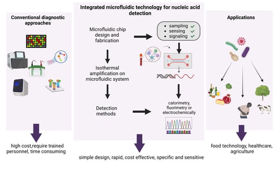
1. Introduction
2. Microfluidic Technology and POCTs
2.1. Significance of POCT in Diagnostics
2.2. Microfluidic POCT Devices
2.3. Materials for Microfluidic Devices
Paper-Based Multiplexed Detection
2.4. Microfluidic Approaches for Plasma Separation in POCT Devices
Limitations of Active and Passive Methods
2.5. Amplification Methods
2.5.1. Non-Isothermal Amplification
2.5.2. Isothermal Amplification
Loop-Mediated Isothermal AMPlification (LAMP)
Helicase Dependent Amplification (HDA)
Rolling Circle Amplification (RCA)
Multiple Displacement Amplification (MDA)
Recombinase Polymerase Amplification (RPA)
Nucleic Acid Sequence Based Amplification (NASBA)
2.6. Strategies for Nucleic Acid Testing
2.6.1. Paper-Based Microfluidics
2.6.2. Polymer-Based Microfluidics
2.6.3. CRISPR-Based Microfluidic Systems
2.6.4. Digital Microfluidics

| Biosensors | Biosensor Type/ Pathogen | Bioanalyte/Amplification Method | Detection Method/ Multiplex (Y/N) | Sensitivity &Detection | Limitations/Future Prospects | Ref. | |
|---|---|---|---|---|---|---|---|
| Polymer-based devices | |||||||
| PDMS-based nanoarray | Cas 12a/HBV, HPV-16, HPV-18 | Nucleic acid(NA)/Amplification freemethod | Surface-enhanced Raman scattering/ Y | Sensitivity: 1 aM Time: 20 min | High cost, only for DNA targets Optimization strategies should be taken in future to increase sensitivity of targets and strength of SERS signal. By reducing Limit of detection and assay time, overall performance of system can be improved shortening the assay time and reduction of the detection limit may also improve the system. Several biomarkers including proteins or RNA can be detected by utilization of Cas13a for RNA targets and aptamers for proteins | [81] | |
| POCKET (POC kit for the full test) | Microfluidic PDMS-based/Mutational analysis of Southeast Asia thalassemia | DNA/RPA | Colorimetric/ Y | Sensitivity: <103 copies/mL Time: <2 h | N/A In future, the variety of sample types will be increased to include those samples that are challenging to prepare, such assputum and feces. An independent and power free method can be developed to control reagent loading procedure in a better way Prevention of cross contamination from positive samples | [8] | |
| Paper-Based devices | |||||||
| Paper-Based device | CRISPR/Ca/SARS-CoV-2 | NA/RT-RPA | Colorimetric/ N | Sensitivity: LOD 100 copies Time: 1 h | N/A Issue of competitive hybridization can be avoided by designing two types of probes for individual identification of each target By optimizing nitrocellulose membrane’s pore size, flow rate of the strip or running buffer can be improved to ensure a better binding efficiency | [82] | |
| SHERLOCKv2 | Cas13, Cas12a and Csm/Dengue, Zika virus | NA/RPA | Fluorescence/Y | Sensitivity: aM Time: 1 h | N/A Solution and colorimetric-based readouts and multiplex lateral flow assays containing multiple test strips for different targets can be developed in future | [83] | |
| CRISPR-based microfluidic LFA chip | Cas12a/ SARS-CoV-2 | NA/RT-RPA | Colorimetric/ N | Sensitivity: 100 copies Time: <2 h | N/A Fully integrated molecular detection platform by adding nucleic acid extraction module to microfluidic chip Incorporation of phase changing material into heater case to control temperature | [84] | |
| CASLFA (CRISPR/Cas9-mediated lateral flow assay) | Cas9/SARS-CoV-2, African swine fever virus (ASFV), Listeria monocytogenes, genetically modified organisms (GMOs) | NA/RPA | Colorimetric/ N | Sensitivity: 100 copies Time:1 h | A number of separation stages are still needed to complete the process Future research may combine microfluidic technology with the developed system to integrate extraction, amplification and detection on a single platform For multiplexing, primers with different labels and variety of test lines will enhance the feasibility of CASLFA to detect multiple targets simultaneously. | [85] | |
| MiSHERLOCK (minimally instrumented SHERLOCK) | Cas-12a/SARS-CoV-2 | RNA/RPA | Fluorescence/Y | Sensitivity: 1240 cp/mL Time: 60 min | Only a few COVID-19 patient samples were examined Owing to lack of resources, SARS-CoV-2 variants could not be tested Screening and diagnosis of disease variants | [86] | |
| SHINE | Cas13a/SARS-CoV-2 | NA/RPA | Fluorescence/ N | Concentration: 10 copies L1 Time: 50 min | Inconsistent measurements with RT-qPCR Lyophilization of reagents would facilitate assay preparation and dissemination while enabling shelf stable testing | [87] | |
| HUDSON(heating unextracted diagnostic samples to obliterate nucleases) | Cas 13-based/Dengue virus, Zika virus, resistance genes, bacteria | NA/RPA | Fluorescence and colorimetric/N | Sensitivity: aM, Time: <2 h | N/A It might be used to detect any type of virus in body fluids, can be used for multiplexed detection, reagents can be lyophilized | [88] | |
| Quartz-glass biochip | |||||||
| CARMEN v.1 | Microfluidic Cas13-based/Detect 169 human-related viruses distinguish between many influenza A subtypes of HIV and Differentiation of SARS-CoV-2 strains | NA/PCR | Fluorescence/ Y | Concentration: Reduced throughput Time: 8–10 h | Outbreak-specific Outbreak specific panels for detection of diseases should be deployed for testing of thousands of samples from a population | [89] | |
| mCARMEN variant identification panel (VIP) | Microfluidic Cas12-based/Detect 21 viruses, including SARS-CoV-2 and influenza strains, with excellent sensitivity | NA/PCR | Fluorescence/ Y | Sensitivity: 102 copies μL−1 Time: 8 h | N/A By integrating RVP (Respiratory Virus Panel) and VIP into a single panel need for manual work and equipment constraint can be reduced In future, the panel can be FDA approved and then commercialized | [90] | |
| SATORI(glass) | Cas13a/SARS-CoV-2 | ssRNA/Amplification free method | Fluorescence/ N | Sensitivity: ~10 fM Time: <5 min | Less sensitive than amplification-based methods (SHERLOCK, DETECTR and qPCR) In future, the SATORI-Cas12a system can be developed to performamplification-free double-stranded DNA detection | [91] | |
| CRISPR dCas 9 | dCas 9-based/SARS-CoV-2, Influenza A virus | NA/Isothermal amplification | colorimetric/ N | Sensitivity:Petamolar(pM) Time: 90 min | N/A In future, it can be used for diagnosis of drug resistant and reemerging viruses | [92] | |
| Other detection platforms | |||||||
| CAS-EXPAR | Cas 9-based/Listeria monocytogenes | NA/Isothermal amplification | Fluorescence/ N | Sensitivity: 0.82 attomolar(aM)Time: 1 h | N/A Detection of single-nucleotide mismatches and any target sequence site | [93] | |
| CRISDA (CRISPR-Cas 9-based nicking endonuclease for initiating strand displacement amplification process) | Cas 9-based/Identification of single nucleotide polymorphisms (SNPs) and homozygous/heterozygous genotypes related to brest cancer | DNA/Isothermal amplification | Peptide nucleic acid (PNA) invasion mediated fluorescence/N | Sensitivity: aM | N/A Highly specific and sensitive detection of SNPs and targeted sequences in POCT devices | [94] | |
| RCH (dCas9-based RCA CRISPR split HRP) | Cas 9-based/Lung cancer bycirculating let-7a biomarker and miRNA | miRNA/RCA | Chemiluminescenc/ N | Sensitivity: Femtomolar(fM) Time: <4 h | For different miRNA, a RCH probe/sgRNA isredesigned and synthesized in days Can be used as a solution or paper-based colorimetric readouts Cost can befurther reducedby industrializing the protein producing procedure. sensitivitycan be improved by implementing alternativeisothermal amplification systems or other reporting systems such as split-GFP, split-luciferase and split-β-galactosidase | [95] | |
2.7. Comparison of Various Detection Methods
2.8. Applications of Microfluidics
3. Limits, Challenges and Policy Recommendations
3.1. On-Site Sample Preparation
3.2. Nonspecific Adsorption
3.3. ComplexityinAdopting Multiplexing Approach
3.4. Introduction of Microfluidic Devices in Wearable Systems
3.5. Signal Readout
4. Conclusions
Author Contributions
Funding
Institutional Review Board Statement
Informed Consent Statement
Data Availability Statement
Conflicts of Interest
References
- Stockmaier, S.; Stroeymeyt, N.; Shattuck, E.C.; Hawley, D.M.; Meyers, L.A.; Bolnick, D.I. Infectious Diseases and Social Distancing in Nature. Science 2021, 371, eabc8881. [Google Scholar] [CrossRef] [PubMed]
- Funk, S.; Salathé, M.; Jansen, V.A.A. Modelling the Influence of Human Behaviour on the Spread of Infectious Diseases: A Review. J. R. Soc. Interface 2010, 7, 1247–1256. [Google Scholar] [CrossRef] [PubMed]
- Khan, S.A.; Ahmad, I.; Khan, H.; Abdullah; Akbar, S. The Contagious Nature of SARS-CoV-2 Omicron Variant and Vaccine Efficacy. Adv. Life Sci. 2022, 9, 429–436. [Google Scholar]
- Lazcka, O.; Campo, F.J.D.; Muñoz, F.X. Pathogen Detection: A Perspective of Traditional Methods and Biosensors. Biosens. Bioelectron. 2007, 22, 1205–1217. [Google Scholar] [CrossRef] [PubMed]
- Rajakaruna, S.J.; Liu, W.-B.; Ding, Y.-B.; Cao, G.-W. Strategy and Technology to Prevent Hospital-Acquired Infections: Lessons from SARS, Ebola, and MERS in Asia and West Africa. Mil. Med. Res. 2017, 4, 32. [Google Scholar] [CrossRef] [PubMed]
- Stone, H.A.; Stroock, A.D.; Ajdari, A. Engineering Flows in Small Devices: Microfluidics Toward a Lab-on-a-Chip. Annu. Rev. Fluid Mech. 2004, 36, 381–411. [Google Scholar] [CrossRef]
- Cavaniol, C.; Cesar, W.; Descroix, S.; Viovy, J.-L. Flowmetering for Microfluidics. Lab Chip 2022, 22, 3603–3617. [Google Scholar] [CrossRef]
- Xu, H.; Xia, A.; Wang, D.; Zhang, Y.; Deng, S.; Lu, W.; Luo, J.; Zhong, Q.; Zhang, F.; Zhou, L.; et al. An Ultraportable and Versatile Point-of-Care DNA Testing Platform. Sci. Adv. 2020, 6, eaaz7445. [Google Scholar] [CrossRef]
- Ortseifen, V.; Viefhues, M.; Wobbe, L.; Grünberger, A. Microfluidics for Biotechnology: Bridging Gaps to Foster Microfluidic Applications. Front. Bioeng. Biotechnol. 2020, 8, 589074. [Google Scholar] [CrossRef]
- Wang, X.; Hong, X.-Z.; Li, Y.-W.; Li, Y.; Wang, J.; Chen, P.; Liu, B.-F. Microfluidics-Based Strategies for Molecular Diagnostics of Infectious Diseases. Mil. Med. Res. 2022, 9, 11. [Google Scholar] [CrossRef]
- Tarim, E.A.; Karakuzu, B.; Oksuz, C.; Sarigil, O.; Kizilkaya, M.; Al-Ruweidi, M.K.A.A.; Yalcin, H.C.; Ozcivici, E.; Tekin, H.C. Microfluidic-Based Virus Detection Methods for Respiratory Diseases. Emergent Mater. 2021, 4, 143–168. [Google Scholar] [CrossRef] [PubMed]
- Lui, C.; Cady, N.; Batt, C. Nucleic Acid-Based Detection of Bacterial Pathogens Using Integrated Microfluidic Platform Systems. Sensors 2009, 9, 3713–3744. [Google Scholar] [CrossRef] [PubMed]
- Silbeglitt, R.; Wong, A. The Global Technology Revolution China, In-Depth Analyses: Emerging Technology Opportunities for the Tianjin Binhai New Area (TBNA) and the Tianjin Technological Development Area (TEDA); Rand Corporation: Santa Monica, CA, USA, 2009; Volume 649. [Google Scholar]
- Yeh, E.-C.; Fu, C.-C.; Hu, L.; Thakur, R.; Feng, J.; Lee, L.P. Self-Powered Integrated Microfluidic Point-of-Care Low-Cost Enabling (SIMPLE) Chip. Sci. Adv. 2017, 3, e1501645. [Google Scholar] [CrossRef] [PubMed]
- Carrell, C.; Kava, A.; Nguyen, M.; Menger, R.; Munshi, Z.; Call, Z.; Nussbaum, M.; Henry, C. Beyond the Lateral Flow Assay: A Review of Paper-Based Microfluidics. Microelectron. Eng. 2019, 206, 45–54. [Google Scholar] [CrossRef]
- Fraser, L.A.; Cheung, Y.; Kinghorn, A.B.; Guo, W.; Shiu, S.C.; Jinata, C.; Liu, M.; Bhuyan, S.; Nan, L.; Shum, H.C.; et al. Microfluidic Technology for Nucleic Acid Aptamer Evolution and Application. Adv. Biosys. 2019, 3, 1900012. [Google Scholar] [CrossRef] [PubMed]
- Ye, X.; Xu, J.; Lu, L.; Li, X.; Fang, X.; Kong, J. Equipment-Free Nucleic Acid Extraction and Amplification on a Simple Paper Disc for Point-of-Care Diagnosis of Rotavirus A. Anal. Chim. Acta 2018, 1018, 78–85. [Google Scholar] [CrossRef]
- Wu, J.; Fang, H.; Zhang, J.; Yan, S. Modular Microfluidics for Life Sciences. J. Nanobiotechnol. 2023, 21, 85. [Google Scholar] [CrossRef]
- Urbaniak, J.; Janowski, D.; Jacewski, B. Isolation of Nucleic Acids Using Silicon Dioxide Powder as a Tool for Environmental Monitoring. Environ. Monit. Assess. 2019, 191, 732. [Google Scholar] [CrossRef]
- Watanabe, R.; Asai, S.; Kakizoe, H.; Saeki, H.; Masukawa, A.; Miyazawa, M.; Ohtagawa, K.; Ravzanaaadii, M.-A.; Doi, M.; Atsumi, H.; et al. Evaluation of the Basic Assay Performance of the GeneSoc® Rapid PCR Testing System for Detection of Severe Acute Respiratory Syndrome Coronavirus 2. PLoS ONE 2021, 16, e0248397. [Google Scholar] [CrossRef]
- Dong, X.; Liu, L.; Tu, Y.; Zhang, J.; Miao, G.; Zhang, L.; Ge, S.; Xia, N.; Yu, D.; Qiu, X. Rapid PCR Powered by Microfluidics: A Quick Review under the Background of COVID-19 Pandemic. TrAC Trends Anal. Chem. 2021, 143, 116377. [Google Scholar] [CrossRef]
- Mendoza-Gallegos, R.A.; Rios, A.; Garcia-Cordero, J.L. An Affordable and Portable Thermocycler for Real-Time PCR Made of 3D-Printed Parts and Off-the-Shelf Electronics. Anal. Chem. 2018, 90, 5563–5568. [Google Scholar] [CrossRef] [PubMed]
- Jafek, A.R.; Harbertson, S.; Brady, H.; Samuel, R.; Gale, B.K. Instrumentation for XPCR Incorporating QPCR and HRMA. Anal. Chem. 2018, 90, 7190–7196. [Google Scholar] [CrossRef] [PubMed]
- Espulgar, W.V.; Saito, M.; Takahashi, K.; Ushiro, S.; Yamamoto, N.; Akeda, Y.; Hamaguchi, S.; Tomono, K.; Tamiya, E. Deskilled and Rapid Drug-Resistant Gene Detection by Centrifugal Force-Assisted Thermal Convection PCR Device. Sensors 2021, 21, 1225. [Google Scholar] [CrossRef] [PubMed]
- Nge, P.N.; Rogers, C.I.; Woolley, A.T. Advances in Microfluidic Materials, Functions, Integration, and Applications. Chem. Rev. 2013, 113, 2550–2583. [Google Scholar] [CrossRef]
- Wei, X.; Tian, T.; Jia, S.; Zhu, Z.; Ma, Y.; Sun, J.; Lin, Z.; Yang, C.J. Target-Responsive DNA Hydrogel Mediated “Stop-Flow” Microfluidic Paper-Based Analytic Device for Rapid, Portable and Visual Detection of Multiple Targets. Anal. Chem. 2015, 87, 4275–4282. [Google Scholar] [CrossRef] [PubMed]
- Hu, J.; Xiao, K.; Jin, B.; Zheng, X.; Ji, F.; Bai, D. Paper-based Point-of-care Test with Xeno Nucleic Acid Probes. Biotechnol. Bioeng. 2019, 116, 2764–2777. [Google Scholar] [CrossRef]
- Chavez-Pineda, O.G.; Rodriguez-Moncayo, R.; Cedillo-Alcantar, D.F.; Guevara-Pantoja, P.E.; Amador-Hernandez, J.U.; Garcia-Cordero, J.L. Microfluidic Systems for the Analysis of Blood-derived Molecular Biomarkers. Electrophoresis 2022, 43, 1667–1700. [Google Scholar] [CrossRef]
- Sun, M.; Khan, Z.S.; Vanapalli, S.A. Blood Plasma Separation in a Long Two-Phase Plug Flowing through Disposable Tubing. Lab Chip 2012, 12, 5225. [Google Scholar] [CrossRef]
- Zhang, X.-B.; Wu, Z.-Q.; Wang, K.; Zhu, J.; Xu, J.-J.; Xia, X.-H.; Chen, H.-Y. Gravitational Sedimentation Induced Blood Delamination for Continuous Plasma Separation on a Microfluidics Chip. Anal. Chem. 2012, 84, 3780–3786. [Google Scholar] [CrossRef]
- Gao, Q.; Chang, Y.; Deng, Q.; You, H. A Simple and Rapid Method for Blood Plasma Separation Driven by Capillary Force with an Application in Protein Detection. Anal. Methods 2020, 12, 2560–2570. [Google Scholar] [CrossRef]
- Jiang, F.; Xiang, N.; Ni, Z. Ultrahigh Throughput Beehive-like Device for Blood Plasma Separation. Electrophoresis 2020, 41, 2136–2143. [Google Scholar] [CrossRef] [PubMed]
- Szydzik, C.; Khoshmanesh, K.; Mitchell, A.; Karnutsch, C. Microfluidic Platform for Separation and Extraction of Plasma from Whole Blood Using Dielectrophoresis. Biomicrofluidics 2015, 9, 064120. [Google Scholar] [CrossRef] [PubMed]
- Lenshof, A.; Ahmad-Tajudin, A.; Järås, K.; Swärd-Nilsson, A.-M.; Åberg, L.; Marko-Varga, G.; Malm, J.; Lilja, H.; Laurell, T. Acoustic Whole Blood Plasmapheresis Chip for Prostate Specific Antigen Microarray Diagnostics. Anal. Chem. 2009, 81, 6030–6037. [Google Scholar] [CrossRef] [PubMed]
- Wilson, C.J.; Clegg, R.E.; Leavesley, D.I.; Pearcy, M.J. Mediation of Biomaterial–Cell Interactions by Adsorbed Proteins: A Review. Tissue Eng. 2005, 11, 1–18. [Google Scholar] [CrossRef] [PubMed]
- Sposito, A.; Hoang, V.; DeVoe, D.L. Rapid Real-Time PCR and High Resolution Melt Analysis in a Self-Filling Thermoplastic Chip. Lab Chip 2016, 16, 3524–3531. [Google Scholar] [CrossRef]
- Kim, T.-H.; Sunkara, V.; Park, J.; Kim, C.-J.; Woo, H.-K.; Cho, Y.-K. A Lab-on-a-Disc with Reversible and Thermally Stable Diaphragm Valves. Lab Chip 2016, 16, 3741–3749. [Google Scholar] [CrossRef]
- Houssin, T.; Cramer, J.; Grojsman, R.; Bellahsene, L.; Colas, G.; Moulet, H.; Minnella, W.; Pannetier, C.; Leberre, M.; Plecis, A.; et al. Ultrafast, Sensitive and Large-Volume on-Chip Real-Time PCR for the Molecular Diagnosis of Bacterial and Viral Infections. Lab Chip 2016, 16, 1401–1411. [Google Scholar] [CrossRef]
- Roy, E.; Stewart, G.; Mounier, M.; Malic, L.; Peytavi, R.; Clime, L.; Madou, M.; Bossinot, M.; Bergeron, M.G.; Veres, T. From Cellular Lysis to Microarray Detection, an Integrated Thermoplastic Elastomer (TPE) Point of Care Lab on a Disc. Lab Chip 2015, 15, 406–416. [Google Scholar] [CrossRef]
- Shin, D.J.; Trick, A.Y.; Hsieh, Y.-H.; Thomas, D.L.; Wang, T.-H. Sample-to-Answer Droplet Magnetofluidic Platform for Point-of-Care Hepatitis C Viral Load Quantitation. Sci. Rep. 2018, 8, 9793. [Google Scholar] [CrossRef]
- Zanoli, L.; Spoto, G. Isothermal Amplification Methods for the Detection of Nucleic Acids in Microfluidic Devices. Biosensors 2012, 3, 18–43. [Google Scholar] [CrossRef]
- Lee, S.-Y.; Huang, J.-G.; Chuang, T.-L.; Sheu, J.-C.; Chuang, Y.-K.; Holl, M.; Meldrum, D.R.; Lee, C.-N.; Lin, C.-W. Compact Optical Diagnostic Device for Isothermal Nucleic Acids Amplification. Sens. Actuators B Chem. 2008, 133, 493–501. [Google Scholar] [CrossRef] [PubMed]
- Lee, S.-Y.; Lee, C.-N.; Mark, H.; Meldrum, D.R.; Lin, C.-W. Efficient, Specific, Compact Hepatitis B Diagnostic Device: Optical Detection of the Hepatitis B Virus by Isothermal Amplification. Sens. Actuators B Chem. 2007, 127, 598–605. [Google Scholar] [CrossRef]
- Ding, X.; Wang, G.; Mu, Y. Single Enzyme-Based Stem-Loop and Linear Primers Co-Mediated Exponential Amplification of Short Gene Sequences. Anal. Chim. Acta 2019, 1081, 193–199. [Google Scholar] [CrossRef] [PubMed]
- Jeong, Y.-J.; Park, K.; Kim, D.-E. Isothermal DNA Amplification in Vitro: The Helicase-Dependent Amplification System. Cell. Mol. Life Sci. 2009, 66, 3325–3336. [Google Scholar] [CrossRef] [PubMed]
- Mahalanabis, M.; Do, J.; ALMuayad, H.; Zhang, J.Y.; Klapperich, C.M. An Integrated Disposable Device for DNA Extraction and Helicase Dependent Amplification. Biomed. Microdevices 2010, 12, 353–359. [Google Scholar] [CrossRef] [PubMed]
- Kolm, C.; Martzy, R.; Führer, M.; Mach, R.L.; Krska, R.; Baumgartner, S.; Farnleitner, A.H.; Reischer, G.H. Detection of a Microbial Source Tracking Marker by Isothermal Helicase-Dependent Amplification and a Nucleic Acid Lateral-Flow Strip Test. Sci. Rep. 2019, 9, 393. [Google Scholar] [CrossRef]
- Mothershed, E.A.; Whitney, A.M. Nucleic Acid-Based Methods for the Detection of Bacterial Pathogens: Present and Future Considerations for the Clinical Laboratory. Clin. Chim. Acta 2006, 363, 206–220. [Google Scholar] [CrossRef]
- Huang, R.; Di, K.; Adeel, K.; Fan, B.; Gu, X.; Xu, H.; Shen, H.; He, N.; Li, Z. The Exploration of Droplet Digital Branched Rolling Circle Amplification Based Ultrasensitive Biosensor for Gastric Cancer Cell-Derived Extracellular Vesicles Detection. Mater. Today Adv. 2022, 16, 100296. [Google Scholar] [CrossRef]
- Hashkavayi, A.B.; Cha, B.S.; Lee, E.S.; Park, K.S. Dual Rolling Circle Amplification-Enabled Ultrasensitive Multiplex Detection of Exosome Biomarkers Using Electrochemical Aptasensors. Anal. Chim. Acta 2022, 1205, 339762. [Google Scholar] [CrossRef]
- Marcy, Y.; Ishoey, T.; Lasken, R.S.; Stockwell, T.B.; Walenz, B.P.; Halpern, A.L.; Beeson, K.Y.; Goldberg, S.M.D.; Quake, S.R. Nanoliter Reactors Improve Multiple Displacement Amplification of Genomes from Single Cells. PLoS Genet. 2007, 3, e155. [Google Scholar] [CrossRef]
- Dean, F.B.; Hosono, S.; Fang, L.; Wu, X.; Faruqi, A.F.; Bray-Ward, P.; Sun, Z.; Zong, Q.; Du, Y.; Du, J.; et al. Comprehensive Human Genome Amplification Using Multiple Displacement Amplification. Proc. Natl. Acad. Sci. USA 2002, 99, 5261–5266. [Google Scholar] [CrossRef] [PubMed]
- Zhang, K.; Martiny, A.C.; Reppas, N.B.; Barry, K.W.; Malek, J.; Chisholm, S.W.; Church, G.M. Sequencing Genomes from Single Cells by Polymerase Cloning. Nat. Biotechnol. 2006, 24, 680–686. [Google Scholar] [CrossRef] [PubMed]
- Wu, N.; Zhang, Y.; Fu, J.; Zhang, R.; Feng, L.; Hu, Y.; Li, X.; Lu, N.; Zhao, X.; Pan, Y.; et al. Performance Assessment of a Novel Two-Step Multiple Displacement Amplification-PCR Assay for Detection of Mycobacterium Tuberculosis Complex in Sputum Specimens. J. Clin. Microbiol. 2012, 50, 1443–1445. [Google Scholar] [CrossRef] [PubMed]
- Hakenberg, S.; Hügle, M.; Weidmann, M.; Hufert, F.; Dame, G.; Urban, G.A. A Phaseguided Passive Batch Microfluidic Mixing Chamber for Isothermal Amplification. Lab Chip 2012, 12, 4576. [Google Scholar] [CrossRef]
- Kersting, S.; Rausch, V.; Bier, F.F.; von Nickisch-Rosenegk, M. Multiplex Isothermal Solid-Phase Recombinase Polymerase Amplification for the Specific and Fast DNA-Based Detection of Three Bacterial Pathogens. Microchim. Acta 2014, 181, 1715–1723. [Google Scholar] [CrossRef]
- Alamolhoda, S.Z.; Zarghami, N.; Kahroba, H.; Mehdipour, A.; Pourhassan-Moghaddam, M.; Jahanban-Esfahlan, R.; Milani, M. Isothermal Amplification of Nucleic Acids Coupled with Nanotechnology and Microfluidic Platforms for Detecting Antimicrobial Drug Resistance and Beyond. Adv. Pharm. Bull. 2021, 1, 58. [Google Scholar] [CrossRef]
- Zhao, X.; Dong, T.; Yang, Z.; Pires, N.; Høivik, N. Compatible Immuno-NASBA LOC Device for Quantitative Detection of Waterborne Pathogens: Design and Validation. Lab Chip 2012, 12, 602–612. [Google Scholar] [CrossRef]
- Hardinge, P.; Murray, J.A.H. Reduced False Positives and Improved Reporting of Loop-Mediated Isothermal Amplification Using Quenched Fluorescent Primers. Sci. Rep. 2019, 9, 7400. [Google Scholar] [CrossRef]
- Dolka, B.; Cisek, A.A.; Szeleszczuk, P. The Application of the Loop-Mediated Isothermal Amplification (LAMP) Method for Diagnosing Enterococcus Hirae-Associated Endocarditis Outbreaks in Chickens. BMC Microbiol. 2019, 19, 48. [Google Scholar] [CrossRef]
- Barreda-García, S.; Miranda-Castro, R.; de-los-Santos-Álvarez, N.; Miranda-Ordieres, A.J.; Lobo-Castañón, M.J. Helicase-Dependent Isothermal Amplification: A Novel Tool in the Development of Molecular-Based Analytical Systems for Rapid Pathogen Detection. Anal. Bioanal. Chem. 2018, 410, 679–693. [Google Scholar] [CrossRef]
- Cao, Y.; Kim, H.; Li, Y.; Kong, H.; Lemieux, B. Helicase-Dependent Amplification of Nucleic Acids. Curr. Protoc. Mol. Biol. 2013, 104, 1–12. [Google Scholar] [CrossRef] [PubMed]
- Murakami, T.; Sumaoka, J.; Komiyama, M. Sensitive Isothermal Detection of Nucleic-Acid Sequence by Primer Generation–Rolling Circle Amplification. Nucleic Acids Res. 2009, 37, e19. [Google Scholar] [CrossRef] [PubMed]
- Pumford, E.A.; Lu, J.; Spaczai, I.; Prasetyo, M.E.; Zheng, E.M.; Zhang, H.; Kamei, D.T. Developments in Integrating Nucleic Acid Isothermal Amplification and Detection Systems for Point-of-Care Diagnostics. Biosens. Bioelectron. 2020, 170, 112674. [Google Scholar] [CrossRef] [PubMed]
- Spits, C.; Le Caignec, C.; De Rycke, M.; Van Haute, L.; Van Steirteghem, A.; Liebaers, I.; Sermon, K. Whole-Genome Multiple Displacement Amplification from Single Cells. Nat. Protoc. 2006, 1, 1965–1970. [Google Scholar] [CrossRef] [PubMed]
- Bleier, S.; Maier, P.; Allgayer, H.; Wenz, F.; Zeller, W.J.; Fruehauf, S.; Laufs, S. Multiple Displacement Amplification Enables Large-Scale Clonal Analysis Following Retroviral Gene Therapy. J. Virol. 2008, 82, 2448–2455. [Google Scholar] [CrossRef]
- Draz, U.; Ali, S.; Firyal, S.; Ali, A.; Saleem, A.H.; Tahir, M.A. Detection of Human Salivary Amylase Level Deposited on Fruits with First Bite Mark. Adv. Life Sci. 2019, 6, 176–181. [Google Scholar]
- Mauk, M.G.; Liu, C.; Sadik, M.; Bau, H.H. Microfluidic Devices for Nucleic Acid (NA) Isolation, Isothermal NA Amplification, and Real-Time Detection. In Mobile Health Technologies; Methods in Molecular Biology; Rasooly, A., Herold, K.E., Eds.; Springer: New York, NY, USA, 2015; Volume 1256, pp. 15–40. ISBN 978-1-4939-2171-3. [Google Scholar]
- Berkenbrock, J.A.; Grecco-Machado, R.; Achenbach, S. Microfluidic Devices for the Detection of Viruses: Aspects of Emergency Fabrication during the COVID-19 Pandemic and Other Outbreaks. Proc. R. Soc. A 2020, 476, 20200398. [Google Scholar] [CrossRef]
- Fang, X.; Liu, Y.; Kong, J.; Jiang, X. Loop-Mediated Isothermal Amplification Integrated on Microfluidic Chips for Point-of-Care Quantitative Detection of Pathogens. Anal. Chem. 2010, 82, 3002–3006. [Google Scholar] [CrossRef]
- Klausner, J.D. Editorial Commentary: The NAAT Is Out of the Bag. Clin. Infect. Dis. 2004, 38, 820–821. [Google Scholar] [CrossRef]
- Hadgu, A.; Dendukuri, N.; Hilden, J. Evaluation of Nucleic Acid Amplification Tests in the Absence of a Perfect Gold-Standard Test: A Review of the Statistical and Epidemiologic Issues. Epidemiology 2005, 16, 604–612. [Google Scholar] [CrossRef]
- Xie, Y.; Li, H.; Chen, F.; Udayakumar, S.; Arora, K.; Chen, H.; Lan, Y.; Hu, Q.; Zhou, X.; Guo, X.; et al. Clustered Regularly Interspaced Short Palindromic Repeats-Based Microfluidic System in Infectious Diseases Diagnosis: Current Status, Challenges, and Perspectives. Adv. Sci. 2022, 9, 2204172. [Google Scholar] [CrossRef]
- Zheng, C.; Wang, K.; Zheng, W.; Cheng, Y.; Li, T.; Cao, B.; Jin, Q.; Cui, D. Rapid Developments in Lateral Flow Immunoassay for Nucleic Acid Detection. Analyst 2021, 146, 1514–1528. [Google Scholar] [CrossRef]
- Hu, J.; Wang, L.; Li, F.; Han, Y.L.; Lin, M.; Lu, T.J.; Xu, F. Oligonucleotide-Linked Gold Nanoparticle Aggregates for Enhanced Sensitivity in Lateral Flow Assays. Lab Chip 2013, 13, 4352. [Google Scholar] [CrossRef] [PubMed]
- Hill, H.D.; Mirkin, C.A. The Bio-Barcode Assay for the Detection of Protein and Nucleic Acid Targets Using DTT-Induced Ligand Exchange. Nat. Protoc. 2006, 1, 324–336. [Google Scholar] [CrossRef] [PubMed]
- Yamada, K.; Henares, T.G.; Suzuki, K.; Citterio, D. Paper-Based Inkjet-Printed Microfluidic Analytical Devices. Angew. Chem. Int. Ed. 2015, 54, 5294–5310. [Google Scholar] [CrossRef] [PubMed]
- Yabuta, T.; Bescher, E.P.; Mackenzie, J.D.; Tsuru, K.; Hayakawa, S.; Osaka, A. Synthesis of PDMS-Based Porous Materials for Biomedical Applications. J. Sol-Gel Sci. Technol. 2003, 26, 1219–1222. [Google Scholar] [CrossRef]
- Musgrave, C.S.A.; Fang, F. Contact Lens Materials: A Materials Science Perspective. Materials 2019, 12, 261. [Google Scholar] [CrossRef]
- Liong, M.; Hoang, A.N.; Chung, J.; Gural, N.; Ford, C.B.; Min, C.; Shah, R.R.; Ahmad, R.; Fernandez-Suarez, M.; Fortune, S.M.; et al. Magnetic Barcode Assay for Genetic Detection of Pathogens. Nat. Commun. 2013, 4, 1752. [Google Scholar] [CrossRef]
- Ng, A.H.C.; Fobel, R.; Fobel, C.; Lamanna, J.; Rackus, D.G.; Summers, A.; Dixon, C.; Dryden, M.D.M.; Lam, C.; Ho, M.; et al. A Digital Microfluidic System for Serological Immunoassays in Remote Settings. Sci. Transl. Med. 2018, 10, eaar6076. [Google Scholar] [CrossRef]
- Qavi, A.J.; Washburn, A.L.; Byeon, J.-Y.; Bailey, R.C. Label-Free Technologies for Quantitative Multiparameter Biological Analysis. Anal. Bioanal. Chem. 2009, 394, 121–135. [Google Scholar] [CrossRef]
- Das, J.; Kelley, S.O. High-Performance Nucleic Acid Sensors for Liquid Biopsy Applications. Angew. Chem. Int. Ed. 2020, 59, 2554–2564. [Google Scholar] [CrossRef] [PubMed]
- Wu, K.; Liu, J.; Saha, R.; Su, D.; Krishna, V.D.; Cheeran, M.C.-J.; Wang, J.-P. Magnetic Particle Spectroscopy for Detection of Influenza A Virus Subtype H1N1. ACS Appl. Mater. Interfaces 2020, 12, 13686–13697. [Google Scholar] [CrossRef] [PubMed]
- Cesewski, E.; Johnson, B.N. Electrochemical Biosensors for Pathogen Detection. Biosens. Bioelectron. 2020, 159, 112214. [Google Scholar] [CrossRef] [PubMed]
- Zhang, Y.; McKelvie, I.D.; Cattrall, R.W.; Kolev, S.D. Colorimetric Detection Based on Localised Surface Plasmon Resonance of Gold Nanoparticles: Merits, Inherent Shortcomings and Future Prospects. Talanta 2016, 152, 410–422. [Google Scholar] [CrossRef]
- Wongkaew, N.; Simsek, M.; Griesche, C.; Baeumner, A.J. Functional Nanomaterials and Nanostructures Enhancing Electrochemical Biosensors and Lab-on-a-Chip Performances: Recent Progress, Applications, and Future Perspective. Chem. Rev. 2019, 119, 120–194. [Google Scholar] [CrossRef]
- Reta, N.; Saint, C.P.; Michelmore, A.; Prieto-Simon, B.; Voelcker, N.H. Nanostructured Electrochemical Biosensors for Label-Free Detection of Water- and Food-Borne Pathogens. ACS Appl. Mater. Interfaces 2018, 10, 6055–6072. [Google Scholar] [CrossRef]
- Sun, Y.; Lu, J. Chemiluminescence-Based Aptasensors for Various Target Analytes. Luminescence 2018, 33, 1298–1305. [Google Scholar] [CrossRef]
- Xiao, Q.; Xu, C. Research Progress on Chemiluminescence Immunoassay Combined with Novel Technologies. TrAC Trends Anal. Chem. 2020, 124, 115780. [Google Scholar] [CrossRef]
- Al Mughairy, B.; Al-Lawati, H.A.J. Recent Analytical Advancements in Microfluidics Using Chemiluminescence Detection Systems for Food Analysis. TrAC Trends Anal. Chem. 2020, 124, 115802. [Google Scholar] [CrossRef]
- Liu, M.; Khan, A.; Wang, Z.; Liu, Y.; Yang, G.; Deng, Y.; He, N. Aptasensors for Pesticide Detection. Biosens. Bioelectron. 2019, 130, 174–184. [Google Scholar] [CrossRef]
- Singh, P.; Pandey, S.K.; Singh, J.; Srivastava, S.; Sachan, S.; Singh, S.K. Biomedical Perspective of Electrochemical Nanobiosensor. Nano-Micro Lett. 2016, 8, 193–203. [Google Scholar] [CrossRef] [PubMed]
- Ansari, S.; Masoum, S. Recent Advances and Future Trends on Molecularly Imprinted Polymer-Based Fluorescence Sensors with Luminescent Carbon Dots. Talanta 2021, 223, 121411. [Google Scholar] [CrossRef] [PubMed]
- Jiang, P.; Guo, Z. Fluorescent Detection of Zinc in Biological Systems: Recent Development on the Design of Chemosensors and Biosensors. Coord. Chem. Rev. 2004, 248, 205–229. [Google Scholar] [CrossRef]
- VanEngelenburg, S.B.; Palmer, A.E. Fluorescent Biosensors of Protein Function. Curr. Opin. Chem. Biol. 2008, 12, 60–65. [Google Scholar] [CrossRef]
- Pazos, E.; Vázquez, O.; Mascareñas, J.L.; Eugenio Vázquez, M. Peptide-Based Fluorescent Biosensors. Chem. Soc. Rev. 2009, 38, 3348. [Google Scholar] [CrossRef]
- Denmark, D.J.; Bustos-Perez, X.; Swain, A.; Phan, M.-H.; Mohapatra, S.; Mohapatra, S.S. Readiness of Magnetic Nanobiosensors for Point-of-Care Commercialization. J. Elec. Mater. 2019, 48, 4749–4761. [Google Scholar] [CrossRef]
- Xianyu, Y.; Wang, Q.; Chen, Y. Magnetic Particles-Enabled Biosensors for Point-of-Care Testing. TrAC Trends Anal. Chem. 2018, 106, 213–224. [Google Scholar] [CrossRef]
- Pashchenko, O.; Shelby, T.; Banerjee, T.; Santra, S. A Comparison of Optical, Electrochemical, Magnetic, and Colorimetric Point-of-Care Biosensors for Infectious Disease Diagnosis. ACS Infect. Dis. 2018, 4, 1162–1178. [Google Scholar] [CrossRef]
- Zhou, J.; Qi, Q.; Wang, C.; Qian, Y.; Liu, G.; Wang, Y.; Fu, L. Surface Plasmon Resonance (SPR) Biosensors for Food Allergen Detection in Food Matrices. Biosens. Bioelectron. 2019, 142, 111449. [Google Scholar] [CrossRef]
- Mahmoudpour, M.; Ezzati Nazhad Dolatabadi, J.; Torbati, M.; Pirpour Tazehkand, A.; Homayouni-Rad, A.; de la Guardia, M. Nanomaterials and New Biorecognition Molecules Based Surface Plasmon Resonance Biosensors for Mycotoxin Detection. Biosens. Bioelectron. 2019, 143, 111603. [Google Scholar] [CrossRef]
- Sepúlveda, B.; Angelomé, P.C.; Lechuga, L.M.; Liz-Marzán, L.M. LSPR-Based Nanobiosensors. Nano Today 2009, 4, 244–251. [Google Scholar] [CrossRef]
- Guo, J.; Zeng, F.; Guo, J.; Ma, X. Preparation and Application of Microfluidic SERS Substrate: Challenges and Future Perspectives. J. Mater. Sci. Technol. 2020, 37, 96–103. [Google Scholar] [CrossRef]
- Vendrell, M.; Maiti, K.K.; Dhaliwal, K.; Chang, Y.-T. Surface-Enhanced Raman Scattering in Cancer Detection and Imaging. Trends Biotechnol. 2013, 31, 249–257. [Google Scholar] [CrossRef] [PubMed]
- Langer, J.; Jimenez de Aberasturi, D.; Aizpurua, J.; Alvarez-Puebla, R.A.; Auguié, B.; Baumberg, J.J.; Bazan, G.C.; Bell, S.E.J.; Boisen, A.; Brolo, A.G.; et al. Present and Future of Surface-Enhanced Raman Scattering. ACS Nano 2020, 14, 28–117. [Google Scholar] [CrossRef]
- Tu, Q.; Chang, C. Diagnostic Applications of Raman Spectroscopy. Nanomed. Nanotechnol. Biol. Med. 2012, 8, 545–558. [Google Scholar] [CrossRef]
- Sengupta, P.; Khanra, K.; Chowdhury, A.R.; Datta, P. Lab-on-a-Chip Sensing Devices for Biomedical Applications. In Bioelectronics and Medical Devices; Elsevier: Amsterdam, The Netherlands, 2019; pp. 47–95. ISBN 978-0-08-102420-1. [Google Scholar]
- Nielsen, J.B.; Hanson, R.L.; Almughamsi, H.M.; Pang, C.; Fish, T.R.; Woolley, A.T. Microfluidics: Innovations in Materials and Their Fabrication and Functionalization. Anal. Chem. 2020, 92, 150–168. [Google Scholar] [CrossRef]
- Merrin, J. Frontiers in Microfluidics, a Teaching Resource Review. Bioengineering 2019, 6, 109. [Google Scholar] [CrossRef]
- Jing, W.; Sui, G. Bioanalysis within Microfluidics: A Review. In ACS Symposium Series; Wang, C., Leblanc, R.M., Eds.; American Chemical Society: Washington, DC, USA, 2015; Volume 1215, pp. 245–268. ISBN 978-0-8412-3120-7. [Google Scholar]
- Narimani, R.; Azizi, M.; Esmaeili, M.; Rasta, S.H.; Khosroshahi, H.T. An Optimal Method for Measuring Biomarkers: Colorimetric Optical Image Processing for Determination of Creatinine Concentration Using Silver Nanoparticles. 3 Biotech. 2020, 10, 416. [Google Scholar] [CrossRef]
- Walgama, C.; Nguyen, M.P.; Boatner, L.M.; Richards, I.; Crooks, R.M. Hybrid Paper and 3D-Printed Microfluidic Device for Electrochemical Detection of Ag Nanoparticle Labels. Lab Chip 2020, 20, 1648–1657. [Google Scholar] [CrossRef]
- Gnoth, C.; Johnson, S. Strips of Hope: Accuracy of Home Pregnancy Tests and New Developments. Geburtshilfe Frauenheilkd 2014, 74, 661–669. [Google Scholar] [CrossRef]
- Oh, K.W. Microfluidic Devices for Biomedical Applications: Biomedical Microfluidic Devices 2019. Micromachines 2020, 11, 370. [Google Scholar] [CrossRef] [PubMed]
- Williams, M.J.; Lee, N.K.; Mylott, J.A.; Mazzola, N.; Ahmed, A.; Abhyankar, V.V. A Low-Cost, Rapidly Integrated Debubbler (RID) Module for Microfluidic Cell Culture Applications. Micromachines 2019, 10, 360. [Google Scholar] [CrossRef] [PubMed]
- Sanjay, S.T.; Zhou, W.; Dou, M.; Tavakoli, H.; Ma, L.; Xu, F.; Li, X. Recent Advances of Controlled Drug Delivery Using Microfluidic Platforms. Adv. Drug Deliv. Rev. 2018, 128, 3–28. [Google Scholar] [CrossRef] [PubMed]
- Martins, J.P.; Torrieri, G.; Santos, H.A. The Importance of Microfluidics for the Preparation of Nanoparticles as Advanced Drug Delivery Systems. Expert Opin. Drug Deliv. 2018, 15, 469–479. [Google Scholar] [CrossRef] [PubMed]
- Arduino, I.; Liu, Z.; Rahikkala, A.; Figueiredo, P.; Correia, A.; Cutrignelli, A.; Denora, N.; Santos, H.A. Preparation of Cetyl Palmitate-Based PEGylated Solid Lipid Nanoparticles by Microfluidic Technique. Acta Biomater. 2021, 121, 566–578. [Google Scholar] [CrossRef] [PubMed]
- Galan, E.A.; Zhao, H.; Wang, X.; Dai, Q.; Huck, W.T.S.; Ma, S. Intelligent Microfluidics: The Convergence of Machine Learning and Microfluidics in Materials Science and Biomedicine. Matter 2020, 3, 1893–1922. [Google Scholar] [CrossRef]
- Niculescu, A.-G.; Chircov, C.; Bîrcă, A.C.; Grumezescu, A.M. Fabrication and Applications of Microfluidic Devices: A Review. Int. J. Mol. Sci. 2021, 22, 2011. [Google Scholar] [CrossRef] [PubMed]
- Wang, D.; Chan, H.N.; Liu, Z.; Micheal, S.; Li, L.; Baniani, D.B.; Tan, M.J.A.; Huang, L.; Wang, J.; Wu, H. Recent Developments in Microfluidic-Based Point-of-care Testing (POCT) Diagnoses. In Nanotechnology and Microfluidics; Jiang, X., Bai, C., Liu, M., Eds.; Wiley: Hoboken, NJ, USA, 2020; pp. 239–280. ISBN 978-3-527-34533-5. [Google Scholar]
- Liu, Y.-Y.; Wang, Y.; Walsh, T.R.; Yi, L.-X.; Zhang, R.; Spencer, J.; Doi, Y.; Tian, G.; Dong, B.; Huang, X.; et al. Emergence of Plasmid-Mediated Colistin Resistance Mechanism MCR-1 in Animals and Human Beings in China: A Microbiological and Molecular Biological Study. Lancet Infect. Dis. 2016, 16, 161–168. [Google Scholar] [CrossRef] [PubMed]
- Shen, F.; Du, W.; Kreutz, J.E.; Fok, A.; Ismagilov, R.F. Digital PCR on a SlipChip. Lab Chip 2010, 10, 2666. [Google Scholar] [CrossRef] [PubMed]
- Schoepp, N.G.; Schlappi, T.S.; Curtis, M.S.; Butkovich, S.S.; Miller, S.; Humphries, R.M.; Ismagilov, R.F. Rapid Pathogen-Specific Phenotypic Antibiotic Susceptibility Testing Using Digital LAMP Quantification in Clinical Samples. Sci. Transl. Med. 2017, 9, eaal3693. [Google Scholar] [CrossRef] [PubMed]
- Tu, J.; Torrente-Rodríguez, R.M.; Wang, M.; Gao, W. The Era of Digital Health: A Review of Portable and Wearable Affinity Biosensors. Adv. Funct. Mater. 2020, 30, 1906713. [Google Scholar] [CrossRef]
- Lee, S.; Oncescu, V.; Mancuso, M.; Mehta, S.; Erickson, D. A Smartphone Platform for the Quantification of Vitamin D Levels. Lab Chip 2014, 14, 1437–1442. [Google Scholar] [CrossRef]
- Zhu, W.-J.; Feng, D.-Q.; Chen, M.; Chen, Z.-D.; Zhu, R.; Fang, H.-L.; Wang, W. Bienzyme Colorimetric Detection of Glucose with Self-Calibration Based on Tree-Shaped Paper Strip. Sens. Actuators B Chem. 2014, 190, 414–418. [Google Scholar] [CrossRef]
- Park, C.; Han, Y.D.; Kim, H.V.; Lee, J.; Yoon, H.C.; Park, S. Double-Sided 3D Printing on Paper towards Mass Production of Three-Dimensional Paper-Based Microfluidic Analytical Devices (3D-ΜPADs). Lab Chip 2018, 18, 1533–1538. [Google Scholar] [CrossRef]
- Parween, S.; Subudhi, P.D.; Asthana, A. An Affordable, Rapid Determination of Total Lipid Profile Using Paper-Based Microfluidic Device. Sens. Actuators B Chem. 2019, 285, 405–412. [Google Scholar] [CrossRef]
- Li, F.; Wang, X.; Liu, J.; Hu, Y.; He, J. Double-Layered Microfluidic Paper-Based Device with Multiple Colorimetric Indicators for Multiplexed Detection of Biomolecules. Sens. Actuators B Chem. 2019, 288, 266–273. [Google Scholar] [CrossRef]
- Chen, X.; Chen, J.; Wang, F.; Xiang, X.; Luo, M.; Ji, X.; He, Z. Determination of Glucose and Uric Acid with Bienzyme Colorimetry on Microfluidic Paper-Based Analysis Devices. Biosens. Bioelectron. 2012, 35, 363–368. [Google Scholar] [CrossRef] [PubMed]


| MicrofluidicApproach | Mechanism | Sample | Target | Material | Ref. |
|---|---|---|---|---|---|
| Sedimentation | Sedimentation | Blood | Plasma | Glass | [28] |
| Microfiltration | Sedimentation | Blood | Plasma | /PDMS/PTFE/Glass | [29] |
| Lateral displacement | Filtration | Blood | Plasma | PMMA | [30] |
| Hydrodynamic | Hydrodynamic | Blood | Plasma | PDMS | [31] |
| Acoustic separation | Dielectrophoretic Hydrodynamic Acoustic | Blood | Plasma | PDMS | [32] |
| Dielectrophoresis | Dielectrophoretic Hydrodynamic Acoustic | Blood | Plasma | PDMS | [33] |
| Compact Disc (CD) format | Sedimentation | Blood | Plasma | COC/PDMS/pMMA | [34] |
| Isothermal Method | Template | Time | Primers | Tm (°C) | Ref. |
|---|---|---|---|---|---|
| LAMP | DNA/RNA | 15–60 min | 3 pairs | 60–65 | [59,60] |
| HDA | DNA, rRNA | 1–1.5 h | 1 pair | 60–65 | [61,62] |
| RCA | cssDNA, RNA, miRNA | 1 h | 1 single primer, 1 padlock probe | 25–37 | [63,64] |
| MDA | dsDNA | 2 h | Random hexamer | 30 | [65,66] |
| RPA | DNA/RNA | 5–7 min | 1 pair | 37–42 | [60,61,62] |
| NASBA | SsRNA, tmRNA, rRNA 1 | 1.5 h | 1 pair | 41 | [63,64] |
| Detection Method | Advantages | Disadvantages | Future Prospects | Refs. |
|---|---|---|---|---|
| Colorimetric | Visualized detection, simplicity | Limited sensitivity and no quantitative detection | In nanoparticle-based colorimetric assays, sensitivity can be increased by increasing size of nanoparticle and by decreasing density of receptor group present on surface of nanoparticle | [84,85,86] |
| Electrochemical | Low cost, robust response, high sensitivity | Interference susceptibility and weak stability | Biocompatibility of nanoparticles can lead to reduction in toxicity detection as the nanoparticles are less reactive against proteins | [87,88,89,90,91,92,93] |
| Chemiluminescence | Simplicity, high sensitivity | Enzyme dependent and time consuming | Designing a multiplex system with increased sample throughput. Quantitative approach should bead opted in future | [89] |
| Fluorescence | Experimental simplicity, flexibility and robust response | Background with a high fluorescence | Development of biosensors based on macro and micromolecule imprinting technology can be considered. Reusable devices based on the green chemistry approach can be designed | [94,95,96,97] |
| Magnetic | Detection of high signal to noise ratio, low cost | Limited availability of miniaturized magnetic readout systems | Integration in point of care testing devices | [98,99,100] |
| SPR-based | Label-free, real-time detection | Expensive, bulky equipment Low limit of detection | Improvement in sensitivity and detection capability Different shapes of nanoparticles can be used to improve sensitivity | [101,102,103] |
| SERS-based | Low background, no photobleaching, good multiplexing capabilities and high sensitivity | Limited view field, Difficulty in fabrication of SERS active substrate in microfluidic chip | Improvement in reproducibility, selectivity, integration of chip and multi-functionality | [104,105,106,107] |
Disclaimer/Publisher’s Note: The statements, opinions and data contained in all publications are solely those of the individual author(s) and contributor(s) and not of MDPI and/or the editor(s). MDPI and/or the editor(s) disclaim responsibility for any injury to people or property resulting from any ideas, methods, instructions or products referred to in the content. |
© 2023 by the authors. Licensee MDPI, Basel, Switzerland. This article is an open access article distributed under the terms and conditions of the Creative Commons Attribution (CC BY) license (https://creativecommons.org/licenses/by/4.0/).
Share and Cite
Mumtaz, Z.; Rashid, Z.; Ali, A.; Arif, A.; Ameen, F.; AlTami, M.S.; Yousaf, M.Z. Prospects of Microfluidic Technology in Nucleic Acid Detection Approaches. Biosensors 2023, 13, 584. https://doi.org/10.3390/bios13060584
Mumtaz Z, Rashid Z, Ali A, Arif A, Ameen F, AlTami MS, Yousaf MZ. Prospects of Microfluidic Technology in Nucleic Acid Detection Approaches. Biosensors. 2023; 13(6):584. https://doi.org/10.3390/bios13060584
Chicago/Turabian StyleMumtaz, Zilwa, Zubia Rashid, Ashaq Ali, Afsheen Arif, Fuad Ameen, Mona S. AlTami, and Muhammad Zubair Yousaf. 2023. "Prospects of Microfluidic Technology in Nucleic Acid Detection Approaches" Biosensors 13, no. 6: 584. https://doi.org/10.3390/bios13060584
APA StyleMumtaz, Z., Rashid, Z., Ali, A., Arif, A., Ameen, F., AlTami, M. S., & Yousaf, M. Z. (2023). Prospects of Microfluidic Technology in Nucleic Acid Detection Approaches. Biosensors, 13(6), 584. https://doi.org/10.3390/bios13060584