Review

Wavelength-Dependent Metal-Enhanced Fluorescence Biosensors via Resonance Energy Transfer Modulation

Department of Applied Chemistry, Institute of Natural Sciences, Kyung Hee University, Yongin-si 17104, Gyeonggi-do, Republic of Korea
*
Author to whom correspondence should be addressed.
Biosensors 2023, 13(3), 376; https://doi.org/10.3390/bios13030376
Submission received: 15 February 2023 / Revised: 10 March 2023 / Accepted: 10 March 2023 / Published: 13 March 2023
(This article belongs to the Special Issue Plasmonic Based Biosensors)

Abstract

:
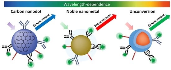
Fluorescence can be enhanced or quenched depending on the distance between the surface of a metal nanoparticle and the fluorophore molecule. Fluorescence enhancement by nearby metal particles is called metal-enhanced fluorescence (MEF). MEF shows promising potential in the field of fluorescence-based biological sensing. MEF-based biosensor systems generally fall into two platform categories: (1) a two/three-dimensional scaffold, or (2) a colloidal suspension. This review briefly summarizes the application studies using wavelength-dependent carbon dots (UV-VIS), noble metals (VIS), and upconversion nanoparticles (NIR to VIS), representative nanomaterials that contribute to the enhancement of fluorescence through the resonance energy transfer modulation and then presents a perspective on this topic.

Graphical Abstract

1. Introduction

Fluorescence is one of the dominant detection/sensing methodologies for the biological and chemical sciences [1]. Fluorescence can act as an excellent sensing probe because fluorescent materials can change emission intensity or wavelengths as they interact with ligands or other chemical agents. In fluorescence-based optical biosensors, fluorescence probes act as effective converters for transmitting fluorescence signals in biometric events collected using various detectors [2]. Despite the attractive features of fluorescence detection, fluorescence signals may not be sufficiently intense when analyte concentrations are low. Thus, new strategies are needed to improve signal collection and to achieve high sensitivity in appropriate dynamic ranges [3].
In 1948, Förster resonance energy transfer (FRET, IUPAC nomenclature) was theoretically established by Theodore Förster [4]. Förster discovered that energy transfer through dipole coupling between molecules depends primarily on two factors: spectral overlap and intermolecular distance, and discovered the R6 distance dependence law for the rate of resonance energy transfer (RET) over short distances [5]. Until now, RET technology has been widely applied to study various intermolecular interactions (fluorescence RET [6], bioluminescence RET [7], chemiluminescence RET [8], nanometal surface energy transfer [9], and plasmon RET [10]). In 2007, Lee’s group showed that the plasmon resonance energy of NPs could be transferred to nearby cytochrome-c. This interaction was called plasmonic resonance energy transfer (PRET) [10]. Gao et al. introduced nanometal surface energy transfer and PRET techniques using plasmonic nanoparticles (NPs) [11]. The physical mechanisms, efficiency measures, and principles of RET have already been summarized in many papers [11,12,13,14]. Metal NPs are associated with luminescence enhancement and quenching phenomena, which are energy transfer pathways. Typically, the signal is quenched when the fluorophores-to-metal separation distance is within about 5 nm and enhanced within the optimal distances typically between 5 and 90 nm [2]. We called the latter metal-enhanced fluorescence (MEF).
With the rapid development of nanotechnology, the combination of fluorescent probes and nanometallic materials has presented an opportunity to further upgrade the reliability and sensitivity of biosensor systems. As a representative example, MEF-based biosensors have the potential to improve current fluorescence-based techniques, such as single-molecule sensing, bioimaging, DNA sequencing, or disease identification [15,16,17]. MEF refers to a system in which the electromagnetic field in the area adjacent to metal NPs enhances the optical signal of nanomolecules or NPs close to the metal surface. This interesting phenomenon effectively combines metal nanostructures with fluorophore molecules used for targeted molecular detection, leading to higher sensitivity and lower detection limits compared to conventional optical biosensors [2,18,19]. Such MEF-based methods to enhance fluorescence detection sensitivity for detecting molecules at ultra-low concentrations are already being applied to biosensor systems [12].
In order to achieve the best enhancement factor, great efforts have been made to design various plasmonic nanomaterial structures that would maximize the radiative emission of the fluorophores and control the distance between the nanomaterial and the fluorophore molecule [12,20,21,22]. Previously, Jeong et al. reviewed MEF’s general approaches and recent developments for biosensors. They reported that the MEF phenomenon can occur in all fluorescent nanomaterials as well as organic fluorophores [2]. In recent years, there has been significant research interest in the development and application of metal nanomaterials for fluorescence enhancement. Among them, wavelength-dependent carbon nanodots (CNDs), noble metals, and upconversion NPs (UCNPs) have been extensively studied due to their ability to modulate RET depending on the spectral range. The optical properties of these metal nanomaterials can be controlled by adjusting their size, surface chemistry, and excitation wavelength, making them highly tunable for specific applications. For instance, wavelength-dependent CNDs can enhance fluorescence in the UV-VIS range, while noble metals such as gold and silver can enhance fluorescence in the VIS range through surface plasmon resonance. UCNPs can convert NIR into VIS light, which is ideal for deep-tissue imaging. These metal nanomaterials have great potential for bioimaging, sensing, and other fields.
This mini-review briefly summarizes application studies using wavelength-dependent CNDs (UV-VIS), noble metals (VIS), and UCNPs (NIR to VIS) that contribute to fluorescence enhancement through RET modulation among numerous metal nanomaterials by classifying a wide spectrum into three ranges. Furthermore, we focused on examples of various technologies designed to control plasmonic nanostructures (nanoarchitecture) that can be used to develop various sensor platforms and control the distance between fluorophores and metal NPs to realize the full potential of MEF technology.

2. Carbon Nanodots in the Ultraviolet-Visible Region
Carbon nanodots (CNDs), used as a “nanolight”, are emerging as an attractive fluorescent carbon material due to their strength, adjustability, photoluminescence and good optoelectronic properties [23,24,25]. Several chemical sensors have been developed to detect biomolecules [26] and metal ions [27,28] based on the quenching and recovery of CND fluorescence. In particular, the emission enhancement of CNDs is of great interest to the development of nanophotonics [29]. However, while plasmon-enhanced fluorescence phenomena for quantum dots (QDs) and fluorophore molecules have been well investigated, low quantum yield (<5%) for CNDs limit their application in biological studies [30,31,32,33,34,35]. Therefore, the important of research on CND-based MEF chemical sensors and light emission improvement is required for biological applications.
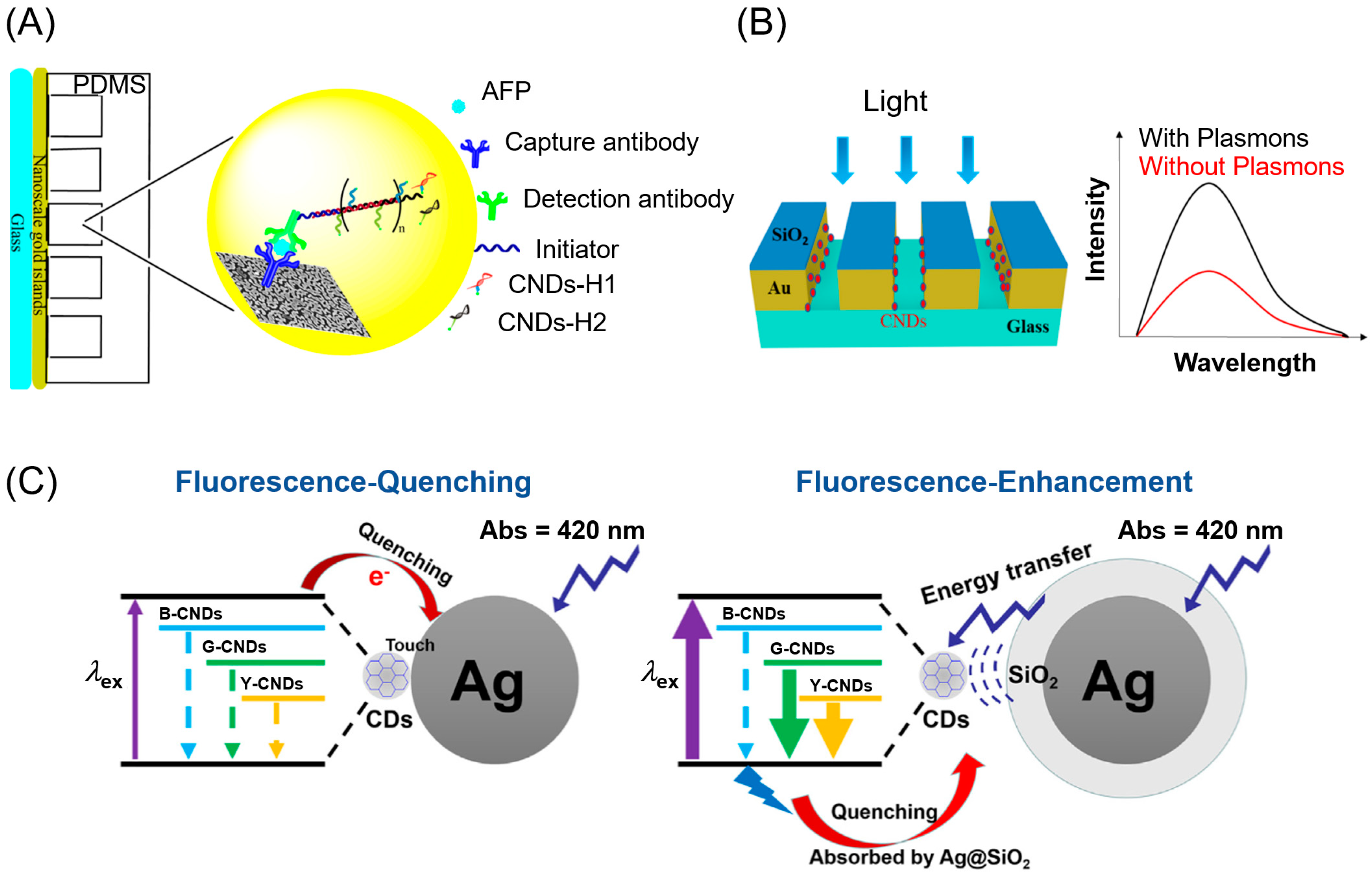
Xu et al. constructed a dual amplification fluorescence sensor based on an immuno-hybridization chain reaction (immuno-HCR) and MEF of CNDs for the detection of α-fetoprotein (AFP) (Figure 1A). A capture antibody-coated Au film slide captured the target molecule, AFP, and a detection antibody-conjugated oligonucleotide initiator was introduced to bind the AFP target. The introduction of a CND-tagged DNA hairpin allowed the DNA hairpin and oligonucleotide initiators to trigger complementary HCRs. Using this gold film and CNDs, the radiative decay rate was greatly improved, increasing the quantum yield and enhancing the fluorescence emission of the CNDs. They achieved a detection limit of 1.35 fM (94.3 fg/mL), as well as a 0.992 coefficient of correlation in the concentration range from 7.14 fM to 71.43 pM (0.0005 to 5 ng/mL). Fluorescence emission of plasmon slides was improved up to nearly seven-fold over non-plasmon slides by the effect of MEF at the same concentrations of AFP [36]. This result is more sensitive than that reported by Wu et al. in 2015, who measured the detection of human AFP using CNDs bound to labeled anti-AFP by cross-linking with glutaraldehyde through a high-throughput well plate-based immunosorbent assay [25]. Bagra et al. designed plasmonic nanoslits to immobilize CNDs and observe the fluorescence enhancement of CNDs. Maximum fluorescence and surface-reflected light intensity enhancement were obtained with a 100 nm nanoslit width, suggesting that plasmon light trapping was responsible for an increased electromagnetic field and plasmon-induced RET (Figure 1B) [29].
Fluorescence enhancements have been demonstrated in the blue-green range by binding CNDs to silver nanoparticles (AgNPs) [37,38,39]. Figure 1C illustrates an electron transition and energy transfer process between a CND and AgNP or AgNPs coated with a SiO2 layer (Ag@SiO2 NP) using a fluorescence quenching or enhancement mechanism, respectively [39]. Plasmonic enhancement was more pronounced when CNDs were nano-hybridized to silver rather than gold due to low intrinsic loss and superposition between the absorption spectra of metallic silver and CNDs [38]. CNDs can be operated like nanoantenna through two-way energy exchange with nearby chromophores. Recently, Sciortino et al. exploited the overlap between the surface plasmon resonance of gold and the electronic transitions of carbon dots to increase the fluorescence by a factor of 5 in the normally very weak orange region [40]. In their study, they elucidated that the underlying mechanism of enhancement was through coherent RET occurring in less than 70 fs from metal nanoantennas to CNDs.
Figure 1. (A) Schematic illustration for the detection of AFP with Immuno-HCR and MEF of CNDs [36]. (B) Illustration of self-assembled monolayer formation and CND immobilization on a gold nanoslit surface [29]. (C) Electron transition process between CNDs and Ag or Ag@SiO2 NP for quenching (left) and enhancement (right) of fluorescence [39]. Reprinted with permission from [29,36,39].
Biosensors 13 00376 g001
Deng et al. synthesized visible-ultraviolet upconversion CNDs to enhance the photocatalytic activity of titanium dioxide [41]. CNDs were synthesized by a hydrothermal method using L-glutamic acid (L-Glu) and m-phenylenediamine (MPD), and they were combined with commercial nano-TiO2 to prepare a CNDs/TiO2 complex, proving that CNDs can convert about 600 nm visible light to 350 nm ultraviolet light. Their successful work has provided an opportunity to further expand the application range of upconversion materials and the utilization efficiency of light sources.
CNDs have indeed been extensively studied due to their unique optical properties, biocompatibility, and potential for various applications including bioimaging, sensing, and drug delivery. In addition, the synthesis of biomass-derived CNDs is an excellent example of the efforts to make CNDs more environmentally friendly [42]. However, the excitation wavelengths of CNDs are limited to their absorption bands, which may be limited in some applications. Furthermore, with regard to its use as a detection probe, several strategies have been proposed to reduce background fluorescence, such as surface immobilization or surface functionalization, but it still can lead to false-positive results due to the high signal-to-noise ratio by autofluorescence in CND.

3. Noble Metals in the Visible Region

Noble metal nanostructures possess unique optical properties as well as good biocompatibility, satisfactory stability, and multiplex functionality. These unique advantages make noble metal nanostructures an ideal medium for developing biosensing and bioimaging methods when used as colloidal suspensions or 2D/3D substrates for MEF studies. Typically, MEF occurs when the fluorophore is positioned at an optimal distance (5–20 nm) from the noble metal [43,44,45], leading to improved quantum yield, reduced fluorescence lifetimes, and increased photostability.
Various techniques have been devised as fluorescent aptasensors by controlling the distance between the fluorophores and the noble NPs according to the MEF principle. Zhou et al. evaluated the fluorophore-metal distance effect and the AgNPs size effect on fluorescence enhancement. Fluorescently labeled molecular beacons were attached to AgNPs using recognition moieties. In addition to reducing the reagent blank signal, AgNPs provided an electric field superimposed on the incident light to increase the fluorescence intensity when the closed hairpin was opened (Figure 2A) [46]. When a 12 nm thick silica spacer shell was introduced between the gold nanoparticles (AuNPs) and gold nanoclusters (AuNCs), the fluorescence enhancement from AuNCs by Au@SiO2NPs was enhanced 3.72 times [47]. Jiang et al. hybridized polyAn-based special nucleic acid aptamers modified on the surface of AuNP with fluorescein amidite (FAM)-labeled single-strand oligonucleotides to enhance fluorescence signals by up to 2.7 times and detect adenosine triphosphate (ATP) with a detection limit of 0.2 nM (Figure 2B) [48]. By placing QDs in the vicinity of metal NPs, the intrinsic excitation and relaxation processes of QDs were significantly modified, resulting in enhanced fluorescence intensity. Recently, Kim et al. used heavy metal-free InP@ZnS core-shell QDs instead of toxic Cd-based QDs and chose Au@Ag@SiO2 core-shell NPs for the localized surface plasmon resonance (LSPR) effect. Systematic control over the thickness of the SiO2 shell enabled enhancements in QD fluorescence in poly(lauryl methacrylate) (PLMA) composite films, up to 12.9 times its original intensity [49]. Lotfi et al. applied the optimized fabricated silver dendritic nanostructure as a platform for the development of a MEF-based aptasensor for thrombin detection. After immobilizing a thiolated 29-mer thrombin-binding aptamer (TBA29(12T)–SH) as a capture aptamer on the surface of the silver dendritic nanostructure, thrombin was sandwiched between the capture aptamer and the Cy5-labeled 15-mer thrombin aptamer (TBA15-Cy5). The aptasensor they developed could detect thrombin at a level of 32 pM [50]. Trotsiuk et al. performed a systematic experimental study of QD fluorescence enhancement near the surface of electrostatically deposited gold nanorods (GNRs) of polyelectrolytes. They found that the tendency of the experimental fluorescence intensity enhancement factor decreased with QD concentration, obeying the a + b/x dependence law. The maximum enhancement factor (up to 11 times) was observed in a complex with a two polyelectrolyte layer spacer between a GNR and QDs, and a QD/GNR ratio = 2.5 [51]. Recently, Shi et al. developed a highly sensitive DNA sensor for detecting HIV DNA fragments based on the signal amplification of plasma resonance fluorescence enhancements using a catalytic hairpin assembly (CHA) and an N-methyl mesoporphyrin IX (NMM)/G-quadruplexes system (Figure 2C). The sensor included a 5′ terminal connected to a triangular gold nanoplate, a 3′ terminal rich in guanine hairpin probes (HP1) and a hairpin probe (HP2) complementary to the partial subsequence of HP1. The plasmon resonance absorption peak of the triangular gold nanoplates overlaps the emission spectrum of the NMM fluorophore, and the fluorescence coupling between the fluorophore and the NP was strong, thus producing a fluorescence enhancement effect. The single-molecule counting method combined with the multiplex signal amplification strategy sufficiently amplified the signal to detect the HIV DNA fragment [52]. Choi et al. developed a CRISPR-Cas12a-based nucleic acid amplification-free fluorescent biosensor to detect cell-free DNA by MEF using DNA-functionalized 20 nm AuNPs. They confirmed the MEF effect according to the DNA length of dsDNA between AuNP and FITC (Figure 2D). The fluorescence signal intensity of the AuNP-dsDNA-FITC complex was maximally enhanced at the optimal distance of 7 nm, and the breast cancer biomarker (BRCA-1) was measured to a femtomolar limit of detection [53]. These structures successfully provided rigid separation distances between metal NPs and fluorophores, thereby converting fluorescence quenching into fluorescence enhancements.
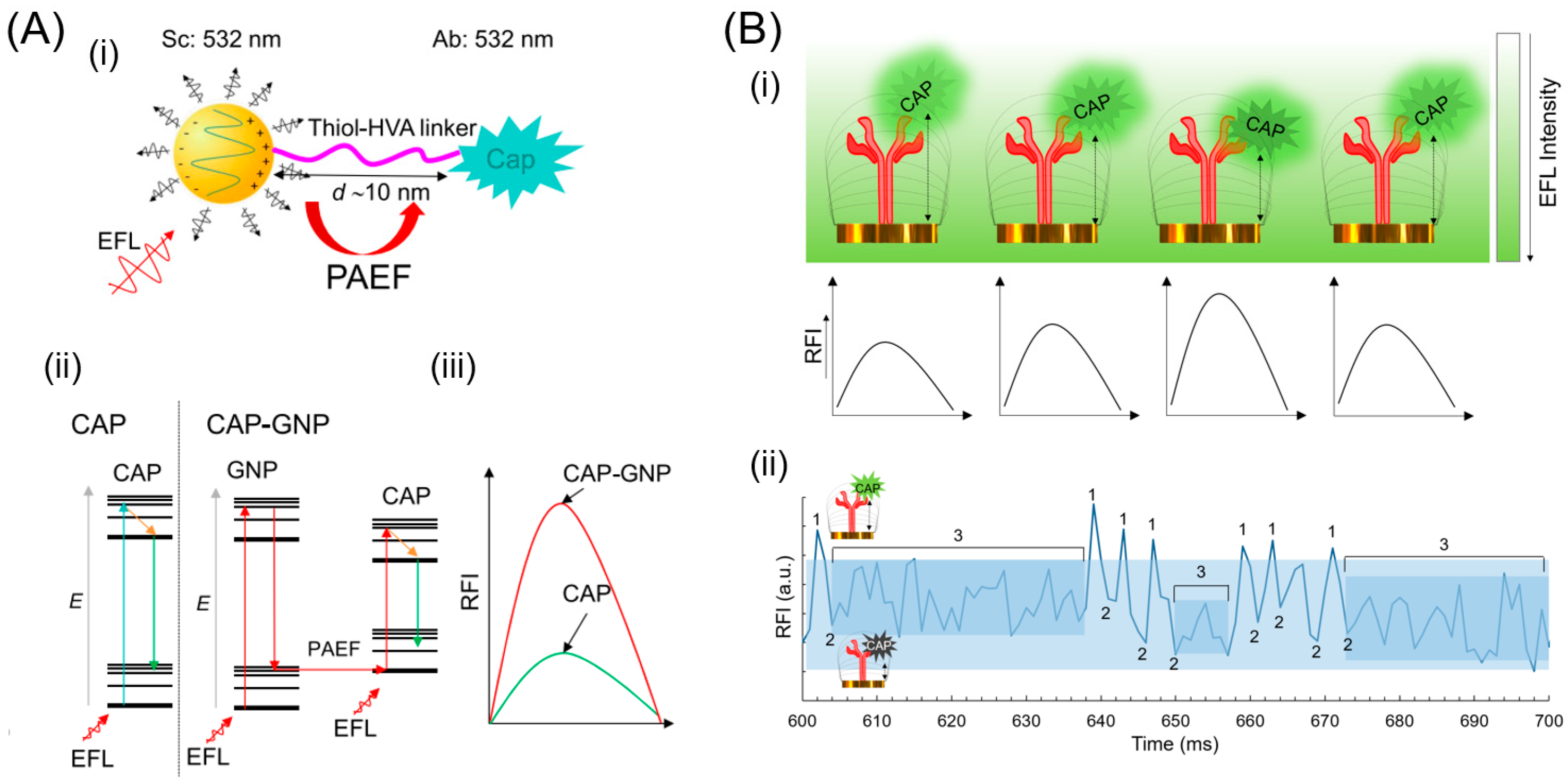
Chakkarapani et al. reported a simple, reliable, and highly sensitive endogenous fluorescence enhancement technique based on a single AuNP that can be used to detect endogenous fluorescent materials. They detected trace capsaicinoid (CAP) at 18 zM via self-endogenous fluorescence enhancement with plasmonic single NPs. Here, the inelastic scattering of plasmons was used as an electromagnetic field enhancer for the highly sensitive detection of ultra-trace amounts of endogenous phosphor. They named the method single nanoparticle plasmon-amplified endogenous fluorescence nanospectroscopic sensing (Figure 3A) [54]. In their recent work, they obtained cross-correlation quantification of CAP based on endogenous single-molecule fluorescence enhancement and quenching interfaces via correlated fluorescence fluctuation optical spectroscopy on plasmonic arrays of gold nanoislands (GNI). The endogenous fluorescence of CAP was dependent on the fluorophore–GNI distance and varied in enhancement, quenching, and equilibrium lifetimes (Figure 3B) [55].
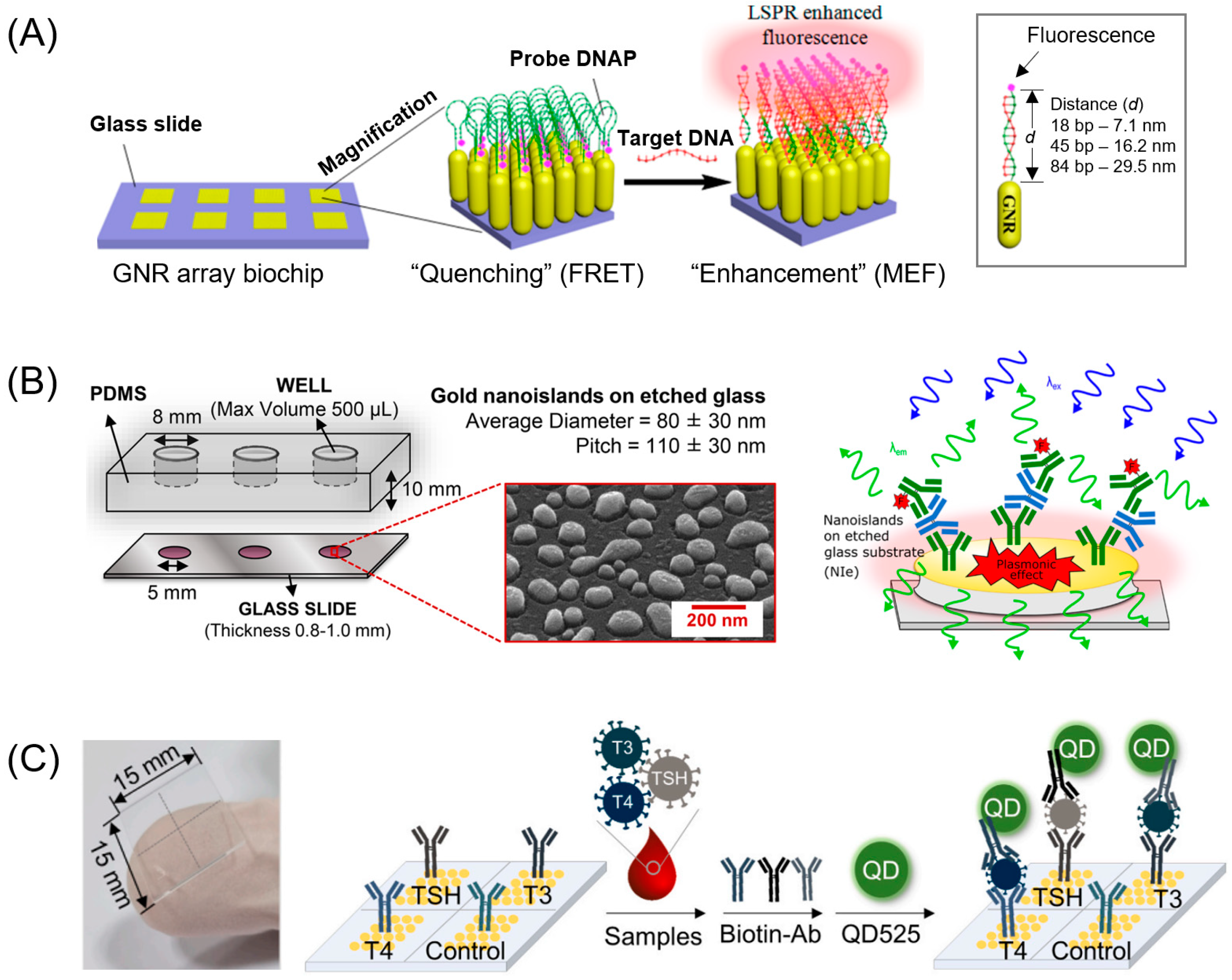
Plasmonic nanostructures have been used to develop various sensor platforms, and analytes can be immobilized on the surface of nanostructures. Signal enhancement depends on the size and shape of the nanostructures (nanoholes, nanoarrays, nano-stripes, etc.) [2]. These MEF-active substrates have been applied to develop biosensors with trace detection limits. In 2013, Gartia et al. [56] demonstrated a metal-coated nano-well array for ultrasensitive refractive index sensing fabricated by an inexpensive large-area nanoreplication technique. Mei and Tang [57] vertically aligned GNRs on a glass surface as a plasmonic substrate to detect a molecular beacon (Figure 4A). This dramatically enhanced the LSPR between adjacent GNRs compared to an ensemble of random GNRs. Inserting an intermediate spacer of appropriate thickness between the fluorophore and the surface of the GNR array resulted in plasmonic coupling enhancement of the fluorescence signal up to 30 nm away. Zhang et al. mass-fabricated Au nanohole arrays by nanoimprinting using a 4-inch nickel mold [58]. These Au nanohole arrays showed high fluorescence enhancement through simultaneous excitation of localized surface plasmon (LSP) and propagating surface plasmon (PSP) modes. Simultaneous excitation of LSP and PSP modes resulted in a detection limit of 140 fM for a prostate-specific antigen (PSA) biomarker. Dual mode detection offered a seven-fold better than when only LSP was resonantly excited on the same substrate. Miranda et al. developed a randomly arranged MEF-based immunosensor of AuNPs that was fabricated through a three-step process to regulate the optical properties of size, interparticle distance, and optimal gold nanostructures (Figure 4B). These AuNPs enhanced the fluorescence emission of three fluorescent dyes (Alexa Fluor 488, Alexa Fluor 546, and PE Cy7) approximately 150-fold on a randomly arranged MEF immunosensor to achieve a detection limit of 28.67 pM (4.3 ng/mL) for immunoglobulin detection [59]. Kang’s group arranged 300 nm gold nanodisks using electron-beam lithography and used them as sensors for the high-sensitivity detection of biomolecules [60]. They designed the analyte to be sandwiched between a capture antibody immobilized on a gold nanodisk and a detection antibody conjugated with QDs. Using this, biogenic amines (histamine (His), tyramine (Tyr), and putrescine (Put)) and thyroid hormone markers (thyroid-stimulating hormone (TSH), triiodothyronine (T3), and thyroxine (T4)) were quantified by multiple simultaneous analysis, showing aM and yM detection limits, respectively (Figure 4C) [61,62]. More recently, Lee et al. proposed a turn-on sensing format based on enhancement of the emission signal of a fluorophore molecule close to plasmonic gold nanodisks rather than the generally used turn-off format, based on competitive reactions, for small-molecule quantification [63].
Most fluorescent nanomaterials or fluorophores close to noble metals such as gold or silver can detect small amounts of target molecules through fluorescence enhancement. However, MEFs rely on surface plasmon resonance (SPR) of noble metals, which can be sensitive to environmental factors such as temperature, pH and salt concentration. This may limit the reproducibility of MEF experiments. In addition, as mentioned above, not only fluorescence enhancement but also fluorescence quenching may occur, so care must be taken when designing experiments.

4. Upconversion Nanoparticles from Near-Infrared to Visible Region

Upconversion nanoparticles (UCNPs), a newly established fluorescent probe type, are excited by near-infrared light and emit visible light [64,65,66]. Compared to organic fluorophores and QDs, it has the property of greatly reducing background autofluorescence, photobleaching, photodamage, and the phototoxicity of biological specimens [67,68,69]. However, quantum yields using UCNPs are relatively low (~1%) due to their small absorption cross sections (ability to absorb photons of a specific wavelength and polarization) [70,71]. Compared to their bulk counterparts, the concentration of dopant ions on the surface of UCNPs is relatively high and can be quenched by surface quenchers, reducing the fluorescence intensity [72,73]. For this reason, research to enhance the upconversion emission of UCNPs is needed. Methods for enhancing the upconversion emission can be divided into two categories: (1) Controlling the absorption cross-section and radiative emission rate by changing the synthesis conditions of UCNPs. (2) Increasing the local photon density of states. In particular, the local field can be improved more than 102 times using a plasmonic nanostructure [74]. In upconversion nanocrystals (UCNCs) that are able to convert lower energy photons (typically NIR) into higher energy photons (usually visible), the excitation and emission wavelengths can be substantially different and usually do not have significant overlap [75,76]. Therefore, it is significant to investigate the interplay between the excitation- and emission-plasmonic resonance coupling, as well as the possibility to distinguish their effects on plasmonic enhancement [77]. In this section, among the various methods to enhance the fluorescence sensitivity of UCNPs, a method using a plasmonic fluorescence enhancement strategy is presented.
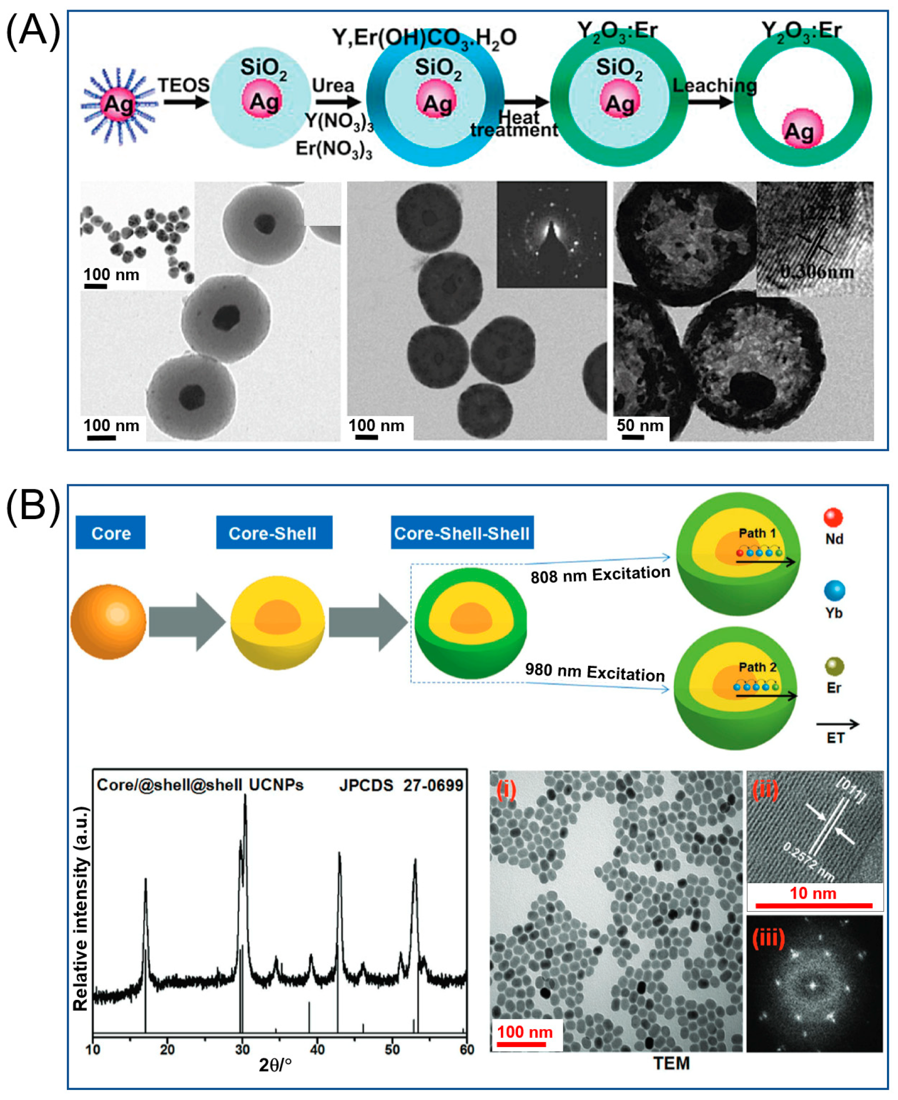
Dispersible plasmonic structures for biological applications can be deployed in bioimaging and therapeutic techniques, such as photodynamic therapy and drug delivery. Zhang et al. [78] easily fabricated a metal core-enhanced Er material based on a core/spacer/shell approach (Figure 5A). They switched the upconversion fluorescence enhancement and quenching phenomena by controlling the silica dielectric spacer thickness and metal core size. The results of this paper showed an optimal PL sensitivity when the space thickness was 30 nm, which was observed to be four times higher than that of the reference Y2O3:Er shell. In contrast, the use of thin SiO2 spacers resulted in PL quenching due to the proximity of the shell and Ag spheres and resulted in efficient nonradiative energy transfer from the luminescent system to the metal surface. Zhang et al. [77] reported a five-fold overall enhancement of upconversion emission in NaYF4:Yb/Er nanocrystals when coupled with gold island films. In contrast to isolated AuNPs [79,80,81,82], continuous gold films are characterized by a plasmonic resonance wavelength in the NIR region where the excitation wavelength (980 nm) for UCNPs is located [83,84,85]. Confocal fluorescence images showed that the upconversion emission increased more than five-fold when combined with the gold film. Kang et al. demonstrated that the excitation and release process of UCNPs can be simultaneously improved using these plasmonic double resonance nanostructures, thereby significantly increasing the UC luminescence (UCL) strength of UCNPs (Figure 5B) [86].
UCNP-based biosensors were used to detect proteins, nucleic acids, small molecules, and amino acids. In 2017, Du et al. demonstrated the usefulness of UCNPs as a bifunctional NP for in vitro cell imaging and latent fingerprint detection by synthesizing Ho3+-activated NaYbF4 UCNPs [87]. In 2019, Wu et al. [88] used an upconversion FRET sensor to detect arginine. They designed positively charged UCNPs as energy donors and negatively charged AgNP as energy acceptors. This method showed a good linear relationship between the fluorescence intensity and the concentration of arginine in 14.42–115.04 μM with a LOD of 2.9 μM. Kumar et al. [89] developed a UCNPs nanoplatform with mesoporous silica functionality to detect dopamine in real-time, wherein the LOD is as low as 0.63 nM. In 2020, Wang et al. [90] designed a new NIR probe based on FRET between UCNPs and Au nanocages for the detection of circulating tumor DNA (ctDNA). They determined excitation and emission at 980 nm and 800 nm, showing a linear range of 5–100 pM and a detection limit of 6.30 pM in serum. In 2021, Hu et al. [91] fabricated a new bilayer of poly(methylmethacrylate) opal photonic crystal (PMMA OPC) with a double photonic stop band, matching the excitation and emission fields of UCNP simultaneously to significantly enhance the upconversion fluorescence intensity. With 980 nm and 808 nm laser excitation, the PSA sensor showed a good linear relationship within the range of 2.94–294.12 pM (0.1–10 ng/mL) and LOD of 29.41 pM (0.01 ng/mL). For the detection of the cancer antigen 125 (CA125) biomarker, Zhang et al. [92] combined UCNPs modified with CA125 aptamer with CNDs as an energy acceptor through π–π stacking interaction. This CA125 sensor showed a linear range of 0.01–100 pM (0.01–100 U/mL) and a detection limit of 9 fM (9 × 10⁻³ U/mL). Recently, Liu et al. [93] reported a quantitative analysis of TSH by RET luminescence mechanism between the UCNPs donor and tetramethylrhodamine receptor. The resulting detection dynamic range and the limit of detection were 0.66–33.26 pM (0.1–5.0 mIU/L) and 0.43 pM (0.065 mIU/L).
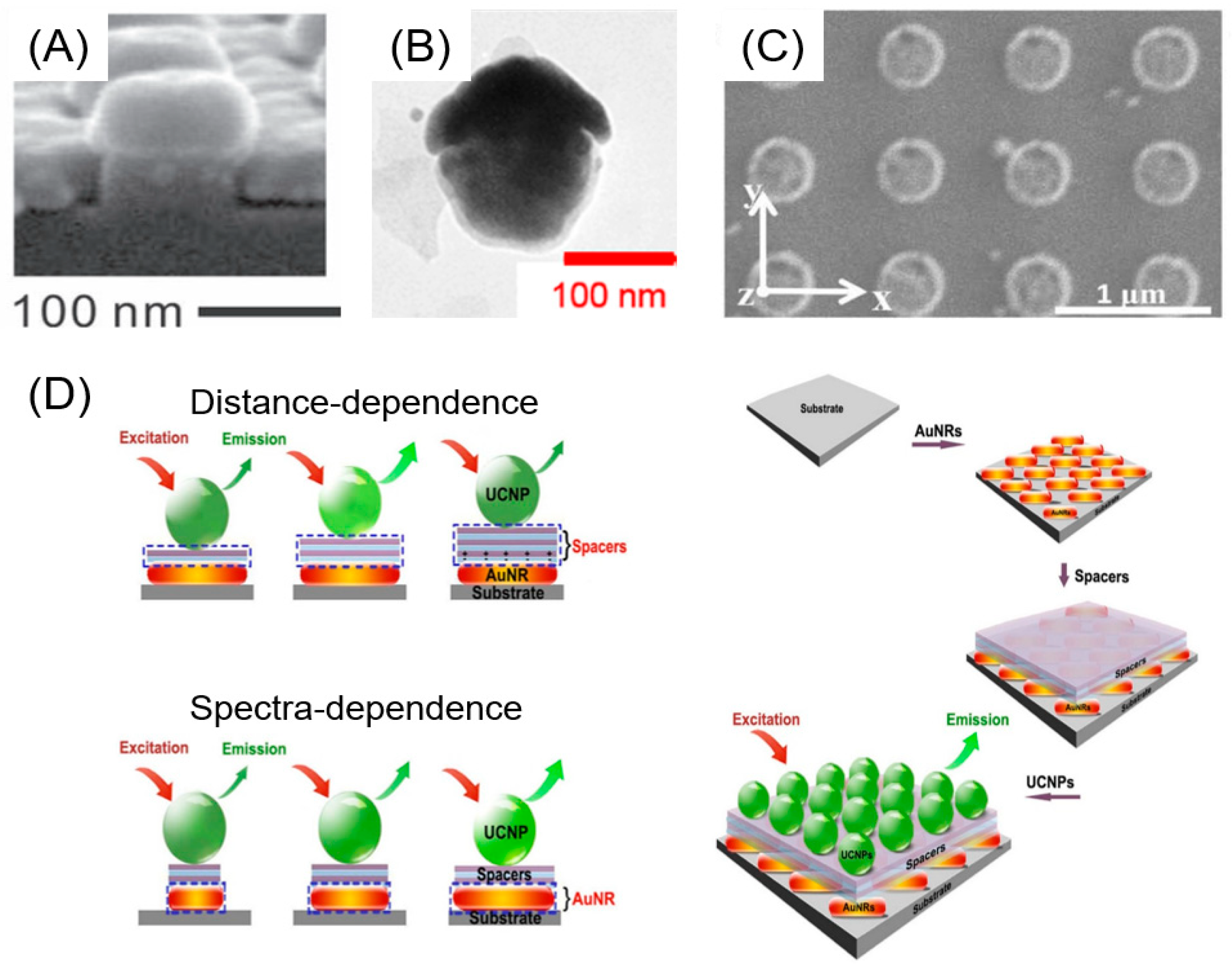
Applications of the non-dispersive plasmonic structures of UCNPs include multicolor flexible displays, fingerprints, solar energy harvesting, temperature sensing, and photocatalysis. The UCL enhancement of NaYF4:Yb3+/Er3+ co-doped nanocrystals was investigated using a disk-coupled dots-on-pillar antenna array (D2PA) with a 3D plasmonic nanoantenna architecture by Zhang et al. [94]. The D2PA structure was fabricated as a 3D nanocavity array, which consisted of Au nanodisks on the top of periodic dielectric pillars, an Au backplane at the feet of the pillars, and dense Au nanodots on the sidewalls (Figure 6A). Tuning the pillar height (h = 75 nm) could optimize the D2PA structure, resulting in a 310-fold uniform enhancement of UCL over a large area and an eight-fold reduction in luminescence decay time. Yamamoto et al. enhanced UCNP green emission by a factor of 23 and red emission by a factor of 43 through upconversion of 30 individual NPs composed of metal (Ag) nanocaps and rare-earth-doped UCNPs (Er- and Yb-doped Y2O3 NPs) [95]. UCNPs with silver nanocaps were formed by depositing a silver layer 10–50 nm thick on a quartz substrate spin-coated with UCNPs, and separating the substrate and UCNP using a polydimethylsiloxane (PDMS) stamp (Figure 6B). The intensity ratio of green and red luminescence was demonstrated to be dependent on the thickness of the Ag nanocaps, and the main cause of the upconversion enhancement is due to the enhancement of the radiative decay rates. Gao et al. placed an emitter layer in a diffractive array of Al nanocylinders to increase the absorption by rare earth ions. The array was designed to confine NIR in the emitter layer through the excitation of plasmon-photon hybrid modes, which are collective resonances of localized surface plasmons in nanocylinders through diffractive coupling, thus increasing the strength of the UCPL by up to seven times (Figure 6C) [96]. Plasmon nanostructures are known to efficiently enhance the fluorescence of surrounding fluorophores by acting as nanoantennae to focus the electric field into nano-spaces. Feng et al. reported a distance-dependent plasmon-enhanced fluorescence system by tuning the spacers between UCNPs and GNRs chosen as plasmon nanoantennae. A maximum upconversion enhancement up to 22.6-fold was achieved when the thickness of spacer polyelectrolyte multilayers was 8 nm and the LSPR absorption wavelength of GNRs overlap (~980 nm) with UCNPs excitation (Figure 6D) [97]. Eriksen et al. applied a rate equation model via a simulation-based approach to study the interaction between near-field enhancement and luminescence quenching over a range of geometries [98]. They used an excitation wavelength of 1523 nm for the simulation rather than the excitation wavelength of 980 nm considered in typical studies for plasmonic enhancement. Because of this, the enhancement factor values predicted by the simulations were lower than those reported in the literature. This suggests that the choice of excitation wavelength has a greater effect on increasing the efficiency of UC than the dependence on various plasmonic geometries.
Most recently, Meng et al. [99] realized single nanocrystal upconversion in a single plasmonic nanocavity mode by in situ controllable coupling and doping. An experimental platform was built to tune the nanocavity field and control the doping concentration of UCNCs. A single UCNC was placed between two GNR nanocavities so that all the activators of UCNC showed the same emission pattern and radiation efficiency. The outermost shell, as a protective layer, suppressed the energy transfer to surface defects, while the spacer layer mitigated the plasmonic quenching by coupling the UCL predominantly to the fundamental dipole mode. A nearly continuous evolution of the Purcell [100,101] enhancement for the same single nanocrystal was realized, which showed the general existence of UCL plasmonic enhancement saturation phenomena, as well as the doping- and coupling-dependent UCL enhancement factors up to 2.3 × 10⁵ [99].
UCNPs are actively pursued as anti-stock emitters for various applications, such as bioimaging, solar energy harvesting, catalysis, displays, anti-counterfeiting, sensing, and lasers [99]. To enhance UCL, researchers improved the internal material properties (composition, doping, and surface structure) of nanocrystals while simultaneously using plasmonic coupling to improve engineered external optical responses. However, despite impressive progress, upconversion brightness still fell short of the level required for commercial applications, and a systematic understanding of plasmon-enhanced upconversion remains a challenge.

5. Conclusions and Perspectives

In this review, we briefly reviewed recently reported papers on MEF-based biosensors designed to address the fundamental limitations of fluorescence-based detection, such as low quantum efficiency, photobleaching, and autofluorescence. Representative excitation wavelength-dependent nanomaterials (i.e., carbon dots, noble metals, upconversion nanoparticles) were introduced and verified to demonstrate great potential in terms of signal enhancement.
Despite these advantages, a selective nanomaterial molecular recognition element that can replace fluorophore molecules is still unavailable. The design and fabrication of nanostructures can potentially enhance fluorescence strength by three orders of magnitude compared to metal colloidal suspension, but it is expensive and time-consuming to manufacture rigid, uniform, and reproducible metal substrates. Thus, efficient nanostructure fabrication remains a task for current scientists in the manufacturing of smart nanomaterials and nanostructured surfaces.
In addition, despite many efforts to devise practical MEF biosensors, there are still limitations to deployment in biological microenvironments (clinical environments). MEF-based fluorescence detection technology with high sensitivity and selectivity will be an important key to advancing the development of portable high-sensitivity field tests that are expected to facilitate early disease diagnosis.

Author Contributions

Conceptualization, writing—original draft preparation, S.L.; supervision, writing—review and editing, S.H.K. All authors have read and agreed to the published version of the manuscript.

Funding

This work was supported by the National Research Foundation of Korea (NRF) grant funded by the Republic of Korea government (MSIT) (No. NRF2019R1A2C2002556 and 2023R1A2C2002623).

Institutional Review Board Statement

Not applicable.

Informed Consent Statement

Not applicable.

Data Availability Statement

Not applicable.

Conflicts of Interest

The authors declare no conflict of interest.

References

  1. Dutta Choudhury, S.; Badugu, R.; Ray, K.; Lakowicz, J.R. Silver–Gold Nanocomposite Substrates for Metal-Enhanced Fluorescence: Ensemble and Single-Molecule Spectroscopic Studies. J. Phys. Chem. C 2012, 116, 5042–5048. [Google Scholar] [CrossRef] [Green Version]
  2. Jeong, Y.; Kook, Y.-M.; Lee, K.; Koh, W.-G. Metal Enhanced Fluorescence (MEF) for Biosensors: General Approaches and a Review of Recent Developments. Biosens. Bioelectron. 2018, 111, 102–116. [Google Scholar] [CrossRef] [PubMed]
  3. Sauer, U. Analytical Protein Microarrays: Advancements Towards Clinical Applications. Sensors 2017, 17, 256. [Google Scholar] [CrossRef] [Green Version]
  4. Förster, T. Intermolecular Energy Migration and Fluorescence. Ann. Phys. 1948, 437, 55. [Google Scholar] [CrossRef]
  5. Jones, G.A.; Bradshaw, D.S. Resonance Energy Transfer: From Fundamental Theory to Recent Applications. Front. Phys. 2019, 7, 100. [Google Scholar] [CrossRef] [Green Version]
  6. Ma, Y.; Yamamoto, Y.; Nicovich, P.R.; Goyette, J.; Rossy, J.; Gooding, J.J.; Gaus, K. A Fret Sensor Enables Quantitative Measurements of Membrane Charges in Live Cells. Nat. Biotechnol. 2017, 35, 363–370. [Google Scholar] [CrossRef]
  7. Ni, Y.; Arts, R.; Merkx, M. Ratiometric Bioluminescent Sensor Proteins Based on Intramolecular Split Luciferase Complementation. ACS Sens. 2019, 4, 20–25. [Google Scholar] [CrossRef]
  8. Li, P.; Liu, L.; Xiao, H.; Zhang, W.; Wang, L.; Tang, B. A New Polymer Nanoprobe Based on Chemiluminescence Resonance Energy Transfer for Ultrasensitive Imaging of Intrinsic Superoxide Anion in Mice. J. Am. Chem. Soc. 2016, 138, 2893–2896. [Google Scholar] [CrossRef]
  9. Darbha, G.K.; Ray, A.; Ray, P.C. Gold Nanoparticle-Based Miniaturized Nanomaterial Surface Energy Transfer Probe for Rapid and Ultrasensitive Detection of Mercury in Soil, Water, and Fish. ACS Nano 2007, 1, 208–214. [Google Scholar] [CrossRef] [PubMed]
  10. Liu, G.L.; Long, Y.-T.; Choi, Y.; Kang, T.; Lee, L.P. Quantized Plasmon Quenching Dips Nanospectroscopy Via Plasmon Resonance Energy Transfer. Nat. Methods 2007, 4, 1015–1017. [Google Scholar] [CrossRef] [PubMed]
  11. Gao, P.F.; Li, Y.F.; Huang, C.Z. Plasmonics-Attended Nset and Pret for Analytical Applications. TrAC Trends Anal. Chem. 2020, 124, 115805. [Google Scholar] [CrossRef]
  12. Semeniak, D.; Cruz, D.F.; Chilkoti, A.; Mikkelsen, M.H. Plasmonic Fluorescence Enhancement in Diagnostics for Clinical Tests at Point-of-Care: A Review of Recent Technologies. Adv. Mater. 2022, 2107986. [Google Scholar] [CrossRef] [PubMed]
  13. Li, J.; Cushing, S.K.; Meng, F.; Senty, T.R.; Bristow, A.D.; Wu, N. Plasmon-Induced Resonance Energy Transfer for Solar Energy Conversion. Nat. Photonics 2015, 9, 601–607. [Google Scholar] [CrossRef]
  14. Choi, J.-H.; Ha, T.; Shin, M.; Lee, S.-N.; Choi, J.-W. Nanomaterial-Based Fluorescence Resonance Energy Transfer (FRET) and Metal-Enhanced Fluorescence (MEF) to Detect Nucleic Acid in Cancer Diagnosis. Biomedicines 2021, 9, 928. [Google Scholar] [CrossRef]
  15. Lakowicz, J.R. Radiative Decay Engineering: Biophysical and Biomedical Applications. Anal. Biochem. 2001, 298, 1–24. [Google Scholar] [CrossRef]
  16. Lakowicz, J.R.; Shen, Y.; D’Auria, S.; Malicka, J.; Fang, J.; Gryczynski, Z.; Gryczynski, I. Radiative Decay Engineering: 2. Effects of Silver Island Films on Fluorescence Intensity, Lifetimes, and Resonance Energy Transfer. Anal. Biochem. 2002, 301, 261–277. [Google Scholar] [CrossRef]
  17. Lakowicz, J.R.; Ray, K.; Chowdhury, M.; Szmacinski, H.; Fu, Y.; Zhang, J.; Nowaczyk, K. Plasmon-Controlled Fluorescence: A New Paradigm in Fluorescence Spectroscopy. Analyst 2008, 133, 1308–1346. [Google Scholar] [CrossRef] [PubMed] [Green Version]
  18. Li, G.-C.; Zhang, Q.; Maier, S.A.; Lei, D. Plasmonic Particle-on-Film Nanocavities: A Versatile Platform for Plasmon-Enhanced Spectroscopy and Photochemistry. Nanophotonics 2018, 7, 1865–1889. [Google Scholar] [CrossRef]
  19. Fothergill, S.M.; Joyce, C.; Xie, F. Metal Enhanced Fluorescence Biosensing: From Ultra-Violet Towards Second near-Infrared Window. Nanoscale 2018, 10, 20914–20929. [Google Scholar] [CrossRef] [PubMed] [Green Version]
  20. Geddes, C.D. Surface Plasmon Enhanced, Coupled and Controlled Fluorescence; John Wiley & Sons: Hoboken, NJ, USA, 2017. [Google Scholar]
  21. Akselrod, G.M.; Argyropoulos, C.; Hoang, T.B.; Ciracì, C.; Fang, C.; Huang, J.; Smith, D.R.; Mikkelsen, M.H. Probing the Mechanisms of Large Purcell Enhancement in Plasmonic Nanoantennas. Nat. Photonics 2014, 8, 835–840. [Google Scholar] [CrossRef] [Green Version]
  22. Li, M.; Cushing, S.K.; Wu, N. Plasmon-Enhanced Optical Sensors: A Review. Analyst 2015, 140, 386–406. [Google Scholar] [CrossRef] [PubMed] [Green Version]
  23. Miao, P.; Han, K.; Tang, Y.; Wang, B.; Lin, T.; Cheng, W. Recent Advances in Carbon Nanodots: Synthesis, Properties and Biomedical Applications. Nanoscale 2015, 7, 1586–1595. [Google Scholar] [CrossRef] [PubMed]
  24. Zeng, Z.; Zhang, W.; Arvapalli, D.M.; Bloom, B.; Sheardy, A.; Mabe, T.; Liu, Y.; Ji, Z.; Chevva, H.; Waldeck, D.H.; et al. A Fluorescence-Electrochemical Study of Carbon Nanodots (CNDs) in Bio- and Photoelectronic Applications and Energy Gap Investigation. Phys. Chem. Chem. Phys. 2017, 19, 20101–20109. [Google Scholar] [CrossRef]
  25. Wu, Y.; Wei, P.; Pengpumkiat, S.; Schumacher, E.A.; Remcho, V.T. Development of a Carbon Dot (C-Dot)-Linked Immunosorbent Assay for the Detection of Human A-Fetoprotein. Anal. Chem. 2015, 87, 8510–8516. [Google Scholar] [CrossRef]
  26. Yu, C.; Li, X.; Zeng, F.; Zheng, F.; Wu, S. Carbon-Dot-Based Ratiometric Fluorescent Sensor for Detecting Hydrogen Sulfide in Aqueous Media and inside Live Cells. Chem. Commun. 2013, 49, 403–405. [Google Scholar] [CrossRef]
  27. Dong, Y.; Wang, R.; Li, G.; Chen, C.; Chi, Y.; Chen, G. Polyamine-Functionalized Carbon Quantum Dots as Fluorescent Probes for Selective and Sensitive Detection of Copper Ions. Anal. Chem. 2012, 84, 6220–6224. [Google Scholar] [CrossRef]
  28. Zhu, A.; Qu, Q.; Shao, X.; Kong, B.; Tian, Y. Carbon-Dot-Based Dual-Emission Nanohybrid Produces a Ratiometric Fluorescent Sensor for in Vivo Imaging of Cellular Copper Ions. Angew. Chem. Int. Ed. 2012, 51, 7185–7189. [Google Scholar] [CrossRef] [PubMed]
  29. Bagra, B.; Zhang, W.; Zeng, Z.; Mabe, T.; Wei, J. Plasmon-Enhanced Fluorescence of Carbon Nanodots in Gold Nanoslit Cavities. Langmuir 2019, 35, 8903–8909. [Google Scholar] [CrossRef] [PubMed]
  30. Yuan, F.; Yuan, T.; Sui, L.; Wang, Z.; Xi, Z.; Li, Y.; Li, X.; Fan, L.; Tan, Z.A.; Chen, A.; et al. Engineering Triangular Carbon Quantum Dots with Unprecedented Narrow Bandwidth Emission for Multicolored LEDs. Nat. Commun. 2018, 9, 2249. [Google Scholar] [CrossRef] [PubMed]
  31. Jiang, K.; Sun, S.; Zhang, L.; Lu, Y.; Wu, A.; Cai, C.; Lin, H. Red, Green, and Blue Luminescence by Carbon Dots: Full-Color Emission Tuning and Multicolor Cellular Imaging. Angew. Chem. Int. Ed. 2015, 54, 5360–5363. [Google Scholar] [CrossRef]
  32. Miao, X.; Yan, X.; Qu, D.; Li, D.; Tao, F.F.; Sun, Z. Red Emissive Sulfur, Nitrogen Codoped Carbon Dots and Their Application in Ion Detection and Theraonostics. ACS Appl. Mater. Interfaces 2017, 9, 18549–18556. [Google Scholar] [CrossRef]
  33. Ding, H.; Wei, J.-S.; Zhong, N.; Gao, Q.-Y.; Xiong, H.-M. Highly Efficient Red-Emitting Carbon Dots with Gram-Scale Yield for Bioimaging. Langmuir 2017, 33, 12635–12642. [Google Scholar] [CrossRef]
  34. Ghosh, S.; Ali, H.; Jana, N.R. Water Dispersible Red Fluorescent Carbon Nanoparticles Via Carbonization of Resorcinol. ACS Sustain. Chem. Eng. 2019, 7, 12629–12637. [Google Scholar] [CrossRef]
  35. Zhang, M.; Su, R.; Zhong, J.; Fei, L.; Cai, W.; Guan, Q.; Li, W.; Li, N.; Chen, Y.; Cai, L.; et al. Red/Orange Dual-Emissive Carbon Dots for Ph Sensing and Cell Imaging. Nano Res. 2019, 12, 815–821. [Google Scholar] [CrossRef]
  36. Xu, D.-D.; Liu, C.; Li, C.-Y.; Song, C.-Y.; Kang, Y.-F.; Qi, C.-B.; Lin, Y.; Pang, D.-W.; Tang, H.-W. Dual Amplification Fluorescence Assay for Alpha Fetal Protein Utilizing Immunohybridization Chain Reaction and Metal-Enhanced Fluorescence of Carbon Nanodots. ACS Appl. Mater. Interfaces 2017, 9, 37606–37614. [Google Scholar] [CrossRef] [PubMed]
  37. Jana, J.; Aditya, T.; Ganguly, M.; Mehetor, S.K.; Pal, T. Fluorescence Enhancement Via Varied Long-Chain Thiol Stabilized Gold Nanoparticles: A Study of Far-Field Effect. Spectrochim. Acta A Mol. Biomol. Spectrosc. 2018, 188, 551–560. [Google Scholar] [CrossRef]
  38. Emam, A.N.; Mostafa, A.A.; Mohamed, M.B.; Gadallah, A.S.; El-Kemary, M. Enhancement of the Collective Optical Properties of Plasmonic Hybrid Carbon Dots Via Localized Surface Plasmon. J. Lumin. 2018, 200, 287–297. [Google Scholar] [CrossRef]
  39. Yuan, K.; Qin, R.; Yu, J.; Li, X.; Li, L.; Yang, X.; Yu, X.; Lu, Z.; Zhang, X.; Liu, H. Effects of Localized Surface Plasmon Resonance of Ag Nanoparticles on Luminescence of Carbon Dots with Blue, Green and Yellow Emission. Appl. Surf. Sci. 2020, 502, 144277. [Google Scholar] [CrossRef]
  40. Sciortino, A.; Panniello, A.; Minervini, G.; Mauro, N.; Giammona, G.; Buscarino, G.; Cannas, M.; Striccoli, M.; Messina, F. Enhancing Carbon Dots Fluorescence Via Plasmonic Resonance Energy Transfer. Mater. Res. Bull. 2022, 149, 111746. [Google Scholar] [CrossRef]
  41. Deng, Y.; Chen, M.; Chen, G.; Zou, W.; Zhao, Y.; Zhang, H.; Zhao, Q. Visible–Ultraviolet Upconversion Carbon Quantum Dots for Enhancement of the Photocatalytic Activity of Titanium Dioxide. ACS Omega 2021, 6, 4247–4254. [Google Scholar] [CrossRef]
  42. Anuar, N.K.K.; Tan, H.L.; Lim, T.P.; So’aib, M.S.; Balar, N.F.A. A Review on multifunctional Carbon-Dots Synthesized From Biomass Waste: Design/Fabrication, Characterization and Application. Front. Energy Res. 2021, 9, 626549. [Google Scholar] [CrossRef]
  43. Anger, P.; Bharadwaj, P.; Novotny, L. Enhancement and Quenching of Single-Molecule Fluorescence. Phys. Rev. Lett. 2006, 96, 113002. [Google Scholar] [CrossRef] [PubMed] [Green Version]
  44. Campion, A.; Gallo, A.R.; Harris, C.B.; Robota, H.J.; Whitmore, P.M. Electronic Energy Transfer to Metal Surfaces: A Test of Classical Image Dipole Theory at Short Distances. Chem. Phys. Lett. 1980, 73, 447–450. [Google Scholar] [CrossRef]
  45. Ranjan Gartia, M.; Eichorst, J.P.; Clegg, R.M.; Logan Liu, G. Lifetime Imaging of Radiative and Non-Radiative Fluorescence Decays on Nanoplasmonic Surface. Appl. Phys. Lett. 2012, 101, 023118. [Google Scholar] [CrossRef]
  46. Zhou, Z.; Huang, H.; Chen, Y.; Liu, F.; Huang, C.Z.; Li, N. A Distance-Dependent Metal-Enhanced Fluorescence Sensing Platform Based on Molecular Beacon Design. Biosens. Bioelectron. 2014, 52, 367–373. [Google Scholar] [CrossRef]
  47. Qin, H.; Ma, D.; Du, J. Distance Dependent Fluorescence Quenching and Enhancement of Gold Nanoclusters by Gold Nanoparticles. Spectrochim. Acta A Mol. Biomol. Spectrosc. 2018, 189, 161–166. [Google Scholar] [CrossRef]
  48. Jiang, L.; Hang, X.; Zhang, P.; Zhang, J.; Wang, Y.; Wang, W.; Ren, L. A Highly Sensitive Fluorescence-Enhanced Aptasensor Based on Polyan-Aptamer Nanostructure. Microchem. J. 2019, 148, 285–290. [Google Scholar] [CrossRef]
  49. Kim, K.-S.; Zakia, M.; Yoon, J.; Yoo, S.I. Metal-Enhanced Fluorescence in Polymer Composite Films with Au@Ag@SiO2 Nanoparticles and Inp@ZnS Quantum Dots. RSC Adv. 2019, 9, 224–233. [Google Scholar] [CrossRef] [Green Version]
  50. Lotfi, A.; Nikkhah, M.; Moshaii, A. Development of Metal-Enhanced Fluorescence-Based Aptasensor for Thrombin Detection Using Silver Dendritic Nanostructures. Plasmonics 2019, 14, 561–568. [Google Scholar] [CrossRef]
  51. Trotsiuk, L.; Muravitskaya, A.; Kulakovich, O.; Guzatov, D.; Ramanenka, A.; Kelestemur, Y.; Demir, H.V.; Gaponenko, S. Plasmon-Enhanced Fluorescence in Gold Nanorod-Quantum Dot Coupled Systems. Nanotechnology 2020, 31, 105201. [Google Scholar] [CrossRef]
  52. Shi, K.; Na, N.; Ouyang, J. Label- and Enzyme-Free Plasmon-Enhanced Single Molecule Fluorescence Detection of Hiv DNA Fragments Based on a Catalytic Hairpin Assembly. Analyst 2022, 147, 604–613. [Google Scholar] [CrossRef]
  53. Choi, J.-H.; Lim, J.; Shin, M.; Paek, S.-H.; Choi, J.-W. CRISPR-Cas12a-Based Nucleic Acid Amplification-Free DNA Biosensor Via Au Nanoparticle-Assisted Metal-Enhanced Fluorescence and Colorimetric Analysis. Nano Lett. 2021, 21, 693–699. [Google Scholar] [CrossRef] [PubMed]
  54. Chakkarapani, S.K.; Lee, S.; Park, B.; Seo, H.-Y.; Kang, S.H. Plasmon-Amplified Endogenous Fluorescence Nanospectroscopic Sensor Based on Inherent Elastic Scattering for Ultratrace Ratiometric Detection of Capsaicinoids. ACS Sens. 2019, 4, 953–960. [Google Scholar] [CrossRef]
  55. Chakkarapani, S.K.; Lee, S.; Kang, S.H. Ultrasensitive Capsaicin Sensor Based on Endogenous Single-Molecule Fluorophore Enhancement and Quenching Interface on Gold Nanoislands. Bull. Korean Chem. Soc. 2021, 42, 1319–1326. [Google Scholar] [CrossRef]
  56. Gartia, M.R.; Hsiao, A.; Pokhriyal, A.; Seo, S.; Kulsharova, G.; Cunningham, B.T.; Bond, T.C.; Liu, G.L. Colorimetric Plasmon Resonance Imaging Using Nano Lycurgus Cup Arrays. Adv. Opt. Mater. 2013, 1, 68–76. [Google Scholar] [CrossRef]
  57. Mei, Z.; Tang, L. Surface-Plasmon-Coupled Fluorescence Enhancement Based on Ordered Gold Nanorod Array Biochip for Ultrasensitive DNA Analysis. Anal. Chem. 2017, 89, 633–639. [Google Scholar] [CrossRef] [PubMed]
  58. Zhang, Q.; Wu, L.; Wong, T.I.; Zhang, J.; Liu, X.; Zhou, X.; Bai, P.; Liedberg, B.; Wang, Y. Surface Plasmon-Enhanced Fluorescence on Au Nanohole Array for Prostate-Specific Antigen Detection. Int. J. Nanomed. 2017, 12, 2307–2314. [Google Scholar] [CrossRef] [Green Version]
  59. Miranda, B.; Chu, K.-Y.; Maffettone, P.L.; Shen, A.Q.; Funari, R. Metal-Enhanced Fluorescence Immunosensor Based on Plasmonic Arrays of Gold Nanoislands on an Etched Glass Substrate. ACS Appl. Nano Mater. 2020, 3, 10470–10478. [Google Scholar] [CrossRef]
  60. Lee, S.; Lee, J.; Batjikh, I.; Yu, H.; Kang, S.H. Ultrasensitive Hypoxia Sensing at the Single-Molecule Level Via Super-Resolution Quantum Dot-Linked Immunosandwich Assay. ACS Sens. 2022, 7, 1372–1380. [Google Scholar] [CrossRef] [PubMed]
  61. Nguyen, H.T.T.; Lee, S.; Lee, J.; Ha, J.-H.; Kang, S.H. Ultrasensitive Biogenic Amine Sensor Using an Enhanced Multiple Nanoarray Chip Based on Competitive Reactions in an Evanescent Field. Sens. Actuators B Chem. 2021, 345, 130354. [Google Scholar] [CrossRef]
  62. Lee, J.; Lee, S.; Lee, G.; Kang, S.H. Simultaneous Quantification of Thyroid Hormones Using an Ultrasensitive Single-Molecule Fourplex Nanoimmunosensor in an Evanescent Field. Biosens. Bioelectron. 2023, 220, 114894. [Google Scholar] [CrossRef] [PubMed]
  63. Lee, S.; Lee, J.; Kang, S.H. Supersensitive Detection of Single-Histamine Molecule on Nanoplates by Turn-On Small Molecule Fluorescence Sandwich Immunoassay. Analyst 2023, 148, 714–718. [Google Scholar] [CrossRef]
  64. Zhou, J.; Liu, Z.; Li, F. Upconversion Nanophosphors for Small-Animal Imaging. Chem. Soc. Rev. 2012, 41, 1323–1349. [Google Scholar] [CrossRef] [PubMed]
  65. Wang, F.; Liu, X. Recent Advances in the Chemistry of Lanthanide-Doped Upconversion Nanocrystals. Chem. Soc. Rev. 2009, 38, 976–989. [Google Scholar] [CrossRef] [PubMed]
  66. Wang, F.; Han, Y.; Lim, C.S.; Lu, Y.; Wang, J.; Xu, J.; Chen, H.; Zhang, C.; Hong, M.; Liu, X. Simultaneous Phase and Size Control of Upconversion Nanocrystals through Lanthanide Doping. Nature 2010, 463, 1061–1065. [Google Scholar] [CrossRef]
  67. Haase, M.; Schäfer, H. Upconverting Nanoparticles. Angew. Chem. Int. Ed. 2011, 50, 5808–5829. [Google Scholar] [CrossRef]
  68. Lin, M.; Zhao, Y.; Wang, S.; Liu, M.; Duan, Z.; Chen, Y.; Li, F.; Xu, F.; Lu, T. Recent Advances in Synthesis and Surface Modification of Lanthanide-Doped Upconversion Nanoparticles for Biomedical Applications. Biotechnol. Adv. 2012, 30, 1551–1561. [Google Scholar] [CrossRef]
  69. Idris, N.M.; Gnanasammandhan, M.K.; Zhang, J.; Ho, P.C.; Mahendran, R.; Zhang, Y. In Vivo Photodynamic Therapy Using Upconversion Nanoparticles as Remote-Controlled Nanotransducers. Nat. Med. 2012, 18, 1580–1585. [Google Scholar] [CrossRef] [PubMed]
  70. Chatterjee, D.K.; Gnanasammandhan, M.K.; Zhang, Y. Small Upconverting Fluorescent Nanoparticles for Biomedical Applications. Small 2010, 6, 2781–2795. [Google Scholar] [CrossRef] [PubMed]
  71. Kolesov, R.; Xia, K.; Reuter, R.; Stöhr, R.; Zappe, A.; Meijer, J.; Hemmer, P.R.; Wrachtrup, J. Optical Detection of a Single Rare-Earth Ion in a Crystal. Nat. Commun. 2012, 3, 1029. [Google Scholar] [CrossRef] [Green Version]
  72. Liu, Y.; Tu, D.; Zhu, H.; Ma, E.; Chen, X. Lanthanide-Doped Luminescent Nano-Bioprobes: From Fundamentals to Biodetection. Nanoscale 2013, 5, 1369–1384. [Google Scholar] [CrossRef]
  73. Zhang, S.-Z.; Sun, L.-D.; Tian, H.; Liu, Y.; Wang, J.-F.; Yan, C.-H. Reversible Luminescence Switching of Nayf4:Yb,Er Nanoparticles with Controlled Assembly of Gold Nanoparticles. Chem. Commun. 2009, 2547–2549. [Google Scholar] [CrossRef]
  74. Das, A.; Bae, K.; Park, W. Enhancement of Upconversion Luminescence Using Photonic Nanostructures. Nanophotonics 2020, 9, 1359–1371. [Google Scholar] [CrossRef]
  75. Auzel, F. Upconversion and Anti-Stokes Processes with F and D Ions in Solids. Chem. Rev. 2004, 104, 139–174. [Google Scholar] [CrossRef]
  76. Gamelin, D.R.; Gudel, H.U. Upconversion Processes in Transition Metal and Rare Earth Metal Systems. In Transition Metal and Rare Earth Compounds: Excited States, Transitions, Interactions Ii; Yersin, H., Ed.; Springer: Berlin/Heidelberg, Germany, 2001; pp. 1–56. [Google Scholar]
  77. Zhang, H.; Xu, D.; Huang, Y.; Duan, X. Highly Spectral Dependent Enhancement of Upconversion Emission with Sputtered Gold Island Films. Chem. Commun. 2011, 47, 979–981. [Google Scholar] [CrossRef] [PubMed]
  78. Zhang, F.; Braun, G.B.; Shi, Y.; Zhang, Y.; Sun, X.; Reich, N.O.; Zhao, D.; Stucky, G. Fabrication of Ag@SiO2@Y2O3:Er Nanostructures for Bioimaging: Tuning of the Upconversion Fluorescence with Silver Nanoparticles. J. Am. Chem. Soc. 2010, 132, 2850–2851. [Google Scholar] [CrossRef]
  79. Murray, W.A.; Barnes, W.L. Plasmonic Materials. Adv. Mater. 2007, 19, 3771–3782. [Google Scholar] [CrossRef]
  80. Xia, Y.; Halas, N.J. Shape-Controlled Synthesis and Surface Plasmonic Properties of Metallic Nanostructures. MRS Bull. 2005, 30, 338–348. [Google Scholar] [CrossRef] [Green Version]
  81. Li, J.F.; Huang, Y.F.; Ding, Y.; Yang, Z.L.; Li, S.B.; Zhou, X.S.; Fan, F.R.; Zhang, W.; Zhou, Z.Y.; Wu, D.Y.; et al. Shell-Isolated Nanoparticle-Enhanced Raman Spectroscopy. Nature 2010, 464, 392–395. [Google Scholar] [CrossRef]
  82. Ni, W.; Ambjörnsson, T.; Apell, S.P.; Chen, H.; Wang, J. Observing Plasmonic−Molecular Resonance Coupling on Single Gold Nanorods. Nano Lett. 2010, 10, 77–84. [Google Scholar] [CrossRef] [PubMed] [Green Version]
  83. Wang, H.; Goodrich, G.P.; Tam, F.; Oubre, C.; Nordlander, P.; Halas, N.J. Controlled Texturing Modifies the Surface Topography and Plasmonic Properties of Au Nanoshells. J. Phys. Chem. B 2005, 109, 11083–11087. [Google Scholar] [CrossRef]
  84. Bai, J.; Huang, S.; Wang, L.; Chen, Y.; Huang, Y. Fluid Assisted Assembly of One-Dimensional Nanoparticle Array inside Inorganic Nanotubes. J. Mater. Chem. 2009, 19, 921–923. [Google Scholar] [CrossRef]
  85. Wang, L.; Bai, J.; Li, Y.; Huang, Y. Multifunctional Nanoparticles Displaying Magnetization and Near-IR Absorption. Angew. Chem. Int. Ed. 2008, 47, 2439–2442. [Google Scholar] [CrossRef] [PubMed]
  86. Kang, F.; He, J.; Sun, T.; Bao, Z.Y.; Wang, F.; Lei, D.Y. Plasmonic Dual-Enhancement and Precise Color Tuning of Gold Nanorod@SiO2 Coupled Core–Shell–Shell Upconversion Nanocrystals. Adv. Funct. Mater. 2017, 27, 1701842. [Google Scholar] [CrossRef]
  87. Du, P.; Zhang, P.; Kang, S.H.; Yu, J.S. Hydrothermal Synthesis and Application of Ho3+-Activated NaYbF4 Bifunctional Upconverting Nanoparticles for in Vitro Cell Imaging and Latent Fingerprint Detection. Sens. Actuators B Chem. 2017, 252, 584–591. [Google Scholar] [CrossRef]
  88. Wu, J.; Wang, H.; Yang, H.; Chen, J.; Yang, H. A Novel Arginine Bioprobe based on Up-Conversion Fluorescence Resonance Energy Transfer. Anal. Chim. Acta 2019, 1079, 200–206. [Google Scholar] [CrossRef] [PubMed]
  89. Kumar, B.; Murali, A.; Giri, S. Upconversion Nanoplatform for FRET-Based Sensing of Dopamine and pH. ChemistrySelect 2019, 4, 5407–5414. [Google Scholar] [CrossRef]
  90. Wang, J.; Hua, G.; Li, L.; Li, D.; Wang, F.; Wu, J.; Ye, Z.; Zhou, X.; Ye, S.; Yang, J.; et al. Upconversion Nanoparticle and Gold Nanocage Satellite Assemblies for Sensitive ctDNA Detection in Serum. Analyst 2020, 145, 5553–5562. [Google Scholar] [CrossRef] [PubMed]
  91. Hu, S.; Xu, H.; Zhou, B.; Xu, S.; Shen, B.; Dong, B.; Yin, Z.; Xu, S.; Sun, L.; Lv, J.; et al. Double Stopband Bilayer Photonic Crystal Based Upconversion Fluorescence PSA Sensor. Sens. Actuators B Chem. 2021, 326, 128816. [Google Scholar] [CrossRef]
  92. Zhang, X.; Wang, Y.; Deng, H.; Xiong, X.; Zhang, H.; Liang, T.; Li, C. An Aptamer Biosensor for CA125 Quantification in Human Serum based on Upconversion Luminescence Resonance Energy Transfer. Microchem. J. 2021, 161, 105761. [Google Scholar] [CrossRef]
  93. Liu, J.; Yu, C.; Han, L.; Shen, Y.; Fang, Y.; Xia, Y.; Yao, X.; Wu, F.; Li, C.; Chen, J.; et al. Upconversion Luminescence–based Aptasensor for the Detection of Thyroid-Stimulating Hormone in Serum. Mikrochim. Acta 2022, 189, 179. [Google Scholar] [CrossRef]
  94. Zhang, W.; Ding, F.; Chou, S.Y. Large Enhancement of Upconversion Luminescence of Nayf4:Yb3+/Er3+ Nanocrystal by 3d Plasmonic Nano-Antennas. Adv. Mater. 2012, 24, OP236–OP241. [Google Scholar] [CrossRef] [PubMed]
  95. Yamamoto, K.; Fujii, M.; Sowa, S.; Imakita, K.; Aoki, K. Upconversion Luminescence of Rare-Earth-Doped Y2O3 Nanoparticle with Metal Nano-Cap. J. Phys. Chem. C 2015, 119, 1175–1179. [Google Scholar] [CrossRef]
  96. Gao, Y.; Murai, S.; Zhang, F.; Tamura, S.; Tomita, K.; Tanaka, K. Enhancing Upconversion Photoluminescence by Plasmonic-Photonic Hybrid Mode. Opt. Express 2020, 28, 886–897. [Google Scholar] [CrossRef] [PubMed]
  97. Feng, A.L.; You, M.L.; Tian, L.; Singamaneni, S.; Liu, M.; Duan, Z.; Lu, T.J.; Xu, F.; Lin, M. Distance-Dependent Plasmon-Enhanced Fluorescence of Upconversion Nanoparticles Using Polyelectrolyte Multilayers as Tunable Spacers. Sci. Rep. 2015, 5, 7779. [Google Scholar] [CrossRef] [Green Version]
  98. Eriksen, E.H.; Madsen, S.P.; Julsgaard, B.; Hofmann, C.L.M.; Goldschmidt, J.C.; Balling, P. Enhanced Upconversion Via Plasmonic Near-Field Effects: Role of the Particle Shape. J. Opt. 2019, 21, 035004. [Google Scholar] [CrossRef]
  99. Meng, Y.; Huang, D.; Li, H.; Feng, X.; Li, F.; Liang, Q.; Ma, T.; Han, J.; Tang, J.; Chen, G.; et al. Bright Single-Nanocrystal Upconversion at Sub 0.5 W cm−2 Irradiance Via Coupling to Single Nanocavity Mode. Nat. Photonics 2023, 17, 73–81. [Google Scholar] [CrossRef]
  100. Purcell, E.M.; Torrey, H.C.; Pound, R.V. Resonance Absorption by Nuclear Magnetic Moments in a Solid. Phys. Rev. 1946, 69, 37–38. [Google Scholar] [CrossRef]
  101. Haroche, S.; Kleppner, D. Cavity Quantum Electrodynamics. Phys. Today 1989, 42, 24–30. [Google Scholar] [CrossRef]
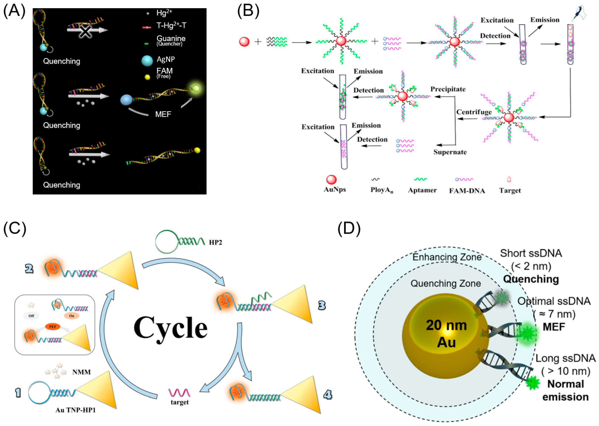
Figure 2. (A) Schematic illustration of the distance-dependent MEF sensing platform based on a molecular beacon [46]. (B) Schematic illustration of the preparation of MEF-based AuNPs@polyAn-aptamer@FAM-DNA nanostructure and target detection [48]. (C) Schematic illustration of target DNA fluorescence detection based on the CHA and plasma resonance fluorescence (PEF) effect [52]. (D) Schematic diagram of the MEF effect dependent on the DNA length of dsDNA between 20-AuNPs and FITC [53]. Reprinted with permission from [46,48,52,53].
Biosensors 13 00376 g002
Figure 3. (A) (i) Conjugation of CAP with gold nanoparticle (GNP) via a thiol-antihomovanillic acid (HVA) linker. (ii) Schematic diagram of CAP fluorescence emission and CAP-GNP fluorescence enhancement emission. (iii) Schematic diagram of the CAP fluorescence enhancement after GNP conjugation [54]. (B) (i) Fluorophore fluctuation in an evanescent field layer after conjugation of GNI with CAP. (ii) Fluctuation-based observation of fluorophore position with regard to proximity of GNI. Numbers 1, 2, and 3 imply fluorescence enhancement, quenching, and equilibrium with regard to the distance between CAP and GNI [55]. Reprinted with permission from [54,55].
Biosensors 13 00376 g003
Figure 4. (A) Practical use of the ordered GNR array chip for DNA detection based on surface plasmon-enhanced fluorescence upon hybridization with the target [57]. (B) Randomly arranged gold nanodisks on an etched glass substrate [59]. (C) Photograph and diagram of the single-molecule immunoreaction process for three thyroid hormones on a fourplex nanoimmunosensor chip [62]. Reprinted with permission from [57,59,62].
Biosensors 13 00376 g004
Figure 5. (A) Synthetic procedure for Ag@SiO2@Y2O3:Er nanostructures and TEM images of the resulting UCNPs [78]. (B) Design of a NaGdF4:Yb3+, Nd3+@NaGdF4:Yb3+, Er3+@NaGdF4 UCNC allowing for two different energy transfer paths [86]. Reprinted with permission from [78,86].
Biosensors 13 00376 g005
Figure 6. (A) SEM image of D2PA structure with different pillar heights on UC [88]. (B) TEM image of Y2O3:Yb, Er NPs with SiO2 spacer layer and Ag nanocap [89]. (C) SEM image of nanocylinder array with the diameter of 440 nm and height of 180 nm arranged in a square lattice [90]. (D) Distance- and spectra-dependence and fabrication process of the UCNPs and GNRs plasmon-enhanced fluorescence system [91]. Reprinted with permission from [89,90,91].
Biosensors 13 00376 g006
Disclaimer/Publisher’s Note: The statements, opinions and data contained in all publications are solely those of the individual author(s) and contributor(s) and not of MDPI and/or the editor(s). MDPI and/or the editor(s) disclaim responsibility for any injury to people or property resulting from any ideas, methods, instructions or products referred to in the content.