Review

2D Metal-Organic Frameworks: Properties, Synthesis, and Applications in Electrochemical and Optical Biosensors

by
Anamika Ghosh
,
Sana Fathima Thanutty Kallungal
and
Sundara Ramaprabhu
*
Alternative Energy and Nanotechnology Laboratory, Department of Physics, Indian Institute of Technology Madras, Chennai 600036, India
*
Author to whom correspondence should be addressed.
These authors contributed equally to this work.
Biosensors 2023, 13(1), 123; https://doi.org/10.3390/bios13010123
Submission received: 29 November 2022 / Revised: 31 December 2022 / Accepted: 9 January 2023 / Published: 11 January 2023

Abstract

:
Two-dimensional (2D) nanomaterials like graphene, layered double hydroxides, etc., have received increasing attention owing to their unique properties imparted by their 2D structure. The newest member in this family is based on metal-organic frameworks (MOFs), which have been long known for their exceptional physicochemical properties—high surface area, tunable pore size, catalytic properties, etc., to list a few. 2D MOFs are promising materials for various applications as they combine the exciting properties of 2D materials and MOFs. Recently, they have been extensively used in biosensors by virtue of their enormous surface area and abundant, accessible active sites. In this review, we provide a synopsis of the recent progress in the field of 2D MOFs for sensor applications. Initially, the properties and synthesis techniques of 2D MOFs are briefly outlined with examples. Further, electrochemical and optical biosensors based on 2D MOFs are summarized, and the associated challenges are outlined.

1. Introduction

Two-dimensional (2D) nanomaterials have sparked great interest among researchers owing to the interesting properties arising from their inherent layered, sheet-like morphology and nanometer regime thickness. The two-dimensionality imparts fascinating features such as mechanical flexibility, interesting charge transfer properties, huge surface areas, optical transparency, and plenty of accessible active sites to them, which finds applications in fields like flexible electronics, catalysis, sensors, gas separation, etc. [1]. Several 2D nanomaterials have been explored to date, the well-known ones being graphene and graphene oxide [2], and others such as metal oxides [3], layered double hydroxides [4], transition metal dichalcogenides [5], borophene [6], graphitic carbon nitride [7], etc. A recent addition to this family is the two-dimensional counterparts of metal-organic frameworks (MOF), which possess unique features and functionalities [8]. Similar to other 2D materials, they possess a huge specific surface area and excellent mechanical properties. Although the electrical conductivity of these structures is low as compared to other 2D materials such as graphene, the easy functional ability, and tunable properties make them interesting materials for diverse applications. A brief overview of some of the properties of 2D MOFs and other 2D nanomaterials are compared and summarized in Table 1 for better understanding.
Metal-organic frameworks (MOFs) are porous, crystalline materials comprising metal-based nodes (ions or clusters) bonded to organic linkers via coordination bonds. They possess unique physical and chemical properties, such as high surface area, tunable porosity, fascinating morphologies, catalytic property, etc. [11]. They are of immense interest owing to the fact that these properties can be tuned based on the choice of metal or organic moiety. For instance, the pore size of MOFs can be altered from micropores to mesopores based on the length of the organic linkers used. Deng et al. [12] demonstrated isoreticular MOF-74 structures with different pore apertures (1.4–9.8 nm) obtained by combining short and long linkers. A more general strategy was developed by Yuan et al. [13] where a method termed linker labilization was used to obtain hierarchical porous structures. Likewise, MOFs provide the unique opportunity to rationally design and tune the properties by modulating the ligands and nodes. Due to these attractive features, MOFs have been widely explored for applications in gas storage [14], sensing [15], catalysis [16], energy storage [17], and much more [18]. The properties and applications of these MOFs are primarily determined by their structure, composition, size, and morphology [19]. A plethora of reports focusing on bulk MOF crystals and 1D MOF materials have been published over the years. However, it was only recently that there had been a surge of research related to 2D MOFs for various applications (Figure 1), as these materials couple the properties of both MOFs and 2D materials [20]. Compared to other materials such as graphene, carbon nanotubes (CNT), metal oxide, and metal particles, 2D MOFs exhibit several exciting properties, suitable for sensing applications. A few of such properties are listed below:
  • Tunable functionalities with varying ligands help to modulate 2D MOF and analyte molecule interaction through different interaction mechanisms such as hydrogen bonding, π-π stacking, electrostatic interactions, etc.
  • High surface area and lateral dimension provide better attachment of the molecules on the 2D MOF surface, leading to higher loading of probe molecules.
  • Adjustable pores for the targeted analyte through tunable building blocks that enable the high selectivity of 2D MOF.
  • Exposed metal sites on the surface of the 2D MOF accelerate the surface catalytic reaction.
  • Exposed metal sites on the surface provide excellent quenching properties, making them suitable for optical nucleic acid (NA) and immunosensor.
A comparison of 2D MOF-based sensors with other reported materials for the most common analyte glucose is summarized in Table 2. This review provides insight into the different 2D MOFs that have been employed in biosensors. Initially, their unique properties, synthesis strategies, and challenges involved are elaborated. Further, specific examples of 2D MOF-based electrochemical and optical biosensors are examined to understand and appreciate the use of these exciting materials for sensing applications. Finally, future perspectives and opportunities related to these materials are briefly discussed.

2. Unique Properties of 2D MOFs

Dimensionality has a strong influence on the properties of a material [29]. As a result, 2D MOFs possess drastically different properties as compared to 3D MOFs and are useful for applications where bulk MOFs are inadequate. For instance, 3D MOFs are limited by their poor electrical conductivity (due to their porosity, crystalline structure, and chemical composition) for applications in electrochemical sensors [30]. Nonetheless, a judicious combination of ligands and metal ions can yield conductive MOFs. Interestingly, the 2D counterparts of these MOFs demonstrate enhanced conductivity owing to the reduced charge transport path lengths [29]. One of the highest conductivity values reported (120 S cm−1) for 2D MOFs is by Low et al. [31] for a Cu-hydroxythiophenolate-based system. Another example is by Ohata et al. [32], where they observed a conductivity of 0.6 S cm−1 for nanosheets of nickel-hexaminotriphenylene (Ni-HITP) MOF. This was the highest observed for HITP-based MOFs with thicknesses less than 100 nm. The conductivities of the bulk and thin film analogs of this MOF were found to be 2 and 40 S cm−1 by Sheberla et al. [33].
2D MOFs also possess enormous surface area (due to the high aspect ratio) and abundant active sites, making them appealing for many applications, particularly sensing [20]. The high surface area provides a stable matrix for the efficient attachment of enzymes and promotes the adsorption of more analyte moieties, thereby increasing the sensitivity. The enhanced adsorption also improves the response time [34]. Additionally, the active sites on its surface are easily accessible owing to its 2D morphology. This enhances their catalytic properties by efficient utilization of these sites, and reduction of the diffusion barrier and mass transfer resistance [29,35,36]. In the case of optical sensors, the presence of more accessible sites facilitates the energy and charge transfer between the fluorophores and quenchers. Han et al. [37] obtained a lower detection limit for metal ions using nanosheets of a zinc-based MOF (ZSB-1) compared to its 3D form. He et al. [38] synthesized ultrathin (1.5 nm thickness) zirconium-based MOF nanosheets. These exhibited superior photocatalytic performance than their bulk counterparts.
Another attractive property of 2D MOFs is their excellent mechanical strength and flexibility, which finds applications in fields such as gas separation and sensor fabrication [39,40]. This is endowed by the presence of coordination bonds within the structure and the nanosheet morphology. Hermosa et al. [40] investigated the mechanical properties of a 2D CuMOF (flakes with the number of layers ranging from 1–50) and observed Young’s modulus of 5 GPa. Although this is lower than the typical value for 2D materials, this was enough to form an isolated, stable 2D structure of this MOF. 2D MOFs are also observed to be considerably more chemically stable than 3D MOFs. The chemical stability of these MOFs is essential for sensor applications where operation in aqueous environments with extreme pH values may be required. Recently, Zhang et al. [41] synthesized very thin (~3.97 nm) indium-porphyrin-based MOF (InTCPP) nanosheets with excellent stability in aqueous solutions with pH values ranging from 2–11. The nanosheets exhibited higher photocatalytic activity towards hydrogen production compared to 3D InTCPP. In a different report [42], bimetallic 2D MOF nanosheets (NiCoBDC) synthesized by the hydrothermal method were found to be stable in alkaline media. The material was soaked in 1 M KOH for 12 h to demonstrate its durability, after which the phase remained unchanged (as determined from X-ray diffraction).
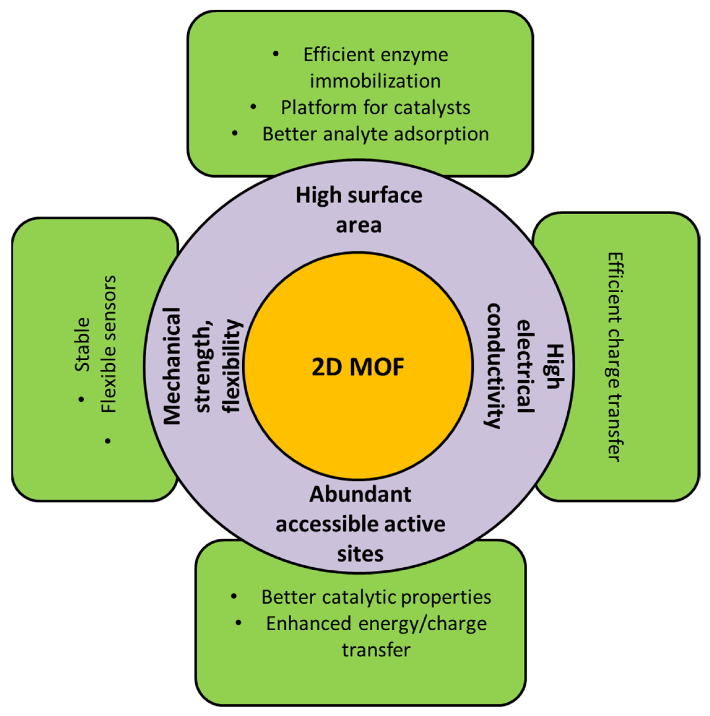
Figure 2 summarizes the exciting properties of 2D MOFs which are appealing for sensor applications. All of the properties of 2D MOFs are predominantly dependent on the synthesis procedure, parameters such as solvent used, temperature, reaction time, etc. In the following section, some of the approaches to synthesizing robust, isolated two-dimensional MOF nanostructures are briefly discussed.

3. Synthesis of 2D MOFs

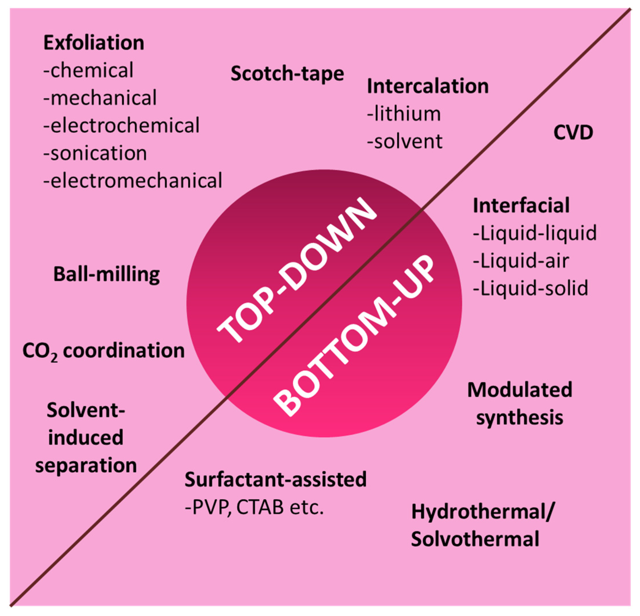
The fundamental challenge associated with 2D MOFs is synthesis, wherein it is necessary to strike a balance between yield, ease of synthesis, crystallinity, and stability. Several groups [39,43,44,45,46,47] have reported a variety of synthesis techniques for the formation of 2D MOFs. As with other 2D nanomaterials, the synthesis of 2D MOFs also follows one of the two main strategies—top-down or bottom-up approaches [8]. Figure 3 summarizes different synthesis routes for the preparation of 2D MOF.

3.1. Top-Down Synthesis

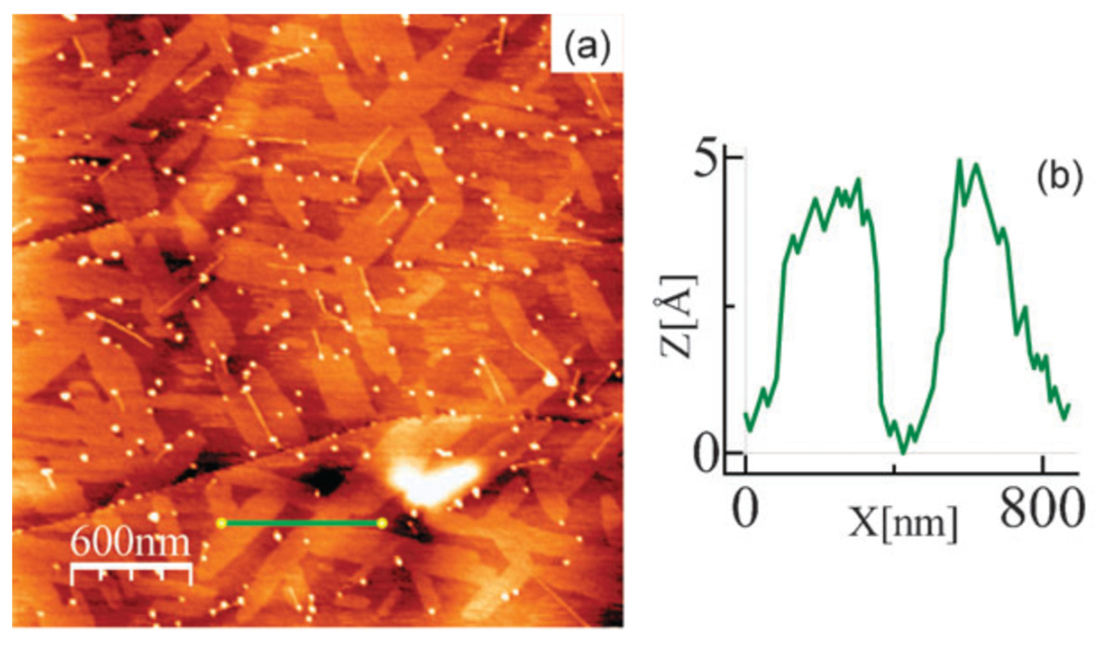
The top-down strategy is a simple yet powerful method that involves delaminating or stripping off layers (exfoliation) of the MOF from a bulk 3D MOF with a stacked-layer structure. The separation of the layers can be brought about by physical or chemical methods such as grinding [48], exfoliation (chemical [49], mechanical [50], electrochemical [51], electromechanical [52], or sonication [53]), intercalation (lithium, solvent) [54,55], etc., resulting in 2D MOF nanosheets with a single or few layers. The primary requirement here is that the MOF should be bonded by strong forces within the layers and weak forces (van der Waals, hydrogen bonding, π-π stacking, etc.) between the layers so as to enable easy separation of the layers [20]. The first report on the synthesis and characterization of a 2D MOF was by Amo-Ochoa et al. [49] (Figure 4). They isolated and studied single-atom-thick, bi-dimensional Cu2Br(IN)2 (IN—isonicotinato) synthesized by sonicating its 3D counterpart (obtained by hydrothermal reaction) in water. The sonication separated the layers by minimizing the interactions between them. Sonication treatment in other solvents such as acetone [56], ethanol [57], methanol [58], N,N-dimethylformamide [59], etc., or a mixture of two or more of these has also been reported [60]. The choice of solvent is a critical parameter and plays a crucial role in exfoliation. The solvents, in addition to separating the layers of the MOF, help stabilize the nanosheets by avoiding restacking. For instance, Tan et al. [61] reported the top-down synthesis of 2D M-DMS and M(2,2-DMS) MOFs (M = Mn, Co, Zn; DMS- dimethyl-succinate) by sonication in different solvents. Ethanol was identified to be the most effective for exfoliation and prevention of restacking. Through systematic studies, they also found that the formation of the 2D structure was primarily dependent on the bulky nature of the organic ligand rather than the cation. Taking a cue from this, another group [43] designed a MOF with bulky ligands (with 3-methoxypropoxy groups) in order to ease the exfoliation in different solvents.
Apart from sonication, other exfoliation techniques such as the Scotch-tape method [62] (mechanical, well-known for graphene synthesis), wet ball-milling [60], solvent-induced separation [63], etc., have also been reported. All of these methods suffer from the limitation of low exfoliation yield. Several groups have reported a combination of these strategies to improve the same. One such example is depicted in Figure 5. As mentioned earlier, these methods are only suitable for 3D MOFs with a layered structure. However, recently Zhou et al. [64] demonstrated the top-down synthesis of a 2D non-layered MOF by CO2-coordination. They proposed that simultaneously dissociating the ligand and coordinating a capping agent in a 3D non-layered MOF can prepare high-quality 2D MOFs. This necessitated the selection of a suitable microenvironment to carry out the defect generation and coordination. They proved this concept by treating 3D non-layered CuBTC (BTC—1,3,5-benzenetricarboxylate) in a solvent mixture containing supercritical CO2. This led to the dissociation of the BTC ligands, followed by the coordination of the CO2 molecules. The resulting Cu(II) dimer was square planar, bonded to two –COO groups of the ligand and two –HCOO groups of the CO2. These groups can attach to trigonal BTC molecules to form the non-layered 2D MOF structure.
Although these approaches are simple and robust, the yield obtained from these methods is too low for practical applications. The exfoliation rate is also low in most cases [8]. Additionally, for layered MOF structures, restacking the layers can threaten the stability of the MOFs, if not accounted for, owing to the weak forces involved. In this context, synthesis approaches different from the top-down strategy need to be explored.

3.2. Bottom-Up Synthesis

The bottom-up approach involves building the 2D structure from the metal ions and ligands by chemical reactions. This method allows more chemical flexibility and tenability [20,65]. For instance, for layered 2D MOFs, the number of layers in the MOF nanosheets can be controlled by tuning the reaction parameters. This is also the widely adopted strategy for non-layered 2D structures. The critical factor in the bottom-up method is suppressing the MOF growth in one direction without affecting the growth in the other two directions [39,66]. For layered-type MOFs, this implies preventing the layers from stacking up in the vertical direction. This can be easily done using interrupters (like surfactants) which can prevent the stacking during the formation process [20]. For non-layered MOFs, selective growth in two directions is challenging yet possible. Most of the strategies to achieve growth restriction involve synthesis in a confined 2D space, creating high surface energy structures, and chemically modifying the surfaces [67]. Based on these concepts, several methods have been reported for synthesizing 2D MOFs—interfacial synthesis, surfactant-assisted synthesis, modulated synthesis, hydro/solvothermal method, sonication synthesis, and so on.
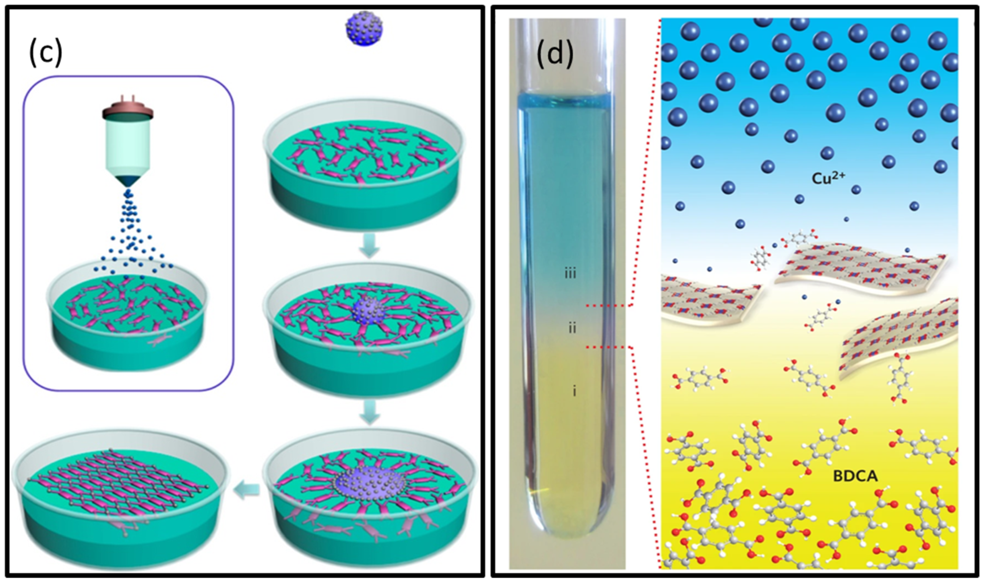
In the interfacial method, the synthesis reaction between the metal nodes and the ligands is confined to a 2D interface region, thereby forming nanosheets [67]. The interface can be liquid-liquid, liquid-air, or liquid-solid. The liquid-liquid [68] interface is formed by two immiscible solvents, one in which the metal is dissolved and another in which the ligand is dissolved. Typical examples of solvent mixtures used are water-ethyl acetate and water-dichloromethane. The formation of MOFs from their constituents occurs on the interface driven by diffusion. In the liquid-air interface method [69], a small quantity of organic solvent is added to a liquid, which evaporates to form the interface. By using a uniform monolayer of ligands, this interface allows the formation of ultra-thin, single-layer MOF nanosheets. Utilizing a Langmuir-Blodgett trough can control the nanosheet thickness as well as maximize the yield. The liquid-solid interface [70] involves layer-by-layer growth of the MOF on a chemically modified surface. Synthesis of MOF nanosheets at gas-solid interfaces has also been reported. However, it requires ultra-high vacuums and flat metal surfaces. Rodenas et al. [39] adopted a three-layer synthesis approach to synthesize copper-1,4-benzenedicarboxylate (CuBDC) MOF. The solutions allow acute control of the diffusion and kinetics of the reactants, thereby producing ultra-thin (5–25 nm) nanosheets with high yield. The thickness was tunable based on the solvent and reaction temperature. The schematic illustrations for different interfacial methods for synthesizing 2D MOFs are given in Figure 6.
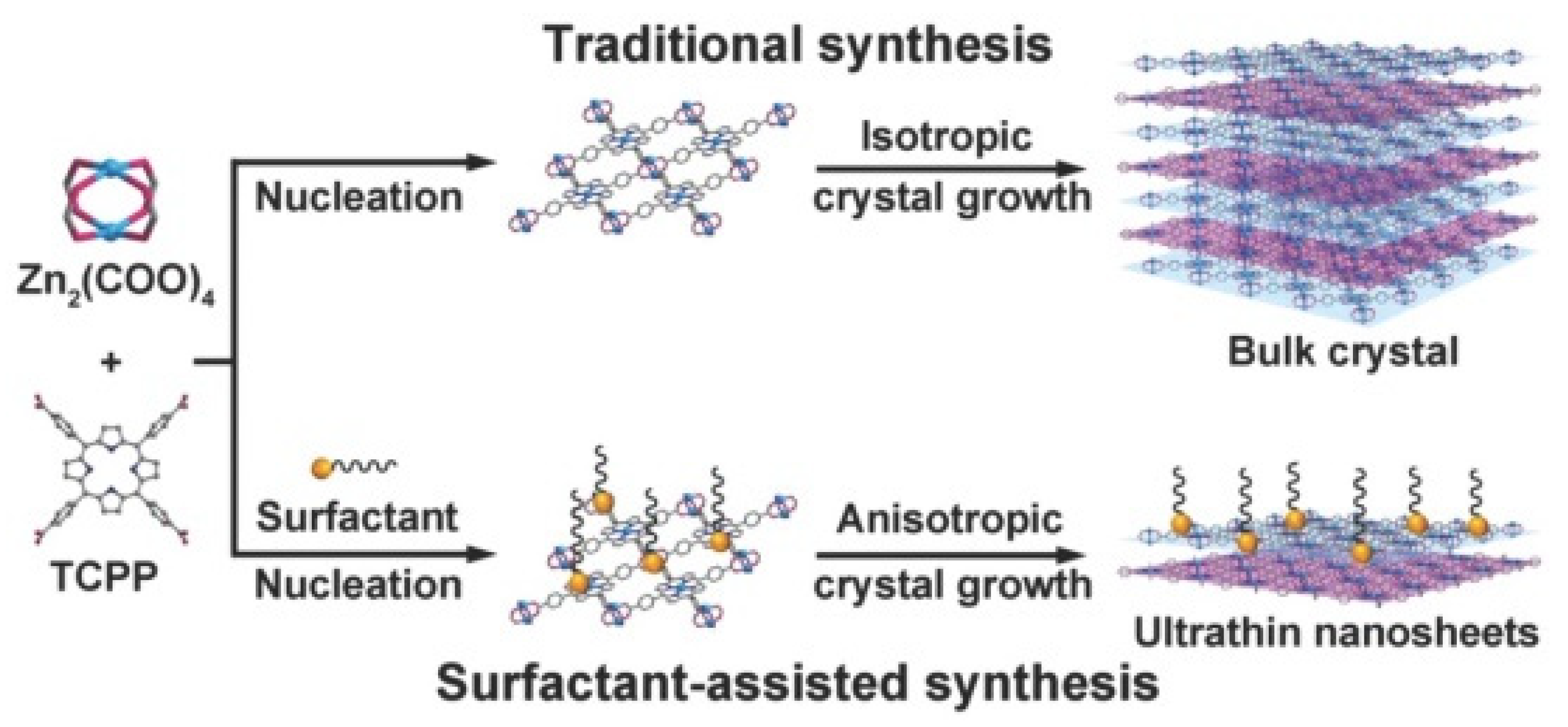
Another widely reported method is the surfactant-assisted synthesis [34,41,47]. The presence of a surfactant (like polyvinylpyrrolidone (PVP), cetyltrimethylammonium bromide (CTAB), etc.) lowers the surface energy of the as-prepared MOF nanosheets by preventing their aggregation. For instance, Zhao et al. [47] utilized PVP for the synthesis of 2D TCPP-based MOFs (Figure 7). The PVP restricted the MOF stacking as well as stabilized the synthesized nanosheets. Instead of surfactants, certain small molecules such as pyridine and acetic acid (termed modulators) have also been employed for 2D MOF synthesis (modulated synthesis) [71]. These molecules possess functional groups similar to that of the linkers, and hence compete to coordinate with the metal nodes. This competition regulates the growth of MOFs. The modulators selectively coordinate to specific planes of the MOFs, which block the growth along that direction. The preparation of non-layered MOFs has also been reported using this method [58].
Multiple other techniques, such as chemical vapor deposition [72], hydro- or solvothermal [73,74] synthesis, etc., have also been reported for the synthesis of MOF nanosheets. In most synthesis methods, there is always a competitive formation of 3D MOF occurring along with the 2D MOF. Oh et al. [75] found that the selective formation of 2D MOFs over their 3D counterparts critically depended on the relative ratio between the metal ions and organic ligands. They also demonstrated a modified bottom-up synthesis of 2D MOF layers by a combination of ultrasonication and surfactant-assisted methods.
Combining the top-down and bottom-up approaches for the synthesis of 2D MOFs is also becoming popular. Chaudhari et al. [76] synthesized nanosheets of a zinc-based MOF using a rapid self-assembly technique followed by sonication. Similarly, Junggeburth et al. [46] employed CTAB-assisted synthesis followed by exfoliation to prepare ~10 nm thick sheets of a zinc-based MOF. In summary, each synthesis method poses its own pros and cons. A judicious choice of the method and thorough optimization of the reaction parameters is necessary to synthesize 2D MOFs with desired properties and suitable for particular applications. Extensive reviews on the synthesis methods can be found elsewhere [20,29,65]. As mentioned in the previous section, 2D MOFs have been utilized in various applications owing to their unique properties. One of the extensively reported applications is in sensors, wherein they have been used for the detection of various analytes such as humidity [77], hydrogen [78], metal ions [37,53], explosives [79], pesticides [80], DNA [47,81], glucose [82,83], proteins [84,85], disease biomarkers [84,86], antibiotics [87,88], and much more. In the following sections, electrochemical and optical sensors employing 2D MOFs for the detection of biological analytes are discussed.

4. 2D MOFs for Electrochemical Biosensor Applications

Recently, MOFs have been widely explored for electrochemical biosensor applications. One of the attractive characteristics of MOF is its tunable pore size by changing the ligand type. Therefore, MOF acts as molecular sieves, improving selectivity towards a specific analyte. Additionally, the accessible pores of the MOF allow the diffusion of the analytes throughout the MOF skeleton, thus improving the catalytic activity of the MOF as an electrochemical biosensor. Often the bulk MOF suffers from poor dispersibility, increasing the possibility of detaching from the catalyst substrate during the sensing measurement. In that case, 2D MOF may overcome the challenges of 3D MOF. 2D MOFs are endowed with a high surface area that encourages the better adsorption of biomolecules. Besides, the exposed metal sites, porous network, enhanced mass transfer, and good dispersion make 2D MOF suitable for electrochemical biosensors. In the below section, we will discuss the recent advancements in the field of 2D-MOF-based electrochemical biosensor.

4.1. 2D MOF as a Nonenzymatic Sensor

The development of 2D MOF as an electrochemical sensor is drawing the attention of research scientists. Moreover, 2D MOFs have recently been probed as nonenzymatic sensors since they offer many advantages over enzymatic ones. Nonenzymatic sensors are cost-effective, stable over time and pH, easy to use, and have no storage issues. Due to their high surface area, ultrathin nanosheet nature, tunable porosity (which contributes toward selectivity), and enhanced electrocatalytic activity, 2D MOFs have the potential for electrochemical biosensing.
Wang et al. tuned the coordination environment in 2D Ni-MOF by a π-conjugated molecule and synthesized MOF@MOF structures (Figure 8). They used conductive 2,3,6,7,10,11-hexahydroxytriphenylene (HHTP) as the organic linker [28]. HHTP acts as an extended π-conjugated molecule which can help in enhancing mobility and conductivity. In this work, the first Ni-MOF nanosheet was synthesized by hydrothermal method using nickel nitrate (Ni(NO3)3) as a precursor for the metal center and benzimidazole and 2-methylimidazole as organic ligands. In the next step, synthesized Ni-MOF and HHTP were dispersed in a mixture of N,N-dimethylformamide (DMF), and deionized (DI) water and kept at 85 °C for 6 h. Through the time-dependent studies, they observed that HHTP particles got attached mainly at the margins or periphery of the NiMOF sheet, and after 2 h, HHTP particles started etching the Ni MOF sheet. In brief, HHTP coordinates with the free Ni+2 of Ni-MOF and simultaneously etches the Ni-MOF nanosheets. Further, they studied the electrochemical glucose sensing properties of porous Ni-MOF@Ni-HHTP-5 (5 indicates 5 mg loading of HHTP) nanosheet in potassium hydroxide (KOH) (0.1 M) solution. At a constant potential of 0.6 V, Ni-MOF@Ni-HHTP-5 electrocatalyst could sense glucose concentrations within 0.5–2665.5 µM with a sensitivity of 2124.90 µA mM−1 cm−2 and LOD of 0.0485 µM. Here, although the glucose-sensing mechanism depends on the change of valence of the Ni metal center, the inclusion of HHTP in the NiMOF nanosheets encourages the better utilization of Ni+2 ions, accelerating the exposure of the metal sites that, in turn, enhances the conductivity and catalytic activity of the sensor.
Stolz et al. studied the electrochemical properties of the different facets of conductive MOF and their application in sensing neurotransmitters such as dopamine (DA) [89]. They synthesized Ni3(HHTP)2 and Co3(HHTP)3 MOF in two different methods: (a) hydrothermal and (b) Interfacial synthesis. Morphology of the hydrothermally synthesized MOFs appeared rod-shaped and defined by {100} family of planes, indicating that terminal edge sites were exposed surface sites. On the other hand, MOF synthesized in interfacial synthesis revealed sheet-type morphology and was defined by {001}, where the dominant surface sites were a basal plane. They further examined the electrochemical performances of {001} and {100} planes of M3(HHTP)2 MOFs in different biological analytes such as DA, uric acid (UA), serotonin (5-HT), ascorbic acid (AA). They observed improved peak current (Ip) for MOF over bare GCE in positively charged DA, 5-HT solution while the performance deteriorated compared to bare glassy carbon electrode (GCE) in case of solution with negative analytes such as UA, AA, etc. However, the Ip value drastically improved in the DA solution with {001} facet of M3(HHTP)2 MOFs compared to the {100} facet. The enhancement of peak current is attributed to probably two factors: (a) complementary Coulombic interactions between positively charged DA and MOF interface, (b) available and favorable adsorption of DA on {001} family of planes compared to {100} of MOF. Their integrated sensor Ni3(HHTP)2/GCE showed linearity of 0–6 µM in 0.1 M PBS and 0–2 µM in simulated cerebrospinal fluid with a LOD of 9.9 ± 2 nM and 214 ± 48 nM, respectively.
Chen et al. fabricated a nonenzymatic sensor of Cu2O-mediated growth of Au nanoparticles (NPs) on 2D MIL-53 (Fe) for real-time monitoring of hydrogen peroxide (H2O2) from living cells (Figure 9). In this work, they prepared Cu2O NPs and MIL-53(Fe) separately, and later, the composite of the two was prepared by sonicating in Nafion solution [90]. MIL-53 (Fe) was synthesized using iron chloride (FeCl3, 6H2O) as the metal source and terephthalic acid (H2BDC) as the organic linker. Hydrothermally synthesized MIL-53 (Fe) showed a 2D stacked sheet-type structure. Further, Au NPs were decorated on Cu2O/MIL-53(Fe)/GCE via electrodeposition method in HAuCl4 solution at −0.2 V. Au@Cu2O/MIL-53(Fe)/GCE sensor could sense H2O2 within a linear range of 0.01–1.52 mM with a sensitivity of 0.351 mA mM−1cm−2 and LOD of 1.01 µM. Sensing of H2O2 is related to the change of valence of Au (Au0 →Au+1); however, 2D MIL-53(Fe) here provides a high surface area and a matrix to anchor agglomeration free NPs. Additionally, pores of MIL-53(Fe) sheets also helped for selective detection of H2O2. Further, Au@Cu2O/MIL-53(Fe) was applied for real-time monitoring of released H2O2 from A549 lung cancer cells, and the sensor could detect an H2O2 concentration of 0.042 fM.
Bottier et al. demonstrated a composite of 2D Co-MOF/Nafion, which shows an enzyme-like activity toward H2O2 sensing. Co-MOF was synthesized in a solvothermal approach using [(Co4O4)(OAc)4(py)4] as a building block [91]. H2O2 sensing performance was further evaluated in pH 7 using voltammetry and chronoamperometry techniques. Voltammetry anodic current vs. H2O2 concentrations plot matched well with the Michaelis-Menten reaction scheme, thus confirming the enzyme mimicking characteristics of the developed sensor. The enzyme mimicking characteristics is probably due to the specific coordination environment of the synthesized 2D MOF, where the metal centers are coordinated by a suitable organic ligand. Thus, the coordinated metal centers mimic the enzyme redox cofactor environment inside the MOF and exhibit ‘enzyme mimic’ characteristics. The sensor displayed linear ranges within 5 µM−1 mM and 1–10 mM with a corresponding sensitivity of 570 ± 5 A cm−2 mM−1 and 395 ± 10 A cm−2 mM−1. Periodically arranged Co metal centers with the optimum length organic ligand in the Co-MOF sheet allow fast diffusion of the electrolyte where exposed metal centers accelerate the redox reaction for H2O2 detection.
Lu et al. designed a heterostructure based on 2D/2D NiCo-MOF/Ti3C2 for nonenzymatic detection of acetaminophen (AP), DA, and UA. In this heterostructure, the lamellar Ni-Co MOF sheet contains abundant unsaturated metal sites that contribute to the catalytic activity [92]. Ti3C2 offers hydrophilicity and conductive matrix support for the agglomeration-free dispersion of NiCo-MOF sheets. The developed sensor can detect AP (working potential 0.346 V vs. SCE), DA (working potential 0.138 V vs. SCE), and UA (working potential 0.266 V vs. SCE) simultaneously in a linear range of 0.01–400 µM, 0.01–300 µM and 0.01–350 µM with a detection limit of 8, 4, and 6 nM, respectively in 0.1 M phosphate buffer solution (PBS pH 7.4). They attributed the electrocatalytic activity of NiCo-MOF/Ti3C2 heterostructure to three factors: (a) metal-O-metal chains create a conductive pathway through the MOF lamellar sheet, (b) exposed unsaturated Ni and Co sites that serve as a catalytic center for AP, UA, DA detection, (c) two-dimensionality of Ni-Co MOF accelerates the electron hopping across the metal centers and also boosts the electrolyte diffusion through the pores of the MOF, and (d) finally Ti3C2 provides a 2D conductive matrix to prevent accumulation of Ni-Co MOF nanosheets, thus enhancing the exposure of more catalytic sites. The proposed sensor could detect AP, UA, and DA from serum and urine samples with a 98.1–102.2% recovery rate.
Wang et al. developed a facile approach to design a 2D array of MOF-based flexible electrode for sweat biosensor via interfacial synthesis and dip coating method [93]. In brief, first, they synthesized amine group functionalized (-NH2) graphene paper (NH2-GOP). Then Cu3(btc)2 MOF (btc-1,3,5-benzenetricarboxylic acid) was prepared through the interfacial emulsion process and later transferred on the NH2-GOP through the dip coating method. This leads to the 2D assembly of Cu3(btc)2 nanocubes on the freestanding graphene paper. The developed NH2-GO-Cu3(btc)2 flexible electrode was further tested to monitor glucose and lactate in sweat. In such MOF-based electrodes, electrochemical sensing is mainly ascribed to the extended conductive network through the MOF skeleton since electron hopping is allowed in the adjacent metal centers of the MOF. The sensor showed linearity for lactate concentrations of 0.05 mM–22.6 mM with a sensitivity of 29 µA cm−2 mM−1 and for glucose concentrations of 0.05–1775.5 µM with a sensitivity of 5.36 mA cm−2 mM−1. Their fabricated sensor also suffers minimum interference from other bioanalytes and has shown promising performance when examined for flexibility and dynamic test.
In another work, Wang et al. developed 2D Ni, Co, and NiCo-based MOF using 1,4- benzene dicarboxylic acid (BDC) as the organic ligand [94]. Synthesized MOFs showed lamellar sheet-type morphology with smooth surfaces. Further, they implemented the synthesized MOFs for urea detection. Urea is a human metabolite, and the content of urea in the blood is an essential indicator of kidney or liver functions. No voltammetric response of Co-MOF with urea addition was observed. Although NiCo MOF and Ni-MOF showed enhanced current response with the addition of urea, the urea sensing response for Ni MOF was more pronounced. This is attributed to the ultra-thin thickness and high surface area of Ni-MOF nanosheets. Ni-MOF nanosheets showed chronoamperometric linear response for urea concentrations of 0.5–832.5 µM with an excellent sensitivity of 1960 µA mM−1 cm−2 in 0.1 M KOH solutions at operating potential 0.45 V.

4.2. Nucleic Acid-Based Sensor

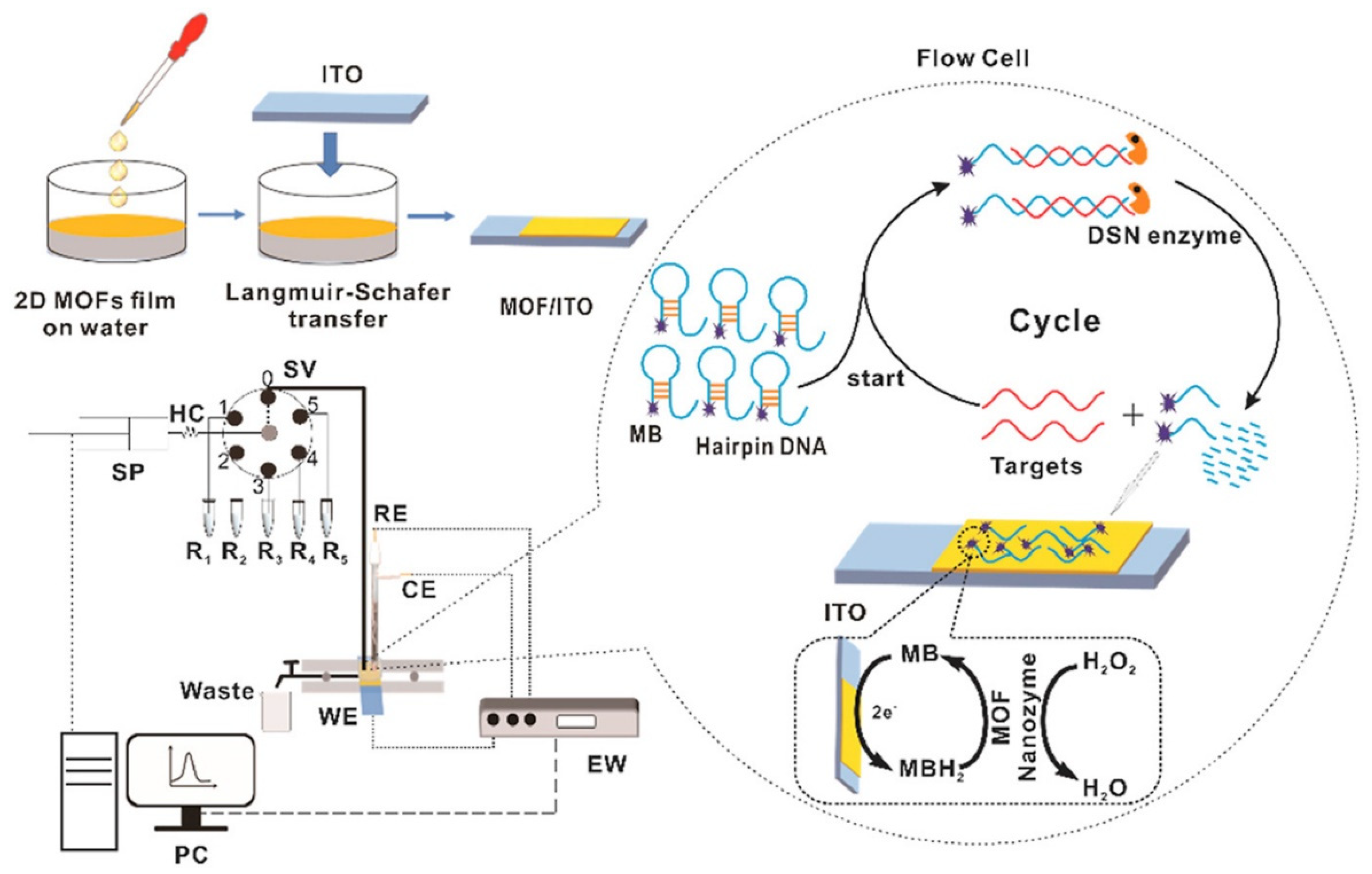
Nucleic acid-based sensors provide massive opportunities for the sensitive and rapid detection of several proteins and biomarkers. As previously discussed, 2D MOF offers a high surface area for the adsorption of biomolecules and sometimes shows peroxidase mimic character; they can be utilized for the nucleic acid-based electrochemical sensor. Apart from these, 2D MOFs possess several other advantages. 2D MOFs allow tight binding with the probe molecule through π-π stacking. Further, they offer a high loading capacity of probe molecules, open metal sites, and tunable organic functionalities that contribute to excellent quenching properties and boost sensing performance. Additionally, the fluorescence quenching efficiency can be effectively improved by modulating the structure of MOF, varying the metal center and ligand, and regulating the ratio of center metal ions and organic ligands. Further, the 2D MOF-based electrochemical NA sensor has also drawn attention owing to the robustness, simplicity, high sensitivity, and rapid detection characteristics of the electrochemical sensing mechanism. In the following section, we will discuss the 2D MOF-based electrochemical NA sensor. Wang et al. designed a flow homogeneous assay based on Co-MOF-based 2D nanozyme for successive microRNA assay [95] (Figure 10). Co-MOF nanosheet was synthesized in an ultrasonication method using Co+2 as metal nodes and H2BDC as an organic ligand. Transmission electron microscopy (TEM) morphology revealed the lateral size of the synthesized nanosheet was about 100–300 nm, while the thickness of the nanosheets below 1 nm was confirmed through (Atomic force microscopy) AFM. The working electrode was prepared by transferring the 2D Co-MOF sheet to the (Indium tin oxide) ITO electrode via the Langmuir-Schafer approach. They further developed a flow homogeneous microRNA assay that doesn’t require DNA recognition elements or functionalization of the working electrode. MOF/ITO electrode could differentiate ssDNA from double strands DNA (dsDNA) due to the π-π stacking. In the flow homogeneous microRNA assay, they used duplex specific nuclease (DSN) enzyme. First, a methylene blue (MB) labeled hairpin DNA (HP) was kept and gave a negligible electrochemical response. In the presence of the target, i.e., microRNA, microRNA hybridized with HP DNA to form an RNA/DNA hybrid system. Further, DNA from the DNA/RNA hybrid system gets hydrolyzed by the DSN enzyme and releases microRNA and short DNA in the solution. MicroRNA can hybridize with HP DNA again, whereas MB-labeled short DNA showed a strong affinity and diffusivity towards 2D MOF/ITO electrode. In the presence of H2O2, oxidized MB gets electrochemically reduced at the MOF/ITO electrode, leading to a distinct enhancement in the electrochemical signal. Thus, based on the above mechanism, the presence of microRNA in repeated cycles yields a large amount of MB labelled short DNA, which amplifies the electrochemical signal. Additionally, the chemical and enzyme-like activity of 2D MOF nanozyme also helped to boost the electrochemical signal. The microRNA assay flow homogeneous system can detect microRNA concentrations ranging from 1 pM to 1 µM with a LOD of 0.12 pM.
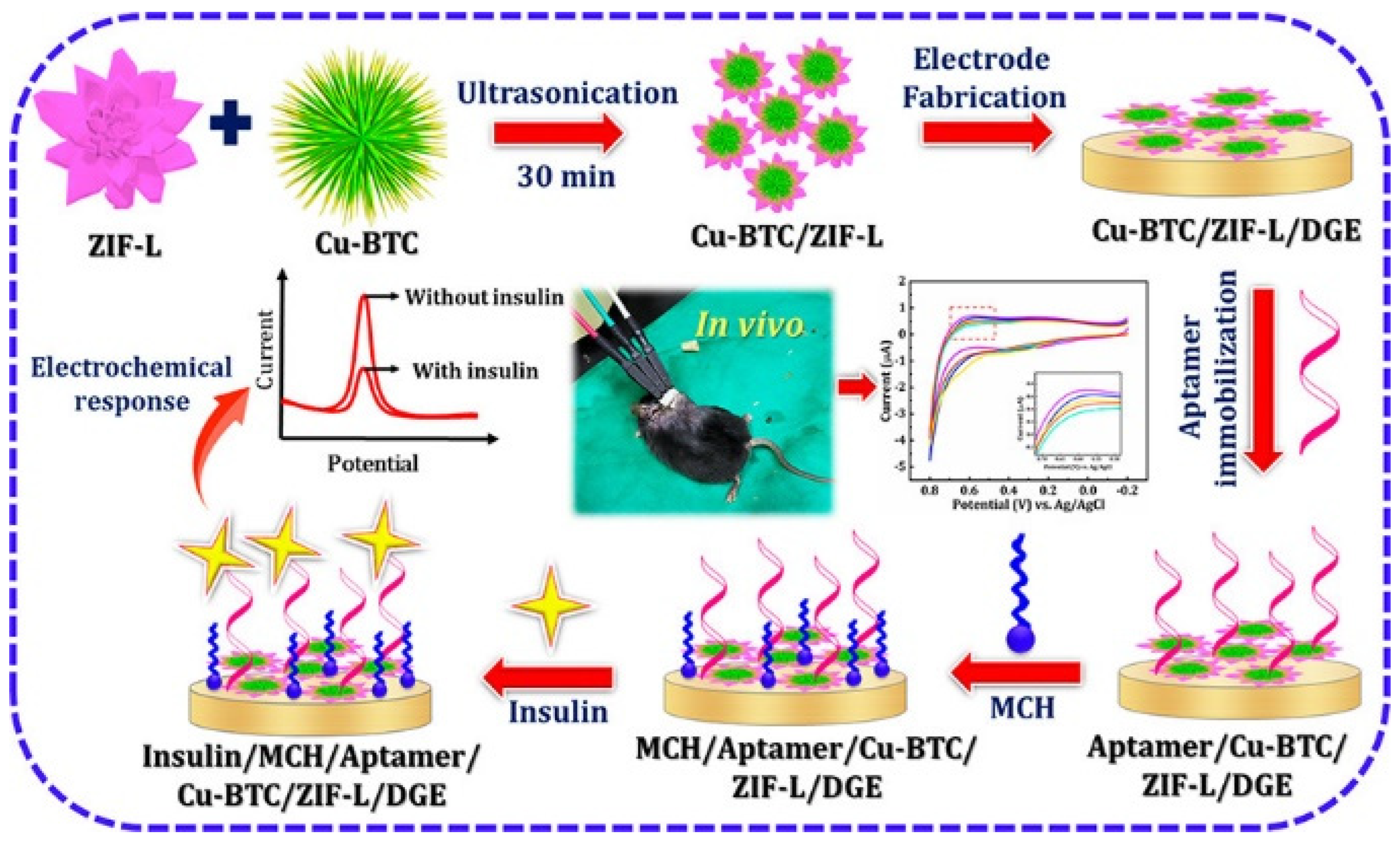
Sakthivel et al. synthesized a composite of two MOFs, such as Cu-BTC nanowire embedded 2D leaf-like ZIF, for the aptamer-based sensor to monitor real-time insulin [96] (Figure 11). Insulin is one of the vital peptide hormones whose primary role is to maintain the normal blood glucose level in the human body. Therefore, fast and sensitive monitoring of insulin levels is very much necessary. To develop the sensor, first, they synthesized CuBTC and Zn-based ZIF-L MOF separately. The morphological studies showed that CuBTC possesses 1D nanowire type morphology while ZIF shows 2D leaf-like morphology with very smooth surfaces. To make the composite, they sonicated CuBTC and ZIF-L in water for 30 min. CuBTC/ZIF-L composite appeared as CuBTC nanowire interconnected 2D leaf-like ZIF-L. To assemble the sensor, the MOF composite was drop cast on disposable Au electrodes (DGE), followed by drop casting insulin aptamer solution onto the modified DGE. Then the mercepto-1-hexanol solution was drop cast to block the non-specific active sites of DGE to minimize the insulin measurement error. Insulin sensing performance was verified by dropping different insulin concentrations on the modified electrode, and the corresponding electrochemical response was measured using differential pulse voltammetry (DPV). This aptamer-based insulin sensor showed linearity within the insulin range of 0.1 pM–5 µM and LOD of 0.027 pM. The sensitivity of the developed aptamer sensor was attributed to the high surface area, enhanced conductivity of 1D/2D MOF, and good electrocatalytic activity. Further, the sensor was grafted in diabetic and non-diabetic mice, and they observed that insulin levels could be efficiently quantified by the developed sensor.
Zhou et al. employed a bimetallic MOF architecture, i.e., Zn MOF on Zr MOF, as an aptamer sensor for sensitive detection of protein tyrosine kinase-7 (PTK7). PTK7 is a vital cancer biomarker that plays a pivotal role in regulating neural development, planar cell polarity in vertebrates, and morphogenetic cell movement during embryonic development [97]. They synthesized Zn MOF using 2-methylimidazole (2-mim) as an organic linker, whereas, for Zr MOF, they used 4′,4′′′,4′′′′′-Nitrilotris [1,1′-biphenyl]-4-carboxylic acid (H3NBB) as an organic ligand. To design the aptamer sensor, MOF solution was first dropped cast on the Au electrode, and then the aptamer solution for detecting PTK7 was immobilized. Generally, linkers in MOF possess different functional groups that offer π-π stacking, hydrogen bonding, and electrostatic interaction with negatively charged nucleic acid sequences. This helps in better immobilization of aptamer on the MOF surface. Later PTK7 sensing performance was evaluated by impedance study and differential pulse voltammetry (DPV). They observed that as the aptamer was immobilized on the MOF-modified Au electrode, impedance increased compared to bare Au or MOF-modified Au. This is because aptamer can ionize into negatively charged species, thus repelling negatively charged Fe(CN)3−/4− and inhibiting the flow of Fe(CN)3−/4− to the electrode surface, thus causing an increment in the charge transfer resistance. Further, in the presence of PTK7 solution, impedance again increased due to the G-quadruplex interaction between PTK7 and aptamer that blocks the path of the redox ions. A similar trend was observed in DPV measurement, and with the increase in PTK7 concentrations, DPV current decreased. With both electrochemical impedance spectroscopy (EIS) and DPV techniques, a linear range of PTK7 within 0.001–1 ng/mL was achieved. However, LOD obtained by the EIS technique was 0.84 pg/mL, whereas from the DPV technique was 0.66 pg mL−1. The developed aptasensor was then implemented in human serum samples, and recovery ranging from 96.6–104.6% was achieved.
Sun et al. developed a black phosphorous nanosheet (BPNS) and thionine (TH) doped 2D Cu-MOF for microRNAs [98]. MicroRNAs are essential biomarkers for many disease diagnoses, such as cancer, heart disease, and many neurological diseases. Therefore, the diagnosis of miRNAs is clinically important. They prepared 2D Cu-MOF using PTTBA as the organic ligand, and TH was doped into Cu-MOF. On the other side, BPNS was prepared by sonicating BP crystals in N-methyl pyrrolidone (NMP). BPNS/TH/Cu-MOF was prepared by slowly adding BPNS dispersion solution into TH/Cu-MOF complex. To prepare the aptamer solution, they first dropped cast Nafion solution on GCE, and then TH/Cu-MOF complex solution was dropped. After drying, ferrocene (Fc) coordinated aptamer solution was dropped on TH/Cu-MOF modified GCE. In the presence of miR3123, due to the formation of a particular complex between Fc aptamer and miR3123, Fc concentration decreased, leading to a decrease in IFc. On the other side, current due thionine ITH didn’t change much. Thus, the ratio of IFc/ITH was utilized as a parameter to evaluate the sensing performance. This strategy led to the selective determination of miR3123, and the sensor showed a wide range linearity of 2 pM–2 µM with a LOD of 0.3 pM.

4.3. Immunosensor

Immunosensors are generally based on antibody-antigen reaction, which is clinically important for early diagnosis of disease-related proteins. Not only the high surface area and conductivity of 2D MOFs, but post-synthesis modifications of 2D MOFs also help to introduce selective molecular recognition element that helps to enhance both selectivity and sensitivity. Other advantages of 2D MOFs are the reduced thickness (~nm) that expedites the electron and mass transport, exposed metal sites serving as the active catalytic sites and their lateral dimension, which permits efficient immobilization of the molecules, and surface-active reactions. Owing to these attractive properties, 2D MOFs serve as a potential candidate for electrochemical immunosensor.
Ahmadi et al. developed an electrochemical immunosensor for detecting cardiac troponin I (CTnI) based on zinc-based 2D Zn-Tetrakis (4-carboxyphenyl) porphyrin (TCPP) MOF functionalized with Fe3O4-COOH and thionine labeled with anti-CTnI monoclonal antibody (Ab1-Zn-MOF/Fe3O4-COOH/Thi) [99] (Figure 12). CTnI is an important biomarker for the early diagnosis of many cardiovascular diseases, especially acute myocardial infarction (AMI). The level of CTnI in healthy humans usually lies below 0.04 ng/mL, while its level above 0.1 ng/mL indicates an increased risk of heart failure. Therefore, it prioritizes the development of a fast and sensitive sensor for CTnI. In the above-said immunosensor, an Au screen printed electrode (SPE) modified with CTAB/DES (deep eutectic solvent of ChCl, urea) and Ab2 (anti-CTnI polyclonal antibody) was used as biosensing surface. The formation of the sensing device was confirmed through the EIS study. Further electrochemical sensing performances were carried out by the DPV technique. DPV response of the developed immunosensor showed an increment in current as the CTnI concentration increased from 0.04 ng/mL to 50 ng/mL. The sensor showed two linear ranges of CTnI: (a) 0.04–0.7 ng/mL and (b)1–50 ng/mL. Consequently, the sensor exhibited a lower LOD and sensitivity of 0.0009 ng/mL and 5.2 µA ng mL−1 cm−2, respectively.
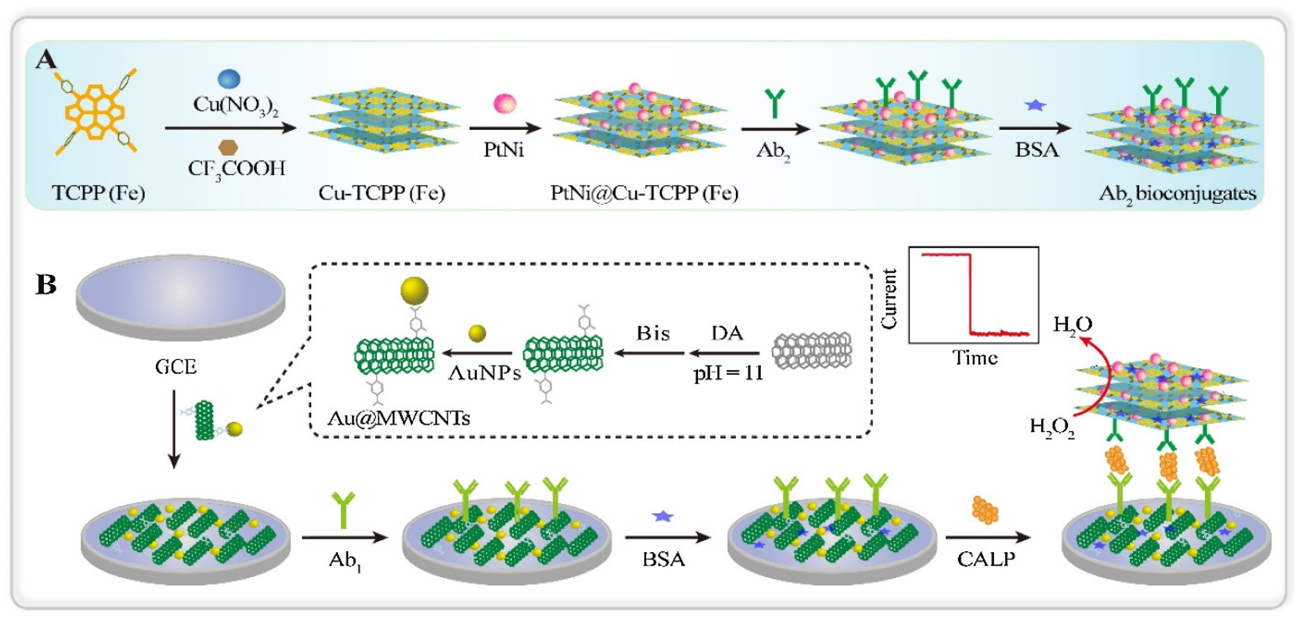
Dong et al. prepared a nonenzymatic calprotectin (CALP) immunosensor based on PtNI NPs functionalized Cu-TCPP(Fe) nanosheets [100] (Figure 13). CALP is a calcium-zinc binding protein that is related to the inflammatory response. It is also a serum biomarker for melioidosis disease. In this work, 2D Cu-TCPP(Fe) nanosheets are employed since, due to nanometer thickness, it allows rapid mass transport. Besides, nanosheets also offer excellent catalytic activity and allow high surface active reactions. Additionally, to enhance conductivity further and prevent the accumulation of nanosheets, PtNi nanospheres were immobilized. Here, antibody (Ab2) conjugated Ab2/PtNi-CuTCPP(Fe) was utilized as a signaling molecule, and Ab1 and Au/MWCNT functionalized GCE as the sensing surface. The increasing trend of amperometric current was observed with increasing CALP concentrations that depicted a potential for enzyme-free detection. This is attributed to the excellent electrocatalytic activity of PtNi/Cu-TCPP(Fe) hybrid nanosheets towards the reduction of H2O2. This developed immunosensor showed a linear range of CALP concentrations within 200 fg/mL–50 ng/mL and a low detection limit of 137.7 fg/mL.
Some of the recent reports on 2D MOF-based electrochemical sensors are summarized in Table 3.
From the above overview, it is understood that 2D MOFs demonstrate appealing properties such as high surface area, flexible porosity, and tunable functionalities, which are advantageous for electrochemical biosensing applications. However, limitations like structural stability and conductivity should be addressed. Structural stability is one of the major concerns for 2D MOF-based sensors. Since electrochemical sensing is done in the aqueous medium, it is essential to design chemically stable 2D MOFs. Therefore, a systematic study on chemical stability with varying functionalities of MOF is required. Further, screening of suitable metal nodes and organic ligands is needed to design chemically stable MOF nanosheets. In this case, theoretical investigations correlating organic ligands and their structural stability in different chemical environments may provide insight into designing such 2D MOF sheets. Although 2D MOFs are more conductive than their bulk counterparts, the conductivity can be further improved by designing redox active MOFs. Another approach to improve conductivity is adjusting the pore aperture. For a conductive 2D MOF, electrocatalytic active centers nearer to each other can accelerate the charge transport through the framework. Therefore, the minimum pore size in the 2D MOF skeleton should be large enough to facilitate the diffusion of ions during electrochemical sensing. At the same time, larger pore sizes are also not always beneficial since they may hinder electron transport across the redox active centers due to increased distance between the active metal sites. Additionally, larger pore sizes will also affect the selectivity of the sensor. Therefore, modulating pore size is necessary to make MOF nanosheets intrinsically conductive and selective towards specific analytes. Alternatively, post-synthesis modification, such as exchanging functional groups, can enhance the stability, conductivity, and surface reactivity of the MOF sheets. As most of the MOF nanosheet-based sensors are employed in the alkaline medium (pH 12), it is necessary to tune the functionalities of 2D MOF for neutral pH (pH 7.4, compatible human serum or blood sample) sensing. Lastly, in-situ spectroscopic techniques (Raman spectroscopy, Fourier transform infrared spectroscopy (FTIR)) need to be performed to understand the MOF-analyte interaction for electrochemical sensing better.

5. 2D MOFs for Optical Biosensors

2D MOFs have also been explored in optical biosensors, although not as extensively as electrochemical biosensors. Optical sensors broadly involve principles of colorimetry, luminescence, surface-enhanced Raman scattering (SERS), and surface plasmon resonance (SPR) [82,86,114]. Detection approaches with and without the use of enzymes can also be found in these sensors. Optical sensors claim to alleviate the drawbacks of electrochemical sensors, such as invasive sample acquisition and signal drifts during in vivo sensing. Colorimetric sensors involve a color change in response to the analyte, whereas luminescence-based sensors exhibit enhancement (turn-on) or quenching of fluorescence in the presence of the analytes [84,115]. For biological analytes, the majority of these sensors utilize MOFs that mimic enzyme action (nanozymes) as these are more stable, cheaper, and suitable in harsh environments as compared to natural enzymes [116]. Such MOFs typically involve porphyrin-based ligands like TCPP(Fe), hemin, etc. [117]. Surface plasmon resonance (SPR) based sensors utilize the change in refractive index occurring due to the interaction of analytes on a metal sensor surface, usually gold [118]. Surface-enhanced Raman scattering (SERS) based sensors involve amplifying the Raman signal of an analyte interacting with the surface plasmon of metals (Au, Ag, etc.) [119].
The colorimetric technique is one of the most straightforward optical sensing strategies, which can provide a visual cue of the occurrence of a reaction between a specific agent and the analyte of interest. The color intensity gives a quantitative measure of the concentration of the analyte in the sensing medium. Zeng et al. [84] employed a 2D Cu-TCPP(Fe) based colorimetric sensor for the quantitative detection of proteins. They demonstrated the sensor using a model analyte- carcinoembryonic antigen (CEA), which is useful for the early detection of colorectal cancer. The MOF nanosheets were modified with Au NPs and DNA aptamers to improve their performance. The Au NPs provide binding sites for the DNA and enhances the enzyme-like activity of the MOF, whereas the DNA aptamer specifically binds to the analyte. For colorimetric sensing, they utilized a 96-well plate filled with CEA antibodies. When the CEA protein is added to the well, it gets captured by the antibody. Further, on the addition of the MOF composite, it binds to the target owing to the specific recognition by the aptamer. This leads to the formation of colored products in the presence of tetramethylbenzidene-hydrogen peroxide (TMB-H2O2) chromogenic substrate, as the MOF has strong horseradish peroxidase (HRP)-like activity. The color change provides a visual cue for the qualitative presence of the protein, whereas the quantitative analysis can be done by measuring the corresponding absorbance wavelength (450 nm). The DNA/Au NP/Cu-TCPP(Fe) sensor showed strong HRP-mimicking activity and excellent selectivity. It exhibited a LOD of 0.742 pg/mL in the linear range of 1 pg/mL to 1000 ng/mL and showed excellent sensing performance in human serum samples.
Luminescence-based sensors offer better sensitivity and lower detection limits than colorimetric techniques and are more popular. These sensors employ the turn-on or -off mechanism (fluorescence enhancement or quenching, respectively) occurring in response to an analyte, with the fluorescence intensity giving a measure of the analyte concentration [115]. The stimuli for the luminescence may be light (fluorescence/photoluminescence), electric current (electroluminescence), chemical reaction (chemiluminescence), an electrochemical reaction (electrochemiluminescence), etc. The fluorescence properties (quenching/enhancement) can be tuned based on the ligand, metal nodes, or guest molecules. Luminescence in MOFs primarily arises from the metal centers (ex.: lanthanides), ligands (ex.: porphyrins), metal-ligand interaction, or additional guest molecules (ex.: fluorescent dyes, quantum dots, lanthanide ions, luminescent complexes) within the MOFs [120].
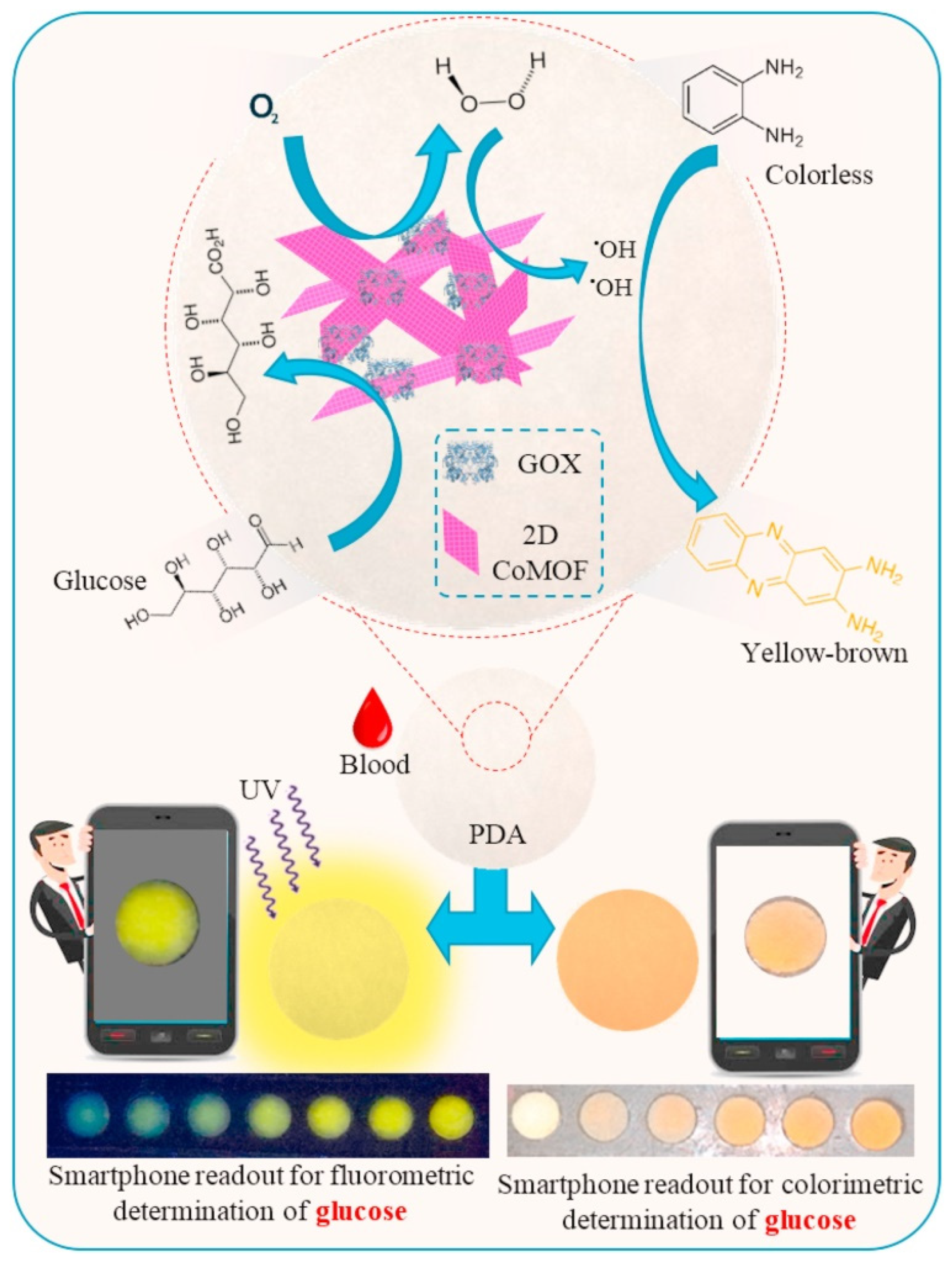
Al Lawati et al. [121] designed a paper-based colorimetric/fluorometric diagnostic device for glucose monitoring by using cobalt-terephthalate MOF (CoMOF) nanosheets acting as the catalyst and enzyme supporting layer (Figure 14). The 2D CoMOF consisted of thin, layered nanosheets of similar thickness, good BET surface area (193.6 m2/g), excellent stability (in aqueous solution), and abundant active sites exposed on its surface. The authors utilized the change in color occurring during the reaction between H2O2 (produced by GOx-assisted oxidation of glucose) and o-phenylenediamine (OPD) to quantify glucose in human blood samples. The oxidized form of OPD (2,3-diaminophenazine) also emitted fluorescence that was proportional to the glucose concentration. The colorimetric and fluorometric signals thus produced were detected by a smartphone-based readout. The sensor exhibited a linear response in the range of 50 µM–15 mM with a LOD of 16.3 µM and 3.2 µM for colorimetric and fluorometric sensing, respectively. The presence of the CoMOF nanosheets served a dual purpose. It catalyzed the peroxide dissociation to hydroxyl radicals as well as provided stable support for the GOx. The GOx exhibited better stability (~60 days) and enhanced glucose oxidation ability (improved reaction speed and sensitivity) in the presence of the MOF, as compared to the presence of horseradish peroxidase (HRP, typically used catalyst). Apart from these, the developed sensor showed a fast response (<1 min), high sensitivity (better than HRP-based systems), high specificity (due to GOx), and excellent real sample recoveries (>95%). In a different example, Qiu et al. [34] developed a multiplex fluorometric sensor for the simultaneous detection of different pathogenic genes by using ultrathin Cu-TCPP nanosheets. The MOF was found to be highly selective towards ssDNA but not dsDNA. This quenches the fluorescence of the labeled DNA. The fluorescence of the label (Texas Red, Cy3, or FAM) is restored when the labeled duplex binds to the target DNA (DNAs of Salmonella enterica, Listeria monocytogenes and Vibrio parahemolyticus used here). Picomolar detection was achieved for the gene segments of all the pathogens. Another interesting work by Liu et al. [81] investigated the effect of pore size and number of layers in 2D cobalt(II)-porphyrin MOF (Co-TTPP MOF, TTPP- 5,10,15,20-tetrakis [4′-(terpyridinyl)phenyl]porphyrin) on sensing DNA by fluorescence method. They precisely controlled the number of layers (1–15 layers) in the MOF by using the Langmuir-Blodgett synthesis technique. They found that highly sensitive DNA detection is achieved for MOFs with an appropriate pore size and an optimum number of layers. At the optimum condition (10 layers), the sensor showed a LOD of 0.1 nM with high selectivity and multiplexing capabilities.
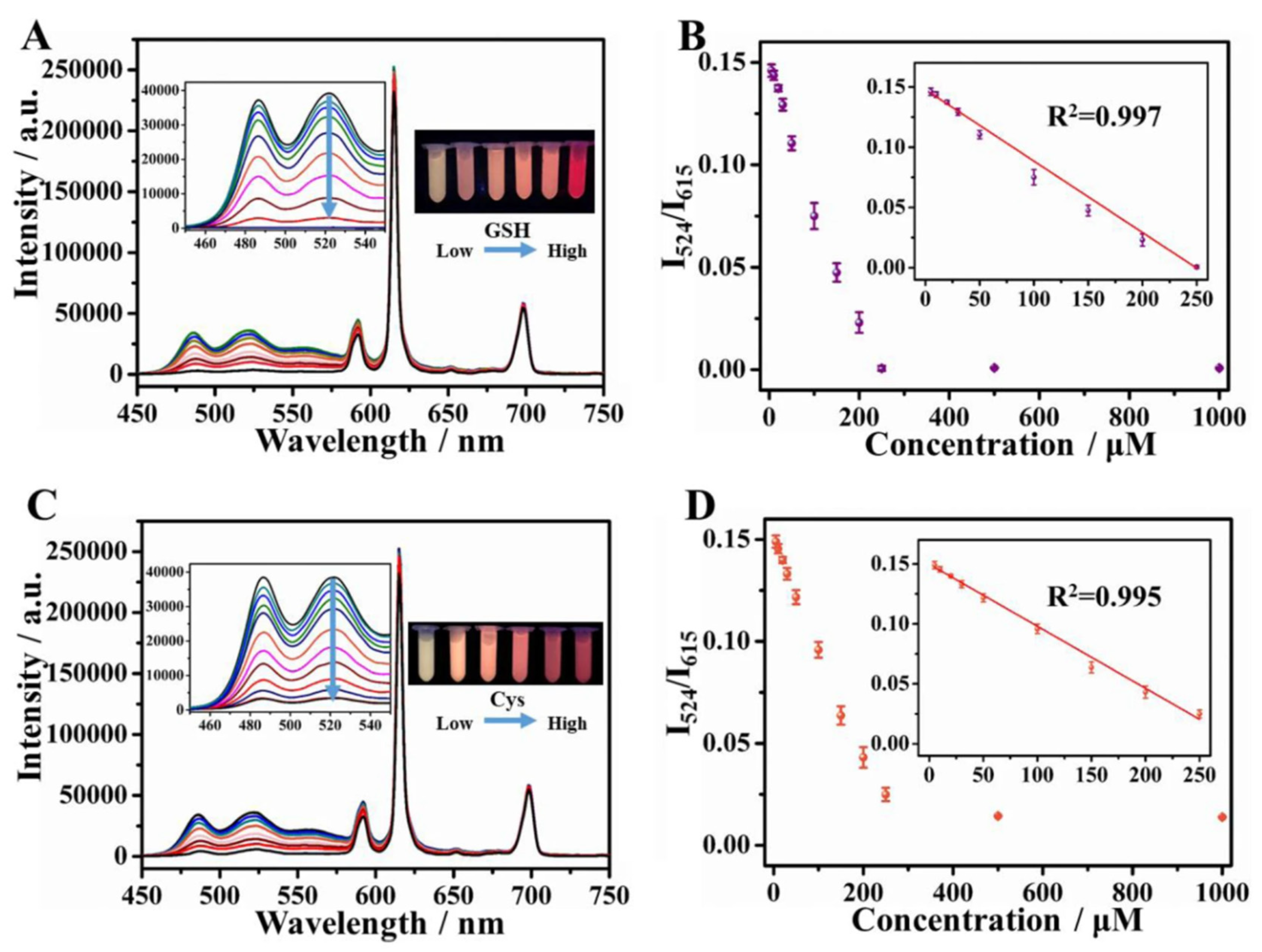
Recently, ratiometric fluorescent sensing has emerged as an effective sensing method as it can evade environmental interferences and manual errors compared to sensing with a single fluorescence intensity. Chen et al. [83] developed a ratiometric fluorescence sensor for the detection of H2O2 and glucose based on water-soluble MIL-53(Al) MOF nanosheets functionalized with amine groups. These nanosheets exhibited fluorescence at 433 nm, which was quenched with the addition of H2O2 to the OPD-HRP system. The OPD oxidation also gave rise to a new fluorescence peak at 564 nm. Hence, they utilized the ratio of the intensities of these fluorescence peaks to detect the peroxide and glucose. They observed LODs of 26.49 nM and 0.041 µM in the linear ranges of 0.5–50 µM and 4–42 µM, respectively, for H2O2 and glucose. Their glucose sensor also exhibited excellent recovery (>97%) in human serum samples. In another work [122], Ni-MOFs were utilized as a stable scaffold for incorporating a luminescent center (Eu3+) (Figure 15). The resulting dual-emitting (emission from Eu3+ and ligand) MOF was used to fabricate a ratiometric fluorescence-based sensor for detecting cysteine (Cys) and glutathione (GSH). The Eu@MOF was also modified with silver (Ag) which lowers the energy transfer efficiency from ligand to Eu3+, resulting in high ligand fluorescence. In the presence of the analyte, the Ag+ ions bind to the –SH groups in them, weakening its influence on the energy transfer process in the MOF. This leads to quenching of the ligand luminescence. The color change was observable by the naked eye. High sensitivity in a wide linear range of 5–250 µM was reported. The LODs obtained were 0.20 µM and 0.17 µM for Cys and GSH, respectively. The sensor also showed good recovery values (92–105%) in human serum samples.
Luminescence sensors employing other excitation events have also been reported. One example is chemiluminescence sensors which involve a chemical reaction that emits light. This method provides ultra-low detection limits with wide detection ranges. Zhu et al. [123] developed a chemiluminescence-based sensor employing cobalt-tetrakis(4-carboxyphenyl)porphyrin (Co-TCPP(Fe)) MOF nanosheets labeled with GOx and luminol for the detection of glucose in human urine. The MOF displayed peroxidase-like activity, which was utilized to catalyze hydrogen peroxide decomposition. The glucose undergoes oxidation in the presence of GOx to form H2O2, which gets decomposed by the 2D MOF, triggering the oxidation of luminol to produce a chemiluminescence signal. The sensor exhibited glucose detection in the linear range of 32–5500 µg/L with a LOD of 10.667 µg/L, good selectivity, and acceptable recoveries (89–121%) in different samples. Another technique that offers high sensitivity and reduced background signals is electrochemiluminescence (ECL), which utilizes light emitted during an electrochemical reaction to detect analytes. These sensors usually exploit MOFs to provide a platform that can efficiently incorporate suitable ECL reagents without particle aggregation. For instance, Wang et al. [124] reported luminol-Ag NPs loaded Co/Ni-MOF nanosheet microflowers for the detection of alpha-fetoprotein. The MOF nanosheets efficiently incorporated the ECL agent, had high catalytic activity, and resisted the aggregation of the nanoparticles. The sensor exhibited a low LOD of 0.417 pg/mL and good performance in human serum samples. The use of ECL reagents as ligands has also been reported. Yan et al. [114] synthesized nanosheets of a zinc-based MOF (RuMOF) using an ECL luminophore as the organic ligand tris(4,4′-dicarboxylic acid-2,2′-bipyridyl)ruthenium-(II) dichloride (Ru(bpy)32+) for ultrasensitive, electrochemiluminescent (ECL) sensing of cardiac troponin I (cTnI). Using Ru(bpy)32+ as a ligand instead of as an ECL guest in typical carriers (silica, 3D MOFs, etc.) avoids the problems associated with the leakage of the luminophore and tedious immobilization steps. Additionally, the nanosheet structure improved the ECL efficiency and posed as an excellent matrix for antibody immobilization. The sensor was fabricated employing sandwich-type immunological recognition and exhibited signal-on ECL behavior. It showed excellent sensitivity in a wide linear range of 1 fg/mL–10 ng/mL with a LOD of 0.48 fg/mL. The sensor also showed excellent recovery in human serum samples (95.14–104.75%), good selectivity, and stability for cTnI detection.
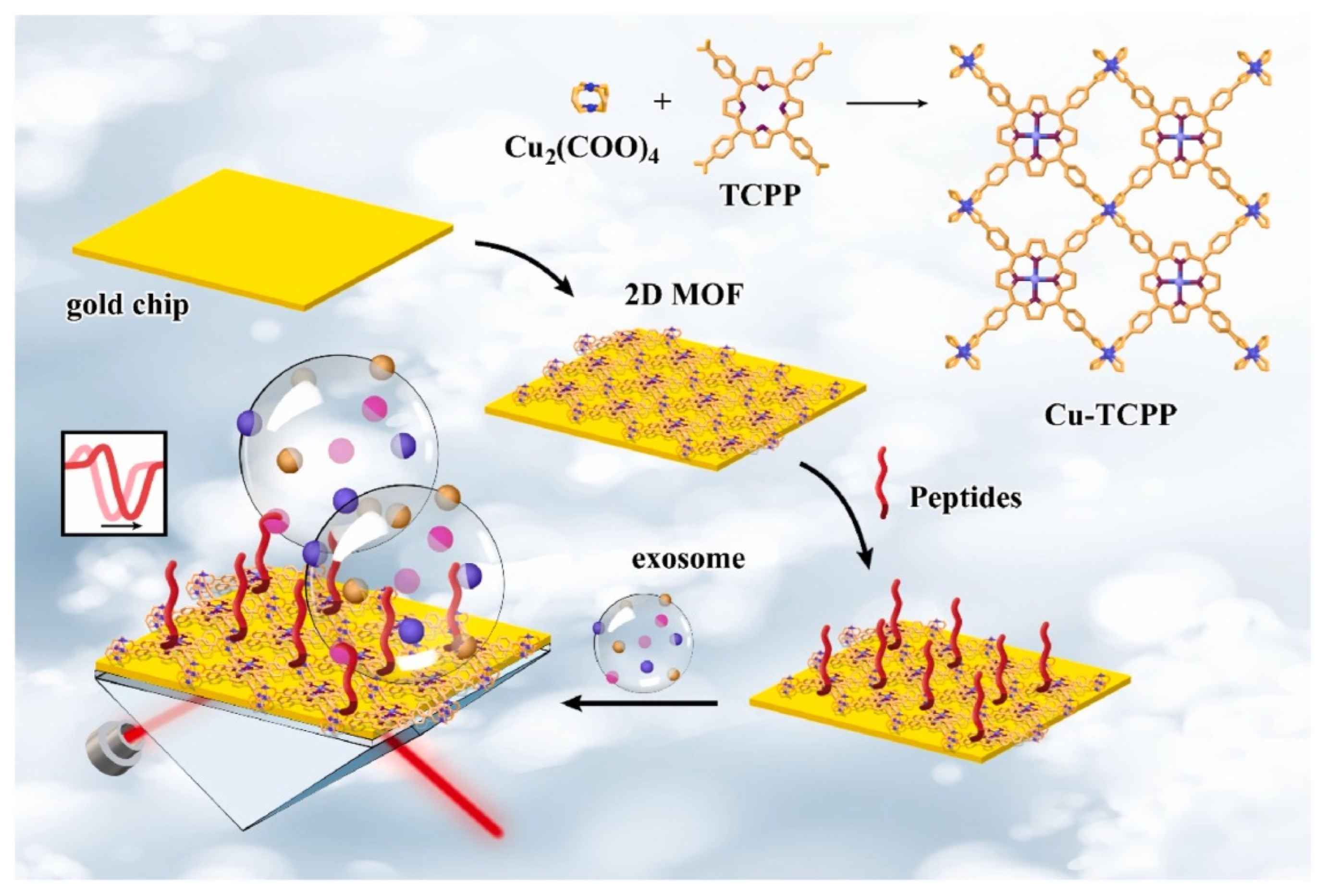
Surface plasmon resonance-based sensors are promising for the ultra-sensitive detection of different analytes. Compared to electrochemical, colorimetric, or luminescent sensors, SPR sensors have the advantage of being simple, label-free, and offering real-time assay. Wang et al. [125] developed an SPR-based sensor to detect programmed death ligand-1 (PD-L1) exosomes using a 2D Cu-TCPP MOF-modified gold chip (Figure 16). They observed enhanced performance for the MOF-modified gold chip compared to the bare one (refractive index (RI) sensitivity increased by 39.57°/RIU after modifying with MOF). The 2D MOF improved the excitation electric field and SPR of the sensing interface owing to its high electrical conductivity, charge mobility, and high-efficiency photogenerated carriers. The signal output was enhanced due to the increased incident light absorption, as the MOF has excellent photoelectric properties. The ordered lamellar arrangement of the MOFs also promoted the coupling of its surface plasmon wave with the SPR of the gold. The sensor also exhibited more signal molecule capture. This was due to the combined effect of the high surface area (more active sites for analyte binding) and the porphyrin ligand structure (affinity to carbon-based biomolecules). The proposed sensor achieved a LOD of 16.7 particles/mL and an excellent recovery of 93.43–102.35% in human serum samples.
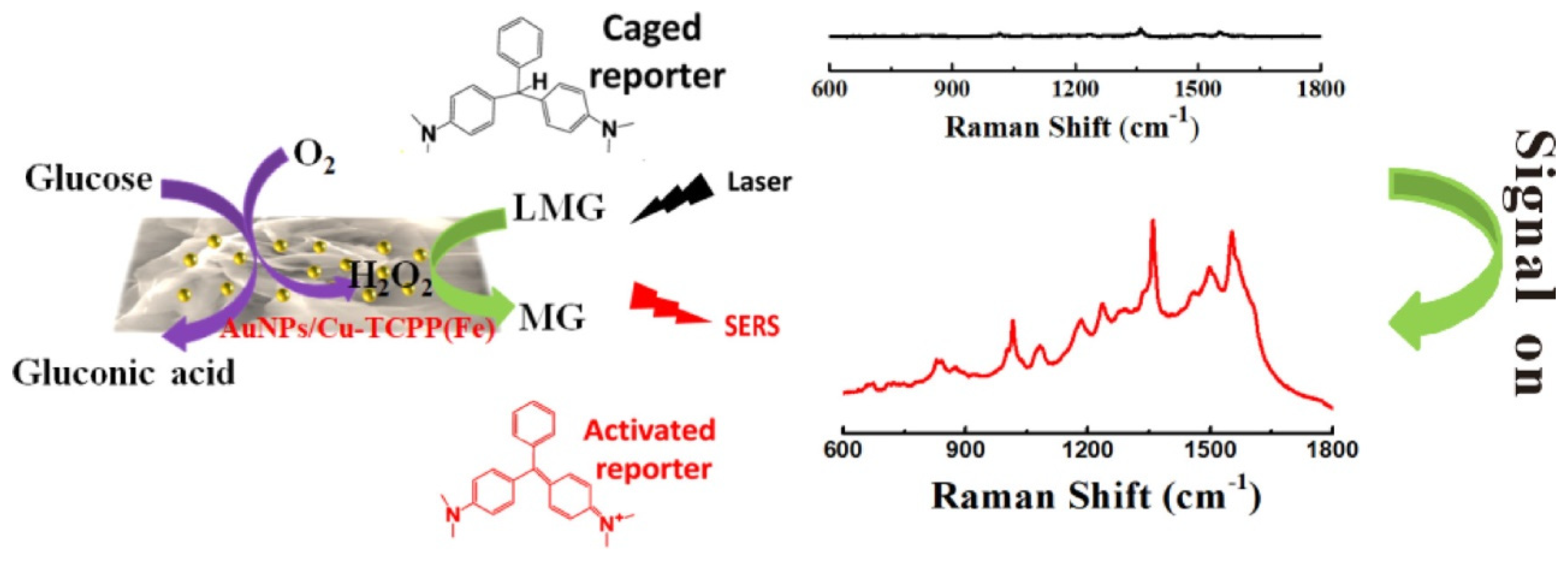
SERS-based biosensors utilize the enhancement of Raman responses of analyte molecules on interaction with localized surface plasmons. It offers ultra-sensitive detection of analytes in trace concentrations (up to single molecule detection). It also provides multiplex detection and excellent detection in biological matrixes. Hu et al. [82] employed 2D porphyrinic MOF (Cu-TCPP(Fe)) for a surface-enhanced Raman scattering (SERS) based non-enzymatic glucose assay (Figure 17). They modified the MOF (with peroxidase-like activity) surface with gold nanoparticles, which had GOx mimicking ability. The combined effect of the Au/MOF catalyzed the reaction of glucose to H2O2 and further the oxidation of leucomalachite green (Raman inactive) to malachite green (Raman active), which produced a Raman signal. A low glucose concentration of 0.16 mM could be detected using this sensor. The selectivity of the sensor was attributed to the gold nanoparticles which exhibited catalysis towards the glucose molecules.
2D MOFs have also been utilized in biosensors based on a photoelectrochemical approach. One of the latest works [85] in this field involves Au NP and aptamer-loaded Yb-TCPP nanosheets to selectively detect SARS-CoV-2. The aptamer bound selectively to the S protein on the virus surface, whereas the MOF and the Au NP were the photoactive components. The Au nanoparticles helped in enhancing the photoactivity of the Yb-TCPP nanosheets owing to the plasmon-induced resonance energy transfer (PIRET) and hot-electron transfer between them. The developed sensor could detect the S protein in a linear range of 0.5–8 µg/mL with a LOD of 72 ng/mL.
Recent literatures employing 2D MOFs for optical biosensors are summarized in Table 4.
In summary, optical sensors employing 2D MOFs exhibit low detection limits and excellent sensitivities in a wide detection range. The ability to tune the optical properties based on the ligand/nodes/guest molecules provides an innovative platform for developing sensors tailored for specific analytes. The high surface area boosts the sensitivity, whereas the specific pore sizes enhance the selectivity toward analytes. The ability of some MOFs to act as nanozymes enables the development of non-enzymatic sensors, which eradicates the drawbacks associated with the use of enzymes in sensors. Optical sensors are non-invasive and hence applicable for in vivo applications. However, elaborate studies on the biocompatibility of 2D MOFs are required before practical use.

6. Conclusions and Future Perspectives

In conclusion, this review provides an extensive overview of the synthesis and properties of 2D MOFs and their applications in optical and electrochemical biosensors. The first section of the review deals with the unique properties of 2D MOFs compared to their bulk counterparts, such as conductivity, surface area, flexibility, mechanical strength, etc. Later, the different synthesis techniques of 2D MOF, such as top-down and bottom-up methods, have been discussed briefly. The top-down synthesis approach deals with the delamination of layers from the 3D MOF through different physical or chemical methods. On the other side, the bottom-up approach involves the growth of MOF from a metal source and organic ligand in a chemical approach. Although the top-down approach has various advantages, it also suffers drawbacks concerning yield and stability. The bottom-up approach offers more scope for controllable growth of MOF nanosheets with tunable morphology, porosity, and surface by tuning synthesis parameters and precursor compositions. Therefore, a wise choice of the synthesis method is essential for successfully developing 2D MOFs.
This review also summarizes the 2D MOFs implemented in optical and electrochemical biosensors. MOF nanosheets possess exciting properties such as high surface area, which provides abundant sites for biomolecule adsorption that is beneficial for both optical and electrochemical sensors. 2D MOF-based optical sensors are based on different optical phenomena such as luminescence, SPR, fluorescence, etc., discussed in this review. High surface area is also advantageous for enhancing mass transfer and thus contributes to the enhanced catalytic activity of the 2D MOF-based electrochemical sensors. Besides, specific pore dimensions can easily be tuned in MOF sheets which further improves the selectivity of the electrochemical sensor. The role of 2D MOF in electrochemical sensors such as bio-analyte (glucose, DA, UA, H2O2, etc.) sensors, nucleic acid sensors, and immunosensors are reviewed in this work.
Although 2D MOFs have unique properties to be applied as biosensors, a few challenges must be addressed for their widespread implementations (Figure 18).
  • One of the major issues with MOFs is their stability in different solution media. Metal centers and organic linkers in MOF nanosheets are very sensitive to the pH as well as the functional groups present in the analyte solution.
  • Although 2D MOFs have shown better conductivity than 3D MOFs, they still suffer from poor conductivity when utilized for sensing without any modifications.
  • Despite several reports, the growth mechanism of 2D MOFs is still poorly understood. Apart from this role of MOF sheets in biosensing applications and the interaction between MOF and biomolecules are not well established.
Despite the above-mentioned challenges and due to the attractive properties of 2D MOFs, they are proposed to be potential candidates for optical and electrochemical biosensing applications. Optical and electrochemical detection of bioanalytes using MOF sheets are still in the infant stage and requires extensive research to investigate the role of MOFs. Suitable synthesis methods with high yield, good stability, and high-quality MOF sheets need to be probed and developed in the future. From the sensing perspective, MOF-biomolecule interaction needs to be thoroughly studied and investigated for the practical implementation of 2D-MOFs in optical and electrochemical biosensors.

Author Contributions

Authors A.G. and S.F.T.K. contributed equally to this manuscript–Conceptualization, Resource collection, Writing–original draft, Writing–review and editing, Visualization S.R.—Supervision. All authors have read and agreed to the published version of the manuscript.

Funding

This research received no external funding.

Institutional Review Board Statement

Not applicable.

Informed Consent Statement

Not applicable.

Data Availability Statement

No new data were created or analysed in this study. Data sharing is not applicable to this article.

Acknowledgments

Authors acknowledge the Indian Institute of Technology Madras (IITM), India and Ministry of Human Resources and Development, Government of India for financial support. One of the authors, thanks the Department of Science and Technology (DST) for the financial support to establish the Nano Functional Materials Technology Centre (NFMTC) through SR/NM/NAT/02−2005 project.

Conflicts of Interest

The authors declare no conflict of interest.

References

  1. Zhang, H.; Cheng, H.M.; Ye, P. 2D Nanomaterials: Beyond Graphene and Transition Metal Dichalcogenides. Chem. Soc. Rev. 2018, 47, 6009–6012. [Google Scholar] [CrossRef]
  2. Catania, F.; Marras, E.; Giorcelli, M.; Jagdale, P.; Lavagna, L.; Tagliaferro, A.; Bartoli, M. A Review on Recent Advancements of Graphene and Graphene-Related Materials in Biological Applications. Appl. Sci. 2021, 11, 614. [Google Scholar] [CrossRef]
  3. Xie, H.; Li, Z.; Cheng, L.; Haidry, A.A.; Tao, J.; Xu, Y.; Xu, K.; Ou, J.Z. Recent Advances in the Fabrication of 2D Metal Oxides. iScience 2022, 25, 1–30. [Google Scholar] [CrossRef] [PubMed]
  4. Chen, Z.; Fan, Q.; Huang, M.; Cölfen, H. Synthesis of Two-Dimensional Layered Double Hydroxides: A Systematic Overview. CrystEngComm 2022, 24, 4639–4655. [Google Scholar] [CrossRef]
  5. Yin, X.; Tang, C.S.; Zheng, Y.; Gao, J.; Wu, J.; Zhang, H.; Chhowalla, M.; Chen, W.; Wee, A.T.S. Recent Developments in 2D Transition Metal Dichalcogenides: Phase Transition and Applications of the (Quasi-)Metallic Phases. Chem. Soc. Rev. 2021, 50, 10087–10115. [Google Scholar] [CrossRef] [PubMed]
  6. Xie, Z.; Meng, X.; Li, X.; Liang, W.; Huang, W.; Chen, K.; Chen, J.; Xing, C.; Qiu, M.; Zhang, B.; et al. Two-Dimensional Borophene: Properties, Fabrication, and Promising Applications. Research 2020, 2020, 2624617. [Google Scholar] [CrossRef]
  7. Vinoth, S.; Shalini Devi, K.S.; Pandikumar, A. A Comprehensive Review on Graphitic Carbon Nitride Based Electrochemical and Biosensors for Environmental and Healthcare Applications. TrAC-Trends Anal. Chem. 2021, 140, 116274. [Google Scholar] [CrossRef]
  8. Li, Y.Z.; Fu, Z.H.; Xu, G. Metal-Organic Framework Nanosheets: Preparation and Applications. Coord. Chem. Rev. 2019, 388, 79–106. [Google Scholar] [CrossRef]
  9. Liu, X.; Zhang, Y.W. Thermal Properties of Transition-Metal Dichalcogenide. Chin. Phys. B 2018, 27, 034402. [Google Scholar] [CrossRef]
  10. Ronchi, R.M.; Arantes, J.T.; Santos, S.F. Synthesis, Structure, Properties and Applications of MXenes: Current Status and Perspectives. Ceram. Int. 2019, 45, 18167–18188. [Google Scholar] [CrossRef]
  11. Butova, V.V.; Soldatov, M.A.; Guda, A.A.; Lomachenko, K.A.; Lamberti, C. Metal-Organic Frameworks: Structure, Properties, Methods of Synthesis and Characterization. Russ. Chem. Rev. 2016, 85, 280–307. [Google Scholar] [CrossRef]
  12. Deng, H.; Grunder, S.; Cordova, K.E.; Valente, C.; Furukawa, H.; Hmadeh, M.; Gándara, F.; Whalley, A.C.; Liu, Z.; Asahina, S.; et al. Large-Pore Apertures in a Series of Metal-Organic Frameworks. Science 2012, 336, 1018–1023. [Google Scholar] [CrossRef] [PubMed] [Green Version]
  13. Yuan, S.; Zou, L.; Qin, J.S.; Li, J.; Huang, L.; Feng, L.; Wang, X.; Bosch, M.; Alsalme, A.; Cagin, T.; et al. Construction of Hierarchically Porous Metal-Organic Frameworks through Linker Labilization. Nat. Commun. 2017, 8, 15356. [Google Scholar] [CrossRef] [PubMed] [Green Version]
  14. Li, H.; Li, L.; Lin, R.-B.; Zhou, W.; Zhang, Z.; Xiang, S.; Chen, B. Porous Metal-Organic Frameworks for Gas Storage and Separation: Status and Challenges. EnergyChem 2019, 1, 100006. [Google Scholar] [CrossRef]
  15. Ghosh, A.; Sana Fathima, T.K.; Ramaprabhu, S. Effect of Coordinated Solvent Molecules in Cu-MOF on Enzyme Free Sensing of Glucose and Lactate in Physiological PH. J. Electrochem. Soc. 2022, 169, 057524. [Google Scholar] [CrossRef]
  16. Yang, D.; Gates, B.C. Catalysis by Metal Organic Frameworks: Perspective and Suggestions for Future Research. ACS Catal. 2019, 9, 1779–1798. [Google Scholar] [CrossRef]
  17. Zhao, Y.; Song, Z.; Li, X.; Sun, Q.; Cheng, N.; Lawes, S.; Sun, X. Metal Organic Frameworks for Energy Storage and Conversion. Energy Storage Mater. 2016, 2, 35–62. [Google Scholar] [CrossRef]
  18. Freund, R.; Zaremba, O.; Arnauts, G.; Ameloot, R.; Skorupskii, G.; Dincă, M.; Bavykina, A.; Gascon, J.; Ejsmont, A.; Goscianska, J.; et al. The Current Status of MOF and COF Applications. Angew. Chem. Int. Ed. 2021, 60, 23975–24001. [Google Scholar] [CrossRef]
  19. Hendon, C.H.; Rieth, A.J.; Korzyński, M.D.; Dincǎ, M. Grand Challenges and Future Opportunities for Metal-Organic Frameworks. ACS Cent. Sci. 2017, 3, 554–563. [Google Scholar] [CrossRef]
  20. Zhao, M.; Huang, Y.; Peng, Y.; Huang, Z.; Ma, Q.; Zhang, H. Two-Dimensional Metal-Organic Framework Nanosheets: Synthesis and Applications. Chem. Soc. Rev. 2018, 47, 6267–6295. [Google Scholar] [CrossRef]
  21. Li, H.; Zhang, L.; Mao, Y.; Wen, C.; Zhao, P. A Simple Electrochemical Route to Access Amorphous Co-Ni Hydroxide for Non-Enzymatic Glucose Sensing. Nanoscale Res. Lett. 2019, 14, 135. [Google Scholar] [CrossRef] [PubMed] [Green Version]
  22. Wang, Q.; Ma, Y.; Jiang, X.; Yang, N.; Coffinier, Y.; Belkhalfa, H.; Dokhane, N.; Li, M.; Boukherroub, R.; Szunerits, S. Electrophoretic Deposition of Carbon Nanofibers/Co(OH)2 Nanocomposites: Application for Non-Enzymatic Glucose Sensing. Electroanalysis 2016, 28, 119–125. [Google Scholar] [CrossRef]
  23. Mohapatra, J.; Ananthoju, B.; Nair, V.; Mitra, A.; Bahadur, D.; Medhekar, N.V.; Aslam, M. Enzymatic and Non-Enzymatic Electrochemical Glucose Sensor Based on Carbon Nano-Onions. Appl. Surf. Sci. 2018, 442, 332–341. [Google Scholar] [CrossRef]
  24. Wang, F.; Chen, X.; Chen, L.; Yang, J.; Wang, Q. High-Performance Non-Enzymatic Glucose Sensor by Hierarchical Flower-like Nickel(II)-Based MOF/Carbon Nanotubes Composite. Mater. Sci. Eng. C 2019, 96, 41–50. [Google Scholar] [CrossRef]
  25. Salah, A.; Al-Ansi, N.; Adlat, S.; Bawa, M.; He, Y.; Bo, X.; Guo, L. Sensitive Nonenzymatic Detection of Glucose at PtPd/Porous Holey Nitrogen-Doped Graphene. J. Alloys Compd. 2019, 792, 50–58. [Google Scholar] [CrossRef]
  26. Gumilar, G.; Kaneti, Y.V.; Henzie, J.; Chatterjee, S.; Na, J.; Yuliarto, B.; Nugraha, N.; Patah, A.; Bhaumik, A.; Yamauchi, Y. General Synthesis of Hierarchical Sheet/Plate-like M-BDC (M = Cu, Mn, Ni, and Zr) Metal-Organic Frameworks for Electrochemical Non-Enzymatic Glucose Sensing. Chem. Sci. 2020, 11, 3644–3655. [Google Scholar] [CrossRef] [Green Version]
  27. Li, Y.; Xie, M.; Zhang, X.; Liu, Q.; Lin, D.; Xu, C.; Xie, F.; Sun, X. Co-MOF Nanosheet Array: A High-Performance Electrochemical Sensor for Non-Enzymatic Glucose Detection. Sens. Actuators B Chem. 2019, 278, 126–132. [Google Scholar] [CrossRef]
  28. Wang, F.; Hu, J.; Liu, Y.; Yuan, G.; Zhang, S.; Xu, L.; Xue, H.; Pang, H. Turning Coordination Environment of 2D Nickel-Based Metal-Organic Frameworks by π-Conjugated Molecule for Enhancing Glucose Electrochemical Sensor Performance. Mater. Today Chem. 2022, 24, 100885. [Google Scholar] [CrossRef]
  29. Liu, W.; Yin, R.; Xu, X.; Zhang, L.; Shi, W.; Cao, X. Structural Engineering of Low-Dimensional Metal–Organic Frameworks: Synthesis, Properties, and Applications. Adv. Sci. 2019, 6, 1802373. [Google Scholar] [CrossRef]
  30. Liu, C.; Li, J.; Pang, H. Metal-Organic Framework-Based Materials as an Emerging Platform for Advanced Electrochemical Sensing. Coord. Chem. Rev. 2020, 410, 213222. [Google Scholar] [CrossRef]
  31. Low, K.H.; Roy, V.A.L.; Chui, S.S.Y.; Chan, S.L.F.; Che, C.M. Highly Conducting Two-Dimensional Copper(i) 4-Hydroxythiophenolate Network. Chem. Commun. 2010, 46, 7328–7330. [Google Scholar] [CrossRef]
  32. Ohata, T.; Nomoto, A.; Watanabe, T.; Hirosawa, I.; Makita, T.; Takeya, J.; Makiura, R. Uniaxially Oriented Electrically Conductive Metal-Organic Framework Nanosheets Assembled at Air/Liquid Interfaces. ACS Appl. Mater. Interfaces 2021, 13, 54570–54578. [Google Scholar] [CrossRef]
  33. Sheberla, D.; Sun, L.; Blood-forsythe, M.A.; Er, S.; Wade, C.R.; Brozek, C.K. High Electrical Conductivity in Ni3(2,3,6,7,10,11-Hexaiminotriphenylene)2,a Semiconducting Metal-Organic Graphene Analogue. J. Am. Chem. Soc. 2014, 136, 8859–8862. [Google Scholar] [CrossRef]
  34. Qiu, Q.; Chen, H.; Ying, S.; Sharif, S.; You, Z.; Wang, Y.; Ying, Y. Simultaneous Fluorometric Determination of the DNAs of Salmonella Enterica, Listeria Monocytogenes and Vibrio Parahemolyticus by Using an Ultrathin Metal-Organic Framework (Type Cu-TCPP). Microchim. Acta 2019, 186, 93. [Google Scholar] [CrossRef]
  35. Nicks, J.; Sasitharan, K.; Prasad, R.R.R.; Ashworth, D.J.; Foster, J.A. Metal–Organic Framework Nanosheets: Programmable 2D Materials for Catalysis, Sensing, Electronics, and Separation Applications. Adv. Funct. Mater. 2021, 31, 2103723. [Google Scholar] [CrossRef]
  36. Varsha, M.V.; Nageswaran, G. Review—2D Layered Metal Organic Framework Nanosheets as an Emerging Platform for Electrochemical Sensing. J. Electrochem. Soc. 2020, 167, 136502. [Google Scholar] [CrossRef]
  37. Han, L.J.; Zheng, D.; Chen, S.G.; Zheng, H.G.; Ma, J. A Highly Solvent-Stable Metal–Organic Framework Nanosheet: Morphology Control, Exfoliation, and Luminescent Property. Small 2018, 14, 1703873. [Google Scholar] [CrossRef]
  38. He, T.; Ni, B.; Zhang, S.; Gong, Y.; Wang, H.; Gu, L.; Zhuang, J.; Hu, W.; Wang, X. Ultrathin 2D Zirconium Metal–Organic Framework Nanosheets: Preparation and Application in Photocatalysis. Small 2018, 14, 1703929. [Google Scholar] [CrossRef]
  39. Rodenas, T.; Luz, I.; Prieto, G.; Seoane, B.; Miro, H.; Corma, A.; Kapteijn, F.; Llabrés i Xamena, F.X.; Gascon, J. Metal-Organic Framework Nanosheets in Polymer Composite Materials for Gas Separation. Nat. Mater. 2015, 14, 48–55. [Google Scholar] [CrossRef]
  40. Hermosa, C.; Horrocks, B.R.; Martínez, J.I.; Liscio, F.; Gómez-Herrero, J.; Zamora, F. Mechanical and Optical Properties of Ultralarge Flakes of a Metal-Organic Framework with Molecular Thickness. Chem. Sci. 2015, 6, 2553–2558. [Google Scholar] [CrossRef]
  41. Zhang, Z.; Wang, Y.; Niu, B.; Liu, B.; Li, J.; Duan, W. Ultra-Stable Two-Dimensional Metal-Organic Frameworks for Photocatalytic H2 Production. Nanoscale 2022, 14, 7146–7150. [Google Scholar] [CrossRef]
  42. Ma, H.M.; Yi, J.W.; Li, S.; Jiang, C.; Wei, J.H.; Wu, Y.P.; Zhao, J.; Li, D.S. Stable Bimetal-MOF Ultrathin Nanosheets for Pseudocapacitors with Enhanced Performance. Inorg. Chem. 2019, 58, 9543–9547. [Google Scholar] [CrossRef]
  43. Foster, J.A.; Henke, S.; Schneemann, A.; Fischer, R.A.; Cheetham, A.K. Liquid Exfoliation of Alkyl-Ether Functionalised Layered Metal-Organic Frameworks to Nanosheets. Chem. Commun. 2016, 52, 10474–10477. [Google Scholar] [CrossRef] [Green Version]
  44. Wang, S.; Xu, D.; Ma, L.; Qiu, J.; Wang, X.; Dong, Q.; Zhang, Q.; Pan, J.; Liu, Q. Ultrathin ZIF-67 Nanosheets as a Colorimetric Biosensing Platform for Peroxidase-like Catalysis. Anal. Bioanal. Chem. 2018, 410, 7145–7152. [Google Scholar] [CrossRef]
  45. Zhao, L.; Si, D.; Zhao, Y.; Wang, L.; An, H.; Ye, H.; Xin, Q.; Zhang, Y. Copper Nanocomposite Decorated Two-Dimensional Metal Organic Frameworks of Metalloporphyrin with Peroxidase-Mimicking Activity. Colloids Surf. A Physicochem. Eng. Asp. 2022, 644, 128876. [Google Scholar] [CrossRef]
  46. Junggeburth, S.C.; Diehl, L.; Werner, S.; Duppel, V.; Sigle, W.; Lotsch, B.V. Ultrathin 2D Coordination Polymer Nanosheets by Surfactant-Mediated Synthesis. J. Am. Chem. Soc. 2013, 135, 6157–6164. [Google Scholar] [CrossRef]
  47. Zhao, M.; Wang, Y.; Ma, Q.; Huang, Y.; Zhang, X.; Ping, J.; Zhang, Z.; Lu, Q.; Yu, Y.; Xu, H.; et al. Ultrathin 2D Metal-Organic Framework Nanosheets. Adv. Mater. 2015, 27, 7372–7378. [Google Scholar] [CrossRef]
  48. Liu, X.; Shi, C.; Zhai, C.; Cheng, M.; Liu, Q.; Wang, G. Cobalt-Based Layered Metal-Organic Framework as an Ultrahigh Capacity Supercapacitor Electrode Material. ACS Appl. Mater. Interfaces 2016, 8, 4585–4591. [Google Scholar] [CrossRef]
  49. Amo-Ochoa, P.; Welte, L.; González-Prieto, R.; Sanz Miguel, P.J.; Gómez-García, C.J.; Mateo-Martí, E.; Delgado, S.; Gómez-Herrero, J.; Zamora, F. Single Layers of a Multifunctional Laminar Cu(i,Ii) Coordination Polymer. Chem. Commun. 2010, 46, 3262–3264. [Google Scholar] [CrossRef] [Green Version]
  50. Abhervé, A.; Mañas-Valero, S.; Clemente-León, M.; Coronado, E. Graphene Related Magnetic Materials: Micromechanical Exfoliation of 2D Layered Magnets Based on Bimetallic Anilate Complexes with Inserted [FeIII(Acac2-Trien)]+ and [FeIII(Sal2-Trien)]+ Molecules. Chem. Sci. 2015, 6, 4665–4673. [Google Scholar] [CrossRef]
  51. Liu, Y.; Wei, Y.; Liu, M.; Bai, Y.; Wang, X.; Shang, S.; Chen, J.; Liu, Y. Electrochemical Synthesis of Large Area Two-Dimensional Metal–Organic Framework Films on Copper Anodes. Angew. Chem. Int. Ed. 2021, 60, 2887–2891. [Google Scholar] [CrossRef]
  52. Liu, X.; Jia, Q.; Fu, Y.; Zheng, T. Exfoliation of Metal-Organic Framework Nanosheets Using Surface Acoustic Waves. Ultrason. Sonochem. 2022, 83, 105943. [Google Scholar] [CrossRef] [PubMed]
  53. Xu, H.; Gao, J.; Qian, X.; Wang, J.; He, H.; Cui, Y.; Yang, Y.; Wang, Z.; Qian, G. Metal-Organic Framework Nanosheets for Fast-Response and Highly Sensitive Luminescent Sensing of Fe3+. J. Mater. Chem. A 2016, 4, 10900–10905. [Google Scholar] [CrossRef]
  54. Ding, Y.; Chen, Y.P.; Zhang, X.; Chen, L.; Dong, Z.; Jiang, H.L.; Xu, H.; Zhou, H.C. Controlled Intercalation and Chemical Exfoliation of Layered Metal-Organic Frameworks Using a Chemically Labile Intercalating Agent. J. Am. Chem. Soc. 2017, 139, 9136–9139. [Google Scholar] [CrossRef] [PubMed]
  55. Ogihara, N.; Ozawa, Y.; Hiruta, O. A Self-Assembled Intercalated Metal-Organic Framework Electrode with Outstanding Area Capacity for High Volumetric Energy Asymmetric Capacitors. J. Mater. Chem. A 2016, 4, 3398–3405. [Google Scholar] [CrossRef] [Green Version]
  56. Li, P.Z.; Maeda, Y.; Xu, Q. Top-down Fabrication of Crystalline Metal-Organic Framework Nanosheets. Chem. Commun. 2011, 47, 8436–8438. [Google Scholar] [CrossRef]
  57. Saines, P.J.; Tan, J.C.; Yeung, H.H.M.; Barton, P.T.; Cheetham, A.K. Layered Inorganic-Organic Frameworks Based on the 2,2-Dimethylsuccinate Ligand: Structural Diversity and Its Effect on Nanosheet Exfoliation and Magnetic Properties. Dalt. Trans. 2012, 41, 8585–8593. [Google Scholar] [CrossRef]
  58. Cliffe, M.J.; Castillo-Martínez, E.; Wu, Y.; Lee, J.; Forse, A.C.; Firth, F.C.N.; Moghadam, P.Z.; Fairen-Jimenez, D.; Gaultois, M.W.; Hill, J.A.; et al. Metal-Organic Nanosheets Formed via Defect-Mediated Transformation of a Hafnium Metal-Organic Framework. J. Am. Chem. Soc. 2017, 139, 5397–5404. [Google Scholar] [CrossRef]
  59. Araki, T.; Kondo, A.; Maeda, K. The First Lanthanide Organophosphonate Nanosheet by Exfoliation of Layered Compounds. Chem. Commun. 2013, 49, 552–554. [Google Scholar] [CrossRef]
  60. Peng, Y.; Li, Y.; Ban, Y.; Jin, H.; Jiao, W.; Liu, X.; Yang, W. Metal-Organic Framework Nanosheets as Building Blocks for Molecular Sieving Membranes. Science 2014, 346, 1356–1359. [Google Scholar] [CrossRef]
  61. Tan, J.C.; Saines, P.J.; Bithell, E.G.; Cheetham, A.K. Hybrid Nanosheets of an Inorganic-Organic Framework Material: Facile Synthesis, Structure, and Elastic Properties. ACS Nano 2012, 6, 615–621. [Google Scholar] [CrossRef] [PubMed]
  62. López-Cabrelles, J.; Mañas-Valero, S.; Vitórica-Yrezábal, I.J.; Bereciartua, P.J.; Rodríguez-Velamazán, J.A.; Waerenborgh, J.C.; Vieira, B.J.C.; Davidovikj, D.; Steeneken, P.G.; van der Zant, H.S.J.; et al. Isoreticular Two-Dimensional Magnetic Coordination Polymers Prepared through Pre-Synthetic Ligand Functionalization. Nat. Chem. 2018, 10, 1001–1007. [Google Scholar] [CrossRef] [PubMed] [Green Version]
  63. Gallego, A.; Hermosa, C.; Castillo, O.; Berlanga, I.; Gõmez-García, C.J.; Mateo-Martí, E.; Martínez, J.I.; Flores, F.; Gõmez-Navarro, C.; Gõmez-Herrero, J.; et al. Solvent-Induced Delamination of a Multifunctional Two Dimensional Coordination Polymer. Adv. Mater. 2013, 25, 2141–2146. [Google Scholar] [CrossRef] [PubMed]
  64. Zhou, Y.; Yan, P.; Zhang, S.; Zhang, Y.; Chang, H.; Zheng, X.; Jiang, J.; Xu, Q. CO2 Coordination-Driven Top-down Synthesis of a 2D Non-Layered Metal–Organic Framework. Fundam. Res. 2022, 2, 674–681. [Google Scholar] [CrossRef]
  65. Liu, Y.L.; Liu, X.Y.; Feng, L.; Shao, L.X.; Li, S.J.; Tang, J.; Cheng, H.; Chen, Z.; Huang, R.; Xu, H.C.; et al. Two-Dimensional Metal–Organic Framework Nanosheets: Synthesis and Applications in Electrocatalysis and Photocatalysis. ChemSusChem 2022, 15, e202102603. [Google Scholar] [CrossRef]
  66. Nian, P.; Liu, H.; Zhang, X. Bottom-up Synthesis of 2D Co-Based Metal-Organic Framework Nanosheets by an Ammonia-Assisted Strategy for Tuning the Crystal Morphology. CrystEngComm 2019, 21, 3199–3208. [Google Scholar] [CrossRef]
  67. Dong, R.; Zhang, T.; Feng, X. Interface-Assisted Synthesis of 2D Materials: Trend and Challenges. Chem. Rev. 2018, 118, 6189–6325. [Google Scholar] [CrossRef]
  68. Li, Y.N.; Wang, S.; Zhou, Y.; Bai, X.J.; Song, G.S.; Zhao, X.Y.; Wang, T.Q.; Qi, X.; Zhang, X.M.; Fu, Y. Fabrication of Metal-Organic Framework and Infinite Coordination Polymer Nanosheets by the Spray Technique. Langmuir 2017, 33, 1060–1065. [Google Scholar] [CrossRef]
  69. Dong, R.; Pfeffermann, M.; Liang, H.; Zheng, Z.; Zhu, X.; Zhang, J.; Feng, X. Large-Area, Free-Standing, Two-Dimensional Supramolecular Polymer Single-Layer Sheets for Highly Efficient Electrocatalytic Hydrogen Evolution. Angew. Chem. Int. Ed. 2015, 54, 12058–12063. [Google Scholar] [CrossRef]
  70. Sakaida, S.; Haraguchi, T.; Otsubo, K.; Sakata, O.; Fujiwara, A.; Kitagawa, H. Fabrication and Structural Characterization of an Ultrathin Film of a Two-Dimensional-Layered Metal-Organic Framework, {Fe(Py)2[Ni(CN)4]} (Py = Pyridine). Inorg. Chem. 2017, 56, 7606–7609. [Google Scholar] [CrossRef]
  71. Schaate, A.; Roy, P.; Godt, A.; Lippke, J.; Waltz, F.; Wiebcke, M.; Behrens, P. Modulated Synthesis of Zr-Based Metal-Organic Frameworks: From Nano to Single Crystals. Chem. A Eur. J. 2011, 17, 6643–6651. [Google Scholar] [CrossRef]
  72. Choe, M.; Koo, J.Y.; Park, I.; Ohtsu, H.; Shim, J.H.; Choi, H.C.; Park, S.S. Chemical Vapor Deposition of Edge-on Oriented 2D Conductive Metal-Organic Framework Thin Films. J. Am. Chem. Soc. 2022, 144, 16726–16731. [Google Scholar] [CrossRef] [PubMed]
  73. Liu, J.; Xiao, J.; Wang, D.; Sun, W.; Gao, X.; Yu, H.; Liu, H.; Liu, Z. Construction and Photocatalytic Activities of a Series of Isostructural Co2+/Zn2+ Metal-Doped Metal-Organic Frameworks. Cryst. Growth Des. 2017, 17, 1096–1102. [Google Scholar] [CrossRef]
  74. Salcedo-Abraira, P.; Vilela, S.M.F.; Babaryk, A.A.; Cabrero-Antonino, M.; Gregorio, P.; Salles, F.; Navalón, S.; García, H.; Horcajada, P. Nickel Phosphonate MOF as Efficient Water Splitting Photocatalyst. Nano Res. 2021, 14, 450–457. [Google Scholar] [CrossRef]
  75. Oh, S.; Park, J.; Oh, M. Competitive Formation between 2D and 3D Metal-Organic Frameworks: Insights into the Selective Formation and Lamination of a 2D MOF. IUCrJ 2019, 6, 681–687. [Google Scholar] [CrossRef] [PubMed] [Green Version]
  76. Chaudhari, A.K.; Kim, H.J.; Han, I.; Tan, J.C. Optochemically Responsive 2D Nanosheets of a 3D Metal–Organic Framework Material. Adv. Mater. 2017, 29, 1701463. [Google Scholar] [CrossRef] [Green Version]
  77. Huo, Y.; Bu, M.; Ma, Z.; Sun, J.; Yan, Y.; Xiu, K.; Wang, Z.; Hu, N.; Li, Y.F. Flexible, Non-Contact and Multifunctional Humidity Sensors Based on Two-Dimensional Phytic Acid Doped Co-Metal Organic Frameworks Nanosheets. J. Colloid Interface Sci. 2022, 607, 2010–2018. [Google Scholar] [CrossRef]
  78. Nagoor Meeran, M.; Saravanan, S.P.; Shkir, M.; Karthik Kannan, S. Fabrication of Transition-Metal (Zn, Mn, Cu)-Based MOFs as Efficient Sensor Materials for Detection of H2 Gas by Clad Modified Fiber Optic Gas Sensor Technique. Opt. Fiber Technol. 2021, 65, 102614. [Google Scholar] [CrossRef]
  79. Singha Mahapatra, T.; Dey, A.; Singh, H.; Hossain, S.S.; Mandal, A.K.; Das, A. Two-Dimensional Lanthanide Coordination Polymer Nanosheets for Detection of FOX-7. Chem. Sci. 2020, 11, 1032–1042. [Google Scholar] [CrossRef] [Green Version]
  80. Lai, H.; Dai, H.; Li, G.; Zhang, Z. Rapid Determination of Pesticide Residues in Fruit and Vegetable Using Au@AgNPs Decorated 2D Ni-MOF Nanosheets as Efficient Surface-Enhanced Raman Scattering Substrate. Sens. Actuators B Chem. 2022, 369, 132360. [Google Scholar] [CrossRef]
  81. Liu, C.; Wang, T.; Ji, J.; Wang, C.; Wang, H.; Jin, P.; Zhou, W.; Jiang, J. The Effect of Pore Size and Layer Number of Metal-Porphyrin Coordination Nanosheets on Sensing DNA. J. Mater. Chem. C 2019, 7, 10240–10246. [Google Scholar] [CrossRef]
  82. Hu, S.; Jiang, Y.; Wu, Y.; Guo, X.; Ying, Y.; Wen, Y.; Yang, H. Enzyme-Free Tandem Reaction Strategy for Surface-Enhanced Raman Scattering Detection of Glucose by Using the Composite of Au Nanoparticles and Porphyrin-Based Metal-Organic Framework. ACS Appl. Mater. Interfaces 2020, 12, 55324–55330. [Google Scholar] [CrossRef] [PubMed]
  83. Chen, J.; Xu, Y.; Xu, F.; Zhang, Q.; Li, S.; Lu, X. Detection of Hydrogen Peroxide and Glucose with a Novel Fluorescent Probe by the Enzymatic Reaction of Amino Functionalized MOF Nanosheets. Anal. Methods 2021, 13, 4228–4237. [Google Scholar] [CrossRef] [PubMed]
  84. Zeng, Y.; Wang, M.; Sun, Z.; Sha, L.; Yang, J.; Li, G. Colorimetric Immunosensor Constructed Using 2D Metal–Organic Framework Nanosheets as Enzyme Mimics for the Detection of Protein Biomarkers. J. Mater. Chem. B 2022, 10, 450–455. [Google Scholar] [CrossRef] [PubMed]
  85. Jiang, Z.W.; Zhao, T.T.; Li, C.M.; Li, Y.F.; Huang, C.Z. 2D MOF-Based Photoelectrochemical Aptasensor for SARS-CoV-2 Spike Glycoprotein Detection. ACS Appl. Mater. Interfaces 2021, 13, 49754–49761. [Google Scholar] [CrossRef]
  86. Moghzi, F.; Soleimannejad, J.; Janczak, J. Dual-Emitting Barium Based Metal-Organic Nanosheets as a Potential Sensor for Temperature and Anthrax Biomarkers. Nanotechnology 2020, 31, 245706. [Google Scholar] [CrossRef]
  87. Qin, G.; Cao, D.; Wan, X.; Wang, X.; Kong, Y. Polyvinylpyrrolidone-Assisted Synthesis of Highly Water-Stable Cadmium-Based Metal-Organic Framework Nanosheets for the Detection of Metronidazole. RSC Adv. 2021, 11, 34842–34848. [Google Scholar] [CrossRef]
  88. Qin, G.; Li, L.; Bai, W.; Liu, Z.; Yuan, F.; Ni, Y. Highly Sensitive and Visualized Sensing of Nitrofurazone on 2D Tb3+@Zn-AIP Ultrathin Nanosheets. Dye. Pigment. 2021, 190, 109309. [Google Scholar] [CrossRef]
  89. Stolz, R.M.; Kolln, A.F.; Rocha, B.C.; Brinks, A.; Eagleton, A.M.; Mendecki, L.; Vashisth, H.; Mirica, K.A. Epitaxial Self-Assembly of Interfaces of 2D Metal–Organic Frameworks for Electroanalytical Detection of Neurotransmitters. ACS Nano 2022, 16, 13869–13883. [Google Scholar] [CrossRef]
  90. Chen, S.; Zhao, P.; Jiang, L.; Zhou, S.; Zheng, J.; Luo, X.; Huo, D.; Hou, C. Cu2O-Mediated Assembly of Electrodeposition of Au Nanoparticles onto 2D Metal-Organic Framework Nanosheets for Real-Time Monitoring of Hydrogen Peroxide Released from Living Cells. Anal. Bioanal. Chem. 2020, 413, 613–624. [Google Scholar] [CrossRef]
  91. Portorreal-Bottier, A.; Gutiérrez-Tarriño, S.; Calvente, J.J.; Andreu, R.; Roldán, E.; Oña-Burgos, P.; Olloqui-Sariego, J.L. Enzyme-like Activity of Cobalt-MOF Nanosheets for Hydrogen Peroxide Electrochemical Sensing. Sens. Actuators B Chem. 2022, 368, 132129. [Google Scholar] [CrossRef]
  92. Lu, X.; Zhang, F.; Sun, Y.; Yu, K.; Guo, W.; Qu, F. A 2D/2D NiCo-MOF/Ti3C2heterostructure for the Simultaneous Detection of Acetaminophen, Dopamine and Uric Acid by Differential Pulse Voltammetry. Dalt. Trans. 2021, 50, 16593–16600. [Google Scholar] [CrossRef] [PubMed]
  93. Wang, Z.; Gui, M.; Asif, M.; Yu, Y.; Dong, S.; Wang, H.; Wang, W.; Wang, F.; Xiao, F.; Liu, H. A Facile Modular Approach to the 2D Oriented Assembly MOF Electrode for Non-Enzymatic Sweat Biosensors. Nanoscale 2018, 10, 6629–6638. [Google Scholar] [CrossRef] [PubMed]
  94. Wang, X.; Liu, B.; Li, J.; Zhai, Y.; Liu, H.; Li, L.; Wen, H. Conductive 2D Metal-Organic Framework (Co, NiCo, Ni) Nanosheets for Enhanced Non-Enzymatic Detection of Urea. Electroanalysis 2021, 33, 1484–1490. [Google Scholar] [CrossRef]
  95. Wang, Z.; Zhang, Y.; Wang, X.; Han, L. Flow-Homogeneous Electrochemical Sensing System Based on 2D Metal-Organic Framework Nanozyme for Successive MicroRNA Assay. Biosens. Bioelectron. 2022, 206, 114120. [Google Scholar] [CrossRef]
  96. Sakthivel, R.; Lin, L.Y.; Duann, Y.F.; Chen, H.H.; Su, C.; Liu, X.; He, J.H.; Chung, R.J. MOF-Derived Cu-BTC Nanowire-Embedded 2D Leaf-like Structured ZIF Composite-Based Aptamer Sensors for Real-Time In Vivo Insulin Monitoring. ACS Appl. Mater. Interfaces 2022, 14, 28639–28650. [Google Scholar] [CrossRef]
  97. Zhou, N.; Su, F.; Guo, C.; He, L.; Jia, Z.; Wang, M.; Jia, Q.; Zhang, Z.; Lu, S. Two-Dimensional Oriented Growth of Zn-MOF-on-Zr-MOF Architecture: A Highly Sensitive and Selective Platform for Detecting Cancer Markers. Biosens. Bioelectron. 2019, 123, 51–58. [Google Scholar] [CrossRef]
  98. Sun, Y.; Jin, H.; Jiang, X.; Gui, R. Black Phosphorus Nanosheets Adhering to Thionine-Doped 2D MOF as a Smart Aptasensor Enabling Accurate Capture and Ratiometric Electrochemical Detection of Target MicroRNA. Sens. Actuators B Chem. 2020, 309, 127777. [Google Scholar] [CrossRef]
  99. Ahmadi, A.; Khoshfetrat, S.M.; Mirzaeizadeh, Z.; Kabiri, S.; Rezaie, J.; Omidfar, K. Electrochemical Immunosensor for Determination of Cardiac Troponin I Using Two-Dimensional Metal-Organic Framework/Fe3O4–COOH Nanosheet Composites Loaded with Thionine and PCTAB/DES Modified Electrode. Talanta 2022, 237, 122911. [Google Scholar] [CrossRef]
  100. Dong, L.; Yin, L.; Tian, G.; Wang, Y.; Pei, H.; Wu, Q.; Cheng, W.; Ding, S.; Xia, Q. An Enzyme-Free Ultrasensitive Electrochemical Immunosensor for Calprotectin Detection Based on PtNi Nanoparticles Functionalized 2D Cu-Metal Organic Framework Nanosheets. Sens. Actuators B Chem. 2020, 308, 127687. [Google Scholar] [CrossRef]
  101. Koyappayil, A.; Yeon, S.H.; Chavan, S.G.; Jin, L.; Go, A.; Lee, M.H. Efficient and Rapid Synthesis of Ultrathin Nickel-Metal Organic Framework Nanosheets for the Sensitive Determination of Glucose. Microchem. J. 2022, 179, 107462. [Google Scholar] [CrossRef]
  102. Li, W.; Lv, S.; Wang, Y.; Zhang, L.; Cui, X. Nanoporous Gold Induced Vertically Standing 2D NiCo Bimetal-Organic Framework Nanosheets for Non-Enzymatic Glucose Biosensing. Sens. Actuators B Chem. 2019, 281, 652–658. [Google Scholar] [CrossRef]
  103. Pan, W.; Zheng, Z.; Wu, X.; Gao, J.; Liu, Y.; Yuan, Q.; Gan, W. Facile Synthesis of 2D/3D Hierarchical NiCu Bimetallic MOF for Non-Enzymatic Glucose Sensor. Microchem. J. 2021, 170, 106652. [Google Scholar] [CrossRef]
  104. Xue, Z.; Jia, L.; Zhu, R.R.; Du, L.; Zhao, Q.H. High-Performance Non-Enzymatic Glucose Electrochemical Sensor Constructed by Transition Nickel Modified Ni@Cu-MOF. J. Electroanal. Chem. 2020, 858, 113783. [Google Scholar] [CrossRef]
  105. Hu, S.; Lin, Y.; Teng, J.; Wong, W.L.; Qiu, B. In Situ Deposition of MOF-74(Cu) Nanosheet Arrays onto Carbon Cloth to Fabricate a Sensitive and Selective Electrocatalytic Biosensor and Its Application for the Determination of Glucose in Human Serum. Microchim. Acta 2020, 187, 670. [Google Scholar] [CrossRef]
  106. Wang, C.; Huang, S.; Luo, L.; Zhou, Y.; Lu, X.; Zhang, G.; Ye, H.; Gu, J.; Cao, F. Ultrathin Two-Dimension Metal-Organic Framework Nanosheets/Multi-Walled Carbon Nanotube Composite Films for the Electrochemical Detection of H2O2. J. Electroanal. Chem. 2019, 835, 178–185. [Google Scholar] [CrossRef]
  107. Shu, Y.; Chen, J.; Xu, Z.; Jin, D.; Xu, Q.; Hu, X. Nickel Metal-Organic Framework Nanosheet/Hemin Composite as Biomimetic Peroxidase for Electrocatalytic Reduction of H2O2. J. Electroanal. Chem. 2019, 845, 137–143. [Google Scholar] [CrossRef]
  108. Liu, B.; Wang, X.; Zhai, Y.; Zhang, Z.; Liu, H.; Li, L.; Wen, H. Facile Preparation of Well Conductive 2D MOF for Nonenzymatic Detection of Hydrogen Peroxide: Relationship between Electrocatalysis and Metal Center. J. Electroanal. Chem. 2020, 858, 113804. [Google Scholar] [CrossRef]
  109. Li, Q.; Zheng, S.; Hu, X.; Shao, Z.; Zheng, M.; Pang, H. Ultrathin Nanosheet Ni-Metal Organic Framework Assemblies for High-Efficiency Ascorbic Acid Electrocatalysis. ChemElectroChem 2018, 5, 3859–3865. [Google Scholar] [CrossRef]
  110. Liu, X.; Hu, M.; Wang, M.; Song, Y.; Zhou, N.; He, L.; Zhang, Z. Novel Nanoarchitecture of Co-MOF-on-TPN-COF Hybrid: Ultralowly Sensitive Bioplatform of Electrochemical Aptasensor toward Ampicillin. Biosens. Bioelectron. 2019, 123, 59–68. [Google Scholar] [CrossRef]
  111. He, L.; Duan, F.; Song, Y.; Guo, C.; Zhao, H.; Tian, J.Y.; Zhang, Z.; Liu, C.S.; Zhang, X.; Wang, P.; et al. 2D Zirconium-Based Metal-Organic Framework Nanosheets for Highly Sensitive Detection of Mucin 1: Consistency between Electrochemical and Surface Plasmon Resonance Methods. 2D Mater. 2017, 4, 025098. [Google Scholar] [CrossRef]
  112. Li, Y.; Wang, C.; Li, Z.; Wang, M.; He, L.; Zhang, Z. Zirconium-Porphyrin Complex as Novel Nanocarrier for Label-Free Impedimetric Biosensing Neuron-Specific Enolase. Sens. Actuators B Chem. 2020, 314, 128090. [Google Scholar] [CrossRef]
  113. Xiao, J.; Hu, X.; Wang, K.; Zou, Y.; Gyimah, E.; Yakubu, S.; Zhang, Z. A Novel Signal Amplification Strategy Based on the Competitive Reaction between 2D Cu-TCPP(Fe) and Polyethyleneimine (PEI) in the Application of an Enzyme-Free and Ultrasensitive Electrochemical Immunosensor for Sulfonamide Detection. Biosens. Bioelectron. 2020, 150, 111883. [Google Scholar] [CrossRef] [PubMed]
  114. Yan, M.; Ye, J.; Zhu, Q.; Zhu, L.; Huang, J.; Yang, X. Ultrasensitive Immunosensor for Cardiac Troponin i Detection Based on the Electrochemiluminescence of 2D Ru-MOF Nanosheets. Anal. Chem. 2019, 91, 10156–10163. [Google Scholar] [CrossRef]
  115. Zhang, Y.; Yuan, S.; Day, G.; Wang, X.; Yang, X.; Zhou, H.C. Luminescent Sensors Based on Metal-Organic Frameworks. Coord. Chem. Rev. 2018, 354, 28–45. [Google Scholar] [CrossRef]
  116. Chen, J.; Shu, Y.; Li, H.; Xu, Q.; Hu, X. Nickel Metal-Organic Framework 2D Nanosheets with Enhanced Peroxidase Nanozyme Activity for Colorimetric Detection of H2O2. Talanta 2018, 189, 254–261. [Google Scholar] [CrossRef]
  117. Chen, J.; Zhu, Y.; Kaskel, S. Porphyrin-Based Metal–Organic Frameworks for Biomedical Applications. Angew. Chem. Int. Ed. 2021, 60, 5010–5035. [Google Scholar] [CrossRef] [Green Version]
  118. Nguyen, H.H.; Park, J.; Kang, S.; Kim, M. Surface Plasmon Resonance: A Versatile Technique for Biosensor Applications. Sensors 2015, 15, 10481–10510. [Google Scholar] [CrossRef] [Green Version]
  119. Lai, H.; Li, G.; Xu, F.; Zhang, Z. Metal-Organic Frameworks: Opportunities and Challenges for Surface-Enhanced Raman Scattering-a Review. J. Mater. Chem. C 2020, 8, 2952–2963. [Google Scholar] [CrossRef]
  120. Cui, Y.; Yue, Y.; Qian, G.; Chen, B. Luminescent Functional Metal-Organic Frameworks. Chem. Rev. 2012, 112, 1126–1162. [Google Scholar] [CrossRef] [PubMed]
  121. Al Lawati, H.A.J.; Hassanzadeh, J. Dual-Function 2D Cobalt Metal-Organic Framework Embedded on Paper as a Point-of-Care Diagnostic Device: Application for the Quantification of Glucose. Anal. Chim. Acta 2020, 1139, 15–26. [Google Scholar] [CrossRef]
  122. Shu, Y.; Dai, T.; Ye, Q.; Jin, D.; Xu, Q.; Hu, X. A Dual-Emitting Two-Dimensional Nickel-Based Metal-Organic Framework Nanosheets: Eu3+/Ag+ Functionalization Synthesis and Ratiometric Sensing in Aqueous Solution. J. Fluoresc. 2021, 31, 1947–1957. [Google Scholar] [CrossRef] [PubMed]
  123. Zhu, N.; Gu, L.; Wang, J.; Li, X.; Liang, G.; Zhou, J.; Zhang, Z. Novel and Sensitive Chemiluminescence Sensors Based on 2D-MOF Nanosheets for One-Step Detection of Glucose in Human Urine. J. Phys. Chem. C 2019, 123, 9388–9393. [Google Scholar] [CrossRef]
  124. Wang, S.; Wang, M.; Li, C.; Li, H.; Ge, C.; Zhang, X.; Jin, Y. A Highly Sensitive and Stable Electrochemiluminescence Immunosensor for Alpha-Fetoprotein Detection Based on Luminol-AgNPs@Co/Ni-MOF Nanosheet Microflowers. Sens. Actuators B Chem. 2020, 311, 127919. [Google Scholar] [CrossRef]
  125. Wang, Y.; Mao, Z.; Chen, Q.; Koh, K.; Hu, X.; Chen, H. Rapid and Sensitive Detection of PD-L1 Exosomes Using Cu-TCPP 2D MOF as a SPR Sensitizer. Biosens. Bioelectron. 2022, 201, 113954. [Google Scholar] [CrossRef]
  126. Li, Y.; Li, J.J.; Zhang, Q.; Zhang, J.Y.; Zhang, N.; Fang, Y.Z.; Yan, J.; Ke, Q. The Multifunctional BODIPY@Eu-MOF Nanosheets as Bioimaging Platform: A Ratiometric Fluorescencent Sensor for Highly Efficient Detection of F-, H2O2 and Glucose. Sens. Actuators B Chem. 2022, 354, 131140. [Google Scholar] [CrossRef]
  127. Ning, D.; Liu, Q.; Wang, Q.; Du, X.M.; Ruan, W.J.; Li, Y. Luminescent MOF Nanosheets for Enzyme Assisted Detection of H2O2 and Glucose and Activity Assay of Glucose Oxidase. Sens. Actuators B Chem. 2019, 282, 443–448. [Google Scholar] [CrossRef]
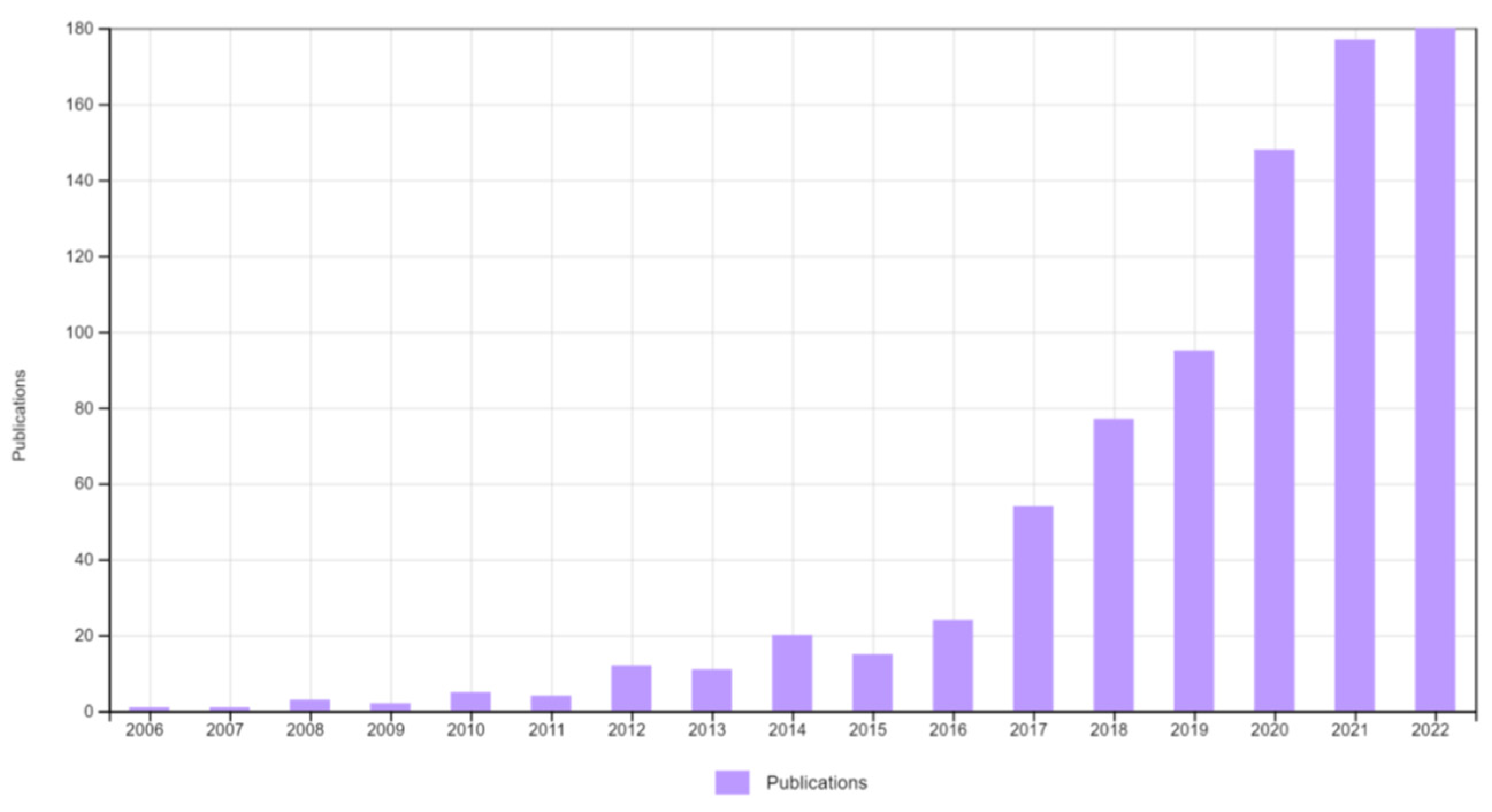
Figure 1. Graph depicting the number of publications on 2D MOFs over the years. Citation Report Graphic is derived from Clarivate Web of Science, Copyright Clarivate 2022. All rights reserved. (Keywords: 2D two-dimensional metal-organic frameworks MOF).
Biosensors 13 00123 g001
Figure 2. Diagram depicting the properties of 2D MOFs that makes it interesting for sensor applications.
Biosensors 13 00123 g002
Figure 3. Schematic illustration of different synthesis routes of 2D MOF.
Biosensors 13 00123 g003
Figure 4. One-atom thick flakes of a Cu-based MOF obtained by top-down sonication method deposited on highly oriented pyrolytic graphite. (a) shows the topography image obtained using Atomic Force Microscope (AFM) and (b) depicts the height profile obtained along the green line in (a). Used with permission of [49]; permission conveyed through Copyright Clearance Center, Inc.
Biosensors 13 00123 g004
Figure 5. Top-down synthesis of Zn2(BIM)4 nanosheets from its corresponding crystals. The scanning electron microscopy (SEM) images of the crystals are shown in (A) with the layered structure clearly visible in the inset. The top-down treatment (wet ball milling + ultrasonication) exfoliates it into nanosheets as observed from the transmission electron microcopy (TEM) images (D). The inset in (D) shows the Tyndall effect exhibited by its colloidal suspension. The phase formation of the prepared MOFs is confirmed by X-ray diffraction (C). The schematic representation of the MOF architecture before and after the exfoliation treatment is shown in (B,E), respectively. (F) represents the space-filling model of a four-membered ring of the MOF nanosheet. From [60]. Reprinted with permission from AAAS.
Biosensors 13 00123 g005
Figure 6. Schematic illustrations of different interfacial methods employed for the synthesis of 2D MOFs: (a) Formation of a single-layer sheet of Ni-based MOF at a liquid-air interface by Langmuir-Blodgett method. Reproduced from [69] Copyright 2015 Wiley Materials. (b) Synthesis of 2D Fe-MOF at a liquid-solid interface. Reprinted (adapted) with permission from [70]. Copyright 2017 American Chemical Society. (c) Formation of MOF nanosheets at a liquid-liquid interface by spraying technique. Reprinted (adapted) with permission from [68]. Copyright 2017 American Chemical Society. (d) Formation of ultrathin nanosheets of CuBDC MOF by a three-layer preparation approach. Reprinted by permission from [Springer Nature Customer Service Centre GmbH]: [Springer Nature] [Nature Materials] [Metal-organic framework nanosheets in polymer composite materials for gas separation, Tania Rodenas et al.] [Copyright 2014 Macmillan Publishers Limited] (2015) [39].
Biosensors 13 00123 g006aBiosensors 13 00123 g006b
Figure 7. Scheme depicting the surfactant-mediated synthesis of 2D MOFs. The traditional synthesis method yields bulk crystals due to isotropic growth. The incorporation of the surfactant on the MOF surface results in anisotropic growth, thereby producing ultrathin nanosheets. Reproduced from [47]. Copyright 2015 Wiley Materials.
Biosensors 13 00123 g007
Figure 8. (a) Schematic representations of Ni-MOF@Ni-HTTP-5 synthesis and its glucose biosensing application, (b,c) SEM and TEM morphologies of Ni-MOF NS, (e,f) SEM and TEM images of Ni-MOF@Ni-HTTP-5, (d,g) STEM and elemental mapping images of Ni-MOF-NS and Ni-MOF@Ni-HTTP-5 NSs. Reprinted from [28], Copyright (2022), with permission from Elsevier.
Biosensors 13 00123 g008
Figure 9. Schematic representation of synthesis of Cu2O mediated Ag/MIL-53(Fe) and real time monitoring of H2O2 from living cell A549. Reprinted by permission from [Springer Nature Customer Service Centre GmbH]:[Springer Nature] [Analytical and Bioanalytical Chemistry] [Cu2O-Mediated Assembly of Electrodeposition of Au Nanoparticles onto 2D Metal-Organic Framework Nanosheets for Real-Time Monitoring of Hydrogen Peroxide Released from Living Cells, Sha Chen et al.] [COPYRIGHT 2020] (2021) [90].
Biosensors 13 00123 g009
Figure 10. Schematic of homogeneous flow assay for microRNA detection, SP-syringe pump, SV-six port multiposition valve, HC-holding coil, WE-working electrode, RE-reference electrode, CE-counter electrode, EW-electrochemical workstation, MB-Methylene Blue. Reprinted from [95], with permission from Elsevier.
Biosensors 13 00123 g010
Figure 11. Schematic representation of Cu−BTC/ZIF−L synthesis and its insulin sensing mechanism. Reprinted (adapted) with permission from [96] Copyright {2022} American Chemical Society.
Biosensors 13 00123 g011
Figure 12. Schematic representation of (A) antibody labelled 2D Zn−TCPP MOF/Fe3O4−COOH/Thi. (B) Deep eutectic solvent, (C) electrochemical sensing mechanism of CTnI. Reprinted from [99], with permission from Elsevier.
Biosensors 13 00123 g012
Figure 13. Scheme of (A) Fabrication procedure of PtNi@Cu-TCPP(Fe)-Ab2 bioconjugates and (B) fabrication of electrochemical immunosensor. Reprinted from [100], Copyright (2020), with permission from Elsevier.
Biosensors 13 00123 g013
Figure 14. Fluorometric and colorimetric glucose detection by a bi-functional 2D Co-MOF. Reprinted from [121], with permission from Elsevier.
Biosensors 13 00123 g014
Figure 15. Ratiometric fluorescence sensing of GSH (A,B) and Cys (C,D) by Ag/Eu@Ni-MOF nanosheets. (A,C) represents the emission spectra of the MOFs in response to different concentrations of the analytes (GSH, Cys). Insets show the photos of the MOF in the solutions under UV irradiation (254 nm). (B,D) indicates the corresponding calibration curves obtained (I524/I615 vs. concentration of analyte) with the linear fits given in the insets. Reprinted by permission from [Springer Nature Customer Service GmbH]: [Springer Nature] [Nature Materials] [Metal-organic framework nanosheets in polymer composite materials for gas separation, Yun Shu, Tao Dai, Qiuyu Ye, Dangqin Jin, Qin Xu, Xiaoya Hu], [The Author(s), under exclusive license to Springer Science+Business Media, LLC, part of Springer Nature 2021] (2021) [122].
Biosensors 13 00123 g015
Figure 16. Schematic illustration of a 2D MOF-based surface plasmon resonance biosensor for the detection of PD-L1 exosomes. Reprinted from [125], Copyright (2022), with permission from Elsevier.
Biosensors 13 00123 g016
Figure 17. Schematic diagram of a 2D MOF−based, enzyme−free, SERS biosensor for glucose detection. Reprinted (adapted) with permission from [82]. Copyright 2020 American Chemical Society.
Biosensors 13 00123 g017
Figure 18. Future perspectives for the development of 2D MOFs as point-of-care sensors.
Biosensors 13 00123 g018
Table 1. Comparison of 2D MOFs with other 2D nanomaterials.
2D MaterialStructureSurface AreaElectrical ConductivityThermal ConductivityStabilityOther PropertiesRef.
Graphenesp2 carbon arranged in a hexagonal, honeycomb lattice High (~2630 m2/g)Very HighHigh (~4000 Wm−1K−1)Stable in most conditionsExcellent mechanical properties (~130 GPa fracture strength).
Excellent optical properties.
[2]
Graphene oxideGraphene structure rich with oxygen functional groupsSmaller than grapheneInsulating/semiconductingHigh; lower than grapheneStable in waterBulk synthesis possible. Easily modifiable surface owing to functional groups.[2]
TMDMX2; a layer of transition metal atoms (M) sandwiched between two layers of chalcogen (X) atomsLow (~16 m2/g for MoS2 nanosheets)Metallic to semiconducting depending on phase2–3 orders lower than grapheneDepends on synthesis and modificationExcellent for optoelectronics.[5,9]
MXeneTransition metal atoms (M) arranged in a hexagonal structure, with the octahedral sites filled by carbon or nitrogen (X)ModerateHighModerateSensitive to water and oxygenExcellent mechanical properties.
Interesting optical and electrochemical properties.
EMI shielding property.
[10]
LDHBrucite-like cationic layers with intercalated anions for charge neutralizationModerateLowLowDependent on type of component ionsEasy synthesis.
Excellent ion exchange capacity, ionic conductivity.
[4]
MOFMetal ions or clusters linked by organic ligandsVery high (~1000–10,000 m2/g) Low; higher than 3D MOFLowDepends on type of MOF; more stable than 3D MOFIn-situ and post-synthetic modification possible.
Tunable pore size.
High porosity.
High mechanical strength and flexibility.
[8]
TMD—Transition metal dichalcogenides. LDH—Layered double hydroxides.
Table 2. Comparison of the nonenzymatic glucose sensing properties of 2D MOFs with other materials.
CatalystMediumSensitivity (µA mM−1cm−2)Linearity (mM)Limit of Detection (µM)Stability/Real Sample (Recovery)References
Amorphous Co-Ni hydroxide0.5 NaOH1911.50.00025–50.1260 days, 102%[21]
CNF/Co(OH)20.1 M NaOH68,0000.01–0.125-[22]
Pt@CNOPBS (pH 7.4)21.62–2890-[23]
Ni(TPA)MOF-SWCNT0.1 M KOH-0.02–4.44.615 days, human serum[24]
Pt-Pd/PHNG0.1 M PBS (pH 7)52.530.1–41.823 weeks, human blood (101.43%)[25]
Hierarchical sheet-like Ni-BDC/GCE0.1 M NaOH6360.01–0.86.68-[26]
Co-MOF nanosheet array/NF0.1 M NaOH10,8860.001–30.00137 days, Fruit juice, human serum[27]
Ni-MOF@Ni-HHTP-50.1 M NaOH2124.900.5–2665.50.02-[28]
CNF: Carbon nano fibre. CNO: Carbon nano-onion. SWCNT: Single-walled carbon nanotube. NF: Nickel foam. PHNG: Porous holey nitrogen doped graphene. HHTP: 2,3,6,7,10,11-hexahydroxytriphenylene.
Table 3. Summary of 2D-MOF based electrochemical biosensors.
SensorMethodAnalyteLinear Range (µM)Sensitivity
(µA mM−1 cm−2)
LODReal Sample/RecoveryRef.
Ultrathin Ni MOFCAGlucose25–3160402.30.6 µMHuman serum (97–104.7%)[101]
Vertical 2D NiCo MOF nanosheetCAGlucose1–8000684.40.29 µMHuman serum (96–106%)[102]
Ni-MOF@HHTPCV and CAGlucose0.5–2665.52124.900.0485-[28]
2D/3D NiCu MOF-6CAGlucose0.02–4.93183215Human serum (94.5–97.3%)[103]
Co-MOF nanosheet array/Ni foamCV and CAGlucose1–300010,8861.3 nMBlood serum, fruit juice[27]
Ni@Cu-MOF nanosheetCVGlucose5–25001703.331.67Human serum (100–104%)[104]
2D MOF-74 (Cu) nanosheetCAGlucose100–100038100.41Human serum[105]
NH2-GP/2D arrays of Cu3(btc)2CV and CAGlucose0.05–1775.5536030 nMHuman sweat sample[93]
NH2-GP/2D arrays of Cu3(btc)2CV and CALactate0.05–22.6 mM295Human sweat sample[93]
2D Cu-TCPP/MWCNTCAH2O21–81591570.70Human serum (104.7%)
Beer sample (103.4%)
[106]
2D Co-MOF@NafionCV and CAH2O25–1000
103–105
570 ± 5 A mM−1cm−2
395 ± 10 A mM−1cm−2
-Commercial lens cleaning (103%) and commercial disinfectant solution (97%)[91]
2D Ni-MOF/HeminCV and DPVH2O21–400380.2Human serum (99.32–101.88%)
Disinfectant water sample
[107]
Co-MOFCAH2O20.5–832.50.0412 (µA µM−1)0.47-[108]
Ni-MOFCAH2O21–33000.041 (µA µM−1)1.58-[108]
NiCo-MOFCAH2O21–8300.045 (µA µM−1)1.07-[108]
Au@Cu2O-MIL 53(Fe)CAH2O210–1520351.571.01A549 cells[90]
Ni-MOFCAAscorbic Acid0.5–8065.52.40.25-[109]
Co-MOFCV and CAUrea500–75005414-[94]
NiCo-MOFCV and CAUrea0.5–332.58606.188Milk sample[94]
Ni-MOFCV and CAUrea0.5–832.519600.471Milk sample[94]
{100} facets of Ni3(HHTP)2CVDopamine--9.9 ± 2 nM (PBS)
214 ± 48 nM (CSF)
-[89]
2D/2D NiCo-MOF/Ti3C2DPVAcetaminophen0.01–4000.0430.008Serum (98.9%) and urine (99.8%)[92]
2D/2D NiCo-MOF/Ti3C2DPVDopamine0.01–3000.10.004Serum (102.2%) and urine (98.3%)[92]
2D/2D NiCo-MOF/Ti3C2DPVUric acid0.01–3500.0520.006Serum (100.6%) and urine (98.3%)[92]
FC labeled ssDNA aptamer/BPNSs/TH/Cu-MOFSWVmicroRNA (miR3123)2 pM–2 µM-0.3 pMHuman serum (97.68–104.4%)[98]
Cu-BTC/ZIF-LDPVInsulin0.1 pM–5 µM-0.027 pMIn vivo animal[96]
2D Zn MOF on Zr MOFEIS, DPVPTK70.001–1 ng/mL-0.84 pg/mL (EIS)
0.66 pg/mL (DPV)
Human serum (96.6–104.6%)[97]
Co-MOF@TPN-COFEISAmpicilin0.001–2000 pg/mL-0.217 × 10−3 pg/mL)Human serum (95.5–99.9%)
River water (98.2–103.4%)
Milk (96.4–102.6%)
[110]
Co-MOF/ITO (flow homogeneous assay)CV and DPVMicroRNA1 pM-1 µM-0.12 pMHuman serum (98.7–109%)[95]
2D Zr-MOF (521-MOF)EISMucin 1 (MUC 1)0.001–0.5 ng/mL-0.12 pg/mLHuman serum (94.8–106.8%)[111]
Ab1-Zn-MOF/Fe3O4-COOH/Thi signal molecule and Ab2/pCTAB/DES as biosensing deviceDPVCardiac troponin (CTnI)0.04 ng/mL–50 ng/mL-0.0009 ng/mLWhole blood sample[99]
PtNi@Cu-TCPP(Fe)CACalprotectin (CALP)200 fg/mL–50 ng/mL-137.7 fg/mLHuman Serum (94–100.9%)[100]
AntiNSE/Zr-TAPPEIS and DPVNeuron specific enolase (NSE)10.0 fg/mL–2.0 ng/mL-7.1 fg/mLHuman serum (93.3–106.9%)[112]
GO@Ab2/Ab1/BSA/Ag/Cu-TCPP(Fe)/MWCNTCASulfonamide1.186–28.051 ng/mL-0.395 ng/mLWater samples (64–118%)[113]
HHTP: 2,3,6,7,10,11-hexahydroxytriphenylene. TCPP: Tetrakis (4-carboxyphenyl) porphyrin. Fc:Ferrocene. BPNS: Black phosphorous nanosheet. TPN: terephthalonitrile. COF: Covalent organic framework. pCTAB: Polymer film of cetyltrimethylammonium bromide. DES: Deep eutectic solvent. TAPP: 5,10,15,20-tetra(4-aminophenyl) porphyrin. Ab2: Polyclonal antibody. Ab1: Monoclonal antibody. BSA:Bovine serum albumin.
Table 4. Summary of 2D-MOF based optical biosensors.
2D MOFAnalyteMethodLinear RangeLODReal Sample Test inRemarksRef.
DNA/Au NP/Cu-TCPP(Fe)Carcinoembryonic antigenColorimetric1 pg/mL to 1000 ng/mL0.742 pg/mLHuman serumMOF has HRP-like activity[84]
BODIPY@Eu-MOFFRatiometric fluorescence0–30 µM0.1737 µMLiving cellsLow cytotoxicity. Also used for bioimaging[126]
H2O20–6 µM6.22 nM
Glucose0–6 µM6.92 nM
Cu@Cu-FeTCPPGlucoseColorimetric0.05–1.25 mM12 µM-High peroxidase mimicking activity[45]
Cu-TCPP/Au chipPD-L1 exosomeSPR104–5 × 10⁶ particles/mL16.7 particles/mLHuman serumHigher RI sensitivity (137.67°/RIU), detection accuracy (0.77), and quality factor (24.81 RIU−1) were enhanced compared to bare gold sensor[126]
NH2- MIL-53(Al)H2O2Ratiometric fluorescence0.5–50 µM26.49 nMHuman serumNH2 groups improve the water-stability of the MOF[83]
glucose0.041 µM
Ag/Eu@Ni-MOFGSHRatiometric fluorescence5–250 µM0.17 µMHuman serum-[122]
Cysteine0.2 µM
Au NP/Cu-TCPP(Fe)GlucoseSERS0.16–8 mM0.16 mMHuman salivaAu: GOx-like activity
2D MOF: peroxidase-like activity
[82]
Co-BDCGlucoseColorimetric50 µM–15 mM16.3 µMHuman blood-[121]
Eu@BCPAnthrax biomarkerFluorescence0–35 µM0.038 nM-Dual-emission[86]
Tb@BCP0.033 nM
Luminol-AgNPs@Co/Ni-MOFAlpha-fetoprotienElectrochemiluminescence1 pg/mL–100 ng/mL0.417 pg/mLHUman plasmaEnhanced ECL performance[124]
Zn-Ru(dcbpy)32+Cardiac troponin IElectrochemiluminescence1 fg/mL–10 ng/mL0.48 fg/mLHuman serumECL luminophore utilized as the organic ligand[114]
Co-TCPP(Fe)GlucoseChemiluminescence32–5500 µg/L10.667 µg/LHuman urinePeroxidase-like catalysis[123]
Co-TTPPDNAFluorescence0–1 nM120 pM-Best performance for MOF with 10 layers[81]
In-aipH2O2Fluorescence0–160 µM0.87 µMHuman serumEnzyme assisted analysis.
2D morphology facilitates efficient capture of unstable intermediates and ensures stable luminescence
[127]
Glucose0–200 µM1.3 µM
Cu-TCPPSalmonella enterica DNAFluorescence0.5–15 nM28 pM-Multiplex detection[34]
Listeria monocytogenes DNA0.1–12 nM35 pM
Vibrio parahemolyticus DNA0.1–9 nM15 pM
Ni-MOFH2O2Colorimetric0.04–160 µM0.008 µMHuman serumMOF exhibited greater affinity towards TMB and H2O2 than HRP[116]
ZIF67H2O2Colorimetric100–1000 mM0.11 mM-Catalytic activity dependent on pH and reaction temperature[44]
DNA—deoxyribonucleic acid. TCPP(Fe)—Fe(III) tetra(4-carboxyphenyl)porphyrin chloride. BODIPY—Boron-dipyrromethene. H2O2—hydrogen peroxide. PD-L1—programmed death ligand-1. BCP—Barium-dipicolinic acid coordination polymer. Ru(dcbpy)32+—tris(4,4′-dicarboxylic acid-2,2′-bipyridyl) ruthenium(II). TTPP—5,10,15,20-tetrakis [40-(terpyridinyl)phenyl]porphyrin. ZIF—Zeolitic imidazolate framework. Aip—aminoisophthalic acid. HRP—horseradish peroxidase. TMB—3,3,5,5-tetramethylbenzidine.
Disclaimer/Publisher’s Note: The statements, opinions and data contained in all publications are solely those of the individual author(s) and contributor(s) and not of MDPI and/or the editor(s). MDPI and/or the editor(s) disclaim responsibility for any injury to people or property resulting from any ideas, methods, instructions or products referred to in the content.