Biomolecule-Based Optical Metamaterials: Design and Applications


Abstract
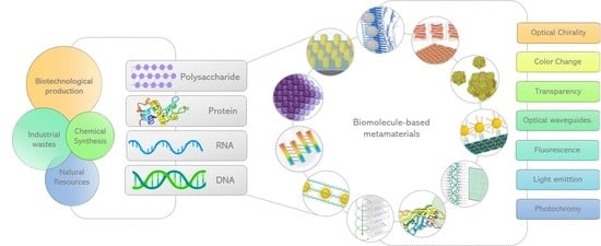
1. Introduction
2. Photonic Biomaterials Found in Nature
2.1. Main Sources to Obtain Polysaccharides
2.1.1. Cellulose
2.1.2. Alginate
2.1.3. Chitosan
2.2. Polysaccharide-Based Optical Metamaterials
2.2.1. Cellulose-Based Optical Material
2.2.2. Chitin-Based Optical Material
2.2.3. Agarose- and Other Polysaccharides-Based Hydrogels
3. Protein Based Metamaterials
3.1. Solvent Free Protein Liquids
3.2. Protein Based Metasurfaces
3.3. Protein Based Metafluids
3.4. Protein Templated Metamaterials
3.5. Protein Hydrogels

| Protein | Configuration | Method | Properties | References |
|---|---|---|---|---|
| Insulin amyloid fibrils | Silver particles (5 nm) follow the helical structure of the biomolecule used as a supporting scaffold. | Amyloid fibrils induction by exposition of insulin to pH 2.0, 70 °C during 7.5 h. Silver NPs were attached to active sites on the insulin fibrils by electroless plating. AgNO3 solution was added to the insulin fibrils (70 mM) and left to incubate overnight. | Optical Chirality. Distribution of NPs deposited along biomolecules, without the use of heavy-metal staining. A general approach for aligning NPs into chains on functional surfaces for applications such as optical waveguides. | [124] |
| Insulin amyloid fibrils | Steady Pt-Pd NP chains templated on Insulin amyloid fibrils with an uniform diameter of about 10 nm and lengths up to several μm. | Amyloid fibrils induction by exposition of insulin to pH 1.6, 70 °C during 7.5 h. Site directed adsorption and subsequent reduction of PtCl4 and Na2PdCl4 during 2 h at room temperature, inducing the growth of Pt-Pd NPs in situ to enable them to align regularly into chains. | Optical Chirality and catalytic activity, CO oxidation and methanol oxidation. | [126] |
| Apoferritin (APO) and -lactoglobulin protein amyloid fibrils | Au, Ag, Pd NPs, Au NRs, Au NSs and magnetic iron oxide NPs. | Amyloid fibrils induction by exposition of Apoferritin and -lactoglobulin to pH 2.0, 90 °C during 24 h. To prepare bioconjugates, AuNSs or AuNRs solution was added to Apoferritin and -lactoglobulin protein amyloid fibrils fibril solution at pH 8 and incubated for 24 h | Highly magnetized nanomaterials (magnetic anisotropy) due to the long-range dipole-dipole coupling and the MNPs alignment along amyloid fibrils. | [132] |
| Streptavidin-biotin system | Isotropic metafluid. Subwavelength structure of strongly coupled nonmagnetic NPs. An individual metamolecule is modeled as 32 Ag-NP (36 nm) surrounding the surface of streptavidin coated polystyrene NP (90 nm), with an average interparticle spacing of 3.8 nm. | First, citrate-capped Ag-NP (36 nm) were synthesized and functionalized with biotin-terminated PEG ligands. The biotin-functionalized Ag-NP were added to a solution of streptavidin coated polystyrene NP (90 nm). | Metafluid exhibited strong optical-frequency magnetism response at visible frequencies. The highly specific chemical recognition Streptavidin-biotin allows the Ag-NP closely and symmetrically pack around. | [115] |
| Bovine serum albumin (BSA) -lactoglobulin from bovine milk (BLG) Conalbumin from chicken egg-white(CA) Recombinant human insulin Cytochrome c from bovine heart (Cyt) | Plasmonic Raspberry-like Core/Satellite Nanoclusters. Spherical Au or AgNPs with a hydrophilic protein shell (as satellites; 5–32 nm) onto large sized metal NPs (as cores; 45–100 nm). | AuNPs or AgNPs were synthesized by the citrate reduction-based seeded-growth method. Citrate-stabilized satellite NPs were coated with a different protein (BSA, BLG, CYTC, and CA) using a simple ligand exchange process at pH 9.0. The citrate-stabilized NP dispersions was added dropwise to protein/citrate solution under vigorous stirring during 24 h at room temperature. | Tuning of the optical/plasmonic properties by changing particle size, composition, or assemblies in a broad range of the visible spectrum. | [143] |
| Lysozyme | Hydrogel | Hydrogel formation by a combination of heating (60 °C) and cooling steps (25 °C) on a lysozyme/TCEP mix at a molar ratio of 4:1. The obtained lysozyme hydrogel was optically transparent. At lower molar ratios of TCEP/lysozyme converted into turbid gels. | Transparent or cloudy optical appearance depending on the concentration of lysozyme or TCEP as well as the effect of the redox, heating and cooling conditions applied on the hydrogel fabrication due to formation of misfolding lysozyme intermediates in the structural organization of the hydrogel. | [138] |
| Silk Fibroin | Periodic nanopatterned 2D lattices in pure silk fibroin protein films. (lattice spacing 300–700 nm, thick film ∼10 μm). | Silk nanostructures were created by nanoscale electron-beam writing of hard metal masks. | Periodic lattices in silk fibroin films feature sizes of hundreds of nanometers that exhibit different colors as a function of varying lattice spacing, angle of illumination and angle of collection. | [98] |
| Silk fabrics | Spherical AuNP in situ synthetized on the surface of silk fabrics (AuNP diameter 28.3–134.7 nm). | AuNP were synthesized in situ. Solutions containing white silk fabric samples and different concentrations of HAuCl4 (0.1–0.6 mM, 50 mL) were shaken for 30 min at room temperature before heating. Subsequently, the solutions were heated at 85 °C for 60 min in a shaking water bath. | AuNP on the surface of fibers leads to yellow, red or brown colors of the treated silk and give rise to enhanced electromagnetic fields for amplification of optical signals. | [99] |
| Silk fibroin | Silk fibroin paper with fibers of 20–120 nm in diameter. Paper thickness varied from 75 μm to 55 μm according to the process applied. | Silk from Bombyx mori was treated to eliminate the sericin protein, degummed fibroin was ground and subsequently homogenized (1, 9, 15, and 30 times). The homogenization system allows fibrillation. Each silk fibroin “pulp paper” was filtrated and dried. | Optically transparent silk fibroin nanofiber paper. Paper thickness, transmittance at 600 nm, transparency varied according to the number of homogenization passes (1 pass to 30 passes). | [104] |
| Recombinant reflectins proteins SoRef2, SoRef1, and SoRef8 | Spin-coated reflectin films. SoRef2 film exhibited a relatively smooth surface with a roughness ∼11.6 nm (without imidazol) and ∼65.4 nm (with imidazol). | Spin coating of reflectin films was performed in a cleanroom. Approximately 250 μL of each protein (SoRef2 at 330 mg/mL, SoRef1 at 416 mg/mL, or SoRef8 at 386 mg/mL) was pipetted onto the center of a pre-cleaned glass slide or polished silicon wafer placed on a spin coater, which was operated at 2000 rpm for 50 s to generate films with different thicknesses. | Reflectin films formed by higher-order assembled structures exhibited dynamic color changes from colorless to white and blue, regardless of film thickness and the type of reflectin protein. | [110] |

3.6. Strategies for Protein Production
4. DNA/RNA Based Metamaterials
4.1. DNA
4.1.1. Spherical Nucleic Acids (SNAs)
4.1.2. Nucleic Acid Biodots
4.1.3. DNA Hydrogel
4.1.4. DNA Lattices
4.1.5. 1D DNA Nanostructures
(A) DNA Nanotubes
(B) DNA Gold Nanorods Nanostructure
(C) DNA Nanowires
(D) DNA-CTMA Nanofibers
(E) DNA-Nanoparticles Chains
4.1.6. 2D DNA Nanostructures
4.1.7. 3D DNA Nanostructures
| Type | Configuration | Method | Optical Properties | References |
|---|---|---|---|---|
| 1D | QDs attached to functionalized DNA origami nanotubes | (a) Self assembly of commercial streptavidin coated QDs on pre-engineered DNA nanostructures that display biotin molecules at selected locations. (b) Hybridization of DNA functionalized QDs to DNA structures carrying capture strands of complementary sequences. | Broad absorption but narrow and symmetric photoluminescence emission spectra, high quantum yield, excellent photostability, and resistance towards chemical degradation. Excellent properties for bio imaging. | [258] |
| DNA-Gold Nanorods (GRN) films | Covalent conjugation of the thiolated gene of enhanced GFP to gold nanorods for the remote control of gene expression in living cells. | Optical switch that allowed induced enhanced green fluorescent receptor expression in HeLa cells after laser exposure. | [259] | |
| DNA Nanowires | Electrochemical synthesis of CdSe NCs with two different ssDNA molecules of 30 base guanine (poly G (30)) and 30 base cytosine (poly C (30)) as templates. | High GXRD peak intensities, excellent optical absorption and control of optical activities, as well as strong phonon confinement. | [260] | |
| DNA-CTMA fibers | Doping of lanthanides chelates into the DNA-CTMA matrix. | Fluorescence and optical amplification properties at suitable pumping wavelengths (612 nm) and low power. | [222] | |
| DNA-NP chains | Adjustment of the number of DNA modified on NPs and control of the assemblies through the design of the hybridized configuration to form dimers, trimers, pyramids, core-satellite, and chains. | Enhancement of EM fields, including SERS and chirality, for the amplification of optical properties. | [261] | |
| 2D | DNA nanoantennas | Consists of the attaching one or two nanoparticles (Au or Ag) to DNA origami structures with available docking sites for a single fluorescent dye next to one NP or in the gap between two NPs. | Provide the possibility of increasing fluorescence by plasmonic effects in the near-field of metal nanostructures more than 100 times the signal of the fluorophore. | [245] |
| DNA nanorulers | DNA origami functionalized by qPAINT method to have a precise spatial control over number, spacing, and arrangement of fluorophores. | Have been used to evaluate the performance of TIRF microscopes and have the potential to be traceable ubiquituous standards for length measurements. | [242] | |
| 3D | Voxels | DNA frames with encapsulated nano-objects were called DNA material that can be coupled with proteins, NPs and QDs. | 3D DNA lattices that show diffraction-limited spectral purity, light-emitting, 3D packaging of desired nano-objects, and the possibility of manipulating and enhancing enzymatic cascade reactions by the lattice architecture. | [252,255] |
| Others | Biodots | Fluorescent nanoparticles (biodots) of several nm in size synthesized from polymeric and monomeric nucleic acids: DNA, RNA, nucleotides, and nucleosides. Individual nucleotides are more suitable for biodot synthesis compared to DNA and RNA. | The fluorescence of nucleic acid biodots is stable in a broad range of pHs and in the presence of physiologically relevant cations. Applications include bioimaging and sensing platforms. | [198] |
| Photoresponsive DNA-cross-linked hydrogels | Incorporation of Azo into the backbone of crosslinker DNA sequences give a Hydrogel phase transition regulated by UV/Vis irradiation. | Under visible light, the Azo molecule was in the trans form and allowed crosslinker DNA to hybridize with DNAs on the polymer side chains forming a 3D hydrogel network. When the gel was irradiated with UV light, the Azo was photoisomerized to the cis form that prevented hybridization and caused the hydrogel to revert to the sol state. | [262] |
4.2. RNA Based Metamaterials
4.3. DNA Production
4.4. DNA Synthesis
4.4.1. In Vitro Methods
(A) Chemical Synthesis
(B) Enzymatic Synthesis
4.4.2. In Vivo Methods
4.4.3. DNA from Natural Sources
4.5. RNA Production
4.5.1. In Vitro and In Vivo Synthesis Methods for RNA
4.5.2. Purification of RNA from Natural Resources
5. Conclusions
Author Contributions
Funding
Data Availability Statement
Acknowledgments
Conflicts of Interest
Abbreviations
| aPCR | Asymmetric polymerase chain reaction |
| AuNPs | Gold nanoparticles |
| BC | Bacterial cellulose |
| BFP | Blue fluorescent protein |
| BLG | -lactoglobulin from bovine milk |
| BSA | Bovine serum albumin |
| CA | Conalbumin from chicken egg-white |
| CD | Chromatic dispersion |
| CLC | Cholesteric liquid crystal |
| CNC | Cellulose nanocrystals |
| CNC-Cu(II) | Copper(II)-doped cellulose nanocrystals |
| CNF | Cellulose nanofibrils |
| CTMA | Cetyltrimethylammonium |
| CYTC | Cytochrome c from bovine heart |
| DG | Diffraction grating |
| dNTP | Deoxynucleotide triphosphate |
| dsDNA | Double-stranded DNA |
| DX | Double-crossover |
| EBL | Electron beam lithography |
| EM | Electron microscopy |
| FESEM | Field-emission scanning electron microscopy |
| FIB | Focused ion beam |
| FML | Functional molecular liquid |
| FP | Fluorescent protein |
| FRET | Fluorescence resonance energy transfer |
| GD | Group delay |
| GFP | Green Fluorescent Protein |
| GNR | Gold Nanorods |
| GXRD | Grazing-incidence X-ray diffraction |
| HOBC | 4′-(hexyloxy)- 4-biphenylcarbonitrile |
| hADMSC | Human adipose tissue-derived mesenchymal stem cell |
| ivTRT | In vitro transcription and reverse transcription |
| IOHG | Inverse opal photonic crystal hydrogel |
| IOPC | Inverse opal photonic crystal |
| LSPR | Localized surface plasmon resonance |
| MCA | Multi-primed chain amplification |
| MIM | Metal-insulator-metal |
| MPA | 3-Mercaptopropyl acid |
| msDNA | Multicopy single-stranded DNA |
| NC | Nanocrystal |
| PAGE | Polyacrylamide gel electrophoresis |
| PC | Photonic crystal |
| PCH | Photonic crystal hydrogel |
| PEG | Polyethylene glycol |
| PER | Primer exchange reaction |
| PLGA | poly lactic-co-glycolic acid |
| POTE | poly (octanoic acid 2-thiophen-3-yl-ethyl ester) |
| pRNA | Packaging RNA |
| QD | Quantum dots |
| RCA | Rolling circle amplification |
| REE | Rare earth elements |
| RNA | Ribonucleic acid |
| SEM | Scanning electron microscopy |
| SF | Silk fibroin |
| SNA | Spherical nucleic acid |
| SPPS | Solid phase peptide synthesis |
| ssRNA | Single-stranded RNA |
| TCEP | Tris(2-carboxyethyl)phosphine |
| TdT | Terminal deoxynucleotide transferase |
| Vis-NIR | Visible-near-infrared |
| WJ | Water-jet |
| YFP | Yellow fluorescent protein |
References
- Kumar, V.; Sinha, N.; Thakur, A.K. Necessity of regulatory guidelines for the development of amyloid based biomaterials. Biomater. Sci. 2021, 9, 4410–4422. [Google Scholar] [CrossRef] [PubMed]
- Higgins, S.G.; Becce, M.; Belessiotis-Richards, A.; Seong, H.; Sero, J.E.; Stevens, M.M. High-Aspect-Ratio Nanostructured Surfaces as Biological Metamaterials. Adv. Mater. 2020, 32, 1903862. Available online: https://onlinelibrary.wiley.com/doi/pdf/10.1002/adma.201903862 (accessed on 29 September 2022). [CrossRef]
- Chen, Y.; Ai, B.; Wong, Z.J. Soft optical metamaterials. Nano Converg. 2020, 7, 18. [Google Scholar] [CrossRef] [PubMed]
- Liang, L.; Fu, Y.; Li, L.; Zheng, H.; Wei, X.; Wei, Y.; Kobayashi, N. RNA-CTMA dielectrics in organic field effect transistor memory. Appl. Sci. 2018, 8, 887. [Google Scholar] [CrossRef]
- Cui, Y.; Fung, K.H.; Xu, J.; Ma, H.; Jin, Y.; He, S.; Fang, N.X. Ultrabroadband light absorption by a sawtooth anisotropic metamaterial slab. Nano Lett. 2012, 12, 1443–1447. [Google Scholar] [CrossRef]
- Cui, Y.; He, Y.; Jin, Y.; Ding, F.; Yang, L.; Ye, Y.; Zhong, S.; Lin, Y.; He, S. Plasmonic and metamaterial structures as electromagnetic absorbers. Laser Photonics Rev. 2014, 8, 495–520. [Google Scholar] [CrossRef]
- Feng, X.; Xu, J.; Liu, Y.; Zhao, W. Visual sensors of an inverse opal hydrogel for the colorimetric detection of glucose. J. Mater. Chem. B 2019, 7, 3576–3581. [Google Scholar] [CrossRef]
- Nizamoglu, S.; Gather, M.C.; Humar, M.; Choi, M.; Kim, S.; Kim, K.S.; Hahn, S.K.; Scarcelli, G.; Randolph, M.; Redmond, R.W.; et al. Bioabsorbable polymer optical waveguides for deep-tissue photomedicine. Nat. Commun. 2016, 7, 10374. [Google Scholar] [CrossRef]
- Salim, A.; Lim, S. Review of recent metamaterial microfluidic sensors. Sensors 2018, 18, 232. [Google Scholar] [CrossRef]
- Lee, H.; Wiegand, D.J.; Griswold, K.; Punthambaker, S.; Chun, H.; Kohman, R.E.; Church, G.M. Photon-directed multiplexed enzymatic DNA synthesis for molecular digital data storage. Nat. Commun. 2020, 11, 5246. [Google Scholar] [CrossRef]
- Yang, Y.; Lee, T.; Kim, M.; Jung, C.; Badloe, T.; Lee, D.; Lee, S.; Lee, H.J.; Rho, J. Dynamic Optical Spin Hall Effect in Chitosan-Coated All-Dielectric Metamaterials for a Biosensing Platform. IEEE J. Sel. Top. Quantum Electron. 2021, 27, 1–8. [Google Scholar] [CrossRef]
- Slocik, J.M.; Govorov, A.O.; Naik, R.R. Plasmonic circular dichroism of peptide-functionalized gold nanoparticles. Nano Lett. 2011, 11, 701–705. [Google Scholar] [CrossRef] [PubMed]
- Urban, M.J.; Dutta, P.K.; Wang, P.; Duan, X.; Shen, X.; Ding, B.; Ke, Y.; Liu, N. Plasmonic toroidal metamolecules assembled by DNA origami. J. Am. Chem. Soc. 2016, 138, 5495–5498. [Google Scholar] [CrossRef] [PubMed]
- Zhang, Y.; Pan, V.; Li, X.; Yang, X.; Li, H.; Wang, P.; Ke, Y. Dynamic DNA Structures. Small 2019, 15, e1900228. [Google Scholar] [CrossRef]
- Vázquez-Guardado, A.; Mehta, F.; Jimenez, B.; Biswas, A.; Ray, K.; Baksh, A.; Lee, S.; Saraf, N.; Seal, S.; Chanda, D. DNA-Modified Plasmonic Sensor for the Direct Detection of Virus Biomarkers from the Blood. Nano Lett. 2021, 21, 7505–7511. [Google Scholar] [CrossRef] [PubMed]
- Kuzyk, A.; Schreiber, R.; Zhang, H.; Govorov, A.O.; Liedl, T.; Liu, N. Reconfigurable 3D plasmonic metamolecules. Nat. Mater. 2014, 13, 862–866. [Google Scholar] [CrossRef]
- Kuzyk, A.; Yang, Y.; Duan, X.; Stoll, S.; Govorov, A.O.; Sugiyama, H.; Endo, M.; Liu, N. A light-driven three-dimensional plasmonic nanosystem that translates molecular motion into reversible chiroptical function. Nat. Commun. 2016, 7, 10591. [Google Scholar] [CrossRef]
- Lin, Q.Y.; Mason, J.A.; Li, Z.; Zhou, W.; O’Brien, M.N.; Brown, K.A.; Jones, M.R.; Butun, S.; Lee, B.; Dravid, V.P.; et al. Building superlattices from individual nanoparticles via template-confined DNA-mediated assembly. Science 2018, 359, 669–672. [Google Scholar] [CrossRef]
- Salim, A.; Lim, S. Recent advances in the metamaterial-inspired biosensors. Biosens. Bioelectron. 2018, 117, 398–402. [Google Scholar] [CrossRef]
- Dogan, E.; Bhusal, A.; Cecen, B.; Miri, A.K. 3D Printing metamaterials towards tissue engineering. Appl. Mater. Today 2020, 20, 100752. [Google Scholar] [CrossRef]
- Xiong, R.; Luan, J.; Kang, S.; Ye, C.; Singamaneni, S.; Tsukruk, V.V. Biopolymeric photonic structures: Design, fabrication, and emerging applications. Chem. Soc. Rev. 2020, 49, 983–1031. [Google Scholar] [CrossRef] [PubMed]
- Ning, P.; Yang, G.; Hu, L.; Sun, J.; Shi, L.; Zhou, Y.; Wang, Z.; Yang, J. Recent advances in the valorization of plant biomass. Biotechnol. Biofuels 2021, 14, 102. [Google Scholar] [CrossRef] [PubMed]
- Zhong, C. Industrial-Scale Production and Applications of Bacterial Cellulose. Front. Bioeng. Biotechnol. 2020, 8, 605374. [Google Scholar] [CrossRef] [PubMed]
- Jonas, R.; Farah, L.F. Production and application of microbial cellulose. Polym. Degrad. Stab. 1998, 59, 101–106. [Google Scholar] [CrossRef]
- Zhong, C.; Zhang, G.C.; Liu, M.; Zheng, X.T.; Han, P.P.; Jia, S.R. Metabolic flux analysis of Gluconacetobacter xylinus for bacterial cellulose production. Appl. Microbiol. Biotechnol. 2013, 97, 6189–6199. [Google Scholar] [CrossRef] [PubMed]
- Czaja, W.K.; Young, D.J.; Kawecki, M.; Brown, R.M. The future prospects of microbial cellulose in biomedical applications. Biomacromolecules 2007, 8, 1–12. [Google Scholar] [CrossRef]
- Urtuvia, V.; Maturana, N.; Acevedo, F.; Peña, C.; Díaz-Barrera, A. Bacterial alginate production: An overview of its biosynthesis and potential industrial production. World J. Microbiol. Biotechnol. 2017, 33, 198. [Google Scholar] [CrossRef]
- Hernández-Carmona, G.; Rodríguez-Montesinos, Y.; Arvizu-Higuera, D.; Reyes-Tisnado, R.; Murillo-Álvarez, J.I.; Muñoz-Ochoa, M. Technological Advance for Alginate Production in Mexico. Ing. Investig. Y Tecnol. 2012, 13, 155–168. [Google Scholar] [CrossRef][Green Version]
- Aziz, S.B.; Hamsan, M.H.; Kadir, M.F.; Woo, H.J. Design of polymer blends based on chitosan:POZ with improved dielectric constant for application in polymer electrolytes and flexible electronics. Adv. Polym. Technol. 2020, 2020, 8586136. [Google Scholar] [CrossRef]
- Hoqani, H.A.S.A.; AL-Shaqsi, N.; Hossain, M.A.; Sibani, M.A.A. Isolation and optimization of the method for industrial production of chitin and chitosan from Omani shrimp shell. Carbohydr. Res. 2020, 492, 108001. [Google Scholar] [CrossRef]
- Colina, M.; Colina, J.A.V.; Zerpa, J.A.M.; Bianchi, J.A.V.; Rodríguez-Monroy, C.; Martínez-Soto, M.E. Evaluation of industrial processes to obtain chitin and chitosan. In Proceedings of the LACCEI International Multi-Conference for Engineering, Education and Technology, Lima, Peru, 18–20 July 2018. [Google Scholar] [CrossRef]
- Mendez-López, M.; Ramos-Hernández, A.; Moreno-Serna, V.; Bonardd, S.; Ramírez, O.; Silva, H.; Inostroza-Rivera, R.; Diaz, D.D.; Leiva, A.; Saldías, C. A facile approach for tuning optical and surface properties of novel biobased Alginate/POTE handleable films via solvent vapor exposure. Int. J. Biol. Macromol. 2021, 193, 258–268. [Google Scholar] [CrossRef] [PubMed]
- Fujiwara, E.; Cabral, T.D.; Sato, M.; Oku, H.; Cordeiro, C.M. Agarose-based structured optical fibre. Sci. Rep. 2020, 10, 7035. [Google Scholar] [CrossRef] [PubMed]
- Lobregas, M.O.S.; Bantang, J.P.O.; Camacho, D.H. Carrageenan-stabilized silver nanoparticle gel probe kit for colorimetric sensing of mercury (II) using digital image analysis. Sens. Bio-Sens. Res. 2019, 26, 100303. [Google Scholar] [CrossRef]
- Santos, M.V.; Tercjak, A.; Gutierrez, J.; Barud, H.S.; Napoli, M.; Nalin, M.; Ribeiro, S.J. Optical sensor platform based on cellulose nanocrystals (CNC) – 4′-(hexyloxy)-4-biphenylcarbonitrile (HOBC) bi-phase nematic liquid crystal composite films. Carbohydr. Polym. 2017, 168, 346–355. [Google Scholar] [CrossRef] [PubMed]
- Song, Y.; Li, Y.; Liu, Z.; Liu, L.; Wang, X.; Su, X.; Ma, Q. A novel ultrasensitive carboxymethyl chitosan-quantum dot-based fluorescence “turn on-off” nanosensor for lysozyme detection. Biosens. Bioelectron. 2014, 61, 9–13. [Google Scholar] [CrossRef]
- Mironenko, A.; Modin, E.; Sergeev, A.; Voznesenskiy, S.; Bratskaya, S. Fabrication and optical properties of chitosan/Ag nanoparticles thin film composites. Chem. Eng. J. 2014, 244, 457–463. [Google Scholar] [CrossRef]
- Meena; Sharma, A. Investigation of structural, optical and sensing performance of starch films reinforced with Ag nanoparticles. Int. J. Polym. Anal. Charact. 2021, 26, 396–410. [Google Scholar] [CrossRef]
- Azeman, N.H.; Arsad, N.; Bakar, A.A.A. Polysaccharides as the sensing material for metal ion detection-based optical sensor applications. Sensors 2020, 20, 3924. [Google Scholar] [CrossRef]
- Zong, A.; Cao, H.; Wang, F. Anticancer polysaccharides from natural resources: A review of recent research. Carbohydr. Polym. 2012, 90, 1395–1410. [Google Scholar] [CrossRef]
- Torres, F.G.; Troncoso, O.P.; Pisani, A.; Gatto, F.; Bardi, G. Natural polysaccharide nanomaterials: An overview of their immunological properties. Int. J. Mol. Sci. 2019, 20, 5092. [Google Scholar] [CrossRef]
- Barzic, A.I.; Barzic, R.F. Optical properties of polysaccharide/polylactide blends. UPB Sci. Bull. Ser. A Appl. Math. Phys. 2015, 77, 293–302. [Google Scholar]
- Shan, D.; Gerhard, E.; Zhang, C.; Tierney, J.W.; Xie, D.; Liu, Z.; Yang, J. Polymeric biomaterials for biophotonic applications. Bioact. Mater. 2018, 3, 434–445. [Google Scholar] [CrossRef] [PubMed]
- Vignolini, S.; Glover, B.; Steiner, U. Photonic structures in plants. In Biomimetics in Photonics; CRC Press: Boca Raton, FL, USA, 2012. [Google Scholar] [CrossRef]
- Omenetto, F.G. Photonics in nature—Opportunities and challenges at the biological interface. APL Photonics 2019, 4, 070402. [Google Scholar] [CrossRef]
- Yu, S.; Qiu, C.W.; Chong, Y.; Torquato, S.; Park, N. Engineered disorder in photonics. Nat. Rev. Mater. 2021, 6, 226–243. [Google Scholar] [CrossRef]
- Caligiuri, V.; Tedeschi, G.; Palei, M.; Miscuglio, M.; Martin-Garcia, B.; Guzman-Puyol, S.; Hedayati, M.K.; Kristensen, A.; Athanassiou, A.; Cingolani, R.; et al. Biodegradable and Insoluble Cellulose Photonic Crystals and Metasurfaces. ACS Nano 2020, 14, 9502–9511. [Google Scholar] [CrossRef] [PubMed]
- Caro-Astorga, J.; Walker, K.T.; Herrera, N.; Lee, K.Y.; Ellis, T. Bacterial cellulose spheroids as building blocks for 3D and patterned living materials and for regeneration. Nat. Commun. 2021, 12, 1–9. [Google Scholar] [CrossRef] [PubMed]
- Cai, H.; Sharma, S.; Liu, W.; Mu, W.; Liu, W.; Zhang, X.; Deng, Y. Aerogel microspheres from natural cellulose nanofibrils and their application as cell culture scaffold. Biomacromolecules 2014, 15, 2540–2547. [Google Scholar] [CrossRef] [PubMed]
- Cennamo, N.; Trigona, C.; Graziani, S.; Zeni, L.; Arcadio, F.; Pasquale, G.D.; Pollicino, A. A Green Slab Waveguide for Plasmonic Sensors Based on Bacterial Cellulose. Proceedings 2019, 15, 36. [Google Scholar] [CrossRef]
- Rull-Barrull, J.; D’Halluin, M.; Grognec, E.L.; Felpin, F.X. Chemically-modified cellulose paper as smart sensor device for colorimetric and optical detection of hydrogen sulfate in water. Chem. Commun. 2016, 52, 2525–2528. [Google Scholar] [CrossRef]
- Orelma, H.; Hokkanen, A.; Leppänen, I.; Kammiovirta, K.; Kapulainen, M.; Harlin, A. Optical cellulose fiber made from regenerated cellulose and cellulose acetate for water sensor applications. Cellulose 2020, 27, 1543–1553. [Google Scholar] [CrossRef]
- SONI, S.; BISHNOI, D.D.; SONI, S.; RAMSWROOP. Liquid crystals and applications of chlosteric liquid crystal in laser. Int. J. Mod. Phys. Conf. Ser. 2013, 22, 1543–1553. [Google Scholar] [CrossRef]
- Ryabchun, A.; Bobrovsky, A. Cholesteric Liquid Crystal Materials for Tunable Diffractive Optics. Adv. Opt. Mater. 2018, 6, 1800335. [Google Scholar] [CrossRef]
- Dai, S.; Prempeh, N.; Liu, D.; Fan, Y.; Gu, M.; Chang, Y. Cholesteric film of Cu(II)-doped cellulose nanocrystals for colorimetric sensing of ammonia gas. Carbohydr. Polym. 2017, 174, 531–539. [Google Scholar] [CrossRef] [PubMed]
- Sergeev, A.A.; Voznesenskiy, S.S.; Bratskaya, S.Y.; Mironenko, A.Y.; Lagutkin, R.V. Investigation of humidity influence upon waveguide features of chitosan thin films. Phys. Procedia 2012, 23, 115–118. [Google Scholar] [CrossRef][Green Version]
- Jang, J.; Kang, K.; Raeis-Hosseini, N.; Ismukhanova, A.; Jeong, H.; Jung, C.; Kim, B.; Lee, J.Y.; Park, I.; Rho, J. Self-Powered Humidity Sensor Using Chitosan-Based Plasmonic Metal–Hydrogel–Metal Filters. Adv. Opt. Mater. 2020, 8, 1901932. [Google Scholar] [CrossRef]
- Shen, P.; Zhang, Y.; Cai, Z.; Liu, R.; Xu, X.; Li, R.; Wang, J.J.; Yang, D. Correction: Three-dimensional/two-dimensional photonic crystal hydrogels for biosensing. J. Mater. Chem. C 2021, 9, 6944. [Google Scholar] [CrossRef]
- Fang, Y.; Ni, Y.; Leo, S.Y.; Taylor, C.; Basile, V.; Jiang, P. Reconfigurable photonic crystals enabled by pressure-responsive shape-memory polymers. Nat. Commun. 2015, 6, 7416. [Google Scholar] [CrossRef]
- Fang, Y.; Ni, Y.; Choi, B.; Leo, S.Y.; Gao, J.; Ge, B.; Taylor, C.; Basile, V.; Jiang, P. Chromogenic Photonic Crystals Enabled by Novel Vapor-Responsive Shape-Memory Polymers. Adv. Mater. 2015, 27, 3696–3704. [Google Scholar] [CrossRef]
- Sun, Y.; Zhang, Y.; Liu, J.; Nie, F. Integrated microfluidic device for the spherical hydrogel pH sensor fabrication. RSC Adv. 2016, 6, 11204–11210. [Google Scholar] [CrossRef]
- Liu, J.; Zhang, Y.; Zhou, R.; Gao, L. Volatile alcohol-responsive visual sensors based on P(HEMA-: Co -MA)-infiltrated SiO2 inverse opal photonic crystals. J. Mater. Chem. C 2017, 5, 6071–6078. [Google Scholar] [CrossRef]
- Tang, W.; Chen, C. Hydrogel-based colloidal photonic crystal devices for glucose sensing. Polymers 2020, 12, 625. [Google Scholar] [CrossRef] [PubMed]
- Wang, Z.; Xue, M.; Zhang, H.; Meng, Z.; Shea, K.J.; Qiu, L.; Ji, T.; Xie, T. Self-assembly of a nano hydrogel colloidal array for the sensing of humidity. RSC Adv. 2018, 8, 9963–9969. [Google Scholar] [CrossRef] [PubMed]
- Huang, G.; Yin, Y.; Pan, Z.; Chen, M.; Zhang, L.; Liu, Y.; Zhang, Y.; Gao, J. Fabrication of 3D photonic crystals from chitosan that are responsive to organic solvents. Biomacromolecules 2014, 15, 4396–4402. [Google Scholar] [CrossRef]
- Jain, A.; Yang, A.H.; Erickson, D. Gel-based optical waveguides with live cell encapsulation and integrated microfluidics. Opt. Lett. 2012, 37, 1472–1474. [Google Scholar] [CrossRef] [PubMed]
- Yetisen, A.K.; Jiang, N.; Fallahi, A.; Montelongo, Y.; Ruiz-Esparza, G.U.; Tamayol, A.; Zhang, Y.S.; Mahmood, I.; Yang, S.A.; Kim, K.S.; et al. Glucose-Sensitive Hydrogel Optical Fibers Functionalized with Phenylboronic Acid. Adv. Mater. 2017, 29, 1606380. [Google Scholar] [CrossRef] [PubMed]
- Hoffman, A.S. Hydrogels for biomedical applications. Adv. Drug Deliv. Rev. 2012, 64, 18–23. [Google Scholar] [CrossRef]
- Cai, Z.; Sasmal, A.; Liu, X.; Asher, S.A. Responsive Photonic Crystal Carbohydrate Hydrogel Sensor Materials for Selective and Sensitive Lectin Protein Detection. ACS Sens. 2017, 2, 1474–1481. [Google Scholar] [CrossRef]
- Hess, A.J.; Funk, A.J.; Liu, Q.; Cruz, J.A.D.L.; Sheetah, G.H.; Fleury, B.; Smalyukh, I.I. Plasmonic Metamaterial Gels with Spatially Patterned Orientational Order via 3D Printing. ACS Omega 2019, 4, 20558–20563. [Google Scholar] [CrossRef]
- Olate-Moya, F.; Arens, L.; Wilhelm, M.; Mateos-Timoneda, M.A.; Engel, E.; Palza, H. Chondroinductive Alginate-Based Hydrogels Having Graphene Oxide for 3D Printed Scaffold Fabrication. ACS Appl. Mater. Interfaces 2020, 12, 4343–4357. [Google Scholar] [CrossRef]
- Yang, J.; Li, Y.; Zheng, Y.; Xu, Y.; Zheng, Z.; Chen, X.; Liu, W. Versatile Aerogels for Sensors. Small 2019, 15, e1902826. [Google Scholar] [CrossRef]
- Druel, L.; Bardl, R.; Vorwerg, W.; Budtova, T. Starch Aerogels: A Member of the Family of Thermal Superinsulating Materials. Biomacromolecules 2017, 18, 4232–4239. [Google Scholar] [CrossRef] [PubMed]
- Bradshaw, D.S.; Leeder, J.M.; Coles, M.M.; Andrews, D.L. Signatures of material and optical chirality: Origins and measures. Chem. Phys. Lett. 2015, 626, 106–110. [Google Scholar] [CrossRef]
- Ikai, T. The dawn of chiral material development using saccharide-based helical polymers. Polym. J. 2017, 49, 355–362. [Google Scholar] [CrossRef]
- Guidetti, G.; Wang, Y.; Omenetto, F.G. Active optics with silk. Nanophotonics 2020, 10, 137–148. [Google Scholar] [CrossRef]
- Dennis, P.B.; Onderko, E.L.; Slocik, J.M.; Bird, L.J.; Phillips, D.A.; Crookes-Goodson, W.J.; Glaven, S.M. Proteins for bioinspired optical and electronic materials. MRS Bull. 2020, 45, 1027–1033. [Google Scholar] [CrossRef]
- Wang, F.; Yang, C.; Hu, X. Advanced protein composite materials. ACS Symp. Ser. 2014, 1175, 177–208. [Google Scholar] [CrossRef]
- Mirzaei, M.; Okoro, O.V.; Nie, L.; Petri, D.F.S.; Shavandi, A. Protein-based 3d biofabrication of biomaterials. Bioengineering 2021, 8, 48. [Google Scholar] [CrossRef]
- Zhang, T.; Hartl, C.; Frank, K.; Heuer-Jungemann, A.; Fischer, S.; Nickels, P.C.; Nickel, B.; Liedl, T. 3D DNA Origami Crystals. Adv. Mater. 2018, 30, e1800273. [Google Scholar] [CrossRef]
- Vallamkondu, J.; Corgiat, E.B.; Buchaiah, G.; Kandimalla, R.; Reddy, P.H. Liquid crystals: A novel approach for cancer detection and treatment. Cancers 2018, 10, 462. [Google Scholar] [CrossRef]
- Viney, C. Liquid Crystalline Phase Behavior of Proteins and Polypeptides. In Protein-Based Materials; Birkhäuser: Boston, MA, USA, 1997; pp. 281–311. [Google Scholar] [CrossRef]
- Sun, Y.L.; Hou, Z.S.; Sun, S.M.; Zheng, B.Y.; Ku, J.F.; Dong, W.F.; Chen, Q.D.; Sun, H.B. Protein-Based Three-Dimensional Whispering-Gallery-Mode Micro-Lasers with Stimulus-Responsiveness. Sci. Rep. 2015, 5, 12852. [Google Scholar] [CrossRef]
- Ghosh, A.; Nakanishi, T. Frontiers of solvent-free functional molecular liquids. Chem. Commun. 2017, 53, 10344–10357. [Google Scholar] [CrossRef] [PubMed]
- Perriman, A.W.; Cölfen, H.; Hughes, R.W.; Barrie, C.L.; Mann, S. Solvent-free protein liquids and liquid crystals. Angew. Chem.-Int. Ed. 2009, 48, 6242–6246. [Google Scholar] [CrossRef] [PubMed]
- Perriman, A.W.; Brogan, A.P.; Cölfen, H.; Tsoureas, N.; Owen, G.R.; Mann, S. Reversible dioxygen binding in solvent-free liquid myoglobin. Nat. Chem. 2010, 2, 622–626. [Google Scholar] [CrossRef] [PubMed]
- Liu, K.; Pesce, D.; Ma, C.; Tuchband, M.; Shuai, M.; Chen, D.; Su, J.; Liu, Q.; Gerasimov, J.Y.; Kolbe, A.; et al. Solvent-free liquid crystals and liquids based on genetically engineered supercharged polypeptides with high elasticity. Adv. Mater. 2015, 27, 2459–2465. [Google Scholar] [CrossRef] [PubMed]
- Zhang, S.; Wong, C.L.; Zeng, S.; Bi, R.; Tai, K.; Dholakia, K.; Olivo, M. Metasurfaces for biomedical applications: Imaging and sensing from a nanophotonics perspective. Nanophotonics 2021, 10, 259–293. [Google Scholar] [CrossRef]
- Torculas, M.; Medina, J.; Xue, W.; Hu, X. Protein-Based Bioelectronics. ACS Biomater. Sci. Eng. 2016, 2, 1211–1223. [Google Scholar] [CrossRef]
- Morse, D.E. Reflectin: Protein Driver of Dynamically Tunable Biophotonics; New Paradigm for Tunably Reconfigurable Materials. 2020. Available online: https://doi.org/10.2172/1719134 (accessed on 29 September 2022).
- Carter, N.A.; Grove, T.Z. Functional protein materials: Beyond elastomeric and structural proteins. Polym. Chem. 2019, 10, 2952–2959. [Google Scholar] [CrossRef]
- Humenik, M.; Winkler, A.; Scheibel, T. Patterning of protein-based materials. Biopolymers 2021, 112, e23412. [Google Scholar] [CrossRef]
- Gromiha, M.M. Chapter 1—Proteins. In Protein Bioinformatics; Gromiha, M.M., Ed.; Academic Press: Singapore, 2010; pp. 1–27. [Google Scholar] [CrossRef]
- Parry, D.A. Chapter Ten—Using Data Mining and Computational Approaches to Study Intermediate Filament Structure and Function. In Intermediate Filament Proteins; Methods in Enzymology; Omary, M.B., Liem, R.K., Eds.; Academic Press: Singapore, 2016; Volume 568, pp. 255–276. [Google Scholar] [CrossRef]
- Kundu, S.C.; Dash, B.C.; Dash, R.; Kaplan, D.L. Natural protective glue protein, sericin bioengineered by silkworms: Potential for biomedical and biotechnological applications. Prog. Polym. Sci. 2008, 33, 998–1012. [Google Scholar] [CrossRef]
- Koh, L.D.; Cheng, Y.; Teng, C.P.; Khin, Y.W.; Loh, X.J.; Tee, S.Y.; Low, M.; Ye, E.; Yu, H.D.; Zhang, Y.W.; et al. Structures, mechanical properties and applications of silk fibroin materials. Prog. Polym. Sci. 2015, 46, 86–110. [Google Scholar] [CrossRef]
- Lawrence, B.D.; Cronin-Golomb, M.; Georgakoudi, I.; Kaplan, D.L.; Omenetto, F.G. Bioactive silk protein biomaterial systems for optical devices. Biomacromolecules 2008, 9, 1214–1220. [Google Scholar] [CrossRef] [PubMed]
- Amsden, J.J.; Perry, H.; Boriskina, S.V.; Gopinath, A.; Kaplan, D.L.; Negro, L.D.; Omenetto, F.G. Spectral analysis of induced color change on periodically nanopatterned silk films. Opt. Express 2009, 17, 21271–21279. [Google Scholar] [CrossRef] [PubMed]
- Liu, J.; Zhou, J.; Tang, B.; Zeng, T.; Li, Y.; Li, J.; Ye, Y.; Wang, X. Surface enhanced Raman scattering (SERS) fabrics for trace analysis. Appl. Surf. Sci. 2016, 386, 296–302. [Google Scholar] [CrossRef]
- Mitropoulos, A.N.; Marelli, B.; Ghezzi, C.E.; Applegate, M.B.; Partlow, B.P.; Kaplan, D.L.; Omenetto, F.G. Transparent, Nanostructured Silk Fibroin Hydrogels with Tunable Mechanical Properties. ACS Biomater. Sci. Eng. 2015, 1, 964–970. [Google Scholar] [CrossRef]
- Gunapu, D.V.K.; Prasad, Y.B.; Mudigunda, V.S.; Yasam, P.; Rengan, A.K.; Korla, R.; Vanjari, S.R.K. Development of robust, ultra-smooth, flexible and transparent regenerated silk composite films for bio-integrated electronic device applications. Int. J. Biol. Macromol. 2021, 176, 498–509. [Google Scholar] [CrossRef]
- Ohgo, K.; Zhao, C.; Kobayashi, M.; Asakura, T. Preparation of non-woven nanofibers of Bombyx mori silk, Samia cynthia ricini silk and recombinant hybrid silk with electrospinning method. Polymer 2002, 44, 841–846. [Google Scholar] [CrossRef]
- Irimia-Vladu, M. “Green” electronics: Biodegradable and biocompatible materials and devices for sustainable future. Chem. Soc. Rev. 2014, 43, 588–610. [Google Scholar] [CrossRef]
- Okahisa, Y.; Yasunaga, Y.; Iwai, K.; Ichi Yagi, S.; Abe, K.; Nishizawa, I.; Ifuku, S. Optically transparent silk fibroin nanofiber paper maintaining native β-sheet secondary structure obtained by cyclic mechanical nanofibrillation process. Mater. Today Commun. 2021, 29, 588–610. [Google Scholar] [CrossRef]
- Guan, Z.; Cai, T.; Liu, Z.; Dou, Y.; Hu, X.; Zhang, P.; Sun, X.; Li, H.; Kuang, Y.; Zhai, Q.; et al. Origin of the Reflectin Gene and Hierarchical Assembly of Its Protein. Curr. Biol. 2017, 27, 2833–2842.e6. [Google Scholar] [CrossRef]
- DeMartini, D.G.; Izumi, M.; Weaver, A.T.; Pandolfi, E.; Morse, D.E. Structures, organization, and function of reflectin proteins in dynamically tunable reflective cells. J. Biol. Chem. 2015, 290, 15238–15249. [Google Scholar] [CrossRef]
- Levenson, R.; DeMartini, D.G.; Morse, D.E. Molecular mechanism of reflectin’s tunable biophotonic control: Opportunities and limitations for new optoelectronics. APL Mater. 2017, 5, 104801. [Google Scholar] [CrossRef]
- Phan, L.; IV, W.G.W.; Ordinario, D.D.; Karshalev, E.; Jocson, J.M.; Burke, A.M.; Gorodetsky, A.A. Reconfigurable infrared camouflage coatings from a cephalopod protein. Adv. Mater. 2013, 25, 104801. [Google Scholar] [CrossRef]
- Dennis, P.B.; Singh, K.M.; Vasudev, M.C.; Naik, R.R.; Crookes-Goodson, W.J. Research Update: A minimal region of squid reflectin for vapor-induced light scattering. APL Mater. 2017, 5, 120701. [Google Scholar] [CrossRef]
- Cai, T.; Han, K.; Yang, P.; Zhu, Z.; Jiang, M.; Huang, Y.; Xie, C. Reconstruction of Dynamic and Reversible Color Change using Reflectin Protein. Sci. Rep. 2019, 9, 5201. [Google Scholar] [CrossRef] [PubMed]
- Wolde-Michael, E.; Roberts, A.D.; Heyes, D.J.; Dumanli, A.G.; Blaker, J.J.; Takano, E.; Scrutton, N.S. Design and fabrication of recombinant reflectin-based multilayer reflectors: Bio-design engineering and photoisomerism induced wavelength modulation. Sci. Rep. 2021, 11, 14580. [Google Scholar] [CrossRef] [PubMed]
- Kim, K.; Yoo, S.; Huh, J.H.; Park, Q.H.; Lee, S. Limitations and Opportunities for Optical Metafluids to Achieve an Unnatural Refractive Index. ACS Photonics 2017, 4, 2298–2311. [Google Scholar] [CrossRef]
- Zhang, W.; Song, Q.; Zhu, W.; Shen, Z.; Chong, P.; Tsai, D.P.; Qiu, C.; Liu, A.Q. Metafluidic metamaterial: A review. Adv. Phys. X 2018, 3, 1417055. [Google Scholar] [CrossRef]
- Horák, M.; Bukvišová, K.; Švarc, V.; Jaskowiec, J.; Křápek, V.; Šikola, T. Comparative study of plasmonic antennas fabricated by electron beam and focused ion beam lithography. Sci. Rep. 2018, 8, 9640. [Google Scholar] [CrossRef]
- Sheikholeslami, S.N.; Alaeian, H.; Koh, A.L.; Dionne, J.A. A metafluid exhibiting strong optical magnetism. Nano Lett. 2013, 13, 4137–4141. [Google Scholar] [CrossRef]
- Huh, J.H.; Kim, K.; Im, E.; Lee, J.; Cho, Y.D.; Lee, S. Exploiting Colloidal Metamaterials for Achieving Unnatural Optical Refractions. Adv. Mater. 2020, 32, e2001806. [Google Scholar] [CrossRef]
- González, M.; Bagatolli, L.A.; Echabe, I.; Arrondo, J.L.; Argaraña, C.E.; Cantor, C.R.; Fidelio, G.D. Interaction of Biotin with Streptavidin. J. Biol. Chem. 1997, 272, 11288–11294. [Google Scholar] [CrossRef] [PubMed]
- Plum, E.; Zhou, J.; Dong, J.; Fedotov, V.A.; Koschny, T.; Soukoulis, C.M.; Zheludev, N.I. Metamaterial with negative index due to chirality. Phys. Rev. B—Condens. Matter Mater. Phys. 2009, 79, 035407. [Google Scholar] [CrossRef]
- Mokashi-Punekar, S.; Zhou, Y.; Brooks, S.C.; Rosi, N.L. Construction of Chiral, Helical Nanoparticle Superstructures: Progress and Prospects. Adv. Mater. 2020, 32, e1905975. [Google Scholar] [CrossRef] [PubMed]
- Dobson, C.M.; Knowles, T.P.; Vendruscolo, M. The amyloid phenomenon and its significance in biology and medicine. Cold Spring Harb. Perspect. Biol. 2020, 12, e1905975. [Google Scholar] [CrossRef]
- Iadanza, M.G.; Jackson, M.P.; Hewitt, E.W.; Ranson, N.A.; Radford, S.E. A new era for understanding amyloid structures and disease. Nat. Rev. Mol. Cell Biol. 2018, 19, 755–773. [Google Scholar] [CrossRef]
- Riek, R.; Eisenberg, D.S. The activities of amyloids from a structural perspective. Nature 2016, 539, 755–773. [Google Scholar] [CrossRef]
- Bansal, A.; Schmidt, M.; Rennegarbe, M.; Haupt, C.; Liberta, F.; Stecher, S.; Puscalau-Girtu, I.; Biedermann, A.; Fändrich, M. AA amyloid fibrils from diseased tissue are structurally different from in vitro formed SAA fibrils. Nat. Commun. 2021, 12, 1013. [Google Scholar] [CrossRef]
- Leroux, F.; Gysemans, M.; Bals, S.; Batenburg, K.J.; Snauwaert, J.; Verbiest, T.; Haesendonck, C.V.; Tendeloo, G.V. Three-dimensional characterization of helical silver nanochains mediated by protein assemblies. Adv. Mater. 2010, 22, 2193–2197. [Google Scholar] [CrossRef]
- Zhang, L.; Gao, F. Self-assembled gold nanochains hybrid based on insulin fibrils. J. Nanoparticle Res. 2012, 14, 1–8. [Google Scholar] [CrossRef]
- Hou, L.; Niu, Y.; Wang, Y.; Jiang, Y.; Chen, R.; Ma, T.; Gao, F. Controlled Synthesis of Pt-Pd Nanoparticle Chains with High Electrocatalytic Activity Based on Insulin Amyloid Fibrils. Nano 2016, 11, 1650063. [Google Scholar] [CrossRef]
- Jurado, R.; Castello, F.; Bondia, P.; Casado, S.; Flors, C.; Cuesta, R.; Domínguez-Vera, J.M.; Orte, A.; Gálvez, N. Apoferritin fibers: A new template for 1D fluorescent hybrid nanostructures. Nanoscale 2016, 8, 9648–9656. [Google Scholar] [CrossRef] [PubMed]
- Mann, B.; Athira, S.; Sharma, R.; Kumar, R.; Sarkar, P. Chapter 14—Bioactive Peptides from Whey Proteins. In Whey Proteins; Deeth, H.C., Bansal, N., Eds.; Academic Press: Cambridge, MA, USA, 2019; pp. 519–547. [Google Scholar] [CrossRef]
- Suo, Z.; Hou, X.; Liu, Y.; Xing, F.; Chen, Y.; Feng, L. β-Lactoglobulin amyloid fibril-templated gold nanoclusters for cellular multicolor fluorescence imaging and colorimetric blood glucose assay. Analyst 2020, 145, 6919–6927. [Google Scholar] [CrossRef] [PubMed]
- Khandelia, R.; Bhandari, S.; Pan, U.N.; Ghosh, S.S.; Chattopadhyay, A. Gold Nanocluster Embedded Albumin Nanoparticles for Two-Photon Imaging of Cancer Cells Accompanying Drug Delivery. Small 2015, 11, 4075–4081. [Google Scholar] [CrossRef] [PubMed]
- Heger, Z.; Skalickova, S.; Zitka, O.; Adam, V.; Kizek, R. Apoferritin applications in nanomedicine. Nanomedicine 2014, 9, 2233–2245. [Google Scholar] [CrossRef]
- Jurado, R.; Gálvez, N. Apoferritin amyloid-fibril directed the in situ assembly and/or synthesis of optical and magnetic nanoparticles. Nanomaterials 2021, 11, 146. [Google Scholar] [CrossRef]
- Antoine, E.E.; Vlachos, P.P.; Rylander, M.N. Tunable collagen I hydrogels for engineered physiological tissue micro-environments. PLoS ONE 2015, 10, e0122500. [Google Scholar] [CrossRef]
- Gelse, K.; Biggemann, J.; Stumpf, M.; Halmheu, M.; Grüneboom, A.; Kleyer, A.; Scholtysek, C.; Pachowsky, M.L.; Hueber, A.; Krönke, G.; et al. Modular Lattice Constructs for Biological Joint Resurfacing. Tissue Eng. Part A 2019, 25, 1053–1062. [Google Scholar] [CrossRef]
- Farajollahi, S.; Dennis, P.B.; Crosby, M.G.; Slocik, J.M.; Pelton, A.T.; Hampton, C.M.; Drummy, L.F.; Yang, S.J.; Silberstein, M.N.; Gupta, M.K.; et al. Disulfide Crosslinked Hydrogels Made From the Hydra Stinging Cell Protein, Minicollagen-1. Front. Chem. 2020, 7, 950. [Google Scholar] [CrossRef]
- Zhu, M.; Yu, H.Y.; Tang, F.; Li, Y.; Liu, Y.; Yao, J.M. Robust natural biomaterial based flexible artificial skin sensor with high transparency and multiple signals capture. Chem. Eng. J. 2020, 394, 124855. [Google Scholar] [CrossRef]
- Tang, J.D.; Mura, C.; Lampe, K.J. Stimuli-Responsive, Pentapeptide, Nanofiber Hydrogel for Tissue Engineering. J. Am. Chem. Soc. 2019, 141, 4886–4899. [Google Scholar] [CrossRef]
- Kumari, A.; Ahmad, B.; Ahmad, B. The physical basis of fabrication of amyloid-based hydrogels by lysozyme. RSC Adv. 2019, 9, 37424–37435. [Google Scholar] [CrossRef]
- David, C.; Foley, S.; Enescu, M. Protein S-S bridge reduction: A Raman and computational study of lysozyme interaction with TCEP. Phys. Chem. Chem. Phys. 2009, 11, 2532–2542. [Google Scholar] [CrossRef] [PubMed]
- Kim, D.W.; Lee, O.J.; Kim, S.W.; Ki, C.S.; Chao, J.R.; Yoo, H.; Il Yoon, S.; Lee, J.E.; Park, Y.R.; Kweon, H.Y.; et al. Novel fabrication of fluorescent silk utilized in biotechnological and medical applications. Biomaterials 2015, 70, 48–56. [Google Scholar] [CrossRef] [PubMed]
- Kim, S.W.; Yun, E.Y.; Choi, K.H.; Kim, S.R.; Park, S.W.; Kang, S.W.; Goo, T.W. Expression of the blue fluorescent protein in fibroin H-chain of transgenic silkworm. J. Sericultural Entomol. Sci. 2014, 52, 25–32. [Google Scholar] [CrossRef]
- Kim, S.W.; Yun, E.Y.; Choi, K.H.; Kim, S.R.; Park, S.W.; Kang, S.W.; Kwon, O.Y.; Goo, T.W. Construction of fluorescent red silk using fibroin H-chain expression system. Korean J. Sericultural Sci. 2012, 50, 87–92. [Google Scholar] [CrossRef][Green Version]
- Höller, R.P.; Dulle, M.; Thomä, S.; Mayer, M.; Steiner, A.M.; Förster, S.; Fery, A.; Kuttner, C.; Chanana, M. Protein-Assisted Assembly of Modular 3D Plasmonic Raspberry-like Core/Satellite Nanoclusters: Correlation of Structure and Optical Properties. ACS Nano 2016, 10, 5740–5750. [Google Scholar] [CrossRef]
- Baiano, A. Recovery of biomolecules from food wastes—A review. Molecules 2014, 19, 14821–14842. [Google Scholar] [CrossRef]
- Banožić, M.; VladiÄ, J.; Banjari, I.; Velić, D.; Aladić, K.; Jokić, S. Spray Drying as a Method of Choice for Obtaining High Quality Products from Food Wastes—A Review. Food Rev. Int. 2021, 1–33. [Google Scholar] [CrossRef]
- Tsopmo, A.; Tsige, F.; Udenigwe, C.C. Utilization of egg byproducts for food and biomaterial applications. In Byproducts from Agriculture and Fisheries: Adding Value for Food, Feed, Pharma and Fuels; John Wiley ans Sons Ltd.: Hoboken, NJ, USA, 2019; pp. 147–167. [Google Scholar] [CrossRef]
- Laca, A.; Paredes, B.; Rendueles, M.; Díaz, M. Egg yolk granules: Separation, characteristics and applications in food industry. LWT 2014, 59, 1–5. [Google Scholar] [CrossRef]
- Costa, C.; Azoia, N.G.; Coelho, L.; Freixo, R.; Batista, P.; Pintado, M. Proteins derived from the dairy losses and by-products as raw materials for non-food applications. Foods 2021, 10, 135. [Google Scholar] [CrossRef]
- Soares, R.M.; Siqueira, N.M.; Prabhakaram, M.P.; Ramakrishna, S. Electrospinning and electrospray of bio-based and natural polymers for biomaterials development. Mater. Sci. Eng. C 2018, 92, 969–982. [Google Scholar] [CrossRef] [PubMed]
- Waldron, K. Handbook of Waste Management and Co-Product Recovery in Food Processing; Woodhead Publishing Limited: Cambridge, UK, 2009; Volume 2. [Google Scholar] [CrossRef]
- Meneguzzo, F.; Zabini, F. Sustainable Exploitation of Agro-FoodWaste. In Agri-food and Forestry Sectors for Sustainable Development: Innovations to Address the Ecosystems-Resources-Climate-Food-Health Nexus; Springer International Publishing: Cham, Switzerland, 2021; pp. 95–111. [Google Scholar] [CrossRef]
- Edlund, A.M.; Jones, J.; Lewis, R.; Quinn, J.C. Economic feasibility and environmental impact of synthetic spider silk production from escherichia coli. New Biotechnol. 2018, 42, 12–18. [Google Scholar] [CrossRef] [PubMed]
- Bowen-MacLean, G.R.; Scott, C.E.; Hillier, N.K. Two-spotted Spider Mites Respond to Chemical Cues Associated with Conspecifics’ Silk When Choosing a Microhabitat. J. Insect Behav. 2021, 34, 271–279. [Google Scholar] [CrossRef]
- Xu, J.; Dong, Q.; Yu, Y.; Niu, B.; Ji, D.; Li, M.; Huang, Y.; Chen, X.; Tan, A. Mass spider silk production through targeted gene replacement in Bombyx mori. Proc. Natl. Acad. Sci. USA 2018, 115, 8757–8762. [Google Scholar] [CrossRef]
- Kronqvist, N.; Sarr, M.; Lindqvist, A.; Nordling, K.; Otikovs, M.; Venturi, L.; Pioselli, B.; Purhonen, P.; Landreh, M.; Biverstål, H.; et al. Efficient protein production inspired by how spiders make silk. Nat. Commun. 2017, 8, 15504. [Google Scholar] [CrossRef]
- Abelein, A.; Chen, G.; Kitoka, K.; Aleksis, R.; Oleskovs, F.; Sarr, M.; Landreh, M.; Pahnke, J.; Nordling, K.; Kronqvist, N.; et al. High-yield Production of Amyloid-β Peptide Enabled by a Customized Spider Silk Domain. Sci. Rep. 2020, 10, 1–10. [Google Scholar] [CrossRef]
- Kulkarni, S.S.; Sayers, J.; Premdjee, B.; Payne, R.J. Rapid and efficient protein synthesis through expansion of the native chemical ligation concept. Nat. Rev. Chem. 2020, 2, 122. [Google Scholar] [CrossRef]
- Mäde, V.; Els-Heindl, S.; Beck-Sickinger, A.G. Automated solid-phase peptide synthesis to obtain therapeutic peptides. Beilstein J. Org. Chem. 2014, 10, 1197–1212. [Google Scholar] [CrossRef]
- Jaradat, D.M. Thirteen decades of peptide synthesis: Key developments in solid phase peptide synthesis and amide bond formation utilized in peptide ligation. Amino Acids 2018, 50, 39–68. [Google Scholar] [CrossRef]
- Amblard, M.; Fehrentz, J.A.; Martinez, J.; Subra, G. Fundamentals of modern peptide synthesis. Methods Mol. Biol. 2005, 298, 3–24. [Google Scholar] [CrossRef]
- Wegner, K.; Barnes, D.; Manzor, K.; Jardine, A.; Moran, D. Evaluation of greener solvents for solid-phase peptide synthesis. Green Chem. Lett. Rev. 2021, 14, 153–164. [Google Scholar] [CrossRef]
- Seeman, N.C.; Sleiman, H.F. DNA nanotechnology. Nat. Rev. Mater. 2018, 3, 17068. [Google Scholar] [CrossRef]
- Yang, D.; Hartman, M.R.; Derrien, T.L.; Hamada, S.; An, D.; Yancey, K.G.; Cheng, R.; Ma, M.; Luo, D. DNA materials: Bridging nanotechnology and biotechnology. Acc. Chem. Res. 2014, 47, 1902–1911. [Google Scholar] [CrossRef] [PubMed]
- Chen, Z.; Liu, C.; Cao, F.; Ren, J.; Qu, X. DNA metallization: Principles, methods, structures, and applications. Chem. Soc. Rev. 2018, 47, 4017–4072. [Google Scholar] [CrossRef] [PubMed]
- Isaacoff, B.P.; Brown, K.A. Progress in Top-Down Control of Bottom-Up Assembly. Nano Lett. 2017, 17, 6508–6510. [Google Scholar] [CrossRef] [PubMed]
- Samanta, D.; Zhou, W.; Ebrahimi, S.B.; Petrosko, S.H.; Mirkin, C.A. Programmable Matter: The Nanoparticle Atom and DNA Bond. Adv. Mater. 2022, 34, 2107875. [Google Scholar] [CrossRef] [PubMed]
- Laramy, C.R.; O’Brien, M.N.; Mirkin, C.A. Crystal engineering with DNA. Nat. Rev. Mater. 2019, 4, 201–224. [Google Scholar] [CrossRef]
- Kuzyk, A.; Jungmann, R.; Acuna, G.P.; Liu, N. DNA Origami Route for Nanophotonics. ACS Photonics 2018, 5, 1151–1163. [Google Scholar] [CrossRef]
- Dai, L.; Liu, P.; Hu, X.; Zhao, X.; Shao, G.; Tian, Y. DNA origami: An outstanding platform for functions in nanophotonics and cancer therapy. Analyst 2021, 146, 1807–1819. [Google Scholar] [CrossRef]
- Silvester, E.; Vollmer, B.; Pražák, V.; Vasishtan, D.; Machala, E.A.; Whittle, C.; Black, S.; Bath, J.; Turberfield, A.J.; Grünewald, K.; et al. DNA origami signposts for identifying proteins on cell membranes by electron cryotomography. Cell 2021, 184, 1110–1121.e16. [Google Scholar] [CrossRef]
- Kuzyk, A.; Schreiber, R.; Fan, Z.; Pardatscher, G.; Roller, E.M.; Högele, A.; Simmel, F.C.; Govorov, A.O.; Liedl, T. DNA-based self-assembly of chiral plasmonic nanostructures with tailored optical response. Nature 2012, 483, 311–314. [Google Scholar] [CrossRef] [PubMed]
- Hensley, A.; Jacobs, W.M.; Rogers, W.B. Self-assembly of photonic crystals by controlling the nucleation and growth of DNA-coated colloids. Proc. Natl. Acad. Sci. USA 2022, 119, e2114050118. [Google Scholar] [CrossRef]
- Kneer, L.M.; Roller, E.M.; Besteiro, L.V.; Schreiber, R.; Govorov, A.O.; Liedl, T. Circular Dichroism of Chiral Molecules in DNA-Assembled Plasmonic Hotspots. ACS Nano 2018, 12, 9110–9115. [Google Scholar] [CrossRef]
- Bagheri, Z.; Ranjbar, B.; Azizi, A.; Latifi, H.; Zibaii, M.I.; Moghadam, T.T. Plasmonic circular dichroism study of gold nanorod-quadruplex nanobioconjugates. Sci. Iran. 2018, 25, 1783–1788. [Google Scholar] [CrossRef]
- Lu, F.; Tian, Y.; Liu, M.; Su, D.; Zhang, H.; Govorov, A.O.; Gang, O. Discrete nanocubes as plasmonic reporters of molecular chirality. Nano Lett. 2013, 13, 3145–3151. [Google Scholar] [CrossRef] [PubMed]
- Lan, X.; Zhou, X.; McCarthy, L.A.; Govorov, A.O.; Liu, Y.; Link, S. DNA-Enabled Chiral Gold Nanoparticle-Chromophore Hybrid Structure with Resonant Plasmon-Exciton Coupling Gives Unusual and Strong Circular Dichroism. J. Am. Chem. Soc. 2019, 141, 19336–19341. [Google Scholar] [CrossRef] [PubMed]
- Murthy, V.; Delong, R.K. Engineering the RNA-nanobio interface. Bioengineering 2017, 4, 13. [Google Scholar] [CrossRef]
- Pu, F.; Ren, J.; Qu, X. Nucleobases, nucleosides, and nucleotides: Versatile biomolecules for generating functional nanomaterials. Chem. Soc. Rev. 2018, 47, 1285–1306. [Google Scholar] [CrossRef]
- Zhang, M.; Lu, W.; Li, J.R.; Bosch, M.; Chen, Y.P.; Liu, T.F.; Liu, Y.; Zhou, H.C. Design and synthesis of nucleobase-incorporated metal-organic materials. Inorg. Chem. Front. 2014, 1, 159–162. [Google Scholar] [CrossRef]
- Seo, D.B.; Kim, S.; Gudala, R.; Challa, K.K.; Hong, K.; Kim, E.T. Synthesis and organic solar cell application of RNA-nucleobase-complexed CdS nanowires. Sol. Energy 2020, 206, 287–293. [Google Scholar] [CrossRef]
- Mirkin, C.A.; Letsinger, R.L.; Mucic, R.C.; Storhoff, J.J. A DNA-based method for rationally assembling nanoparticles into macroscopic materials. Nature 1996, 382, 607–609. [Google Scholar] [CrossRef] [PubMed]
- Cutler, J.I.; Auyeung, E.; Mirkin, C.A. Spherical nucleic acids. J. Am. Chem. Soc. 2012, 134, 1376–1391. [Google Scholar] [CrossRef] [PubMed]
- Song, Y.; Song, W.; Lan, X.; Cai, W.; Jiang, D. Spherical nucleic acids: Organized nucleotide aggregates as versatile nanomedicine. Aggregate 2022, 3, e120. [Google Scholar] [CrossRef] [PubMed]
- Rogers, W.B.; Manoharan, V.N. Programming colloidal phase transitions with DNA strand displacement. Science 2015, 347, 639–642. [Google Scholar] [CrossRef]
- Tian, Y.; Zhang, Y.; Wang, T.; Xin, H.L.; Li, H.; Gang, O. Lattice engineering through nanoparticle-DNA frameworks. Nat. Mater. 2016, 15, 654–661. [Google Scholar] [CrossRef] [PubMed]
- Ross, M.B.; Mirkin, C.A.; Schatz, G.C. Optical Properties of One-, Two-, and Three-Dimensional Arrays of Plasmonic Nanostructures. J. Phys. Chem. C 2016, 120, 816–830. [Google Scholar] [CrossRef]
- Kravets, V.G.; Kabashin, A.V.; Barnes, W.L.; Grigorenko, A.N. Plasmonic Surface Lattice Resonances: A Review of Properties and Applications. Chem. Rev. 2018, 118, 5912–5951. [Google Scholar] [CrossRef] [PubMed]
- Sarfraz, N.; Khan, I. Plasmonic Gold Nanoparticles (AuNPs): Properties, Synthesis and their Advanced Energy, Environmental and Biomedical Applications. Chem.—Asian J. 2021, 16, 720–742. [Google Scholar] [CrossRef]
- Miao, Z.; Gao, Z.; Chen, R.; Yu, X.; Su, Z.; Wei, G. Surface-bioengineered Gold Nanoparticles for Biomedical Applications. Curr. Med. Chem. 2018, 25, 1920–1944. [Google Scholar] [CrossRef]
- Jeevanandam, J.; Barhoum, A.; Chan, Y.S.; Dufresne, A.; Danquah, M.K. Review on nanoparticles and nanostructured materials: History, sources, toxicity and regulations. Beilstein J. Nanotechnol. 2018, 9, 1050–1074. [Google Scholar] [CrossRef]
- Wagner, A.M.; Knipe, J.M.; Orive, G.; Peppas, N.A. Quantum dots in biomedical applications. Acta Biomater. 2019, 94, 44–63. [Google Scholar] [CrossRef] [PubMed]
- Cotta, M.A. Quantum Dots and Their Applications: What Lies Ahead? ACS Appl. Nano Mater. 2020, 3, 4920–4924. [Google Scholar] [CrossRef]
- Li, H.; Huang, Y.; Wei, Z.; Wang, W.; Yang, Z.; Liang, Z.; Li, Z. An oligonucleotide synthesizer based on a microreactor chip and an inkjet printer. Sci. Rep. 2019, 9, 5058. [Google Scholar] [CrossRef]
- Singh, S.; Mishra, A.; Kumari, R.; Sinha, K.K.; Singh, M.K.; Das, P. Carbon dots assisted formation of DNA hydrogel for sustained release of drug. Carbon 2017, 114, 169–176. [Google Scholar] [CrossRef]
- Borovaya, M.; Horiunova, I.; Plokhovska, S.; Pushkarova, N.; Blume, Y.; Yemets, A. Synthesis, properties and bioimaging applications of silver-based quantum dots. Int. J. Mol. Sci. 2021, 22, 12202. [Google Scholar] [CrossRef] [PubMed]
- Guo, C.X.; Xie, J.; Wang, B.; Zheng, X.; Yang, H.B.; Li, C.M. A new class of fluorescent-dots: Long luminescent lifetime bio-dots self-assembled from DNA at low temperatures. Sci. Rep. 2013, 3, srep02957. [Google Scholar] [CrossRef]
- Wang, M.; Tsukamoto, M.; Sergeyev, V.G.; Zinchenko, A. Fluorescent nanoparticles synthesized from DNA, RNA, and nucleotides. Nanomaterials 2021, 11, 2265. [Google Scholar] [CrossRef]
- Wang, M.; Tsukamoto, M.; Sergeyev, V.G.; Zinchenko, A. Metal ions sensing by biodots prepared from dna, rna, and nucleotides. Biosensors 2021, 11, 333. [Google Scholar] [CrossRef]
- Khajouei, S.; Ravan, H.; Ebrahimi, A. DNA hydrogel-empowered biosensing. Adv. Colloid Interface Sci. 2020, 275, 102060. [Google Scholar] [CrossRef]
- Kahn, J.S.; Hu, Y.; Willner, I. Stimuli-Responsive DNA-Based Hydrogels: From Basic Principles to Applications. Acc. Chem. Res. 2017, 50, 680–690. [Google Scholar] [CrossRef]
- Jeong, J.Y.; Do, J.Y.; Hong, C.A. Target DNA- and pH-responsive DNA hydrogel–based capillary assay for the optical detection of short SARS-CoV-2 cDNA. Microchim. Acta 2022, 189, 1–9. [Google Scholar] [CrossRef] [PubMed]
- Žuržul, N.; Stokke, B.T. Dna aptamer functionalized hydrogels for interferometric fiber-optic based continuous monitoring of potassium ions. Biosensors 2021, 11, 266. [Google Scholar] [CrossRef] [PubMed]
- Chen, M.; Grazon, C.; Sensharma, P.; Nguyen, T.T.; Feng, Y.; Chern, M.; Baer, R.C.; Varongchayakul, N.; Cook, K.; Lecommandoux, S.; et al. Hydrogel-embedded quantum dot-transcription factor sensors for quantitative progesterone detection. ACS Appl. Mater. Interfaces 2020, 12, 43513–43521. [Google Scholar] [CrossRef] [PubMed]
- Meng, X.; Zhang, K.; Dai, W.; Cao, Y.; Yang, F.; Dong, H.; Zhang, X. Multiplex microRNA imaging in living cells using DNA-capped-Au assembled hydrogels. Chem. Sci. 2018, 9, 7419–7425. [Google Scholar] [CrossRef]
- Choi, M.; Choi, J.W.; Kim, S.; Nizamoglu, S.; Hahn, S.K.; Yun, S.H. Light-guiding hydrogels for cell-based sensing and optogenetic synthesis in vivo. Nat. Photonics 2013, 7, 987–994. [Google Scholar] [CrossRef]
- Jiang, S.; Zhang, F.; Yan, H. Complex assemblies and crystals guided by DNA. Nat. Mater. 2020, 19, 694–700. [Google Scholar] [CrossRef]
- Teschome, B.; Facsko, S.; Gothelf, K.V.; Keller, A. Alignment of Gold Nanoparticle-Decorated DNA Origami Nanotubes: Substrate Prepatterning versus Molecular Combing. Langmuir 2015, 31, 12823–12829. [Google Scholar] [CrossRef]
- Wang, P.; Gaitanaros, S.; Lee, S.; Bathe, M.; Shih, W.M.; Ke, Y. Programming Self-Assembly of DNA Origami Honeycomb Two-Dimensional Lattices and Plasmonic Metamaterials. J. Am. Chem. Soc. 2016, 138, 7733–7740. [Google Scholar] [CrossRef]
- Schulz, F.; Pavelka, O.; Lehmkühler, F.; Westermeier, F.; Okamura, Y.; Mueller, N.S.; Reich, S.; Lange, H. Structural order in plasmonic superlattices. Nat. Commun. 2020, 11, 3821. [Google Scholar] [CrossRef]
- Stephanopoulos, N.; Freeman, R.; North, H.A.; Sur, S.; Jeong, S.J.; Tantakitti, F.; Kessler, J.A.; Stupp, S.I. Bioactive DNA-peptide nanotubes enhance the differentiation of neural stem cells into neurons. Nano Lett. 2015, 15, 603–609. [Google Scholar] [CrossRef]
- Lo, P.K.; Karam, P.; Aldaye, F.A.; McLaughlin, C.K.; Hamblin, G.D.; Cosa, G.; Sleiman, H.F. Loading and selective release of cargo in DNA nanotubes with longitudinal variation. Nat. Chem. 2010, 2, 319–328. [Google Scholar] [CrossRef] [PubMed]
- Zhang, D.Y.; Hariadi, R.F.; Choi, H.M.; Winfree, E. Integrating DNA strand-displacement circuitry with DNA tile self-assembly. Nat. Commun. 2013, 4, 1965. [Google Scholar] [CrossRef] [PubMed]
- Wang, T.; Schiffels, D.; Cuesta, S.M.; Fygenson, D.K.; Seeman, N.C. Design and characterization of 1D nanotubes and 2D periodic arrays self-assembled from DNA multi-helix bundles. J. Am. Chem. Soc. 2012, 134, 1606–1616. [Google Scholar] [CrossRef] [PubMed]
- Cha, Y.J.; Kim, D.S.; Yoon, D.K. Highly Aligned Plasmonic Gold Nanorods in a DNA Matrix. Adv. Funct. Mater. 2017, 27, 1703790. [Google Scholar] [CrossRef]
- Wang, H.; Huff, T.B.; Zweifel, D.A.; He, W.; Low, P.S.; Wei, A.; Cheng, J.X. In vitro and in vivo two-photon luminescence imaging of single gold nanorods. Proc. Natl. Acad. Sci. USA 2005, 102, 15752–15756. [Google Scholar] [CrossRef]
- Chang, W.; Wang, J.; Zhang, J.; Ling, Q.; Li, Y.; Wang, J. High Performance Gold Nanorods@DNA Self-Assembled Drug-Loading System for Cancer Thermo-Chemotherapy in the Second Near-Infrared Optical Window. Pharmaceutics 2022, 14, 1110. [Google Scholar] [CrossRef]
- Mariyappan, K.; Park, S.; Nanda, S.S.; Kokkiligadda, S.; Jo, S.; Lee, J.; Tandon, A.; Yi, D.K.; Park, S.H. Fibres and films made from DNA and CTMA-modified DNA embedded with gold nanorods and organic light-emitting materials. Colloids Surf. B Biointerfaces 2022, 211, 112291. [Google Scholar] [CrossRef]
- Huang, Y.; Duan, X.; Lieber, C.M. Semiconductor nanowires nanoscale electronics and optoelectronics. In Dekker Encyclopedia of Nanoscience and Nanotechnology, 2nd ed.; CRC Press: Boca Raton, FL, USA, 2009. [Google Scholar]
- Jian, P.C.J.; Liu, T.F.; Tsai, C.M.; Tsai, M.S.; Chang, C.C. Ni2+ doping DNA: A semiconducting biopolymer. Nanotechnology 2008, 19, 355703. [Google Scholar] [CrossRef]
- Pandian, S.R.K.; Yuan, C.J.; Lin, C.C.; Wang, W.H.; Chang, C.C. DNA-based nanowires and nanodevices. Adv. Phys. X 2017, 2, 22–34. [Google Scholar] [CrossRef]
- Aryal, B.R.; Westover, T.R.; Ranasinghe, D.R.; Calvopiña, D.G.; Uprety, B.; Harb, J.N.; Davis, R.C.; Woolley, A.T. Four-Point Probe Electrical Measurements on Templated Gold Nanowires Formed on Single DNA Origami Tiles. Langmuir 2018, 34, 15069–15077. [Google Scholar] [CrossRef]
- Wang, L.; Ishihara, K.; Izumi, H.; Wada, M.; Zhang, G.; Ishikawa, T.; Watanabe, A.; Horinouchi, S.; Ogata, N. Strongly luminescent rare-earth-ion-doped DNA-CTMA complex film and fiber materials. In Proceedings of the Materials and Devices for Optical and Wireless Communications, Shanghai, China, 29 August 2002; Chang-Hasnain, C.J., Xia, Y., Iga, K., Eds.; International Society for Optics and Photonics, SPIE: Bellingham, WA, USA, 2002; Volume 4905, pp. 143–153. [Google Scholar] [CrossRef]
- Rau, I.; Grote, J.G.; Kajzar, F.; Pawlicka, A. DNA—Novel nanomaterial for applications in photonics and in electronics. Comptes Rendus Phys. 2012, 13, 853–864. [Google Scholar] [CrossRef]
- Catherall, T.; Huskisson, D.; McAdams, S.; Vijayaraghavan, A. Self-assembly of one dimensional DNA-templated structures. J. Mater. Chem. C 2014, 2, 6895–6920. [Google Scholar] [CrossRef]
- Ner, Y.; Grote, J.G.; Stuart, J.A.; Sotzing, G.A. White luminescence from multiple-dye-doped electrospun DNA nanofibers by fluorescence resonance energy transfer. Angew. Chem. Int. Ed. 2009, 48, 5134–5138. [Google Scholar] [CrossRef] [PubMed]
- Long, W.; Zou, W.; Li, X.; Jiang, W.; Li, X.; Chen, J. Tracing photon transmission in dye-doped DNA-CTMA optical nanofibers. Opt. Express 2014, 22, 5134–5138. [Google Scholar] [CrossRef] [PubMed]
- Persano, L.; Szukalski, A.; Gaio, M.; Moffa, M.; Salvadori, G.; Sznitko, L.; Camposeo, A.; Mysliwiec, J.; Sapienza, R.; Mennucci, B.; et al. Dye Stabilization and Wavelength Tunability in Lasing Fibers Based on DNA. Adv. Opt. Mater. 2020, 8, 2001039. [Google Scholar] [CrossRef] [PubMed]
- Suzuki, T.; Kawabe, Y. Light amplification in DNA-surfactant complex films stained by hemicyanine dye with immersion method. Opt. Mater. Express 2014, 4, 1411–1419. [Google Scholar] [CrossRef]
- Radko, A.; Nizioł, J.; Makyła-Juzak, K.; Ekiert, R.; Górska, N.; Górecki, A.; Marzec, M. Properties of DNA-CTMA monolayers obtained by Langmuir-Blodgett technique. Mater. Sci. Eng. B Solid-State Mater. Adv. Technol. 2021, 263, 114859. [Google Scholar] [CrossRef]
- Zhao, Y.; Xu, L.; Liz-Marzán, L.M.; Kuang, H.; Ma, W.; Asenjo-García, A.; Abajo, F.J.G.D.; Kotov, N.A.; Wang, L.; Xu, C. Alternating plasmonic nanoparticle heterochains made by polymerase chain reaction and their optical properties. J. Phys. Chem. Lett. 2013, 4, 641–647. [Google Scholar] [CrossRef]
- Ke, Y.; Castro, C.; Choi, J.H. Structural DNA Nanotechnology: Artificial Nanostructures for Biomedical Research. Annu. Rev. Biomed. Eng. 2018, 20, 375–401. [Google Scholar] [CrossRef]
- Suo, Z.; Chen, J.; Hu, Z.; Liu, Y.; Xing, F.; Feng, L. Recent Advances in Novel DNA Guiding Nanofabrication and Nanotechnology. Nanofabrication 2018, 4, 32–52. [Google Scholar] [CrossRef]
- Pàmies, P. Lego sets from DNA bricks. Nat. Mater. 2013, 12, 3. [Google Scholar] [CrossRef]
- Jin, Z.; Sun, W.; Ke, Y.; Shih, C.J.; Paulus, G.L.; Wang, Q.H.; Mu, B.; Yin, P.; Strano, M.S. Metallized DNA nanolithography for encoding and transferring spatial information for graphene patterning. Nat. Commun. 2013, 4, 1663. [Google Scholar] [CrossRef] [PubMed]
- Kabusure, K.M.; Piskunen, P.; Yang, J.; Kataja, M.; Chacha, M.; Ojasalo, S.; Shen, B.; Hakala, T.K.; Linko, V. Optical characterization of DNA origami-shaped silver nanoparticles created through biotemplated lithography. Nanoscale 2022, 14, 9648–9654. [Google Scholar] [CrossRef] [PubMed]
- Hui, L.; Nixon, R.; Tolman, N.; Mukai, J.; Bai, R.; Wang, R.; Liu, H. Area-selective atomic layer deposition of metal oxides on DNA nanostructures and its applications. ACS Nano 2020, 14, 13047–13055. [Google Scholar] [CrossRef]
- Diagne, C.T.; Brun, C.; Gasparutto, D.; Baillin, X.; Tiron, R. DNA Origami Mask for Sub-Ten-Nanometer Lithography. ACS Nano 2016, 10, 6458–6463. [Google Scholar] [CrossRef]
- Steinhauer, C.; Jungmann, R.; Sobey, T.L.; Simmel, F.C.; Tinnefeld, P. DNA origami as a nanoscopic ruler for superresolution microscopy. Angew. Chem. Int. Ed. 2009, 48, 8870–8873. [Google Scholar] [CrossRef]
- Woehrstein, J.B.; Strauss, M.T.; Ong, L.L.; Wei, B.; Zhang, D.Y.; Jungmann, R.; Yin, P. Sub–100-nm metafluorophores with digitally tunable optical properties self-assembled from DNA. Sci. Adv. 2017, 3, e1602128. [Google Scholar] [CrossRef]
- Jungmann, R.; Avendaño, M.S.; Dai, M.; Woehrstein, J.B.; Agasti, S.S.; Feiger, Z.; Rodal, A.; Yin, P. Quantitative super-resolution imaging with qPAINT. Nat. Methods 2016, 13, 439–442. [Google Scholar] [CrossRef]
- Unterauer, E.M.; Jungmann, R. Quantitative Imaging With DNA-PAINT for Applications in Synaptic Neuroscience. Front. Synaptic Neurosci. 2022, 13, 798267. [Google Scholar] [CrossRef]
- Jungmann, R.; Steinhauer, C.; Scheible, M.; Kuzyk, A.; Tinnefeld, P.; Simmel, F.C. Single-molecule kinetics and super-resolution microscopy by fluorescence imaging of transient binding on DNA origami. Nano Lett. 2010, 10, 4756–4761. [Google Scholar] [CrossRef]
- Raab, M.; Jusuk, I.; Molle, J.; Buhr, E.; Bodermann, B.; Bergmann, D.; Bosse, H.; Tinnefeld, P. Using DNA origami nanorulers as traceable distance measurement standards and nanoscopic benchmark structures. Sci. Rep. 2018, 8, 1780. [Google Scholar] [CrossRef] [PubMed]
- Du, Y.; Jiang, Q.; Beziere, N.; Song, L.; Zhang, Q.; Peng, D.; Chi, C.; Yang, X.; Guo, H.; Diot, G.; et al. DNA-Nanostructure–Gold-Nanorod Hybrids for Enhanced In Vivo Optoacoustic Imaging and Photothermal Therapy. Adv. Mater. 2016, 28, 10000–10007. [Google Scholar] [CrossRef] [PubMed]
- Acuna, G.P.; Möller, F.M.; Holzmeister, P.; Beater, S.; Lalkens, B.; Tinnefeld, P. Fluorescence enhancement at docking sites of DNA-directed self-assembled nanoantennas. Science 2012, 338, 506–510. [Google Scholar] [CrossRef] [PubMed]
- Tapio, K.; Mostafa, A.; Kanehira, Y.; Suma, A.; Dutta, A.; Bald, I. A Versatile DNA Origami-Based Plasmonic Nanoantenna for Label-Free Single-Molecule Surface-Enhanced Raman Spectroscopy. ACS Nano 2021, 15, 7065–7077. [Google Scholar] [CrossRef] [PubMed]
- Glembockyte, V.; Grabenhorst, L.; Trofymchuk, K.; Tinnefeld, P. DNA Origami Nanoantennas for Fluorescence Enhancement. Acc. Chem. Res. 2021, 54, 3338–3348. [Google Scholar] [CrossRef]
- Xu, A.; Harb, J.N.; Kostiainen, M.A.; Hughes, W.L.; Woolley, A.T.; Liu, H.; Gopinath, A. DNA origami: The bridge from bottom to top. MRS Bull. 2017, 42, 943–950. [Google Scholar] [CrossRef]
- Ullah, N.; Zhao, R.; Huang, L. Recent Advancement in Optical Metasurface: Fundament to Application. Micromachines 2022, 13, 1025. [Google Scholar] [CrossRef]
- Meng, Y.; Chen, Y.; Lu, L.; Ding, Y.; Cusano, A.; Fan, J.A.; Hu, Q.; Wang, K.; Xie, Z.; Liu, Z.; et al. Optical meta-waveguides for integrated photonics and beyond. Light Sci. Appl. 2021, 10, 235. [Google Scholar] [CrossRef]
- Shen, B.; Linko, V.; Tapio, K.; Pikker, S.; Lemma, T.; Gopinath, A.; Gothelf, K.V.; Kostiainen, M.A.; Toppari, J.J. Plasmonic nanostructures through DNA-assisted lithography. Sci. Adv. 2018, 4, eaap8978. [Google Scholar] [CrossRef]
- Tian, Y.; Lhermitte, J.R.; Bai, L.; Vo, T.; Xin, H.L.; Li, H.; Li, R.; Fukuto, M.; Yager, K.G.; Kahn, J.S.; et al. Ordered three-dimensional nanomaterials using DNA-prescribed and valence-controlled material voxels. Nat. Mater. 2020, 19, 789–796. [Google Scholar] [CrossRef]
- Wang, S.; Xie, X.; Chen, Z.; Ma, N.; Zhang, X.; Li, K.; Teng, C.; Ke, Y.; Tian, Y. DNA-grafted 3D superlattice self-assembly. Int. J. Mol. Sci. 2021, 22, 7558. [Google Scholar] [CrossRef] [PubMed]
- Sun, S.; Yang, S.; Xin, H.L.; Nykypanchuk, D.; Liu, M.; Zhang, H.; Gang, O. Valence-programmable nanoparticle architectures. Nat. Commun. 2020, 11, 2279. [Google Scholar] [CrossRef] [PubMed]
- Wang, S.T.; Minevich, B.; Liu, J.; Zhang, H.; Nykypanchuk, D.; Byrnes, J.; Liu, W.; Bershadsky, L.; Liu, Q.; Wang, T.; et al. Designed and biologically active protein lattices. Nat. Commun. 2021, 12, 3702. [Google Scholar] [CrossRef] [PubMed]
- Sun, W.; Boulais, E.; Hakobyan, Y.; Wang, W.L.; Guan, A.; Bathe, M.; Yin, P. Casting inorganic structures with DNA molds. Science 2014, 346, 1258361. [Google Scholar] [CrossRef] [PubMed]
- Yang, H.; Li, H.; Tang, P.; Lan, X. Progress and perspective on chiral plasmonic nanostructures enabled by DNA programming methodology. Mater. Adv. 2021, 2, 7336–7349. [Google Scholar] [CrossRef]
- Samanta, A.; Banerjee, S.; Liu, Y. DNA nanotechnology for nanophotonic applications. Nanoscale 2015, 7, 2210–2220. [Google Scholar] [CrossRef]
- Stone, J.; Jackson, S.; Wright, D. Biological applications of gold nanorods. Wiley Interdiscip. Rev. Nanomed. Nanobiotechnol. 2011, 3, 100–109. [Google Scholar] [CrossRef]
- Sarangi, S.N.; Rath, S.; Goswami, K.; Nozaki, S.; Sahu, S.N. DNA template driven CdSe nanowires and nanoparticles: Structure and optical properties. Phys. E Low-Dimens. Syst. Nanostruct. 2010, 42, 1670–1674. [Google Scholar] [CrossRef]
- Wu, X.; Hao, C.; Kumar, J.; Kuang, H.; Kotov, N.A.; Liz-Marzán, L.M.; Xu, C. Environmentally responsive plasmonic nanoassemblies for biosensing. Chem. Soc. Rev. 2018, 47, 4677–4696. [Google Scholar] [CrossRef]
- Kang, H.; Liu, H.; Zhang, X.; Yan, J.; Zhu, Z.; Peng, L.; Yang, H.; Kim, Y.; Tan, W. Photoresponsive DNA-cross-linked hydrogels for controllable release and cancer therapy. Langmuir 2011, 27, 399–408. [Google Scholar] [CrossRef]
- Bhatti, G.K.; Khullar, N.; Sidhu, I.S.; Navik, U.S.; Reddy, A.P.; Reddy, P.H.; Bhatti, J.S. Emerging role of non-coding RNA in health and disease. Metab. Brain Dis. 2021, 36, 1119–1134. [Google Scholar] [CrossRef] [PubMed]
- Marek, M.S.; Johnson-Buck, A.; Walter, N.G. The shape-shifting quasispecies of RNA: One sequence, many functional folds. Phys. Chem. Chem. Phys. 2011, 13, 11524–11537. [Google Scholar] [CrossRef] [PubMed]
- Binzel, D.W.; Li, X.; Burns, N.; Khan, E.; Lee, W.J.; Chen, L.C.; Ellipilli, S.; Miles, W.; Ho, Y.S.; Guo, P. Thermostability, Tunability, and Tenacity of RNA as Rubbery Anionic Polymeric Materials in Nanotechnology and Nanomedicine-Specific Cancer Targeting with Undetectable Toxicity. Chem. Rev. 2021, 121, 7398–7467. [Google Scholar] [CrossRef] [PubMed]
- Afonin, K.A.; Grabow, W.W.; Walker, F.M.; Bindewald, E.; Dobrovolskaia, M.A.; Shapiro, B.A.; Jaeger, L. Design and self-assembly of siRNA-functionalized RNA nanoparticles for use in automated nanomedicine. Nat. Protoc. 2011, 6, 2022–2034. [Google Scholar] [CrossRef] [PubMed]
- Grabow, W.W.; Zakrevsky, P.; Afonin, K.A.; Chworos, A.; Shapiro, B.A.; Jaeger, L. Self-assembling RNA nanorings based on RNAI/II inverse kissing complexes. Nano Lett. 2011, 11, 878–887. [Google Scholar] [CrossRef]
- Li, H.; Lee, T.; Dziubla, T.; Pi, F.; Guo, S.; Xu, J.; Li, C.; Haque, F.; Liang, X.J.; Guo, P. RNA as a stable polymer to build controllable and defined nanostructures for material and biomedical applications. Nano Today 2015, 10, 631–655. [Google Scholar] [CrossRef]
- Guo, P.X.; Zhang, C.L.; Chen, C.P.; Garver, K.; Trottier, M. Inter-RNA interaction of phage phi 29 pRNA to form a hexameric complex for viral DNA transportation. Mol. Cell 1998, 2, 149–155. [Google Scholar] [CrossRef]
- Leontis, N.B.; Lescoute, A.; Westhof, E. The building blocks and motifs of RNA architecture. Curr. Opin. Struct. Biol. 2006, 16, 279–287. [Google Scholar] [CrossRef]
- Liu, X.; Duan, D.; Wang, Y.; Liu, J.; Duan, D. Advancements in 3WJ-based RNA nanotechnology and its application for cancer diagnosis and therapy. Front. Biosci. Landmark 2022, 27, 61. [Google Scholar] [CrossRef]
- Han, D.; Qi, X.; Myhrvold, C.; Wang, B.; Dai, M.; Jiang, S.; Bates, M.; Liu, Y.; An, B.; Zhang, F.; et al. Single-stranded DNA and RNA origami. Science 2017, 358, 358. [Google Scholar] [CrossRef]
- Chen, S.; Hermann, T. RNA-DNA hybrid nanoshapes that self-assemble dependent on ligand binding. Nanoscale 2020, 12, 3302–3307. [Google Scholar] [CrossRef] [PubMed]
- Ohno, H.; Osada, E.; Saito, H. Design, assembly, and evaluation of RNA–Protein nanostructures. Methods Mol. Biol. 2015, 1297, 197–211. [Google Scholar] [CrossRef]
- Agarwal, S.; Franco, E. Enzyme-Driven Assembly and Disassembly of Hybrid DNA-RNA Nanotubes. J. Am. Chem. Soc. 2019, 141, 7831–7841. [Google Scholar] [CrossRef] [PubMed]
- Liu, D.; Geary, C.W.; Chen, G.; Shao, Y.; Li, M.; Mao, C.; Andersen, E.S.; Piccirilli, J.A.; Rothemund, P.W.; Weizmann, Y. Branched kissing loops for the construction of diverse RNA homooligomeric nanostructures. Nat. Chem. 2020, 12, 249–259. [Google Scholar] [CrossRef]
- Ishikawa, J.; Furuta, H.; Ikawa, Y. RNA Tectonics (tectoRNA) for RNA nanostructure design and its application in synthetic biology. Wiley Interdiscip. Rev. RNA 2013, 4, 651–664. [Google Scholar] [CrossRef]
- Geary, C.; Chworos, A.; Verzemnieks, E.; Voss, N.R.; Jaeger, L. Composing RNA Nanostructures from a Syntax of RNA Structural Modules. Nano Lett. 2017, 17, 7095–7101. [Google Scholar] [CrossRef] [PubMed]
- Ohno, H.; Saito, H. Chapter Six—RNA and RNP as Building Blocks for Nanotechnology and Synthetic Biology. In Nanotechnology Tools for the Study of RNA; Progress in Molecular Biology and Translational Science; Yoshizawa, S., Ed.; Academic Press: Cambridge, MA, USA, 2016; Volume 139, pp. 165–185. [Google Scholar] [CrossRef]
- Severcan, I.; Geary, C.; Verzemnieks, E.; Chworos, A.; Jaeger, L. Square-shaped RNA particles from different RNA folds. Nano Lett. 2009, 9, 1270–1277. [Google Scholar] [CrossRef] [PubMed]
- Graczyk, A.; Pawlowska, R.; Chworos, A. Gold Nanoparticles as Carriers for Functional RNA Nanostructures. Bioconjug. Chem. 2021, 32, 1667–1674. [Google Scholar] [CrossRef]
- Chworos, A.; Severcan, I.; Koyfman, A.Y.; Weinkam, P.; Oroudjev, E.; Hansma, H.G.; Jaeger, L. Building programmable jigsaw puzzles with RNA. Science 2004, 306, 2068–2072. [Google Scholar] [CrossRef]
- Geary, C.; Chworos, A.; Jaeger, L. Promoting RNA helical stacking via A-minor junctions. Nucleic Acids Res. 2011, 39, 1066–1080. [Google Scholar] [CrossRef]
- Afonin, K.A.; Bindewald, E.; Yaghoubian, A.J.; Voss, N.; Jacovetty, E.; Shapiro, B.A.; Jaeger, L. In vitro assembly of cubic RNA-based scaffolds designed in silico. Nat. Nanotechnol. 2010, 5, 676–682. [Google Scholar] [CrossRef] [PubMed]
- Severcan, I.; Geary, C.; Chworos, A.; Voss, N.; Jacovetty, E.; Jaeger, L. A polyhedron made of tRNAs. Nat. Chem. 2010, 2, 772–779. [Google Scholar] [CrossRef] [PubMed]
- Haque, F.; Pi, F.; Zhao, Z.; Gu, S.; Hu, H.; Yu, H.; Guo, P. RNA versatility, flexibility, and thermostability for practice in RNA nanotechnology and biomedical applications. Wiley Interdiscip. Rev. RNA 2018, 9, e1452. [Google Scholar] [CrossRef] [PubMed]
- Yourston, L.; Rolband, L.; West, C.; Lushnikov, A.; Afonin, K.A.; Krasnoslobodtsev, A.V. Tuning properties of silver nanoclusters with RNA nanoring assemblies. Nanoscale 2020, 12, 16189–16200. [Google Scholar] [CrossRef] [PubMed]
- Han, D.; Park, Y.; Kim, H.; Lee, J.B. Self-assembly of free-standing RNA membranes. Nat. Commun. 2014, 5, 4367. [Google Scholar] [CrossRef] [PubMed]
- Ding, L.; Li, J.; Wu, C.; Yan, F.; Li, X.; Zhang, S. A self-assembled RNA-triple helix hydrogel drug delivery system targeting triple-negative breast cancer. J. Mater. Chem. B 2020, 8, 3527–3533. [Google Scholar] [CrossRef]
- Shi, Z.; Li, S.K.; Charoenputtakun, P.; Liu, C.Y.; Jasinski, D.; Guo, P. RNA nanoparticle distribution and clearance in the eye after subconjunctival injection with and without thermosensitive hydrogels. J. Control. Release 2018, 270, 14–22. [Google Scholar] [CrossRef]
- Huang, Z.; Kangovi, G.N.; Wen, W.; Lee, S.; Niu, L. An RNA Aptamer Capable of Forming a Hydrogel by Self-Assembly. Biomacromolecules 2017, 18, 2056–2063. [Google Scholar] [CrossRef]
- Ghasemi, M.; Jeong, H.; Kim, D.; Kim, B.; Jang, J.I.; Oh, K. Linear and nonlinear optical properties of transfer ribonucleic acid (tRNA) thin solid films. RSC Adv. 2022, 12, 8661–8667. [Google Scholar] [CrossRef]
- Hughes, R.A.; Ellington, A.D. Synthetic DNA synthesis and assembly: Putting the synthetic in synthetic biology. Cold Spring Harb. Perspect. Biol. 2017, 9, a023812. [Google Scholar] [CrossRef]
- Yoo, E.; Choe, D.; Shin, J.; Cho, S.; Cho, B.K. Mini review: Enzyme-based DNA synthesis and selective retrieval for data storage. Comput. Struct. Biotechnol. J. 2021, 19, 2468–2476. [Google Scholar] [CrossRef] [PubMed]
- Hao, M.; Qiao, J.; Qi, H. Current and emerging methods for the synthesis of single-stranded DNA. Genes 2020, 11, 116. [Google Scholar] [CrossRef] [PubMed]
- Ma, S.; Tang, N.; Tian, J. DNA synthesis, assembly and applications in synthetic biology. Curr. Opin. Chem. Biol. 2012, 16, 260–267. [Google Scholar] [CrossRef] [PubMed]
- Kosuri, S.; Church, G.M. Large-scale de novo DNA synthesis: Technologies and applications. Nat. Methods 2014, 11, 499–507. [Google Scholar] [CrossRef] [PubMed]
- Palluk, S.; Arlow, D.H.; Rond, T.D.; Barthel, S.; Kang, J.S.; Bector, R.; Baghdassarian, H.M.; Truong, A.N.; Kim, P.W.; Singh, A.K.; et al. De novo DNA synthesis using polymerasenucleotide conjugates. Nat. Biotechnol. 2018, 36, 645–650. [Google Scholar] [CrossRef] [PubMed]
- Casini, A.; Storch, M.; Baldwin, G.S.; Ellis, T. Bricks and blueprints: Methods and standards for DNA assembly. Nat. Rev. Mol. Cell Biol. 2015, 16, 568–576. [Google Scholar] [CrossRef] [PubMed]
- Song, L.F.; Deng, Z.H.; Gong, Z.Y.; Li, L.L.; Li, B.Z. Large-Scale de novo Oligonucleotide Synthesis for Whole-Genome Synthesis and Data Storage: Challenges and Opportunities. Front. Bioeng. Biotechnol. 2021, 9, 689797. [Google Scholar] [CrossRef]
- Pease, A.C.; Solas, D.; Sullivan, E.J.; Cronin, M.T.; Holmes, C.P.; Fodor, S.P. Light-generated oligonucleotide arrays for rapid DNA sequence analysis. Proc. Natl. Acad. Sci. USA 1994, 91, 5022–5026. [Google Scholar] [CrossRef]
- Singh-Gasson, S.; Green, R.D.; Yue, Y.; Nelson, C.; Blattner, F.; Sussman, M.R.; Cerrina, F. Maskless fabrication of light-directed oligonucleotide microarrays using a digital micromirror array. Nat. Biotechnol. 1999, 17, 974–978. [Google Scholar] [CrossRef]
- Gao, X.; LeProust, E.; Zhang, H.; Srivannavit, O.; Gulari, E.; Yu, P.; Nishiguchi, C.; Xiang, Q.; Zhou, X. A flexible light-directed DNA chip synthesis gated by deprotection using solution photogenerated acids. Nucleic Acids Res. 2001, 29, 4744–4750. [Google Scholar] [CrossRef]
- Ghindilis, A.L.; Smith, M.W.; Schwarzkopf, K.R.; Roth, K.M.; Peyvan, K.; Munro, S.B.; Lodes, M.J.; Stöver, A.G.; Bernards, K.; Dill, K.; et al. CombiMatrix oligonucleotide arrays: Genotyping and gene expression assays employing electrochemical detection. Biosens. Bioelectron. 2007, 22, 1853–1860. [Google Scholar] [CrossRef] [PubMed]
- Motea, E.A.; Berdis, A.J. Terminal deoxynucleotidyl transferase: The story of a misguided DNA polymerase. Biochim. Biophys. Acta-Proteins Proteom. 2010, 1804, 1151–1166. [Google Scholar] [CrossRef] [PubMed]
- Mathews, A.S.; Yang, H.; Montemagno, C. 3′-O-Caged 2′-Deoxynucleoside Triphosphates for Light-Mediated, Enzyme-Catalyzed, Template-Independent DNA Synthesis. Curr. Protoc. Nucleic Acid Chem. 2017, 71, 13.17.1–13.17.38. [Google Scholar] [CrossRef] [PubMed]
- Murgha, Y.E.; Rouillard, J.M.; Gulari, E. Methods for the preparation of large quantities of complex single-stranded oligonucleotide libraries. PLoS ONE 2014, 9, e94752. [Google Scholar] [CrossRef] [PubMed]
- Miura, H.; Quadros, R.M.; Gurumurthy, C.B.; Ohtsuka, M. Easi-CRISPR for creating knock-in and conditional knockout mouse models using long ssDNA donors. Nat. Protoc. 2018, 13, 195–215. [Google Scholar] [CrossRef] [PubMed]
- Codner, G.F.; Mianné, J.; Caulder, A.; Loeffler, J.; Fell, R.; King, R.; Allan, A.J.; Mackenzie, M.; Pike, F.J.; McCabe, C.V.; et al. Application of long single-stranded DNA donors in genome editing: Generation and validation of mouse mutants. BMC Biol. 2018, 16, 70. [Google Scholar] [CrossRef]
- Marimuthu, C.; Tang, T.H.; Tominaga, J.; Tan, S.C.; Gopinath, S.C. Single-stranded DNA (ssDNA) production in DNA aptamer generation. Analyst 2012, 137, 1307–1315. [Google Scholar] [CrossRef]
- Veneziano, R.; Ratanalert, S.; Zhang, K.; Zhang, F.; Yan, H.; Chiu, W.; Bathe, M. Designer nanoscale DNA assemblies programmed from the top down. Science 2016, 352, 1534. [Google Scholar] [CrossRef]
- Veneziano, R.; Shepherd, T.R.; Ratanalert, S.; Bellou, L.; Tao, C.; Bathe, M. In vitro synthesis of gene-length single-stranded DNA. Sci. Rep. 2018, 8, 6548. [Google Scholar] [CrossRef] [PubMed]
- Ali, M.M.; Li, F.; Zhang, Z.; Zhang, K.; Kang, D.K.; Ankrum, J.A.; Le, X.C.; Zhao, W. Rolling circle amplification: A versatile tool for chemical biology, materials science and medicine. Chem. Soc. Rev. 2014, 43, 3324–3341. [Google Scholar] [CrossRef]
- Nelissen, F.H.; Goossens, E.P.; Tessari, M.; Heus, H.A. Enzymatic preparation of multimilligram amounts of pure single-stranded DNA samples for material and analytical sciences. Anal. Biochem. 2015, 475, 68–73. [Google Scholar] [CrossRef] [PubMed]
- Ducani, C.; Kaul, C.; Moche, M.; Shih, W.M.; Högberg, B. Enzymatic production of ’monoclonal stoichiometric’ single-stranded DNA oligonucleotides. Nat. Methods 2013, 10, 647–652. [Google Scholar] [CrossRef]
- Gu, H.; Breaker, R.R. Production of single-stranded DNAs by self-cleavage of rolling-circle amplification products. BioTechniques 2013, 54, 337–343. [Google Scholar] [CrossRef] [PubMed]
- Bush, J.; Singh, S.; Vargas, M.; Oktay, E.; Hu, C.H.; Veneziano, R. Synthesis of DNA origami scaffolds: Current and emerging strategies. Molecules 2020, 25, 3386. [Google Scholar] [CrossRef] [PubMed]
- Kishi, J.Y.; Schaus, T.E.; Gopalkrishnan, N.; Xuan, F.; Yin, P. Programmable autonomous synthesis of single-stranded DNA. Nat. Chem. 2018, 10, 155–164. [Google Scholar] [CrossRef] [PubMed]
- Hollenstein, M. DNA Synthesis by Primer Exchange Reaction Cascades. ChemBioChem 2018, 19, 422–424. [Google Scholar] [CrossRef]
- Bezryadin, A.; Remeika, M. Nanowire Structures and Electrical Devices. 2010. Available online: https://www.osti.gov/biblio/1015642 (accessed on 29 September 2022).
- Hassanien, R.; Al-Hinai, M.; Al-Said, S.A.F.; Little, R.; Šiller, L.; Wright, N.G.; Houlton, A.; Horrocks, B.R. Preparation and characterization of conductive and photoluminescent DNA-templated polyindole nanowires. ACS Nano 2010, 4, 2149–2159. [Google Scholar] [CrossRef] [PubMed]
- Pearson, A.C.; Liu, J.; Pound, E.; Uprety, B.; Woolley, A.T.; Davis, R.C.; Harb, J.N. DNA origami metallized site specifically to form electrically conductive nanowires. J. Phys. Chem. B 2012, 116, 10551–10560. [Google Scholar] [CrossRef]
- Yanisch-Perron, C.; Vieira, J.; Messing, J. Improved M13 phage cloning vectors and host strains: Nucleotide sequences of the M13mpl8 and pUC19 vectors. Gene 1985, 33, 103–119. [Google Scholar] [CrossRef]
- Rothemund, P.W. Folding DNA to create nanoscale shapes and patterns. Nature 2006, 440, 297–302. [Google Scholar] [CrossRef]
- Douglas, S.M.; Dietz, H.; Liedl, T.; Högberg, B.; Graf, F.; Shih, W.M. Self-assembly of DNA into nanoscale three-dimensional shapes. Nature 2009, 459, 414–418. [Google Scholar] [CrossRef] [PubMed]
- Nickels, P.C.; Ke, Y.; Jungmann, R.; Smith, D.M.; Leichsenring, M.; Shih, W.M.; Liedl, T.; Högberg, B. DNA origami structures directly assembled from intact bacteriophages. Small 2014, 10, 1765–1769. [Google Scholar] [CrossRef] [PubMed]
- Kick, B.; Hensler, S.; Praetorius, F.; Dietz, H.; Weuster-Botz, D. Specific growth rate and multiplicity of infection affect high-cell-density fermentation with bacteriophage M13 for ssDNA production. Biotechnol. Bioeng. 2017, 114, 777–784. [Google Scholar] [CrossRef] [PubMed]
- Qi, H.; Lu, H.; Qiu, H.J.; Petrenko, V.; Liu, A. Phagemid vectors for phage display: Properties, characteristics and construction. J. Mol. Biol. 2012, 417, 129–143. [Google Scholar] [CrossRef] [PubMed]
- Andersen, E.S.; Dong, M.; Nielsen, M.M.; Jahn, K.; Subramani, R.; Mamdouh, W.; Golas, M.M.; Sander, B.; Stark, H.; Oliveira, C.L.; et al. Self-assembly of a nanoscale DNA box with a controllable lid. Nature 2009, 459, 73–76. [Google Scholar] [CrossRef]
- Zadegan, R.M.; Jepsen, M.D.; Thomsen, K.E.; Okholm, A.H.; Schaffert, D.H.; Andersen, E.S.; Birkedal, V.; Kjems, J. Construction of a 4 Zeptoliters switchable 3D DNA box origami. ACS Nano 2012, 6, 10050–10053. [Google Scholar] [CrossRef]
- Ranjbar, R.; Hafezi-Moghadam, M.S. Design and construction of a DNA origami drug delivery system based on MPT64 antibody aptamer for tuberculosis treatment. Electron. Physician 2016, 8, 1857–1864. [Google Scholar] [CrossRef][Green Version]
- Chen, X.; Wang, Q.; Peng, J.; Long, Q.; Yu, H.; Li, Z. Self-Assembly of Large DNA Origami with Custom-Designed Scaffolds. ACS Appl. Mater. Interfaces 2018, 10, 24344–24348. [Google Scholar] [CrossRef]
- Nafisi, P.M.; Aksel, T.; Douglas, S.M. Construction of a novel phagemid to produce custom DNA origami scaffolds. Synth. Biol. 2018, 3, ysy015. [Google Scholar] [CrossRef]
- Marchi, A.N.; Saaem, I.; Vogen, B.N.; Brown, S.; Labean, T.H. Toward larger DNA origami. Nano Lett. 2014, 14, 5740–5747. [Google Scholar] [CrossRef]
- Praetorius, F.; Kick, B.; Behler, K.L.; Honemann, M.N.; Weuster-Botz, D.; Dietz, H. Biotechnological mass production of DNA origami. Nature 2017, 552, 84–87. [Google Scholar] [CrossRef] [PubMed]
- Chasteen, L.; Ayriss, J.; Pavlik, P.; Bradbury, A.R. Eliminating helper phage from phage display. Nucleic Acids Res. 2006, 34, e145. [Google Scholar] [CrossRef] [PubMed]
- Jiang, W.; Rao, G.S.; Aman, R.; Butt, H.; Kamel, R.; Sedeek, K.E.M.; Mahfouz, M.M. High-Efficiency Retron-Mediated Single-Stranded DNA Production in Plants. 2021. Available online: https://doi.org/10.22541/au.163717150.01158451/v1 (accessed on 29 September 2022).
- Farzadfard, F.; Lu, T.K. Genomically encoded analog memory with precise in vivo dna writing in living cell populations. Science 2014, 346, 1256272. [Google Scholar] [CrossRef] [PubMed]
- Schubert, M.G.; Goodman, D.B.; Wannier, T.M.; Kaur, D.; Farzadfard, F.; Lu, T.K.; Shipman, S.L.; Church, G.M. High-throughput functional variant screens via in vivo production of single-stranded DNA. Proc. Natl. Acad. Sci. USA 2021, 118, e2018181118. [Google Scholar] [CrossRef] [PubMed]
- Elbaz, J.; Yin, P.; Voigt, C.A. Genetic encoding of DNA nanostructures and their self-assembly in living bacteria. Nat. Commun. 2016, 7, 11179. [Google Scholar] [CrossRef]
- Garcillán-Barcia, M.P.; Pluta, R.; Lorenzo-Díaz, F.; Bravo, A.; Espinosa, M. The Facts and Family Secrets of Plasmids That Replicate via the Rolling-Circle Mechanism. Microbiol. Mol. Biol. Rev. 2022, 86, e0022220. [Google Scholar] [CrossRef]
- Bruand, C.; Ehrlich, S.D. UvrD-dependent replication of rolling-circle plasmids in Escherichia coli. Mol. Microbiol. 2000, 35, 204–210. [Google Scholar] [CrossRef]
- Wang, D.; Cui, J.; Gan, M.; Xue, Z.; Wang, J.; Liu, P.; Liu, P.; Hu, Y.; Pardo, Y.; Hamada, S.; et al. Transformation of Biomass DNA into Biodegradable Materials from Gels to Plastics for Reducing Petrochemical Consumption. J. Am. Chem. Soc. 2020, 142, 10114–10124. [Google Scholar] [CrossRef]
- Delgado, F.F.; Cermak, N.; Hecht, V.C.; Son, S.; Li, Y.; Knudsen, S.M.; Olcum, S.; Higgins, J.M.; Chen, J.; Grover, W.H.; et al. Intracellular Water Exchange for Measuring the Dry Mass, Water Mass and Changes in Chemical Composition of Living Cells. PLoS ONE 2013, 8, e67590. [Google Scholar] [CrossRef]
- Kudo, K.; Sakai, M. Fabrication and Characterization of Organic Devices. In Electronic Processes in Organic Electronics: Bridging Nanostructure, Electronic States and Device Properties; Ishii, H., Kudo, K., Nakayama, T., Ueno, N., Eds.; Springer: Tokyo, Japan, 2015; pp. 159–184. [Google Scholar] [CrossRef]
- Rau, I.; Kajzar, F. Biopolymers for application in photonics. In Analysis and Performance of Engineering Materials: Key Research and Development; Apple Academic Press: Cambridge, MA, USA, 2015. [Google Scholar] [CrossRef]
- Kobayashi, N.; Nakamura, K. DNA Electronics and Photonics. In Electronic Processes in Organic Electronics: Bridging Nanostructure, Electronic States and Device Properties; Ishii, H., Kudo, K., Nakayama, T., Ueno, N., Eds.; Springer: Tokyo, Japan, 2015; pp. 253–281. [Google Scholar] [CrossRef]
- Arvanitoyannis, I.S.; Kassaveti, A. Fish industry waste: Treatments, environmental impacts, current and potential uses. Int. J. Food Sci. Technol. 2008, 43, 726–745. [Google Scholar] [CrossRef]
- Coppola, D.; Lauritano, C.; Esposito, F.P.; Riccio, G.; Rizzo, C.; de Pascale, D. Fish Waste: From Problem to Valuable Resource. Mar. Drugs 2021, 19, 116. [Google Scholar] [CrossRef] [PubMed]
- Grote, J.G.; Heckman, E.M.; Diggs, D.E.; Hagen, J.A.; Yaney, P.P.; Steckl, A.J.; Clarson, S.J.; He, G.S.; Zheng, Q.; Prasad, P.N.; et al. DNA-based materials for electro-optic applications: Current status. In Proceedings of the Nonlinear Optical Transmission and Multiphoton Processes in Organics III, San Diego, CA, USA, 18 August 2005; Yeates, A.T., Ed.; International Society for Optics and Photonics, SPIE: Bellingham, WA, USA, 2005; Volume 5934, p. 593406. [Google Scholar] [CrossRef]
- Ramakrishnan, G.S.; Fathima, A.A.; Ramya, M. A rapid and efficient DNA extraction method suitable for marine macroalgae. 3 Biotech 2017, 7, 364. [Google Scholar] [CrossRef] [PubMed]
- Cha, Y.J.; Park, S.M.; You, R.; Kim, H.; Yoon, D.K. Microstructure arrays of DNA using topographic control. Nat. Commun. 2019, 10, 2512. [Google Scholar] [CrossRef]
- Takahashi, Y.; Kondo, K.; Miyaji, A.; Umeo, M.; Honma, T.; Asaoka, S. Recovery and separation of rare earth elements using columns loaded with DNA-filter hybrid. Anal. Sci. 2012, 28, 985–992. [Google Scholar] [CrossRef] [PubMed]
- Takahashi, Y.; Kondo, K.; Miyaji, A.; Watanabe, Y.; Fan, Q.; Honma, T.; Tanaka, K. Recovery and separation of rare earth elements using salmon milt. PLoS ONE 2014, 9, e114858. [Google Scholar] [CrossRef] [PubMed]
- Yamada, M.; Kato, K.; Nomizu, M.; Ohkawa, K.; Yamamoto, H.; Nishi, N. UV-irradiated DNA matrixes selectively bind endocrine disruptors with a planar structure. Environ. Sci. Technol. 2002, 36, 949–954. [Google Scholar] [CrossRef]
- Rau, I.; Czaplicki, R.; Derkowska, B.; Grote, J.G.; Kajzar, F.; Krupka, O.; Sahraoui, B. Nonlinear optical properties of functionalized DNA-CTMA complexes. Nonlinear Opt. Quantum Opt. 2011, 42, 283–323. [Google Scholar]
- Reddeppa, M.; Mitta, S.B.; Park, B.G.; Kim, S.G.; Park, S.H.; Kim, M.D. DNA-CTMA functionalized GaN surfaces for NO2 gas sensor at room temperature under UV illumination. Org. Electron. 2019, 65, 334–340. [Google Scholar] [CrossRef]
- Pawlicka, A.; Sentanin, F.; Firmino, A.; Grote, J.G.; Kajzar, F.; Rau, I. Ionically conducting DNA-based membranes for eletrochromic devices. Synth. Met. 2011, 161, 2329–2334. [Google Scholar] [CrossRef]
- Takeshima, T.; Sun, L.; Wang, Y.; Yamada, Y.; Nishi, N.; Yonezawa, T.; Fugetsu, B. Salmon milt DNA as a template for the mass production of Ag nanoparticles. Polym. J. 2014, 46, 36–41. [Google Scholar] [CrossRef]
- Zinchenko, A.; Sergeyev, V.G. DNA-based materials as chemical reactors for synthesis of metal nanoparticles. Polym. Sci. Ser. C 2017, 59, 18–28. [Google Scholar] [CrossRef]
- Ongaro, A.; Griffin, F.; Beecher, P.; Nagle, L.; Iacopino, D.; Quinn, A.; Redmond, G.; Fitzmaurice, D. DNA-templated assembly of conducting gold nanowires between gold electrodes on a silicon oxide substrate. Chem. Mater. 2005, 17, 1959–1964. [Google Scholar] [CrossRef]
- Kundu, S.; Jayachandran, M. The self-assembling of DNA-templated Au nanoparticles into nanowires and their enhanced SERS and catalytic applications. RSC Adv. 2013, 3, 16486–16498. [Google Scholar] [CrossRef]
- Chopade, P.; Dugasani, S.R.; Kesama, M.R.; Yoo, S.; Gnapareddy, B.; Lee, Y.W.; Jeon, S.; Jeong, J.H.; Park, S.H. Phase, current, absorbance, and photoluminescence of double and triple metal ion-doped synthetic and salmon DNA thin films. Nanotechnology 2017, 28, 405702. [Google Scholar] [CrossRef]
- Caruthers, M. A brief review of DNA and RNA chemical synthesis. Biochem. Soc. Trans. 2011, 39, 575–580. Available online: https://portlandpress.com/biochemsoctrans/article-pdf/39/2/575/547654/bst0390575.pdf (accessed on 29 September 2022). [CrossRef]
- Baronti, L.; Karlsson, H.; Marušič, M.; Petzold, K. A guide to large-scale RNA sample preparation. Anal. Bioanal. Chem. 2018, 410, 3239–3252. [Google Scholar] [CrossRef]
- Beckert, B.; Masquida, B. Synthesis of RNA by in vitro transcription. Methods Mol. Biol. 2011, 703, 29–41. [Google Scholar] [CrossRef]
- Kanwal, F.; Chen, T.; Zhang, Y.; Simair, A.; Lu, C. A Modified In Vitro Transcription Approach to Improve RNA Synthesis and Ribozyme Cleavage Efficiency. Mol. Biotechnol. 2019, 61, 469–476. [Google Scholar] [CrossRef]
- Ponchon, L.; Dardel, F. Large scale expression and purification of recombinant RNA in Escherichia coli. Methods 2011, 54, 267–273. [Google Scholar] [CrossRef]
- Nelissen, F.H.; Leunissen, E.H.; Laar, L.V.D.; Tessari, M.; Heus, H.A.; Wijmenga, S.S. Fast production of homogeneous recombinant RNA-towards large-scale production of RNA. Nucleic Acids Res. 2012, 40, e102. [Google Scholar] [CrossRef]
- Liu, Y.; Stepanov, V.G.; Strych, U.; Willson, R.C.; Jackson, G.W.; Fox, G.E. DNAzyme-mediated recovery of small recombinant RNAs from a 5S rRNA-derived chimera expressed in Escherichia coli. BMC Biotechnol. 2010, 10, 85. [Google Scholar] [CrossRef] [PubMed]
- Zhang, X.; Potty, A.S.; Jackson, G.W.; Stepanov, V.; Tang, A.; Liu, Y.; Kourentzi, K.; Strych, U.; Fox, G.E.; Willson, R.C. Engineered 5S ribosomal RNAs displaying aptamers recognizing vascular endothelial growth factor and malachite green. J. Mol. Recognit. 2009, 22, 154–161. [Google Scholar] [CrossRef] [PubMed]
- Deana, A.; Belasco, J.G. Lost in translation: The influence of ribosomes on bacterial mRNA decay. Genes Dev. 2005, 19, 2526–2533. [Google Scholar] [CrossRef] [PubMed]
- Santer, L.; Bär, C.; Thum, T. Circular RNAs: A Novel Class of Functional RNA Molecules with a Therapeutic Perspective. Mol. Ther. 2019, 27, 1350–1363. [Google Scholar] [CrossRef]
- Chen, X.; Lu, Y. Circular RNA: Biosynthesis in vitro. Front. Bioeng. Biotechnol. 2021, 9, 787881. [Google Scholar] [CrossRef]
- Petkovic, S.; Müller, S. RNA circularization strategies in vivo and in vitro. Nucleic Acids Res. 2015, 43, 2454–2465. [Google Scholar] [CrossRef]
- Walker, S.E.; Lorsch, J. Chapter Nineteen—RNA Purification—Precipitation Methods. In Laboratory Methods in Enzymology: RNA; Methods in Enzymology; Lorsch, J., Ed.; Academic Press: Cambridge, MA, USA, 2013; Volume 530, pp. 337–343. [Google Scholar] [CrossRef]
- ThermoFisher Scientific. RNA Content Can Vary Widely between Tissues, Cell-Types, Physiological State, etc. Available online: https://web.archive.org/web/20220928233326/https://www.thermofisher.com/us/en/home/references/ambion-tech-support/rna-isolation/general-articles/rna-yields-from-tissues-and-cells.html (accessed on 28 September 2022).
- Qiagen. FAQ-How Much RNA Does a Typical Mammalian Cell Contain? Available online: https://web.archive.org/web/20220928235756/https://www.qiagen.com/ie/resources/faq?id=06a192c2-e72d-42e8-9b40-3171e1eb4cb8&lang=en (accessed on 28 September 2022).
- Coate, J.E.; Doyle, J.J. Variation in transcriptome size: Are we getting the message? Chromosoma 2015, 124, 27–43. [Google Scholar] [CrossRef]
- Xiao, H.; Kim, W.S.; Meng, B. A highly effective and versatile technology for the isolation of RNAs from grapevines and other woody perennials for use in virus diagnostics. Virol. J. 2015, 12, 171. [Google Scholar] [CrossRef]
- Patel, J.S.; Upadhyay, R.S.; Singh, H.B.; Kharwar, R.N.; Ahmed, M.; Sarma, B.K. A Simple and efficient protocol for isolation of RNA from different tissues of chickpea and pea. Int. J. Agric. Environ. Biotechnol. 2016, 9, 39. [Google Scholar] [CrossRef]
- Yockteng, R.; Almeida, A.M.R.; Yee, S.; Andre, T.; Hill, C.; Specht, C.D. A method for extracting high-quality RNA from diverse plants for next-generation sequencing and gene expression analyses. Appl. Plant Sci. 2013, 1, 1300070. [Google Scholar] [CrossRef]
- Rivera, M.C.; Maguire, B.; Lake, J.A. Isolation of ribosomes and polysomes. Cold Spring Harb. Protoc. 2015, 2015, 293–299. [Google Scholar] [CrossRef] [PubMed]
- Petrov, A.; Wu, T.; Puglisi, E.V.; Puglisi, J.D. Chapter Seventeen—RNA Purification by Preparative Polyacrylamide Gel Electrophoresis. In Laboratory Methods in Enzymology: RNA; Methods in Enzymology; Lorsch, J., Ed.; Academic Press: Cambridge, MA, USA, 2013; Volume 530, pp. 315–330. [Google Scholar] [CrossRef]
- McCarthy, S.M.; Gilar, M.; Gebler, J. Reversed-phase ion-pair liquid chromatography analysis and purification of small interfering RNA. Anal. Biochem. 2009, 390, 181–188. [Google Scholar] [CrossRef] [PubMed]
- Martins, R.; Queiroz, J.A.; Sousa, F. Ribonucleic acid purification. J. Chromatogr. A 2014, 1355, 1–14. [Google Scholar] [CrossRef] [PubMed]
- Hage, D.S.; Matsuda, R. Affinity chromatography: A historical perspective. Methods Mol. Biol. 2015, 1286, 1–9. [Google Scholar] [CrossRef]
- Lukavsky, P.J.; Puglisi, J.D. Large-scale preparation and purification of polyacrylamide-free RNA oligonucleotides. RNA 2004, 10, 889–893. [Google Scholar] [CrossRef]
- Olmedo, F.; Iturbe, F.; Gomez-Hernández, J.; López-Munguía, A. Continuous production of 5′-ribonucleotides from yeast RNA by hydrolysis with immobilized 5′-phosphodiesterase and 5′-adenylate deaminase. World J. Microbiol. Biotechnol. 1994, 10, 36–40. [Google Scholar] [CrossRef]
- Ren, H.; Chen, X.; Cao, J.; Li, L.; Bai, J.; Chen, Y.; Xiong, J.; Ying, H. Determination of optimal conditions for ribonucleic acid production by Candida tropicalis no. 121. Korean J. Chem. Eng. 2011, 28, 1721–1726. [Google Scholar] [CrossRef]
- Khatun, F.; Kurata, K.; Chuwattanakul, V.; Sugiyama, M.; Kaneko, Y.; Harashima, S. Increased transcription of RPL40A and RPL40B is important for the improvement ofRNA production in Saccharomyces cerevisiae. J. Biosci. Bioeng. 2013, 116, 423–432. [Google Scholar] [CrossRef]
- Chen, H.; Wang, J.; Li, Q.; Xu, X.; Niu, C.; Zheng, F.; Liu, C. Fed-Batch Fermentation of Saccharomyces pastorianus with High Ribonucleic Acid Yield. Foods 2022, 11, 2742. [Google Scholar] [CrossRef]
- Danilevich, V.N.; Sorokin, V.V.; Moiseev, Y.P.; Sizova, S.V. Preparation and Properties of Nanoparticles, tRNA–Bivalent Metal Cation (Me2+) Complexes, and Prospects of Their Practical Use. Dokl. Biochem. Biophys. 2018, 479, 118–122. [Google Scholar] [CrossRef]


| Carbohydrate | Configuration | Method | Properties | References |
|---|---|---|---|---|
| Alginate | Optical films from Alginate/ POTE 1 | Films were prepared directly from stock solutions of POTE and alginate through a solution-casting method. Mixed solutions were stirred for 12 h, placed into Petri dishes and vacuum-dried at 40 °C. | Tuning UV-visible absorbance and wettability behavior. | [32] |
| Agarose | Optical fibers | A melted agarose solution was poured into a glass mold with six internal rods. After cooling, the rods were removed to obtain air holes, and the solidified waveguide was released and cut. | Exhibit transmittance at 633 nm, which can be modulated for sensing purposes. | [33] |
| Carrageenan | Carrageenan-Ag Nanoparticles | A colloidal solution of carrageenan and AgNP was obtained by continuously stirring 1% w/v carrageenan in water and 50 mM AgNO3 for 24 h at 60 °C. | Dark brown to white shift color. | [34] |
| Cellulose | Biphasic nematic liquid crystal composite films | Obtained by combining cellulose nanocrystals with a low molecular weight nematic liquid crystal from HOB 2. | Iridescence under unpolarized room light. | [35] |
| Chitosan | MPA-Chitosan Quantum Dots (QD) | To prepare the QD, a solution of 1.25 mM CdCl2 and MPA 3 was poured into a flask under N2 atmosphere; in parallel a solution of NaHTe was obtained. Both CdCl2 and NaHTe solutions were mixed and subjected to a reflux at 100 °C under open-air conditions, thus obtaining water- compatible MPA-capped QDs. Finally, 3.56 nM QDs was added into 10 mL carboxymethyl chitosan solution, the mix was sonicated for 5 min, stirred, and vibrated overnight at room temperature in the dark. | Turn on-off fluorescence properties depending on the interaction with the analyte. | [36] |
| Chitosan | Planar optical waveguide from chitosan-Ag composite | Chitosan stock solutions were stirred with either a citric acid or an acetic acid solution, heated for 6 h, filtered and centrifuged. The chitosan solutions were spun onto the substrates in a spin coater. Later the films were air dried, and deprotonated by immersion into 3% ammonia solution. | Turning its refractive index through the silver ions reduction directly in controlled thickness chitosan films. | [37] |
| Starch | Starch-Ag nanocomposite films. | Fabricated via solution casting. | SPR at 438–449 nm. | [38] |
Publisher’s Note: MDPI stays neutral with regard to jurisdictional claims in published maps and institutional affiliations. |
© 2022 by the authors. Licensee MDPI, Basel, Switzerland. This article is an open access article distributed under the terms and conditions of the Creative Commons Attribution (CC BY) license (https://creativecommons.org/licenses/by/4.0/).
Share and Cite
Torres-Huerta, A.L.; Antonio-Pérez, A.; García-Huante, Y.; Alcázar-Ramírez, N.J.; Rueda-Silva, J.C. Biomolecule-Based Optical Metamaterials: Design and Applications. Biosensors 2022, 12, 962. https://doi.org/10.3390/bios12110962
Torres-Huerta AL, Antonio-Pérez A, García-Huante Y, Alcázar-Ramírez NJ, Rueda-Silva JC. Biomolecule-Based Optical Metamaterials: Design and Applications. Biosensors. 2022; 12(11):962. https://doi.org/10.3390/bios12110962
Chicago/Turabian StyleTorres-Huerta, Ana Laura, Aurora Antonio-Pérez, Yolanda García-Huante, Nayelhi Julieta Alcázar-Ramírez, and Juan Carlos Rueda-Silva. 2022. "Biomolecule-Based Optical Metamaterials: Design and Applications" Biosensors 12, no. 11: 962. https://doi.org/10.3390/bios12110962
APA StyleTorres-Huerta, A. L., Antonio-Pérez, A., García-Huante, Y., Alcázar-Ramírez, N. J., & Rueda-Silva, J. C. (2022). Biomolecule-Based Optical Metamaterials: Design and Applications. Biosensors, 12(11), 962. https://doi.org/10.3390/bios12110962