Abstract
Over the last few decades, biosensors have made significant advances in detecting non-invasive biomarkers of disease-related body fluid substances with high sensitivity, high accuracy, low cost and ease in operation. Among various two-dimensional (2D) materials, MXenes have attracted widespread interest due to their unique surface properties, as well as mechanical, optical, electrical and biocompatible properties, and have been applied in various fields, particularly in the preparation of biosensors, which play a critical role. Here, we systematically introduce the application of MXenes in electrochemical, optical and other bioanalytical methods in recent years. Finally, we summarise and discuss problems in the field of biosensing and possible future directions of MXenes. We hope to provide an outlook on MXenes applications in biosensing and to stimulate broader interests and research in MXenes across different disciplines.
1. Introduction
The main two-dimensional (2D) material is a solid crystal consisting of a single or several atomic layers, a sheet thickness of 1–10 Å, and a lateral size ranging from 100 nm to several μm [1]. Two-dimensional materials with properties such as large specific surface area and unique electronics are focuses of research in many research fields [2]. Since 2004, Novoselov et al. performed exfoliation to obtain graphene nanostructures; since then, the two-dimensional material has attracted much attention [3]. In 2011, Gogotsi et al. prepared a two-dimensional Ti3C2 nanosheet named MXenes [4]. MXenes are typically a few μm laterally and 1 nm thick or less [5]. It shows superior physicochemical properties compared to other two-dimensional nanomaterials [6].
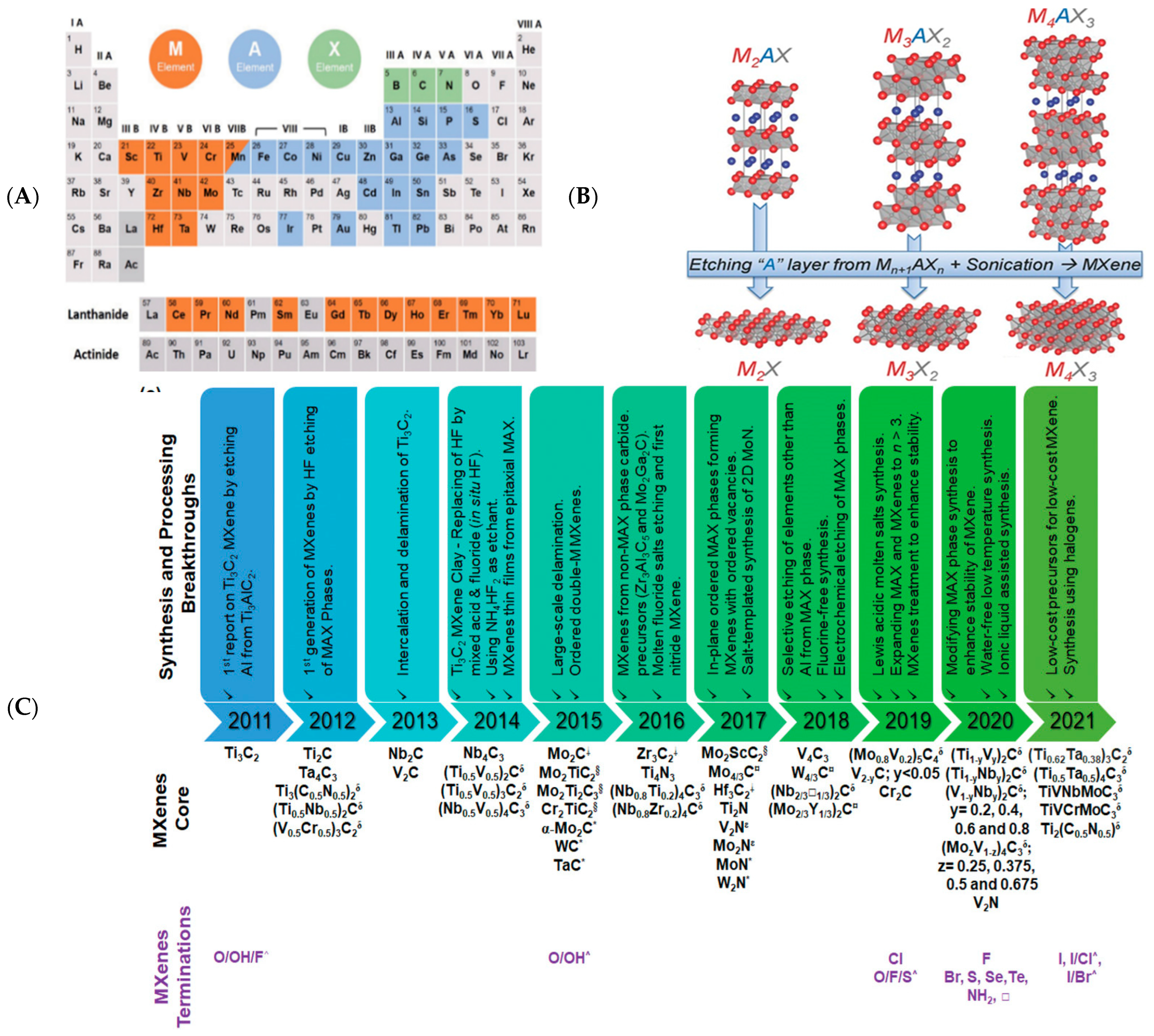
The precursor of MXenes is the MAX phase. MAX consists of Mn+1Xn units and an alternately stacked “A” element single atomic plane, expressed as Mn+1AXn. The unique crystal structure of the MAX phase combines the excellent properties of ceramics and metals [7]. Etching the “A” element of the MAX phase yields two-dimensional nanomaterial MXenes with a structural formula of Mn+1XnTx [8]. MXenes can be expressed as M2XF2, M2X(OH)2, M2XO2, etc. M is a transition metal; “A” is an element of Groups 13 and 14 of the periodic table; X is boron, carbon, or nitrogen; n includes integers from 1 to 3; Tx denotes surface groups [9] (Figure 1A,B). A list of the significant syntheses and processes in the field of MXenes research over the last decade, as well as the development of new MXenes core components and surface group control techniques, is illustrated in Figure 1C. Compared to the precursor MAX phase, derivative MXenes retain metallic and electrical conductivity benefits of MAX but also offer smaller lateral dimensions and thicknesses, as well as unique physical and chemical properties [10,11].
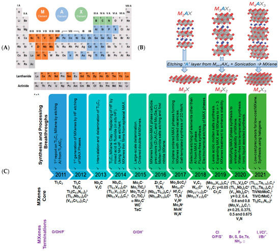
Figure 1.
(A) Elements represented by M, A, and X in the MAX phase. (B) The structure of the MAX phase and its corresponding MXenes. Reprinted with permission from Ref. [9]. Copyright 2013, Wiley-VCH. (C) Chronological presentations of progress in the field of MXenes. List of the main synthesis and processing breakthroughs over the first 10 years of MXenes’ research and new MXenescore compositions discovered in that decade and progress in surface terminations control. δ Solid solution MXenes; □ MXenesfrom non-MAX phase precursors; § out-of-plane ordered double transition metal MXene; ¤ MXenes from in-plane ordered double transition metal MAX phase analogues; * 2D carbides and nitrides produced by bottom-up approaches; ε nitride MXenes produced by the post-synthesis treatment of carbide MXene; □ vacancy; ^ mixed terminations. Reprinted with permission from Ref. [12]. Copyright 2021, Wiley-VCH.
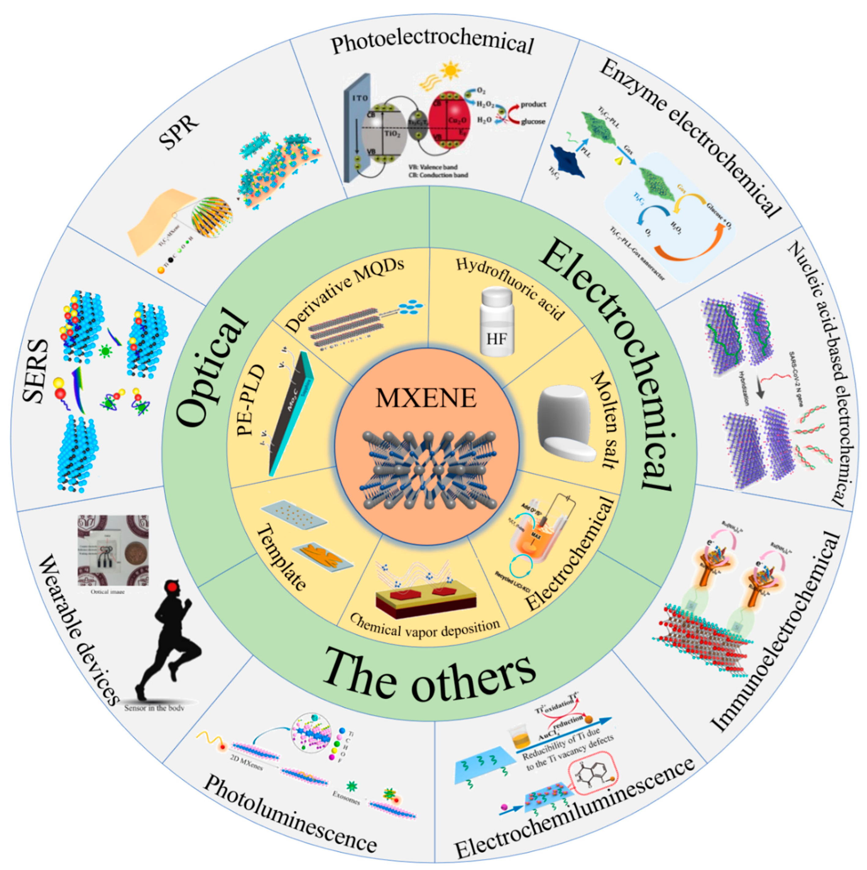
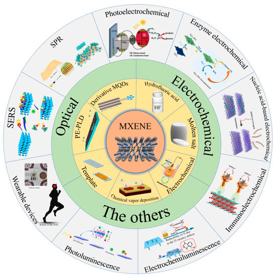
The central area of current advanced biosensing research studies is developing biosensors for detecting biological and chemical molecules that affect disease or are damaging to the human body. The most advanced biosensors can accurately and rapidly detect the target, predict the onset of the disease in time, and receive immediate medical attention [13]. Hence, high sensitivity and selectivity are significant for the design of biosensors. Due to its unique mechanical, hydrophilic, biocompatibility, and other excellent properties, MXenes are frequently used as a new biosensing platform. Electrochemical biosensors are essential for biological, environmental, and pharmaceutical fields. It offers high sensitivity, long-term reliability and high accuracy, rapidity, low cost, and easy miniaturisation [14]. In addition, electrochemical biosensors offer a further path for creating next-generation point-of-care testing devices [15]. With advancing nanotechnology with respect to MXene-based optical biosensors, unprecedented progress has been made in optical analysis. Optical analysis has advantages of high sensitivity, high selectivity, fast analysis, and good reproducibility. It has been widely used in biochemistry and biomedical and environmental analysis and has received increasing attention [16]. The synthesis of MXenes and their application in biosensing are reflected in Scheme 1. We will review and summarize published studies on biosensing since the development of MXenes, including those mainly classifying biosensors into electrochemical, optical biosensors and some derivative biosensors. In addition, we will also discuss the challenges of MXenes in preparing biosensors and future perspectives on applying MXenes in biosensing.
Scheme 1.
MXenes cover both top-down and bottom-up methods of synthesis. They play an irreplaceable position in enzyme-, nucleic-acid-, immune-based electrochemical biosensing; photoluminescence; electrochemiluminescence; photoelectric effect-based optical biosensors; and other biosensors such as wearable biosensors, surface plasmon resonance, and surface-enhanced Raman spectroscopy.
2. Synthesis and Structures of MXenes
2.1. Synthesis of MXenes
There are two methods for the synthesis of MXenes. The top-down method is the most commonly used, which can be used to exfoliate multilayer materials into a few-layer or single-layer MXenes sheet. The second method is a bottom-up approach, which focuses on the growth of Mxenes from atoms or molecules [17,18].
2.1.1. Top-Down Method
Selective etching disintegrates the strong covalent bonds between the MX and the A layers in the MAX phase. The primary method is etching with hydrofluoric acid (HF), molten salts, etc. In this process, oxygen (O), hydroxyl (OH), and fluorine (F) replace the M-A strong metal bond [17]. There are two main steps to gain 2D MXenes by HF: etching and exfoliation. Although the direct use of HF is straightforward and practical, it causes environmental pollution and damages to the human body [4]. In situ HF can be obtained by reacting a fluorinated salt with mild acid, which is less toxic to MXenes surfaces [19]. Researchers explored new synthetic methods (Figure 2). The typical chemical reaction equation for the synthesis of MXenes in the MAX phase is as follows [9].
MXenes must undergo an exfoliation process to obtain nanosheet structures: The surface groups of MXenes result in the layers being linked by hydrogen and Van der Waals forces [3]. Exfoliation enhances the interlayer spacing by weakening interactions between layers using various molecular and ionic processes [20].
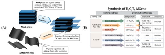
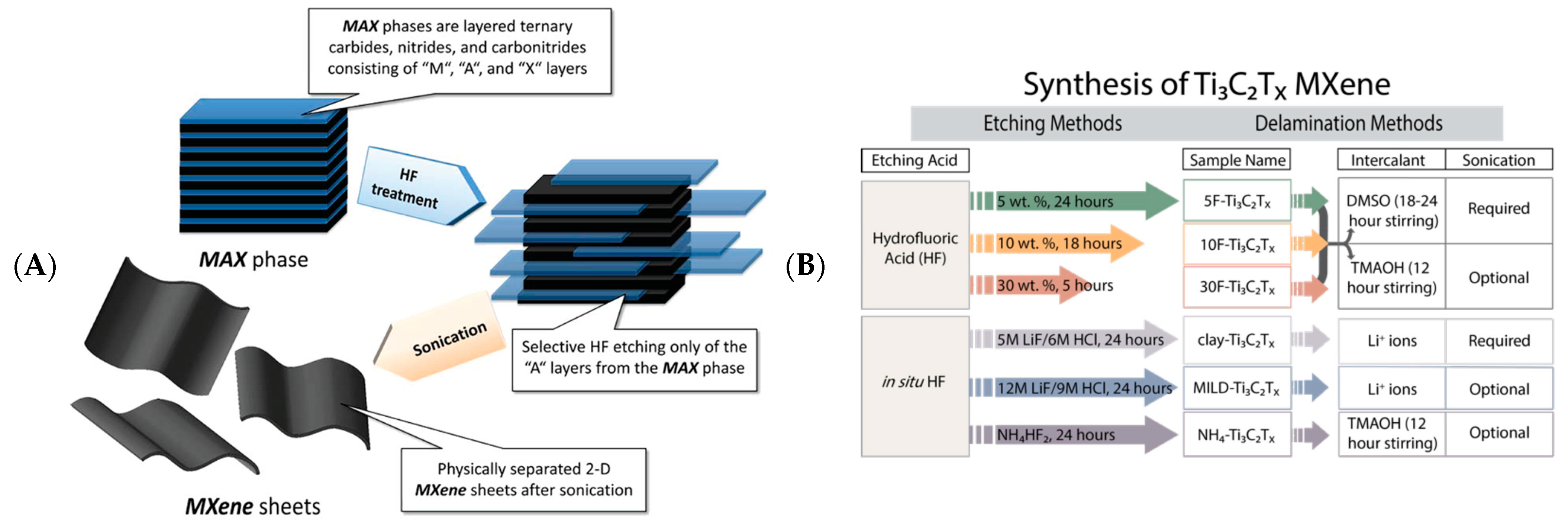

Figure 2.
(A) Schematic diagram of the process of preparing MXenes by HF. Reprinted with permission from Ref. [21]. Copyright 2012, American Chemical Society. (B) A guide to Ti3C2 MXenes synthesis using HF. Reprinted with permission from Ref. [22]. Copyright 2017, American Chemical Society.
Figure 2.
(A) Schematic diagram of the process of preparing MXenes by HF. Reprinted with permission from Ref. [21]. Copyright 2012, American Chemical Society. (B) A guide to Ti3C2 MXenes synthesis using HF. Reprinted with permission from Ref. [22]. Copyright 2017, American Chemical Society.

The molten salt method uses fluorinated molten salts, Lewis salts [23]. The synthesis does not involve fluoride, reducing the risk of synthesis [8,24]. The mechanism of MXenes formation in molten salts is similar to that of conventional HF methods: ZnCl2 and CuCl2 high-temperature molten salts strip a more comprehensive range of MAX phase materials [8] (Figure 3A). In the molten salt of Lewis acids, Zn2+, Cu2+, and Cl− are consistent with acting H+ and F− in HF. Minimally intensive layer delamination (MILD) and electrochemical etching can also be used for MAX etching, producing high-quality, non-toxic MXenes [25,26].
2.1.2. Bottom-Up Method
Bottom-up synthesis methods have been reported, such as chemical vapor deposition (CVD) [27], template [28], and plasma-enhanced pulsed laser deposition (PE-PLD) [29] (Figure 3B). MXenes produced by this method possess good crystalline quality and controllable structure and size [18].


Figure 3.
(A) Preparation mechanism of Ti3C2Cl2 etched by ZnCl2. Reprinted with permission from Ref. [30]. Copyright 2019, American Chemical Society. (B) Bottom-up approach to obtain MXenes. Atomic layer deposition method: steps to prepare Ti3AlC2 MAX films by sputtering Ti, Al and C on a sapphire substrate (a), schematic diagram of Ti3C2Tx (b) and STEM images (c). CVD method: schematic diagram of the Mo2C synthesis process (d), AFM images of hexagonal ultra-thin Mo2C crystals (e) and STEM images (f). Reprinted with permission from Ref. [27]. Copyright 2020, American Chemical Society.

Xu et al. used CVD to synthesize high-quality Mo2C crystals [27]. The synthesis of Mo2C MXene/graphene heterostructures and Mo2C MXene-graphene hybrid films by this method has been reported [29,31]. Compared to CVD, the template method has a relatively high yield of MXenes. Two-dimensional MXenes are mainly obtained by carbonizing or nitriding two-dimensional transition metal oxide (TMO) nanosheet templates. Xia et al. prepared hexagonal-structured 2D h-MoN nanosheets using precursor MoO2 nanosheets [28]. PE-PLD is a successful method for preparing large-area ultra-thin face-centered cubic (FCC) Mo2C MXene [29].
The stability of MXenes is an important property and limits its application to a certain extent. Researchers have tried to improve its stability. High concentrations of HF accelerate the degradation of MXenes and affect its structure, so relatively mild reaction conditions are necessary [32]. Organic solvents mitigate the oxidation of MXenes. Contact with water should be avoided as much as possible to prevent oxidation [33]. The oxidation of MXenes is quicker in liquid media than in solid media, and this degradation process is exacerbated by photocatalysis and thermocatalysis [34]. The storage of MXenes in Ar-sealed vials at 4 °C exhibits high stability at room temperatures [35].
2.2. Strustures of Mxenes
The crystal structure within a 2D material can affect its properties [18]. There are six types of MXenes structures (Figure 4A): (1) single transition metal MXenes (Ti3C2 and Nb4C3); (2) solid solution MXenes ((Ti, V)3C2 and (Cr, V)3C2); (3) sequential planar internal and external bimetal MXenes with one transition metal occupying the outer layer (Cr and Mo); the central metal is another metal (Nb and Ta) [36,37]; (4) ordered double-transition metals MXenes ((Cr2V) C2); (5) orrderly double vacancy MXenes (Mo1.33CTx) [38]; (6) random empty space MXenes (Nb1.33CTx) [39].
Computational simulation studies have been reported to identify novel stable MXenes structures, contributing to exploratory studies [40]. The properties and applications of these materials can be adapted by various parameters for composition, surface modification by heat treatment or chemical pathways, and structural adjustments [41]. MXenes have two—dimensional structures (a), one—dimensional structures (b) and (c), three—dimensional structures (d), and zero—dimensional structures (e) (Figure 4B).


Figure 4.
(A) Different types of MXenes structures. Reprinted with permission from Ref. [42]. Copyright 2019, Elsevier. (B) 2D, 1D, 3D, and 0D structures of MXenes. Adapted with permission from Ref. [18]. Copyright 2021, Wiley-VCH.
Figure 4.
(A) Different types of MXenes structures. Reprinted with permission from Ref. [42]. Copyright 2019, Elsevier. (B) 2D, 1D, 3D, and 0D structures of MXenes. Adapted with permission from Ref. [18]. Copyright 2021, Wiley-VCH.

3. MXenes in Biosensing
Several strategies involving MXenes in analytical nanoscience, biosensing, and other areas have been reported. MXenes exhibit hydrophilicity due to surface groups such as OH, O, and F. Its surface can interact with most biomolecules through hydrogen bonding, Van der Waals forces, electrostatic interactions, and ligand binding, rendering it an excellent carrier for biosensors applications [43,44,45]. Several different MXenes compositions have been proved to be biocompatible and non-cytotoxic [46,47].
We summarized the composition and analytical performance of some MXene-based electrochemical biosensors, optical biosensors, and other biosensors and attached them to the subsections. These cases demonstrate the broad applicability of MXenes in the fabrication of biosensors. Readers can easily extract MXene-based biosensing research and measurement data from these tables.
3.1. Electrochemical Biosensing
Due to their high electronic conductivity, MXenes can drive most electrochemical reactions, which is of great significance for the application in electrochemical biosensing [48]. The electrical properties of MXenes can be improved by changing elemental compositions and surface groups [18]. In particular, the external transition metal layer of MXenes plays a more critical role in the electronic properties than the internal layer [49]. The number and thickness of the layers of MXenes also affect electrical properties [3,50].
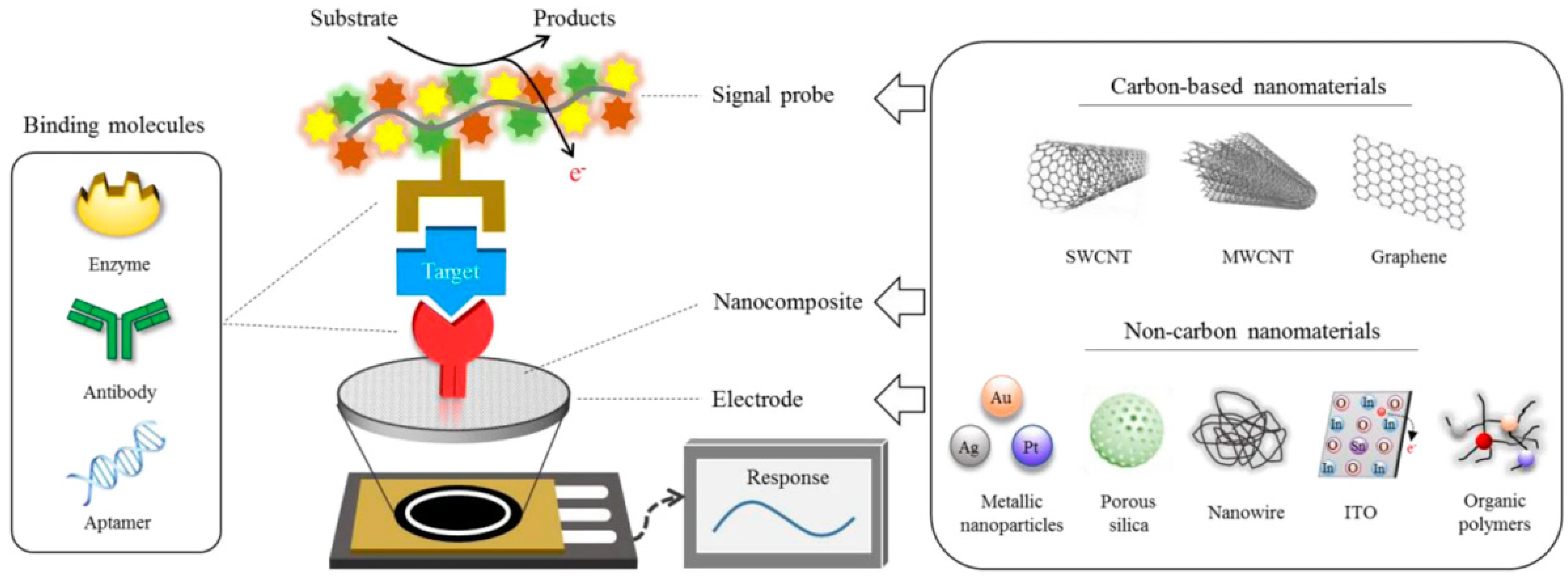
Biosensors based on electrical signals change the electrochemical properties of the sensor surface by binding to essential substances in the organism, such as proteins, amino acids, nucleic acids, antibodies, etc. (Figure 5). The development of MXenes for electrochemical biosensors has been intensively investigated because of their excellent properties, such as high conductivity, electrochemical activity, and large surface area. The classification of electrochemical biosensors is as follows: enzyme electrochemical, nucleic acid electrochemical, and immunoelectrochemical biosensing.
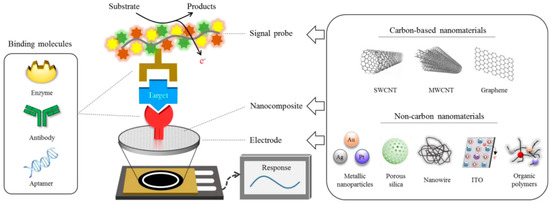
Figure 5.
Schematic diagram of the analytical principle of an electrochemical biosensor. Reprinted with permission from Ref. [51]. Copyright 2020, Springer Nature.
3.1.1. Enzyme-Based Electrochemical Biosensing
Enzyme electrochemical biosensors with higher efficiencies and substrate specificities in mild conditions have been extensively explored over the last few years (Table 1). The basic principle is the direct electron transfer (DET) process between the enzyme and the electrode. The immobilisation of enzymes on the bare electrode surface can render the enzymes biologically inactive, making it extremely difficult to perform DET on the electrode’s surface [52]. MXenes can be used as a strategy for enhancing DET because of their large specific surface area, excellent electrical conductivity, and good biocompatibility.
Much of the literature has shown that MXenes or MXenes composite materials can maintain the activity of enzymes after complexing enzymes due to the various properties and unique structures of MXenes. This demonstrates that MXenes can be magnificent structures for enzyme-based biosensors. Xu et al. mixed Ti3C2 MXene and HRP enzyme directly to fabricate a biosensor for the detection of H2O2 to analyse the levels of serum samples from AMI patients before and after surgery [53]. Ma et al. fabricated a low detection limit enzyme biosensor for the detection of H2O2 using a chitosan complex of Ti3C2 MXene-loaded HRP enzyme and successfully used it to detect trace amounts of H2O2 in foods [54].


Table 1.
Enzyme-based electrochemical biosensors for identifying units, target, and analytical parameters.
Table 1.
Enzyme-based electrochemical biosensors for identifying units, target, and analytical parameters.
| MXenes Composite | Identify Units | Target | LOD | Range | Ref. |
|---|---|---|---|---|---|
| Au/Ti3C2 | glucose oxidase | glucose | 5.9 μM | 0.1–18 mM | [55] |
| PLL/Ti3C2 | glucose oxidase | glucose | 2.6 μM | 4.0–20 µM | [56] |
| PEDOT: SCX/Ti3C2Tx | glucose oxidase | glucose | 22.5 μM | 0.5–8 mM | [57] |
| Ti3C2/Nafions | Horse Radish Peroxidase | H2O2 | 1 μM | 5–8000 μM | [53] |
| MXene/chitosan | Horse Radish Peroxidase | H2O2 | 0.74 μM | 5–1650 μM | [54] |
| Chit/ChOx/Ti3C2Tx | cholesterol oxidase | cholesterol | 0.11 nM | 0.3–4.5 nM | [58] |
| Ti3C2 | tyrosinase | phenol | 12 nmol L−1 | 50 nM–15.5 μM | [59] |
| CS-Ti3C2Tx | acetylcholinesterase | acetylthiocholine chloride | 3 fM | 10 nM–10 fM | [60] |
| GA/Nb2CTx | acetylcholinesterase | phosmet | 144 pM | 200 pM–1 μM | [26] |
In addition, there are several reports on using MXenes in different compound types of enzymes, such as glucose oxidase [57], cholesterol oxidase [58], acetylcholinesterase [60], tyrosinase [59], etc. Wu et al. proposed a hybrid PLL/Ti3C2/glucose oxidase glucose biosensor that accelerates the breakdown of H2O2 generated during glucose oxidation by catalysing a cascade reaction [56] (Figure 6A). Xia et al. developed a Chit/cholesterol oxidase/Ti3C2Tx composite cholesterol oxidase biosensor [55]. Chit/Ti3C2Tx served as a support matrix for immobilising the enzyme. Gold nanoparticles anchored on Ti3C2Tx MXene nanosheets enhanced the electron transfer between the enzyme and the electrode. The relative current sensitivity and LOD were 0.3–4.5 nM and 0.11 nM, respectively. Song et al. derived electrochemical etching to derive fluorine-free Nb2CTx with low cytotoxicity and constructed a Nb2CTx/acetylcholinesterase biosensor to detect sulfoxide [26] (Figure 6B). Moreover, the sensor’s enzymatic activity and electron transfer are superior to the corresponding V2C and Ti3C2 MXenes biosensors. Wu et al. used Ti3C2 MXene as a new substrate to immobilise tyrosinase and facilitated the direct electron transfer process for the sensitive and rapid detection of phenol [59]. Therefore, Ti3C2 MXene can be a phenolic biosensor with high recovery and long-term stability. The biosensor exhibits good analytical performance over a wide linear range of 0.05–15.5 μM, with detection limits as low as 12 nM.

Figure 6.
(A) Schematic diagram of the detection of the Ti3C2-PLL-Gox nanoreactor Reprinted with permission from Ref. [56]. Copyright 2020, Elsevier. (B) Schematic illustration of the enzymatic inhibition of sulfoxide detection by the HF-free Nb2CTx/AChE biosensor Reprinted with permission from Ref. [26]. Copyright 2020, Wiley-VCH.
The above examples and the contents show that it is feasible to combine enzymes directly on MXenes or with other materials to improve the performance of enzyme electrochemical biosensors.
3.1.2. Nucleic Acid-Based Electrochemical Biosensing
Using nucleic acids as recognition elements allows the specific recognition of the target and the generation of some signal changes [61]. Nucleic acid is a stable and easy-to-handle biomolecule, so it has excellent detection performances [62]. Nucleic-acid-based electrochemical biosensors offer advantages of both nucleic acid probes and electrochemical detection, enabling the sensitive detection of analytes such as nucleic acid, ref. [63] proteins [64], biological molecules [65], inorganic ions [66], and cells [67] (Table 2). Nucleic acid electrochemical biosensors are based on five conformations: double-stranded, triple-stranded, quadruple-stranded, DNA nanostructures, and single-stranded DNA functionalisation (hairpin structure, aptamers, and DNAzyme) [68]. Unlike enzymes, nucleic acids possess little redox capacity. The development of nucleic acid electrochemical biosensors generally relies on molecules with redox properties, such as methylene blue (MB) and ferrocene (Fc), or through charge changes that occur during nucleic acid hybridisation [69]. The nucleic acid electrochemical biosensor has various applications in genetics, clinical medicine, and biosensing due to its rapid detection, simple experimental procedures, high sensitivity, and low cost [70]. There are two types of nucleic acid biosensors.
The first type of nucleic acid electrochemical biosensor follows the Watson–Crick pairing principle, which hybridizes a nucleic acid sequence with a complementary nucleic acid sequence through base pairing [61]. The detection principle works by immobilising nucleic acids on the electrode’s surface to capture complementary nucleic acid sequences, thus obtaining an altered electrical signal for specific detection [71]. There are many reports using specific nucleic acid sequences to create biosensors for the detection of disease-predicting miRNAs and DNA, and some electrochemical biosensors have been validated for point-of-care detection. Duan et al. developed a Ti3C2/FePc QDs MXene nanocomposite nucleic acid biosensor with good biocompatibility [72]. The Ti3C2/FePc QDs composite material was used as a carrier to detect miRNA-155 by using a change in electrochemical impedance caused by DNA modifications. Mohammadniaei et al. used double screen-printed gold electrodes modified with MXenes and AuNPs and single-stranded DNA-functionalised magnetic particles to detect miRNA-21 and miRNA-141 by using duplex-specific nuclease (DSN) amplification assay strategy [73] (Figure 7A). This biosensor can continue to be upgraded to quantify more analytes, forming a device for point-of-care testing (POC) cancer screening. Chen et al. fabricated a DNA electrochemical biosensor using MXene-based [74]. The surface groups were covered using ssDNA adsorbed on Ti3C2 MXene to attenuate conductivity. When target DNA and ssDNA are hybridized and desorbed from Ti3C2 MXene, the fast, simple, and sensitive detection of N-gene sequences in SARS-Cov-2 was possible (Figure 7B). The feasibility of DNA-functionalised MXenes in developing real-time monitoring diagnostic devices for clinical testing can be demonstrated.


Table 2.
Nucleic acid-based electrochemical biosensors identify units, target, and analytical parameters.
Table 2.
Nucleic acid-based electrochemical biosensors identify units, target, and analytical parameters.
| MXenes Composite | Identify Units | Target | LOD | Range | Ref. |
|---|---|---|---|---|---|
| MoS2 /Au NPs/Ti3C2 | DNA probe | miRNA-182 | 0.43 fM | 1 fM–0.1 nM | [63] |
| Au/Ti3C2 | DNA probe | miRNA-21, 141 | 204 aM 138 aM | 500 aM–50 nM | [73] |
| Ti3C2Tx @FePcQDs | DNA probe | miRNA-155 | 4.3 aM | 0.01 fM–10 pM | [72] |
| MCH/CP/AuNPs/Ti3C2Tx | DNA probe | BCR/ABL fusion gene | 0.05 fM | 0.2 fM–20 nM | [75] |
| Ti3C2Tx | DNA probe | SARS-Cov-2 N gene | 105 copies mL−1 | 105–109 copies mL−1 | [74] |
| PMo12/PPy@Ti3C2Tx | Aptamer | Osteopontin | 0.98 fg mL−1 | 0.05–10,000 pg mL−1 | [64] |
| AuNPs/Ti3C2 | Aptamer | Mucin 1 | 0.72 pg mL−1 | 5 pg mL−1–50 ng mL−1 | [76] |
| Ti3C2 | Aptamer | gliotoxin | 5 pM | 5 pM–10 nM | [65] |
| Ti3C2 | Aptamer | HER2-positive CTCs | 47 cell mL−1 | 20–200 cells mL−1 | [77] |
| CoCu-ZIF@ Ti3C2 CDs | Aptamer | B16-F10 cell | 33 cells∙mL−1 | 1 × 10²–1× 10⁵ cells∙mL−1 | [67] |
| Au@Nb4C3Tx | Aptamer | Pb2+ | 4 nM | 10 nM–5 μM | [66] |
The second nucleic acid electrochemical biosensor uses single-stranded DNA (ssDNA) or RNA to bind to various biomolecules for analyte detection, including proteins, small biomolecules, cells, etc. [78,79]. Electrochemical biosensors made up of aptamers are easy, reliable, quick in responding, low in price, and possess acceptable repeatability [80]. Geng Xue of our research group cleverly used conformational changes of aptamers before and after capturing serotonin to construct an aptamer biosensor [81]. The interaction between aptamer and serotonin was destroyed by guanidine hydrochloride, and 98.2% of the signal was recovered, showing acceptable repeatability. Zhou et al. synthesized intercalating polypyrrole (PPy) Ti3C2Tx MXene and phosphomolybdic acid (PMo12) composites with a strong synergistic effect, promoting the anchoring of RNA aptamers on the composites [64] (Figure 7C). The G-quadruplex formed by osteopontin (OPN) and aptamer exhibits stable and high sensitivity, which proves the excellent performance of this MXene composite aptamer biosensor. Li et al. created a nuclease-driven DNA walker cascade signal amplification strategy to construct electrochemical aptamer biosensors on Au nanoparticles/MXene-modified electrodes for mucin 1 [76]. A DNA nanostructure-modified Ti3C2 MXene nanosheet biosensor was developed by Wang et al. for the detection of gliotoxins [65]. Tetrahedral DNA nanostructures were quickly immobilised on the surface of MXenes nanosheets, thus avoiding the tedious and expensive modification of DNA probes. HB5 aptamer immobilised on the MXenes layer via electrostatic interactions was highly selective for HER-2-positive cells, as reported by Vajhadin et al. Sandwich-like structures formed between magnetically captured cells, and functionalised MXenes electrodes effectively shield electron transfers, allowing quantitative cell detection with changes in the current [77] (Figure 7D).
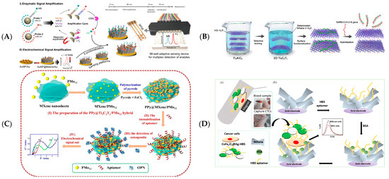
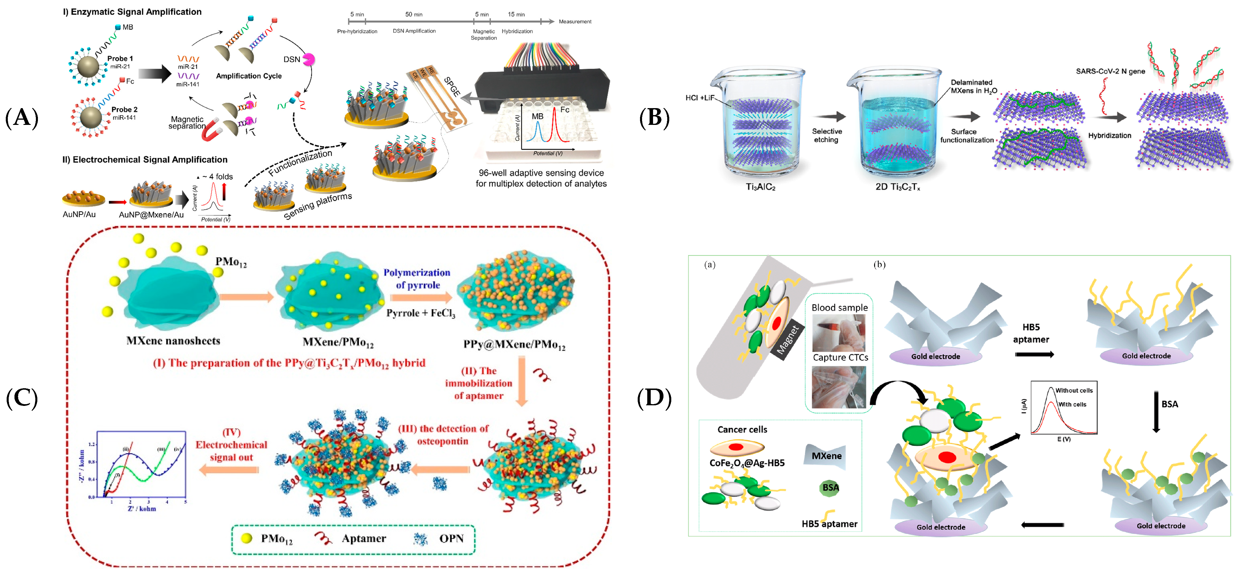

Figure 7.
(A) Schematic diagram representing the entire assay procedure for multiplex and concurrent detections of miRNA. Reprinted with permission from Ref. [73]. Copyright 2020, Elsevier. (B) Schematic of the ssDNA/Ti3C2Tx for the detection of the SARS-Cov-2 nucleocapsid gene. Reprinted with permission from Ref. [74]. Copyright 2022, American Chemical Society. (C) Schematic diagram of PPy@Ti3C2/PMo12 aptamer biosensor for OPN detection. Reprinted with permission from Ref. [64]. Copyright 2019, Elsevier. (D) Schematic diagram of the MXenes based cell sensor for the detection of SK-BR-3 cells: magnetic cell separation using CoFe2O4@Ag-HB5 (a) and electrochemical cell detection on a functionalised MXenes surface (b). Adapted with permission from Ref. [77]. Copyright 2021, Elsevier.

3.1.3. Immunoelectrochemical Biosensing
Electrochemical immunosensors are coupled to the sensor via antigen–antibody interactions. The accessibility of antibody to a wide range of molecules and the high selectivity and sensitivity renders immunochemical methods valuable for clinical diagnosis. These electrochemical biosensors for bioanalysis have advantages of small reagent volumes, high sensitivity and specificity, and portability [82]. As observed from the contents, immunoelectrochemical biosensors offer tremendous advantages in the specific detection of biomolecules (Table 3).

Table 3.
Immunoelectrochemical biosensors identify units, target, and analytical parameters.
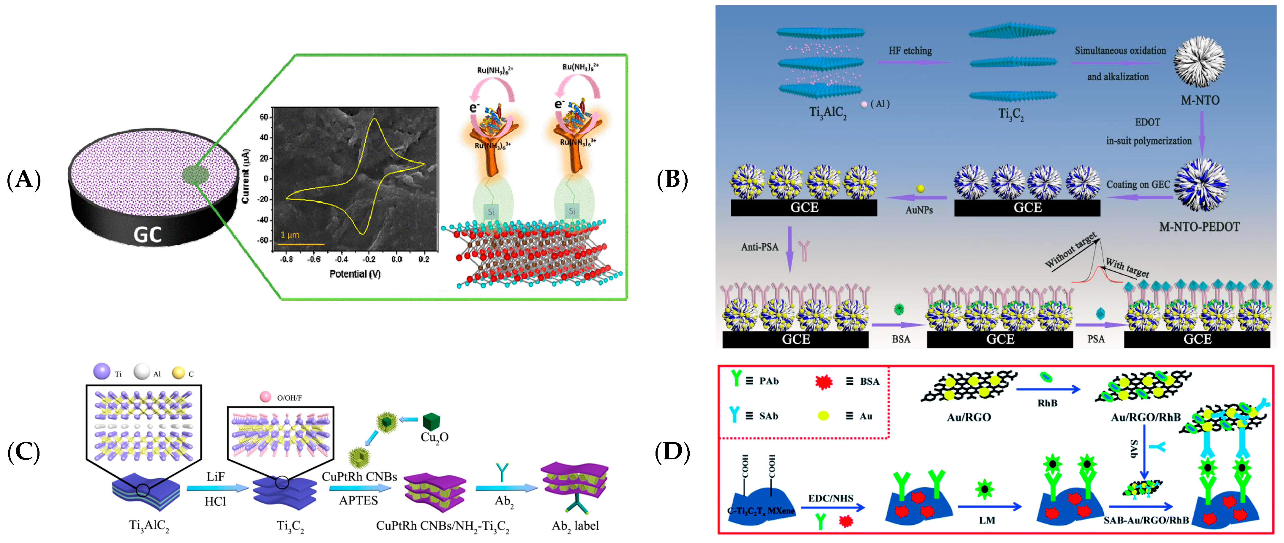
In 2018, Kumar et al. fabricated the first MXene-based immunoelectrochemical sensor to detect carcinoembryonic antigens (CEAs) [84]. Aminosilane-functionalised MXenes offered more binding sites for bioreceptors than GCE, and the CEA antigen is better immobilised on Ti3C2 MXene (Figure 8A). Xu et al. synthesized a composite of 3D sodium titanate nanoribbons, anchored poly(3,4-ethylene dioxythiophene), and gold nanoparticles by oxidizing and alkalizing Ti3C2 Mxene [86]. The composites described above were used to immobilise prostate-specific substance (PSA) antibodies to create a facile electrochemical label-free immunosensor for the sensitive detection of PSA (Figure 8B).
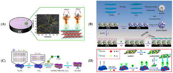
Figure 8.
(A) Schematic diagram of the detection mechanism of electrochemical CEA. Reprinted with permission from Ref. [84]. Copyright 2018, Elsevier. (B) The fabrication and detection steps of the immunosensor. Reprinted with permission from Ref. [86]. Copyright 2020, Elsevier. (C) Schematic diagram of the fabrication of the working electrode of the immunosensor. Reprinted with permission from Ref. [87]. Copyright 2020, American Chemical Society. (D) Illustrations for constructing the sandwich-like immunosensor of LM based on Ti3C2Tx MXene. Reprinted with permission from Ref. [88]. Copyright 2021, Royal Society of Chemistry.
Dong et al. used a CuPtRh/NH2-Ti3C2 nanocomposite composed of trimetallic hollow CuPtRh cubic nanoboxes (CNBs) and laminated ammoniated Ti3C2 flakes to fabricate a sandwich-type immunosensor to detect cardiac troponin I (CTnI) [87]. Aminated Ti3C2 provides abundant binding sites for both CuPtRh CNBs and antibodies, while CuPtRh CNBs can prevent Ti3C2 from stacking again (Figure 8C). In addition, MXenes can serve to detect bacteria. Niu et al. constructed a sensing platform with carboxylated Ti3C2Tx MXene and rhodamine B/gold/reduced graphene oxide as the signal [88] (Figure 8D). A sandwich electrochemical immunosensing platform for detecting Listeria monocytogenes was also developed by them.
3.2. Optical Biosensing
Optical properties, including absorption, transmission, photoluminescence, scattering, and emission, are essential for applying MXenes. The surface groups, doping, and defects affect the energy band’s structure [89]. A thin layer of Ti3C2Tx has been reported to absorb photons in the UV-visible region between 300 and 500 nm with a transmission of 91%. O-functionalised Ti3C2 MXene has a higher light absorption efficiency [90]. The optical properties are also affected by the thickness of the film and the distance between MXenes layers. Intercalation with hydrazine, urea, methyl ammonium hydroxide, and DMSO changes the interlayer distance of Ti3C2Tx, decreasing light transmittance [50].
MXenes have excellent hydrophilicity, biocompatibility, and optical characteristics, making them appropriate for all sorts of biosensing applications. It was discovered as a fluorescence quenching agent and a carrier for biomedical and imaging applications, contributing to high-performance optical biosensors. The interaction of light and materials is central to the optical inspection principle. It identifies samples by non-destructively monitoring changes in the intensity or spectral shift of light [91]. This section will summarize MXenes biosensing applications in photoluminescence, electrochemiluminescence, and photoelectrochemical applications.
3.2.1. Photoluminescence (PL)
MXenes possess features that make MXenes excellent for fluorescence biosensors, such as larger absorption bands, higher energy levels, etc., which causes fluorescence quenches in fluorescent substances [91]. Hence, changes in fluorescence intensity can be employed as indications for biological analytes detection (Table 4). MXene quantum dots (MQDs) are luminous, extremely water-soluble, dispersible, and biocompatible [92]. As a consequence, searching for photoluminescent biosensors based on MXenes and MQDs has emerged as a widespread research issue [93,94].

Table 4.
MXene-based photoluminescence biosensors identify units, target, and analytical parameters.
Because of MXenes’ strong and broad absorption in the visible and near-infrared regions, MXenes generally act as an acceptor designed to quench the fluorescence signal emitted by sensing probes, such as metal nanoclusters, quantum dots, fluorescent dyes, etc. [94]. Due to the two-dimensional planer structure and hydrophilic surface groups of MXenes, the abundant binding sites and hydrophilic groups on MXenes provide more possibilities for biomolecular interactions [44].
Shi et al. detected glutathione by combining copper nanoclusters (Cu NCs)-functionalised MXenes [99]. MXenes quenches the fluorescence of Cu NCs through the internal filtering effect (IFE), and glutathione can analyze MXenes and Cu NCs, resulting in fluorescence recovery. Ti3C2 MXene nanosheets combined with red-emitting carbon dots (RCDs) area unit effective and selective fluorescence stimulant sensors were used for glucose detection by Zhu et al. Ti3C2 nanosheets impassively quenched the fluorescence intensity of RCDs (>96%) through IFE [94] (Figure 9A). Kalkal et al. constructed a fluorescent biosensing system based on Ag/Ti3C2 to quench the fluorescence signal on antibody/amino-graphene quantum dots [97]. The fluorescence recovered when antigen was added. It can be used to detect neuron-specific enolase with good reproducibility.

Figure 9.
(A) Schematic diagram of MXene-based glucose oxidase fluorescent biosensor. Reprinted with permission from Ref. [94]. Copyright 2019, Royal Society of Chemistry. (B) Illustration of analysis of HPV-18 type DNA using Ti3C2 MXene. Reprinted with permission from Ref. [95]. Copyright 2019, Elsevier.
MXenes are used as fluorescence quenchers to construct optical sensors for monitoring enzyme activity and biomolecules. Similarly to the previous section, the Fc and MB of the nucleic acid biosensor can be replaced with some fluorescent materials that can be used, which are more practical for this type of biosensor. Zhu et al. reported a Ti3C2 MXene-based fluorescent biosensor to detect phospholipase D by FRET quenching of rhodamine B (RhB)-labeled phospholipids [98]. Phospholipase D cleaves phospholipids, causing RhB-labeled phospholipids to detach from Ti3C2 MXenes and re-reflorescence. Peng et al. used the affinity difference between single-stranded and double-stranded DNA on MXenes to construct fluorescent signal detection for human papillomavirus HPV-18 DNA on ultra-thin Ti3C2 MXene [95] (Figure 9B). Wang et al. presented dual-signal-labelled DNA-functionalised Ti3C2 MXene nanoprobes to achieve a dual analysis of MUC 1 and miRNA-21 at low concentrations in vitro, and the in situ imaging of MCF-7 breast cancer cells [96]. Furthermore, cell imaging can provide multiple layers of information, such as biomarkers’ expression levels and spatial distribution.
When the thickness dimensions of 2D nanomaterials are less than 100 nm, MXenes can be converted into quantum dots with quantum confinement and optical properties [105]. MQDs, with an average lateral size ranging from 1.8 to 16 nm, can be obtained by hydrothermal processes [100], acidic oxidation, and chemical stripping [106]. Charge transfer is enhanced, and fluorescence is enhanced by utilizing heteroatom doping [101]. MQDs have similar properties to MXenes, such as high dispersion and good biocompatibility. The small band gaps of MXenes can expand their band gap through quantum effects, contributing to their strong fluorescence effect [107]. Some researchers synthesized MQDs that exhibited different fluorescence effects in different solvents under 365 nm UV light irradiation [93,104].
On account of their tunable size, photoluminescence, and photostability, MQDs can be applied as fluorescent probes and can also be functionalised with natural biomolecules [107]. The performance of MQDs as fluorescent agents or signals can be improved, and the application of MXenes in biosensing can be widely expanded [91,108]. MQDs have contributed enormously to detecting metal ions, biomolecules, and cellular imaging. The first MQD-based fluorescence sensor is based on the coordination of Zn2+ through hydroxyl groups on the surface of MQDs with selective quenching [100] (Figure 10B). Heteroatom-doped MQDs can be the detector for the fluorescence detection of different metal particles, such as Cu2+ [101], Ag+, and Mn2+ [109].

Figure 10.
(A) Schematic diagram of fluorescence assay of alkaline phosphatase activity of MQDs. Reprinted with permission from Ref. [102]. Copyright 2018, Royal Society of Chemistry. (B) Schematic diagram of hydrothermal preparation of MQDs. Reprinted with permission from Ref. [100]. Copyright 2017, Wiley-VCH.
MQDs can be implemented to detect some biomolecules because they have absorption bands that overlap with the excitation and/or emission spectra of MQDs. Guo et al. designed an MQD-based fluorometric strategy for alkaline phosphatase activities and embryonic stem cell identification [102] (Figure 10A). The effective quenching of MQD fluorescence was obtained by p-nitrophenol produced by the alkaline phosphatase-catalysed dephosphorylation of p-nitrophenyl phosphate. It can also be used as an IFE-based method to analyse ESC biomarker ALP in ESC lysates accurately. Liu et al. described a fluorescent platform for detecting cytochrome c and trypsin [103]. The fluorescence of MQDs was burst by cytochrome c through the IFE. Meanwhile, cytochrome could be degraded by trypsin, and MQDs’ fluorescence could be restored. Chen et al. constructed a fluorescent sensor with the pH-dependent emission of blue fluorescence from MQDs for ratiometric MQDs probes to detect cellular pH [104].
3.2.2. Electrochemiluminescence (ECL)
As a mixture of electrochemistry and optics, electrochemiluminescence is a new method for evaluations and detections. Because of its low background signal, excellent sensitivity, controllability, speed, and low cost, it is frequently employed in biochemistry for proteins, nucleic acids, enzymes, and clinical diagnostics [91]. MXenes have been proven viable as working electrodes for ECL biosensors, with improved ECL characteristics compared to glassy carbon electrodes [110]. The ECL biosensor is well suited for the analysis of nucleic acids or gene fragments, biomolecules, biomarkers, and even cells (Table 5).
In 2018, Fang et al. fabricated an ECL biosensor of Ru(bpy)32+ functionalised Ti3C2Tx MXene to detect unlabelled single nucleotide mismatches in human urine, using tripropylamine as a co-reactant [111]. Exposed bases in mismatched DNA bind to Ru(bpy)32+ on the Ti3C2Tx MXene and are more prone to electrochemical oxidation in enhancing ECL intensities. Zhuang et al. constructed ECL nanoprobes via Ti3C2Tx-mediated in situ formations of Au NPs and the anchoring of luminol and utilised the catalytic hairpin assembly (CHA) amplification of signalling to fabricate ECL biosensors for miRNA-155 detection [112] (Figure 11A). Yao et al. detected the SARS-Cov-2 gene by MXenes/PEI adsorbed Au and Ru(bpy)32+ DNA walkers [113]. After the DNA walker excised hairpin DNA under the action of Nb.BbvCl endonuclease, template DNA-Ag hybridized with hairpin DNA and decreased the signal of ECL (Figure 11B). Zhang et al. modified DNA probes on MXenes/PEI composites by Ru(bpy)32+ and AuNP and used the CRISPR-Cas12a strategy to construct an ECL signal on/off biosensor for the detection of SARS-Cov-2 (RdRp) gene [114].


Table 5.
MXene-based electrochemiluminescence biosensors identify units, target, and analytical parameters.
Table 5.
MXene-based electrochemiluminescence biosensors identify units, target, and analytical parameters.
| MXenes Composite | Identify Units | Target | LOD | Range | Ref. |
|---|---|---|---|---|---|
| g-C3N4/Ti3C2 | Ti3C2 | Protein Kinase | 1.0 mU mL−1 | 0.015–40 U mL−1 | [115] |
| Ti3C2Tx | Ru(bpy)32+ | nucleotide mismatch | 5 nM | - | [111] |
| Au@Ti3C2@PEI-Ru(dcbpy)32+ | Model DNA-AgNCs | SARS-Cov-2 Gene | 0.21 fM | 1 fM–100 pM | [113] |
| AuNPs/Ti3C2/Luminol | sDNA | miRNA-155 | 0.15 fM | 0.3 fM–1 nM | [112] |
| Ru@Ti3C2@AuNPs | Fc-DNA | SARS-Cov-2 gene | 12.8 aM | - | [114] |
| Ti3C2/PEI | aptamer | MCF-7 | 125 particles μL−1 | 5 × 10²–5 × 10⁶ particles μL−1 | [116] |
| Ti3C2/Au | aptamer | CD63 | 30 particles μL−1 | 102–105 particles μL−1 | [117] |
| AuNPs/Ti3C2 | aptamer | cardiac troponin I | 0.04 fM | 0.1 fM–1 pM | [118] |
| AuNPs-Ru-Arg@Ti3C2 | antibody | CEA | 1.5 pg mL−1 | 0.01–150 ng mL−1 | [119] |
| R6G-Ti3C2Tx@AuNRs/ABEI | antibody | Vibrio vulnificus | 1 CFU mL−1 | 1–108 CFU mL−1 | [120] |
Strategies for detecting biomolecules can be implemented with aptamers, resulting in higher ECL signal intensities. Sun et al. proposed PEI-functionalised MXenes and g-C3N4 composites as detection probes, and kemptide chelated with Ti in the composites after protein kinase A (PKA) phosphorylation to promote electron transfers at the electrode’s interface, enhancing strategies for ECL signalling [115]. Moreover, this biosensor enables inhibitor screening and PKA activity monitoring in MCF-7 cell lysates. Mi et al. reported a method for the quantitative detection of cardiac troponin (CTnI) by electrochemical and ECL dual signals using tetrahedral DNAs (TDs) and in situ hybrid chain reaction (HPR) on Au/Ti3C2 MXene [118] (Figure 11C). Both ECL Dox-Luminol/Current Dox and Current MB/Current Dox dual signals can be used for the quantitative detection of CTnI, which is expected to be used in screen critically ill patients with COVID-19. Zhang et al. used MXenes to generate AuNPs in situ and modified aptamers and constructed an ECL biosensor to detect exosomes CD63 [117]. Zhang and colleagues developed an exosome-selective ECL biosensor using aptamer-modified Ti3C2 MXenes as probes with an LOD of 125 μL particles−1 [116].
Immunochemical methods are also highly selective and sensitive in the field of ECL. Luo et al. constructed an MXene-based substrate using [Ru(bpy)2(mcpbpy)]Cl2 and L-arginine as co-reactants to detect carcinoembryonic proteins (CEA) by antigen [119]. Upon antigen binding to the antibody, spatial site resistance leads to a decline in the rate of electron transfer and electrolyte diffusion at the electrode’s surface, resulting in a decrease in ECL signal intensities (Figure 11D). Wei et al. constructed an ECL/SERS dual-signal biosensor to detect the causative agent of Vibrio vulnificus (VV) [120]. The pathogenic bacteria. VV is captured by Fe3O4@Ab1 as the capture unit. Ab2, R6G, and ABEI bind to AuNR as the signal unit to capture VV through Au-S and Au-N, forming a Faraday cage structure.


Figure 11.
(A) Schematic diagram of the preparation of Au@Ti3C2@PEI-Ru(dcbpy)32+ nanocomposites (a); Combined unilateral DNA walker amplification strategy based on nanocomposites for ECL biosensor detection of SARS-Cov-2 RdRp gene (b). Reprinted with permission from Ref. [113]. Copyright 2021, American Chemistry Society. (B) Strategy of stable luminol-Au NPs-Ti3C2 (a) and construction of the proposed ECL biosensor (b). Reprinted with permission from Ref. [112]. Copyright 2021, Springer Nature. (C) Schematic representation of specific target recognition and BFP release (a) and the ratiometric biosensing mechanism of cTnI (b). Reprinted with permission from Ref. [118]. Copyright 2021, Elsevier. (D) Schematic representation showing the detection principle of the prepared ECL biosensor. Reprinted with permission from Ref. [119]. Copyright 2022, Royal Society of Chemistry.


3.2.3. Photoelectrochemical (PEC)
Similarly to electrochemiluminescence sensing, photoelectrochemical sensing is a practical analytical method that integrates optical and electrochemical analyses. MXenes also promise photoelectrochemical sensors with their excellent optical and electronic properties [121] (Table 6).

Table 6.
MXene-based photoelectrochemical biosensors identify units, target, and analytical parameters.
Li et al. took advantage of Ti3C2 MXene, readily forming PN junctions with photosensitive semiconductors and, therefore, used Ti3C2/Cu2O heterostructures for the high-sensitivity detection of glucose [122]. The in situ growth of Cu2O on MXenes improves photoelectrochemical performances compared to pure Cu2O. In order to improve the photocurrent conversion efficiency and detection sensitivity of glucose, a Z-type heterostructure based on TiO2/Ti3C2Tx/Cu2O was proposed in addition to the construction of a Schottky-junction-based PEC sensor [123]. A DNA probe can also achieve the label-free PEC determination of methyltransferase (MTase) for label-free Bi2S3/Ti3C2 PN junctions [124]. For exosomes, the enzyme-induced deposition of CdS on Ti3C2 MXene forms a Ti3C2 MXene/CdS composite, creating a built-in electric field in the tight interface between CdS and Ti3C2 MXene, enabling highly accurate detection [125]. In addition to this, the use of Ti3C2@ReS2 to immobilise DNA probes and perform specific PEC detections of miRNA-141 has excellent performance [126]. For the detection of 5hmCTP on APTES/Ti3C2, the use of antibodies for PEC is also feasible [127]. Chen et al. developed a photoelectrochemical biosensor for the sensitive and selective detection of glutathione based on MQDs [128].
3.3. Other Biosensing
3.3.1. Wearable Biosensing
The covalent between the M transition metal and the X element in MXenes, the terminal surface groups, and the thickness of the atomic layers resulted in excellent mechanical properties [3,18]. Numerous theoretical findings on the mechanical properties of MXenes have been reported. Kurtoglu et al. predicted a higher elasticity coefficients for various pristine and functionalised MXenes than their precursors due to the greater density of charge density in the Mn+1Xn layer [129]. The excellent mechanical properties provide favourable conditions for fabricating wearable biosensors.
Some studies have used wearable nanoelectronics to detect health-related physiological activities, such as physical or chemical stimulation, micropressure, and changes in physiological signals. Stretchable mechanical properties, high gauge factor, flexible materials integrating flexible bio-electronic interfaces, and miniaturized signalling systems need to be investigated to meet the required sensitivity of sensor devices and to improve the usability of wearable devices [130]. Recently, ultrathin MXenes comprised high-performance materials for stretchable and bendable conductive coatings [131] (Table 7). Conductive and conformal MXenes multilayers can withstand up to 2.5 mm bending and 40% tensile stretching, with recoverable electrical resistances, while maintaining a conductivity of 2000 S m−1 [10].

Table 7.
MXene-based wearable biosensors identify units, target, and analytical parameters.
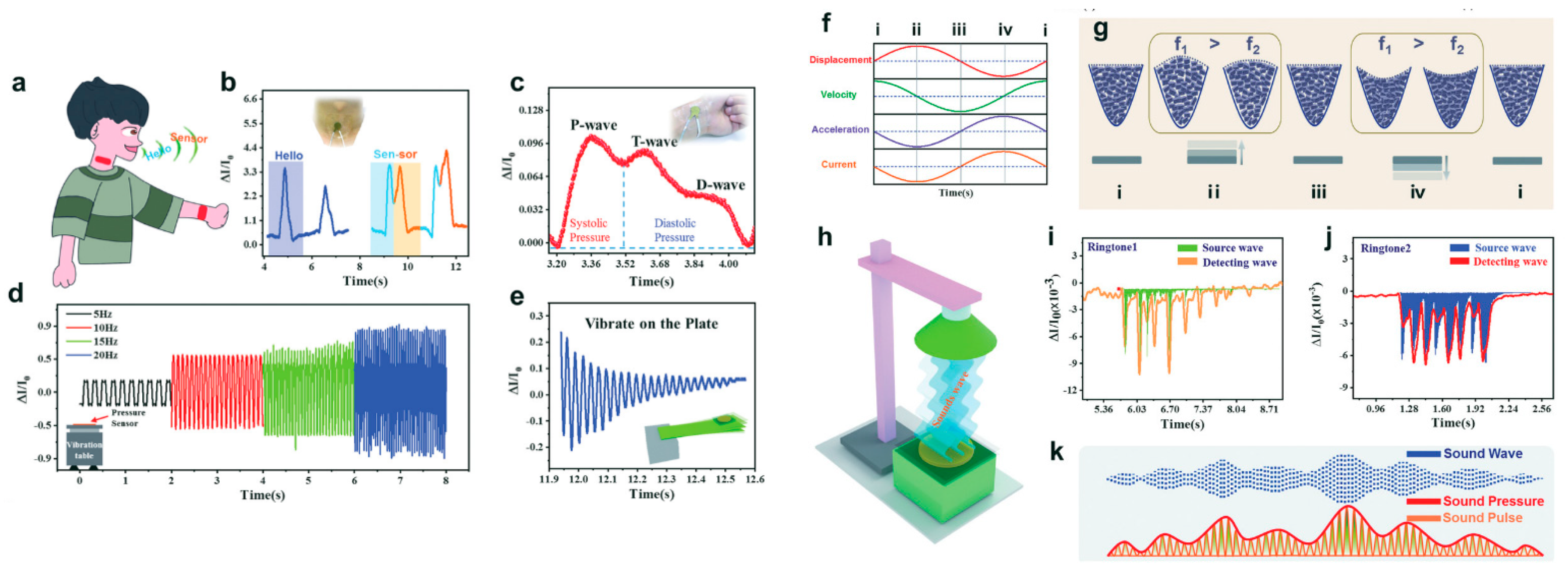
Piezoresistive wearable biosensors are designed to detect weak movements of the human body using stretch changes in materials. The development of MXene-based piezoresistive biosensors has been reported to change the resistance of the biosensor by varying the MXene’s layer spacing under external pressure [138]. The biosensor can monitor physical stimulation processes, such as blinking, throat swallowing, and knee bending release through electrical current. Strain sensors were also fabricated by using Ti3C2 MXenes nanocomposites with single-walled carbon nanotubes (SWCNT) obtained by layer-by-layer (LBL) spraying [139]. The multifunctional force-sensing sensor for acoustic monitoring consists of two Au electrodes on a polyethylene terephthalate (PET) substrate at the top and bottom, an intermediate MXenes layer, and a fingerprint structure on the substrate in a combined arrangement [140]. The manufactured sensor is versatile and capable of sensing sound, micro-motion, and acceleration in a single device. This biosensor can be flexibly attached to a person’s throat and wrist and is used to detect a person’s vocalisation and pulse. The sensor can record relevant peaks when saying “hello” and “sensor” or when detecting a steady heartbeat pulse signal. The biosensor has shown excellent sensitivity in detecting subtle human activity and other weak stresses (Figure 12). It offers a new research direction for portable and wearable sensing devices in biosensing and human behaviour analyses.
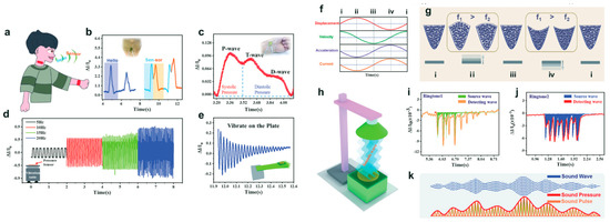
Figure 12.
Schematic diagram of a pressure sensor as a wearable device for physiological signal detection (a). Corresponding signals for saying “hello” and “sensor” (b). Corresponding signal for the pulse on the medial wrist (c). Corresponding signal of the pressure sensor vibrating at different frequencies on the shaker (d). Corresponding signals of the pressure sensor vibrating on plate (e). Corresponding relationship between current values, displacement, velocity and acceleration in the vibration mode (f). Schematic diagram of the density of MXenes at different vibration stages (g). Diagram of a sound wave hitting a pressure transducer (h). Schematic diagram of the acoustic pressure on the sensor surface associated with sound waves and sound pulses (i). Corresponding detection signals and source waves for the two ringtones (j–k). Reprinted with permission from Ref. [140]. Copyright 2020, Wiley-VCH.
Wearable microfluidic biosensors were originally designed to integrate biological identifiers (enzymes, nucleic acids, enzymes, or cellular receptors) into the sensor operation. Non-invasive biomarker detection platforms via biofluids such as sweat, saliva, tears, or interstitial fluid are more practical [141]. Such wearable sensors provide real-time biochemical information about the wearer’s health and offer effective disease detection and body function management [142]. A 3D electrode network electrochemical impedance immunosensor based on loaded laser-burned graphene (LBG) loaded with Ti3C2Tx was fabricated for the non-invasive monitoring of cortisol biomarkers in human sweat [132]. The sensor has a detection limit and linearity of 88 pM and 0.01–100 nM, respectively (Figure 13). In addition, microfluidic wearable biosensors can be used to detect K+, Na+ ions [135,136], glucose, lactate [134,143], pH, and other human biochemical information in biological fluids [137].

Figure 13.
(A) Schematic representation of the Ti3C2Tx MXene-loaded/LBG-based cortisol biomarker assay. (B) Patch sensor attached to the body position (a), optical image of the wearable patch (b) and fabrication sequence of a wearable patch cortisol sensor integrated with a microfluidic system (c). Reprinted with permission from Ref. [132]. Copyright 2020, Elsevier.
In various wearable biosensors, sensing electrodes play an essential role in the design of wearable biosensors. MXenes offer the ability to immobilise biomolecules as a sensitive detection platform. Nevertheless, the mechanical friction and deformation of wearable devices against human skin over time leads to mechanical failure and requires a re-structuring of the device. Moreover, the attainment of signal-to-noise ratios and the stability required to achieve this device are highly challenging [133].
3.3.2. Surface-Enhanced Raman Spectroscopy (SERS)
The hydrophilic nature of the MXenes surface provides a good site for Raman labelling. It serves as a potential material for SERS and provides an effective method for the ultra-sensitive determination of targets (Table 8). Sarycheva et al. showed that composites of metals and MXenes could be used as SERS subestrates for rhodamine 6G [144]. Integrating noble metal nanoparticles with MXenes exhibits an empathetic, sensitive SERS response in detecting several common dye molecules. This extends MXenes composites for visible light SERS in the sensor field. A reliable substrate for MXenes/AuNR composites was prepared by Xie et al. with high sensitivities for determining common organic dyes, such as Rh6G, crystalline violet, and peacock green [145]. It can detect organic contaminants and shows high sensitivities for more complex organic pesticides and contaminants. A ratiometric SERS aptamer sensor for ochratoxin A was developed by Zhao et al. 2-Mercaptobenzimidazole-5-carboxylic acid ligands and Au-Ag Janus nanoparticles were used as Raman signal molecules to amplify the SERS signal efficiently [146]. Liu et al. used the Ti3C2Tx-PDDA-Ag NPs hybrid platform as a sensitive and homogeneous biosensor for the label-free quantification of the biomolecule adenine based on the SERS method [147]. These studies demonstrate that MXenes can be well-suited for SERS.

Table 8.
MXene-based other biosensors identify units, target, and analytical parameters.
3.3.3. Surface Plasmon Resonance (SPR)
SPR sensing is a non-destructive, label-free, real-time detection method. Nanomaterials modify the sensor’s surface to enhance the signal and demonstrate high sensitivities to low concentration targets [148] (Table 9). MXenes-based SPR biosensors for the ultra-sensitive detection of carcinoembryonic antigens (CEAs) were fabricated by Wu et al. These SPR biosensors demonstrate good reproducibility and high selectivity in human serum samples, providing a potential method for early clinical diagnoses and cancer surveillance [149]. Their group also constructed SPR biosensors for CEA using amino-functionalised MXenes [150]. MXenes were assembled on Au films to immobilise monoclonal anti-CEA antibody sensing materials covalently. MXenes were used as a substrate for binding hollow gold nanoparticles (HGNPs), and they were modified with SPA. MXenes/HGNPs/SPA/Ab2 nanocomplexes act as signal enhancers for SPR-sensing components. The sensor offers a wide linear detection range, ultra-low detection limits, and good selectivity for CEA in human serums. Chen et al. anchored targeting aptamers with thiol-modified niobium carbide MXene quantum dots and could specifically bind the SARS-Cov-2 N gene, resulting in a change in the SPR signal for laser irradiation at a wavelength of 633 nm [151]. The above studies indicate the development of MXene-based biomarker sensing chips or devices in the field of SPR to broaden the area of MXenes biosensing applications.

Table 9.
MXene-based SPR biosensors identify units, target, and analytical parameters.
4. Conclusions and Outlook
From its discovery to the present moment, MXenes, an emerging two-dimensional biosensing material, has achieved unprecedented rapid development. Although the development time is relatively short, synthesis methods of MXenes are constantly innovating and developing towards the direction of green environmental protection, simplicity and convenience, and controllable surface groups. MXenes are known for their tunable surface groups; good hydrophilicity and biocompatibility; excellent mechanical, electrical, and optical properties; and specific morphological structures. They have become a focus of research in electrochemical and optical biosensing.
HF synthesis is undoubtedly convenient and quick, giving access to abundant hydrophilic surface groups. However, using F− can cause a certain amount of pollution to humans, the environment, and even the ecosystem. For the synthesis of MXenes, synthetic research has always progressed in the direction of less or no fluorine. Different synthetic methods and raw materials can adjust different surface groups. The structure determines the properties, and mechanical, hydrophilic, biocompatible, electrical, and optical properties can be adjusted according to different research needs using different synthetic routes.
From its large specific surface area, high electrical conductivity, large surface groups, and fast electron transfer properties, MXenes are often used as modification and immobilisation materials in electrochemical biosensors to improve electrocatalytic performances and detection sensitivities and to reduce redox potentials in order to obtain high-performance composites. However, most of the current MXene-based electrochemical biosensors use composite materials adsorbed on MXene’s surface. There are relatively few reports on surface groups on MXenes as the adsorption media for the adsorption of biomolecules to prepare electrochemical biosensors. With the development of the tunable functionalisation of MXenes surface groups, we believe there is more significant potential for the direct binding of MXenes surfaces to biomolecules, such as proteins and nucleic acids. Although optical biosensors have been less studied than electrochemical biosensors, optical sensing is also a vital detection strategy in biosensing. By benefitting from the fluorescence quenching effect of MXenes and MQD’s fluorescence enhancement effect, photoluminescence has been reported relatively more often in optical biosensing work. From the above studies, it can be concluded that nucleic acid-based biosensors are less selective than enzyme and immune-based biosensors. However, nucleic acids are smaller than antibodies and enzymes, so nucleic-acid-based biosensors have a higher density of surface modifications and have a higher sensitivity and reproducibility.
In contrast, SPR, SERS, and other optical sensing have been reported less often. Even so, the sparseness and singularity of functional groups on MXene’s surface led to the reduced binding of biomolecules or other organic molecules. Meanwhile, the study of MQDs with high fluorescence efficiency, quantum yield, and different luminescence wavelengths has become an urgent problem.
About the main challenges mentioned above, we believe that several aspects can be studied for future developments and trends in MXenes biosensing.
- (1)
- MXenes and MQDs synthesis methods are supposed to develop with adjustable size and surface groups. It is necessary to study further study the interaction between the composition, structure, properties, and biomolecules of MXenes in order to understand the appropriate MXene-adaptation-related biosensors that can be developed according to the actual situation. Furthermore, it can provide theoretical support for the development and development of MXenes morphological structure, size, and surface groups, and the study of interaction with biomolecules with the help of machine learning methods.
- (2)
- The direct construction of biosensors on MXenes surfaces: The abundant surface groups of MXenes can be complexed or combined with many biomolecules, and enzyme-based electrochemical sensors have been partially verified. Nevertheless, there are relatively few studies on nucleic acid, antibodies, and other biomolecules combined with MXenes and constructed in sensors. Moreover, the natural binding process can shorten the experimental time and reduce errors caused by too many experimental procedures. At the same time, it is also very convenient for developed point-of-care tests. During the COVID-19 epidemic, point-of-care was necessary for rapid diagnoses and the timely treatment of patients.
- (3)
- Utilise the catalytic properties and the reduction of MXenes: For electrochemical biosensing, MXenes can reduce precious metal nanoparticles in situ. They can also be used to catalyse redox reactions of O2 in systems that promote free radical reactions in ECL, and improving the ECL signal is well worth exploring. MXenes are also used as semiconductor materials during electrochemical reactions and can be used to enhance the electrochemical signal. Therefore, reducing the detection limit, extending the detection range, and improving the sensitivity during electrochemical biosensing are possible.
- (4)
- Develop MQDs with high fluorescence efficiency, quantum yield, and different fluorescence emission wavelengths: As a result, the sensitivity and detection limit of fluorescence detections can be improved, and they can be used in optical biosensors with different wavelengths. MQDs can be extended to cell imaging, photothermal therapy, and other biomedical tissue applications.
Author Contributions
Conceptualization, D.L. and H.Z.; writing—original draft preparation, D.L.; writing—review and editing, D.L., X.Z. and H.Z.; project administration, Y.C. and L.F.; supervision, L.F. All authors have read and agreed to the published version of the manuscript.
Funding
This work was supported by the National Natural Science Foundation of China (No. 21705106) and the National Science Foundation of China (No. 22177067); the Program for Professor of Special Appointment (Eastern Scholar) at Shanghai Institutions of Higher Learning (No. TP2016023); and the Shanghai Sailing Program (No. 20YF1413000).
Institutional Review Board Statement
Not applicable.
Informed Consent Statement
Not applicable.
Data Availability Statement
Not applicable.
Conflicts of Interest
The authors declare no conflict of interest.
References
- Yang, F.; Song, P.; Ruan, M.; Xu, W. Recent progress in two-dimensional nanomaterials: Synthesis, engineering, and applications. FlatChem 2019, 18, 100133. [Google Scholar] [CrossRef]
- Choi, S.-J.; Kim, I.-D. Recent Developments in 2D Nanomaterials for Chemiresistive-Type Gas Sensors. Electron. Mater. Lett. 2018, 14, 221–260. [Google Scholar] [CrossRef]
- Deshmukh, K.; Kovářík, T.; Khadheer Pasha, S.K. State of the art recent progress in two dimensional MXenes based gas sensors and biosensors: A comprehensive review. Coord. Chem. Rev. 2020, 424, 213514. [Google Scholar] [CrossRef]
- Naguib, M.; Kurtoglu, M.; Presser, V.; Lu, J.; Niu, J.; Heon, M.; Hultman, L.; Gogotsi, Y.; Barsoum, M.W. Two-dimensional nanocrystals produced by exfoliation of Ti3AlC2. Adv. Mater. 2011, 23, 4248–4253. [Google Scholar] [CrossRef] [PubMed]
- Huang, X.; Wu, P. A Facile, High-Yield, and Freeze-and-Thaw-Assisted Approach to Fabricate MXene with Plentiful Wrinkles and Its Application in On-Chip Micro-Supercapacitors. Adv. Funct. Mater. 2020, 30, 1910048. [Google Scholar] [CrossRef]
- Ma, Y.; Li, B.; Yang, S. Ultrathin two-dimensional metallic nanomaterials. Mater. Chem. Front. 2018, 2, 456–467. [Google Scholar] [CrossRef]
- Ghazaly, A.E.; Ahmed, H.; Rezk, A.R.; Halim, J.; Persson, P.O.Å.; Yeo, L.Y.; Rosen, J. Ultrafast, One-Step, Salt-Solution-Based Acoustic Synthesis of Ti3C2 MXene. ACS Nano 2021, 15, 4287–4293. [Google Scholar] [CrossRef]
- Qing, H.; Mian, L.I. Recent Progress and Prospects of Ternary Layered Carbides/Nitrides MAX Phases and Their Derived Two-Dimensional Nanolaminates MXenes. J. Inorg. Mater. 2019, 35, 1–7. [Google Scholar] [CrossRef]
- Naguib, M.; Mochalin, V.N.; Barsoum, M.W.; Gogotsi, Y. 25th anniversary article: MXenes: A new family of two-dimensional materials. Adv. Mater. 2014, 26, 992–1005. [Google Scholar] [CrossRef]
- Li, X.; Lu, Y.; Liu, Q. Electrochemical and optical biosensors based on multifunctional MXene nanoplatforms: Progress and prospects. Talanta 2021, 235, 122726. [Google Scholar] [CrossRef]
- Wang, L.; Zhang, H.; Zhuang, T.; Liu, J.; Sojic, N.; Wang, Z. Sensitive electrochemiluminescence biosensing of polynucleotide kinase using the versatility of two-dimensional Ti3C2TX MXene nanomaterials. Anal. Chim. Acta 2022, 1191, 339346. [Google Scholar] [CrossRef] [PubMed]
- Naguib, M.; Barsoum, M.W.; Gogotsi, Y. Ten Years of Progress in the Synthesis and Development of MXenes. Adv. Mater. 2021, 33, 2103393. [Google Scholar] [CrossRef] [PubMed]
- Roointan, A.; Ahmad Mir, T.; Ibrahim Wani, S.; Mati Ur, R.; Hussain, K.K.; Ahmed, B.; Abrahim, S.; Savardashtaki, A.; Gandomani, G.; Gandomani, M.; et al. Early detection of lung cancer biomarkers through biosensor technology: A review. J. Pharm. Biomed. Anal. 2019, 164, 93–103. [Google Scholar] [CrossRef] [PubMed]
- Zhou, Y.; Kubota, L.T. Trends in Electrochemical Sensing. ChemElectroChem 2020, 7, 3684–3685. [Google Scholar] [CrossRef]
- Mohammadniaei, M.; Nguyen, H.V.; Tieu, M.V.; Lee, M.-H. 2D Materials in Development of Electrochemical Point-of-Care Cancer Screening Devices. Micromachines 2019, 10, 662. [Google Scholar] [CrossRef]
- Wrobel, T.P.; Bhargava, R. Infrared Spectroscopic Imaging Advances as an Analytical Technology for Biomedical Sciences. Anal. Chem. 2018, 90, 1444–1463. [Google Scholar] [CrossRef]
- Kumar, J.A.; Prakash, P.; Krithiga, T.; Amarnath, D.J.; Premkumar, J.; Rajamohan, N.; Vasseghian, Y.; Saravanan, P.; Rajasimman, M. Methods of synthesis, characteristics, and environmental applications of MXene: A comprehensive review. Chemosphere 2022, 286 Pt 1, 131607. [Google Scholar] [CrossRef]
- Huang, J.; Li, Z.; Mao, Y.; Li, Z. Progress and biomedical applications of MXenes. Nano Sel. 2021, 2, 1480–1508. [Google Scholar] [CrossRef]
- Zhang, J.; Li, Y.; Duan, S.; He, F. Highly electrically conductive two-dimensional Ti3C2 Mxenes-based 16S rDNA electrochemical sensor for detecting Mycobacterium tuberculosis. Anal. Chim. Acta 2020, 1123, 9–17. [Google Scholar] [CrossRef]
- Lu, L.; Han, X.; Lin, J.; Zhang, Y.; Qiu, M.; Chen, Y.; Li, M.; Tang, D. Ultrasensitive fluorometric biosensor based on Ti3C2 MXenes with Hg(2+)-triggered exonuclease III-assisted recycling amplification. Analyst 2021, 146, 2664–2669. [Google Scholar] [CrossRef]
- Naguib, M.; Mashtalir, O.; Carle, J.; Presser, V.; Lu, J.; Hultman, L.; Gogotsi, Y.; Barsoum, M.W. Two-Dimensional Transition Metal Carbides. ACS Nano 2012, 6, 1322–1331. [Google Scholar] [CrossRef] [PubMed]
- Alhabeb, M.; Maleski, K.; Anasori, B.; Lelyukh, P.; Clark, L.; Sin, S.; Gogotsi, Y. Guidelines for Synthesis and Processing of Two-Dimensional Titanium Carbide (Ti3C2Tx MXene). Chem. Mater. 2017, 29, 7633–7644. [Google Scholar] [CrossRef]
- Ghidiu, M.; Lukatskaya, M.R.; Zhao, M.Q.; Gogotsi, Y.; Barsoum, M.W. Conductive two-dimensional titanium carbide ’clay’ with high volumetric capacitance. Nature 2014, 516, 78–81. [Google Scholar] [CrossRef]
- Sang, X.; Xie, Y.; Lin, M.W.; Alhabeb, M.; Van Aken, K.L.; Gogotsi, Y.; Kent, P.R.C.; Xiao, K.; Unocic, R.R. Atomic Defects in Monolayer Titanium Carbide (Ti3C2Tx) MXene. ACS Nano 2016, 10, 9193–9200. [Google Scholar] [CrossRef] [PubMed]
- Shahzad, F.; Alhabeb, M.; Hatter Christine, B.; Anasori, B.; Man Hong, S.; Koo Chong, M.; Gogotsi, Y. Electromagnetic interference shielding with 2D transition metal carbides (MXenes). Science 2016, 353, 1137–1140. [Google Scholar] [CrossRef] [PubMed]
- Song, M.; Pang, S.Y.; Guo, F.; Wong, M.C.; Hao, J. Fluoride-Free 2D Niobium Carbide MXenes as Stable and Biocompatible Nanoplatforms for Electrochemical Biosensors with Ultrahigh Sensitivity. Adv. Sci 2020, 7, 2001546. [Google Scholar] [CrossRef] [PubMed]
- Gao, L.; Li, C.; Huang, W.; Mei, S.; Lin, H.; Ou, Q.; Zhang, Y.; Guo, J.; Zhang, F.; Xu, S.; et al. MXene/Polymer Membranes: Synthesis, Properties, and Emerging Applications. Chem. Mater. 2020, 32, 1703–1747. [Google Scholar] [CrossRef]
- Tian, Z.; Wei, C.; Sun, J. Recent advances in the template-confined synthesis of two-dimensional materials for aqueous energy storage devices. Nanoscale Adv. 2020, 2, 2220–2233. [Google Scholar] [CrossRef]
- Zhang, F.; Zhang, Z.; Wang, H.; Chan, C.H.; Chan, N.Y.; Chen, X.X.; Dai, J.-Y. Plasma-enhanced pulsed-laser deposition of single-crystalline Mo2C ultrathin superconducting films. Phys. Rev. Mater. 2017, 1, 034002. [Google Scholar] [CrossRef]
- Li, M.; Lu, J.; Luo, K.; Li, Y.; Chang, K.; Chen, K.; Zhou, J.; Rosen, J.; Hultman, L.; Eklund, P.; et al. Element Replacement Approach by Reaction with Lewis Acidic Molten Salts to Synthesize Nanolaminated MAX Phases and MXenes. J. Am. Chem. Soc. 2019, 141, 4730–4737. [Google Scholar] [CrossRef]
- Sun, W.; Wang, X.; Feng, J.; Li, T.; Huan, Y.; Qiao, J.; He, L.; Ma, D. Controlled synthesis of 2D Mo2C/graphene heterostructure on liquid Au substrates as enhanced electrocatalytic electrodes. Nanotechnology 2019, 30, 385601. [Google Scholar] [CrossRef] [PubMed]
- Srivastava, P.; Mishra, A.; Mizuseki, H.; Lee, K.R.; Singh, A.K. Mechanistic Insight into the Chemical Exfoliation and Functionalization of Ti3C2 MXene. ACS Appl Mater. Interfaces 2016, 8, 24256–24264. [Google Scholar] [CrossRef] [PubMed]
- Li, X.; Huang, Z.; Zhi, C. Environmental Stability of MXenes as Energy Storage Materials. Front. Mater. 2019, 6, 312. [Google Scholar] [CrossRef]
- Huang, S.; Mochalin, V.N. Hydrolysis of 2D Transition-Metal Carbides (MXenes) in Colloidal Solutions. Inorg. Chem. 2019, 58, 1958–1966. [Google Scholar] [CrossRef]
- Zhang, C.J.; Pinilla, S.; McEvoy, N.; Cullen, C.P.; Anasori, B.; Long, E.; Park, S.-H.; Seral-Ascaso, A.; Shmeliov, A.; Krishnan, D.; et al. Oxidation Stability of Colloidal Two-Dimensional Titanium Carbides (MXenes). Chem. Mater. 2017, 29, 4848–4856. [Google Scholar] [CrossRef]
- Tan, T.L.; Jin, H.M.; Sullivan, M.B.; Anasori, B.; Gogotsi, Y. High-Throughput Survey of Ordering Configurations in MXene Alloys Across Compositions and Temperatures. ACS Nano 2017, 11, 4407–4418. [Google Scholar] [CrossRef] [PubMed]
- Cheng, Y.-W.; Dai, J.-H.; Zhang, Y.-M.; Song, Y. Two-Dimensional, Ordered, Double Transition Metal Carbides (MXenes): A New Family of Promising Catalysts for the Hydrogen Evolution Reaction. J. Phys. Chem. C 2018, 122, 28113–28122. [Google Scholar] [CrossRef]
- Tao, Q.; Dahlqvist, M.; Lu, J.; Kota, S.; Meshkian, R.; Halim, J.; Palisaitis, J.; Hultman, L.; Barsoum, M.W.; Persson, P.O.A.; et al. Two-dimensional Mo1.33C MXene with divacancy ordering prepared from parent 3D laminate with in-plane chemical ordering. Nat. Commun. 2017, 8, 14949. [Google Scholar] [CrossRef]
- Halim, J.; Palisaitis, J.; Lu, J.; Thörnberg, J.; Moon, E.J.; Precner, M.; Eklund, P.; Persson, P.O.Å.; Barsoum, M.W.; Rosen, J. Synthesis of Two-Dimensional Nb1.33C (MXene) with Randomly Distributed Vacancies by Etching of the Quaternary Solid Solution (Nb2/3Sc1/3)2AlC MAX Phase. ACS Appl. Nano Mater. 2018, 1, 2455–2460. [Google Scholar] [CrossRef]
- Zhang, P.; Yang, X.J.; Li, P.; Zhao, Y.; Niu, Q.J. Fabrication of novel MXene (Ti3C2)/polyacrylamide nanocomposite hydrogels with enhanced mechanical and drug release properties. Soft Matter 2020, 16, 162–169. [Google Scholar] [CrossRef]
- Khaledialidusti, R.; Anasori, B.; Barnoush, A. Temperature-dependent mechanical properties of Tin+1CnO2 (n = 1, 2) MXene monolayers: A first-principles study. Phys. Chem Chem Phys. 2020, 22, 3414–3424. [Google Scholar] [CrossRef]
- Ronchi, R.M.; Arantes, J.T.; Santos, S.F. Synthesis, structure, properties and applications of MXenes: Current status and perspectives. Ceram. Int. 2019, 45, 18167–18188. [Google Scholar] [CrossRef]
- Wu, X.; Ma, P.; Sun, Y.; Du, F.; Song, D.; Xu, G. Application of MXene in Electrochemical Sensors: A Review. Electroanalysis 2021, 33, 1827–1851. [Google Scholar] [CrossRef]
- Manzanares-Palenzuela, C.L.; Pourrahimi, A.M.; Gonzalez-Julian, J.; Sofer, Z.; Pykal, M.; Otyepka, M.; Pumera, M. Interaction of single- and double-stranded DNA with multilayer MXene by fluorescence spectroscopy and molecular dynamics simulations. Chem. Sci. 2019, 10, 10010–10017. [Google Scholar] [CrossRef]
- Huang, Z.; Liu, B.; Liu, J. Mn(2+)-Assisted DNA Oligonucleotide Adsorption on Ti2C MXene Nanosheets. Langmuir 2019, 35, 9858–9866. [Google Scholar] [CrossRef]
- Vural, M.; Zhu, H.; Pena-Francesch, A.; Jung, H.; Allen, B.D.; Demirel, M.C. Self-Assembly of Topologically Networked Protein–Ti3C2Tx MXene Composites. ACS Nano 2020, 14, 6956–6967. [Google Scholar] [CrossRef]
- Huang, R.; Chen, X.; Dong, Y.; Zhang, X.; Wei, Y.; Yang, Z.; Li, W.; Guo, Y.; Liu, J.; Yang, Z.; et al. MXene Composite Nanofibers for Cell Culture and Tissue Engineering. ACS Appl. Bio Mater. 2020, 3, 2125–2131. [Google Scholar] [CrossRef]
- Shahzad, F.; Zaidi, S.A.; Naqvi, R.A. 2D Transition Metal Carbides (MXene) for Electrochemical Sensing: A Review. Crit Rev. Anal. Chem. 2020, 52, 848–864. [Google Scholar] [CrossRef] [PubMed]
- Anasori, B.; Xie, Y.; Beidaghi, M.; Lu, J.; Hosler, B.C.; Hultman, L.; Kent, P.R.C.; Gogotsi, Y.; Barsoum, M.W. Two-Dimensional, Ordered, Double Transition Metals Carbides (MXenes). ACS Nano 2015, 9, 9507–9516. [Google Scholar] [CrossRef]
- Verger, L.; Natu, V.; Carey, M.; Barsoum, M.W. MXenes: An Introduction of Their Synthesis, Select Properties, and Applications. Trends Chem. 2019, 1, 656–669. [Google Scholar] [CrossRef]
- Cho, I.H.; Kim, D.H.; Park, S. Electrochemical biosensors: Perspective on functional nanomaterials for on-site analysis. Biomater. Res. 2020, 24, 6. [Google Scholar] [CrossRef] [PubMed]
- Chia, H.L.; Mayorga-Martinez, C.C.; Antonatos, N.; Sofer, Z.; Gonzalez-Julian, J.J.; Webster, R.D.; Pumera, M. MXene Titanium Carbide-based Biosensor: Strong Dependence of Exfoliation Method on Performance. Anal. Chem. 2020, 92, 2452–2459. [Google Scholar] [CrossRef]
- Xu, W.; Sakran, M.; Fei, J.; Li, X.; Weng, C.; Yang, W.; Zhu, G.; Zhu, W.; Zhou, X. Electrochemical Biosensor Based on HRP/Ti3C2/Nafion Film for Determination of Hydrogen Peroxide in Serum Samples of Patients with Acute Myocardial Infarction. ACS Biomater. Sci. Eng. 2021, 7, 2767–2773. [Google Scholar] [CrossRef]
- Ma, B.K.; Li, M.; Cheong, L.Z.; Weng, X.C.; Shen, C.; Huang, Q. Enzyme-MXene Nanosheets: Fabrication and Application in Electrochemical Detection of H2O2. J. Inorg. Mater. 2020, 35, 131. [Google Scholar] [CrossRef]
- Rakhi, R.B.; Nayak, P.; Xia, C.; Alshareef, H.N. Novel amperometric glucose biosensor based on MXene nanocomposite. Sci. Rep. 2016, 6, 36422. [Google Scholar] [CrossRef] [PubMed]
- Wu, M.; Zhang, Q.; Fang, Y.; Deng, C.; Zhou, F.; Zhang, Y.; Wang, X.; Tang, Y.; Wang, Y. Polylysine-modified MXene nanosheets with highly loaded glucose oxidase as cascade nanoreactor for glucose decomposition and electrochemical sensing. J. Colloid Interface Sci. 2021, 586, 20–29. [Google Scholar] [CrossRef] [PubMed]
- Murugan, P.; Annamalai, J.; Atchudan, R.; Govindasamy, M.; Nallaswamy, D.; Ganapathy, D.; Reshetilov, A.; Sundramoorthy, A.K. Electrochemical Sensing of Glucose Using Glucose Oxidase/PEDOT:4-Sulfocalix [4]arene/MXene Composite Modified Electrode. Micromachines 2022, 13, 304. [Google Scholar] [CrossRef] [PubMed]
- Xia, T.; Liu, G.; Wang, J.; Hou, S.; Hou, S. MXene-based enzymatic sensor for highly sensitive and selective detection of cholesterol. Biosens. Bioelectron. 2021, 183, 113243. [Google Scholar] [CrossRef] [PubMed]
- Wu, L.; Lu, X.; Dhanjai; Wu, Z.-S.; Dong, Y.; Wang, X.; Zheng, S.; Chen, J. 2D transition metal carbide MXene as a robust biosensing platform for enzyme immobilization and ultrasensitive detection of phenol. Biosens. Bioelectron. 2018, 107, 69–75. [Google Scholar] [CrossRef] [PubMed]
- Zhou, L.Y.; Zhang, X.; Ma, L.; Gao, J.; Jiang, Y.J. Acetylcholinesterase/chitosan-transition metal carbides nanocomposites-based biosensor for the organophosphate pesticides detection. Biochem. Eng. J. 2017, 128, 243–249. [Google Scholar] [CrossRef]
- Du, Y.; Dong, S. Nucleic Acid Biosensors: Recent Advances and Perspectives. Anal. Chem. 2017, 89, 189–215. [Google Scholar] [CrossRef]
- Wu, Y.; Arroyo-Currás, N. Advances in nucleic acid architectures for electrochemical sensing. Curr. Opin. Electrochem. 2021, 27, 100695. [Google Scholar] [CrossRef]
- Liu, L.; Wei, Y.; Jiao, S.; Zhu, S.; Liu, X. A novel label-free strategy for the ultrasensitive miRNA-182 detection based on MoS2/Ti3C2 nanohybrids. Biosens. Bioelectron. 2019, 137, 45–51. [Google Scholar] [CrossRef] [PubMed]
- Zhou, S.; Gu, C.; Li, Z.; Yang, L.; He, L.; Wang, M.; Huang, X.; Zhou, N.; Zhang, Z. Ti3C2Tx MXene and polyoxometalate nanohybrid embedded with polypyrrole: Ultra-sensitive platform for the detection of osteopontin. Appl. Surf. Sci. 2019, 498, 143889. [Google Scholar] [CrossRef]
- Wang, H.; Li, H.; Huang, Y.; Xiong, M.; Wang, F.; Li, C. A label-free electrochemical biosensor for highly sensitive detection of gliotoxin based on DNA nanostructure/MXene nanocomplexes. Biosens. Bioelectron. 2019, 142, 111531. [Google Scholar] [CrossRef]
- Rasheed, P.A.; Pandey, R.P.; Jabbar, K.A.; Mahmoud, K.A. Nb4C3Tx(MXene)/Au/DNA Aptasensor for the Ultraselective Electrochemical Detection of Lead in Water Samples. Electroanalysis 2022, 34, 1–8. [Google Scholar] [CrossRef]
- Liu, Y.; Huang, S.J.; Li, J.N.; Wang, M.H.; Wang, C.B.; Hu, B.; Zhou, N.; Zhang, Z.H. 0D/2D heteronanostructure-integrated bimetallic CoCu-ZIF nanosheets and MXene-derived carbon dots for impedimetric cytosensing of melanoma B16-F10 cells. Microchim. Acta 2021, 188, 69. [Google Scholar] [CrossRef]
- Hu, Z.; Suo, Z.; Liu, W.; Zhao, B.; Xing, F.; Zhang, Y.; Feng, L. DNA conformational polymorphism for biosensing applications. Biosens. Bioelectron. 2019, 131, 237–249. [Google Scholar] [CrossRef]
- Yoon, J.; Shin, M.; Lim, J.; Lee, J.Y.; Choi, J.W. Recent Advances in MXene Nanocomposite-Based Biosensors. Biosensors 2020, 10, 185. [Google Scholar] [CrossRef]
- Khan, R.; Ben Aissa, S.; Sherazi, T.A.; Catanante, G.; Hayat, A.; Marty, J.L. Development of an Impedimetric Aptasensor for Label Free Detection of Patulin in Apple Juice. Molecules 2019, 24, 1017. [Google Scholar] [CrossRef]
- Zhu, C.; Yang, G.; Li, H.; Du, D.; Lin, Y. Electrochemical sensors and biosensors based on nanomaterials and nanostructures. Anal. Chem. 2015, 87, 230–249. [Google Scholar] [CrossRef]
- Duan, F.; Guo, C.; Hu, M.; Song, Y.; Wang, M.; He, L.; Zhang, Z.; Pettinari, R.; Zhou, L. Construction of the 0D/2D heterojunction of Ti3C2Tx MXene nanosheets and iron phthalocyanine quantum dots for the impedimetric aptasensing of microRNA--155. Sens. Actuators B Chem. 2020, 310, 127844. [Google Scholar] [CrossRef]
- Mohammadniaei, M.; Koyappayil, A.; Sun, Y.; Min, J.; Lee, M.H. Gold nanoparticle/MXene for multiple and sensitive detection of oncomiRs based on synergetic signal amplification. Biosens. Bioelectron. 2020, 159, 112208. [Google Scholar] [CrossRef]
- Chen, W.Y.; Lin, H.; Barui, A.K.; Gomez, A.M.U.; Wendt, M.K.; Stanciu, L.A. DNA-Functionalized Ti3C2Tx MXenes for Selective and Rapid Detection of SARS-CoV-2 Nucleocapsid Gene. ACS Appl. Nano Mater. 2021, 5, 1902–1910. [Google Scholar] [CrossRef]
- Yu, R.J.; Xue, J.; Wang, Y.; Qiu, J.F.; Huang, X.Y.; Chen, A.Y.; Xue, J.J. Novel Ti3C2Tx MXene nanozyme with manageable catalytic activity and application to electrochemical biosensor. J. Nanobiotechnol. 2022, 20, 119. [Google Scholar] [CrossRef] [PubMed]
- Li, Z.Y.; Li, D.Y.; Huang, L.; Hu, R.; Yang, T.; Yang, Y.H. An electrochemical aptasensor based on intelligent walking DNA nanomachine with cascade signal amplification powered by nuclease for Mucin 1 assay. Anal. Chim. Acta 2022, 1214, 339964. [Google Scholar] [CrossRef]
- Vajhadin, F.; Mazloum-Ardakani, M.; Shahidi, M.; Moshtaghioun, S.M.; Haghiralsadat, F.; Ebadi, A.; Amini, A. MXene-based cytosensor for the detection of HER2-positive cancer cells using CoFe2O4@Ag magnetic nanohybrids conjugated to the HB5 aptamer. Biosens. Bioelectron. 2022, 195, 113626. [Google Scholar] [CrossRef] [PubMed]
- Fu, Z.; Xiang, J. Aptamers, the Nucleic Acid Antibodies, in Cancer Therapy. Int. J. Mol. Sci. 2020, 21, 2793. [Google Scholar] [CrossRef]
- Ranallo, S.; Porchetta, A.; Ricci, F. DNA-Based Scaffolds for Sensing Applications. Anal. Chem. 2019, 91, 44–59. [Google Scholar] [CrossRef]
- Wei, X.; Wang, S.; Zhan, Y.; Kai, T.; Ding, P. Sensitive Identification of Microcystin-LR via a Reagent-Free and Reusable Electrochemical Biosensor Using a Methylene Blue-Labeled Aptamer. Biosensors 2022, 12, 556. [Google Scholar] [CrossRef] [PubMed]
- Geng, X.; Zhang, M.; Long, H.; Hu, Z.; Zhao, B.; Feng, L.; Du, J. A reusable neurotransmitter aptasensor for the sensitive detection of serotonin. Anal. Chim. Acta 2021, 1145, 124–131. [Google Scholar] [CrossRef]
- Fan, Y.; Shi, S.; Ma, J.; Guo, Y. A paper-based electrochemical immunosensor with reduced graphene oxide/thionine/gold nanoparticles nanocomposites modification for the detection of cancer antigen 125. Biosens. Bioelectron. 2019, 135, 1–7. [Google Scholar] [CrossRef] [PubMed]
- Zhang, M.M.; Mei, L.S.; Zhang, L.; Wang, X.; Liao, X.C.; Qiao, X.W.; Hong, C.L. Ti3C2 MXene anchors CuAu-LDH multifunctional two-dimensional nanomaterials for dual-mode detection of CEA in electrochemical immunosensors. Bioelectrochemistry 2021, 142, 107943. [Google Scholar] [CrossRef] [PubMed]
- Kumar, S.; Lei, Y.; Alshareef, N.H.; Quevedo-Lopez, M.A.; Salama, K.N. Biofunctionalized two-dimensional Ti3C2 MXenes for ultrasensitive detection of cancer biomarker. Biosens. Bioelectron. 2018, 121, 243–249. [Google Scholar] [CrossRef] [PubMed]
- Medetalibeyoglu, H.; Kotan, G.; Atar, N.; Yola, M.L. A novel and ultrasensitive sandwich-type electrochemical immunosensor based on delaminated MXene@AuNPs as signal amplification for prostate specific antigen (PSA) detection and immunosensor validation. Talanta 2020, 220, 121403. [Google Scholar] [CrossRef]
- Xu, Q.; Xu, J.K.; Jia, H.Y.; Tian, Q.Y.; Liu, P.; Chen, S.X.; Cai, Y.; Lu, X.Y.; Duan, X.M.; Lu, L.M. Hierarchical Ti3C2 MXene-derived sodium titanate nanoribbons/PEDOT for signal amplified electrochemical immunoassay of prostate specific antigen. J. Electroanal. Chem. 2022, 860, 113869. [Google Scholar] [CrossRef]
- Dong, H.; Cao, L.; Tan, Z.; Liu, Q.; Zhou, J.; Zhao, P.; Wang, P.; Li, Y.; Ma, W.; Dong, Y. A Signal Amplification Strategy of CuPtRh CNB-Embedded Ammoniated Ti3C2 MXene for Detecting Cardiac Troponin I by a Sandwich-Type Electrochemical Immunosensor. ACS Appl. Bio Mater. 2020, 3, 377–384. [Google Scholar] [CrossRef]
- Niu, H.M.; Cai, S.M.; Liu, X.K.; Huang, X.M.; Chen, J.; Wang, S.L.; Zhang, S.H. A novel electrochemical sandwich-like immunosensor based on carboxyl Ti3C2Tx MXene and rhodamine b/gold/reduced graphene oxide for Listeria monocytogenes. Anal. Methods 2022, 14, 843–849. [Google Scholar] [CrossRef]
- Jiang, X.; Kuklin, A.V.; Baev, A.; Ge, Y.; Ågren, H.; Zhang, H.; Prasad, P.N. Two-dimensional MXenes: From morphological to optical, electric, and magnetic properties and applications. Phys. Rep. 2020, 848, 1–58. [Google Scholar] [CrossRef]
- Bai, Y.; Zhou, K.; Srikanth, N.; Pang, J.H.L.; He, X.; Wang, R. Dependence of elastic and optical properties on surface terminated groups in two-dimensional MXene monolayers: A first-principles study. RSC Adv. 2016, 6, 35731–35739. [Google Scholar] [CrossRef]
- Zhu, X.; Zhang, Y.; Liu, M.; Liu, Y. 2D titanium carbide MXenes as emerging optical biosensing platforms. Biosens. Bioelectron. 2021, 171, 112730. [Google Scholar] [CrossRef]
- Xu, Q.; Ding, L.; Wen, Y.; Yang, W.; Zhou, H.; Chen, X.; Street, J.; Zhou, A.; Ong, W.-J.; Li, N. High photoluminescence quantum yield of 18.7% by using nitrogen-doped Ti3C2 MXene quantum dots. J. Mater. Chem. C 2018, 6, 6360–6369. [Google Scholar] [CrossRef]
- Xu, G.; Niu, Y.; Yang, X.; Jin, Z.; Wang, Y.; Xu, Y.; Niu, H. Preparation of Ti3C2Tx MXene-Derived Quantum Dots with White/Blue-Emitting Photoluminescence and Electrochemiluminescence. Adv. Opt. Mater. 2018, 6, 1800951. [Google Scholar] [CrossRef]
- Zhu, X.; Pang, X.; Zhang, Y.; Yao, S. Titanium carbide MXenes combined with red-emitting carbon dots as a unique turn-on fluorescent nanosensor for label-free determination of glucose. J. Mater. Chem. B 2019, 7, 7729–7735. [Google Scholar] [CrossRef]
- Peng, X.; Zhang, Y.; Lu, D.; Guo, Y.; Guo, S. Ultrathin Ti3C2 nanosheets based “off-on” fluorescent nanoprobe for rapid and sensitive detection of HPV infection. Sens. Actuators B Chem. 2019, 286, 222–229. [Google Scholar] [CrossRef]
- Wang, S.; Wei, S.; Wang, S.; Zhu, X.; Lei, C.; Huang, Y.; Nie, Z.; Yao, S. Chimeric DNA-Functionalized Titanium Carbide MXenes for Simultaneous Mapping of Dual Cancer Biomarkers in Living Cells. Anal. Chem. 2019, 91, 1651–1658. [Google Scholar] [CrossRef] [PubMed]
- Kalkal, A.; Kadian, S.; Kumar, S.; Manik, G.; Sen, P.; Kumar, S.; Packirisamy, G. Ti3C2-MXene decorated with nanostructured silver as a dual-energy acceptor for the fluorometric neuron specific enolase detection. Biosens. Bioelectron. 2022, 195, 113620. [Google Scholar] [CrossRef] [PubMed]
- Zhu, X.; Fan, L.; Wang, S.; Lei, C.; Huang, Y.; Nie, Z.; Yao, S. Phospholipid-Tailored Titanium Carbide Nanosheets as a Novel Fluorescent Nanoprobe for Activity Assay and Imaging of Phospholipase D. Anal. Chem. 2018, 90, 6742–6748. [Google Scholar] [CrossRef]
- Shi, Y.E.; Han, F.; Xie, L.; Zhang, C.; Li, T.; Wang, H.; Lai, W.F.; Luo, S.; Wei, W.; Wang, Z.; et al. A MXene of type Ti3C2Tx functionalized with copper nanoclusters for the fluorometric determination of glutathione. Microchim. Acta 2019, 187, 38. [Google Scholar] [CrossRef]
- Xue, Q.; Zhang, H.; Zhu, M.; Pei, Z.; Li, H.; Wang, Z.; Huang, Y.; Huang, Y.; Deng, Q.; Zhou, J.; et al. Photoluminescent Ti3C2 MXene Quantum Dots for Multicolor Cellular Imaging. Adv. Mater. 2017, 29. [Google Scholar] [CrossRef]
- Guan, Q.; Ma, J.; Yang, W.; Zhang, R.; Zhang, X.; Dong, X.; Fan, Y.; Cai, L.; Cao, Y.; Zhang, Y.; et al. Highly fluorescent Ti3C2 MXene quantum dots for macrophage labeling and Cu(2+) ion sensing. Nanoscale 2019, 11, 14123–14133. [Google Scholar] [CrossRef]
- Guo, Z.; Zhu, X.; Wang, S.; Lei, C.; Huang, Y.; Nie, Z.; Yao, S. Fluorescent Ti3C2 MXene quantum dots for an alkaline phosphatase assay and embryonic stem cell identification based on the inner filter effect. Nanoscale 2018, 10, 19579–19585. [Google Scholar] [CrossRef]
- Liu, M.; Zhou, J.; He, Y.; Cai, Z.; Ge, Y.; Zhou, J.; Song, G. epsilon-Poly-L-lysine-protected Ti3C2 MXene quantum dots with high quantum yield for fluorometric determination of cytochrome c and trypsin. Microchim Acta 2019, 186, 770. [Google Scholar] [CrossRef] [PubMed]
- Chen, X.; Sun, X.; Xu, W.; Pan, G.; Zhou, D.; Zhu, J.; Wang, H.; Bai, X.; Dong, B.; Song, H. Ratiometric photoluminescence sensing based on Ti3C2 MXene quantum dots as an intracellular pH sensor. Nanoscale 2018, 10, 1111–1118. [Google Scholar] [CrossRef] [PubMed]
- Xu, Y.; Wang, X.; Zhang, W.L.; Lv, F.; Guo, S. Recent progress in two-dimensional inorganic quantum dots. Chem. Soc. Rev. 2018, 47, 586–625. [Google Scholar] [CrossRef]
- Shao, B.; Liu, Z.; Zeng, G.; Wang, H.; Liang, Q.; He, Q.; Cheng, M.; Zhou, C.; Jiang, L.; Song, B. Two-dimensional transition metal carbide and nitride (MXene) derived quantum dots (QDs): Synthesis, properties, applications and prospects. J. Mater. Chem. A 2020, 8, 7508–7535. [Google Scholar] [CrossRef]
- Sinha, A.; Dhanjai; Zhao, H.; Huang, Y.; Lu, X.; Chen, J.; Jain, R. MXene: An emerging material for sensing and biosensing. TrAC Trends Anal. Chem. 2018, 105, 424–435. [Google Scholar] [CrossRef]
- Wang, Z.; Xuan, J.; Zhao, Z.; Li, Q.; Geng, F. Versatile Cutting Method for Producing Fluorescent Ultrasmall MXene Sheets. ACS Nano 2017, 11, 11559–11565. [Google Scholar] [CrossRef]
- Desai, M.L.; Basu, H.; Singhal, R.K.; Saha, S.; Kailasa, S.K. Ultra-small two dimensional MXene nanosheets for selective and sensitive fluorescence detection of Ag+ and Mn2+ ions. Colloids Surf. A Physicochem. Eng. Asp. 2019, 565, 70–77. [Google Scholar] [CrossRef]
- Zhang, J.; Kerr, E.; Usman, K.A.S.; Doeven, E.H.; Francis, P.S.; Henderson, L.C.; Razal, J.M. Cathodic electrogenerated chemiluminescence of tris(2,2’-bipyridine)ruthenium(ii) and peroxydisulfate at pure Ti3C2Tx MXene electrodes. Chem. Commun. 2020, 56, 10022–10025. [Google Scholar] [CrossRef] [PubMed]
- Fang, Y.; Yang, X.; Chen, T.; Xu, G.; Liu, M.; Liu, J.; Xu, Y. Two-dimensional titanium carbide (MXene)-based solid-state electrochemiluminescent sensor for label-free single-nucleotide mismatch discrimination in human urine. Sens. Actuators B Chem. 2018, 263, 400–407. [Google Scholar] [CrossRef]
- Zhuang, T.T.; Zhang, H.X.; Wang, L.; Yu, L.H.; Wang, Z.H. Anchoring luminol based on Ti3C2-mediated in situ formation of Au NPs for construction of an efficient probe for miRNA electrogenerated chemiluminescence detection. Anal. Bioanal. Chem. 2021, 413, 6963–6971. [Google Scholar] [CrossRef]
- Yao, B.; Zhang, J.; Fan, Z.; Ding, Y.; Zhou, B.; Yang, R.; Zhao, J.; Zhang, K. Rational Engineering of the DNA Walker Amplification Strategy by Using a Au@Ti3C2@PEI-Ru(dcbpy)3(2+) Nanocomposite Biosensor for Detection of the SARS-CoV-2 RdRp Gene. ACS Appl. Mater. Interfaces 2021, 13, 19816–19824. [Google Scholar] [CrossRef] [PubMed]
- Zhang, K.; Fan, Z.; Huang, Y.; Ding, Y.; Xie, M. A strategy combining 3D-DNA Walker and CRISPR-Cas12a trans-cleavage activity applied to MXene based electrochemiluminescent sensor for SARS-CoV-2 RdRp gene detection. Talanta 2022, 236, 122868. [Google Scholar] [CrossRef] [PubMed]
- Sun, Y.; Zhang, Y.; Zhang, H.; Liu, M.; Liu, Y. Integrating Highly Efficient Recognition and Signal Transition of g-C3N4 Embellished Ti3C2 MXene Hybrid Nanosheets for Electrogenerated Chemiluminescence Analysis of Protein Kinase Activity. Anal. Chem. 2020, 92, 10668–10676. [Google Scholar] [CrossRef]
- Zhang, H.; Wang, Z.; Zhang, Q.; Wang, F.; Liu, Y. Ti3C2 MXenes nanosheets catalyzed highly efficient electrogenerated chemiluminescence biosensor for the detection of exosomes. Biosens. Bioelectron. 2019, 124–125, 184–190. [Google Scholar] [CrossRef]
- Zhang, H.; Wang, Z.; Wang, F.; Zhang, Y.; Wang, H.; Liu, Y. In Situ Formation of Gold Nanoparticles Decorated Ti3C2 MXenes Nanoprobe for Highly Sensitive Electrogenerated Chemiluminescence Detection of Exosomes and Their Surface Proteins. Anal. Chem. 2020, 92, 5546–5553. [Google Scholar] [CrossRef] [PubMed]
- Mi, X.; Li, H.; Tan, R.; Feng, B.; Tu, Y. The TDs/aptamer cTnI biosensors based on HCR and Au/Ti3C2-MXene amplification for screening serious patient in COVID-19 pandemic. Biosens. Bioelectron. 2021, 192, 113482. [Google Scholar] [CrossRef] [PubMed]
- Luo, W.; Ye, Z.; Ma, P.; Wu, Q.; Song, D. Preparation of a disposable electrochemiluminescence sensor chip based on an MXene-loaded ruthenium luminescent agent and its application in the detection of carcinoembryonic antigens. Analyst 2022, 147, 1986–1994. [Google Scholar] [CrossRef] [PubMed]
- Wei, W.; Lin, H.; Hao, T.; Su, X.; Jiang, X.; Wang, S.; Hu, Y.; Guo, Z. Dual-mode ECL/SERS immunoassay for ultrasensitive determination of Vibrio vulnificus based on multifunctional MXene. Sens. Actuators B Chem. 2021, 332, 129525. [Google Scholar] [CrossRef]
- Yan, Z.; Wang, Z.; Miao, Z.; Liu, Y. Dye-Sensitized and Localized Surface Plasmon Resonance Enhanced Visible-Light Photoelectrochemical Biosensors for Highly Sensitive Analysis of Protein Kinase Activity. Anal. Chem. 2016, 88, 922–929. [Google Scholar] [CrossRef]
- Li, M.; Wang, H.; Wang, X.; Lu, Q.; Li, H.; Zhang, Y.; Yao, S. Ti3C2/Cu2O heterostructure based signal-off photoelectrochemical sensor for high sensitivity detection of glucose. Biosens. Bioelectron. 2019, 142, 111535. [Google Scholar] [CrossRef]
- Chen, G.; Wang, H.; Wei, X.; Wu, Y.; Gu, W.; Hu, L.; Xu, D.; Zhu, C. Efficient Z-Scheme heterostructure based on TiO2/Ti3C2Tx/Cu2O to boost photoelectrochemical response for ultrasensitive biosensing. Sens. Actuators B Chem. 2020, 312, 127951. [Google Scholar] [CrossRef]
- Fu, Y.; Ding, F.; Chen, J.; Liu, M.; Zhang, X.; Du, C.; Si, S. Label-free and near-zero-background-noise photoelectrochemical assay of methyltransferase activity based on a Bi2S3/Ti3C2 Schottky junction. Chem. Commun. 2020, 56, 5799–5802. [Google Scholar] [CrossRef] [PubMed]
- Qiu, Z.L.; Fan, D.C.; Xue, X.H.; Zhang, J.Y.; Xu, J.L.; Lyu, H.X.; Chen, Y.T. Ti3C2 MXene-anchored photoelectrochemical detection of exosomes by in situ fabrication of CdS nanoparticles with enzyme-assisted hybridization chain reaction. RSC Adv. 2022, 12, 14260–14267. [Google Scholar] [CrossRef]
- Liu, L.; Yao, Y.; Ma, K.J.; Shangguan, C.J.; Jiao, S.L.; Zhu, S.Y.; Xu, X.X. Ultrasensitive photoelectrochemical detection of cancer-related miRNA-141 by carrier recombination inhibition in hierarchical Ti3C2@ReS2. Sens. Actuators B Chem. 2021, 331, 129470. [Google Scholar] [CrossRef]
- Zheng, Y.L.; Zhou, Y.L.; Cui, X.T.; Yin, H.S.; Ai, S.Y. Enhanced photoactivity of CdS nanorods by MXene and ZnSnO3: Application in photoelectrochemical biosensor for the effect of environmental pollutants on DNA hydroxymethylation in wheat tissues. Mater. Today Chem. 2022, 24, 100878. [Google Scholar] [CrossRef]
- Chen, X.; Li, J.; Pan, G.; Xu, W.; Zhu, J.; Zhou, D.; Li, D.; Chen, C.; Lu, G.; Song, H. Ti3C2 MXene quantum dots/TiO2 inverse opal heterojunction electrode platform for superior photoelectrochemical biosensing. Sens. Actuators B Chem. 2019, 289, 131–137. [Google Scholar] [CrossRef]
- Kurtoglu, M.; Naguib, M.; Gogotsi, Y.; Barsoum, M.W. First principles study of two-dimensional early transition metal carbides. MRS Commun. 2012, 2, 133–137. [Google Scholar] [CrossRef]
- Kim, J.; Campbell, A.S.; de Ávila, B.E.-F.; Wang, J. Wearable biosensors for healthcare monitoring. Nat. Biotechnol. 2019, 37, 389–406. [Google Scholar] [CrossRef]
- An, H.; Habib, T.; Shah, S.; Gao, H.; Radovic, M.; Green, M.J.; Lutkenhaus, J.L. Surface-agnostic highly stretchable and bendable conductive MXene multilayers. Sci. Adv. 2018, 4, eaaq0118. [Google Scholar] [CrossRef]
- Nah, J.S.; Barman, S.C.; Zahed, M.A.; Sharifuzzaman, M.; Yoon, H.; Park, C.; Yoon, S.; Zhang, S.; Park, J.Y. A wearable microfluidics-integrated impedimetric immunosensor based on Ti3C2Tx MXene incorporated laser-burned graphene for noninvasive sweat cortisol detection. Sens. Actuators B Chem. 2021, 329, 129206. [Google Scholar] [CrossRef]
- Lei, Y.; Zhao, W.; Zhang, Y.; Jiang, Q.; He, J.-H.; Baeumner, A.J.; Wolfbeis, O.S.; Wang, Z.L.; Salama, K.N.; Alshareef, H.N. A MXene-Based Wearable Biosensor System for High-Performance In Vitro Perspiration Analysis. Small 2019, 15, 1901190. [Google Scholar] [CrossRef]
- Li, M.; Wang, L.; Liu, R.; Li, J.; Zhang, Q.; Shi, G.; Li, Y.; Hou, C.; Wang, H. A highly integrated sensing paper for wearable electrochemical sweat analysis. Biosens. Bioelectron. 2021, 174, 112828. [Google Scholar] [CrossRef]
- Zhang, S.; Zahed, M.A.; Sharifuzzaman, M.; Yoon, S.; Hui, X.; Chandra Barman, S.; Sharma, S.; Yoon, H.S.; Park, C.; Park, J.Y. A wearable battery-free wireless and skin-interfaced microfluidics integrated electrochemical sensing patch for on-site biomarkers monitoring in human perspiration. Biosens. Bioelectron. 2021, 175, 112844. [Google Scholar] [CrossRef]
- Saleh, A.; Wustoni, S.; Bihar, E.; El-Demellawi, J.K.; Zhang, Y.; Hama, A.; Druet, V.; Yudhanto, A.; Lubineau, G.; Alshareef, H.N.; et al. Inkjet-printed Ti3C2Tx MXene electrodes for multimodal cutaneous biosensing. J. Phys. Mater. 2020, 3, 044004. [Google Scholar] [CrossRef]
- Chen, L.; Chen, F.; Liu, G.; Lin, H.; Bao, Y.; Han, D.; Wang, W.; Ma, Y.; Zhang, B.; Niu, L. Superhydrophobic Functionalized Ti3C2Tx MXene-Based Skin-Attachable and Wearable Electrochemical pH Sensor for Real-Time Sweat Detection. Anal. Chem. 2022, 94, 7319–7328. [Google Scholar] [CrossRef]
- Ma, Y.; Liu, N.; Li, L.; Hu, X.; Zou, Z.; Wang, J.; Luo, S.; Gao, Y. A highly flexible and sensitive piezoresistive sensor based on MXene with greatly changed interlayer distances. Nat. Commun. 2017, 8, 1207. [Google Scholar] [CrossRef] [PubMed]
- Cai, Y.; Shen, J.; Ge, G.; Zhang, Y.; Jin, W.; Huang, W.; Shao, J.; Yang, J.; Dong, X. Stretchable Ti3C2Tx MXene/Carbon Nanotube Composite Based Strain Sensor with Ultrahigh Sensitivity and Tunable Sensing Range. ACS Nano 2018, 12, 56–62. [Google Scholar] [CrossRef]
- Gao, Y.; Yan, C.; Huang, H.; Yang, T.; Tian, G.; Xiong, D.; Chen, N.; Chu, X.; Zhong, S.; Deng, W.; et al. Microchannel-Confined MXene Based Flexible Piezoresistive Multifunctional Micro-Force Sensor. Adv. Funct. Mater. 2020, 30, 1909603. [Google Scholar] [CrossRef]
- Mathew, M.; Rout, C.S. Electrochemical biosensors based on Ti3C2Tx MXene: Future perspectives for on-site analysis. Curr. Opin. Electrochem. 2021, 30, 100782. [Google Scholar] [CrossRef]
- Heikenfeld, J.; Jajack, A.; Rogers, J.; Gutruf, P.; Tian, L.; Pan, T.; Li, R.; Khine, M.; Kim, J.; Wang, J.; et al. Wearable sensors: Modalities, challenges, and prospects. Lab. Chip 2018, 18, 217–248. [Google Scholar] [CrossRef]
- Lei, Y.; Cui, Y.; Huang, Q.; Dou, J.; Gan, D.; Deng, F.; Liu, M.; Li, X.; Zhang, X.; Wei, Y. Facile preparation of sulfonic groups functionalized Mxenes for efficient removal of methylene blue. Ceram. Int. 2019, 45, 17653–17661. [Google Scholar] [CrossRef]
- Sarycheva, A.; Makaryan, T.; Maleski, K.; Satheeshkumar, E.; Melikyan, A.; Minassian, H.; Yoshimura, M.; Gogotsi, Y. Two-Dimensional Titanium Carbide (MXene) as Surface-Enhanced Raman Scattering Substrate. J. Phys. Chem. C 2017, 121, 19983–19988. [Google Scholar] [CrossRef]
- Xie, H.; Li, P.; Shao, J.; Huang, H.; Chen, Y.; Jiang, Z.; Chu, P.K.; Yu, X.F. Electrostatic Self-Assembly of Ti3C2Tx MXene and Gold Nanorods as an Efficient Surface-Enhanced Raman Scattering Platform for Reliable and High-Sensitivity Determination of Organic Pollutants. ACS Sens. 2019, 4, 2303–2310. [Google Scholar] [CrossRef] [PubMed]
- Zheng, F.; Ke, W.; Shi, L.; Liu, H.; Zhao, Y. Plasmonic Au-Ag Janus Nanoparticle Engineered Ratiometric Surface-Enhanced Raman Scattering Aptasensor for Ochratoxin A Detection. Anal. Chem. 2019, 91, 11812–11820. [Google Scholar] [CrossRef]
- Liu, R.Y.; Jiang, L.; Yu, Z.Z.; Jing, X.F.; Liang, X.; Wang, D.; Yang, B.; Lu, C.X.; Zhou, W.; Jin, S.Z. MXene (Ti3C2Tx)-Ag nanocomplex as efficient and quantitative SERS biosensor platform by in-situ PDDA electrostatic self-assembly synthesis strategy. Sens. Actuators B Chem. 2021, 333, 129581. [Google Scholar] [CrossRef]
- Su, S.; Sun, Q.; Gu, X.; Xu, Y.; Shen, J.; Zhu, D.; Chao, J.; Fan, C.; Wang, L. Two-dimensional nanomaterials for biosensing applications. TrAC Trends Anal. Chem. 2019, 119, 115610. [Google Scholar] [CrossRef]
- Wu, Q.; Li, N.; Wang, Y.; Liu, Y.; Xu, Y.; Wei, S.; Wu, J.; Jia, G.; Fang, X.; Chen, F.; et al. A 2D transition metal carbide MXene-based SPR biosensor for ultrasensitive carcinoembryonic antigen detection. Biosens. Bioelectron. 2019, 144, 111697. [Google Scholar] [CrossRef]
- Wu, Q.; Li, N.; Wang, Y.; Xu, Y.; Wu, J.; Jia, G.; Ji, F.; Fang, X.; Chen, F.; Cui, X. Ultrasensitive and Selective Determination of Carcinoembryonic Antigen Using Multifunctional Ultrathin Amino-Functionalized Ti3C2-MXene Nanosheets. Anal. Chem. 2020, 92, 3354–3360. [Google Scholar] [CrossRef] [PubMed]
- Chen, R.Y.; Kan, L.; Duan, F.H.; He, L.H.; Wang, M.H.; Cui, J.; Zhang, Z.H.; Zhang, Z.H. Surface plasmon resonance aptasensor based on niobium carbide MXene quantum dots for nucleocapsid of SARS-CoV-2 detection. Microchim. Acta 2021, 188, 316. [Google Scholar] [CrossRef] [PubMed]
Publisher’s Note: MDPI stays neutral with regard to jurisdictional claims in published maps and institutional affiliations. |
© 2022 by the authors. Licensee MDPI, Basel, Switzerland. This article is an open access article distributed under the terms and conditions of the Creative Commons Attribution (CC BY) license (https://creativecommons.org/licenses/by/4.0/).

