Communication

Room-Temperature Synthesis of Air-Stable Near-Infrared Emission in FAPbI3 Nanoparticles Embedded in Silica

1
Department of Electro-Optical Engineering, National Taipei University of Technology, Taipei 106344, Taiwan
2
Department of Electronic Engineering, Ming Chi University of Technology, New Taipei City 24301, Taiwan
3
Giant-Tek Corporation, Miaoli County 35048, Taiwan
4
Center for Organic Photonics and Electronics Research (OPERA), Kyushu University, 744 Motooka, Nishi, Fukuoka 819-0395, Japan
5
Department of Mathematic and Physical Sciences, General Education, R.O.C. Air Force Academy, Kaohsiung 82047, Taiwan
*
Authors to whom correspondence should be addressed.
Biosensors 2021, 11(11), 440; https://doi.org/10.3390/bios11110440
Submission received: 13 September 2021 / Revised: 2 November 2021 / Accepted: 3 November 2021 / Published: 4 November 2021

Abstract

:
Hybrid organic−inorganic and all-inorganic metal halide perovskite nanoparticles (PNPs) have shown their excellent characteristics for optoelectronic applications. We report an atmospheric process to embed formamidinium CH(NH2)2PbI3 (FAPbI3) PNPs in silica protective layer at room temperature (approximately 26 °C) employing (3-aminopropyl) triethoxysilane (APTES). The resulting perovskite nanocomposite (PNCs) achieved a high photoluminescence (PL) quantum yield of 58.0% and good stability under atmospheric moisture conditions. Moreover, the PNCs showed high PL intensity over 1 month of storage (approximately 26 °C) and more than 380 min of PNCs solutions in DI water. The studied near-infrared (NIR) light-emitting diode (LED) combined a NIR-emitting PNCs coating and a blue InGaN-based chip that exhibited a 788 nm electroluminescence spectrum of NIR-LEDs under 2.6 V. This may be a powerful tool to track of muscle and disabled patients in the detection of a blood vessel.

1. Introduction

Organic–inorganic Formamidinium lead halide (CH(NH2)2PbX3 or FAPbX3, X = Cl, Br, I) perovskite nanoparticles (PNPs) have been regarded as novel materials for many optoelectronic applications owing to their advanced class of direct bandgap and excellent photophysical properties, such as strong absorption coefficient, narrow emission width, ease of size control, and so on [1,2,3,4,5,6,7]. The applications of such PNPs have also been shown in different fields, including solar cells [8], sensitive photodetectors [9,10,11,12,13], low threshold lasers [14,15], laser diodes [16], and light emitting diodes [16,17,18]. Compared to the all-inorganic Cs- or organic–inorganic CH3NH3-based PNPs, the organic–inorganic formamidinium-based PNPs have higher stability such as higher chemical, thermal, and moisture stability [15,19,20,21,22,23,24]. Nevertheless, the poor stability of organic–inorganic hybrid perovskites against oxygen, water, and thermal treatment has restricted their actual applications [25].
Several methods were presented to improve the stability of the PNPs. For example, enclosing PNPs in poly (methyl methacrylate) [26,27], polyhedral oligomeric silsesquioxane (POSS) [28] and inorganic SiO2 network structure were used to effectively keep optical and chemical stabilities of the PNPs. Compared with the organic encapsulation coating, the inorganic SiO2 encapsulation was widely used to prevent the influence of atmospheric moisture and oxygen for PNPs [29,30]. In addition, silica-wrapped PNPs could be applied in phosphor powders and light conversion films to exchange the light-emitting color. Hu et al., reported the silica-coated process to encapsulate CdSe/ZnS QDs in 2009 [31], but silica-coated CsPbX3 (X = Cl, Br and I) PNPs compounds were fabricated until 2016 [32,33]. Subsequently, APTES [34,35,36], tetraethylorthosilicate (TEOS), and Tetramethoxysilane (TMOS) were utilized to form silica-coated CsPbX3 (X = Cl, Br and I) PNPs.
The PNPs were synthesized by a typical hot injection process and a post treatment for encapsulation, which exhibits a low throughput. Sun and coworkers used a one-pot method to prepare silica-coated CsPbX3 (X = Cl, Br and I) PNP, which added a little number of APTES during the hot injection process. This is an easy and effective method to improve stability [34]. Organic–inorganic CH3NH3PbBr3 PNPs were also prepared in a facile room-temperature one-pot method employing (3-aminopropyl) trimethoxysilane (APTMS) [37], which ensures high luminescence and stability using an easy and rapid strategy. It is highly desirous to develop a near-infrared (NIR) light for the tracking of muscle or disabled patients in the detection of blood vessel, because 650–950 nm wavelengths in NIR are less significantly absorbed by human skin, and can therefore penetrate deeper into the body [38]. Therefore, a one-pot method is necessary for silica-wrapped NIR FAPbI3 PNPs at room temperature in open air.
Herein, a fast, simple, and efficiency strategy to synthesize high-stability PNPs embedded into silica by air synthesis at room temperature was demonstrated. The perovskite nanocomposites (PNCs) were prepared via a APTES hydrolysis encapsulation strategy. The NIR PNCs was very stable in several rigorous conditions, such as storing in the humid air and ultrasonication in water. In addition, NIR-LED devices were also prepared by FAPbI3 PNCs as the light-conversion materials coated on the commercial blue InGaN chip. The PNCs exhibits well moisture-resistant and air stability with a long operating lifetime compared to FAPbI3 PNPs.

2. Materials and Methods

2.1. Air-Synthesis of NIR-FAPbI3 PNPs and PNCs

First, 0.1 mmol of formamidine acetate (99%) was dissolved in 10 mL OCTA and stirred 10 min at room temperature (25 °C) in open air for preparation FA precursor as the first step. Then, 0.1 mmol of lead (II) iodide (PbI2, 99.999%) were dissolved in a mixture of 10 mL of toluene (98%), 0.8 mL of oleic acid (OA, 90%), 1.2 mL of oleylamine (OAM, 90%), and 1 mL of APTES (99%) at room temperature in the air under stirring for 1 h until PbI2 was completely dissolved. Subsequently, 2 mL of FA precursor solution was added into the mixture and vigorously stirred for 30 min. The mixture solution was added to hexane (95%) and centrifuged at 9000 rpm for 5 min and the hexane was used to disperse the precipitates. After the second centrifugation, the powders of the NIR-FAPbI3 PNCs can be obtained by removing the hexane under the airflow at room temperature.

2.2. Manufacture of NIR-LEDs and Characterization

The NIR-FAPbI3 NCs powders and the UV resin (weight ratio = 1:2) were mixed, coated on blue LED chips (wavelength = 455 nm), and baked at 70 °C for 5 min in an oven. Consequently, UV curing for 30 s in air used a 365 nm UV lamp to obtain the color-converted layers. Electroluminescence (EL) performances were measured using an LQ-100R spectrometer (Enlitech, Kaohsiung, Taiwan). Photoluminescence quantum yield (PLQY) and photoluminescence (PL) were obtained using F-7000 (Hitachi, Tokyo, Japan). The surface morphologies of samples were observed using JEM-2100 (JEOL, Tokyo, Japan) and JSM-7610F (JEOL). FTIR spectra was measured using spectrum one (PerkinElner, Waltham, MA, USA). X-ray diffractometer (XRD) patterns were measured using a D8 ADVANCE (Bruker, Billerica, MA, USA).

3. Results and Discussion

PNCs were obtained through the air synthesis at room temperature. The simple reaction system, PbI2, OA, OAM, toluene, and APTES in one pot, was stirred 30 min at room temperature (28 °C) in open air (Figure 1). The FA precursor was then rapidly injected into the mixture, and the colorless solution turned dark red immediately, which indicates the constitution of FAPbI3 PNCs (Video S1, Supporting Information).
The APTES molecule provides Si–O bonds which generate the Si–O–Si ligands through hydrolysis and dehydration in the reaction to package PNPs. This protects PNPs from environmental factors [39,40,41]. Therefore, to verify Si–O–Si ligands on the surface of PNCs, a FTIR spectrum was used to prove the silica wrapping (Figure 2). The absorption peak at 914 and 1108 cm−1 can be observed in the FAPbI3 PNCs sample, which is attributed to Si–OH bonds caused by the hydrolysis condensation of APTES and asymmetrical Si-O-Si groups, respectively. These two peaks at 914 and 1108 cm−1 indicate that APTES is well bonded to FAPbI3 PNCs. In addition, there is a strong stretching vibration at 1710 cm−1 due to C=N from FA+. The C–H stretching vibrations of CH2 and CH3 were detected from 2800 to 3000 cm−1 [41,42,43].
In order to verify that the PNPs embedded in silica, and confirm the real PNCs structure, the morphological features of the PNCs were observed by TEM. HRTEM images (Figure 3a) show that the as-synthesized FAPbI3 PNPs have a cubic shape. Figure 3b shows the HRTEM image of the as-prepared FAPbI3 PNCs; the PNPs embedded into a shapeless material can be clearly seen, which suggests the presence of SiO2 materials. These SiO2 shells protect the PNPs from the influence of atmospheric moisture and oxygen [29,30]. The particle sizes have provided in Figure S1. Si and O elements can be detected by Energy dispersive spectroscopy (EDS) of Figure 3b (Figure S2), which is the evidence for the silica presence. The particle sizes of PNPs and PNCs were established to be 16.8 and 10.6 nm, respectively. The smaller size of PNCs may be due to the fact that the Si–O–Si ligands inhibit contacts between FAPbI3, leading to limited particle growth. Similar results were observed in X-ray diffraction (XRD) patterns, as shown in Figure S3. Both samples only showed the cubic phase of FAPbI3, indicating amorphous SiO2. Compared with PNPs, PNCs exhibited weaker XRD intensity, which was attributed to smaller particle size and lower perovskite particle density in the powder. Meanwhile, compared with air, the higher refractive index of SiO2 can enhance the light extraction from PNCs.
Figure 4 shows the FESEM images of the PNPs and the PNCs powders. Figure 4a shows that the larger grain size (approximately a few hundred nanometers) in the PNP powders is much greater than the TEM observation, which indicates that the PNPs aggregate without SiO2 protection. The larger particles in Figure 4b were attributed to the SiO2 matrix growth and network covalent solid of SiO2. Thus, the abovementioned results evidence that the PNPs and PNCs can be obtained using our simple room-temperature synthesis method.
The PL spectrum of 0.25 mL APTES exhibits a narrow symmetric emission band with a peak at 795 nm, with a longer wavelength because of the scattering effect of large particles, as shown in Figure 5. However, an inadequate number of ligands leads to low PLQY (ca. 23%). When the APTES concentration increases to 0.5 mL, the highest PLQY (58.0 %) was obtained with a slight blue-shift emission. Although the emission could be further blue-shifted, the PLQY of NCs reduced. It is known that with high ligand concentrations, the rate of the reactive molecules’ delivery through the silica-wrapped layer becomes slower due to the steric hindrance of Si–O–Si, resulting in smaller particles and the reduced PLQY [38]. Figure 5c shows the as-prepared PNPs and PNCs powders.
To confirm that PNCs effectively blocks moisture and oxygen in the atmosphere, the PL spectra of the respective powders stored at approximately 26 °C with a relative humidity of approximately 75 % were measured for the different storage times. The PL intensities of the FAPbI3 PNPs showed an obvious decay after 16 days, which is in agreement with previous reports [34,39,43], as shown in Figure 6a. In contrast, Figure 6b exhibits a slow decrease in PL intensity which suggests a good stability in the moist air for the FAPbI3 PNCs. Furthermore, the water stability of FAPbI3 PNCs was recorded by 1 mL of FAPbI3 PNP and PNC solutions injecting to 2 mL of DI water. Figure 6c shows the PL intensities of FAPbI3 PNP and PNC solutions in DI water; the dark red fluorescence of FAPbI3 PNPs solution decayed swiftly after 16 min in DI water. However, the FAPbI3 PNC solution still showed dark red light in the DI water even after 32 min, as shown in Figure 6c. It also remained 25% of initial PL intensity after 384 min. In contrast, the FAPbI3 PNCs, revealed better water stability for the FAPbI3 PNCs.
The NIR FAPbI3 PNCs powder was coated on blue InGaN chip (wavelength = 455 nm) and NIR-LEDs were fabricated, as displayed in Figure 7a. Figure 7b shows a typical EL spectrum of NIR LEDs located at 788 nm under 2.6 V, indicating NIR emission. This may have a potential as a NIR light source to detect a blood vessel. Our results indicate that moisture-resistant and air-stability FAPbI3 PNCs synthesis at room temperature is a promising material in bio-optoelectronic devices.

4. Conclusions

In conclusion, we successfully synthesized FAPbI3 embedded into silica at room temperature in open air by a facile method. The air-synthesized PNCs at room temperature treatments still display high stability under ambient exposure and a narrow emission in the PL spectra. In particular, the SiO2 protective layer provides high PL intensity after 32 days of storage atmosphere (28 °C) and stability in DI water. The NIR-LEDs based on the NIR-emitting FAPbI3 PNCs powder coated on the blue LED have a 788 nm EL spectra. We hope our results can be further applied in biomedical lighting applications and devices based.

Supplementary Materials

The following are available online at https://www.mdpi.com/article/10.3390/bios11110440/s1, Figure S1: The corresponding particle sizes of Figure 3 for PNPs and PNCs; Figure S2: Energy dispersive spectroscopy (EDS) of Figure 3b; Figure S3: X-ray diffractometer (XRD) patterns of FAPbI3 PNP and PNC powders; Figure S4: PL intensities of the studied PNCs under (a) UV radiation (365 nm; ~0.1 W/cm2) and (b) heating treatment (100 °C) for different times.

Author Contributions

Conceptualization, Z.-L.T. and L.-C.C.; formal analysis, L.-W.C., C.-H.H. and Y.-T.L.; investigation, L.-W.C., C.-Y.X., Z.-M.X. and C.-C.L.; methodology, C.-H.H. and Y.-T.L.; resources, Z.-L.T. and C.-C.L.; writing—original draft, L.-C.C. All authors have read and agreed to the published version of the manuscript.

Funding

This work was supported by the Ministry of Science and Technology, Taiwan, under Grant No. MOST 110-2221-E-131-028-.

Institutional Review Board Statement

Not applicable.

Informed Consent Statement

Not applicable.

Acknowledgments

The authors would like to extend their heartfelt thanks for the use of the TEM (JEM-2100) apparatus at the Advanced Instrument Center of National Yunlin University of Science and Technology.

Conflicts of Interest

The authors declare no conflict of interest.

References

  1. Bekenstein, Y.; Koscher, B.A.; Eaton, S.W.; Yang, P.; Alivisatos, A.P. Highly luminescent colloidal nanoplates of perovskite cesium lead halide and their oriented assemblies. J. Am. Chem. Soc. 2015, 137, 16008–16011. [Google Scholar] [CrossRef] [PubMed] [Green Version]
  2. Zhang, D.; Eaton, S.W.; Yu, Y.; Dou, L.; Yang, P. Solution-phase synthesis of cesium lead halide perovskite nanowires. J. Am. Chem. Soc. 2015, 137, 9230–9233. [Google Scholar] [CrossRef]
  3. Li, X.; Cao, F.; Yu, D.; Chen, J.; Sun, Z.; Shen, Y.; Zhu, Y.; Wang, L.; Wei, Y.; Wu, Y. All inorganic halide perovskites nanosystem: Synthesis, structural features, optical properties and optoelectronic applications. Small 2017, 13, 1603996. [Google Scholar] [CrossRef]
  4. Song, J.; Cui, Q.; Li, J.; Xu, J.; Wang, Y.; Xu, L.; Xue, J.; Dong, Y.; Tian, T.; Sun, H. Ultralarge all-inorganic perovskite bulk single crystal for high-performance visible–infrared dual-modal photodetectors. Adv. Opt. Mater. 2017, 5, 1700157. [Google Scholar] [CrossRef]
  5. Sun, S.; Yuan, D.; Xu, Y.; Wang, A.; Deng, Z. Ligand-mediated synthesis of shape-controlled cesium lead halide perovskite nanocrystals via reprecipitation process at room temperature. ACS Nano 2016, 10, 3648–3657. [Google Scholar] [CrossRef] [PubMed]
  6. Swarnkar, A.; Chulliyil, R.; Ravi, V.K.; Irfanullah, M.; Chowdhury, A.; Nag, A. Colloidal CsPbBr3 perovskite nanocrystals: Luminescence beyond traditional quantum dots. Angew. Chem. 2015, 127, 15644–15648. [Google Scholar] [CrossRef]
  7. Swarnkar, A.; Ravi, V.K.; Nag, A. Beyond colloidal cesium lead halide perovskite nanocrystals: Analogous metal halides and doping. ACS Energy Lett. 2017, 2, 1089–1098. [Google Scholar] [CrossRef]
  8. Swarnkar, A.; Marshall, A.R.; Sanehira, E.M.; Chernomordik, B.D.; Moore, D.T.; Christians, J.A.; Chakrabarti, T.; Luther, J.M. Quantum dot–induced phase stabilization of α-CsPbI3 perovskite for high-efficiency photovoltaics. Science 2016, 354, 92–95. [Google Scholar] [CrossRef] [PubMed] [Green Version]
  9. Ramasamy, P.; Lim, D.-H.; Kim, B.; Lee, S.-H.; Lee, M.-S.; Lee, J.-S. All-inorganic cesium lead halide perovskite nanocrystals for photodetector applications. Chem. Commun. 2016, 52, 2067–2070. [Google Scholar] [CrossRef]
  10. Lv, L.; Xu, Y.; Fang, H.; Luo, W.; Xu, F.; Liu, L.; Wang, B.; Zhang, X.; Yang, D.; Hu, W. Generalized colloidal synthesis of high-quality, two-dimensional cesium lead halide perovskite nanosheets and their applications in photodetectors. Nanoscale 2016, 8, 13589–13596. [Google Scholar] [CrossRef] [PubMed]
  11. Jang, D.M.; Park, K.; Kim, D.H.; Park, J.; Shojaei, F.; Kang, H.S.; Ahn, J.-P.; Lee, J.W.; Song, J.K. Reversible halide exchange reaction of organometal trihalide perovskite colloidal nanocrystals for full-range band gap tuning. Nano Lett. 2015, 15, 5191–5199. [Google Scholar] [CrossRef] [PubMed]
  12. Saidaminov, M.I.; Haque, M.A.; Savoie, M.; Abdelhady, A.L.; Cho, N.; Dursun, I.; Buttner, U.; Alarousu, E.; Wu, T.; Bakr, O.M. Perovskite photodetectors operating in both narrowband and broadband regimes. Adv. Mater. 2016, 28, 8144–8149. [Google Scholar] [CrossRef] [PubMed]
  13. Saidaminov, M.I.; Adinolfi, V.; Comin, R.; Abdelhady, A.L.; Peng, W.; Dursun, I.; Yuan, M.; Hoogland, S.; Sargent, E.H.; Bakr, O.M. Planar-integrated single-crystalline perovskite photodetectors. Nat. Commun. 2015, 6, 1–7. [Google Scholar] [CrossRef] [Green Version]
  14. Yakunin, S.; Protesescu, L.; Krieg, F.; Bodnarchuk, M.I.; Nedelcu, G.; Humer, M.; De Luca, G.; Fiebig, M.; Heiss, W.; Kovalenko, M.V. Low-threshold amplified spontaneous emission and lasing from colloidal nanocrystals of caesium lead halide perovskites. Nat. Commun. 2015, 6, 1–9. [Google Scholar]
  15. Fu, Y.; Zhu, H.; Schrader, A.W.; Liang, D.; Ding, Q.; Joshi, P.; Hwang, L.; Zhu, X.; Jin, S. Nanowire lasers of formamidinium lead halide perovskites and their stabilized alloys with improved stability. Nano Lett. 2016, 16, 1000–1008. [Google Scholar] [CrossRef] [PubMed]
  16. Colella, S.; Mazzeo, M.; Rizzo, A.; Gigli, G.; Listorti, A. The bright side of perovskites. J. Phys. Chem. Lett. 2016, 7, 4322–4334. [Google Scholar] [CrossRef]
  17. Aygüler, M.F.; Weber, M.D.; Puscher, B.M.; Medina, D.D.; Docampo, P.; Costa, R.D. Light-emitting electrochemical cells based on hybrid lead halide perovskite nanoparticles. J. Phys. Chem. C 2015, 119, 12047–12054. [Google Scholar] [CrossRef] [Green Version]
  18. Bade, S.G.R.; Li, J.; Shan, X.; Ling, Y.; Tian, Y.; Dilbeck, T.; Besara, T.; Geske, T.; Gao, H.; Ma, B. Fully printed halide perovskite light-emitting diodes with silver nanowire electrodes. ACS Nano 2016, 10, 1795–1801. [Google Scholar] [CrossRef] [PubMed]
  19. Manser, J.S.; Christians, J.A.; Kamat, P.V. Intriguing optoelectronic properties of metal halide perovskites. Chem. Rev. 2016, 116, 12956–13008. [Google Scholar] [CrossRef] [PubMed]
  20. Protesescu, L.; Yakunin, S.; Bodnarchuk, M.I.; Bertolotti, F.; Masciocchi, N.; Guagliardi, A.; Kovalenko, M.V. Monodisperse formamidinium lead bromide nanocrystals with bright and stable green photoluminescence. J. Am. Chem. Soc. 2016, 138, 14202–14205. [Google Scholar] [CrossRef] [PubMed] [Green Version]
  21. Eperon, G.E.; Stranks, S.D.; Menelaou, C.; Johnston, M.B.; Herz, L.M.; Snaith, H.J. Formamidinium lead trihalide: A broadly tunable perovskite for efficient planar heterojunction solar cells. Energy Environ. Sci. 2014, 7, 982–988. [Google Scholar] [CrossRef]
  22. Song, J.; Hu, W.; Wang, X.-F.; Chen, G.; Tian, W.; Miyasaka, T. HC(NH2)2 PbI3 as a thermally stable absorber for efficient ZnO-based perovskite solar cells. J. Mater. Chem. A 2016, 4, 8435–8443. [Google Scholar] [CrossRef]
  23. Smecca, E.; Numata, Y.; Deretzis, I.; Pellegrino, G.; Boninelli, S.; Miyasaka, T.; La Magna, A.; Alberti, A. Stability of solution-processed MAPbI3 and FAPbI3 layers. Phys. Chem. Chem. Phys. 2016, 18, 13413–13422. [Google Scholar] [CrossRef]
  24. Binek, A.; Hanusch, F.C.; Docampo, P.; Bein, T. Stabilization of the trigonal high-temperature phase of formamidinium lead iodide. J. Phys. Chem. Lett. 2015, 6, 1249–1253. [Google Scholar] [CrossRef] [PubMed]
  25. Leijtens, T.; Eperon, G.E.; Noel, N.K.; Habisreutinger, S.N.; Petrozza, A.; Snaith, H.J. Stability of metal halide perovskite solar cells. Adv. Energy Mater. 2015, 5, 1500963. [Google Scholar] [CrossRef]
  26. Chen, L.-C.; Tien, C.-H.; Tseng, Z.-L.; Dong, Y.-S.; Yang, S. Influence of PMMA on all-inorganic halide perovskite CsPbBr3 quantum dots combined with polymer matrix. Materials 2019, 12, 985. [Google Scholar] [CrossRef] [PubMed] [Green Version]
  27. Huang, J.; Lei, T.; Siron, M.; Zhang, Y.; Yu, S.; Seeler, F.; Dehestani, A.; Quan, L.N.; Schierle-Arndt, K.; Yang, P. Lead-free cesium europium halide perovskite nanocrystals. Nano Lett. 2020, 20, 3734–3739. [Google Scholar] [CrossRef]
  28. Huang, H.; Chen, B.; Wang, Z.; Hung, T.F.; Susha, A.S.; Zhong, H.; Rogach, A.L. Water resistant CsPbX3 nanocrystals coated with polyhedral oligomeric silsesquioxane and their use as solid state luminophores in all-perovskite white light-emitting devices. Chem. Sci. 2016, 7, 5699–5703. [Google Scholar] [CrossRef] [Green Version]
  29. Zhang, F.; Shi, Z.-F.; Ma, Z.-Z.; Li, Y.; Li, S.; Wu, D.; Xu, T.-T.; Li, X.-J.; Shan, C.-X.; Du, G.-T. Silica coating enhances the stability of inorganic perovskite nanocrystals for efficient and stable down-conversion in white light-emitting devices. Nanoscale 2018, 10, 20131–20139. [Google Scholar] [CrossRef]
  30. Tang, X.; Chen, W.; Liu, Z.; Du, J.; Yao, Z.; Huang, Y.; Chen, C.; Yang, Z.; Shi, T.; Hu, W. Ultrathin, core–shell structured SiO2 coated Mn2+-doped perovskite quantum dots for bright white light-emitting diodes. Small 2019, 15, 1900484. [Google Scholar]
  31. Hu, X.; Zrazhevskiy, P.; Gao, X. Encapsulation of single quantum dots with mesoporous silica. Ann. Biomed. Eng. 2009, 37, 1960–1966. [Google Scholar] [CrossRef] [PubMed] [Green Version]
  32. Wang, H.C.; Lin, S.Y.; Tang, A.C.; Singh, B.P.; Tong, H.C.; Chen, C.Y.; Lee, Y.C.; Tsai, T.L.; Liu, R.S. Mesoporous silica particles integrated with all-inorganic CsPbBr3 perovskite quantum-dot nanocomposites (MP-PQDs) with high stability and wide color gamut used for backlight display. Angew. Chem. Int. Ed. 2016, 55, 7924–7929. [Google Scholar] [CrossRef]
  33. Dirin, D.N.; Protesescu, L.; Trummer, D.; Kochetygov, I.V.; Yakunin, S.; Krumeich, F.; Stadie, N.P.; Kovalenko, M.V. Harnessing defect-tolerance at the nanoscale: Highly luminescent lead halide perovskite nanocrystals in mesoporous silica matrixes. Nano Lett. 2016, 16, 5866–5874. [Google Scholar] [CrossRef] [PubMed]
  34. Sun, C.; Zhang, Y.; Ruan, C.; Yin, C.; Wang, X.; Wang, Y.; Yu, W.W. Efficient and stable white LEDs with silica-coated inorganic perovskite quantum dots. Adv. Mater. 2016, 28, 10088–10094. [Google Scholar] [CrossRef] [PubMed]
  35. Chen, W.; Shi, T.; Du, J.; Zang, Z.; Yao, Z.; Li, M.; Sun, K.; Hu, W.; Leng, Y.; Tang, X. Highly stable silica-wrapped Mn-doped CsPbCl3 quantum dots for bright white light-emitting devices. ACS Appl. Mater. Interfaces 2018, 10, 43978–43986. [Google Scholar] [CrossRef] [PubMed]
  36. Zeng, F.-L.; Yang, M.; Qin, J.-L.; Teng, F.; Wang, Y.-Q.; Chen, G.-X.; Wang, D.-W.; Peng, H.-S. Ultrastable luminescent organic–inorganic perovskite quantum dots via surface engineering: Coordination of methylammonium bromide and covalent silica encapsulation. ACS Appl. Mater. Interfaces 2018, 10, 42837–42843. [Google Scholar] [CrossRef] [PubMed]
  37. Yang, M.; Peng, H.-S.; Zeng, F.-L.; Teng, F.; Qu, Z.; Yang, D.; Wang, Y.-Q.; Chen, G.-X.; Wang, D.-w. In situ silica coating-directed synthesis of orthorhombic methylammonium lead bromide perovskite quantum dots with high stability. J. Colloid Interface Sci. 2018, 509, 32–38. [Google Scholar] [CrossRef]
  38. Luo, B.; Pu, Y.C.; Lindley, S.A.; Yang, Y.; Lu, L.; Li, Y.; Li, X.; Zhang, J.Z. Organolead halide perovskite nanocrystals: Branched capping ligands control crystal size and stability. Angew. Chem. Int. Ed. 2016, 55, 8864–8868. [Google Scholar] [CrossRef] [PubMed] [Green Version]
  39. Smith, A.M.; Mancini, M.C.; Nie, S. Second window for in vivo imaging. Nat. Nanotechnol. 2009, 4, 710–711. [Google Scholar] [CrossRef] [Green Version]
  40. He, K.; Shen, C.; Zhu, Y.; Chen, X.; Bi, Z.; Marimuthu, T.; Xu, G.; Xu, X. Stable Luminescent CsPbI3 Quantum Dots Passivated by (3-Aminopropyl) triethoxysilane. Langmuir 2020, 36, 10210–10217. [Google Scholar] [CrossRef] [PubMed]
  41. Zhang, C.; Zhang, A.; Liu, T.; Zhou, L.; Zheng, J.; Zuo, Y.; He, Y.; Li, J. A facile method for preparing Yb3+-doped perovskite nanocrystals with ultra-stable near-infrared light emission. RSC Adv. 2020, 10, 17635–17641. [Google Scholar] [CrossRef]
  42. Sun, C.; Shen, X.; Zhang, Y.; Wang, Y.; Chen, X.; Ji, C.; Shen, H.; Shi, H.; Wang, Y.; William, W.Y. Highly luminescent, stable, transparent and flexible perovskite quantum dot gels towards light-emitting diodes. Nanotechnology 2017, 28, 365601. [Google Scholar] [CrossRef] [PubMed]
  43. Zhong, Q.; Cao, M.; Hu, H.; Yang, D.; Chen, M.; Li, P.; Wu, L.; Zhang, Q. One-pot synthesis of highly stable CsPbBr3@ SiO2 core–shell nanoparticles. ACS Nano 2018, 12, 8579–8587. [Google Scholar] [CrossRef] [PubMed]
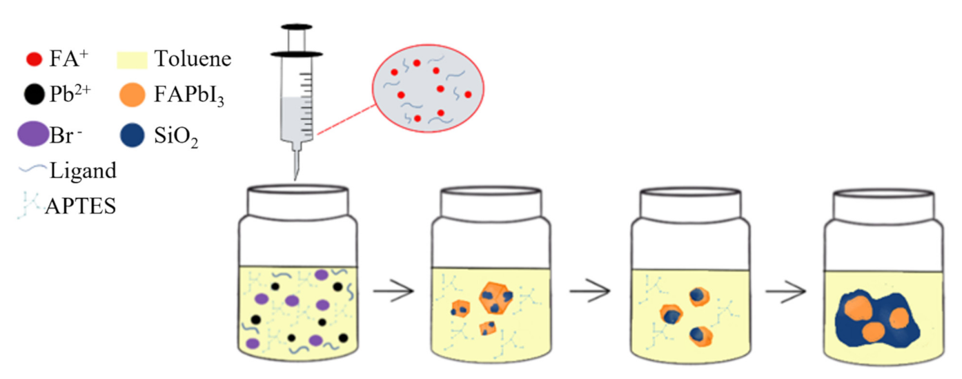
Figure 1. Schematic of the air-synthesis method for preparation of FAPbI3 NCs.
Biosensors 11 00440 g001
Figure 2. FTIR spectra of APTES and FAPbI3 NCs.
Biosensors 11 00440 g002
Figure 3. HRTEM images of (a) PNPs and (b) PNCs.
Biosensors 11 00440 g003
Figure 4. FESEM images of (a) PNP and (b) PNC powders.
Biosensors 11 00440 g004
Figure 5. (a) Photographs of FAPbI3 NCs solvent (from left to right: 0.25–1.0 mL of APTES) under room light and the UV light respectively; (b) the PL spectra of FAPbI3 NCs with different amount of APTES, and (c) photographs of FAPbI3 and FAPbI3NCs powders under room light and UV light.
Biosensors 11 00440 g005
Figure 6. The PL spectra of FAPbI3 (a) PNP and (b) PNC powders stored in air after different days; (c) the intensity of the PL peaks under the DI water as a function of times for FAPbI3 NPN and PNC-dispersed solutions. The insets show the photographs of the FAPbI3 NPNs and PNCs added into water after 16 min.
Biosensors 11 00440 g006
Figure 7. The photographs of the blue chip (455 nm) and the blue chip consisting of FAPbI3NCs under the (a) room light and (b) the NIR-LED devices EL spectrum.
Biosensors 11 00440 g007
Publisher’s Note: MDPI stays neutral with regard to jurisdictional claims in published maps and institutional affiliations.