Antimicrobial Synergistic Effect Between Ag and Zn in Ag-ZnO·mSiO2 Silicate Composite with High Specific Surface Area

 ,
, 
Abstract
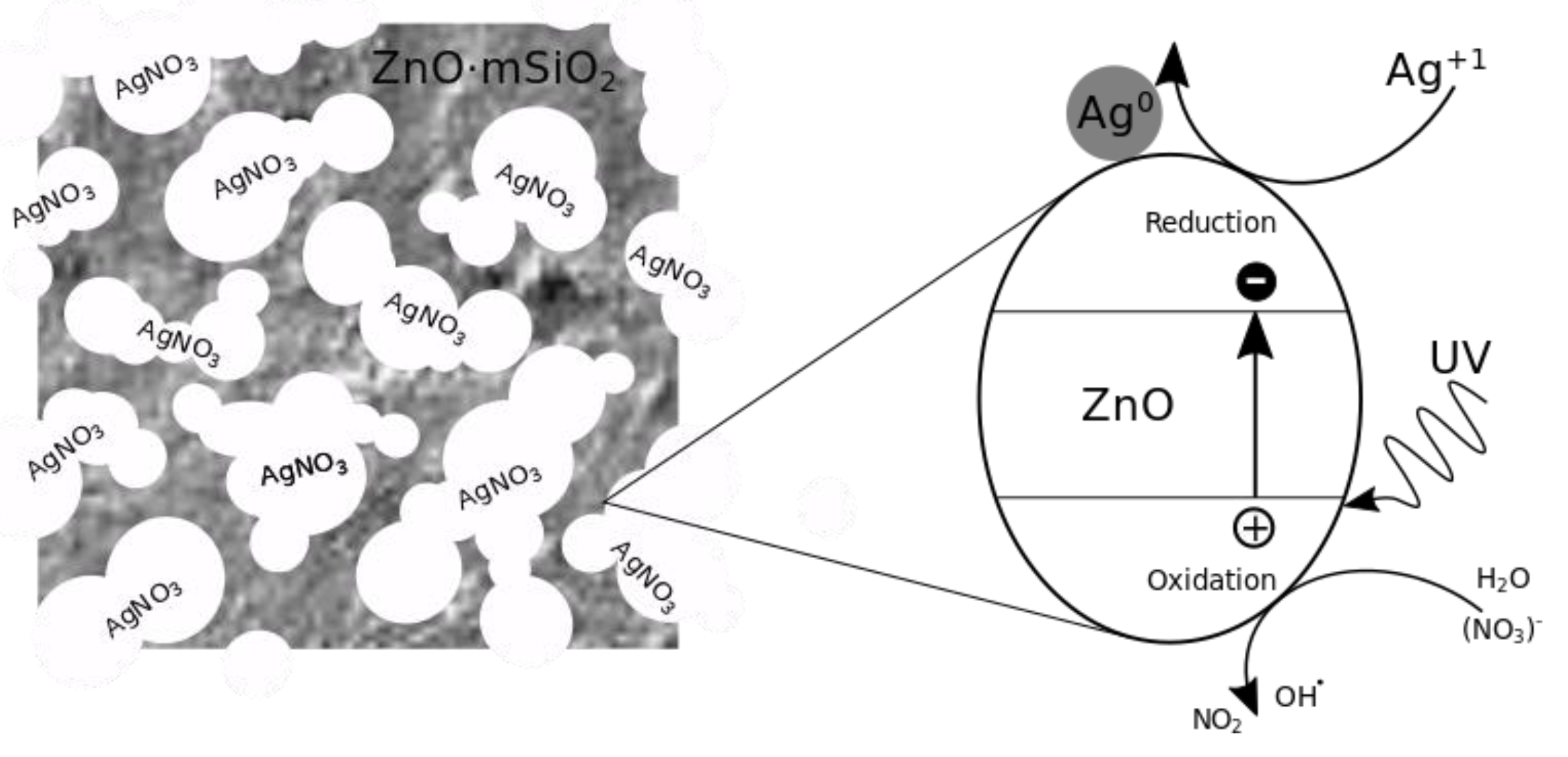
:1. Introduction
2. Materials and Methods
3. Results
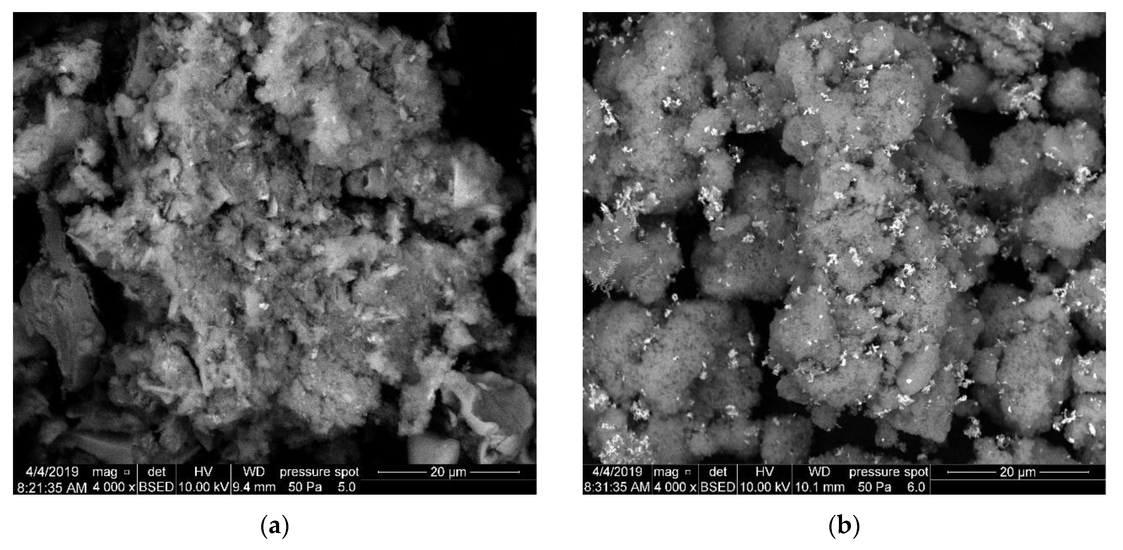
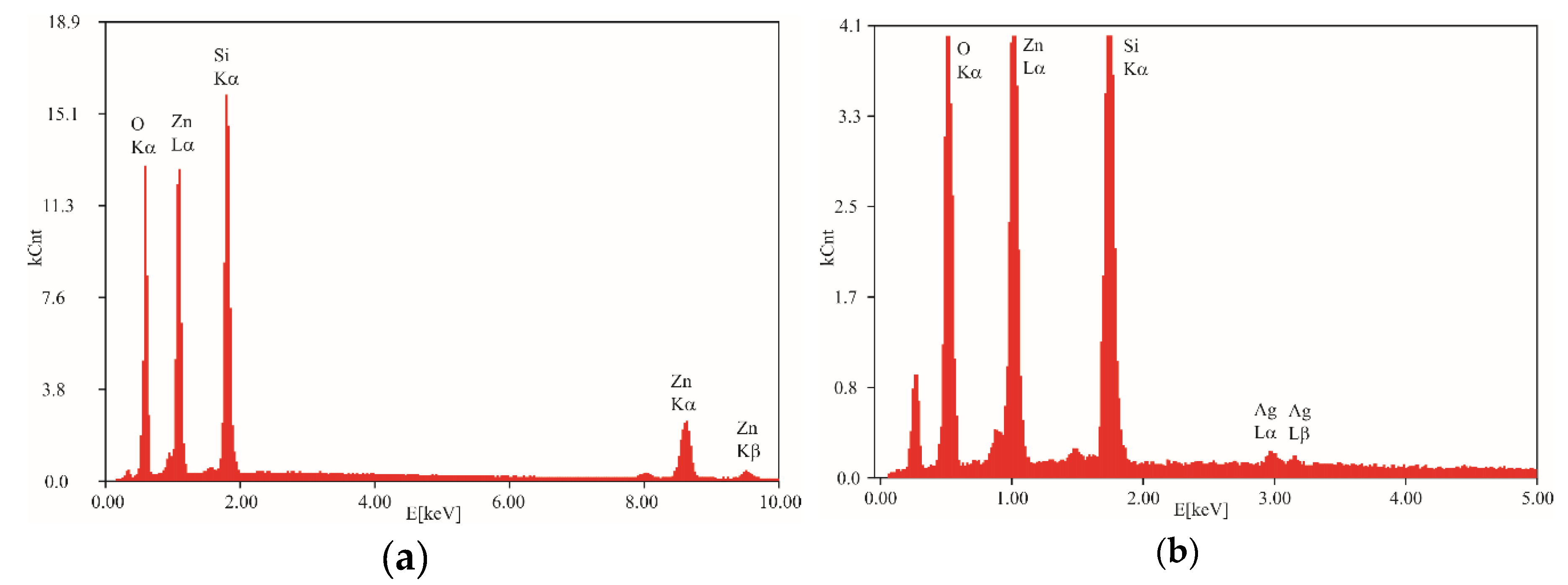
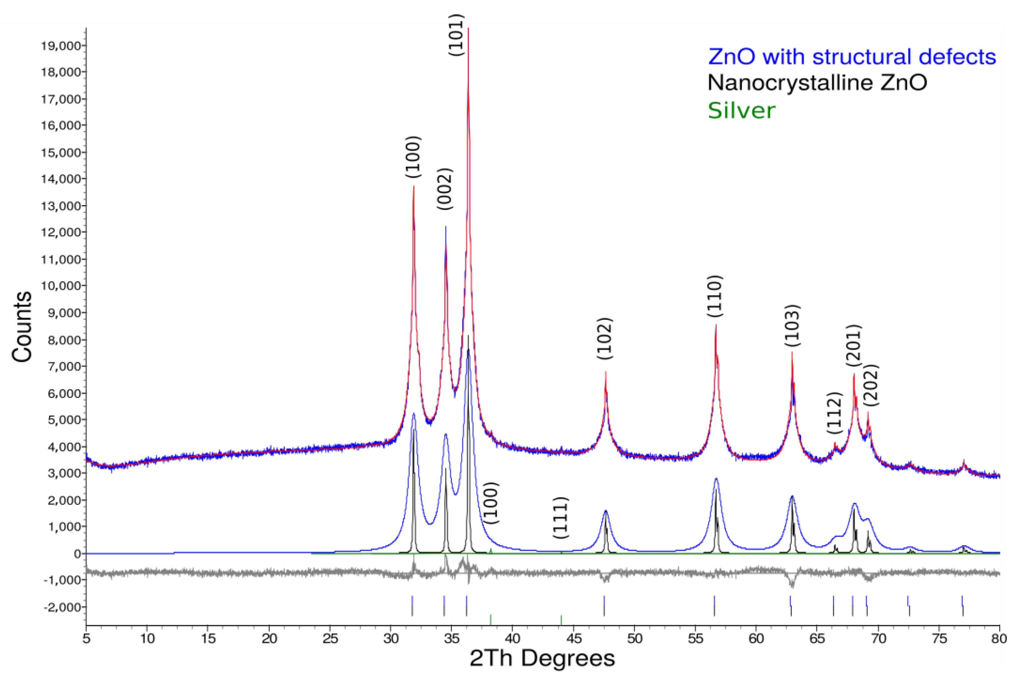
3.1. Scanning Electron Microscopy, Energy Disperse X-Ray Spectroscopy and Specific Surface Area Analysis
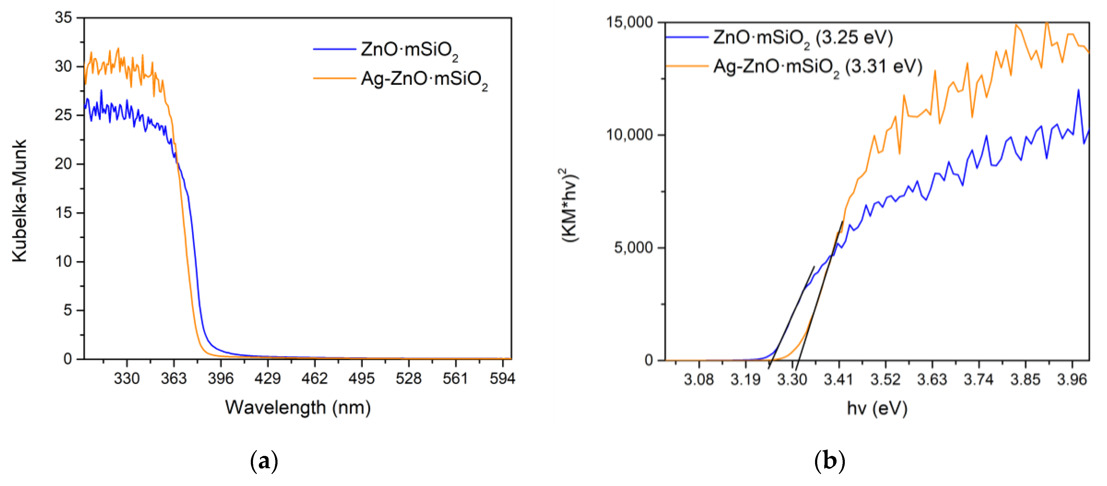
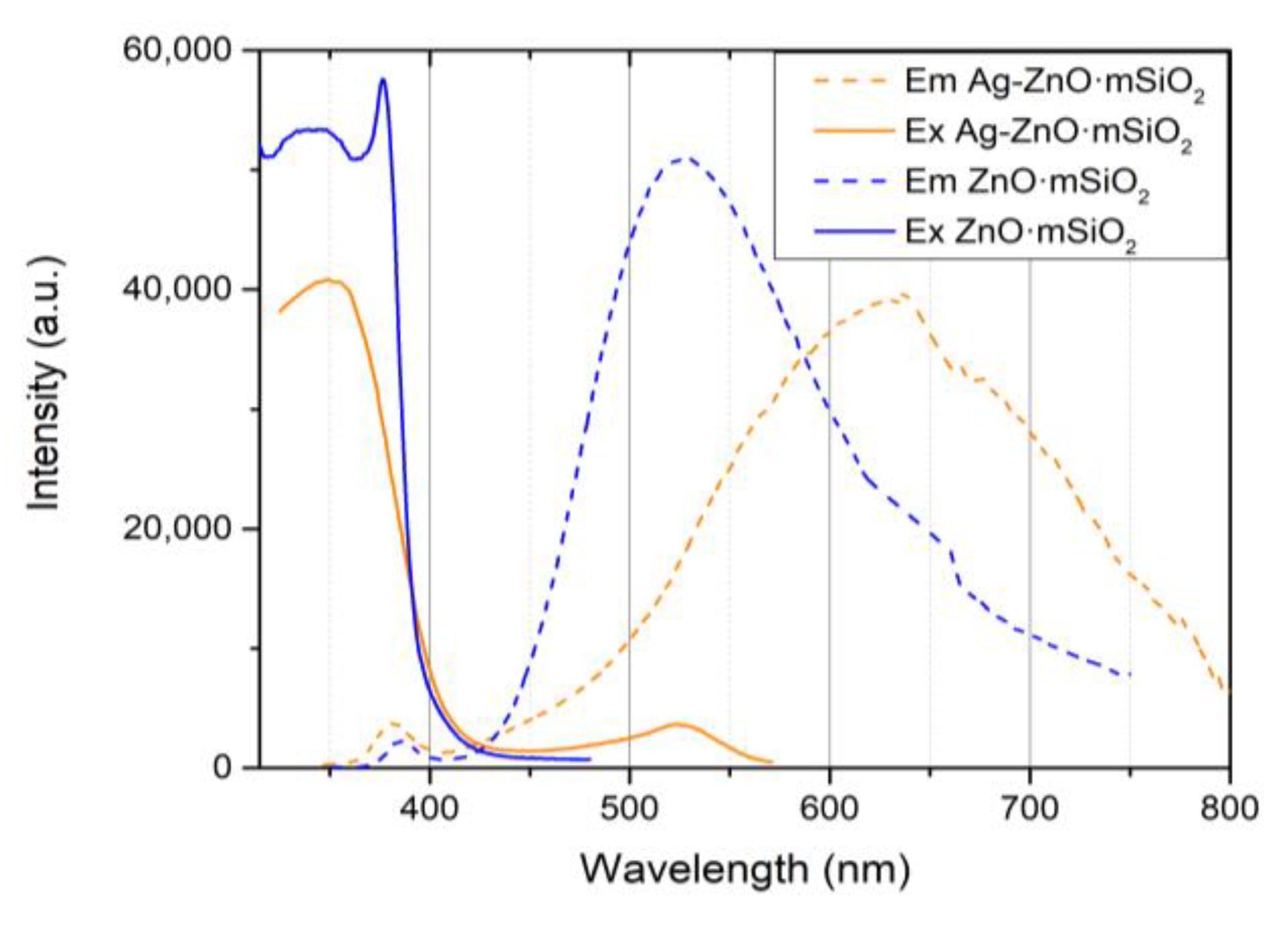
3.2. Optical Analysis
3.3. Antimicrobial Activity of Materials
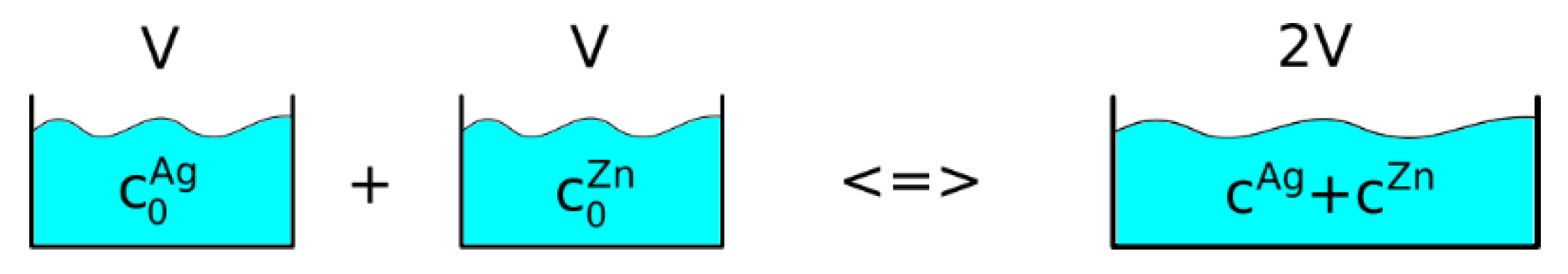
3.4. Calculation of Synergistic Effect between Ag and Zn in Ag-ZnO·mSiO2 Composite
4. Discussion
Author Contributions
Funding
Acknowledgments
Conflicts of Interest
References
- Barra Caracciolo, A.; Topp, E.; Grenni, P. Pharmaceuticals in the environment: Biodegradation and effects on natural microbial communities. A review. J. Pharm. Biomed. Anal. 2015, 106, 25–36. [Google Scholar] [CrossRef]
- Cycoń, M.; Mrozik, A.; Piotrowska-Seget, Z. Antibiotics in the Soil Environment—Degradation and Their Impact on Microbial Activity and Diversity. Front. Microbiol. 2019, 10, 338. [Google Scholar] [CrossRef]
- Li, B.; Webster, T.J. Bacteria antibiotic resistance: New challenges and opportunities for implant-associated orthopedic infections. J. Orthop. Res. 2017, 36, 22–32. [Google Scholar] [CrossRef]
- Zhang, X.F.; Liu, Z.G.; Shen, W.; Gurunathan, S. Silver Nanoparticles: Synthesis, Characterization, Properties, Applications and Therapeutic Approaches. Int. J. Mol. Sci. 2016, 17, 1534. [Google Scholar] [CrossRef]
- Percival, S.L.; Bowler, P.G.; Russell, D. Bacterial resistance to silver in wound care. J. Hosp. Infect. 2005, 60, 1–7. [Google Scholar] [CrossRef]
- Zhang, C.; Hu, Z.; Deng, B. Silver nanoparticles in aquatic environments: Physiochemical behavior and antimicrobial mechanisms. Water Res. 2016, 88, 403–427. [Google Scholar] [CrossRef] [Green Version]
- Durán, N.; Durán, M.; de Jesus, M.B.; Seabra, A.B.; Fávaro, W.J.; Nakazato, G. Silver nanoparticles: A new view on mechanistic aspects on antimicrobial activity. Nanomed. Nanotechnol. Biol. Med. 2016, 12, 789–799. [Google Scholar] [CrossRef]
- Hatchett, D.W.; White, H.S. Electrochemistry of Sulfur Adlayers on the Low-Index Faces of Silver. J. Phys. Chem. 1996, 100, 9854–9859. [Google Scholar] [CrossRef]
- Feng, Q.L.; Wu, J.; Chen, G.Q.; Cui, F.Z.; Kim, T.N.; Kim, J.O. A mechanistic study of the antibacterial effect of silver ions onEscherichia coli andStaphylococcus aureus. J. Biomed. Mater. Res. 2000, 52, 662–668. [Google Scholar] [CrossRef]
- Dakal, T.C.; Kumar, A.; Majumdar, R.S.; Yadav, V. Mechanistic Basis of Antimicrobial Actions of Silver Nanoparticles. Front. Microbiol. 2016, 7, 7831. [Google Scholar] [CrossRef]
- Berger, T.J.; Spadaro, J.A.; Chapin, S.E.; Becker, R.O. Electrically generated silver ions: Quantitative effects on bacterial and mammalian cells. Antimicrob. Agents Chemother. 1976, 9, 357–358. [Google Scholar] [CrossRef]
- Nowack, B.; Krug, H.F.; Height, M. 120 Years of Nanosilver History: Implications for Policy Makers. Environ. Sci. Technol. 2011, 45, 1177–1183. [Google Scholar] [CrossRef]
- Bosetti, M.; Masse, A.; Tobin, E.; Cannas, M. Silver coated materials for external fixation devices: In vitro biocompatibility and genotoxicity. Biomaterials 2002, 23, 887–892. [Google Scholar] [CrossRef]
- Knetsch, M.L.W.; Koole, L.H. New Strategies in the Development of Antimicrobial Coatings: The Example of Increasing Usage of Silver and Silver Nanoparticles. Polymers (Basel) 2011, 3, 340–366. [Google Scholar] [CrossRef]
- Kharaghani, D.; Khan, M.; Shahzad, A.; Inoue, Y.; Yamamoto, T.; Rozet, S.; Tamada, Y.; Kim, I. Preparation and In-Vitro Assessment of Hierarchal Organized Antibacterial Breath Mask Based on Polyacrylonitrile/Silver (PAN/AgNPs) Nanofiber. Nanomaterials 2018, 8, 461. [Google Scholar] [CrossRef]
- Olmos, D.; Pontes-Quero, G.; Corral, A.; González-Gaitano, G.; González-Benito, J. Preparation and Characterization of Antimicrobial Films Based on LDPE/Ag Nanoparticles with Potential Uses in Food and Health Industries. Nanomaterials 2018, 8, 60. [Google Scholar] [CrossRef]
- Wang, K.; Wu, Y.; Li, H.; Li, M.; Zhang, D.; Feng, H.; Fan, H. Synthesis, characterization and antimicrobial activity of silver nanoparticles: Agn(NALC)m and Agn(GSHR)m. RSC Adv. 2014, 4, 5130. [Google Scholar] [CrossRef]
- Coseri, S.; Spatareanu, A.; Sacarescu, L.; Rimbu, C.; Suteu, D.; Spirk, S.; Harabagiu, V. Green synthesis of the silver nanoparticles mediated by pullulan and 6-carboxypullulan. Carbohydr. Polym. 2015, 116, 9–17. [Google Scholar] [CrossRef]
- Guzman, M.; Dille, J.; Godet, S. Synthesis and antibacterial activity of silver nanoparticles against gram-positive and gram-negative bacteria. Nanomed. Nanotechnol. Biol. Med. 2012, 8, 37–45. [Google Scholar] [CrossRef]
- Prabhu, S.; Poulose, E.K. Silver nanoparticles: Mechanism of antimicrobial action, synthesis, medical applications and toxicity effects. Int. Nano Lett. 2012, 2, 32. [Google Scholar] [CrossRef]
- Huang, S.; Wang, J.; Zhang, Y.; Yu, Z.; Qi, C. Quaternized Carboxymethyl Chitosan-Based Silver Nanoparticles Hybrid: Microwave-Assisted Synthesis, Characterization and Antibacterial Activity. Nanomaterials 2016, 6, 118. [Google Scholar] [CrossRef]
- Cataldo, F.; Ursini, O.; Angelini, G. Synthesis of silver nanoparticles by radiolysis, photolysis and chemical reduction of AgNO3 in Hibiscus sabdariffa infusion (karkadé). J. Radioanal. Nucl. Chem. 2016, 307, 447–455. [Google Scholar] [CrossRef]
- Chopade, B.; Patil, S.; Ahire, M.; Kitture, R.; Jabgunde, A.; Kale, S.; Pardesi, K.; Cameotra, S.S.; Bellare, J.; Dhavale, D.D.; et al. Synthesis of silver nanoparticles using Dioscorea bulbifera tuber extract and evaluation of its synergistic potential in combination with antimicrobial agents. Int. J. Nanomed. 2012, 7, 483–496. [Google Scholar] [CrossRef] [Green Version]
- Rhim, J.W.; Wang, L.F.; Hong, S.I. Preparation and characterization of agar/silver nanoparticles composite films with antimicrobial activity. Food Hydrocoll. 2013, 33, 327–335. [Google Scholar] [CrossRef]
- Venkatesan, J.; Kim, S.K.; Shim, M. Antimicrobial, Antioxidant, and Anticancer Activities of Biosynthesized Silver Nanoparticles Using Marine Algae Ecklonia cava. Nanomaterials 2016, 6, 235. [Google Scholar] [CrossRef]
- Sharma, V.K.; Yngard, R.A.; Lin, Y. Silver nanoparticles: Green synthesis and their antimicrobial activities. Adv. Colloid Interface Sci. 2009, 145, 83–96. [Google Scholar] [CrossRef]
- Raza, M.; Kanwal, Z.; Rauf, A.; Sabri, A.; Riaz, S.; Naseem, S. Size- and Shape-Dependent Antibacterial Studies of Silver Nanoparticles Synthesized by Wet Chemical Routes. Nanomaterials 2016, 6, 74. [Google Scholar] [CrossRef]
- Piella, J.; Bastús, N.G.; Puntes, V. Size-Controlled Synthesis of Sub-10-nanometer Citrate-Stabilized Gold Nanoparticles and Related Optical Properties. Chem. Mater. 2016, 28, 1066–1075. [Google Scholar] [CrossRef]
- Hassanien, A.S.; Khatoon, U.T. Synthesis and characterization of stable silver nanoparticles, Ag-NPs: Discussion on the applications of Ag-NPs as antimicrobial agents. Phys. B Condens. Matter 2019, 554, 21–30. [Google Scholar] [CrossRef]
- Otari, S.V.; Yadav, H.M.; Thorat, N.D.; Patil, R.M.; Lee, J.K.; Pawar, S.H. Facile one pot synthesis of core shell Ag@SiO2 nanoparticles for catalytic and antimicrobial activity. Mater. Lett. 2016, 167, 179–182. [Google Scholar] [CrossRef]
- Bakr, E.A.; El-Attar, H.G.; Salem, M.A. Colloidal Ag Pd core–shell nanoparticles showing fast catalytic eradication of dyes from water and excellent antimicrobial behavior. Res. Chem. Intermed. 2019, 45, 1509–1526. [Google Scholar] [CrossRef]
- Tudose, M.; Culita, D.C.; Musuc, A.M.; Marinescu, G.; Somacescu, S.; Munteanu, C.; Bleotu, C.; Chifiriuc, M.C. Multifunctional Silver Nanoparticles-Decorated Silica Functionalized with Retinoic Acid with Anti-Proliferative and Antimicrobial Properties. J. Inorg. Organomet. Polym. Mater. 2016, 26, 1043–1052. [Google Scholar] [CrossRef]
- Jędrzejczyk, R.J.; Turnau, K.; Jodłowski, P.J.; Chlebda, D.K.; Łojewski, T.; Łojewska, J. Antimicrobial Properties of Silver Cations Substituted to Faujasite Mineral. Nanomaterials 2017, 7, 240. [Google Scholar] [CrossRef]
- Soto-Quintero, A.; Romo-Uribe, Á.; Bermúdez-Morales, V.; Quijada-Garrido, I.; Guarrotxena, N. 3D-Hydrogel Based Polymeric Nanoreactors for Silver Nano-Antimicrobial Composites Generation. Nanomaterials 2017, 7, 209. [Google Scholar] [CrossRef]
- Vi, T.; Rajesh Kumar, S.; Rout, B.; Liu, C.H.; Wong, C.B.; Chang, C.W.; Chen, C.H.; Chen, D.; Lue, S. The Preparation of Graphene Oxide-Silver Nanocomposites: The Effect of Silver Loads on Gram-Positive and Gram-Negative Antibacterial Activities. Nanomaterials 2018, 8, 163. [Google Scholar] [CrossRef]
- Das, M.R.; Sarma, R.K.; Saikia, R.; Kale, V.S.; Shelke, M.V.; Sengupta, P. Synthesis of silver nanoparticles in an aqueous suspension of graphene oxide sheets and its antimicrobial activity. Colloids Surf. B Biointerfaces 2011, 83, 16–22. [Google Scholar] [CrossRef]
- Zhang, M.; Zhao, Y.; Yan, L.; Peltier, R.; Hui, W.; Yao, X.; Cui, Y.; Chen, X.; Sun, H.; Wang, Z. Interfacial Engineering of Bimetallic Ag/Pt Nanoparticles on Reduced Graphene Oxide Matrix for Enhanced Antimicrobial Activity. ACS Appl. Mater. Interfaces 2016, 8, 8834–8840. [Google Scholar] [CrossRef]
- Mirahmadi-Zare, S.Z.; Allafchian, A.R.; Jalali, S.A.H. Core-shell fabrication of an extra-antimicrobial magnetic agent with synergistic effect of substrate ligand to increase the antimicrobial activity of Ag nanoclusters. Environ. Prog. Sustain. Energy 2019, 38, 237–245. [Google Scholar] [CrossRef]
- Allafchian, A.R.; Jalali, S.A.H.; Amiri, R.; Shahabadi, S. Antibacterial activity of new magnetic Ag/TiO2 nanocomposite in silane sol–gel matrix. J. Mater. Sci. Mater. Electron. 2017, 28, 12312–12319. [Google Scholar] [CrossRef]
- Ye, J.; Cheng, H.; Li, H.; Yang, Y.; Zhang, S.; Rauf, A.; Zhao, Q.; Ning, G. Highly synergistic antimicrobial activity of spherical and flower-like hierarchical titanium dioxide/silver composites. J. Colloid Interface Sci. 2017, 504, 448–456. [Google Scholar] [CrossRef] [Green Version]
- Naik, K.; Chatterjee, A.; Prakash, H.; Kowshik, M. Mesoporous TiO2 Nanoparticles Containing Ag Ion with Excellent Antimicrobial Activity at Remarkable Low Silver Concentrations. J. Biomed. Nanotechnol. 2013, 9, 664–673. [Google Scholar] [CrossRef]
- Xiao, G.; Zhang, X.; Zhang, W.; Zhang, S.; Su, H.; Tan, T. Visible-light-mediated synergistic photocatalytic antimicrobial effects and mechanism of Ag-nanoparticles chitosan–TiO2 organic–inorganic composites for water disinfection. Appl. Catal. B Environ. 2015, 170–171, 255–262. [Google Scholar] [CrossRef]
- D’Agostino, A.; Taglietti, A.; Desando, R.; Bini, M.; Patrini, M.; Dacarro, G.; Cucca, L.; Pallavicini, P.; Grisoli, P. Bulk Surfaces Coated with Triangular Silver Nanoplates: Antibacterial Action Based on Silver Release and Photo-Thermal Effect. Nanomaterials 2017, 7, 7. [Google Scholar] [CrossRef]
- Shende, P.; Oza, B.; Gaud, R.S. Silver-doped titanium dioxide nanoparticles encapsulated in chitosan–PVA film for synergistic antimicrobial activity. Int. J. Polym. Mater. Polym. Biomater. 2018, 67, 1080–1086. [Google Scholar] [CrossRef]
- Jia, Q.; Shan, S.; Jiang, L.; Wang, Y.; Li, D. Synergistic antimicrobial effects of polyaniline combined with silver nanoparticles. J. Appl. Polym. Sci. 2012, 125, 3560–3566. [Google Scholar] [CrossRef]
- Kang, C.; Ahn, D.; Roh, C.; Kim, S.S.; Lee, J. Development of Synergistic Antimicrobial Coating of p-Aramid Fibers Using Ag Nanoparticles and Glycidyltrimethylammonium Chloride (GTAC) without the Aid of a Cross-Linking Agent. Polymers (Basel) 2017, 9, 357. [Google Scholar] [CrossRef]
- Hamed, S.; Emara, M.; Shawky, R.M.; El-domany, R.A.; Youssef, T. Silver nanoparticles: Antimicrobial activity, cytotoxicity, and synergism with N-acetyl cysteine. J. Basic Microbiol. 2017, 57, 659–668. [Google Scholar] [CrossRef]
- Croes, S.; Stobberingh, E.E.; Stevens, K.N.J.; Knetsch, M.L.W.; Koole, L.H. Antimicrobial and Anti-Thrombogenic Features Combined in Hydrophilic Surface Coatings for Skin-Penetrating Catheters. Synergy of Co-embedded Silver Particles and Heparin. ACS Appl. Mater. Interfaces 2011, 3, 2543–2550. [Google Scholar] [CrossRef]
- Savić, N.D.; Milivojevic, D.R.; Glišić, B.D.; Ilic-Tomic, T.; Veselinovic, J.; Pavic, A.; Vasiljevic, B.; Nikodinovic-Runic, J.; Djuran, M.I. A comparative antimicrobial and toxicological study of gold(III) and silver(i) complexes with aromatic nitrogen-containing heterocycles: Synergistic activity and improved selectivity index of Au(III)/Ag(i) complexes mixture. RSC Adv. 2016, 6, 13193–13206. [Google Scholar] [CrossRef]
- Fakhri, A.; Tahami, S.; Naji, M. Synthesis and characterization of core-shell bimetallic nanoparticles for synergistic antimicrobial effect studies in combination with doxycycline on burn specific pathogens. J. Photochem. Photobiol. B Biol. 2017, 169, 21–26. [Google Scholar] [CrossRef]
- Garza-Cervantes, J.A.; Chávez-Reyes, A.; Castillo, E.C.; García-Rivas, G.; Antonio Ortega-Rivera, O.; Salinas, E.; Ortiz-Martínez, M.; Gómez-Flores, S.L.; Peña-Martínez, J.A.; Pepi-Molina, A.; et al. Synergistic Antimicrobial Effects of Silver/Transition-metal Combinatorial Treatments. Sci. Rep. 2017, 7, 903. [Google Scholar] [CrossRef]
- Krylova, G.; Eremenko, A.; Smirnova, N.; Eustis, S. Structure and spectra of photochemically obtained nanosized silver particles in presence of modified porous silica. Int. J. Photoenergy 2005, 7, 193–198. [Google Scholar] [CrossRef]
- Xu, L.; Li, S.; Li, F.; Zhang, H.; Wang, D.; Chen, M.; Chen, F. Ultraviolet light-induced photochemical reaction for controlled fabrication of Ag nano-islands on ZnO nanosheets: An advanced inexpensive substrate for ultrasensitive surface-enhanced Raman scattering analysis. Opt. Mater. Express 2017, 7, 3137. [Google Scholar] [CrossRef]
- Kantipudi, S.; Sunkara, J.R.; Rallabhandi, M.; Thonangi, C.V.; Cholla, R.D.; Kollu, P.; Parvathaneni, M.K.; Pammi, S.V.N. Enhanced wound healing activity of Ag–ZnO composite NPs in Wistar Albino rats. IET Nanobiotechnology 2018, 12, 473–478. [Google Scholar] [CrossRef]
- Bednář, J.; Mančík, P.; Svoboda, L.; Dvorsky, R. Enhanced Disintegration of Silicon Particles due to their Mutual Impact Caused by Ultrasonic Cavitation Bubbles. Key Eng. Mater. 2019, 810, 131–136. [Google Scholar] [CrossRef]
- Richard, D. A Method Of The Preparation Of Fibrillar And Lamellar Porous Microstructures And Nanostructures By Means Of Controlled Vacuum Freeze-drying Of Liquid Nanoparticles Dispersions. Patent Application No: US9410739B2, 7 Mar 2013. [Google Scholar]
- Dvorský, R.; Bednář, J.; Mančík, P.; Svoboda, L.; Trojková, J.; Matýsek, D.; Peikertová, P. Synthesis of composite photocatalytic nanoparticles ZnO·mSiO2 using new aerosol method. Hut List 2016, 6, 68–72. [Google Scholar]
- Clinical and Laboraty Standards Institutes. Methods for Determining Bactericidal Activity of Antimicrobial Agents; National Committee for Clinical Laboratory Standards: Wayne, PA, USA, 1999; ISBN 1-56238-384-1. [Google Scholar]
- Cockerill, F. Methods for Dilution Antimicrobial Susceptibility Tests for Bacteria that Grow Aerobically: Approved Standard; Clinical and Laboratory Standards Institute: Wayne, Pa, USA, 2012; ISBN 1-56238-784-7. [Google Scholar]
- Balouiri, M.; Sadiki, M.; Ibnsouda, S.K. Methods for in vitro evaluating antimicrobial activity: A review. J. Pharm. Anal. 2016, 6, 71–79. [Google Scholar] [CrossRef]
- Qing, Y.; Cheng, L.; Li, R.; Liu, G.; Zhang, Y.; Tang, X.; Wang, J.; Liu, H.; Qin, Y. Potential antibacterial mechanism of silver nanoparticles and the optimization of orthopedic implants by advanced modification technologies. Int. J. Nanomed. 2018, 13, 3311–3327. [Google Scholar] [CrossRef]
- Malachová, K.; Praus, P.; Rybková, Z.; Kozák, O. Antibacterial and antifungal activities of silver, copper and zinc montmorillonites. Appl. Clay Sci. 2011, 53, 642–645. [Google Scholar] [CrossRef]
- Silva Santos, K.; Barbosa, A.; Pereira da Costa, L.; Pinheiro, M.; Oliveira, M.; Ferreira Padilha, F. Silver Nanocomposite Biosynthesis: Antibacterial Activity against Multidrug-Resistant Strains of Pseudomonas aeruginosa and Acinetobacter baumannii. Molecules 2016, 21, 1255. [Google Scholar] [CrossRef]
- Malachová, K.; Praus, P.; Pavlíčková, Z.; Turicová, M. Activity of antibacterial compounds immobilised on montmorillonite. Appl. Clay Sci. 2009, 43, 364–368. [Google Scholar] [CrossRef]
- Dvorsky, R.; Svoboda, L.; Bednář, J.; Mančík, P.; Matýsek, D.; Pomiklová, M. Deposition of Sorption and Photocatalytic Material on Nanofibers and Fabric by Controlled Sublimation. Mater. Sci. Forum 2018, 936, 63–67. [Google Scholar] [CrossRef]


| m = 3 | m = 2.5 | m = 2 | m = 1.5 |
|---|---|---|---|
| ZnO∙3SiO2 | 2ZnO∙5SiO2 | ZnO∙2SiO2 | 2ZnO∙3SiO2 |
| Microbial Strains | MIC (mg·cm−3) | ||
|---|---|---|---|
| AgNO3 | Ag-ZnO·mSiO2 | ZnO·mSiO2 | |
| E. coli (G−) | 0.005 | 2.9 ± 0.1 | 10.6 |
| Pseudomonas aeruginosa (G−) | 0.005 | 3.9 ± 1.4 | 26.5 |
| Streptococcus salivarius (G+) | 0.16 | 5.9 ± 1.3 | 21.2 ± 2.7 |
| Staphylococcus aureus (G+) | 0.16 | 5.9 ± 0.1 | 21.2 |
| Candida albicans | 0.16 | 23.5 ± 0.5 | N/A |
| Microbial Strains | Ratio | ||
|---|---|---|---|
| E. coli (G−) | 0.003 | 0.008 | 2.4 |
| Pseudomonas aeruginosa (G−) | 0.003 | 0.010 | 3.3 |
| Streptococcus salivarius (G+) | 0.102 | 0.016 | 0.2 |
| Staphylococcus aureus (G+) | 0.102 | 0.016 | 0.2 |
| Candida albicans | 0.102 | 0.063 | 0.6 |
| Microbial Strains | Material Saving (%) | ||||
|---|---|---|---|---|---|
| E. coli (G−) | 0.003 | 2.979 | 0.008 | 0.809 | 45.20 |
| Pseudomonas aeruginosa (G−) | 0.003 | 7.447 | 0.010 | 1.090 | 70.45 |
| Streptococcus salivarius (G+) | 0.102 | 5.957 | 0.016 | 1.652 | 44.93 |
| Staphylococcus aureus (G+) | 0.102 | 5.957 | 0.016 | 1.652 | 44.93 |
© 2019 by the authors. Licensee MDPI, Basel, Switzerland. This article is an open access article distributed under the terms and conditions of the Creative Commons Attribution (CC BY) license (http://creativecommons.org/licenses/by/4.0/).
Share and Cite
Bednář, J.; Svoboda, L.; Rybková, Z.; Dvorský, R.; Malachová, K.; Stachurová, T.; Matýsek, D.; Foldyna, V. Antimicrobial Synergistic Effect Between Ag and Zn in Ag-ZnO·mSiO2 Silicate Composite with High Specific Surface Area. Nanomaterials 2019, 9, 1265. https://doi.org/10.3390/nano9091265
Bednář J, Svoboda L, Rybková Z, Dvorský R, Malachová K, Stachurová T, Matýsek D, Foldyna V. Antimicrobial Synergistic Effect Between Ag and Zn in Ag-ZnO·mSiO2 Silicate Composite with High Specific Surface Area. Nanomaterials. 2019; 9(9):1265. https://doi.org/10.3390/nano9091265
Chicago/Turabian StyleBednář, Jiří, Ladislav Svoboda, Zuzana Rybková, Richard Dvorský, Kateřina Malachová, Tereza Stachurová, Dalibor Matýsek, and Vladimír Foldyna. 2019. "Antimicrobial Synergistic Effect Between Ag and Zn in Ag-ZnO·mSiO2 Silicate Composite with High Specific Surface Area" Nanomaterials 9, no. 9: 1265. https://doi.org/10.3390/nano9091265
APA StyleBednář, J., Svoboda, L., Rybková, Z., Dvorský, R., Malachová, K., Stachurová, T., Matýsek, D., & Foldyna, V. (2019). Antimicrobial Synergistic Effect Between Ag and Zn in Ag-ZnO·mSiO2 Silicate Composite with High Specific Surface Area. Nanomaterials, 9(9), 1265. https://doi.org/10.3390/nano9091265