Article

Manganese/Yttrium Codoped Strontium Nanohexaferrites: Evaluation of Magnetic Susceptibility and Mossbauer Spectra

by
Munirah Abdullah Almessiere
1,2,*,
Yassine Slimani
3,
Hakan Güngüneş
4,
Abdulhadi Baykal
2,
S.V. Trukhanov
5,6,7 and
A.V. Trukhanov
5,6,7
1
Department of Physics, College of Science, Imam Abdulrahman Bin Faisal University, P.O. Box 1982, Dammam 31441, Saudi Arabia
2
Department of Nano-Medicine Research, Institute for Research & Medical Consultations (IRMC), Imam Abdulrahman Bin Faisal University, P.O. Box 1982, Dammam 31441, Saudi Arabia
3
Department of Biophysics, Institute for Research & Medical Consultations (IRMC), Imam Abdulrahman Bin Faisal University, P.O. Box 1982, Dammam 31441, Saudi Arabia
4
Department of Physics, Hitit University, Çevre Yolu Bulvarı-Çorum 19030, Turkey
5
Scientific-Practical Materials Research Centre NAS of Belarus, 19 P. Brovki Street, 220072 Minsk, Belarus
6
Department of Electronic Materials Technology, National University of Science and Technology MISiS, Leninsky Prospekt, 4, Moscow 119049, Russia
7
Laboratory of Crystal Growth, South Ural State University, Lenin Prospect, 76, Chelyabinsk 454080, Russia
*
Author to whom correspondence should be addressed.
Nanomaterials 2019, 9(1), 24; https://doi.org/10.3390/nano9010024
Submission received: 12 November 2018 / Revised: 15 December 2018 / Accepted: 19 December 2018 / Published: 25 December 2018
(This article belongs to the Collection Applications of Magnetic Nanomaterials)

Abstract

:
Manganese (Mn)- and yttrium (Y)-substituted Sr-nanohexaferrites (MYSNHFs) of composition Sr1−xMnxFe12−xYxO19 (with 0.0 ≤ x ≤ 0.5) were prepared by citrate sol-gel autocombustion method. As-prepared MYSNHFs were characterized via diverse analytical techniques to determine the influence of Mn and Y cosubstitution on their microstructures and magnetic properties. 57Fe Mössbauer spectra of the MYSNHFs were used to evaluate the variation in the line width, isomer shift, quadrupole splitting, and hyperfine magnetic field values. It was shown that the dopant ions could preferentially occupy the 12k, 4f2, and 2b sites. Furthermore, the observed shift in the blocking temperatures of the studied MYSNHFs towards lower values with rising Mn2+ and Y3+ contents was attributed to the overall particles size reduction. Meanwhile, the AC susceptibility of the proposed MYSNHFs revealed that the magnetic interactions were weakened with the increase in dopant contents which was ascribed to the replacement of both Sr2+ and Fe3+ ions by the Mn2+ and Y3+ dopants.

Graphical Abstract

1. Introduction

Nanotechnology science has created great excitement and expectations in the last few years. By its very nature, the subject is of immense academic interest, having to do with very tiny objects in the nanometer regime. There has already been much progress in the synthesis, assembly, and fabrication of nanomaterials, and, of equal importance, in the potential applications of these materials in a wide variety of technologies [1,2,3].
In recent times, strontium (Sr) hexaferrites (hereafter named as Sr–HFs) have been intensively studied due to their effectiveness towards microwave (MW) absorption, magnetic recording media, signal processing, telecommunication, MW filtering, audio systems, magneto-optic media, and so forth [4,5,6,7,8]. However, the practical application of such Sr-nanohexaferrites (Sr-nHFs) is strictly affected by the synthesis techniques and preferential site occupation by the dopant ions among the five different Fe3+ sublattices such as tetrahedral (4f1), trigonal bipyramidal (2b), and octahedral (12k, 2a and 4f2) in the hexagonal structure [9]. Meanwhile, Sr–HF systems appear unique wherein their structures allow the favorable substitution of all Fe3+ ions by trivalent ions without secondary phase formation [10]. This in turn leads to the procession of varied magnetic traits depending on the nature of dopants’ including magnetic, nonmagnetic and rare-earths and their contents [11].
Over the last decades, various strategies have been developed to modify the magnetic and electric properties of Sr–HFs via the partial substitution of Fe3+‏ or Sr2+‏ ions. W.M.S. Silva et al. [12] investigated the Mn substitution on the structure and magnetic properties of SrFe12O19 nanoparticles prepared by sol-gel method. They showed that the crystal lattice constants did not change significantly with Mn substitution. Room temperature Mössbauer investigations indicated that Mn ions preferentially occupied the 12k, 4f1, 4f2, and 2a sites. On the other hand, yttrium (Y)-substituted SrFe12O19 hexaferrites were prepared through a solid-state reaction technique by S. Jiang et al. [13]. It is found that the single magnetoplumite phase structure transformed into a multiphase structure with the increase of Y content, where a small amount of hematite phase existed in M-type phase. The magnetization measurements showed that the saturation magnetization (Ms) first increases and then decreases with the increasing of Y content. However, the value of coercivity (Hc) increases with the increasing of Y content. X.F. Niu and coworkers [14] investigated the structural and magnetic properties of Y-doped Sr–HF. The obtained results revealed that the lattice constant ‘a’ increased first of all and then decreased and ‘c’ increases slowly with increasing Y content. Magnetization investigations indicated that Hc and maximum energy product (BHmax) are first increased and then decreased. Also, D. Shekhawat and P.K. Roy [15] reported the influence of Y substitution on the structural, dielectric, and magnetic properties of Sr–HFs synthesized by the autocombustion approach. The structural analysis indicates that the Y ions reorganize themselves without troubling the parent lattice. Ms and Mr magnetizations are decreased, however Hc and Curie temperature (Tc) are improved with increasing Y content. The optimized value of BHmax was obtained for Sr–HFs substituted with Y.
In recent years, several studies tried to greatly improve the properties of M-type Sr–HFs via the cosubstitution of Ce–Y [16], Nd–Zn [17], La–Co [18], La–Cu [19], Zr–Mn [20], Nd–Co [21], La–Zn [22], Nd–Zn [23], Gd–Sn [24], Pr–Ni [25], Bi–Cr [26], Co–Zr [27], Co–W [28], and Mn–Zn [29]. However, the role of both Mn and Y cosubstitution on the various properties of M-type Sr–HFs has not yet been studied. Accordingly, we studied in the present work the effect of Mn and Y cosubstitution on the structural, morphological, microstructural, and magnetic properties (Mössbauer spectra, AC susceptibility, Magnetization versus applied field) of Sr-nHFs was investigated deeply. So, a series of Sr1−xMnxFe12−yYyO19 (with varying x = y), where Sr2+ and Fe3+ ions were partially cosubstituted via Mn2+ of and Y3+ cations, was prepared.

2. Experimental

Analytical grade chemical reagents (purity 99.99%, Sigma-Aldrich, St. Louis, MO, USA) of strontium nitrate [Sr(NO3)2], extra pure iron nitrate [Fe(NO3)3] manganese nitrate [Mn(NO3)2] and yttrium oxide [Y2O3] were utilized as initial materials to prepare Mn/Y codoped Sr1−xMnxFe12−xYxO19 under changing stoichiometric contents (0.0 ≤ x ≤ 0.5) (hereafter designated at MYSNHFs) by sol-gel autocombustion technique. First, stoichiometric amounts of different metal nitrates were dissolved in deionized water using a magnetic stirrer at 80 °C. Next, yttrium oxide was dissolved in 10 mL of HCl at 200 °C by magnetic stirrer to achieve a transparent solution and then added to the nitrate solution under magnetic stirrer at 80 °C for 1 h. Additionally, citric acid (C6H8O7) was added to the resultant mixture as fuel, wherein the pH was adjusted at 7 by incorporating ammonia solution at 150 °C for 30 min, after which the temperature was increased to 320 °C until the solution transformed into a gel then burnt to black powder. Finally, the produced powder was calcinated at 1100 °C for 5 h with heating rate of 10 °C/min to obtain Sr-nHFs phase.
Structures of as-prepared Sr-nHFs were analyzed using X-ray powder diffraction measurement (XRD; Rigaku Benchtop Miniflex, Tokyo, Japan) operated with Cu Kα line at the angular range of 2θ = 20–70°. Scanning/transmission electron microscope (SEM/TEM; FEI Titan 80 – 300kV FEG S/TEM, Hillsboro, OR, USA) equipped with energy dispersive X-ray (EDX) spectroscopy were used for morphology analysis and to determine the chemical elements present in the sample and elemental mapping. Fourier transform infrared (FTIR; Bruker alpha-II FTIR spectrophotometer attached with a diamond ATR, MA, USA) spectra in the wavenumber range of 4000 to 400 cm−1 were recorded to confirm the formation of M-type hexaferrite metal-oxygen bond. AC magnetic susceptibilities and dc magnetizations of all prepared products were measured using a superconducting quantum interference device (PPMS DynaCool, Quantum Design, San Diego, CA, USA). The Mössbauer spectra were performed at room temperature using a conventional Mössbauer spectrometer (Fast Com Tec PC-moss II, Oberhaching, Germany) under constant accelerations mode using 57Fe in Rh matrix with an approximate activity of 10 m Ci. The recorded spectra were analyzed and fitted to inbuilt Win-Normos fitting software (WISSEL company, Germany).

3. Results and Discussion

3.1. Structural Properties

Figure 1a,b shows the XRD patterns of the studied MYSNHFs, which revealed single hexaferrite phase consistent with the JCPDS Card number 96-100-8857 that implemented through the Rietveld refinements by match3! Software (Crystal Impact, Bonn, Germany). At high dopant (Mn/Y) concentrations, XRD patterns displayed a minor peak assigned to α-Fe2O3 phase. Rietveld refinements was used to evaluate the cell parameters (a and c) and crystallite size (D) of prepared MYSNHFs as enlisted in Table 1. The value of a was almost reminiscent of the same values with the increase in dopant concentration. However, the observed fluctuation in the c values was attributed to the ionic radii mismatch of Fe3+ (0.64 Å) and Y3+ (0.90 Å) cations that caused a variation in the crystalline microstrain and the exchange energy of the MYSNHFs [30,31]. The crystallite sizes of the obtained MYSNHFs were calculated by Scherrer’s formula wherein full width at half maximum (FWHM) of the most intense XRD peak was selected.

3.2. Morphology

Figure 2 illustrates the FESEM images of the two selected (x = 0.0, 0.2, 0.4, and 0.5) as-synthesized MYSNHFs, where the surface was consisted of some aggregates of hexagonal plate-like structures. The particles are nanoscale in thickness and microscale in diameter (1–5 µm), so it can be said that the Sr-ferrite particles tend more to grow in the direction parallel to hexagonal plane than that of vertical to the plane [32,33,34].
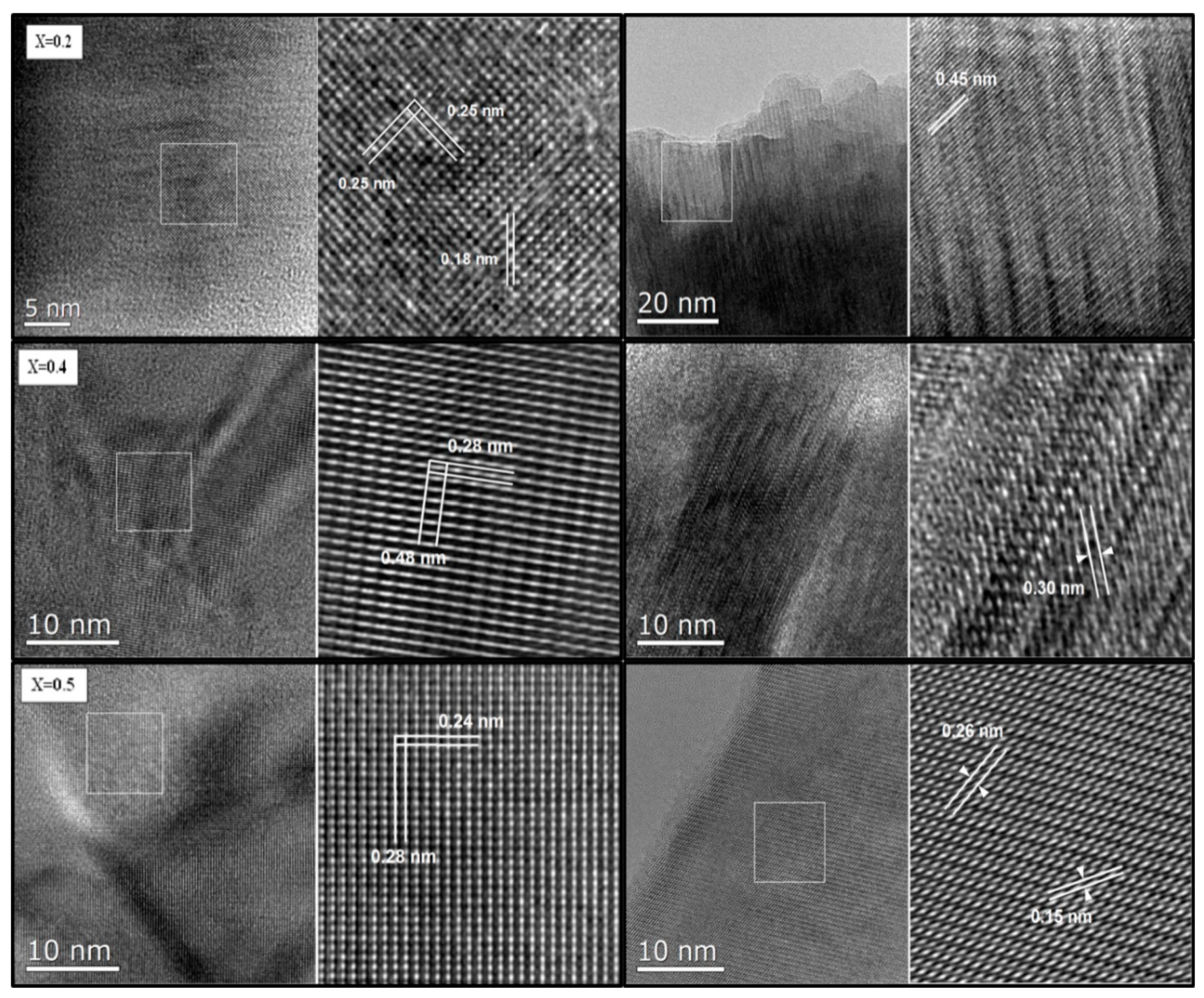
Figure 3 depicts the HRTEM images of three selected (x = 0.2, 0.4, and 0.5) MYSNHFs together with their lattice spacing. The values of lattice spacing were found between 0.15 to 0.48 nm. The estimated lattice spacing were tallied to the (307), (209), (203), (108), (114), (008), (106), (102), and (101) orientations of M type hexagonal atomic planes (in accordance to JCPDS card number) for the respective dopant content as indicated in the Figure 3.
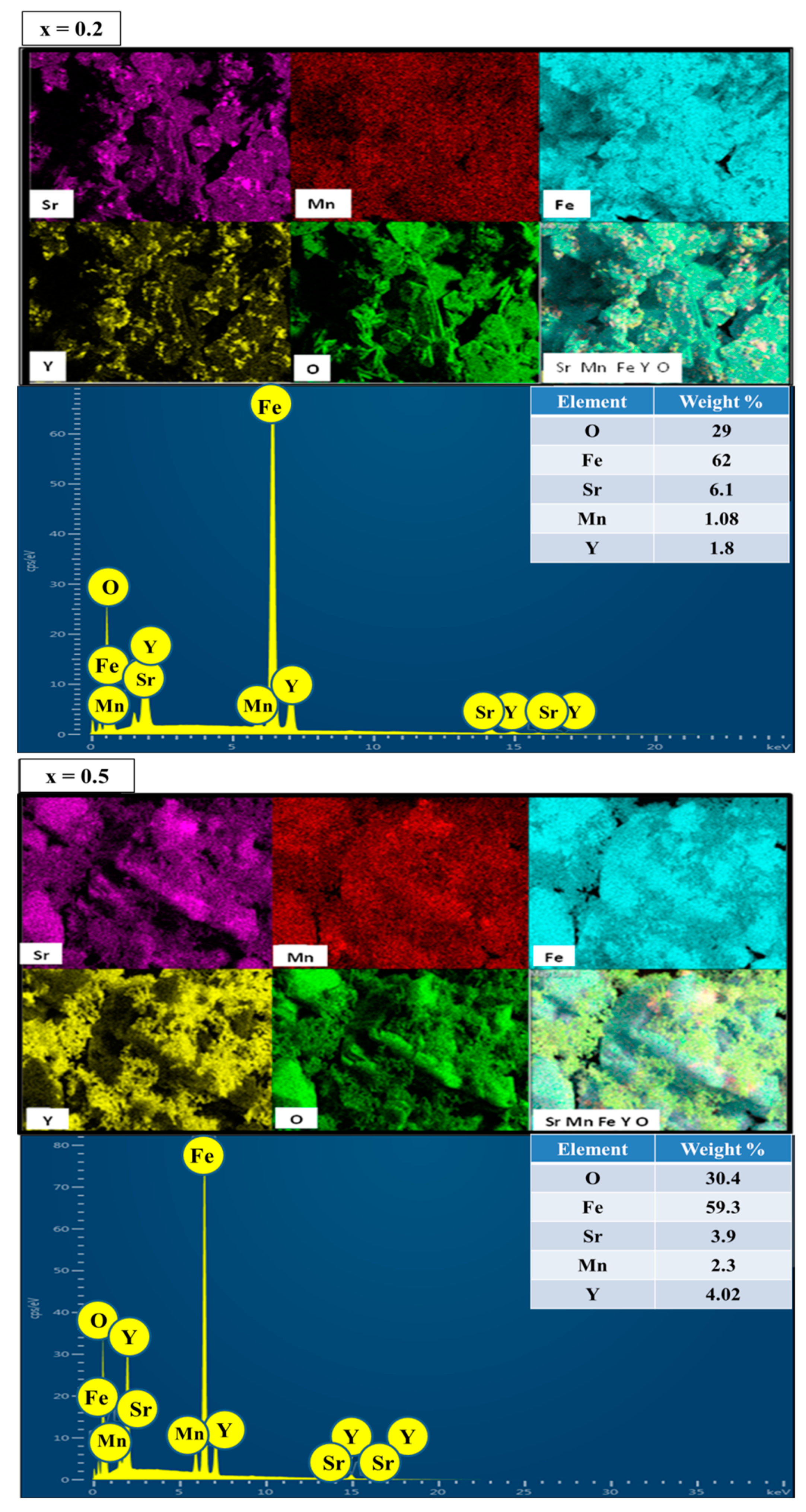
Figure 4 displays the EDX spectra and elemental maps of two selected (x = y = 0.2 and 0.5) MYSNHFs, which revealed the appropriate traces of elements (correct stoichiometric ratios) as indicated in the inset. This observation clearly confirmed the incorporation of Mn/Y into the Sr–HFs lattice structures.

3.3. FTIR Spectra

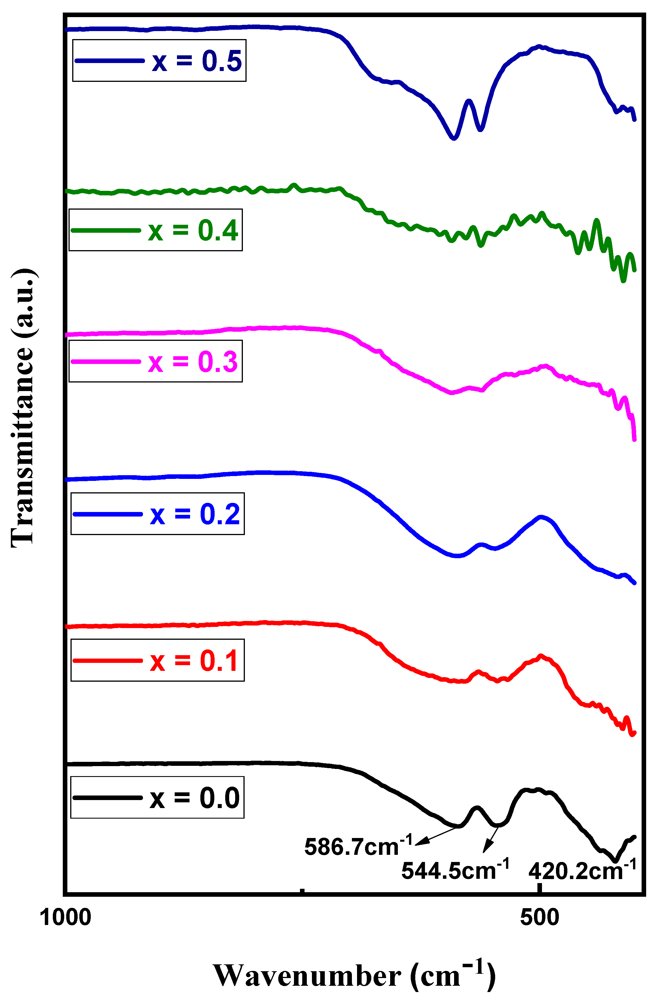
Figure 5 presents the FTIR spectra of obtained MYSNHFs, where the spectral features of all samples were nearly the same. The observed absorption bands at ~420.2, ~544.5, and ~586.7 cm−1 were assigned to the asymmetric stretching of MYSNHFs linkages and out-of-plane bending vibrations of octahedral as well as tetrahedral sites [35,36]. The appeared bands at around 426.34 and 589.67 cm−1 were allocated to the Fe–O bending vibration and Fe–O stretching vibrations. Meanwhile, the observed band at 548.84 cm−1 was approved to the Sr–O bending vibration [37]. Besides, all the absorption bands were broadened accompanied by slight shift (so called bands position disorder) with the increase in the Mn2+ and Y3+ contents in the MYSNHFs [38].

3.4. Mössbauer Spectral Analysis

Figure 6a-f shows the room temperature Mössbauer spectra of achieved MYSNHFs. Mössbauer spectral analyses (using 57Fe) were carried out to determine the relationship between the structure and magnetic properties of the proposed MYSNHFs. It provided useful information about the preferred lattice site occupancy of each type of dopant (cations distribution) in the achieved MYSNHFs.
Table 2 summarizes the fitted parameters of MYSNHFs such as the hyperfine field (Bhf), the quadrupole shift (QS), the isomer shift (IS), the line width (W), and percentage relative area (RA) of the dopant components. Mössbauer spectra were fitted with five discrete sextets corresponding to the octahedral (12k, 4f2 and 2a), the tetrahedral (4f1), and the trigonal bipyramidal (2b) iron sites. For Fe3+ ions, MYSNHFs structure consisted of three spin-up (2a, 2b, and 12k) and two spin-down (4f1 and 4f2) sublattices [39,40]. The 12k position in the Mössbauer spectra of MYSNHFs was split into 12k and 12k1, which were assigned to the perturbation of 12k sites by the presence of Mn2+ and Y3+ ions in the neighboring sites. One superparamagnetic doublet was created in the codoped sample (for x = y = 0.1 and 0.2) beside the ferromagnetic sextets. For uniform distribution of Fe3+ ions, the statistical occupancy corresponding to 12k, 4f1, 4f2, 2a, and 2b sublattice sites in terms of area must be 50:17:17:8:8 [35]. According to results on relative area of undoped Ba-hexaferrite, 12k, 4f1, and 2b positions are close to theoretical values [41]. The 2a position was heavily populated but the 4f2 site was less occupied.
Values of isomer shift (I.S) for MYSNHFs provided the information about the nature of chemical bonding of the iron as well as valence state of Fe cations. The values of I.S were in the range of 0.26 to 0.401 mm/s for all sextets and corresponded to the characteristic charge states of Fe3+. Furthermore, the isomer shift of 4f1 and 2a contributions were increased (Table 2) with the increase in doping levels. The isomer shift of other sites remained unaltered with the addition of dopants. These showed that the s electron density of Fe3+ ions at the 4f1 and 2a sites decreased but others were not affected by Mn2+ and Y3+ substitution.
Values of quadrupole splitting (Q.S) of studied MYSNHFs provided the basic insight about the symmetry of crystal lattice and local distortions. As dopant contents were increased, the Q.S of 4f1, 4f2 and 2b sites were slightly reduced, which was attributed to the symmetry perturbation around these sites due to Mn2+ and Y3+ cation substitution. The room temperature ranking of the hyperfine fields for MYSNHFs containing dopants contents of 0, 0.1, 0.2, 0.3, 0.4, and 0.5 for the five Fe sublattices followed the trend of Bhf (12k1) < Bhf (2b) < Bhf (12k) < Bhf (4f1) < Bhf (2a) < Bhf (4f2) except x, y = 0.4, 0.5. Moreover, the hyperfine fields of 2a site was bigger than that of 4f2 site for x, y = 0.4, 0.5. The hyperfine magnetic field (Table 2) of all sites was slightly reduced with the increase in dopants contents. Meanwhile, the hyperfine magnetic field of 2a site was reduced up to x, y = 0.3 and then enhanced. The observed reduction in the hyperfine magnetic field of the proposed MYSNHFs with substitution of dopants was ascribed to nonmagnetic nature of Y3+ cations that replaced Fe3+ ions in the lattice.
Figure 7 presents the relative area (RA) distribution of all sextets for different Mn2+/Y3+ contents in the obtained MYSNHFs. The value of RA for these sextets was found to be directly proportional to the number of Fe3+ cations in the respective site. Besides, the values of RA for 12k, 2b, and 4f2 sites were reduced and for the 2a site was increased up to x = 0.3 and further increased thereafter. It was argued that such reduction in RA up to x = 0.3 was due to the preferential occupation of Mn2+ and Y3+ ions in the 12k, 2b, and 4f2 sites. Beyond x = 0.3, the observed increase in the RA value was due to the transfer of some Mn2+ and Y3+ cations from 4f2 site to 2a site. Auwal et al. [42] acknowledged the preferred occupation of Y3+ cations at the bipyramidal 2b sites in SrBixLaxYxFe12−3xO19 hexaferrites.

3.5. AC Magnetic Susceptibility

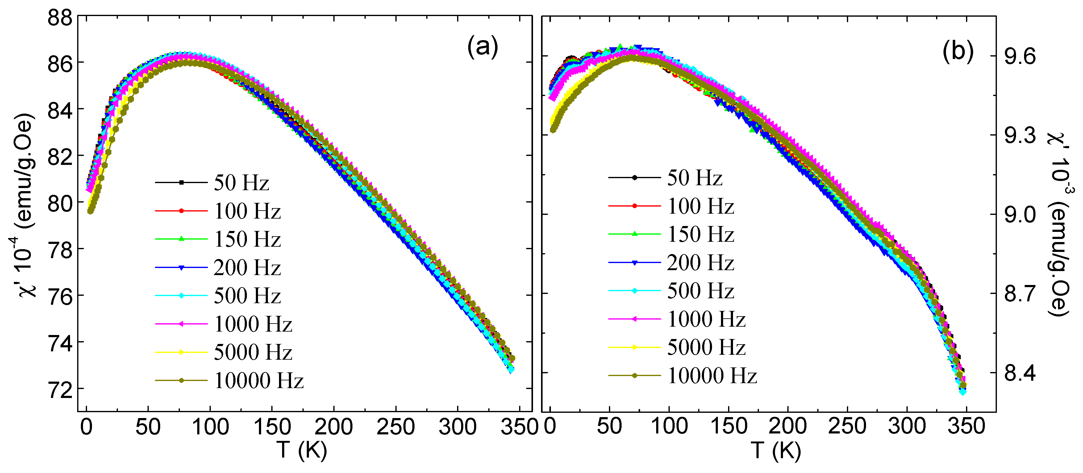
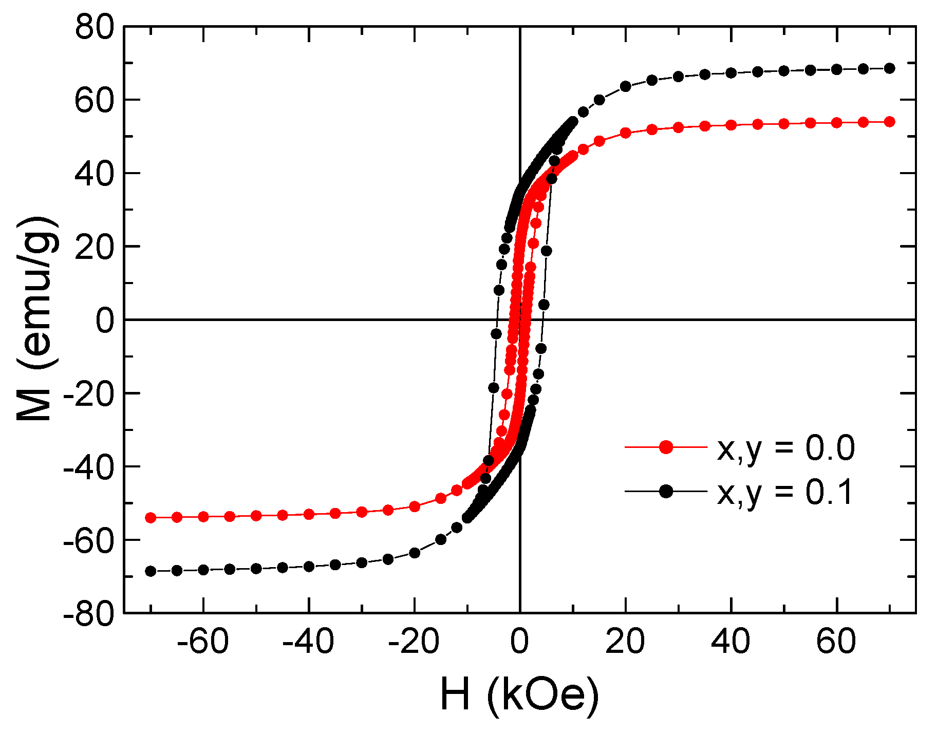
The dynamical magnetic properties and the indirect exchange interactions between Fe3+ and Mn2+ cations in the synthesized MYSNHFs were evaluated using the AC susceptibility data. Figure 8 illustrates the temperature dependent variation in the real part of the AC-magnetic susceptibility (χ′) for two selected MYSNHFs (with x = 0 and 0.1) subjected to the applied AC-field of 10 Oe over the frequency range of 50 to 104 Hz. The values of χ′ for the Mn2+ and Y3+ substituted specimens were reduced significantly compared to the undoped (SrFe12O19) compound, which agreed well with the measurements of magnetization versus applied magnetic field, M(H), as shown in Figure 9. The M(H) hysteresis loops measured at room temperature indicated that the SrFe12O19 and Sr0.9Mn0.1Fe11.9Y0.1O19 nanohexaferrites exhibit ferrimagnetic (FM) behavior. It can be clearly seen from the M(H) results that the magnetization is reduced with Mn and Y substitutions. Magnetic parameters including the saturation magnetization, remanence and coercive field were found to decrease with the increase in Mn2+ and Y3+ contents. Moreover, both samples showed a single peak in χ′(T) curves at a specific temperature TB called blocking temperature, suggesting stabilization of the magnetic phase due to Mn2+ and Y3+ cation substitutions.
The observed shape of χ′ curve at TB was attributed to the emergence of superparamagnetism (SPM) in MYSNHFs which is shown by numerous spin glass (SG) like states [43,44,45]. In this case, the magnetic moments of the hexaferrite nanoparticles were blocked or frozen at T < T B , otherwise behaved freely like paramagnetic state at T > T B . However, T B is seldom represents the fundamental character of a material but often is determined by the microstructure of the sample. The value of TB for Sr0.9Mn0.1Fe11.9Y0.1O19 sample (Figure 8) was shifted slightly to higher temperatures compared to the undoped SrFe12O19 one. This observation was primarily ascribed to the lowering in the anisotropy barrier energy ( E a ) that could determine the SPM state from the blocking region, leading to the grains size shrinkage for x = y = 0.1 [43,44,45]. Additionally, the value of E a was strongly determined by the average particles size and volume ( E a = K eff V , where K eff is the effective anisotropy constant and V is the particles volume). The observed shift in TB position towards higher values with the increase in frequency was also reported for numerous spin-frustrated materials [46,47,48].
A small frequency dispersion in χ’ as evidenced on the left-hand side of the freezing peak. In the present study, the behavior of the χ’ clearly indicated the presence of magnetic inhomogeneity of the studied MYSNHFs. Simultaneously, a weak spin relaxation with varying frequency of the external magnetic field occurred. Another evidence of the presence of magnetic inhomogeneity in the prepared MYSNHFs may be the multipeak character of the imaginary part of the ac-susceptibility. However, the absence of significant decrease as well as shift in the peak value of χ’ with the increase in frequency indicated the deficiency of classical spin glass state in the studied MYSNHFs.
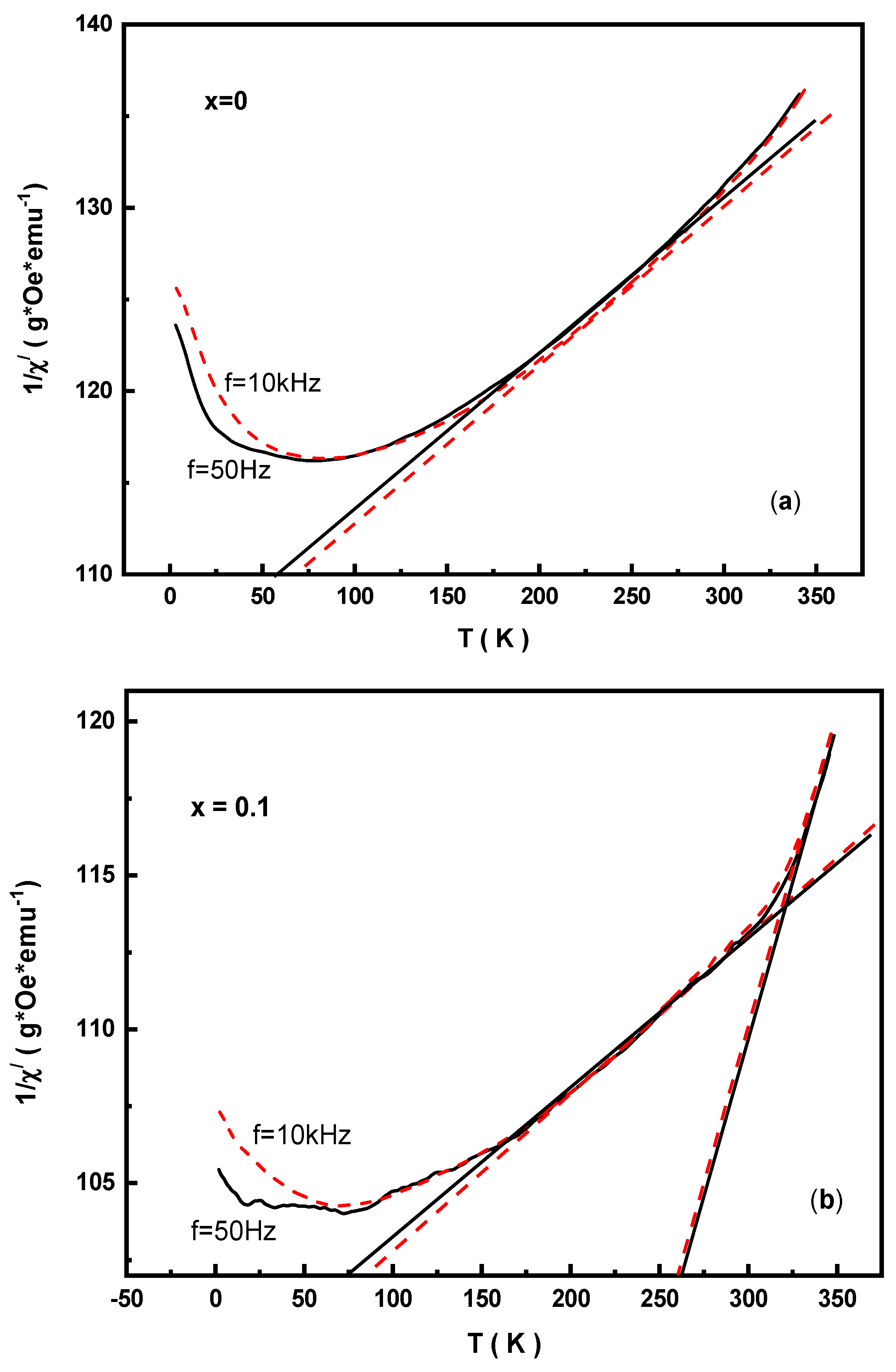
Figure 10 shows the temperature dependent variation in 1/χ’, wherein some interesting features in the studied MYSNHFs were detected from detailed analysis. The behavior of 1/χ’ versus T was found to be strictly of Curie–Weiss type, in which the paramagnetic Curie temperature (ϴp) was revealed above the Curie point (≈730 K). The attainment of positive ϴp implied the presence of predominant indirect exchange interactions in the MYSNHFs. In the temperature range of 50 to 350 K, the 1/χ’ curve for SrFe12O19 was bent downwards, suggesting a continuous change in ϴpi at each point. This in turn indicated the occurrences of a set of positive indirect exchange interactions of different intensities in the studied MYSNHFs.
It is known that in the orbital disordered state, the Mn2+(6)–O–Fe3+(6) super-exchange interactions for the octahedral coordination of Mn and Fe cations are positive, whereas for the Mn2+(5,6)–O–Fe3+(6,5) pentahedral coordination they are negative [49,50,51,52]. Thus, due to Mn2+ and Y3+ cations doping the competitive exchange interactions between the antiferromagnetic and ferromagnetic ordered domains may lead to frustrating exchange coupling and thereby the formation of spin glass state (SGS). The realization of this spin-glass mechanism in the studied MYSNHFs was confirmed by the behavior of 1/χ′ (Figure 10). The linear extrapolation of 1/χ’(T) curve above TB provided two different ϴp values, indicating that the presence of exchange interactions of different strength in the prepared MYSNHFs. Besides, the value of TB also determined the average diameter of the ferromagnetic domains.
The magnetic behavior of particles at nanoscale is known to obey the activation energy of noninteracting magnetic systems likely as in SPM state. Thus, the mechanism of SPM relaxation may be interpreted using the Neel–Arrhenius (N–A) law with the expression [43,44]
τ = τ o   exp ( E a / k B T B )
where, τ denotes the measured time related to the applied frequency ( f = 1 / τ ) and τ 0 denotes the jump attempt time of the nanoparticles magnetic moments between opposite orientations (spin flip-flop) of easy axis magnetization, which varies from 10−9 to 10−13 s for SPM systems. Therefore, the analysis involving the temperature peak shift on the χ′ curves can be regarded as an effective tool to extract the values of E a and K eff .
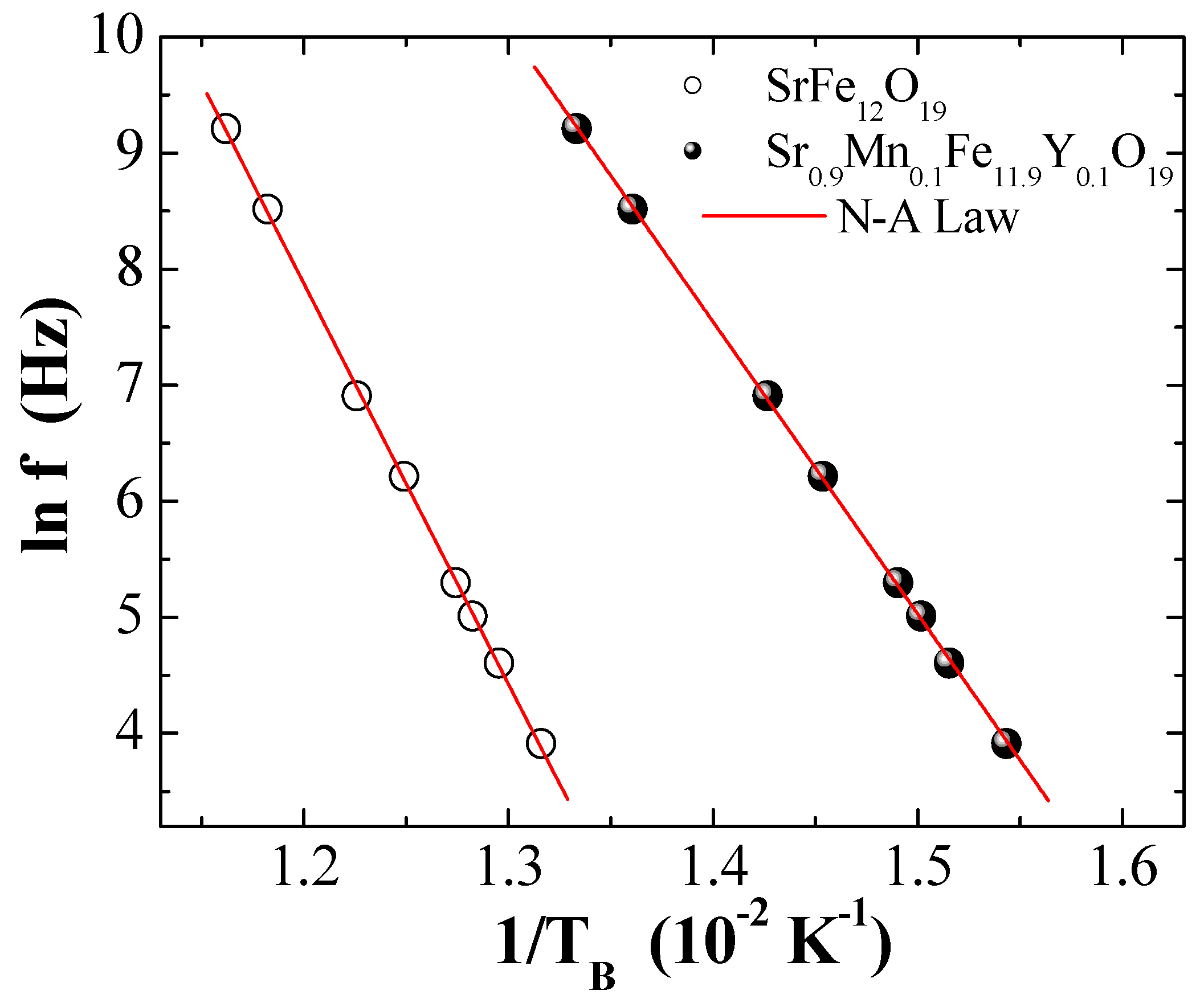
Figure 11 illustrates the dependence of ln ( f ) on 1/TB for two selected MYSNHFs (SrFe12O19 and Sr0.9Mn0.1Fe11.9Y0.1O19), wherein the revelation of linear behavior clearly indicated the involvement of thermally activated processes. The slope and the intercept of the ln ( f ) ~1/TB curves produced the values of E a and f o = 1 / τ 0 , respectively. The values of K e f f were obtained from the expression of E a = K eff V . Table 3 enlists the calculated values of f o , τ 0 , E a / k B , and K eff for the indicated MYSNHFs. In the present work, despite the accurate fitting of the experimental data to the N–A law larger values of τ o was achieved, which were unphysical and occurred outside the characteristic range shown by SPM systems, signifying the manifestation of strong interaction among MYSNHFs nanoparticles. In short, it was affirmed that N–A theory was deficient to interpret the magnetic traits in these materials.
To get better insight involving the collective response of the magnetic indirect exchange interactions the Vogel–Fulcher (V–F) law was applied [43,44]. In this law, the behavior of the interacting MYSNHFs nanoparticles is given by
τ = τ o   exp [ E a / k B ( T B T 0 ) ]
where T 0 denotes the V–F temperature that renders useful information related to the interaction’s intensity between magnetic nanoparticles, k B is the Boltzmann constant, and the other parameters have their usual meaning ( τ o ≈ 10−9 − 10−13 s).
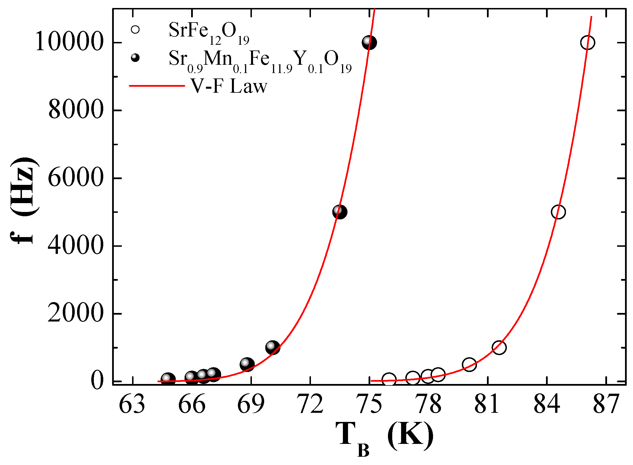
Figure 12 presents the dependence of f on TB for two selected MYSNHFs (SrFe12O19 and Sr0.9Mn0.1Fe11.9Y0.1O19) together with the V–F law fitting. Table 3 outlines the calculated magnetic parameters, such as the f o , τ 0 , E a / k B , and K eff for the indiacted MYSNHFs. The disclosure of somewhat realistic τ 0 values in the allowed range of 10−9–10−13 s clearly approved the validity of V–F law to describe the achieved magnetic behavior of MYSNHFs in a better way compared to the N–A law. Furthermore, the attainment of non-negligible T0 values compared to TB was majorly ascribed to the strong interactions among MYSNHFs nanoparticles in the studied hexaferrites [37]. Meanwhile, the observed shortening in the τ 0 values with the inclusion of Mn2+ and Y3+ cations in the MYSNHFs was mainly attributed to the shrinkage of magnetic nanoparticles and subsequent reduction in the exchange coupling strength among tiny nanoparticles. The values of K eff and T 0 for Sr0.9Mn0.1Fe11.9Y0.1O19 significantly decreased compared to the undoped MYSNHFs (SrFe12O19). The observed reduction in the K eff and T 0 values due to Mn2+ and Y3+ cations substitution was attributed to the weakening in the magnetic anisotropy or indirect exchange interactions between MYSNHFs nanoparticles [43,44].

4. Conclusions

A series of MYSNHFs with stoichiometric composition of Sr1−xMnxFe12−xYxO19 (0.0 ≤ x ≤ 0.5) were prepared using the citrate sol-gel autocombustion technique. The influence of Mn2+ and Y3+ ions substitution on the evolution of morphology, microstructure, and magnetic properties of synthesized MYSNHFs was examined. As-prepared samples were characterized using XRD, FESEM, EDX, HRTEM, FTIR, 57Fe Mössbauer spectroscopy, and PPMS-VSM measurements. The XRD pattern, FESEM, and HRTEM images confirmed the evolution of M-type hexagonal phases in the achieved MYSNHFs. Mössbauer spectral analyses revealed the preferred occupation of the substitution ions into 12k, 4f2, and 2b sites of the hexagonal sublattice in MYSNHFs. The AC susceptibility (real part) of the proposed MYSNHFs disclosed strong frequency dependent magnetic response. The observed shift in the AC susceptibility peak towards lower TB value with increasing Mn2+and Y3+ ions substitution levels was attributed to the shrinkage of magnetic nanoparticles in the studied MYSNHFs. It was established that the magnetic interactions were weakened due to the inclusion of Mn2+and Y3+ ions into prepared MYSNHFs, wherein Sr2+ and Fe3+ ions were replaced by respective Mn2+ and Y3+ ions. In short, present knowledge may contribute towards the development of Mn2+ and Y3+ ions substituted MYSNHF-based device applications.

Author Contributions

Synthesis of the sample, writing—original draft preparation, investigation magnetic properties and writing, investigation and analysis the Mossbauer Spectra, Supervision, review and editing; participated in drafting the article or revising it critically for important intellectual content; gave final approval of the version to be submitted and any revised version; participated sufficiently in the work to take public responsibility for appropriate portions of the content.
(1).
Synthesis of the sample, writing—original draft preparation, Munirah Abdullah Almessiere;
(2).
Investigation magnetic properties and writing, Yassine Slimani
(3).
Investigation and analysis the Mossbauer Spectra, Hakan Güngüneş
(4).
(Supervision, Abdulhadi Baykal and A.V. Trukhanov
(5).
Review and Editing, S.V. Trukhanov

Funding

The authors are grateful to the Institute for Research & Medical Consultations (IRMC) of Imam Abdulrahman Bin Faisal University (IAU—Saudi Arabia) for the financial assistance to pursue this research through the Projects application number: (2018-IRMC-S-1); (2018-IRMC-S-2), and (2017-IRMC-S-3). This work was carried out with financial support in part from the Ministry of Education and Science of the Russian Federation in the framework of Increase Competitiveness Program of NUST «MISiS» implemented by a governmental degree dated 16th of March 2013, No. 211, among the leading world scientific and educational centers: grant No. П02-2017-2-4 and additionally grants No. К4-2017-041 and No. К3-2018-026. The partial support by the Ministry of Education and Science of the Russian Federation with Government task SUSU 5.5523.2017/8.9 and the framework of the Increase Competitiveness Program of MISiS G02-2017-2-4 is appreciated.

Acknowledgments

The authors highly acknowledged the Institute for Research & Medical Consultations (IRMC) of Imam Abdulrahman Bin Faisal University (IAU—Saudi Arabia). The technical assistance provided by Core Labs of King Abdullah University of Science and Technology (KAUST) is highly appreciated.

Conflicts of Interest

The authors declare no conflicts of interest.

References

  1. Rao, C.N.R.; Cheetham, A.K. Science and technology of nanomaterials: Current status and future prospects. J. Mat. Chem. 2001, 12, 2887–2894. [Google Scholar] [CrossRef]
  2. Wang, X.X.; Ma, T.; Shu, J.C.; Cao, M.S. Confinedly tailoring Fe3O4 clusters-NG to tune electromagnetic parameters and Microwave absorption with broadened bandwidth. Chem. Eng. J. 2018, 332, 321–3300. [Google Scholar] [CrossRef]
  3. Gu, Z.; Yan, L.; Tian, G.; Li, S.; Chai, Z.; Zhao, Y. Recent Advances in Design and Fabrication of Upconversion Nanoparticles and Their Safe Theranostic Applications. Adv. Mater. 2013, 25, 3758–3779. [Google Scholar] [CrossRef] [PubMed]
  4. Katlakunta, S.; Meena, S.S.; Srinath, S.; Bououdina, M.; Sandhya, R.; Praveena, K. Improved magnetic properties of Cr3+ doped SrFe12O19 synthesized via microwave hydrothermal route. Mater. Res. Bull. 2015, 63, 58–66. [Google Scholar] [CrossRef]
  5. Jacobo, S.E.; Bercoff, P.G.; Herme, C.A.; Vives, L.A. Sr hexaferrite/Ni ferrite nanocomposites magnetic behavior and microwave absorbing properties in the X-band. Mater. Chem. Phys. 2015, 157, 124–129. [Google Scholar] [CrossRef]
  6. Auwal, I.A.; Baykal, A.; Güner, S.; Sertkol, M.; Sözeri, H. Magneto-optical properties BaBixLaxFe12−2xO19 (0.0 ≤ x ≤ 0.5) hexaferrites. J. Magn. Magn. Mater. 2016, 409, 92–98. [Google Scholar] [CrossRef]
  7. Harker, S.; Stewart, G.; Hutchison, W.; Amiet, A.; Tucker, D. Microwave absorption and 57Fe Mössbauer properties of Ni-Ti doped barium hexaferrite. Hyperfine Interact. 2015, 230, 205–211. [Google Scholar] [CrossRef]
  8. Dishovski, N.; Petkov, A.; Nedkov, I.; Razkazov, I. Hexaferrite contribution to microwave absorbers characteristics. IEEE Trans. Magn. 1994, 30, 969–971. [Google Scholar] [CrossRef]
  9. Iqbal, M.J.; Ashiq, M.N.; Gomez, P.H.; Munoz, J.M. Synthesis, physical, magnetic and electrical properties of Al–Ga substituted co-precipitated nanocrystalline strontium hexaferrite. J. Magn. Magn. Mater. 2008, 320, 881–886. [Google Scholar] [CrossRef]
  10. Obradors, X.; Collomb, A.; Pernet, M.; Joubert, J.C.; Isalgue, A. Structural and magnetic properties of BaFe12−xMnxO19 hexagonal ferrites. J. Magn. Magn. Mater. 1984, 44, 118–128. [Google Scholar] [CrossRef]
  11. Mocuta, H.; Lechevallier, L.; le Breton, J.M.; Wang, J.F.; Harris, I.R. Structural and magnetic properties of hydrothermally synthesized Sr1−xNdxFe12O19 hexagonal ferrites. J. Alloy. Compd. 2004, 364, 48–52. [Google Scholar] [CrossRef]
  12. Silva, W.M.S.; Ferreira, N.S.; Soares, J.M.; da Silva, R.B.; Macêdo, M.A. Investigation of structural and magnetic properties of nanocrystalline Mn-doped SrFe12O19 prepared by proteic sol–gel process. J. Magn. Magn. Mater. 2015, 395, 263–270. [Google Scholar] [CrossRef]
  13. Jiang, S.; Liu, X.; Rehman, K.M.U.; Li, M.; Wu, Y. Synthesis and characterization of Sr1−xYxFe12O19 hexaferrites prepared by solid-state reaction method. J. Mater. Sci. Mater. Electron. 2016, 27, 12919–12924. [Google Scholar] [CrossRef]
  14. Niu, X.F.; Zhang, M.Y. Structure and magnetic properties of yttrium-doped M-type strontium ferrite. Asian J. Chem. 2014, 26, 6783–6786. [Google Scholar] [CrossRef]
  15. Shekhawat, D.; Roy, P.K. Impact of yttrium on the physical, electro-magnetic and dielectric properties of auto-combustion synthesized nanocrystalline strontium hexaferrite. J. Mater. Sci. Mater. Electron. 2018. [Google Scholar] [CrossRef]
  16. Almessiere, M.A.; Slimani, Y.; el Sayed, H.S.; Baykal, A. Structural and magnetic properties of Ce-Y substituted strontium nanohexaferrites. Ceram. Int. 2018, 44, 12511–12519. [Google Scholar] [CrossRef]
  17. Almessiere, M.A.; Slimani, Y.; Baykal, A. Impact of Nd-Zn co-substitution on microstructure and magnetic properties of SrFe12O19 nanohexaferrite. Ceram. Int. 2019, 45, 963–969. [Google Scholar] [CrossRef]
  18. Peng, L.; Li, L.Z.; Wang, R.; Hu, Y.; Tu, X.Q.; Zhong, X.X. Effect of La–Co substitution on the crystal structure and magnetic properties of low temperature sintered Sr1−xLaxFe12−xCoxO19 (x=0–0.5) ferrites. J. Magn. Magn. Mater. 2015, 393, 399–403. [Google Scholar] [CrossRef]
  19. Yang, Y.J.; Liu, X.S. Microstructure and magnetic properties of La– Cu doped M-type strontium ferrites prepared by ceramic process. Mater. Technol. Adv. Perform. Mater. 2014, 29, 232–236. [Google Scholar] [CrossRef]
  20. Yang, Y.; Wang, F.; Shao, J.; Batoo, K.M. Microstructure and magnetic properties of Zr–Mn substituted M-type SrLa hexaferrites. Appl. Phys. A 2017, 123, 1–8. [Google Scholar] [CrossRef]
  21. Zhang, Z.Y.; Liu, X.X.; Wang, X.J.; Peng, Y.; Li, R. Effect of Nd–Co substitution on magnetic and microwave absorption properties of SrFe12O19 hexaferrites. J. Alloy. Compd. 2012, 525, 114–119. [Google Scholar] [CrossRef]
  22. Lee, S.W.; An, S.Y. In-Bo Shim, Chul Sung Kim, Mossbauer studies of La–Zn substitution effect in strontium ferrite nanoparticles. J. Magn. Magn. Mater. 2005, 290–291, 231–233. [Google Scholar] [CrossRef]
  23. Yang, Y.; Wang, F.; Shao, J.; Huang, D.; He, H.; Trukhanov, A.V.; Trukhanov, S.V. Influence of Nd-NbZn co-substitution on structural, spectral and magnetic properties of M-type calcium-strontium hexaferrites Ca0.4Sr0.6-xNdxFe12.0-x(Nb0.5Zn0.5)xO19. J. Alloy. Compd. 2018, 765, 616–623. [Google Scholar] [CrossRef]
  24. Ashiq, M.N.; Shakoor, S.; Najam-ul-Haq, M.; Warsi, M.F.; Ali, I.; Shakir, I. Structural, electrical, dielectric and magnetic properties of Gd-Sn substituted Sr-hexaferrite synthesized by sol–gel combustion method. J. Magn. Magn. Mater. 2015, 374, 173–178. [Google Scholar] [CrossRef]
  25. Iqbal, M.J.; Farooq, S. Impact of Pr–Ni substitution on the electrical and magnetic properties of chemically derived nanosized strontium–barium hexaferrites. J. Alloy. Compd. 2010, 505, 560–567. [Google Scholar] [CrossRef]
  26. Shakoor, S.; Ashiq, M.N.; Marlana, M.A.; Mahmood, A.; Warsi, M.F.; Najam-ul-Haq, M.; Karamat, N. Electrical, dielectric and magnetic characterization of Bi–Cr substituted M-type strontium hexaferrite nanomaterials. J. Magn. Magn. Mater. 2014, 362, 110–114. [Google Scholar] [CrossRef]
  27. Kaur, P.; Chawla, S.K.; Meena, S.S.; Yusuf, S.M.; Pubby, K.; Narang, S.B. Modulation of physico-chemical, magnetic, microwave and electromagnetic properties of nanocrystalline strontium hexaferrite by Co-Zr doping synthesized using citrate precursor sol-gel method. Ceram. Int. 2017, 43, 590–598. [Google Scholar] [CrossRef]
  28. Joshi, R.; Singh, C.; Kaur, D.; Zaki, H.; Narang, S.B.; Jotania, R.; Mishra, S.; Singh, J.; Dhruv, P.; Ghimiree, M. Structural and magnetic properties of Co2+-W4+ ions doped M-type Ba-Sr hexaferrites synthesized by a ceramic method. J. Alloy. Compd. 2017, 695, 909. [Google Scholar] [CrossRef]
  29. Yang, Y.J.; Shao, J.X.; Wang, F.H.; Liu, X.S.; Huang, D.H. Impacts of MnZn doping on the structural and magnetic properties of M-type SrCaLa hexaferrites. Appl. Phys. A 2017, 123, 309. [Google Scholar] [CrossRef]
  30. Baniasadi, A.; Ghasemi, A.; Nemati, A.; Ghadikolaei, M.A.; Paimozd, E. Effect of Ti–Zn substitution on structural, magnetic and microwave absorption characteristics of strontium hexaferrite. J. Alloy. Compd. 2014, 583, 325–328. [Google Scholar] [CrossRef]
  31. Rostami, M.; Vahdani, M.R.K.; Moradi, M.; Mardani, R. Structural, magnetic, and microwave absorption properties of Mg–Ti–Zr–Co-substituted barium hexaferrites nanoparticles synthesized via sol–gel auto-combustion method. J. Sol-Gel Sci. Technol. 2017, 82, 783–794. [Google Scholar] [CrossRef]
  32. Zhang, T.; Peng, X.; Li, J.; Yang, Y.; Xu, J.; Wang, P.; Jin, D.; Jin, H.; Hong, B.; Wang, X.; et al. Platelet-like hexagonal SrFe12O19 particles: Hydrothermal synthesis and their orientation in a magnetic field. J. Magn. Magn. Mater. 2016, 412, 102–106. [Google Scholar] [CrossRef]
  33. Rezaie, E.; Rezanezhad, A.; Ghadimi, L.S.; Hajalilou, A.; Arsalani, N. Effect of calcination on structural and supercapacitance properties of hydrothermally synthesized plate-like SrFe12O19 hexaferrite nanoparticles. Ceram. Int. 2018, 44, 20285–20290. [Google Scholar] [CrossRef]
  34. Annapureddya, V.; Kang, J.H.; Palneedi, H.; Kima, J.W.; Ahna, C.W.; Choi, S.Y.; Johnson, S.D.; Ryu, J. Growth of self-textured barium hexaferrite ceramics by normal sintering process and their anisotropic magnetic properties. J. Eur. Ceram. Soc. 2017, 37, 4701–4706. [Google Scholar] [CrossRef]
  35. Pradeep, A.; Chandrasekaran, G. FTIR study of Ni, Cu and Zn substituted nano-particles of MgFe2O4. Mater. Lett. 2006, 60, 371–374. [Google Scholar] [CrossRef]
  36. Pereira, F.M.M.; Junior, C.A.R.; Santos, M.R.P.; Sohn, R.S.T.M.; Freire, F.N.A.; Sasaki, J.M.; De-Paiva, J.A.C.; Sombra, A.S.B. Structural and dielectric spectroscopy studies of the M-type barium strontium hexaferrite alloys (BaxSr1−xFe12O19). J. Mater. Sci. Mater. Electron. 2008, 19, 627–638. [Google Scholar] [CrossRef]
  37. Thakur, A.; Singh, R.R.; Barman, P.B. Synthesis and characterizations of Nd3+ doped SrFe12O19 nanoparticles. Mater. Chem. Phys. 2013, 141, 562–569. [Google Scholar] [CrossRef]
  38. Tenorio-Gonzalez, F.N.; Bolarín-Miro, A.M.; Jesús, F.Sa.; Vera-Serna, P.; Menendez-Gonzalez, N.; Sanchez-Marcos, J. Crystal structure and magnetic properties of high Mn-doped strontium hexaferrite. J. Alloy. Compd. 2017, 695, 2083–2090. [Google Scholar] [CrossRef]
  39. Solovyova, E.D.; Pashkova, E.V.; Ivanitski, V.P.; Vyunov, O.I.; Belous, A.G. Mössbauer and X-ray diffraction study of Co2+–Si4+ substituted M-type barium hexaferrite BaFe12−2xCoxSixO19±γ. J. Magn. Magn. Mater. 2013, 330, 72–75. [Google Scholar] [CrossRef]
  40. Belous, A.G.; Vyunov, O.I.; Pashkova, E.V.; Ivanitski, V.P.; Gavrilenko, O.N. Mössbauer Study and Magnetic Properties of M-Type Barium Hexaferrite Doped with Co + Ti and Bi + Ti Ions. Phys. Chem. B 2006, 110, 26477–26481. [Google Scholar] [CrossRef]
  41. Chawla, S.K.; Mudsainiyan, R.K.; Meena, S.S.; Yusuf, S.M. Sol–gel synthesis, structural and magnetic properties of nanoscale M-type barium hexaferrites BaCoxZrxFe(12−2x)O19. J. Magn. Magn. Mater. 2014, 350, 23–29. [Google Scholar] [CrossRef]
  42. Auwal, I.A.; Güngüneş, H.; Güner, S.; Shirsath, S.E.; Sertkol, M.; Structural, A.B. magneto-optical properties and cation distribution of SrBixLaxYxFe12−3xO19 (0.0 ≤ x ≤ 0.33) hexaferrites. Mater. Res. Bull. 2016, 80, 263–272. [Google Scholar] [CrossRef]
  43. Almessiere, M.A.; Slimani, Y.; Güngüneş, H.; el Sayed, H.S.; Baykal, A. AC susceptibility and Mossbauer study of Ce3+ ion substituted SrFe12O19 nanohexaferrites. Ceram. Int. 2018. [Google Scholar] [CrossRef]
  44. Almessiere, M.A.; Slimani, Y.; el Sayed, H.S.; Baykal, A. Ce-Y co-substituted Strontium nanohexaferrites: AC susceptibility and Mossbauer studies. Ceram. Int. 2018. [Google Scholar] [CrossRef]
  45. Slimani, Y.; Baykal, A.; Manikandan, A. Effect of Cr3+ substitution on AC susceptibility of Ba hexaferrite nanoparticles. J. Magn. Magn. Mater. 2018, 458, 204–212. [Google Scholar] [CrossRef]
  46. Mohapatra, J.; Mitra, A.; Bahadur, D.; Aslam, M. Superspin glass behavior of self-interacting CoFe2O4 nanoparticles. J. Alloy. Compd. 2015, 628, 416–423. [Google Scholar] [CrossRef]
  47. Kaul, S.N.; Methfessel, S. Effect of field and frequency on the temperature dependence of a.c. susceptibility of the (La, Gd) Ag spin-glass. Solid State Comm. 1983, 47, 147–151. [Google Scholar] [CrossRef]
  48. Fiorani, D.; Viticoli, S.; Dormann, J.L.; Tholence, J.L.; Murani, A.P. Spin-glass behavior in an antiferromagnetic frustrated spinel: ZnCr1.6Ga0.4O4. Phys. Rev. B 1984, 30, 2776–2786. [Google Scholar] [CrossRef]
  49. Trukhanov, S.V.; Troyanchuk, I.O.; Fita, I.M.; Szymczak, H.; Bärner, K. Comparative study of the magnetic and electrical properties of Pr1−xBaxMnO3-δ manganites depending on the preparation conditions. J. Magn. Magn. Mater. 2001, 237, 276–282. [Google Scholar] [CrossRef]
  50. Trukhanov, S.V.; Lobanovski, L.S.; Bushinsky, M.V.; Khomchenko, V.A.; Pushkarev, N.V.; Tyoyanchuk, I.O.; Maignan, A.; Flahaut, D.; Szymczak, H.; Szymczak, R. Influence of oxygen vacancies on the magnetic and electrical properties of La1−xSrxMnO3−x/2 manganites. Eur. Phys. J. B 2004, 42, 51–61. [Google Scholar] [CrossRef]
  51. Trukhanov, S.V.; Trukhanov, A.V.; Vasiliev, A.N.; Szymczak, H. Frustrated exchange interactions formation at low temperatures and high hydrostatic pressures in La0.70Sr0.30MnO2.85. J. Exp. Theor. Phys. 2010, 111, 209–214. [Google Scholar] [CrossRef]
  52. Trukhanov, S.V.; Trukhanov, A.V.; Turchenko, V.A.; Kostishyn, V.G.; Panina, L.V.; Kazakevich, I.S.; Balagurov, A.M. Structure and magnetic properties of BaFe11.9In0.1O19 hexaferrite in a wide temperature range. J. Alloy. Compd. 2016, 689, 383–393. [Google Scholar] [CrossRef]
Figure 1. XRD patterns with Rietveld refinement for the various Sr1−xMnxFe12−xYxO19 (0.0 ≤ x ≤ 0.5) nanohexaferrites.
Nanomaterials 09 00024 g001
Figure 2. Scanning electron microscope (SEM) images of prepared Sr1−xMnxFe12−xYxO19 (x = 0.0, 0.2, 0.4, and 0.5) nanohexaferrites.
Nanomaterials 09 00024 g002
Figure 3. High-resolution transmission electron microscope (HRTEM) images of prepared Sr1−xMnxFe12−xYxO19 (x = 0.2, 0.4, and 0.5) nanohexaferrites.
Nanomaterials 09 00024 g003
Figure 4. Elemental mapping and energy-dispersive X-ray diffraction spectroscopy (EDX) spectra of Sr1−xMnxFe12−xYxO19 (x = 0.2 and 0.5) nanohexaferrites.
Nanomaterials 09 00024 g004
Figure 5. Fourier-transform infrared spectroscopy (FTIR) spectra of proposed Sr1−xMnxFe12−xYxO19 (0.0 ≤ x = y ≤ 0.5) nanohexaferrites.
Nanomaterials 09 00024 g005
Figure 6. Room temperature Mössbauer spectra of all studied Sr1−xMnxFe12−xYxO19 (x = 0.0, 0.1, 0.2, 0.3, 0.4 and 0.5) nanohexaferrites.
Nanomaterials 09 00024 g006
Figure 7. Mn and Y contents dependent relative area variation of Sr1−xMnxFe12−xYxO19 (0.0 ≤ x ≤ 0.5) nanohexaferrites.
Nanomaterials 09 00024 g007
Figure 8. Temperature-dependent AC susceptibility (real part) for (a) SrFe12O19 and (b) Sr0.9Mn0.1Fe11.9Y0.1O19.
Nanomaterials 09 00024 g008
Figure 9. M–H curves of synthesized Sr1−xMnxFe12−yYyO19 (x = y = 0.0 and 0.1) nanohexaferrites performed at room temperature.
Nanomaterials 09 00024 g009
Figure 10. Inverse of real part of the AC susceptibility versus temperature for (a) SrFe12O19 and (b) Sr0.9Mn0.1Fe11.9Y0.1O19.
Nanomaterials 09 00024 g010
Figure 11. ln(f) against 1/TB for the two selected SrFe12O19 (i.e., x = y = 0.0) and Sr0.9Mn0.1Fe11.9Y0.1O19 (i.e., x = y = 0.1) where solid line presents Neel–Arrhenius (N-A) model fit.
Nanomaterials 09 00024 g011
Figure 12. Frequency dependent TB for two selected Sr1−xMnxFe12−yYyO19 (x = y = 0.0 and 0.1) nanohexaferrites where solid line presents Vogel–Fulcher (V–F) law fit.
Nanomaterials 09 00024 g012
Table 1. Structural parameters of all the synthesized Sr1−xMnxFe12−xYxO19 (0.0 ≤ x ≤ 0.5) nanohexaferrites.
xa = b (Å)c (Å)DXRD (nm)χ2
0.05.88123.04855.11.8
0.15.88123.02169.12.1
0.25.88223.02355.92.4
0.35.88323.03959.82.6
0.45.88323.02949.52.6
0.55.88423.05037.43.2
Table 2. Mössbauer spectral parameters of the studied Sr1−xMnxFe12−xYxO19 (0.0 ≤ x ≤ 0.5) nanohexaferrites including hyperfine magnetic field (Bhf), isomer shift (I.S), quadrupole splitting (Q.S), line width (W), and relative area percent (RA%) with estimated uncertainties.
x SiteBhf (T)I.S (mm/s)Q.S (mm/s)W (mm/s)RA (%)
(±0.01)(±0.002)(±0.001)(±0.006)
012k41.1830.3530.3960.27748.381
4f149.1570.2590.1760.23817.771
4f251.8350.3790.2920.24413.924
2a50.8850.3230.0160.36311.679
2b40.9370.2792.2790.9888.2449
0.112k41.130.3530.4010.24943.331
12k138.7610.3050.2230.3484.1509
4f149.1060.2610.1790.24816.983
4f251.8960.3750.2850.25412.616
2a50.5580.3420.0690.31113.347
2b40.9520.2922.2470.2515.3067
Db-0.2320.7036804.2661
0.212k41.0890.3510.3990.25139.926
12k138.5570.3050.2710.2667.995
4f148.9940.2610.1710.26718.46
4f251.8140.3620.3170.14610.953
2a50.4640.3560.0580.31315.042
2b40.8790.3052.2160.2644.3767
Db-0.2930.8710.8143.2476
0.312k41.1540.3540.40.29433.051
12k138.6520.2840.2220.2349.0202
4f149.080.2790.1270.34319.982
4f252.5510.422−0.0410.28.7147
2a50.8850.3840.0150.44923.917
2b40.9830.2972.2440.2683.5987
Db-0.4380.8350.8121.7174
0.412k41.1130.3460.3890.30228.237
12k138.7620.270.2170.30416.523
4f148.7320.2920.1110.40221.383
4f252.5550.423−0.0780.185.1586
2a50.8160.3730.0890.47124.509
2b40.9060.2822.1870.2493.2939
Db-0.2490.390.920.89625
0.512k41.1030.350.3850.46325.222
12k138.5890.2580.2250.46120.37
4f148.4640.3150.0630.51122.301
4f252.2210.403−0.070.46710.131
2a50.5090.3760.040.54719.531
2b40.9140.2842.140.212.4454
Table 3. Physical parameters of the selected Sr1−xMnxFe12−yYyO19 (x = y = 0.0 and 0.1) nanohexaferrites obtained from Neel–Arrhenius and Vogel–Fulcher model fitting.
ModelsParametersValues
SrFe12O19Sr0.9Mn0.1Fe11.9Y0.1O19
Neel–Arrhenius τ o (s)3.85 × 10−222.58 × 10⁻¹⁹
E a / k B (K)3452.592518.3
K eff (erg/cm3)1.25 × 1032.02 × 103
Vogel–Fulcher τ o (s)1.18 × 10−117.39 × 10⁻¹¹
E a / k B (K)587.83226.66
T o (K)49.2251.26
K eff (erg/cm3)213.33181.94