Intestinal Microfold Cells Play a Critical Role in the Uptake and Oral Tolerance Mediated by Lysophosphatidylserine-Containing Lipidic Nanoparticles


Abstract
1. Introduction
2. Materials
2.1. SPF Conditional RANK Knockout Mice
2.2. Preparation of Nanoparticles
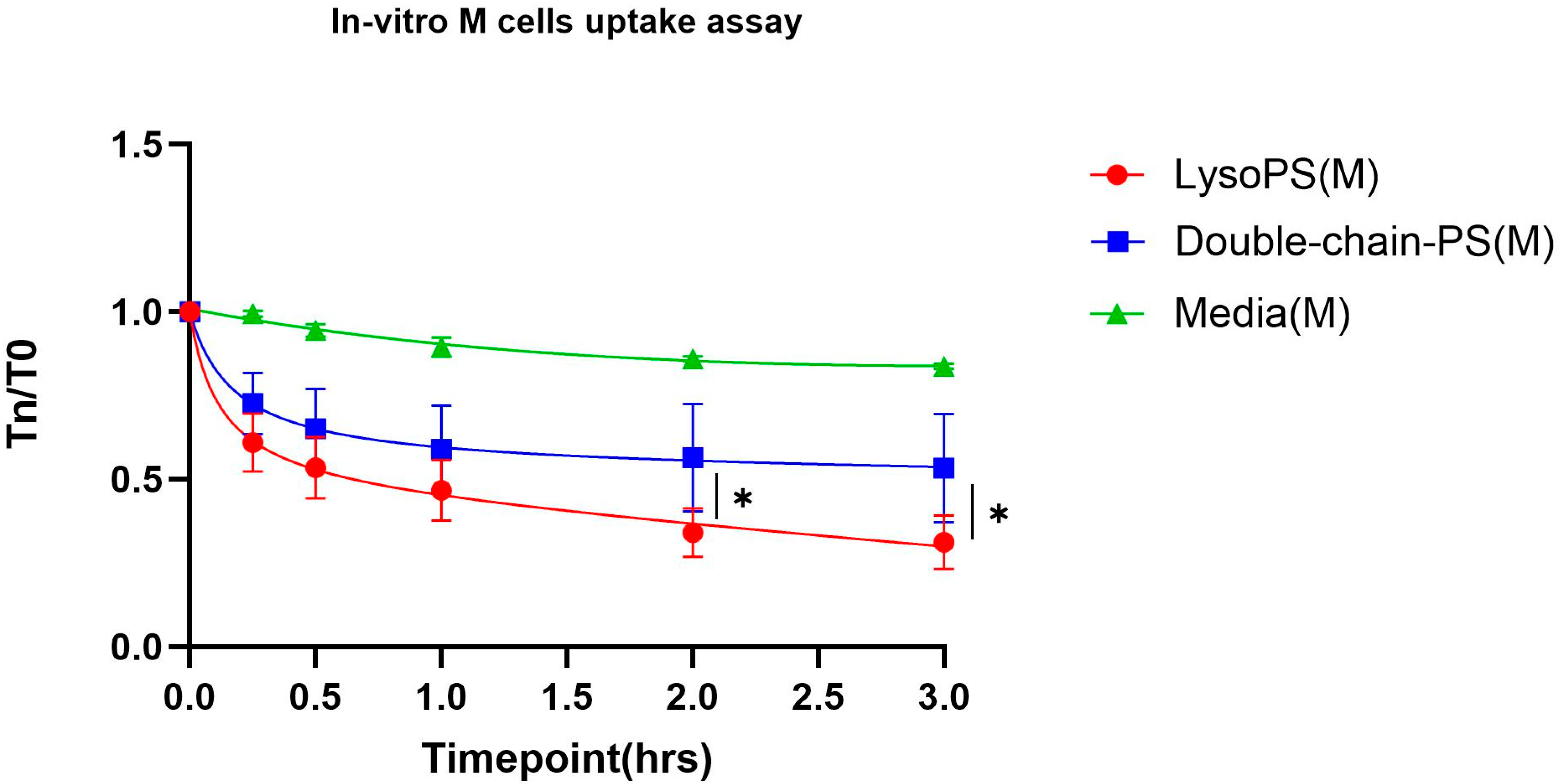
2.3. In Vitro M Cell Uptake Study
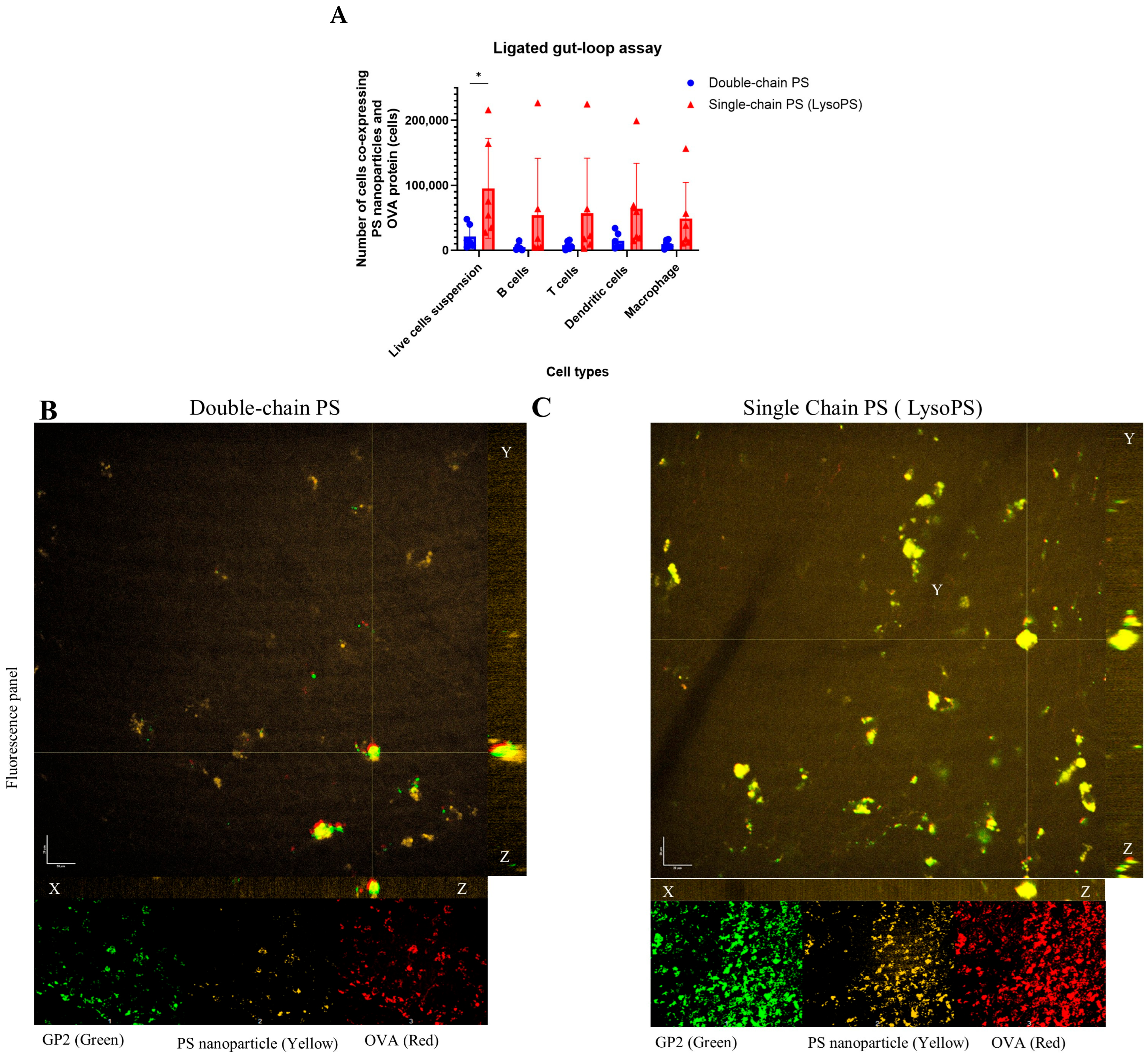
2.4. PS Nanoparticle Exposure in the Intestinal M-Cells and MLN
2.5. Oral Tolerance Study
2.6. Anti-OVA IgG Antibody ELISA
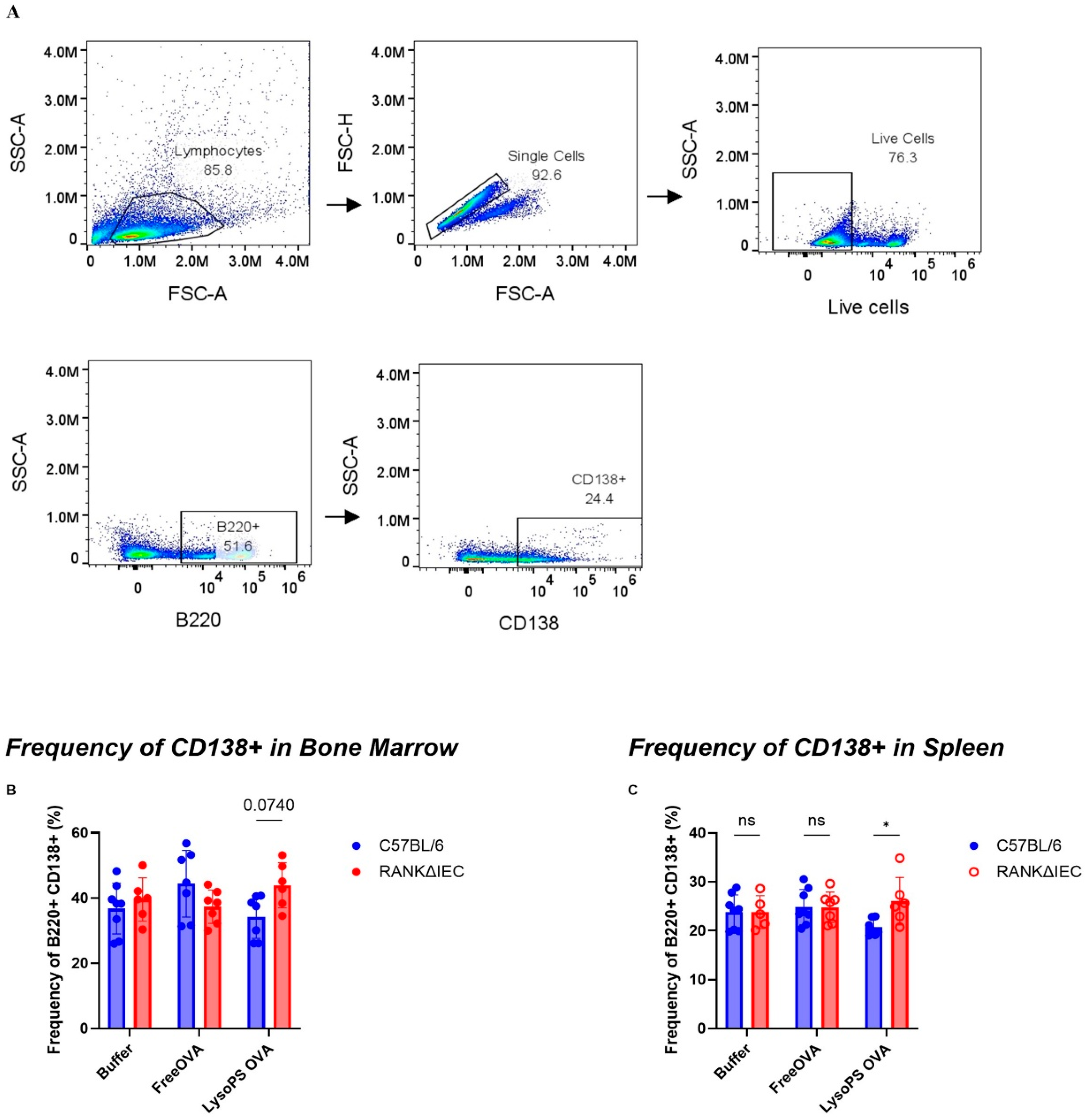
2.7. Flow Cytometry
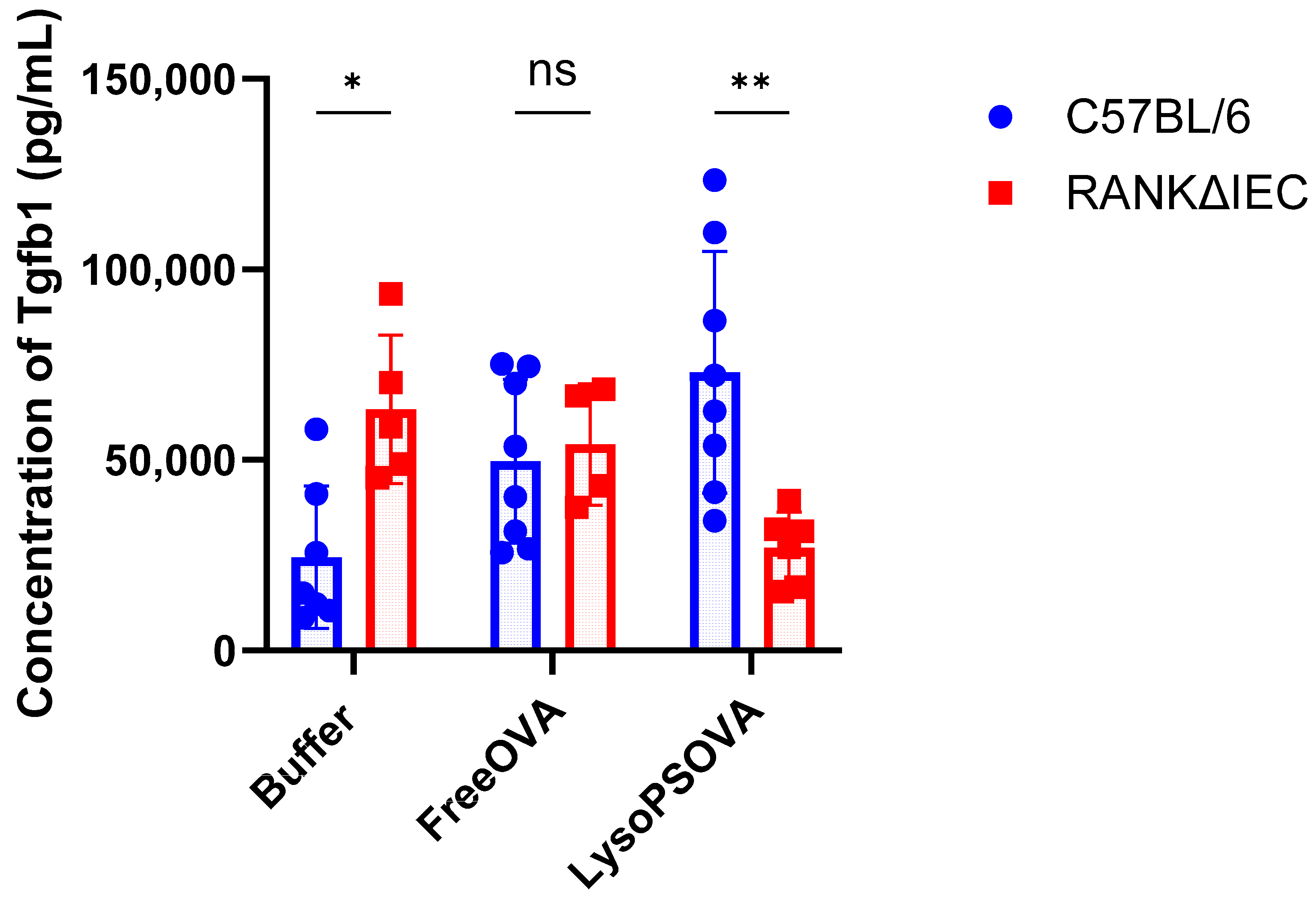
2.8. Multiplex TGF-Beta Cytokines Assay
2.9. Statistical Analysis
3. Results
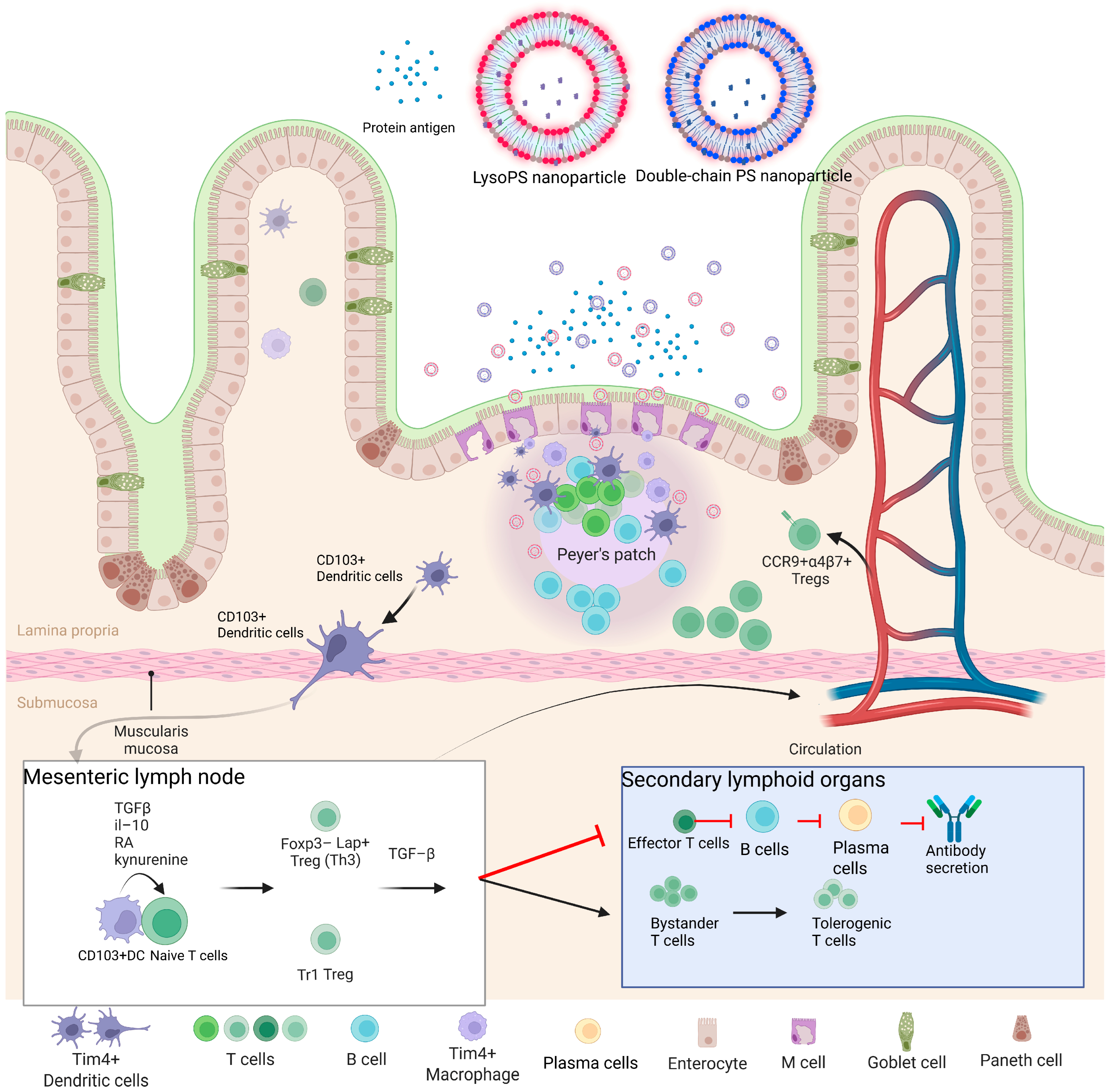
3.1. LysoPS Nanoparticles Enhance Intestinal M-Cells Absorption for Oral Tolerance
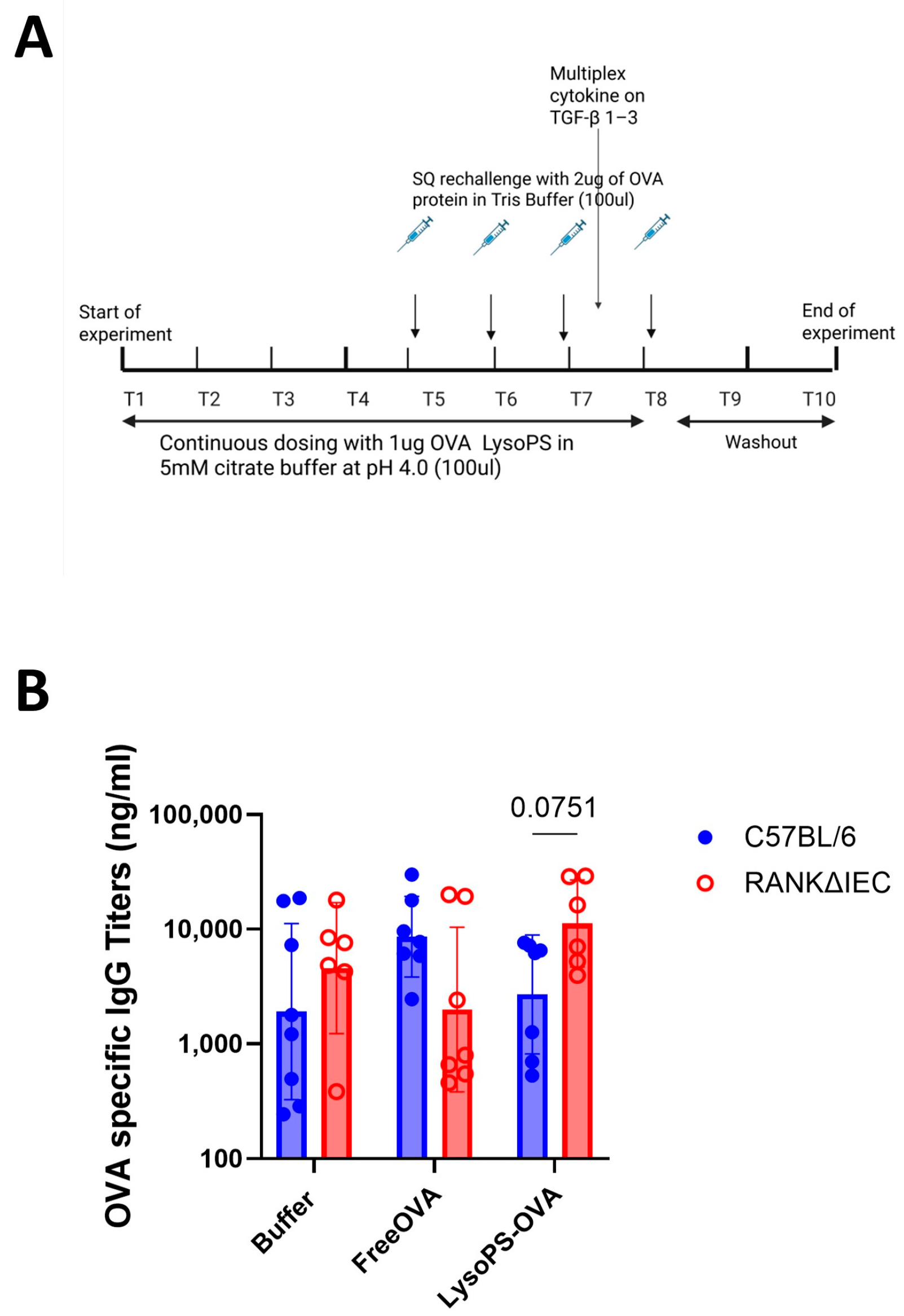
3.2. LysoPS-OVA Induced OVA Specific IgG Antibody in RANKΔIEC Mice
3.3. LysoPS Suppressed Plasma Cell Differentiation
3.4. LysoPS Induced Immunosuppressive Cytokine—Transforming Growth Factor-Beta (TGF-β)
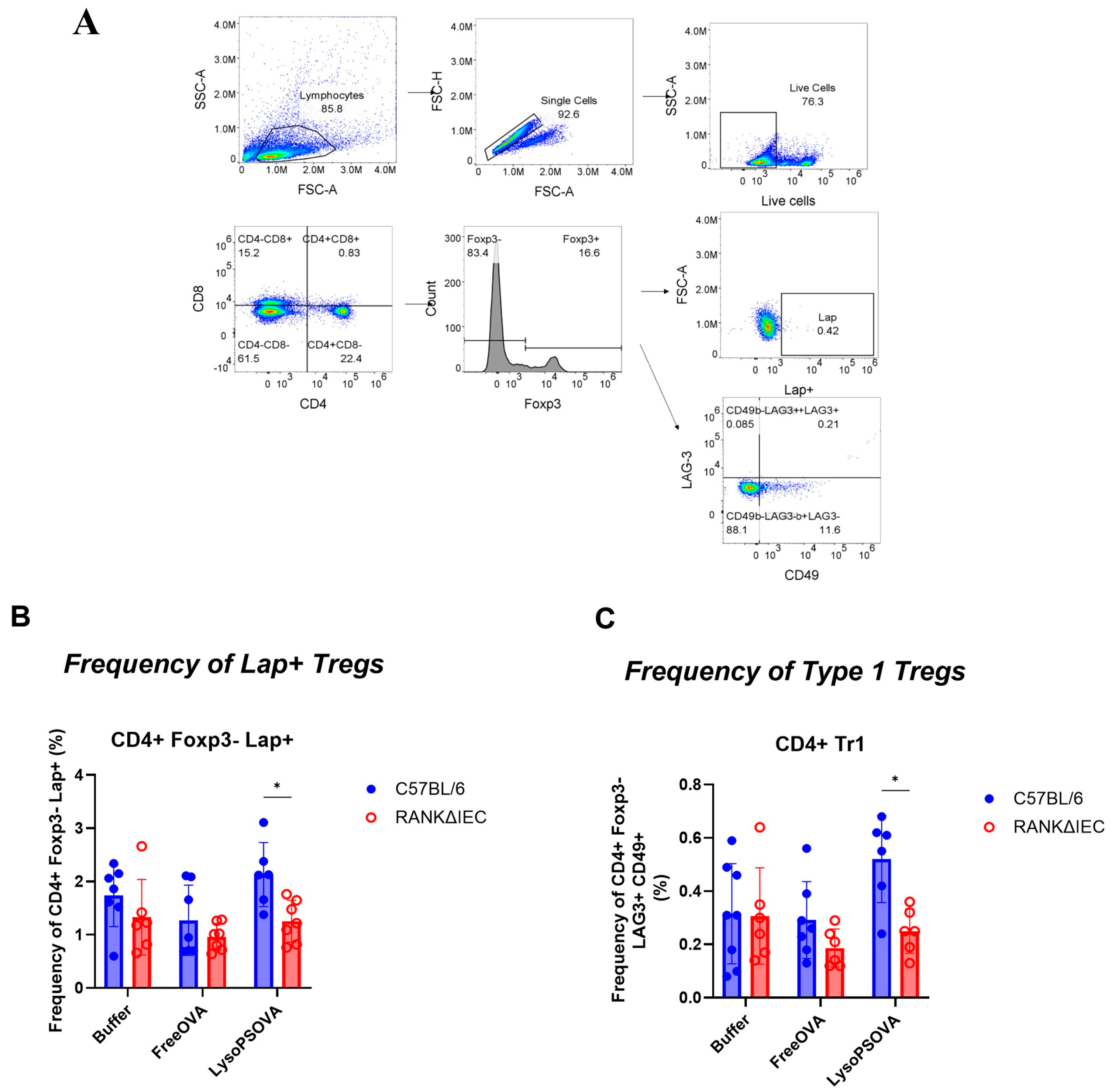
3.5. LysoPS-OVA Induced Regulatory T Cells (Tregs)
4. Discussion
5. Conclusions
Supplementary Materials
Author Contributions
Funding
Institutional Review Board Statement
Data Availability Statement
Acknowledgments
Conflicts of Interest
References
- Chaurio, R.A.; Janko, C.; Munoz, L.E.; Frey, B.; Herrmann, M.; Gaipl, U.S. Phospholipids: Key players in apoptosis and immune regulation. Molecules 2009, 14, 4892–4914. [Google Scholar] [CrossRef] [PubMed]
- Fadeel, B.; Quinn, P.; Xue, D.; Kagan, V. Fat(al) attraction: Oxidized lipids act as “eat-me” signals. HFSP J. 2007, 1, 225–229. [Google Scholar] [CrossRef] [PubMed]
- Hoffmann, P.R.; Kench, J.A.; Vondracek, A.; Kruk, E.; Daleke, D.L.; Jordan, M.; Marrack, P.; Henson, P.M.; Fadok, V.A. Interaction between phosphatidylserine and the phosphatidylserine receptor inhibits immune responses in vivo. J. Immunol. 2005, 174, 1393–1404. [Google Scholar] [CrossRef]
- Kay, J.G.; Grinstein, S. Phosphatidylserine-mediated cellular signaling. Adv. Exp. Med. Biol. 2013, 991, 177–193. [Google Scholar] [CrossRef]
- Gaitonde, P.; Ramakrishnan, R.; Chin, J.; Kelleher, R.J., Jr.; Bankert, R.B.; Balu-Iyer, S.V. Exposure to factor VIII protein in the presence of phosphatidylserine induces hypo-responsiveness toward factor VIII challenge in hemophilia A mice. J. Biol. Chem. 2013, 288, 17051–17056. [Google Scholar] [CrossRef]
- Ramani, K.; Miclea, R.D.; Purohit, V.S.; Mager, D.E.; Straubinger, R.M.; Balu-Iyer, S.V. Phosphatidylserine containing liposomes reduce immunogenicity of recombinant human factor VIII (rFVIII) in a murine model of hemophilia A. J. Pharm. Sci. 2008, 97, 1386–1398. [Google Scholar] [CrossRef]
- Glassman, F.Y.; Schneider, J.L.; Ramakrishnan, R.; Dingman, R.K.; Ramanathan, M.; Bankert, R.B.; Balu-Iyer, S.V. Phosphatidylserine Is Not Just a Cleanup Crew but Also a Well-Meaning Teacher. J. Pharm. Sci. 2018, 107, 2048–2054. [Google Scholar] [CrossRef]
- Glassman, F.Y.; Balu-Iyer, S.V. Subcutaneous administration of Lyso-phosphatidylserine nanoparticles induces immunological tolerance towards Factor VIII in a Hemophilia A mouse model. Int. J. Pharm. 2018, 548, 642–648. [Google Scholar] [CrossRef]
- Nguyen, N.H.; Glassman, F.Y.; Dingman, R.K.; Shenoy, G.N.; Wohlfert, E.A.; Kay, J.G.; Bankert, R.B.; Balu-Iyer, S.V. Rational design of a nanoparticle platform for oral prophylactic immunotherapy to prevent immunogenicity of therapeutic proteins. Sci. Rep. 2021, 11, 17853. [Google Scholar] [CrossRef]
- Rezende, R.M.; Weiner, H.L. History and mechanisms of oral tolerance. Semin. Immunol. 2017, 30, 3–11. [Google Scholar] [CrossRef] [PubMed]
- Morbe, U.M.; Jorgensen, P.B.; Fenton, T.M.; von Burg, N.; Riis, L.B.; Spencer, J.; Agace, W.W. Human gut-associated lymphoid tissues (GALT); diversity, structure, and function. Mucosal Immunol. 2021, 14, 793–802. [Google Scholar] [CrossRef]
- Kobayashi, N.; Takahashi, D.; Takano, S.; Kimura, S.; Hase, K. The Roles of Peyer’s Patches and Microfold Cells in the Gut Immune System: Relevance to Autoimmune Diseases. Front. Immunol. 2019, 10, 2345. [Google Scholar] [CrossRef]
- Jung, C.; Hugot, J.P.; Barreau, F. Peyer’s Patches: The Immune Sensors of the Intestine. Int. J. Inflam. 2010, 2010, 823710. [Google Scholar] [CrossRef]
- Rynda, A.; Maddaloni, M.; Mierzejewska, D.; Ochoa-Reparaz, J.; Maslanka, T.; Crist, K.; Riccardi, C.; Barszczewska, B.; Fujihashi, K.; McGhee, J.R.; et al. Low-dose tolerance is mediated by the microfold cell ligand, reovirus protein sigma1. J. Immunol. 2008, 180, 5187–5200. [Google Scholar] [CrossRef]
- Suzuki, H.; Sekine, S.; Kataoka, K.; Pascual, D.W.; Maddaloni, M.; Kobayashi, R.; Fujihashi, K.; Kozono, H.; McGhee, J.R.; Fujihashi, K. Ovalbumin-protein sigma 1 M-cell targeting facilitates oral tolerance with reduction of antigen-specific CD4+ T cells. Gastroenterology 2008, 135, 917–925. [Google Scholar] [CrossRef]
- Rios, D.; Wood, M.B.; Li, J.; Chassaing, B.; Gewirtz, A.T.; Williams, I.R. Antigen sampling by intestinal M cells is the principal pathway initiating mucosal IgA production to commensal enteric bacteria. Mucosal Immunol. 2016, 9, 907–916. [Google Scholar] [CrossRef]
- Fossati, P. Phosphate determination by enzymatic colorimetric assay. Anal. Biochem. 1985, 149, 62–65. [Google Scholar] [CrossRef] [PubMed]
- Beloqui, A.; Brayden, D.J.; Artursson, P.; Preat, V.; des Rieux, A. A human intestinal M-cell-like model for investigating particle, antigen and microorganism translocation. Nat. Protoc. 2017, 12, 1387–1399. [Google Scholar] [CrossRef] [PubMed]
- Kraus, T.A.; Brimnes, J.; Muong, C.; Liu, J.H.; Moran, T.M.; Tappenden, K.A.; Boros, P.; Mayer, L. Induction of mucosal tolerance in Peyer’s patch-deficient, ligated small bowel loops. J. Clin. Investig. 2005, 115, 2234–2243. [Google Scholar] [CrossRef] [PubMed]
- Dillon, A.; Lo, D.D. M Cells: Intelligent Engineering of Mucosal Immune Surveillance. Front. Immunol. 2019, 10, 1499. [Google Scholar] [CrossRef]
- Komban, R.J.; Stromberg, A.; Biram, A.; Cervin, J.; Lebrero-Fernandez, C.; Mabbott, N.; Yrlid, U.; Shulman, Z.; Bemark, M.; Lycke, N. Activated Peyer’s patch B cells sample antigen directly from M cells in the subepithelial dome. Nat. Commun. 2019, 10, 2423. [Google Scholar] [CrossRef]
- Lelouard, H.; Fallet, M.; de Bovis, B.; Meresse, S.; Gorvel, J.P. Peyer’s patch dendritic cells sample antigens by extending dendrites through M cell-specific transcellular pores. Gastroenterology 2012, 142, 592–601.e593. [Google Scholar] [CrossRef] [PubMed]
- Coombes, J.L.; Siddiqui, K.R.; Arancibia-Carcamo, C.V.; Hall, J.; Sun, C.M.; Belkaid, Y.; Powrie, F. A functionally specialized population of mucosal CD103+ DCs induces Foxp3+ regulatory T cells via a TGF-beta and retinoic acid-dependent mechanism. J. Exp. Med. 2007, 204, 1757–1764. [Google Scholar] [CrossRef] [PubMed]
- Burton, O.T.; Logsdon, S.L.; Zhou, J.S.; Medina-Tamayo, J.; Abdel-Gadir, A.; Noval Rivas, M.; Koleoglou, K.J.; Chatila, T.A.; Schneider, L.C.; Rachid, R.; et al. Oral immunotherapy induces IgG antibodies that act through FcgammaRIIb to suppress IgE-mediated hypersensitivity. J. Allergy Clin. Immunol. 2014, 134, 1310–1317 e1316. [Google Scholar] [CrossRef]
- Sugimoto, M.; Kamemura, N.; Nagao, M.; Irahara, M.; Kagami, S.; Fujisawa, T.; Kido, H. Differential response in allergen-specific IgE, IgGs, and IgA levels for predicting outcome of oral immunotherapy. Pediatr. Allergy Immunol. 2016, 27, 276–282. [Google Scholar] [CrossRef]
- Salmivesi, S.; Paassilta, M.; Huhtala, H.; Nieminen, R.; Moilanen, E.; Korppi, M. Changes in biomarkers during a six-month oral immunotherapy intervention for cow’s milk allergy. Acta Paediatr. 2016, 105, 1349–1354. [Google Scholar] [CrossRef] [PubMed]
- Chak, V.; Bybel, M.; Harne, S.; Kay, J.G.; Wohlfert, E.; Balu-Iyer, S.V. Phosphatidylserine exposure on tolerogenic nanoparticle correlate with oral tolerance. Sci. Rep. 2025, 16, 2487. [Google Scholar] [CrossRef]
- Nguyen, N.H.; Chen, M.; Chak, V.; Balu-Iyer, S.V. Biophysical Characterization of Tolerogenic Lipid-Based Nanoparticles Containing Phosphatidylcholine and Lysophosphatidylserine. J. Pharm. Sci. 2022, 111, 2072–2082. [Google Scholar] [CrossRef]
- Chak, V.; Ovalle, E.J.; Bard, J.; Wohlfert, E.A.; Kay, J.G.; Balu-Iyer, S. Single-Cell Transcriptional Analysis of Murine Mesenteric Lymph Nodes Following Oral Lyso-phosphatidylserine Nanoparticle Administration Reveals Cellular Heterogeneity in Tolerance Features. J. Pharm. Sci. 2025, 114, 103919. [Google Scholar] [CrossRef]
- Corr, S.C.; Gahan, C.C.; Hill, C. M-cells: Origin, morphology and role in mucosal immunity and microbial pathogenesis. FEMS Immunol. Med. Microbiol. 2008, 52, 2–12. [Google Scholar] [CrossRef]
- Jang, M.H.; Sougawa, N.; Tanaka, T.; Hirata, T.; Hiroi, T.; Tohya, K.; Guo, Z.; Umemoto, E.; Ebisuno, Y.; Yang, B.G.; et al. CCR7 is critically important for migration of dendritic cells in intestinal lamina propria to mesenteric lymph nodes. J. Immunol. 2006, 176, 803–810. [Google Scholar] [CrossRef]
- Sun, L.Z.; Elsayed, S.; Aasen, T.B.; Van Do, T.; Aardal, N.P.; Florvaag, E.; Vaali, K. Comparison between ovalbumin and ovalbumin peptide 323-339 responses in allergic mice: Humoral and cellular aspects. Scand. J. Immunol. 2010, 71, 329–335. [Google Scholar] [CrossRef] [PubMed]
- Kawasaki, Y.; Nakagawa, A.; Nagaosa, K.; Shiratsuchi, A.; Nakanishi, Y. Phosphatidylserine binding of class B scavenger receptor type I, a phagocytosis receptor of testicular sertoli cells. J. Biol. Chem. 2002, 277, 27559–27566. [Google Scholar] [CrossRef] [PubMed]
- Rigotti, A.; Acton, S.L.; Krieger, M. The class B scavenger receptors SR-BI and CD36 are receptors for anionic phospholipids. J. Biol. Chem. 1995, 270, 16221–16224. [Google Scholar] [CrossRef] [PubMed]
- Cunin, P.; Beauvillain, C.; Miot, C.; Augusto, J.F.; Preisser, L.; Blanchard, S.; Pignon, P.; Scotet, M.; Garo, E.; Fremaux, I.; et al. Clusterin facilitates apoptotic cell clearance and prevents apoptotic cell-induced autoimmune responses. Cell Death Dis. 2016, 7, e2215. [Google Scholar] [CrossRef]
- Futter, C.E.; White, I.J. Annexins and endocytosis. Traffic 2007, 8, 951–958. [Google Scholar] [CrossRef]
- Kehrl, J.H.; Roberts, A.B.; Wakefield, L.M.; Jakowlew, S.; Sporn, M.B.; Fauci, A.S. Transforming growth factor beta is an important immunomodulatory protein for human B lymphocytes. J. Immunol. 1986, 137, 3855–3860. [Google Scholar] [CrossRef]
- Davidson, T.S.; DiPaolo, R.J.; Andersson, J.; Shevach, E.M. Cutting Edge: IL-2 Is Essential for TGF-β-Mediated Induction of Foxp3+ T Regulatory Cells. J. Immunol. 2007, 178, 4022–4026. [Google Scholar] [CrossRef]
- Weiner, H.L.; da Cunha, A.P.; Quintana, F.; Wu, H. Oral tolerance. Immunol. Rev. 2011, 241, 241–259. [Google Scholar] [CrossRef]
- Wang, J.; Zhao, X.; Wan, Y.Y. Intricacies of TGF-beta signaling in Treg and Th17 cell biology. Cell. Mol. Immunol. 2023, 20, 1002–1022. [Google Scholar] [CrossRef] [PubMed]
- Scurr, M.; Ladell, K.; Besneux, M.; Christian, A.; Hockey, T.; Smart, K.; Bridgeman, H.; Hargest, R.; Phillips, S.; Davies, M.; et al. Highly prevalent colorectal cancer-infiltrating LAP+ Foxp3− T cells exhibit more potent immunosuppressive activity than Foxp3+ regulatory T cells. Mucosal Immunol. 2014, 7, 428–439. [Google Scholar] [CrossRef]
- Wu, H.Y.; Quintana, F.J.; da Cunha, A.P.; Dake, B.T.; Koeglsperger, T.; Starossom, S.C.; Weiner, H.L. In Vivo induction of Tr1 cells via mucosal dendritic cells and AHR signaling. PLoS ONE 2011, 6, e23618. [Google Scholar] [CrossRef]
- Bertolini, T.B.; Biswas, M.; Terhorst, C.; Daniell, H.; Herzog, R.W.; Pineros, A.R. Role of orally induced regulatory T cells in immunotherapy and tolerance. Cell. Immunol. 2021, 359, 104251. [Google Scholar] [CrossRef] [PubMed]
- Pierini, A.; Nishikii, H.; Baker, J.; Kimura, T.; Kwon, H.S.; Pan, Y.; Chen, Y.; Alvarez, M.; Strober, W.; Velardi, A.; et al. Foxp3+ regulatory T cells maintain the bone marrow microenvironment for B cell lymphopoiesis. Nat. Commun. 2017, 8, 15068. [Google Scholar] [CrossRef]
- Gargaro, M.; Scalisi, G.; Manni, G.; Briseno, C.G.; Bagadia, P.; Durai, V.; Theisen, D.J.; Kim, S.; Castelli, M.; Xu, C.A.; et al. Indoleamine 2,3-dioxygenase 1 activation in mature cDC1 promotes tolerogenic education of inflammatory cDC2 via metabolic communication. Immunity 2022, 55, 1032–1050 e1014. [Google Scholar] [CrossRef] [PubMed]
- Sasaki, I.; Kato, T.; Hemmi, H.; Fukuda-Ohta, Y.; Wakaki-Nishiyama, N.; Yamamoto, A.; Kaisho, T. Conventional Type 1 Dendritic Cells in Intestinal Immune Homeostasis. Front. Immunol. 2022, 13, 857954. [Google Scholar] [CrossRef]
- Campos Canesso, M.C.; de Castro, T.B.R.; Nakandakari-Higa, S.; Lockhart, A.; Luehr, J.; Bortolatto, J.; Parsa, R.; Esterhazy, D.; Lyu, M.; Liu, T.T.; et al. Identification of antigen-presenting cell-T cell interactions driving immune responses to food. Science 2025, 387, eado5088. [Google Scholar] [CrossRef] [PubMed]
- Bonnardel, J.; Da Silva, C.; Henri, S.; Tamoutounour, S.; Chasson, L.; Montanana-Sanchis, F.; Gorvel, J.P.; Lelouard, H. Innate and adaptive immune functions of peyer’s patch monocyte-derived cells. Cell Rep. 2015, 11, 770–784. [Google Scholar] [CrossRef]
- Bakdash, G.; Vogelpoel, L.T.; van Capel, T.M.; Kapsenberg, M.L.; de Jong, E.C. Retinoic acid primes human dendritic cells to induce gut-homing, IL-10-producing regulatory T cells. Mucosal Immunol. 2015, 8, 265–278. [Google Scholar] [CrossRef]
- Cassani, B.; Villablanca, E.J.; Quintana, F.J.; Love, P.E.; Lacy-Hulbert, A.; Blaner, W.S.; Sparwasser, T.; Snapper, S.B.; Weiner, H.L.; Mora, J.R. Gut-tropic T cells that express integrin alpha4beta7 and CCR9 are required for induction of oral immune tolerance in mice. Gastroenterology 2011, 141, 2109–2118. [Google Scholar] [CrossRef]




Disclaimer/Publisher’s Note: The statements, opinions and data contained in all publications are solely those of the individual author(s) and contributor(s) and not of MDPI and/or the editor(s). MDPI and/or the editor(s) disclaim responsibility for any injury to people or property resulting from any ideas, methods, instructions or products referred to in the content. |
© 2026 by the authors. Licensee MDPI, Basel, Switzerland. This article is an open access article distributed under the terms and conditions of the Creative Commons Attribution (CC BY) license.
Share and Cite
Chak, V.; Harne, S.; Kay, J.G.; Wohlfert, E.; Balu-Iyer, S.V. Intestinal Microfold Cells Play a Critical Role in the Uptake and Oral Tolerance Mediated by Lysophosphatidylserine-Containing Lipidic Nanoparticles. Nanomaterials 2026, 16, 412. https://doi.org/10.3390/nano16070412
Chak V, Harne S, Kay JG, Wohlfert E, Balu-Iyer SV. Intestinal Microfold Cells Play a Critical Role in the Uptake and Oral Tolerance Mediated by Lysophosphatidylserine-Containing Lipidic Nanoparticles. Nanomaterials. 2026; 16(7):412. https://doi.org/10.3390/nano16070412
Chicago/Turabian StyleChak, Vincent, Sujay Harne, Jason G. Kay, Elizabeth Wohlfert, and Sathy V. Balu-Iyer. 2026. "Intestinal Microfold Cells Play a Critical Role in the Uptake and Oral Tolerance Mediated by Lysophosphatidylserine-Containing Lipidic Nanoparticles" Nanomaterials 16, no. 7: 412. https://doi.org/10.3390/nano16070412
APA StyleChak, V., Harne, S., Kay, J. G., Wohlfert, E., & Balu-Iyer, S. V. (2026). Intestinal Microfold Cells Play a Critical Role in the Uptake and Oral Tolerance Mediated by Lysophosphatidylserine-Containing Lipidic Nanoparticles. Nanomaterials, 16(7), 412. https://doi.org/10.3390/nano16070412