Article

Systematic Evaluation of Zn2+, Ca2+, and Co2+ Doping for Tailoring the Thermal, Structural, Morphological and Magnetic Performance of CdBi0.1Fe1.9O4@SiO2 Nanocomposites


1
Faculty of Science, Technical University of Cluj-Napoca, 76 Victoriei Street, 430122 Baia Mare, Romania
2
Faculty of Chemistry and Chemical Engineering, Babes-Bolyai University, 11 Arany Janos Street, 400084 Cluj-Napoca, Romania
3
INCDO-INOE 2000, Research Institute for Analytical Instrumentation, 67 Donath Street, 400293 Cluj-Napoca, Romania
*
Author to whom correspondence should be addressed.
Nanomaterials 2026, 16(4), 259; https://doi.org/10.3390/nano16040259
Submission received: 9 January 2026 / Revised: 28 January 2026 / Accepted: 14 February 2026 / Published: 16 February 2026

Abstract

The influence of Zn2+, Ca2+ and Co2+ doping on the thermal, structural, morphological, and magnetic characteristics of CdBi0.1Fe1.9O4 nanoparticles synthetized via the sol–gel technique and calcined at 300, 600, 900 and 1200 °C was investigated. Thermal analysis revealed the initial formation of metallic glyoxylates up to 300 °C, followed by their decomposition into metal oxides and subsequent ferrite formation. X-ray diffraction revealed that the ferrites were poorly crystallized at lower temperatures, whereas at higher calcination temperatures all nanocomposites exhibited well-crystalized ferrites coexisting with the SiO2 matrix, except for the Co0.1Cd0.9Bi0.1Fe1.9O4@SiO2 nanocomposite, which formed a single, well-defined crystalline phase. Atomic force microscopy images revealed spherical ferrite particles encapsulated within an amorphous layer, with particle size, surface area, and coating thickness influenced by both the type of dopant ion and the calcination temperature. The structural parameters estimated by X-ray diffraction, as well as the magnetic characteristics, were strongly influenced by the dopant type and thermal treatment. These results demonstrate that the structural and magnetic characteristics of CdBi0.1Fe1.9O4 ferrites can be effectively tuned through controlled doping and calcination, providing insights for the design of tailored functional applications.

1. Introduction

Nanostructured ferrites exhibit advantageous magnetic characteristics, such as reduced coercivity (Hc), high saturation magnetization (Ms), elevated magnetic permeability, large surface area, high electrical resistivity, and low dielectric and eddy current losses. In addition, their mechanical rigidity, chemical and thermal stability, non-toxicity, physical flexibility, corrosion resistance, and cost effectiveness further enhance their technological significance [1,2,3,4,5,6,7,8,9]. Nevertheless, the structural and magnetic characteristics of ferrites are strongly dependent on their chemical composition, precursor selection, synthesis strategy, thermal processing conditions, and the incorporation of dopants, impurities, or secondary phases [6,7,8,9,10,11,12]. Owing to their multifunctional characteristics, ferrites find applications across several technological sectors, including (i) biomedical applications such as anticancer and antibacterial therapies, bioimaging, biosensors), (ii) energy and environmental applications, including energy storage, microwave absorption, magnetic refrigeration, corrosion protection, and water decontamination, (iii) electronic and magnetic technologies, such as spintronics, humidity sensors, ferrofluids, high-capacity information storage media, and (iv) industrial applications, including ceramic pigments, ferroceramics, and magnetic inks [13,14,15,16,17,18].
CdFe2O4 crystallizes in a normal spinel structure, in which Cd2+ ions preferentially occupy the tetrahedral (A) sites and Fe3+ ions on the octahedral (B) sites within a face-centered cubic lattice [1,2,3,4]. Moreover, CdFe2O4 is a typical example of geometrically frustrated antiferromagnet, exhibiting unique magnetic behavior that arise from superexchange interactions among its magnetic ions [1,2,3,4,5]. Although CdFe2O4 is non-magnetic, the relatively large radius of the Cd2+ ion induces a lattice distortion in the spinel structure, which can lead to spin canting and perturbation of the magnetic moment [1,3,5,8]. Compared to other magnetic materials, CdFe2O4 exhibits high electrical resistivity, a characteristic that is particularly advantageous for the design of soft ferrites, as it minimizes eddy-current losses and provides a clear advantage over magnetic materials such as metal alloys and metal oxides [1,2,3,4,5,6,7]. Owing to the synergy of its chemical stability, structural, mechanical integrity, and multifunctional electrical, magnetic, and optical characteristics, CdFe2O4 is suitable for a wide range of applications including transformer core materials, microwave components, and biomedical technologies [1,2,3,4,5,6].
The incorporation of non-magnetic Cd2+ ions into the spinel ferrite lattice is known to modify the cation distribution and enhance magnetic performance, notably by increasing the net magnetic moment and Ms, enabling applications in loading coils, magnetic recording heads, antenna cores and microwave devices [3,5,6]. Furthermore, co-doping with divalent ions Zn2+ or Co2+ can further improve magnetic characteristics, electrical conductivity, permeability, density, photocatalytic activity, and biological activity [7,8,9]. Additionally, substituting Bi3+ or Fe3+ ions with other transition metal ions can induce lattice distortions, modify the cycloidal spin structure and enhance ferrimagnetism [19,20,21,22].
Numerous synthetic strategies have been developed for ferrite preparation, including wet chemical techniques (co-precipitation, hydrothermal/solvothermal methods), sonochemical, sol-gel, solid-state methods (solid-state reaction, ball milling/mechanochemical processing), mechanical treatments (milling, grinding), thermal and combustion-based methods (decomposition, flash combustion, auto-combustion), as well as vapor phase and aerosol-assisted techniques (vapor phase deposition and surfactant-mediated aerosol processing) [23,24,25,26,27,28,29]. Each method provides distinct advantages in tuning particle size, morphology, and functional characteristics, with wet-chemical routes favoring nanosized particles and solid-state routes typically used for bulk ferrite systems [23,27,28]. Among these approaches, the sol-gel method provides a versatile and cost-effective strategy for producing ferrites with well-controlled composition, structural uniformity, and tailored functional characteristics [23,27,28,29,30,31]. Nanoparticle agglomeration, which can hinder the formation of single-phase ferrites, can be mitigated by applying homogenous ultrathin coatings or dispersing particles within non-magnetic matrices. In this context, the sol-gel method using tetraethylorthosilicate (TEOS) enables the formation of robust inorganic networks with moderate reactivity, facilitating the incorporation of a wide range of organic and inorganic species. Among potential coating materials, mesoporous SiO2 is particularly attractive due to its hydrophilicity, chemical stability, non-toxicity and biocompatibility, and its ability to control particle growth and size distribution [23,27,28]. Embedding ferrite nanoparticles within a SiO2 matrix suppresses agglomeration, promotes controlled crystallite development, and can enhance magnetic performance. Moreover, the bio-inert SiO2 matrix improves the biocompatibility of nanoferrites and mitigates inflammatory risks in biological environments [23,27,28].
The magnetic behavior of nanoferrites is primarily determined by the combined effects of cation distribution at the A and B sites and surface-related spin disorder, both of which are influenced by particle size, synthesis method, and post-synthesis thermal processing conditions [32,33,34]. Additionally, the magnetic characteristics of CoFe2O4, ZnFe2O4 and CdFe2O4 are influenced by size, shape, purity, and magnetic stability of the constituent particles [27]. Multiferroic systems have attracted significant interest due to the coexistence of switchable ferroelectric, ferrimagnetic, and ferroelastic order parameters within a single phase [19,35]. However, when ferrimagnetic ferrite particles decrease below a critical size, thermal fluctuations can disrupt single-domain magnetic ordering, leading to a substantially reduction in Ms and coercivity (Hc) [27].
This study investigates the influence of Zn2+ (Zn0.1Cd0.9Bi0.1Fe1.9O4), Ca2+ (Ca0.1Cod0.9Bi0.1Fe1.9O4), and Co2+ (Co0.1Cd0.9Bi0.1Fe1.9O4) doping on the structural, morphological, and magnetic characteristics of CdBi0.1Fe1.9O4 nanoparticles embedded within a SiO2 matrix, synthetized via the sol-gel route and calcined at various temperatures. The thermal evolution and decomposition of glyoxylate precursors were analyzed using thermal analysis and Fourier transform infrared (FT-IR) spectroscopy. The crystalline phase development and crystallinity were assessed using X-ray diffraction (XRD) which was used to study, while particle shape, morphology, size and agglomeration were investigated using atomic force microscopy (AFM). The magnetic characteristics, including the variation of Ms vs. Hc were evaluated for both undoped and Zn-, Ca- and Co-doped CdBi0.1Fe1.9O4 samples.
The novelty of the present study arises from (i) a comprehensive assessment of the combined effect of the SiO2 matrix and divalent dopant ions on the formation of metal-glyoxylate precursors and their subsequent thermal transformation into ferrite phases embedded in the SiO2 matrix; (ii) a systematic comparative investigation of Zn2+, Ca2+, and Co2+ doping in CdBi0.1Fe1.9O4@SiO2 nanocomposites (NCs) to elucidate their influence on morphological, structural, and magnetic characteristics, identifying pathways to enhance functional performance and broaden potential applications; (iii) the development of new trimetallic ferrites embedded in a SiO2 matrix; (iv) achieving single-phase crystalline ferrites at relatively low calcination temperatures (300 °C) for both undoped and doped systems, demonstrating the efficiency of the proposed synthesis; and (v) showing that dopant incorporation, particularly Co2+ ion, suppresses the formation of secondary phases at high calcination temperatures (1200 °C), highlighting the stabilizing role of selective doping.

2. Materials and Methods

2.1. Reagents

Zinc nitrate hexahydrate (Zn(NO3)2∙6H2O, 98%), calcium nitrate tetrahydrate (Ca(NO3)2∙4H2O, 99%), cobalt nitrate hexahydrate (Co(NO3)2∙6H2O, 98%), and cadmium nitrate tetrahydrate (Cd(NO3)2∙4H2O, 99%), were purchased from Carlo Erba (Milan, Italy). Ferric nitrate nonahydrate (Fe(NO3)3∙9H2O, 98%), bismuth nitrate pentahydrate (Bi(NO3)2·5H2O (98%)), ethylene glycol (EG, 99%), TEOS (99%) and ethanol (96%) were obtained from Merck, Darmstadt, Germany.

2.2. Synthesis

CdBi0.1Fe1.9O4 and M0.1Cd0.9Bi0.1Fe1.9O4 (M = Zn, Ca, Co) embedded within a SiO2 matrix were prepared using a sol-gel technique. The resulting NCs consisted of 45 wt.% ferrite and 55 wt.% SiO2. Appropriate molar ratios of metal nitrates were selected to achieve M/Cd/Bi/Fe of 0/10/1/19 for the undoped composition and 1/9/1//19 for the Zn-, Ca-, and Co-doped samples. The precursor sols were prepared by dissolving the corresponding metal nitrates in a mixed solvent system containing EG, TEOS, and ethanol using a NO3/EG/TEOS molar ratio of 1/1/1. The solutions were magnetically stirred for 30 min to ensure homogeneity and subsequently aged under ambient conditions until complete gelation, which occurred after approximately 30 days. The resulting gels entrapped uniformly distributed metal ions within the organic–inorganic network. To enhance the phase purity and promote the crystallite growth, the dried gels were finely ground and subjected to a thermal pretreatment at 40 °C for 4 h. Calcination was then carried out subsequentially at 300, 600, 900 and 1200 °C for 4 h at each temperature using a Nabertherm LT9 muffle furnace (Lilienthal, Germany).

2.3. Characterization

The thermal evolution of the samples was investigated by thermogravimetric (TG) and differential thermal analysis (DTA) in air, using a heating rate of at 10 °C·min−1 up to 1200 °C; measurements were performed SDT Q600 analyzer (TA Instruments, New Castle, DE, USA), employing alumina as the reference material. FT-IR spectra were recorded on KBr pellets containing 1 wt.% of the sample using a Prestige-21 (Shimadzu, Tokyo, Japan). Phase identification was performed by XRD using a D8 Advance diffractometer (Bruker, Karlsruhe, Germany) with CuKα radiation (λ = 1.5418 Å) operated at 40 kV and 35 mA. Atomic force microscopy (AFM) measurements were performed using a high-resolution scanning probe microscope JSPM 4210 produced by JEOL Company (Tokyo, Japan) operating in tapping mode with a NSC 15 Hard cantilever (resonant frequency of 325 kHz and a force constant of 40 N/m). The acquired topographic images were analyzed with WinSPM software (version 2.0, JEOL, Tokyo, Japan) to determine surface height, roughness, and particle diameter. The magnetic characterization was carried out using a 7400 vibrating-sample magnetometer (VSM, Lake Shore, Carson, CA, USA). Hysteresis loops were measured at room temperature under applied fields up to 2 T, while the magnetization measurements were performed in fields up to 5 T.

3. Results and Discussion

3.1. Formation and Decomposition of Glyoxylate Precursor and SiO2 Development

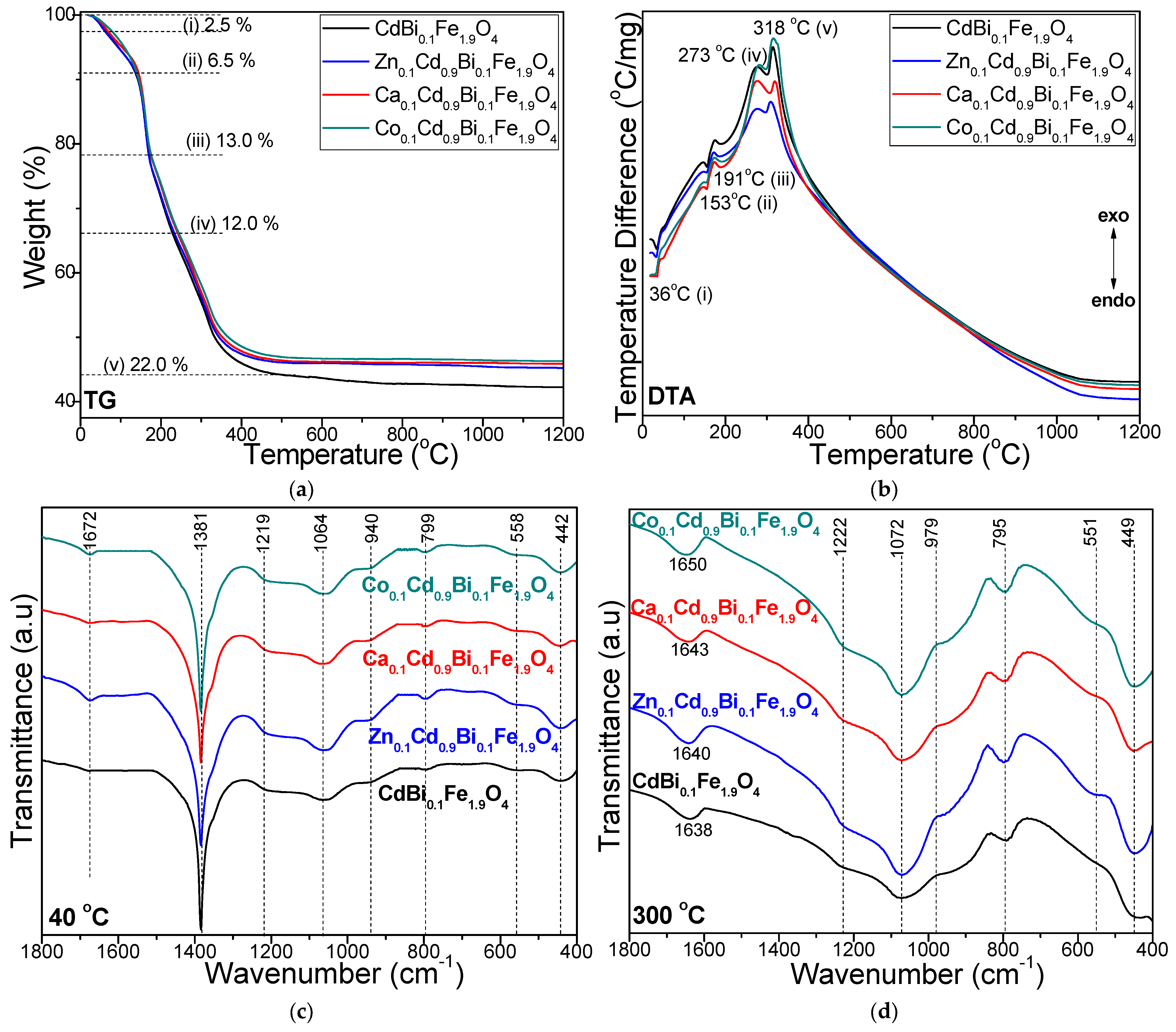
Figure 1 presents the formation and decomposition of metal-glyoxylate precursors within the ferrite system, as well as the development of the SiO2 matrix, as evidenced by the TG-DTA profiles of the gels heated at 40 °C and the comparative FT-IR spectra of the gels heated at 40 and 200 °C.
The TG and DTA curves of all gels reveal five distinct thermal processes: (i) removal of residual water, including physically adsorbed moisture, manifested by an endothermic peak at approximatively 36 °C, corresponding to a mass loss of 2.5%; (ii) formation of Cd, Zn-, Ca-, Co- glyoxylates, evidenced by an endothermic effect near 153 °C with a mass loss of ~6.5%, accompanied by the release of H2O and NO (these redox reactions likely yield a homogenous mixture of homonuclear Fe3+, Bi3+ and Cd2+, Zn2+, Ca2+, Co2+ glyoxylates); (iii) formation of Fe- and Bi- glyoxylates occurring around 191 °C, marked by an endothermic effect and a mass loss of ~13%, due to the release of crystallization water from the nitrates and volatile species (H2O, NO2), driving redox reactions [13,15]; (iv) oxidative decomposition of Cd-, Zn-, Ca-, Co- glyoxylates into their corresponding metal oxides (CdO, ZnO, CaO, CoO) along with the concomitant formation of ferrites, appearing as an exothermic effect near 273 °C with the release of CO, CO2 and H2O, corresponding to a mass loss ~12%; and (v) oxidative decomposition of Fe- and Bi- glyoxylates into Fe2O3 and Bi2O3 occurring around 318 °C, producing an exothermic effect with a mass loss ~22% due to the elimination of CO, CO2 and H2O (Figure 1a,b) [13,14,15,28].
The two-stage progress (at 153 °C and 191 °C) of the redox reaction between metal nitrates and EG is attributed to the higher acidity of the [Fe(H2O)6]3+ and [Bi(H2O)6]3+ cations compared to [Cd(H2O)6]2+, [Zn (H2O)6]2+, [Ca(H2O)6]2+ and [Co(H2O)6]2+ cations [13,15]. Consequently, the redox reaction leading to the formation of the Fe carboxylates occurs at lower temperatures, producing a mixture of homonuclear Fe3+ and Co2+ carboxylates [13,15]. In contrast, the Fe3+ and Bi3+ glyoxylates form at higher temperatures [13,15]. All other synthesized gels display similar TG and DTA curves, with the position and intensity of the exothermic effects depending on the specific Fe(NO3)3/Bi(NO3)3/Co(NO3)2, Fe(NO3)3/Bi(NO3)3/Co(NO3)2/M2+(NO3) (M = Zn, Ca or Co) ratio. The highest total mass loss is observed for CdBi0.1Fe1.9O4@SiO2 (58.0%), while slightly lower mass losses are recorded for Zn0.1Cd0.9Bi0.1Fe1.9O4@SiO2 (54.7%), Ca0.1Cd0.9Bi0.1Fe1.9O4@SiO2 (54.3%), Co0.1Cd0.9Bi0.1Fe1.9O4@SiO2 (53.7%). During thermal treatment, the SiO2 matrix undergoes multiple transformations, which makes it difficult to make the distinction between processes associated with the formation and decomposition of glyoxylate precursors [13,14,15,28].
The FT-IR spectra indicate the formation of metal glyoxylate precursors from metal nitrates, as evidenced by the disappearance of the nitrate-related band at 1381 cm−1 upon calcination at 300 °C (Figure 1c,d) [13,14,15,28]. The SiO2 matrix already begins to develop at 40 °C and becomes more clearly defined after calcination at 300 °C, as confirmed by the presence of Si–O–Si stretching vibrations observed around 1064 cm−1 (40 °C) and 1072 cm−1 (300 °C), together with Si–O vibrational vibrations appearing as shoulders near 1219 cm−1 (40 °C) and 1222 cm−1 (300 °C) [13,14,15,16,23,24,25,26,27,28]. Deformation vibrations of the Si–OH groups, arising from the hydrolysis of –Si(OCH2CH3)4 (TEOS), are observed at 940 cm−1 (40 °C) and 972 cm−1 (300 °C), while Si–O stretching within SiO4 tetrahedra appear at 799 cm−1 (40 °C) and 795 cm−1 (300 °C) [13,14,15,16,23,24,25,26,27,28]. O–H vibrations from EG and adsorbed water appear at 1672 cm−1 at 40 °C; after calcination at 300 °C, these bands broaden and shift to 1638–1650 cm−1, increasing in intensity by Zn2+, Ca2+, or Co2+ doping. This behavior is attributed to –COO vibrations, indicating the formation of chelated complexes through coordination of glyoxylate ligands with the metal ions [13,14,15,16,23,24,25,26,27,28].
The FT-IR spectra exhibit absorption bands at 558 cm−1 (40 °C) and 551 cm−1 (300 °C), which are assigned to the stretching vibrations of tetrahedral Cd–O, Zn–O, Ca–O, and Co–O bonds, as well as to cyclic Si–O–Si structures. The bands observed at 442 and 449 cm−1 are associated with the octahedral Fe–O, Bi–O, and Si–O vibrational modes [13,14,15,16,23,24,25,26,27,28]. The shifts in the A- and B- site band positions with different dopants and calcination temperatures indicate lattice distortions arising from changes in M–O bond lengths [35,36]. These variations are in full agreement with the inverse relationship between bond length, reduced atomic mass, and stretching vibration frequency [37,38,39].

3.2. Formation of the Crystalline Structure of Ferrites

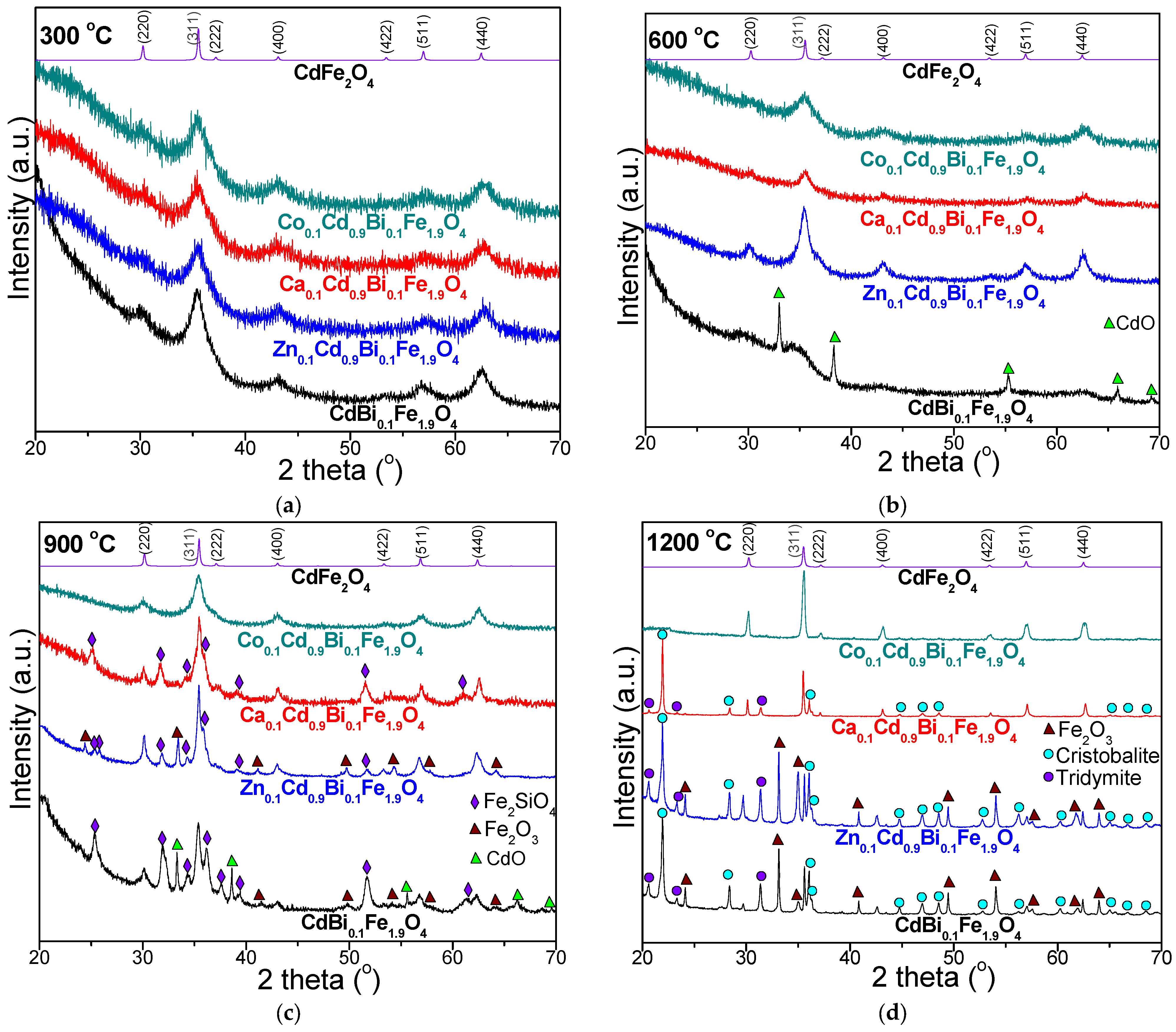
At low calcination temperatures, oxidic phases typically exhibit poor crystallinity or may remain partially amorphous; thus, the development of the desired surface characteristics and crystallinity strongly depends on optimizing the calcination conditions [13,14,16,19]. In addition, the pronounced chemical reactivity of the amorphous SiO2 matrix facilitates its participation in various chemical transformations during the calcination process. Figure 2 shows the XRD patterns of the NCs calcined at 300, 600, 900, and 1200 °C.
At 300 °C, the diffraction peaks corresponding to the (220), (311), (222), (400), (422), (511), and (440) planes confirm the formation of single, low-crystallized CdBi0.1Fe1.9O4 phase, consistent with CdFe2O4 (JCPDS card no. 65-3115) and Bi2Fe4O9 (JCPDS card no. 74-1098), exhibiting a cubic spinel structure with space group Fd3m [26,27,28]. No secondary or impurity-phases are remarked at this temperature, indicating that the dopant ions are successfully incorporated into the spinel ferrite lattice without compromising its structural integrity.
At 600 °C, the XRD pattern of the undoped CdBi0.1Fe1.9O4@SiO2 NC is dominated by the CdO phase (JCPDS card no. 05-0640), while the characteristic diffraction peaks of the CdBi0.1Fe1.9O4 phase appear only weakly, indicating incomplete ferrite formation and poor crystallinity. In contrast, doping CdBi0.1Fe1.9O4 with Zn2+, Ca2+, and Co2+ ions promotes the formation of low crystallized single-phase ferrites. Among the doped samples, Zn0.1Cd0.9Bi0.1Fe1.9O4@SiO2 NC exhibits the most intense diffraction peaks, indicating the highest degree of crystallinity. These results suggest that incorporation of Zn2+, Ca2+, and Co2+ ions at 600 °C promotes the formation of single-phase ferrite structures and improves structural ordering compared with the undoped sample. The presence of secondary phases can be attributed to the higher cation mobility and lattice strain variations induced during calcination, which also slightly shift the 2θ positions, broadens the peaks, and increases crystallite sizes [37,40]. At 600 °C, doping CdBi0.1Fe1.9O4 with Zn2+, Ca2+, or Co2+ ions, restricts Cd2+ diffusion and enhances thermodynamic stability of Zn0.1Cd0.9Bi0.1Fe1.9O4, Ca0.1Cd0.9Bi0.1Fe1.9O4, Co0.1Cd0.9Bi0.1Fe1.9O4 phases embedded in SiO2 matrix. The homogeneity of the metal oxide particles may also contribute to an increased defect density and higher pore volume in the final composites [40,41].
At 900 °C, the XRD pattern of the undoped CdBi0.1Fe1.9O4@SiO2 NC reveals the coexistence of the crystalline ferrite phase with secondary crystalline phases of CdO, α-Fe2O3 (JCPDS card no. 33-0664), and Fe2SiO4 (JCPDS card no. 70-1861). For the Zn-doped CdBi0.1Fe1.9O4@SiO2 NC, CdO is no longer detected, while the diffraction pattern consists of the crystalline ferrite phase accompanied by α-Fe2O3 and Fe2SiO4. In the case of Ca-doped CdBi0.1Fe1.9O4@SiO2, α-Fe2O3 also disappears, resulting in a diffraction pattern containing the ferrite phase together with Fe2SiO4 as single secondary phase. The decomposition of carboxylate precursors generates a reducing environment that partially converts Fe3+ ions into Fe2+ ions, which subsequently interact with the SiO2 matrix, resulting in the formation of Fe2SiO4 at 900 °C in all NCs [27,28,39]. The formation of α-Fe2O3 instead of pure crystalline ferrite may result from the incomplete embedding of the ferrite within the SiO2 matrix and from suboptimal calcinations conditions [19,20]. The presence of α-Fe2O3 indicates the decomposition of Fe(NO3)3 into α-Fe2O3, which promotes spinel ferrite formation, while the excess of metal oxides in insoluble secondary phases may enhance densification by increasing pore volume and generating demagnetizing fields [37,40]. The formation of secondary phases is further influenced by the cation mobility and the lattice strain induced during the calcination process. These effects also display as peak broadening, increased crystallite size, and slight shifts in 2θ positions [14,27,28].
Following calcination at 600 and 900 °C, the ferrite phases remain poorly crystalline, while higher calcination temperatures promote the formation of larger crystallites and accelerate the crystallite growth due to improved nucleation and growth kinetics [27,28]. The presence of sharp diffraction peaks at elevated temperature confirms the development of well-crystallized spinel ferrite structures. At 1200 °C, both undoped CdBi0.1Fe1.9O4@SiO2 and Zn0.1Cd0.9Bi0.1Fe1.9O4@SiO2 display four crystalline phases: CdBi0.1Fe1.9O4, α-Fe2O3, cristobalite (JCPDS card no. 74-9378), and tridymite (JCPDS card no. 42-1401), indicating partial crystallization of the SiO2 matrix in the absence of ferrite. Ca2+ doping of CdBi0.1Fe1.9O4@SiO2 leads to the disappearance of the α-Fe2O3 phase, resulting in the ferrite phase coexisting only with the crystalline SiO2 phases (cristobalite and tridymite). In contrast, Co2+ doping results in the formation of a single-phase ferrite, Co0.1Cd0.9Bi0.1Fe1.9O4, corresponding to CoFe2O4 (JCPDS card no. 22-1086), CdFe2O4 (JCPDS card no. 65-3115), and Bi2Fe4O9 (JCPDS card no. 74-1098). This behavior is attributed to the atomic level cation mixing, which promotes the formation of a dense, fine-grained, and homogeneous ferrite structure [40,41,42,43].
The diffraction peaks of the ferrite phases become more intense at 1200 ◦C, indicating high crystallinity, uniform nucleation, and efficient crystal growth, with minimal influence of from surface coatings [14,27,28]. Variations in peak intensity and signal-to-noise ratio corresponds to the variations in crystallite size and degree of crystallinity [32].
Table 1 summarizes the structural parameters of the NCs calcined at 300, 600, 900 and 1200 °C, including average crystallite size (DCS), lattice parameter (a), unit cell volume (V), physical (dp) and X-ray (dXRD) densities, hopping length at the A (dA) and B (dB) sites, and porosity (P), as determined by XRD [44,45].
Across all calcination temperatures, the undoped CdBi0.1Fe1.9O4 exhibits the smallest DCS, whereas doping with Zn2+, Ca2+, and Co2+ ions lead to a noticeable increase in DCS (Table 1). The calcination temperature-driven growth of DCS can be explained by (i) elevated thermal energy, which enhances atomic diffusion, promotes particle coalescence, and favors the formation of larger crystallites; (ii) agglomeration of incompletely crystallized particles, which reduces the effective nucleation density and supports the formation of single, well-defined crystals; and (iii) variations in cation valence and ionic radii, which induce lattice strain, generate additional nucleation sites, and facilitate crystallite formation [14,27,28]. Additionally, peak broadening, lattice strain and the constraining effect of the SiO2 matrix can further modulate the crystallite growth. DCS is a key determinant of the magnetic behavior of the NCs, particularly when the particle size nears the crystallite size [14,23,24]. These results highlight that the synthesis strategy and calcination conditions critically influence the structural and magnetic characteristics of the NCs. The Reference Intensity Ratio (RIR) method was used to determine the quantitative phase analysis (Table 1) [45,46].
The lattice parameter (a) exhibits the highest values in undoped CdBi0.1Fe1.9O4 across all calcination temperatures. Doping with Zn2+, Ca2+, or Co2+ ions leads to a reduction in the lattice parameter, ascribed to the smaller ionic radii of the dopants, namely Zn2+ (0.74 Å) [3,9,46,47], Ca2+ (0.95 Å) [39], Co2+ (0.75 Å) [19,21,25] compared to Cd2+ (0.97 Å) [4,5,10,12]. The lattice parameter increases with calcination temperature from 8.344 to 8.498 Å, reflecting the combined effect of ionic radii, lattice distortions, and structural defects [14,16,23,27]. Differences between experimental and theoretical lattice parameters arise from the idealized assumption that ions behave as rigid spheres within a perfectly ordered lattice. In the CdBi0.1Fe1.9O4 structure, the Cd2+ ions and dopant (Zn2+, Ca2+ and Co2+) ions predominantly occupy the A- sites, whereas Fe3+ and Bi3+ ions are distributed across both A- and B- sites, depending on the degree of cation disorder and specific synthesis conditions [10,17,18]. Lattice shrinkage is primarily governed by the cation charge distribution, surface tension, and dipole interactions within the nanocrystallites, which also limit grain growth. The internal stresses associated with variations in the lattice parameter can hinder grain development further during calcination [16,23,27].
The cell volume (V) gradually increases with calcination temperature, ranging from 581 to 614 Å3. Doping CdBi0.1Fe1.9O4@SiO2 with Zn2+, Ca2+, Co2+ ions causes a slight reduction in cell volume, likely due to the combined effects of higher molecular mass, the formation of oxygen vacancies, and decreased crystallization temperature. These factors jointly offset the expected volumetric contraction associated with the densification [14,27,28].
The hopping lengths for the tetrahedral A (dA) and octahedral B (dB) sites are closely correlated to the lattice parameter. Lattice expansion is primarily driven by the greater ionic radii of Cd2+, Ca2+, and Bi3+ ions compared to those of Zn2+, Co2+, and Fe3+ ions. Moreover, the Cd2+ ions and the dopant ions (Zn2+, Ca2+, Co2+) show a limited preference for the A sites, whereas Fe3+ and Bi3+ ions are unequally distributed between the A- and B- sites, depending on the composition of undoped and doped CdBi0.1Fe1.9O4@SiO2 [14,27,28].
Higher dA and dB indicate greater cation separation, which increases the energy barrier for charge carrier transfer and influences the magnetic characteristics. Since the crystal field splitting at (A) sites is smaller than at the B sites, variations in cation distribution, particularly of Zn2+, Ca2+, Co2+, Cd2+, and Fe3+ ions, and changes in the degree of inversion can modify the 3d orbitals energy levels, resulting in band gap modifications [14,27,28,45]. Furthermore, replacing magnetic Cd2+ ions with Zn2+, Ca2+, or Co2+ ions adjust the cation distribution within the ferrite lattice, thereby affecting the A-B superexchange interactions, and enabling tunable magnetic behavior [45].
The dXRD reflects the dislocation density per unit volume in a crystalline material [11,13] and is primarily influenced by the lattice parameter and molecular mass [15]. In this study, the dXRD increases with Zn2+, Ca2+, Co2+ ions doping in CdBi0.1Fe1.9O4@SiO2 NCs, whereas it gradual decrease with the increasing calcination temperature (from 6.855 to 6.513 g/cm3), which could be attributed to the pore formation during the thermal treatment [15]. Variations in dXRD associated with changes in the lattice parameter are attributed to modified cation distribution between the A- and B- sites. Replacing Cd2+ ions with Zn2+, Ca2+, or Co2+ ions reduces grains compactness and promotes grain growth, likely due to the pore development through calcination and subsequent sample processing. Additionally, disparities in ionic radii introduce lattice strain and structural disorder, thereby perturbing the spinel framework of the ferrite [11,13].
The porosity (P) of undoped CdBi0.1Fe1.9O4@SiO2 (22.1–16.7%) was higher than that of Zn0.1Cd0.9Bi0.1Fe1.9O4@SiO2 (21.7–16%), Ca0.1Cd0.9Bi0.1Fe1.9O4@SiO2 (20.2–14.5%), Co0.1Cd0.9Bi0.1Fe1.9O4@SiO2 (21.2–15.6%) NCs. This trend is attributed to enhanced vacancy generation and pore formation during synthesis, whereas higher calcination temperatures facilitate densification, resulting in a progressive reduction in porosity [48,49]. Low porosity, as observed for Co0.1Cd0.9Bi0.1Fe1.9O4@SiO2 calcined at 1200 °C, is essential for achieving superior magnetic performance, whereas higher porosity levels can negatively affect magnetic behavior [14,27,28,48,49]. Additionally, doping with Zn2+, Ca2+, Co2+ ions influences porosity by promoting the formation of oxygen vacancies, which slightly expand the lattice while maintaining the structural symmetry of the ferrite framework [14,27,28,48,49].

3.3. AFM Analysis

XRD analysis indicates that the calcination temperatures above 600 °C promotes the development of well-defined ferrite crystallites, while the samples calcined at 300 °C remain amorphous. High calcination temperatures also enhance the tendency of adjacent particles to coalesce. Such behavior has been reported for fine clay particles [50], where physical attraction without chemical bonds governs aggregation; dispersion in an aqueous medium ensures proper particle separation and individualization [51]. Thus, the NCs calcined at 600, 900 and 1200 °C were dispersed into the ultra-pure water by magnetic agitation at 3000 rpm for 15 min. Afterward, glass slides were vertically immersed into the dispersions, allowing the Brownian motion of nanoparticles to promote adsorption onto the slide surface. The literature indicates that an adsorption time of 10–30 s is sufficient to ensure a uniform a thin film of nanoparticles transferred onto the solid substrate [51]. Following this guideline, the ferrite nanoparticles were adsorbed for 15 s, followed by the gentle extraction of the glass slide followed by natural drying. This procedure ensures a proper individualization of the adsorbed nanoparticles and results in thin films with topographical features that depend on the sample’s composition.
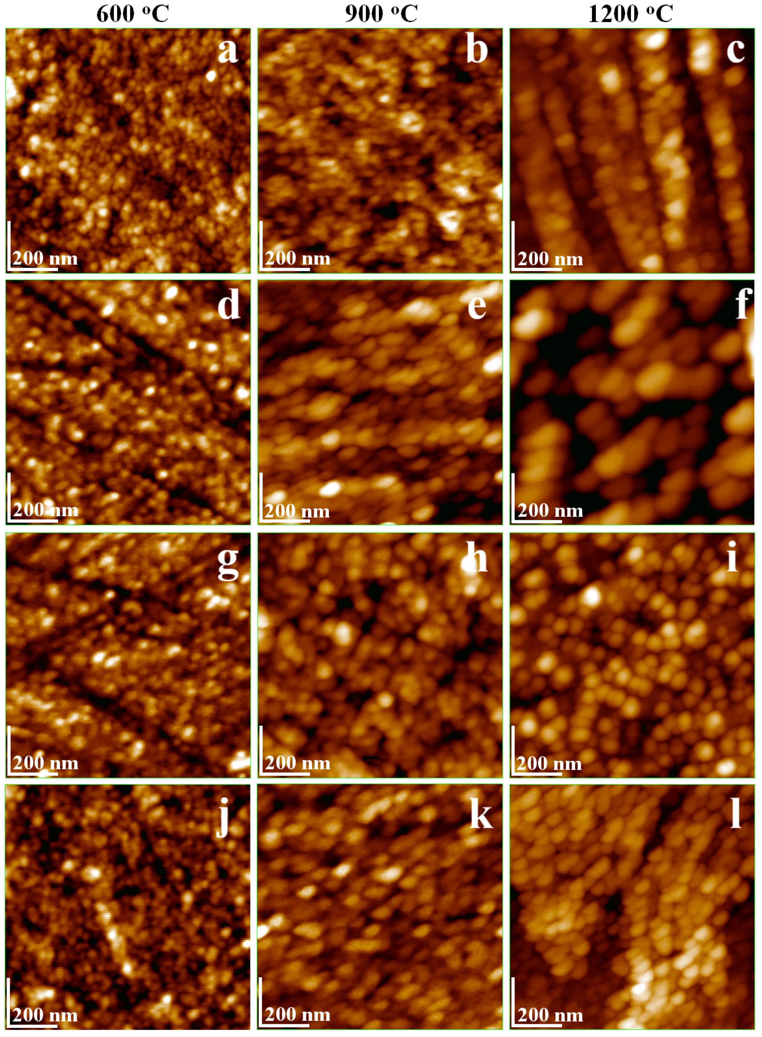
The CdBi0.1Fe1.9O4@SiO2 consists of very small, rounded nanoparticles after calcination at 600 °C, with a diameter of 18 nm (Figure 3a). This diameter is slightly larger than the corresponding crystallite size of 14.9 nm, which can be attributed to the amorphous SiO2 shell embedding the ferrite crystallite as the composite core. Ferrite core increases with the calcination temperature developing the rectangle shape characteristics of its crystal structure. Thus, the particles calcined at 900 °C exhibit partial alteration of the rounded shape due to the ferrite core growth alongside the amorphous SiO2 coating, leading to a slight increase in diameter to about 35 nm (Figure 3b). This trend is further accentuated after calcination at 1200 °C (Figure 3c) where the nanoparticles become slightly elongated due to the extensive development of the ferrite crystallite core, resulting in diameters up to 52 nm. These observations are consistent with literature reports on CdFe2O4 [52], where nanoparticle elongation is evident in SEM images [53]. Moreover, the perovskite structure of BiFeO3 has a square-to-elongated crystal lattice, in agreement with the AFM observations [54], with particle sizes increasing with the calcination temperature [55].
Previous studies have shown that doping BiFeO3 with Cd2+ and Mg2+ ions significantly improves its microstructural and magnetic properties by stabilizing the spinel phase, thereby enhancing both dielectric and magnetic performance [56]. The AFM images indicate a synergistic effect of Cd and Bi substitution on the growth and development of the ferrite core. Nayak et.al emphasizes that structural changes within BiFeO3 play a key role in tailoring its magnetic behavior [57]. Building on this, we propose that additional tuning of the ferrite characteristics can be achieved through the strategic incorporation of minor dopants. This hypothesis is supported by the observed improvements in the crystal lattice, as observed in XRD patterns presented in Figure 2.
Zn2+ doping of CdBi0.1Fe1.9O4 exhibits a pronounced effect, promoting well-developed particles even at a relatively low calcination temperature (600 °C, Figure 3d). The nanoparticles display a diameter of approximately 22 nm and a predominantly rounded shape. The increase in calcination temperature facilitates optimal growth of the ferrite core, which alters the nanoparticle morphology: the particles become slightly elongated after calcination at 900 °C and develop a blockier, boulder-like shape with softened square corners after calcination at 1200 °C, partially moderated by the amorphous SiO2 shell (Figure 3e,f). A similar evolution in morphology is observed on the NCs doped with Ca2+ (Figure 3g–i).
Co2+ doping of CdBi0.1Fe1.9O4 exhibit hybrid behavior, revealing small, rounded nanoparticles after calcination at 600 °C (Figure 3j) with a diameter of approximatively 19 nm, closely resembling the undoped CdBi0.1Fe1.9O4. The doping effect on the nanoparticle structure becomes apparent after calcination at 900 °C (Figure 3k), inducing a significant increase of the particle size to about 38 nm and pronounced elongation due to the enhanced growth of the ferrite core. Further increasing the calcination temperature increases to 1200 °C leads to only a minor increase in particle diameter to 54 nm, but evidence a strong consolidation of the ferrite core, ensuring optimal stability and well-defined individualization of the NC structure.
Considering the approximate rounded shape of the nanoparticles and ferrite crystallite cores, the thickness of the amorphous SiO2 shell is estimated to about 2 nm. This value is consistent with the expectations of the synthesis procedure. It should be noted, however, that AFM cannot reliably differentiate between the ferrite core and the amorphous SiO2 shell, whereas XRD analysis allows for this differentiation. This represents a limitation of the present study.
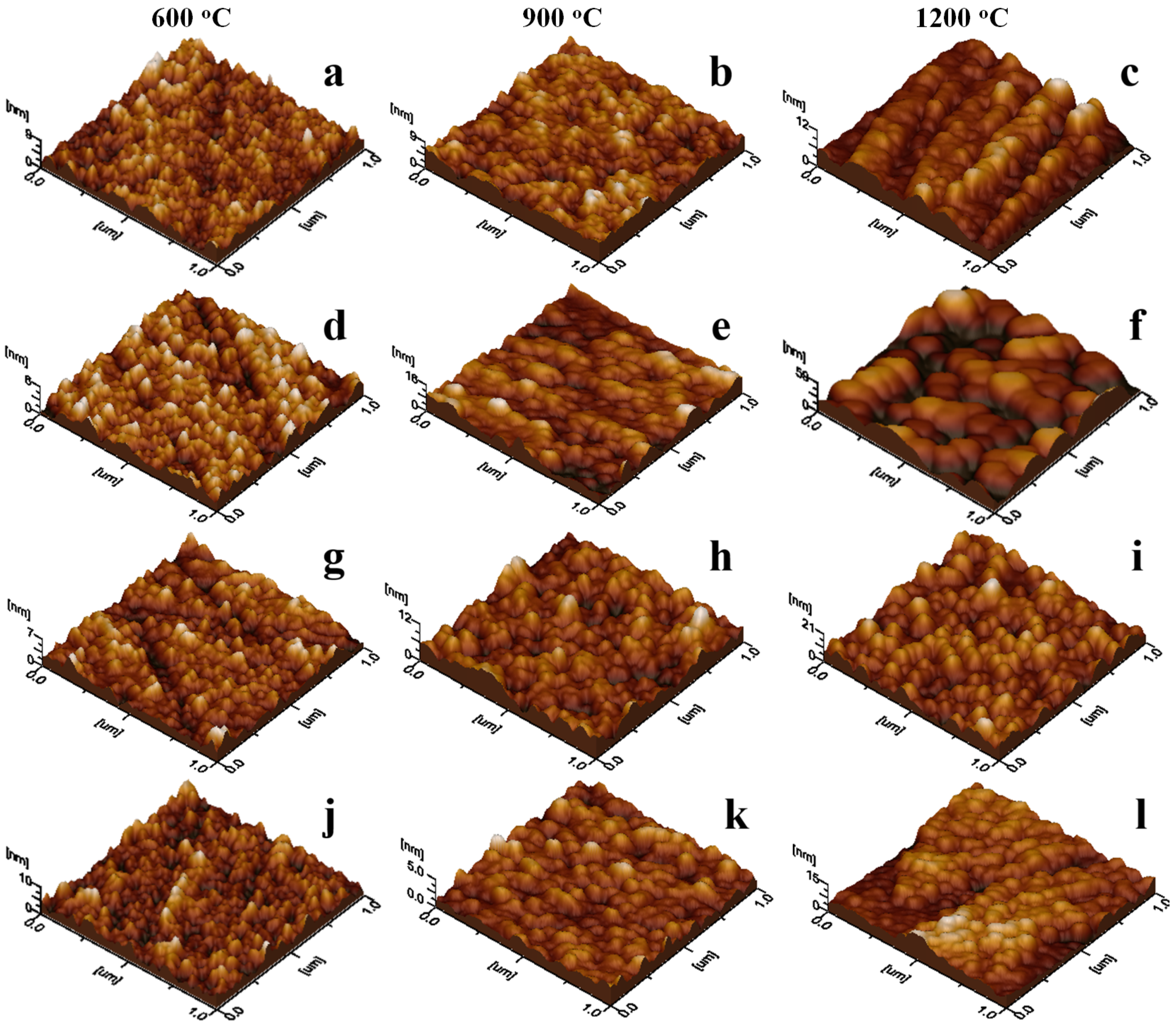
Amorphous SiO2 shell provides an optimal insulation of the ferrite nuclei during the ferrite crystallization, preventing the coalescence between adjacent crystallites. This facilitates the optimal encapsulation of the ferrite core into well individualized composite nanoparticles [58,59,60]. The optimal encapsulation promotes uniform dispersion of the nanoparticles into the aqueous environment which favors the uniform adsorption onto the solid substrate (e.g., glass slide) (Figure 4). The roughness of the surface strongly depends on the nanoparticle’s diameter and the uniformity of adsorbed layer. For example, the samples calcined at 600 °C exhibit low roughness and minimal local height variations ensuring a thin and compact film (Figure 4a,d,g,j). The continuous growth of the ferrite core partly alters the nanoparticle shapes (Figure 4b,e,h,k) leading to the relative increase of the surface roughness (Table 2). The slightly roughness enhancement facilitates the nanoparticle functionalization with biomedical agents (e.g., anticancer drug for targeted delivery onto the tumor sites) according to the literature data [61,62]. Moreover, the higher calcination temperature promotes optimal crystallization of the ferrite core and further increases surface roughness due to the adsorption of larger and more irregularly shaped nanoparticles. Despite this, the resulting thin films remain relatively uniform owing to the well-individualized nanoparticles, which prevent coalescence. The literature highlights the versatility and potential of such ferrites due to their structural stability in composite materials, while also noting associated environmental concerns [63]. In our study, the well-dispersed nanoparticles observed in the adsorbed thin films enable efficient recovery from environmental media via magnetic separation, allowing applications such as oil spill remediation [64] or degradation of dye pollutants [65].

3.4. Magnetic Behavior

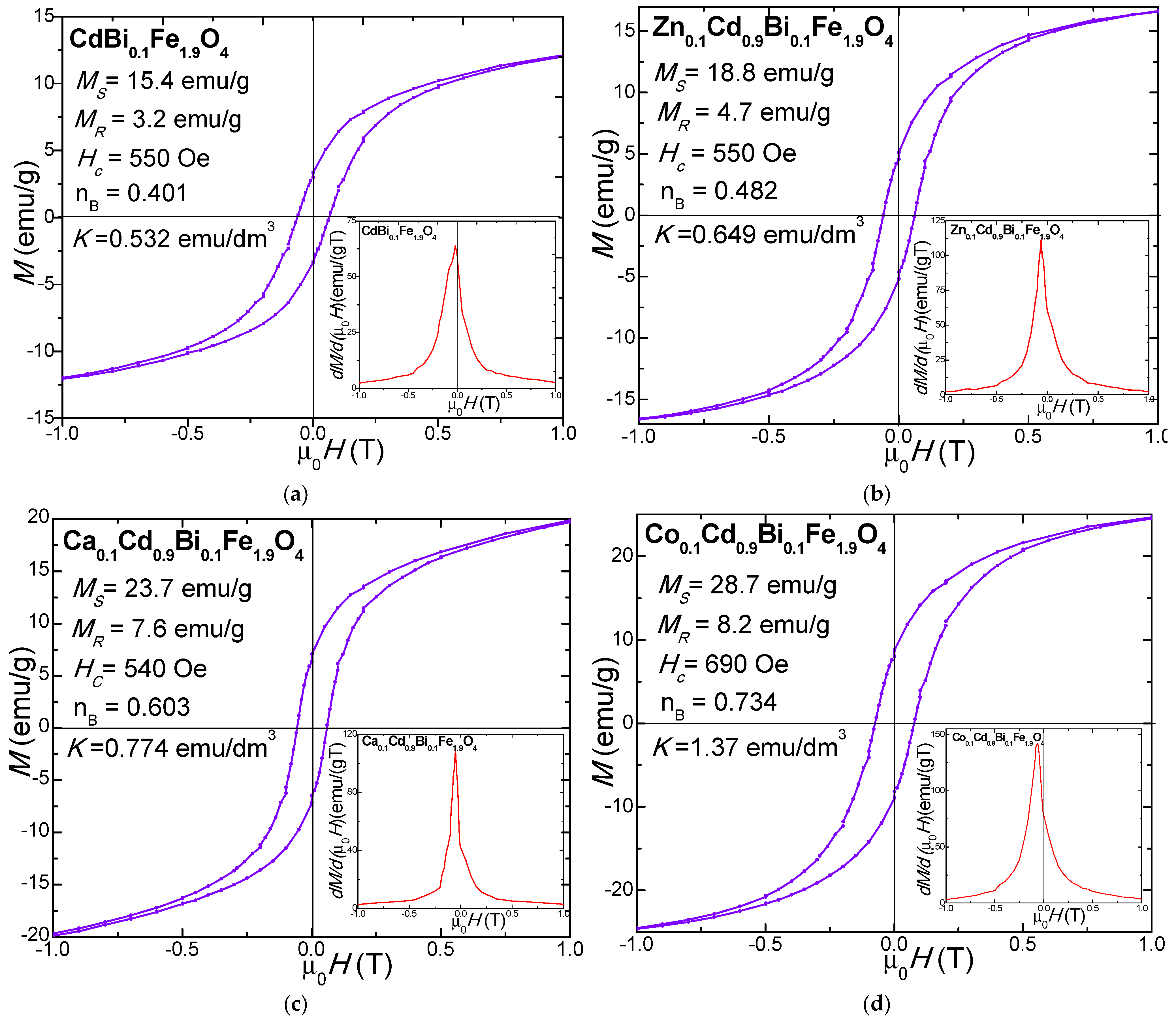
The magnetic hysteresis loops M(µ0H) and their first derivatives (dM/d(μ0H) of NCs calcined at 1200 °C exhibit typical soft ferrimagnetic behavior (Figure 5). At the nanoscale, the high surface-to-volume ratio positions many atoms at or near the particle surface, where broken bonds, defects, spin disorder, and spin canting generate surface spin effects that reduce magnetization and broaden the Hc distribution [66]. As surface contributions increase, surface-dominated interactions outweigh core effects, causing deviations from bulk behavior. Thus, the magnetic characteristics are strongly governed by particle size, shape, and morphology, which depend on synthesis and thermal treatment conditions [27,28,66,67].
All samples exhibit characteristic S-shaped hysteresis loops (Figure 5), which indicated that relatively high magnetocrystalline anisotropy (K) requires stronger fields for saturation. The moderate Hc observed is attributed to partial particle coalescence, which enhances interparticle magnetic coupling and overall magnetization. However, diminished magnetic performance may result from a magnetically inactive surface layer associated with defects, altered cation distribution, incomplete saturation, and lattice defects [14,16,27].
The derivative curves dM/d(μ0H) vs. μ0H reveal magnetization reversal dynamics. For the NCs calcined at 1200 °C, a dominant peak near Hc indicates a single magnetic phase [14,16,23], while a broad peak reflects a distribution of particle sizes and Hc. The minimal peak shift from the origin indicates a narrow Hc distribution due to strong interparticle coupling [68,69]. Sharp peaks are associated with well-crystallized, magnetically homogeneous samples, whereas broader features indicate increased size dispersion and internal defects [14,27]. These results confirm the formation of uniform, well-crystallized nanocrystals.
Doping CdBi0.1Fe1.9O4@SiO2 with non-magnetic Zn2+, Ca2+, and Co2+ ions modifies the magnetocrystalline anisotropy and particle size, leading to enhanced MS, remnant magnetization (MR), magnetic moments per unit cell (nB), HC and K compared to undoped CdBi0.1Fe1.9O4 [14,27,28]. This enhancement is attributed to the dopant ions which promote soft ferrimagnetic behavior [14,16,23,27,67].
Ms of ferrites is primarily governed by the superexchange interactions between the A- and B- sites. An increase in Dc enhances Ms by reducing surface-related defects and improving magnetic ordering [67]. In this study, the highest Ms value (28.7 emu/g) was observed for Co0.1Cd0.9Bi0.1Fe1.9O4@SiO2 NC, whereas the undoped CdBi0.1Fe1.9O4@SiO2 NC exhibited lower Ms values (15.4 emu/g). The surface phenomena such as broken chemical bonds, deviations from the ideal cation distribution, lattice defects, and randomly oriented magnetic moments, contribute to reducing the overall magnetic performance. Additionally, the structural disorder at the particle surface can disrupt the alignment of magnetic moments, a phenomenon typically associated with single-domain configurations [14,16,23,27,67]. Partial substitution of Cd2+ with Zn2+, Ca2+, Co2+ ions may also reduce Ms due to the spin canting arising from the triangular spin configuration within the sublattice, as described by the Yaffet–Kittel [14,16,23,27]. Overall, the enhanced characteristics of Co0.1Cd0.9Bi0.1Fe1.9O4@SiO2 NC, including its relatively high Ms and favorable combination of structural and morphological characteristics, make it a promising candidate for technological applications in communication devices [22,26,32].
The remanent magnetization (MR) of undoped CdBi0.1Fe1.9O4@SiO2 is 3.2 emu/g. The doping with Zn2+ ions increases MR to 4.7 emu/g, while Co2+ ions doping results in an even greater increase, reaching 8.2 emu/g, demonstrating the significant effect of the dopant type on the MR of ferrites.
The K value was calculated under the assumption that the spinel ferrite nanoparticles are approximatively spherical. The K is governed by crystal symmetry, intrinsic magneto crystalline anisotropy, as well as particle size and shape [14,16,23,27]. Among the studied NCs calcined at 1200 °C, Co0.1Cd0.9Bi0.1Fe1.9O4@SiO2 NC exhibits the highest K value of 1.34 erg/dm3. The increase in K for the CdBi0.1Fe1.9O4@SiO2 NC with the addition of dopant ions, reaching the highest value for Co0.1Cd0.9Bi0.1Fe1.9O4@SiO2 NC, follows a trend similar to MS, although no clear linear or colinear correlation between MS and K is observed. Generally, K values increase with doping, which can be attributed to several factors: (i) partial pinning of surface spins in magnetically disordered surface layers, requiring stronger magnetic fields to achieve saturation, and (ii) the presence of randomly oriented grains and lattice vacancies, which further contribute to magnetic disorder. Additionally, crystalline K is affected by the lattice strain arising from the partial substitution of Cd2+ by Zn2+, Ca2+, or Co2+ ions, whichalters the local structural environment and contributes to K enhancement [14,16,23,27].
The lowest nB value is observed in undoped CdBi0.1Fe1.9O4 (0.401) increasing upon doping with Zn2+ (0.482), Ca2+ (0.603) or Co2+ (0.734) doping, which indicates an enhancement of non-collinear spin structures as described by the Yafet–Kittel model. The low Ms in undoped CdBi0.1Fe1.9O4 is attributed to spin canting in the B- sublattice, Jahn-Teller cations, surface magnetic dead layers, cation distribution deviations, incomplete saturation, random moment orientation, and lattice defects. Magnetization in larger particles is dominated by domain wall motion, while smaller particles form single domains aligned with the applied field [69].
The modest increase in Hc values for the doped NCs can be attributed to the contributions from magnetocrystalline anisotropy, microstrain effects, particle size distribution, and reduction in magnetic domain dimensions [31]. The undoped CdBi0.1Fe1.9O4@SiO2 NC exhibits an Hc value of 530 Oe, whereas Co0.1Cd0.9Bi0.1Fe1.9O4@SiO2 NC exhibit the highest HC of 690 Oe. This enhancement is likely related to the variations in Dc, K and the formation of particle agglomerates that increase the particle size beyond the single-domain threshold, resulting in multi-domain structures and reduced domain wall pinning at grain boundaries [14,16,23,27,67]. Conversely, lower Hc values suggest surface spin distortion arising from variations in the magnetocrystalline K. In addition, an increased density of surface lattice defects, such as the atom displacement in the surface layers, could develop local energy barriers that further contribute to increased HC [14,16,23,27,67].
Embedding of magnetic ferrites within the SiO2 matrix stabilizes and organizes the magnetic domains, leading to well-defined magnetic nano-crystallites within the NC. This structural reinforcement improves the mechanical integrity and processability, enabling potential applications in magnetic coatings, thin films, and other nanostructured devices [2,3,6,32].

4. Conclusions

The influence of Zn2+, Ca2+, Co2+ ions doping on the structural, morphological, surface, and magnetic characteristics of CdBi0.1Fe1.9O4 was systematically investigated. At 300 °C, the ferrites exhibited poorly crystallinity, whereas Co0.1Cd0.9Bi0.1Fe1.9O4@SiO2 calcined at 1200 °C displayed a well-defined single-phase ferrite structure. Doping not only enhanced ferrite crystallinity but also promoted the development of crystalline SiO2 phases (silicate, cristobalite, and tridymite). Although all NCs maintained a cubic spinel structure, XRD analysis revealed that the dopant incorporation induced variations in the structural parameters. AFM imaging confirmed that the ferrite crystallites observed by XRD are encapsulated within amorphous SiO2, forming well-individualized nanoparticles. At lower calcination temperatures, the nanoparticles are predominantly rounded, whereas higher calcination temperatures enhance ferrite core growth, leading to distorted particle shapes and increased sizes. The surface roughness of the adsorbed thin films strongly correlates with nanoparticle size and morphology: small, rounded particles yield low roughness, while larger, irregular nanoparticles significantly increase surface roughness, making them suitable for targeted functionalization. The MS (15.4–28.7 emu/g), MR (3.2–8.2 emu/g), Hc (530–690 Oe) and K (0.532–1.24 erg/dm3) diminished with the incorporation of dopant ions, approaching soft ferrimagnetic-like behavior. The analysis using Néel’s collinear and Yafet–Kittel spin-canting models, considering the surface spin effects, indicates that doping and thermal treatment are critical factors in governing the magnetic behavior and particle size distribution of the nanocomposites.

Author Contributions

Conceptualization, T.D.; methodology, T.D., I.P. and O.C.; software, T.D.; validation, T.D., I.P. and O.C.; formal analysis, T.D., I.P. and O.C.; investigation, T.D.; resources, T.D., I.P. and O.C.; data curation, T.D.; writing—original draft preparation, T.D., I.P. and O.C.; writing—review and editing, T.D. and O.C.; visualization, T.D., I.P. and O.C.; supervision, T.D. All authors have read and agreed to the published version of the manuscript.

Funding

This research received no external funding.

Data Availability Statement

Data are available on request from the corresponding author.

Acknowledgments

O.C. acknowledges the support of Innovation Plan 2022–2027 (PNCDI IV), European and International Cooperation Program, contr. no. 3BMTR⁄2025.

Conflicts of Interest

The authors declare no conflicts of interest.

References

  1. Bagade, A.V.; Nagwade, P.A.; Nagawade, A.V.; Thopate, S.R.; Pund, S.N. A review on synthesis, characterization and applications of cadmium ferrite and its doped variants. Orient. J. Chem. 2022, 38, 1–5. [Google Scholar] [CrossRef]
  2. Shakil, M.; Inayat, U.; Arshad, M.I.; Nabi, G.; Khalid, N.R.; Tariq, N.H.; Shah, A.; Iqbal, M.Z. Influence of zinc and cadmium co-doping on optical and magnetic properties of cobalt ferrites. Ceram. Int. 2020, 46, 7767–7773. [Google Scholar] [CrossRef]
  3. Nandanwar, A.K.; Chaodhary, D.L.; Kamde, S.N.; Choudhary, D.S.; Rewatkar, K.G. Study of structural and magnetic properties of zinc-substituted cadmium ferrite nanocrystals. Mater. Todays Proc. 2020, 29, 951–955. [Google Scholar] [CrossRef]
  4. Siddique, M.A.; Irfan, M.; Rasool, R.T.; Arshad, M.; Ashraf, G.A.; Akhtar, M.N.; Manzoor, S.; Raza, M.R.; Khan, M.A. Role of Zn cations substitution on structural, vibrational, elastic, dielectric, bandwidth, and microwave absorbance of Co–Cd ferrites at high frequencies. Mater. Chem. Phys. 2024, 315, 128972. [Google Scholar] [CrossRef]
  5. Kaur, H.; Singh, A.; Kumar, V.; Ahlawat, D.S. Structural, thermal and magnetic investigations of cobalt ferrite doped with Zn2+ and Cd2+ synthesised by auto combustion method. J. Magn. Magn. Mater. 2019, 474, 505–511. [Google Scholar] [CrossRef]
  6. Ghorbani, H.; Eshraghi, M.; Dodaran, A.A.S. Structural and magnetic properties of cobalt ferrite nanoparticles doped with cadmium. Phys. B Condens. Matter 2022, 634, 413816. [Google Scholar] [CrossRef]
  7. Aakash, P.; Nordblad, R.; Mohan, S. Mukherjee, Structural, magnetic and hyperfine characterisations of nanocrystalline Zn-Cd doped nickel ferrites. J. Magn. Magn. Mater. 2017, 441, 710–717. [Google Scholar] [CrossRef]
  8. Sanad, M.M.S.; Azab, A.A.; Taha, T.A. Introduced oxygen vacancies in cadmium ferrite anode materials via Zn2+ incorporation for high performance lithium-ion batteries. Mater. Sci. Semicond. Proc. 2022, 143, 106567. [Google Scholar] [CrossRef]
  9. Ghatak, S.; Chakraborty, G.; Sinha, M.; Pradhan, S.K.; Meikap, A.K. Dielectric relaxation and magnetic field dependent alternating current conductivity of nanocrystalline cadmium–zinc ferrite below room temperature. Phys. B 2011, 406, 3261–3266. [Google Scholar] [CrossRef]
  10. Sinha, M.; Pradhan, S.K. Synthesis of nanocrystalline Cd–Zn ferrite by ball milling and its stability at elevated temperatures. J. Alloys Compd. 2010, 489, 91–98. [Google Scholar] [CrossRef]
  11. Parajuli, D.; Taddesse, P.; Murali, N.; Samatha, K. Study of structural, electromagnetic and dielectric properties of cadmium substituted Ni–Zn nanosized ferrites. J. Indian Chem. Soc. 2022, 99, 100380. [Google Scholar] [CrossRef]
  12. Gupta, M.; Gupta, M.; Anu Mudsainiyan, R.K.; Randhawa, B.S. Physico-chemical analysis of pure and Zn doped Cd ferrites (Cd1- xZnxFe2O4) nanofabricated by Pechini sol-gel method. J. Anal. Appl. Pyrol. 2015, 116, 75–85. [Google Scholar] [CrossRef]
  13. Stefănescu, M.; Stoia, M.; Caizer, C.; Dippong, T.; Barvinschi, P. Preparation of CoxFe3−xO4 nanoparticles by thermal decomposition of some organo-metallic precursors. J. Therm. Anal. Calorim. 2009, 97, 245–250. [Google Scholar] [CrossRef]
  14. Dippong, T.; Cadar, O.; Deac, I.G.; Lazar, M.; Borodi, G.; Levei, E.A. Influence of ferrite to silica ratio and thermal treatment on porosity, surface, microstructure and magnetic properties of Zn0.5Ni0.5Fe2O4/SiO2 nanocomposites. J. Alloys Compd. 2020, 828, 154409. [Google Scholar] [CrossRef]
  15. Dippong, T.; Levei, E.A.; Cadar, O.; Goga, F.; Barbu Todoran, L.; Borodi, G. Size and shape-controlled synthesis and characterization of CoFe2O4 nanoparticles embedded in a PVA-SiO2 hybrid matrix. J. Anal. Appl. Sci. 2017, 128, 121–130. [Google Scholar] [CrossRef]
  16. Dippong, T.; Levei, E.A.; Diamandescu, L.; Bibicu, I.; Leostean, C.; Borodi, G.; Barbu-Tudoran, L. Structural and magnetic properties of CoxFe3-xO4 versus Co/Fe molar ratio. J. Magn. Magn. Mater. 2015, 394, 111–116. [Google Scholar] [CrossRef]
  17. Kulkarni, A.B.; Mathad, S.N. Variation in structural and mechanical properties of Cd-doped Co-Zn ferrites. Mater. Sci. Energy Technol. 2019, 2, 455–462. [Google Scholar] [CrossRef]
  18. Hcini, S.; Kouki, N.; Omri, A.; Dhahri, A.; Bouazizi, M.L. Effect of sintering temperature on structural, magnetic, magnetocaloric and critical behaviors of Ni-Cd-Zn ferrites prepared using sol-gel method. J. Magn. Magn. Mater. 2018, 464, 91–102. [Google Scholar] [CrossRef]
  19. Chinchay-Espino, H.A.; Montes-Albino, G.M.; Morales-Cruz, C.M.; Dobbertin-Sanchez, S.E.; Rojas-Flores, S. Effect of cobalt substitution on the structural and magnetic properties of bismuth ferrite powders. Crystals 2022, 12, 1058. [Google Scholar] [CrossRef]
  20. Panwar, M.N.; Khan, H.M.; Bano, N.; Zahid, M.; Mazhar, M.E.; Bilal, M.; Saleem, M.I.; Solre, G.F.B.; Gomez, P.H.; Li, Z. Influence of bismuth and cobalt doping on structural, dielectric, and magnetic properties of M-type calcium hexagonal ferrites. J. Alloys Compd. 2024, 994, 174623. [Google Scholar] [CrossRef]
  21. Patil, S.; Meti, S.; Kanavi, P.S.; Bhajantri, R.F.; Anandalli, M.; Mondal, R.; Karmakar, S.; Muhiuddin, M.; Rahman, M.R.; Kumar, B.C.; et al. A study on solubility of bismuth cations in nickel cobalt ferrite nanoparticles and their influence on dielectric and magnetic properties. Mater. Sci. Eng. B 2023, 296, 116570. [Google Scholar] [CrossRef]
  22. Vijaya, K.; Kumar, N.; Kumar, S. Electrical properties of bismuth doped CoBixFe2-xO4 nano ferrites. Mater. Today Proc. 2016, 3, 4193–4198. [Google Scholar] [CrossRef]
  23. Dippong, T.; Cadar, O.; Levei, E.A.; Deac, I.G. Microstructure, porosity and magnetic properties of Zn0.5Co0.5Fe2O4/SiO2 nanocomposites prepared by sol-gel method using different polyols. J. Magn. Magn. Mater. 2020, 498, 166168. [Google Scholar] [CrossRef]
  24. Zahir, R.; Chowdhury, F.U.Z.; Uddin, M.M.; Hakim, M.A. Structural, magnetic and electrical characterisation of Cd-substituted Mg ferrites synthesised by double sintering technique. J. Magn. Magn. Mater. 2016, 410, 55–62. [Google Scholar] [CrossRef]
  25. Ahmad, R.; Hussain Gul, I.; Zarrar, M.; Anwar, H.; Niazi, M.B.K.; Khan, A. Improved electrical properties of cadmium substituted cobalt ferrites nano-particles for microwave application. J. Magn. Magn. Mater. 2016, 405, 28–35. [Google Scholar] [CrossRef]
  26. Valan, M.F.; Manikandan, A.; Arul Antony, S. Microwave combustion synthesis and characterization studies of magnetic Zn1–xCdxFe2O4 (0 ≤ x ≤ 0.5) nanoparticles. J. Nanosci. Nanotechnol. 2014, 15, 4543–4551. [Google Scholar] [CrossRef]
  27. Dippong, T.; Levei, E.A.; Cadar, O.; Mesaros, A.; Borodi, G. Sol-gel synthesis of CoFe2O4:SiO2 nanocomposites—Insights into the thermal decomposition process of precursors. J. Anal. Appl. Pyrol. 2017, 125, 169–177. [Google Scholar] [CrossRef]
  28. Dippong, T.; Levei, E.A.; Cadar, O.; Goga, F.; Toloman, D.; Borodi, G. Thermal behavior of Ni, Co and Fe succinates embedded in silica Matrix. J. Therm. Anal. Calorim. 2019, 136, 1587–1596. [Google Scholar] [CrossRef]
  29. Divakara, S.G.; Mahesh, B. A comprehensive review on current trends in greener and sustainable synthesis of ferrite nanoparticles and their promising applications. Results Eng. 2024, 21, 101702. [Google Scholar]
  30. Ali, S.S.L.; Selvaraj, S.; Batoo, K.M.; Chauhan, A.; Rana, G.; Kalaichelvan, S.; Radhakrishnan, A. Green synthesis of cubic spi-nel ferrites and their potential biomedical applications. Ceram. Int. 2024, 50, 52159–52189. [Google Scholar] [CrossRef]
  31. Abidin, M.Z.U.; Ikram, M.; Moeen, S.; Nazir, G.; Kanoun, M.B.; Goumri-Said, S. A comprehensive review on the synthesis of ferrite nanomaterials via bottom-up and top-down approaches advantages, disadvantages, characterizations and computa-tional insights. Coord. Chem. Rev. 2024, 520, 216158. [Google Scholar] [CrossRef]
  32. Badr, M.H.; Kudrevatykh, N.V.; Hassan, M.A.; Moustafa, M.; Rammah, Y.S.; Abouhaswa, A.S.; EL-Hamalawy, A.A. Structural and magnetic analysis of Cd-Zn spinel ferrite nanoparticles. Phys. Scr. 2023, 98, 025823. [Google Scholar] [CrossRef]
  33. Rao, P.A.; Raghavendra, V.; Suryanarayana, B.; Paulos, T.; Murali, N.; Varma, P.V.S.K.P.; Prasad, R.G.; Ramakrishna, Y.; Chandramouli, K. Cadmium substitution effect on structural, electrical and magnetic properties of Ni-Zn nano ferrites. Res. Phys. 2020, 19, 103487. [Google Scholar]
  34. Amer, M.A.; Meaz, T.M.; El-Kestawy, M.; Ghoneim, A.I. Magnetic and structural studies of trivalent Co-substituted Cd–Mn ferrites. J. Magn. Magn. Mater. 2016, 405, 137–144. [Google Scholar] [CrossRef]
  35. Gonçalves, M.J.; Faria, V.L.; Nascimento, A.B.; Germscheidt, R.L.; Patra, S.; Saravaria, L.P.H.; Bonacin, A.J.; Munoz, R.A.A.; Angnes, L. Sensing performances of spinel ferrites MFe2O4 (M = Mg, Ni, Co, Mn, Cu and Zn) based electrochemical sensors: A review. Anal. Chim. Acta 2022, 1233, 340362. [Google Scholar] [CrossRef]
  36. Panda, R.K.; Muduli, R.; Behera, D. Electric and magnetic properties of Bi substituted cobalt ferrite nanoparticles: Evolution of grain effect. J. Alloys Compd. 2015, 634, 239–245. [Google Scholar] [CrossRef]
  37. Kumar, H.; Singh, J.P.; Srivastava, R.C.; Negi, P.; Agrawal, H.M.; Asokan, K. FTIR and electrical study of dysprosium doped cobalt ferrite nanoparticles. J. Nanosci. 2014, 2014, 862415. [Google Scholar] [CrossRef]
  38. Rodriguez-Carvajal, J. Recent advances in magnetic structure determination by neutron powder diffraction. Phys. B Condens. Matter 1993, 192, 55–69. [Google Scholar] [CrossRef]
  39. Dhankhar, S.; Tiwari, P.; Baskar, K.; Basu, S.; Singh, S. Effect of low cobalt doping on morphology and properties of calcium ferrite and its application as cathode in solid oxide fuel cell. J. Curr. Appl. Phys. 2017, 17, 467–473. [Google Scholar] [CrossRef]
  40. Huili, H.; Grindi, B.; Viau, G.; Tahar, L.B. Influence of the stoichiometry and grain morphology on the magnetic properties of Co substituted Ni–Zn nanoferrites. Ceram. Int. 2016, 42, 17594–17604. [Google Scholar] [CrossRef]
  41. Ramesh, S.; Sekhar, B.C.; Rao, P.S.V.S.; Rao, B.P. Microstructural and magnetic behavior of mixed Ni–Zn–Co and Ni–Zn–Mn ferrites. Ceram. Int. 2014, 40, 8729–8735. [Google Scholar] [CrossRef]
  42. Arulmurugan, R.; Vaidyanathan, G.; Sendhilnathan, S.; Jeyadevan, B. Mn-Zn ferrite nanoparticles for ferrofluid preparation: Study on thermalemagnetic properties. J. Magn. Magn. Mater. 2006, 298, 83–94. [Google Scholar] [CrossRef]
  43. Zheng, Z.G.; Zhong, X.C.; Zhang, Y.H.; Yu, J.Y.; Zeng, D.C. Synthesis, structure and magnetic properties of nanocrystalline ZnxMn1-xFe2O4 prepared by ball milling. J. Alloys Compd. 2008, 466, 377–382. [Google Scholar] [CrossRef]
  44. Raji, P.; Balachandra Kumar, K. Structural, elastic and magnetic properties of Ca doped copper ferrite nanoparticles. Phys. B Condens. Matter. 2022, 632, 413759. [Google Scholar] [CrossRef]
  45. Mohamed, W.S.; Alzaid, M.; Abdelbaky, M.S.M.; Amghouz, Z.; García-Granda, S.; Abu-Dief, A.M. Impact of Co2+ substitution on microstructure and magnetic properties of CoxZn1-xFe2O4 nanoparticles. Nanomat 2019, 9, 1602. [Google Scholar] [CrossRef] [PubMed]
  46. Hubbard, C.R.; Snyder, R.L. RIR—Measurement and use in quantitative XRD. Powder Diffr. 1988, 3, 74–77. [Google Scholar] [CrossRef]
  47. Mahmood, A.; Maqsood, A.; Tayyab, S.; Ali, M.; Ul Abideen, Z. Effect of cadmium on the fabrication and thermally tailoring electrical transport properties of zinc ferrite nanoparticles. Ceram. Int. 2026. [Google Scholar] [CrossRef]
  48. Ge, Y.; Li, C.; Waterhouse, G.I.N.; Zhang, Z.; Yu, L. ZnFe2O4@SiO2@Polypyrrole nanocomposites with efficient electromagnetic wave absorption properties in the K and Ka band regions. Ceram. Int. 2021, 47, 1728–1739. [Google Scholar] [CrossRef]
  49. Powar, R.R.; Phadtare, V.D.; Parale, V.G.; Park, H.H.; Pathak, S.; Kamble, P.R.; Piste, P.B.; Zambare, D.N. Structural, morphological, and magnetic properties of ZnxCo1-xFe2O4 (0 < x < 1) prepared using a chemical co-precipitation method. Ceram. Int. 2018, 44, 20782–20789. [Google Scholar]
  50. Avram, S.E.; Birle, B.V.; Cosma, C.; Tudoran, L.B.; Moldovan, M.; Cuc, S.; Borodi, G.; Petean, I. Ecological building material obtained through the moderate thermal consolidation of ceramic slurry collected from industrial waste waters. Materials 2025, 18, 1715. [Google Scholar] [CrossRef]
  51. Avram, S.E.; Tudoran, L.B.; Borodi, G.; Petean, I. Microstructural Characterization of the Mn Lepidolite Distribution in Dark Red Clay Soils. Appl. Sci. 2025, 15, 6445. [Google Scholar] [CrossRef]
  52. Jdidi, A.R.; Nouira, W.; Selmi, A.; Drissi, N.; Aissa, M.; Hcini, S.; Gassoumi, M. Impact of Calcination Temperature on the Properties and Photocatalytic Efficiency of Cd0.6Mg0.2Cu0.2Fe2O4 Spinel Ferrites Synthesized via the Sol–Gel Method. Crystals 2025, 15, 457. [Google Scholar] [CrossRef]
  53. Al-Kadhi, N.S.; Basha, M.T. Enhanced removal of Cd(II) ions from aqueous media via adsorption on facilely synthesized copper ferrite nanoparticles. Molecules 2024, 29, 3711. [Google Scholar] [CrossRef] [PubMed]
  54. Mallek-Zouari, I.; Kaddar, Y.; Ben Taazayet, W.; Mounkachi, O.; Hlil, E.-K.; Thabet Mliki, N.; El Moutaouakil, A. first-principles investigation of structural, electronic, and magnetic properties of BiFeO3 and Bi2Fe4O9 nanostructures. Int. J. Mol. Sci. 2025, 26, 4671. [Google Scholar] [CrossRef] [PubMed]
  55. Petrukhin, D.; Salnikov, V.; Nikitin, A.; Sidane, I.; Slimani, S.; Alberti, S.; Peddis, D.; Omelyanchik, A.; Rodionova, V. Effect of bismuth ferrite nanoparticles on physicochemical properties of polyvinylidene fluoride-based nanocomposites. J. Compos. Sci. 2024, 8, 329. [Google Scholar] [CrossRef]
  56. Gan, G.; Zhang, H.; Li, Q.; Li, J.; Huang, X.; Xie, F.; Xu, F.; Zhang, Q.; Li, M.; Liang, T.; et al. Low loss, enhanced magneto-dielectric properties of Bi2O3 doped Mg-Cd ferrites for high frequency antennas. J. Alloys Compd. 2018, 735, 2634–2639. [Google Scholar] [CrossRef]
  57. Nayak, S.; Agnihotri, P.; Nayak, J.P.; Joshi, C.; Rai, R. Exploring bismuth ferrite: Structural changes and future applications. Curr. Appl. Phys. 2025, 73, 49–76. [Google Scholar] [CrossRef]
  58. Melis, N.; Loche, D.; Thakkar, S.V.; Cutrufello, M.G.; Sini, M.F.; Sedda, G.; Pilia, L.; Frongia, A.; Casula, M.F. Magnetic aerogels for room-temperature catalytic production of bis(indolyl)methane derivatives. Molecules 2024, 29, 2223. [Google Scholar] [CrossRef]
  59. Omelyanchik, A.; Salvador, M.; D’Orazio, F.; Mameli, V.; Cannas, C.; Fiorani, D.; Musinu, A.; Rivas, M.; Rodionova, V.; Varvaro, G.; et al. magnetocrystalline and surface anisotropy in CoFe2O4 nanoparticles. Nanomaterials 2020, 10, 1288. [Google Scholar] [CrossRef]
  60. Virumbrales, M.; Saez-Puche, R.; Torralvo, M.J.; Blanco-Gutierrez, V. Mesoporous silica matrix as a tool for minimizing dipolar interactions in NiFe2O4 and ZnFe2O4 Nanoparticles. Nanomaterials 2017, 7, 151. [Google Scholar] [CrossRef]
  61. Kharat, P.B.; Somvanshi, S.B.; Dawi, E.A.; Ibrahim, R.M.; Mopari, A.M. Surface-functionalized nanoscale nickel-ferrites (NiFe2O4) nanoparticles as heating agents for cancer theranostics. Inorg. Chem. Commun. 2025, 182, 115569. [Google Scholar] [CrossRef]
  62. Giannousi, K.; Toursidis, P.D.; Chatzatoglou, E.; Dendrinou-Samara, C. Manganese ferrite nanoparticles functionalized with gallic acid for inhibition of insulin amyloids formation and mitigation of oxidative stress. Colloids Surf. A Physicochem. Eng. Asp. 2025, 727, 138114. [Google Scholar] [CrossRef]
  63. Chakraborty, S.; Roy, S.; Ahmaruzzaman, M.d. Advancing the frontier of spinel ferrites in sustainable remediation: Fundamentals, synthetic strategies, and mechanistic insights. J. Water Process Eng. 2026, 81, 109276. [Google Scholar] [CrossRef]
  64. Košak, A.; Hadela, A.; Poberžnik, M.; Lobnik, A. magnetic separation of oil spills from water using cobalt ferrite nanoparticles with fluorocarbon functionalization. Int. J. Mol. Sci. 2025, 26, 6562. [Google Scholar] [CrossRef]
  65. Fernandes, R.J.C.; Cardoso, B.D.; Rodrigues, A.R.O.; Pires, A.; Pereira, A.M.; Araújo, J.P.; Pereira, L.; Coutinho, P.J.G. Zinc/magnesium ferrite nanoparticles functionalized with silver for optimized photocatalytic removal of malachite green. Materials 2024, 17, 3158. [Google Scholar] [CrossRef]
  66. Rao, S.; Choudary, G.S.V.R.K.; Rao, R.H.; Sujatha, C. Structural and magnetic properties of ultrafine CoFe2O4 nanoparticles. Proc. Mat. Sci. 2015, 10, 19–27. [Google Scholar] [CrossRef]
  67. Haque, S.U.; Saikia, K.K.; Murugesan, G.; Kalainathan, S. A study on dielectric and magnetic properties of lanthanum substituted cobalt ferrite. J. Alloys Compd. 2017, 701, 612–618. [Google Scholar] [CrossRef]
  68. Ghosh, M.P.; Murkherjee, S. Microstructural, magnetic and hyperfine characterizations of Cu-doped cobalt ferrite nanoparticles. J. Am. Chem. Soc. 2019, 102, 7509–7520. [Google Scholar] [CrossRef]
  69. Ansari, S.M.; Suryawanshi, S.R.; More, M.A.; Sen, D.; Kolekar, Y.D.; Ramana, C.V. Field emission properties of nano-structured cobalt ferrite (CoFe2O4) synthesized by low-temperature chemical method. Chem. Phys. Lett. 2018, 701, 151–156. [Google Scholar] [CrossRef]
Figure 1. (a) TG and (b) DTA curves of CdBi0.1Fe1.9O4, Zn0.1Cd0.9Bi0.1Fe1.9O4, Ca0.1Cod0.9Bi0.1Fe1.9O4, Co0.1Cd0.9Bi0.1Fe1.9O4 gels heated at 40 °C; (c) FT-IR spectra of gels heated at 40 °C and (d) FT-IR spectra of gels calcinated at 300 °C.
Nanomaterials 16 00259 g001
Figure 2. XRD patterns of CdBi0.1Fe1.9O4@SiO2, Zn0.1Cd0.9Bi0.1Fe1.9O4@SiO2, Ca0.1Cd0.9Bi0.1Fe1.9O4@SiO2, Co0.1Cd0.9Bi0.1Fe1.9O4@SiO2 NCs calcined at (a) 300 °C, (b) 600 °C, (c) 900 °C and (d) 1200 °C.
Nanomaterials 16 00259 g002
Figure 3. AFM topographic images of the powders’ thin films transferred onto solid substrate: CdBi0.1Fe1.9O4@SiO2 NC calcined at (a) 600 °C, (b) 900 °C, (c) 1200 °C; Zn0.1Cd0.9Bi0.1Fe1.9O4@SiO2 NC calcined at (d) 600 °C, (e) 900 °C, (f) 1200 °C; Ca0.1Cd0.9Bi0.1Fe1.9O4@SiO2 NC calcined at (g) 600 °C, (h) 900 °C, (i) 1200 °C; Co0.1Cd0.9Bi0.1Fe1.9O4@SiO2 NC calcined at (j) 600 °C, (k) 900 °C, (l) 1200 °C.
Nanomaterials 16 00259 g003
Figure 4. AFM tridimensional profiles of the powders’ thin films transferred onto solid substrate: CdBi0.1Fe1.9O4@SiO2 NC calcined at (a) 600 °C, (b) 900 °C, (c) 1200 °C; Zn0.1Cd0.9Bi0.1Fe1.9O4@SiO2 NC calcined at (d) 600 °C, (e) 900 °C, (f) 1200 °C; Ca0.1Cd0.9Bi0.1Fe1.9O4@SiO2 NC calcined at (g) 600 °C, (h) 900 °C, (i) 1200 °C; Co0.1Cd0.9Bi0.1Fe1.9O4@SiO2 NC calcined at (j) 600 °C, (k) 900 °C, (l) 1200 °C.
Nanomaterials 16 00259 g004
Figure 5. Magnetic hysteresis loops and magnetization derivative of (a) CdBi0.1Fe1.9O4@SiO2, (b) Zn0.1Cd0.9Bi0.1Fe1.9O4@SiO2, (c) Ca0.1Cd0.9Bi0.1Fe1.9O4@SiO2, and (d) Co0.1Cd0.9Bi0.1Fe1.9O4@SiO2 NCs calcined at 1200 °C.
Nanomaterials 16 00259 g005
Table 1. Structural parameters and quantitative analysis of CdBi0.1Fe1.9O4@SiO2, Zn0.1Cd0.9Bi0.1Fe1.9O4@SiO2, Ca0.1Cd0.9Bi0.1Fe1.9O4@SiO2, Co0.1Cd0.9Bi0.1Fe1.9O4@SiO2 NCs estimated by XRD.
SampleTemp
[°C]
DCS
[nm]
a
(Å)
V
3)
dA
(Å)
dB
(Å)
dp
(g/cm3)
dXRD
(g/cm3)
P
(%)
Quantitative Analysis
CdBi0.1Fe1.9O4@SiO23007.38.3795883.6282.9625.3426.85522.1100% CdBi0.1Fe1.9O4
60014.98.4155963.6442.9755.3796.76320.574% CdO/26% CdBi0.1Fe1.9O4
90031.68.4576053.6622.9905.4116.66218.834%CdBi0.1Fe1.9O4/23% Fe2SiO4/6%Fe2O3/37%CdO
120048.28.4986143.6803.0045.4676.56416.732% CdBi0.1Fe1.9O4/35% Fe2O3/33% SiO2
Zn0.1Cd0.9Bi0.1Fe1.9O4@SiO23008.38.3445813.6132.9505.3496.83021.7100% Zn0.1Cd0.9Bi0.1Fe1.9O4
60016.58.3765883.6272.9615.3936.74920.0100% Zn0.1Cd0.9Bi0.1Fe1.9O4
90034.88.4235983.6482.9785.4316.63618.273% Zn0.1Cd0.9Bi0.1Fe1.9O4/ 22% Fe2SiO4/5%Fe2O3
120052.48.4686073.6672.9945.4896.53816.034% Zn0.1Cd0.9Bi0.1Fe1.9O4/35% Fe2O3/31% SiO2
Ca0.1Cd0.9Bi0.1Fe1.9O4@SiO23009.68.3665863.6232.9585.3596.71420.2100% Ca0.1Cd0.9Bi0.1Fe1.9O4
60015.98.4065943.6402.9725.4086.62418.4100% Ca0.1Cd0.9Bi0.1Fe1.9O4
90036.28.4466033.6572.9865.4496.52516.572% Ca0.1Cd0.9Bi0.1Fe1.9O4/18% Fe2SiO4
120056.58.4886123.6753.0015.4976.42914.571% Ca0.1Cd0.9Bi0.1Fe1.9O4/29% SiO2
Co0.1Cd0.9Bi0.1Fe1.9O4@SiO23007.98.3535833.6172.9535.3526.79221.2100% Co0.1Cd0.9Bi0.1Fe1.9O4
60015.78.3895903.6332.9665.3986.71119.6100% Co0.1Cd0.9Bi0.1Fe1.9O4
90033.08.4356003.6522.9825.4406.59917.6100% Co0.1Cd0.9Bi0.1Fe1.9O4
120049.88.4736083.6692.9965.4956.51315.6100% Co0.1Cd0.9Bi0.1Fe1.9O4
Table 2. Topographic parameters and particle diameter measured with AFM.
SampleTemp, °CHeight, nmRa, nmD, nm
CdBi0.1Fe1.9O4@SiO260090.8918
90091.1435
1200121.8752
Zn0.1Cd0.9Bi0.1Fe1.9O4@SiO260080.8622
900162.2543
1200599.0672
Ca0.1Cd0.9Bi0.1Fe1.9O4@SiO260070.9020
900121.5441
1200213.4064
Co0.1Cd0.9Bi0.1Fe1.9O4@SiO2600101.1019
90050.6338
1200152.4454
Disclaimer/Publisher’s Note: The statements, opinions and data contained in all publications are solely those of the individual author(s) and contributor(s) and not of MDPI and/or the editor(s). MDPI and/or the editor(s) disclaim responsibility for any injury to people or property resulting from any ideas, methods, instructions or products referred to in the content.