1. Introduction
Antibiotic resistance (ABR) is a pressing global issue that has compromised the effectiveness of traditional antibiotic treatment strategies and contributed to rising mortality, morbidity and economic burdens [
1,
2]. The emergence and spread of multidrug-resistant (MDR) bacteria, which are resistant to multiple antibiotics, have made this problem even worse and have triggered the need for new and sustainable approaches to alternative therapy [
2].
MDR infections are a significant public health crisis worldwide. Epidemiological evidence suggests that MDR bacterial infections are a major cause of morbidity and mortality in the world, and the costs of treatment are growing significantly [
3,
4]. People with MDR infections suffer significantly longer hospital stays, higher treatment expenses and higher death rates than people with susceptible infections [
1,
2]. The three primary MDR pathogens addressed in this study, such as
Staphylococcus aureus,
Escherichia coli, and
Pseudomonas spp., are among the most pathologically relevant microorganisms causing diverse clinical infections [
5,
6].
S. aureus is responsible for serious life-threatening skin and soft tissue infections, hospital-acquired pneumonia, bacteremia and endocarditis; methicillin-resistant
S. aureus (MRSA) is responsible for much of the hospital-acquired infection problem worldwide.
E. coli is a major cause of urinary tract infections, sepsis, and meningitis, and ESBL-producing strains are becoming more common in clinical practice.
Pseudomonas aeruginosa is responsible for chronic respiratory infections in hospitalized and immunocompromised patients, wound infections in burn patients, and chronic lung infections in patients with Cystic Fibrosis. The resistance mechanisms, such as drug inactivation, alteration of drug targets, reduced membrane permeability, and efflux pumps [
4], make multiple classes of antibiotics ineffective for the treatment of these infections, making the need for alternative antimicrobial strategies a pressing priority as advocated by the World Health Organization [
7].
In this regard, nanotechnology has emerged as an interdisciplinary technology to tackle the issue of antimicrobial resistance. Controlling the manipulation of matter at the nanoscale provides distinct physicochemical properties such as high surface area, increased reactivity and enhanced cellular uptake, which contribute to enhanced antimicrobial properties [
8,
9]. Nanoparticles can interact with microbial cells in several ways, including cell wall destruction, generation of reactive oxygen species (ROS) and disruption of key cellular processes, thereby reducing the resistance and MDR phenomenon [
10]. Additionally, the combination of nanotechnology and biology has led to the emergence of green nanotechnology with an emphasis on environmentally, economically and sustainably safe methods of synthesis. Plant-mediated biosynthesis of nanoparticles has gained considerable attention due to its simplicity, non-toxicity and absence of toxic chemicals, and offers a safe alternative to conventional physicochemical strategies [
11,
12,
13].
Momordica charantia (bitter gourd) is an important bioresource among plants with a range of phytochemicals and medicinal properties.
M. charantia has been traditionally used as an antioxidant, anti-diabetic, anti-inflammatory and antimicrobial agent [
14]. It contains a range of phytoconstituents like flavonoids, phenolic acids, alkaloids and proteins like momordin that are responsible for its therapeutic properties [
15,
16]. These phytoconstituents play a crucial role in the green synthesis of nanoparticles, acting as reducing and capping agents, which affect their size, shape, and activity. Using
M. charantia extract for nanoparticle synthesis not only improves biocompatibility but also offers potential synergistic antimicrobial properties.
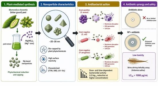
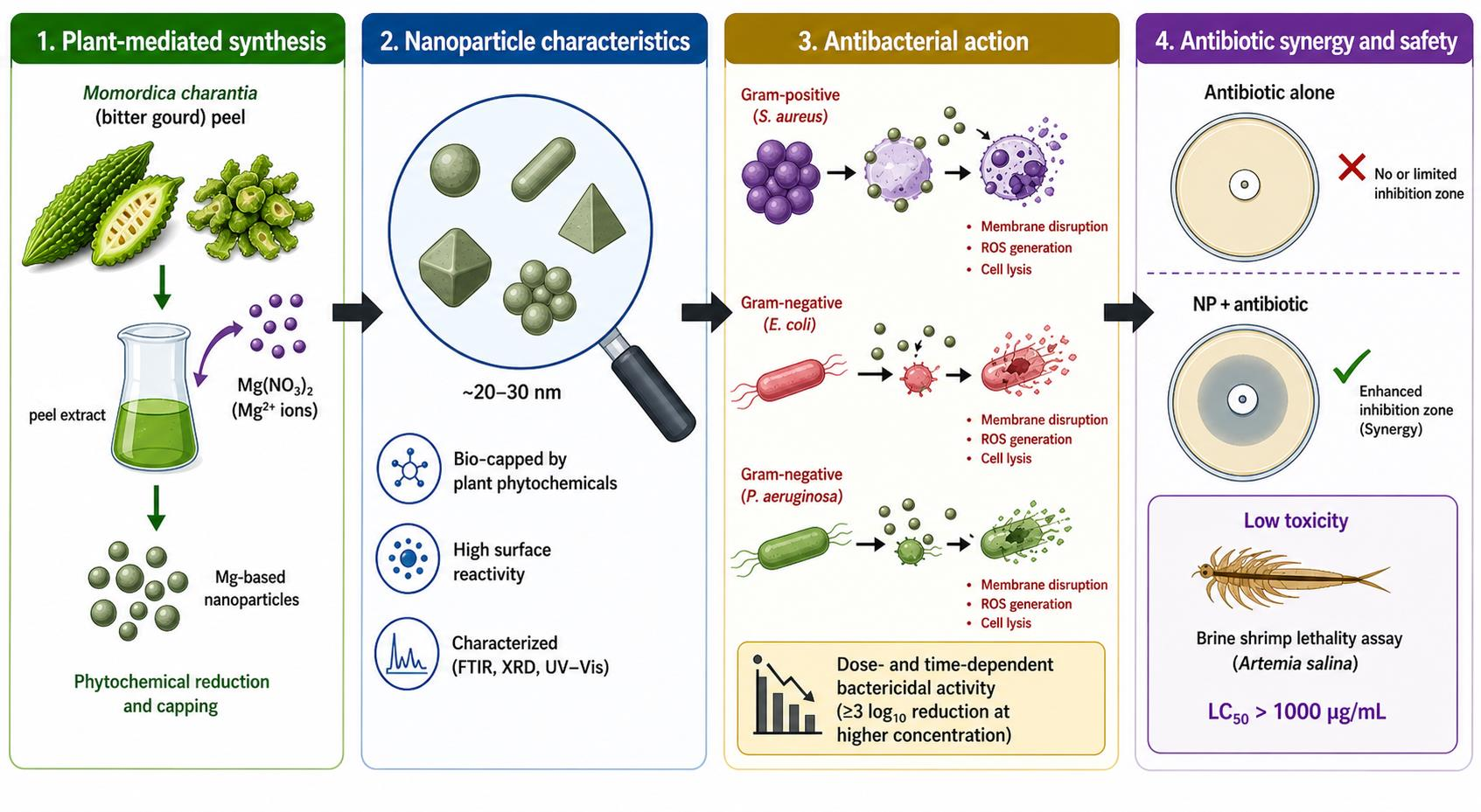
The current study aims to fill the existing gaps in the evaluation of nanoparticle-based antimicrobials using qualitative, quantitative, and safety assessments. Although qualitative zone of inhibition methods have been commonly used in previous studies, such methods fail to offer accurate information on antimicrobial and bactericidal activity. Thus, alongside traditional disc diffusion assays, this study includes minimum inhibitory concentration (MIC), minimum bactericidal concentration (MBC) and growth rate analyses to allow a more quantitative assessment of antimicrobial activity. While magnesium nitrate nanoparticles [Mg(NO3)2-NPs] hold the promise of effective antimicrobials, little is known about their toxicity, even in preliminary biological systems. To overcome this concern, the current study evaluated Mg(NO3)2-NPs toxicity using the Brine Shrimp Lethality test as a fast and efficient model for biocompatibility. Therefore, the present study intends to (i) prepare Mg(NO3)2-NPs using M. charantia peel extract, (ii) assess its antibacterial activity against MDR bacterial pathogens using qualitative and quantitative approaches, (iii) determine its synergism with conventional antibiotics, and (iv) assess its preliminary brine shrimp toxicity. This interdisciplinary approach gives more insight into the medicinal properties and safety of eco-friendly nanoparticles.
2. Materials and Methods
2.1. Collection and Identification of MDR Bacterial Isolates
Previously identified bacterial isolates, such as
E. coli,
S. aureus and
Pseudomonas spp., were collected from the culture bank of the Microbiology Research Laboratory (MRL), Kohat University of Science and Technology (KUST), Kohat. The isolates were reactivated and sub-cultured on nutrient agar medium before being used. Bacterial identification was confirmed by Gram staining and standard biochemical tests (oxidase, catalase, indole, citrate and triple sugar iron (TSI) tests) following standard microbiological techniques [
17]. The MDR nature of the isolates was confirmed by antibiotic susceptibility tests, as per the guidelines of the Clinical and Laboratory Standards Institute [
18].
2.2. Plant Material and Extract Preparation
The fruits of M. charantia were obtained from the local market in Jand, District Attock, Punjab, Pakistan. The fruit was washed with distilled water, sliced and dried in the shade for 5–6 days. The dried powder was pulverized into a fine powder with the help of a blender and stored in airtight containers at room temperature for future use.
2.2.1. Aqueous Extract of M. charantia
The aqueous extract of
M. charantia was obtained by adding 25 g of powdered plant to 100 mL of distilled water. The suspension was heated for 1 h at approximately 80–90 °C with frequent shaking for the extraction of its therapeutic components. It was cooled to room temperature and filtered using Whatman No. 1 filter paper. This was then evaporated to semi-concentrated form (50 °C) using a rotary evaporator and was used for the preparation of nanoparticles and antibacterial activity [
19,
20].
2.2.2. Methanolic Extract of M. charantia
The methanolic extract of
M. charantia was prepared by extracting 25 g of powdered plant sample with 150 mL of methanol and left at room temperature for 24 h with occasional shaking. The mixture was filtered through Whatman filter paper, and the solvent was evaporated in a rotary evaporator under reduced pressure (40 °C) to get a concentrated crude extract. The concentrated extract was dried at 50 °C in a hot air oven to remove traces of solvent and stored for analysis [
19,
20].
2.3. Phytochemical Analysis of M. charantia Extracts
The qualitative phytochemical analysis of aqueous and methanolic extracts was carried out to identify the major bioactive compounds present in them. Different bioactive compounds such as saponins, phenolics, flavonoids, tannins, and terpenoids were screened using standard colourimetric and precipitation-based methods as described by Harith et al. [
21] by looking for color change and/or froth formation.
2.4. Antibacterial Activity of M. charantia Extracts
Antibacterial activity of
M. charantia was tested by the agar well diffusion method [
22]. Bacterial suspensions subjected to standard turbidity were spread on the surface of Mueller–Hinton agar (MHA) plates. Wells of approximately 6 mm diameter were bored in the agar, and 80 µL of aqueous and methanolic extracts were added to each well. Dimethyl sulfoxide (DMSO) was used as a negative control, and antibiotic discs as a positive control. The plates were incubated at 37 °C for 24 h, and zones of inhibition were measured in millimetres.
2.5. Green Synthesis of Mg(NO3)2-NPs
Mg(NO3)2-NPs were prepared by a green synthesis method using M. charantia peel extract. About 2 g of magnesium nitrate was mixed with 100 mL of deionized water. The extract was added slowly while stirring with a magnetic stirrer. The pH of the reaction solution was maintained at around 10 by the gradual addition of NaOH. The mixture was then stirred for 2 h until the appearance of a whitish-yellow precipitate, which indicated the formation of nanoparticles. The solution was then centrifuged at 4000 rpm for 15 min, and the precipitate was washed with distilled water. The nanoparticles were then dried at 80 °C and stored for future use.
2.6. Characterization of Mg(NO3)2-NPs
Scanning electron microscopy (SEM), energy-dispersive X-ray spectroscopy (EDAX), X-ray diffraction (XRD), and Fourier transform infrared spectroscopy (FTIR) were carried out at the Centralized Resource Laboratory (CRL) and National Centre of Excellence in Geology (NCEG), University of Peshawar, to characterize the synthesized Mg(NO3)2-NPs. For SEM analysis, a small amount of dried nanoparticle powder was spread uniformly on a carbon-coated aluminum stub. The stubs were gold-coated using a vacuum sputter coater to improve sample conductivity and prevent charging artefacts. The sputter-coated samples were then analyzed by SEM at accelerating voltages ranging from 10 to 20 kV. Images were taken to study the structure, size and dispersion of the nanoparticles.
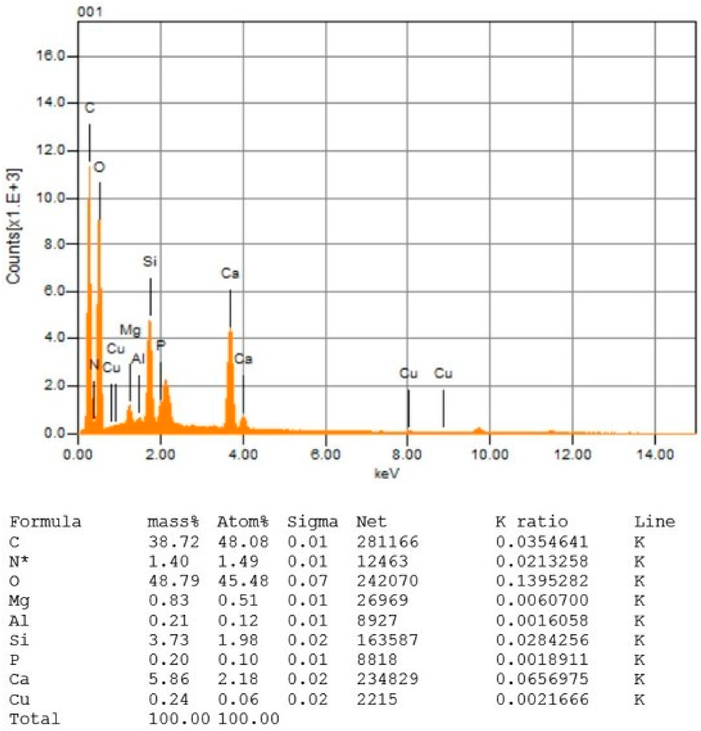
EDAX was also conducted along with SEM to identify the elemental composition of the samples. In SEM, the bombardment of the sample by the incident electron beam resulted in the emission of X-rays. These emissions were captured and used to confirm the presence of magnesium and other elements, and the weight percentage of these elements was determined from the EDAX spectra.
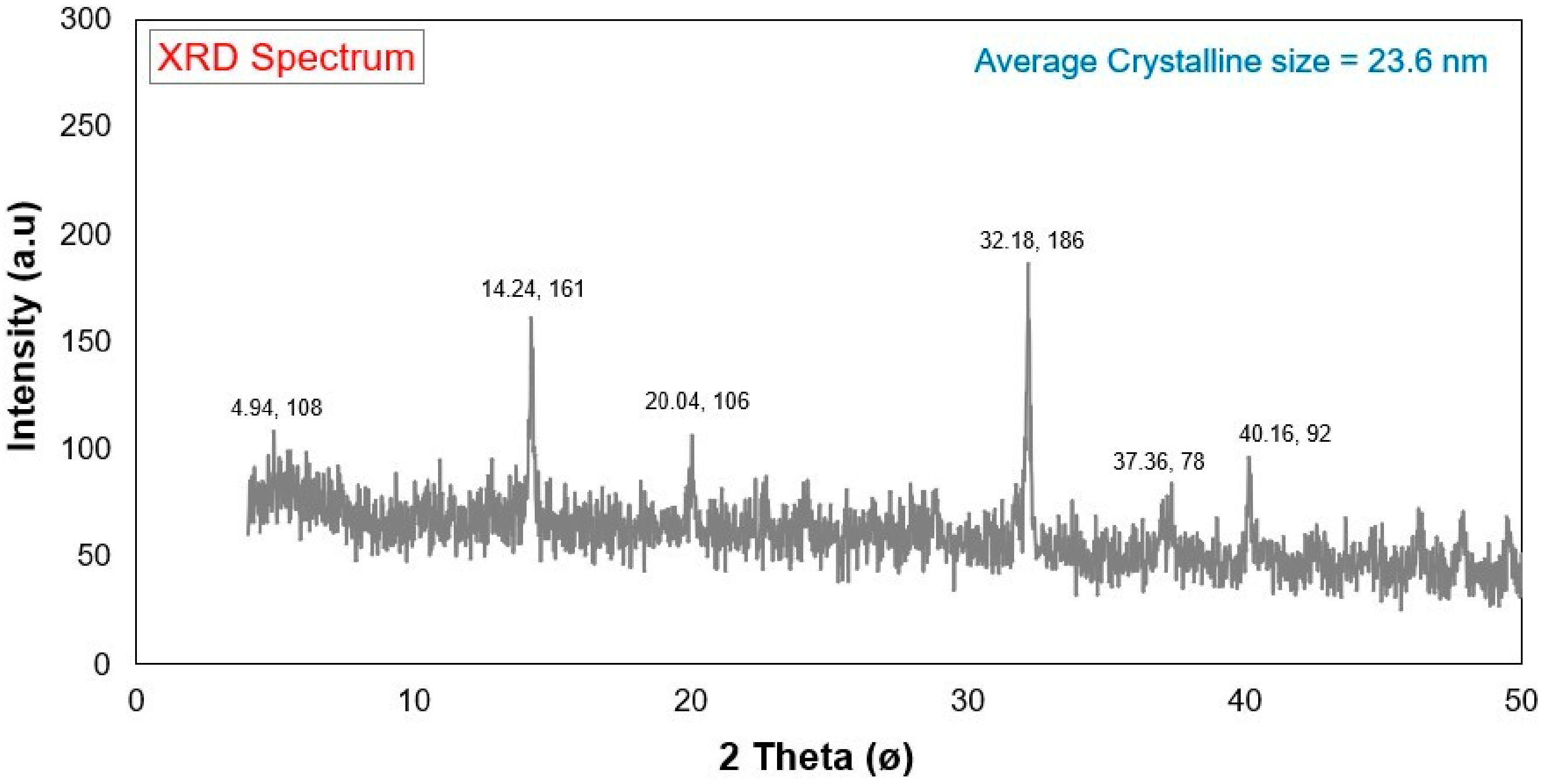
In the XRD analysis, the dried nanoparticle sample was ground and prepared on a sample holder with a flat surface. Further, an X-ray diffractometer with Cu-Kα radiation (λ = 1.5406 Å) was used. The X-ray diffraction data were collected over a 2θ range of around 10° to 80° at a rate of around 2°/min. The diffraction peaks were matched with reference patterns of the Joint Committee on Powder Diffraction Standards (JCPDS database) to establish the crystallinity, purity and average size of the nanoparticles.
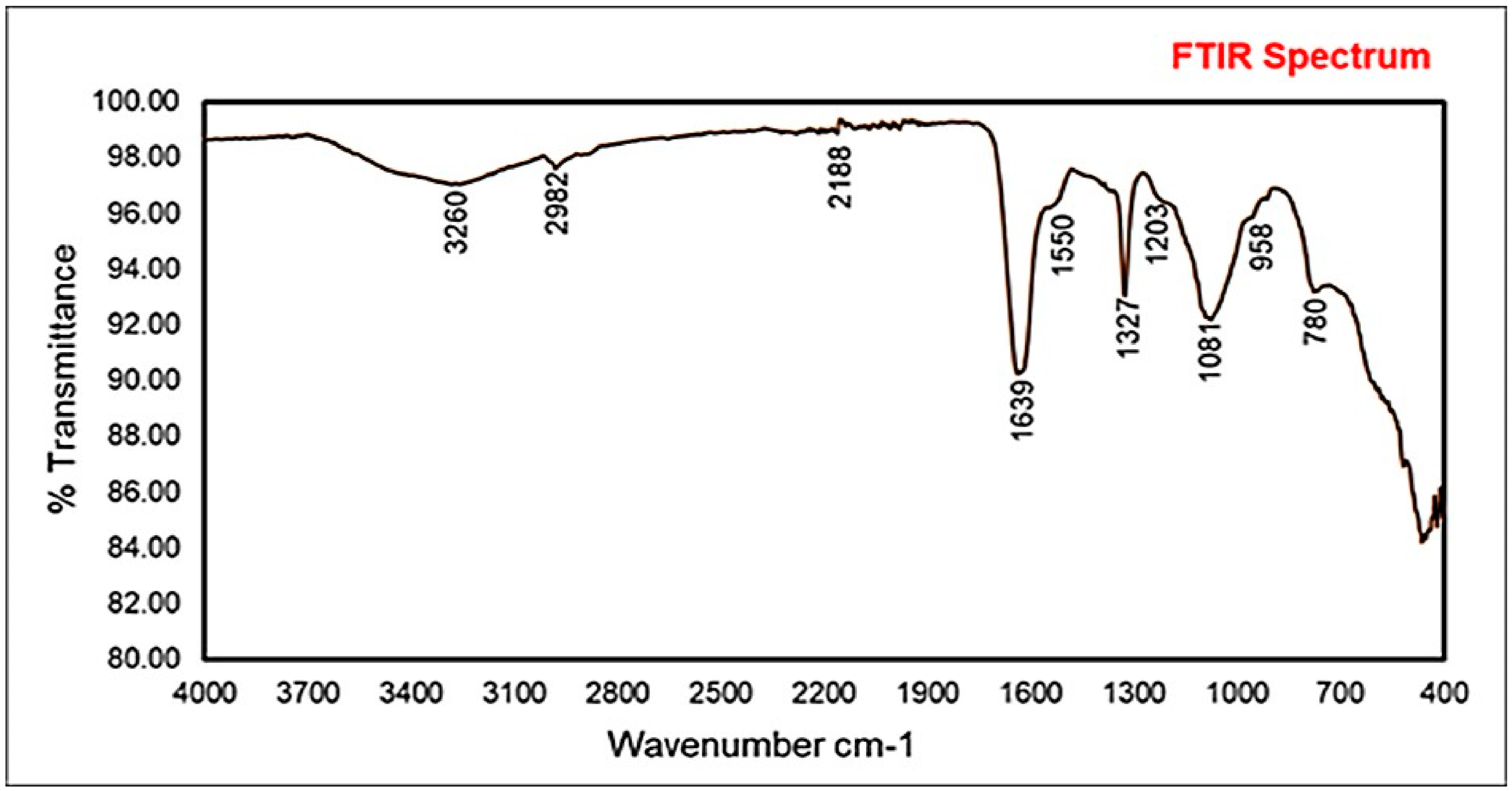
FTIR analysis was employed to study the functional groups on the nanoparticle surface and the involvement of biomolecules of the plant extract in the synthesis and stability of the nanoparticles. The spectra were recorded using the KBr pellet method, where a small quantity of the dried nanoparticles was mixed with KBr powder and pressed into a pellet. The measurements were taken between 4000 and 400 cm−1. The absorption peaks were used to determine the presence of functional groups responsible for the reduction and capping of nanoparticles.
2.7. Antibiotics Used in the Study
Antibiotics used in this study were erythromycin (5 µg/disc), ceftazidime (30 µg/disc), penicillin (15 µg/disc) and oxacillin (5 µg/disc). Antibiotic susceptibility was tested following the CLSI [
18] guidelines.
2.8. Preparation of Mg(NO3)2-NPs Coated Filter Paper Discs
Sterile filter paper discs (about 6 mm diameter) were made from Whatman No. 1 filter paper by autoclaving at 121 °C for 15 min. Different concentrations (5–25 mg/mL) of Mg(NO3)2-NPs were prepared in DMSO. The discs were soaked in the suspension and dried in a sterile environment at 80 °C.
2.9. Antibacterial Activity of Mg(NO3)2-NPs Coated Filter Discs Against MDR Bacteria
The antibacterial effects of the Mg(NO
3)
2-NPs-coated filter discs against MDR bacteria were assessed by the Kirby–Bauer disc diffusion method as recommended by CLSI [
18]. MHA plates were inoculated with different MDR bacterial strains, and filter paper discs coated with nanoparticles were placed on them. The plates were incubated at 37 °C for 24 h. The diameter of the zone of inhibition was measured in millimetres. DMSO was used as the negative control, and antibiotic discs as the positive controls.
2.10. Determination of MIC and MBC of Mg(NO3)2-NPs
The MIC of Mg(NO3)2-NPs was measured by the broth microdilution method in sterile 96-well micro-titer plates according to the principles of antimicrobial susceptibility testing. In this assay, Mg(NO3)2-NPs were dispersed in sterile Mueller–Hinton broth and twofold diluted to get concentrations of 25, 12.5, 6.25, 3.125, 1.56, 0.78, 0.39 and 0.19 mg/mL. Fresh overnight cultures of E. coli, S. aureus and Pseudomonas spp. at approximately 0.5 McFarland standard were diluted to a final inoculum concentration of 5 × 10⁵ CFU/mL in each well. Broth and bacterial inoculum were used as growth control; broth only was used as sterility control, while DMSO was used as solvent control.
The plates were incubated at 37 °C for 24 h. The lowest concentration of nanoparticles at which no visible growth was observed was taken as the MIC. The MBC was determined by taking 10 µL of samples from wells with no visible growth and subculturing them on MHA plates, followed by incubation at 37 °C for 24 h. The MBC was recorded as having the lowest concentration with no colony formation on MHA plates. Both MIC and MBC experiments were conducted in triplicate.
2.11. Growth Kinetics Analysis of Mg(NO3)2-NPs
To assess the bactericidal and bacteriostatic activity of Mg(NO3)2-NPs against the MDR bacteria (E. coli, S. aureus and Pseudomonas spp.), the time-kill assay was conducted. Bacterial suspension was prepared in Mueller–Hinton broth and standardized to 106 CFU/mL. The concentrations of the Mg(NO3)2-NPs used for each suspension were equal to 0.5×, 1×, and 2× MIC determined by broth microdilution according to CLSI guidelines. Cultures not treated but incubated under the same conditions were used as growth controls. All tubes were cultured for 24 h at 37 °C on an orbital shaker with shaking (150 rpm). A total of 100 µL samples were aseptically collected at regular intervals (0, 2, 4, 6, 8, 12 and 24 h), serially diluted in sterile phosphate-buffered saline (pH 7.4), and spot-plated on MHA plates in triplicate. After 18 to 24 h of incubation, colonies were counted and reported as log10 (CFU/mL). Bactericidal activity was a reduction in viable cell count of ≥3 log10 compared to the initial inoculum. Each experiment was repeated three times (n = 3), and results are presented as mean ± standard deviation (SD). The statistical analysis was carried out by applying one-way ANOVA with Tukey’s post hoc test (IBM SPSS Statistics, version 25) and a p-value < 0.05 was regarded as statistically significant.
2.12. Preparation of Nanoparticle-Coated Antibiotic Discs
A solution of 0.25 mg/mL of Mg(NO3)2-NPs was prepared in distilled water. A volume of 5 mL of the nanoparticle suspension was poured onto antibiotic discs and dried at 80 °C to produce nanoparticle-loaded antibiotic discs.
2.13. Comparative Antibacterial Assay for Mg(NO3)2-NPs Coated and Uncoated Antibiotics
The standard disc diffusion method was used to assess the antibacterial activity of nanoparticle-coated and uncoated antibiotic discs [
18]. MHA plates were inoculated with bacteria, and discs were placed on the plates for 24 h at 37 °C. The diameter of the zones of inhibition was determined and compared to evaluate the activity of the antibiotic.
2.14. Toxicity Assessment Using Brine Shrimp Lethality Assay (BSLA)
The Brine Shrimp Lethality Assay (BSLA) was used to evaluate the preliminary toxicity of Mg(NO
3)
2-NPs. The eggs of
Artemia salina were incubated in artificial seawater for 24–48 h with continuous light and aeration. The motile nauplii were transferred to test tube plates with 5 mL of artificial seawater. Mg(NO
3)
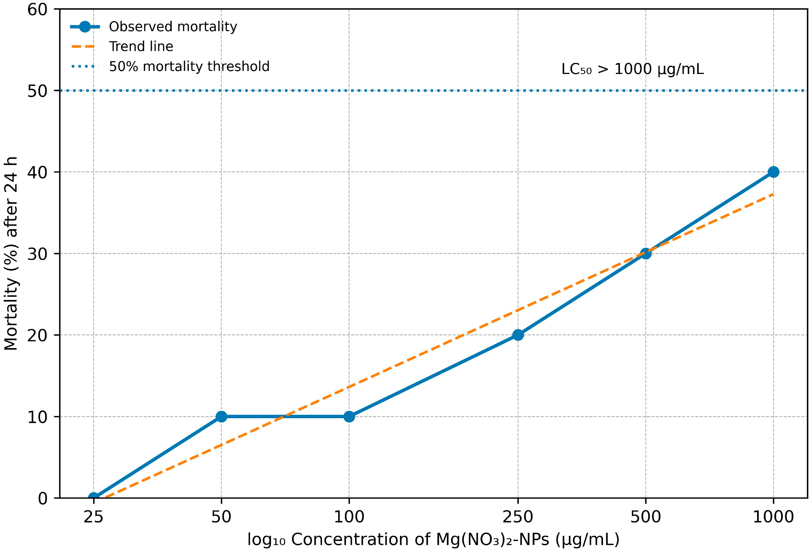
2-NPs were tested at concentrations of 25, 50, 100, 250, 500, and 1000 µg/mL. Five nauplii were added to each concentration. Artificial seawater was used as a negative control, and potassium dichromate was used as a positive control. Following 24 h incubation at room temperature, the mortality of nauplii was recorded. The percentage mortality was calculated as:
The median lethal concentration (LC50) was determined by regression/probit analysis. The sample was considered practically non-toxic if the LC50 was greater than 1000 µg/mL in the preliminary screening procedure. The lethality of brine shrimp, with the calculation of LC50 values, was used as a rapid toxicity screening and the values were calculated based on mortality against log concentration after 24 h exposure.
2.15. Statistical Analysis
Each experiment was repeated three times, and the results were presented as mean ± standard error (SE). Data were analyzed using one-way analysis of variance (ANOVA) using statistical software (IBM SPSS Statistics, version 25). Data with p-values less than 0.05 were considered statistically significant data.
4. Discussion
The MDR phenotypes of
E. coli,
S. aureus and
Pseudomonas spp. in this study are like the well-described resistance mechanisms of these bacteria, which include drug target modification, drug inactivation, efflux and biofilm tolerance. These processes decrease the concentration of drugs and their efficacy in the cell [
23]. All strains were completely resistant to multiple antibiotics, which indicates a very selective environment, and this is consistent with previous studies reporting the rapid dissemination of MDR phenotypes through horizontal gene transfer and plasmid transfer [
24].
Based on the phytochemical study of
M. charantia extracts, tannins, flavonoids and terpenoids were found in the methanolic extract, while phenolics and saponins were present in the aqueous extract. The selective extraction is due to the polarity and solubility of secondary metabolites. Flavonoids and phenolic compounds are known to have antimicrobial activity by disrupting cell membranes, chelating metal ions, blocking nucleic acid synthesis and disrupting energy production. The lipophilic properties of terpenoids allow them to penetrate the cell membrane, disrupting its integrity, leading to the leakage of cell contents. Previous studies have shown these effects to be antibacterial [
25]. The chemical constituents in
M. charantia peel extract, identified from qualitative screening (
Table 1), specifically flavonoids, phenolics, tannins and terpenoids, have dual functional activities as both reducing and stabilizing agents in the green synthesis of Mg(NO
3)
2-NPs, which has been directly confirmed and explained by FTIR analysis (
Figure 3,
Table S2). Specifically, the hydroxyl (O-H) and phenolic groups identified in the FTIR directly correlated with the phenolic compounds identified in the phytochemical screening and act as electron donors to reduce the Mg
2+ ion to produce metallic nanoparticles in the synthesis reaction. Amine and protein-derived groups were detected at 2982 cm
−1, which are related to terpenoids and alkaloids and play a role in nucleation and initiation of nanoparticle synthesis. These same phytochemical components contribute to the stabilization of nanoparticles in several ways: hydroxyl and phenolic functionalities form hydrogen bonding with the surfaces of the nanoparticles; aromatic functional groups stabilize the nanoparticles through π-π stacking interactions; carboxylic acids (1327 cm
−1) and amine (2982 cm
−1) functional groups provide electrostatic stabilization; and C-O stretching vibrations (1081 cm
−1) indicate the presence of ether linkages capable of coordinating with metal ions. Carboxylic acids and fluorinated compounds (1203 cm
−1) detected in the FTIR are used as surface capping agents in the synthesis of nanoparticles, which build steric barriers to avoid aggregation and coalescence of nanoparticles. The role of the phytochemical-mediated stabilization mechanism is one of the reasons that plant-synthesized nanoparticles have superior biocompatibility to chemically synthesized nanoparticles, since the organic coating formed through plant phytochemicals prevents the interaction between the nanoparticles and cells without losing antimicrobial activity. The strong correlation of phytochemical classes with the identified functional groups (FTIR,
Figure 3) and the resulting properties of the nanoparticles indicates that
M. charantia peel extract is a multifunctional biological matrix that can simultaneously reduce the metal ions, initiate the formation of nanoparticles, stabilize metal nanoparticles from aggregation, and provide biocompatibility, which is the most comprehensive mechanism for green synthesis compared to a single-component chemical reducing agent.
The antibacterial activity of
M. charantia extracts against MDR bacteria, with higher activity of methanolic extracts, agrees with the various phytochemicals extracted by methanol. The susceptibility of
S. aureus to antimicrobial agents is different than that of the Gram-negative bacteria, which may be due to varying cell wall properties. Lipopolysaccharides constitute the outer membrane in Gram-negative bacteria, which hinders penetration of active molecules, but are not seen in Gram-positive bacteria. These results are consistent with the results of Jindal and Kaushalendra [
26].
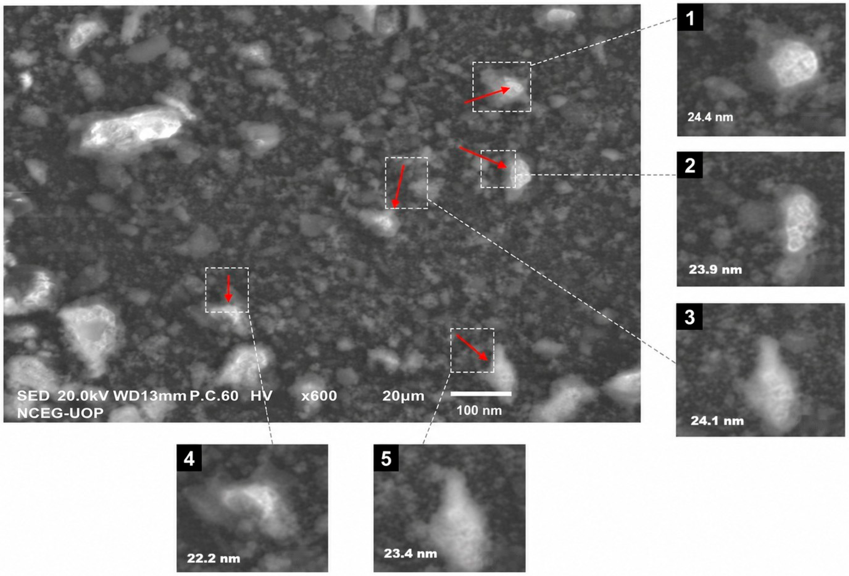
This study reveals the capacity of M. charantia peel extract to serve as a reducing, stabilizing and capping agent in the biosynthesis of Mg(NO3)2-NPs, indicating that plant phytochemicals play an active role in this process. The SEM micrograph showed a fine granular matrix with partially agglomerated and polydispersed nanoparticles distributed in it. The morphology of the particles deviated from a perfect spherical shape, with a more irregular and multi-faceted form, suggesting anisotropic nucleation and growth in the plant-mediated synthesis.
The enlarged SEM areas gave representative particle sizes in the range of 22–25 nm corresponding to the XRD-determined average crystallite size of 23.6 nm. This agreement validates that the synthesized Mg(NO
3)
2-NPs are at the nanoscale level. The variation in size and irregular morphology could be due to the preferential adsorption of phytochemicals like phenolic compounds, flavonoids and proteins on certain crystal planes, which would control directional growth. However, the observed localized agglomeration in the SEM image might be attributed to the high surface energy of nanoscale particles and interactions between the surfaces coated with the phytochemicals. The plant-mediated magnesium-based nanoparticles with similar morphological variations have been reported by Ahmad et al. [
27] and Gatou et al. [
28].
EDAX analysis has confirmed that nanoparticles are mainly composed of magnesium, nitrogen, carbon and oxygen. The presence of carbon and oxygen also indicates that the nanoparticles are capped by organic molecules. These organic capping agents stabilize the nanoparticles due to steric effects. This observation has been reported for the biosynthesis of nanoparticles using plant sources [
29], where organic capping impacts nanoparticle stability and characteristics.
The crystallinity of nanoparticles, as confirmed by XRD (average crystallite size 23.6 nm), is an important factor in explaining the physical and biological behaviour of nanoparticles. Smaller crystallites have a higher surface/volume, thus a higher surface reactivity, and interaction with microorganisms. The smaller the size of the nanoparticles, the greater their penetration efficiency through the cell wall and membrane and the better their antimicrobial properties. The study reported by Ramezani-Farani et al. [
29] indicates that the crystallite size of the magnesium nanoparticles is quite similar, and the reactivity is increased, which reflects the present study. Furthermore, the average crystallite size of 23.6 nm, obtained from XRD data, is quantitatively validated with the visual inspection of the nanoscale dimensions in the SEM micrograph. This XRD analysis is corroborated by the claimed particle size range of 1–100 nm based on SEM image visualization, and the calculated average crystallite size of 23.6 nm is within the wider range of the SEM visualization, confirming the nanoscale nature of the synthesized nanoparticles. The SEM morphological characterization (qualitative visual range of 1–100 nm) and the XRD crystallite sizing (quantitative average of 23.6 nm) offer complementary and comprehensive evidence for the successful synthesis of nanoparticles. Both qualitative SEM and quantitative XRD are used in this way as the standard approach for characterizing nanomaterials, and the Debye–Scherrer method is a well-established and internationally recognized method for determining dimensions of nanoparticles [
29,
30]. As a result, our XRD-based crystallographic analysis and SEM-based visual observations validate that Mg(NO
3)
2-NPs are indeed nanoscale.
FTIR spectra showed the presence of functional groups (hydroxyl, amine and aromatic groups), which implies the involvement of plant biomolecules in the formation of nanoparticles. They are groups that can donate electrons to reduce the Mg
2+ ions and coordinate with the nanoparticles through coordination and adsorption. The role played by these functional groups in the dispersion of nanoparticles and in preventing nanoparticle aggregation to conserve biological activity is of great importance. Similar FTIR patterns have been reported by Al-Harbi et al. [
30], which highlights the stabilizing properties of the phytochemicals on the nanoparticles.
The dose-dependent antibacterial activity of Mg(NO
3)
2-NPs suggests a concentration-dependent effect. The antibacterial action of magnesium nanoparticles may include disruption of the cell membrane, ROS formation, and interaction with biomolecules inside the cells, such as proteins and DNA. The greater susceptibility of
S. aureus to Gram-negative bacteria reflects the weak cell wall of Gram-positive bacteria. Increasing nanoparticle concentration leads to higher surface interaction with bacteria, which results in cell membrane damage and cell death [
31].
MIC and MBC are quantitative tests that are useful to supplement diffusion tests. In the present study, the MIC and MBC of Mg(NO
3)
2-NPs against
S. aureus (Gram-positive) were lower than those of Gram-negative bacteria, which indicates higher sensitivity of Gram-positive bacteria. This finding is also in line with the reports of higher sensitivity of Gram-positive bacteria because of the absence of an outer membrane and hence higher penetration of nanoparticles [
29,
32]. In contrast, Gram-negative bacteria such as Pseudomonas spp. have an outer membrane (lipopolysaccharides) and efflux pumps, which reduce antibiotic penetration and enhance resistance [
24]. The MBC/MIC ratio was also indicative of bactericidal activity, especially against the tested bacteria
S. aureus and
E. coli, and the MBC was two times lower than the MIC observed. Other studies have found similar bactericidal effects on magnesium nanoparticles, with greater reactivity and small size of the nanoparticles making it easier to interact with bacterial surfaces and inside bacterial cells [
29]. The study validates the antibacterial ability of Mg(NO
3)
2-NPs, in addition to the studies on inhibition and proposes their use as an antimicrobial agent.
Bacterial growth kinetics give important information for the dynamic effect of antimicrobials. The present study showed that Mg(NO
3)
2-NPs exhibited a definite concentration-dependent antibacterial response against the MDR bacterial isolates, with progressive decreases in viable cell counts noted as the concentration of the Mg(NO
3)
2-NPs increased from 0.5× to 2× MIC. All isolates showed a significantly delayed onset of the exponential growth phase (
p < 0.05) and gradual bacteriostatic pressure (
p < 0.05) at sub-inhibitory concentrations (0.5× MIC), which represented a significant antibacterial pressure but not lethal effects.
S. aureus was the most susceptible, producing near-total growth inhibition within 8–12 h (
p < 0.001);
E. coli gave a progressive growth inhibition that was bacteriostatic, and
Pseudomonas spp. gave only partial growth inhibition and continued to grow through the 24 h period. A decrease of ≥3 log
10 in CFU/mL, reflecting bactericidal activity, was achieved at 2x MIC;
S. aureus had the lowest MIC, followed by
E. coli (
p < 0.001 for both strains) and
Pseudomonas spp., which had the highest MIC. Similar time-kill curves have been found with metal-based nanoparticles, where a higher concentration leads to a more rapid killing of bacteria because of the increased damage to the bacterial membrane and induction of oxidative stress [
32]. The inability of Mg(NO
3)
2-NPs to attain bactericidal concentrations against
Pseudomonas spp. at 2x MIC, show a log
10 reduction that never exceeded the bactericidal threshold over 24 h. The relative tolerance of
Pseudomonas spp. to Mg(NO
3)
2-NPs is due to several inter-related intrinsic resistance mechanisms inherent in Gram-negative bacteria. The higher MIC (25 mg/mL) than that of Gram-positive
S. aureus (6.25 mg/mL) may be attributed to the lipopolysaccharide-rich outer membrane of Gram-negative
Pseudomonas spp., which acts as a selective permeability barrier that slows down the diffusion and penetration of nanoparticles to the inner cell membrane [
24]. Second,
Pseudomonas spp. constitutively expresses resistance–nodulation–division (RND) type efflux pumps (
MexAB-OprM,
MexCD-OprJ) and ATP-binding cassette (ABC) transporters that actively extrude antimicrobial agents and potentially nanoparticles from the cytoplasm, reducing intracellular accumulation [
24]. The time-kill kinetics data we compiled (
Table 5) further support this active resistance mechanism:
S. aureus was able to achieve near-complete growth inhibition within 8–12 h at 1× MIC, while
Pseudomonas spp. showed only partial growth inhibition with subsequent bacterial proliferation during the 24 h exposure, which is indicative of continued efflux pump activity to overcome the stress induced by exposure to the nanoparticles. Third, under antimicrobial stress,
Pseudomonas spp. can adaptively change the composition of LPS and the outer membrane charge density, which decreases the adhesion of nanoparticles and their uptake by cells [
24].
Nanoparticles are active against bacteria through different mechanisms, including the production of ROS, denaturation of proteins and DNA replication, leading to a progressive decrease in cell viability [
10]. Therefore, the growth kinetics results not only corroborate the MIC/MBC results but also reflect the dynamic interaction between nanoparticles and bacteria, including time- and concentration-dependent killing.
The other important aspect of this study is the remarkable improvement in the efficacy of antibiotics when coated with Mg(NO
3)
2-NPs. This synergistic effect can be attributed to the ability of nanoparticles to enhance the delivery of antibiotics and to bypass resistance mechanisms. Nanoparticles may increase the permeability of cell membranes, inhibit efflux pumps and penetrate biofilms, facilitating the entry of antibiotics into the cell. In addition, nanoparticles can be used as a delivery system for antibiotics. This combination effect has been reported by Salman et al. [
31] and Das et al. [
32], where nanoparticles and antibiotics were found to have a synergistic effect on the antibacterial activity against MDR bacteria. In addition, the
M. charantia phytochemicals, including triterpenes, alkaloids and phenolics, may have a synergistic effect on the antibacterial effect of nanoparticles and antibiotics [
14,
33]. The synergistic effect of phytochemicals, nanoparticles and antibiotics is a multi-targeted approach to fight drug resistance.
It is necessary to determine the toxicity of nanoparticle-based antimicrobials to evaluate their potential biomedical applications. In the present study, Mg(NO
3)
2-NPs were nontoxic in the BSLA with an LC
50 of over 1000 µg/mL, indicating good preliminary biocompatibility. BSLA has been extensively used as a rapid, cost-effective model for the assessment of acute toxicity of nanoparticles and has been shown to be a good predictor of cytotoxicity to higher organisms [
34]. Recently, green-synthesized nanoparticles have shown low toxicity in the BSLA, and it has been concluded that the phytochemicals derived from the plant used for nanoparticle synthesis are responsible for the biocompatibility of the nanoparticles [
35]. These natural surfactants protect against interactions between nanoparticles and cells and subsequent biological activity. In addition, nanoparticle toxicity has been reported to increase with an increase in concentration in
A. salina, with higher mortality due to increased nanoparticle uptake and ROS generation [
36]. This dose-dependent mortality at higher concentrations observed in the current study confirms the toxicity of Mg(NO
3)
2-NPs. However, low mortality at low concentrations suggests that the nanoparticles may selectively exert their antimicrobial property without being toxic. While BSLA has multiple benefits, it only provides preliminary toxicity data and further studies in mammalian cells and in vivo models must be conducted to establish the safety and therapeutic potential of these nanoparticles [
37].
This study has some limitations. The in vitro analysis of antibacterial activity, including the MIC, MBC and growth kinetics, provides a quantitative and comprehensive result of the antibacterial effects of Mg(NO3)2-NPs, but it may not fully predict the response in the complex biological system. Growth kinetics revealed clear-time and dose-dependent killing effects, while more advanced mechanistic studies, such as ROS, membrane permeability and gene expression profiling, were required to elucidate the mechanism of action. The BSLA toxicity analysis indicated low toxicity and acceptable biocompatibility (at low concentrations) but only gives a preliminary estimate of the toxicity and may not predict toxicity in higher animals. Thus, additional studies with mammalian cell lines, in vivo studies and advanced mechanistic and toxicity studies are needed to confirm the clinical and therapeutic utility of Mg(NO3)2-NPs as an antibacterial and an adjuvant to antibiotics.




 ,
,