Next-Generation SERS Probes: Engineering Hotspots, Intelligent Molecular Targeting, and AI-Driven Spectral Analysis for Emerging Applications

 , , ,
, , ,  , and
, and 
Abstract
1. Introduction: Fundamentals of SERS Enhancement
1.1. Electromagnetic vs. Chemical Enhancement
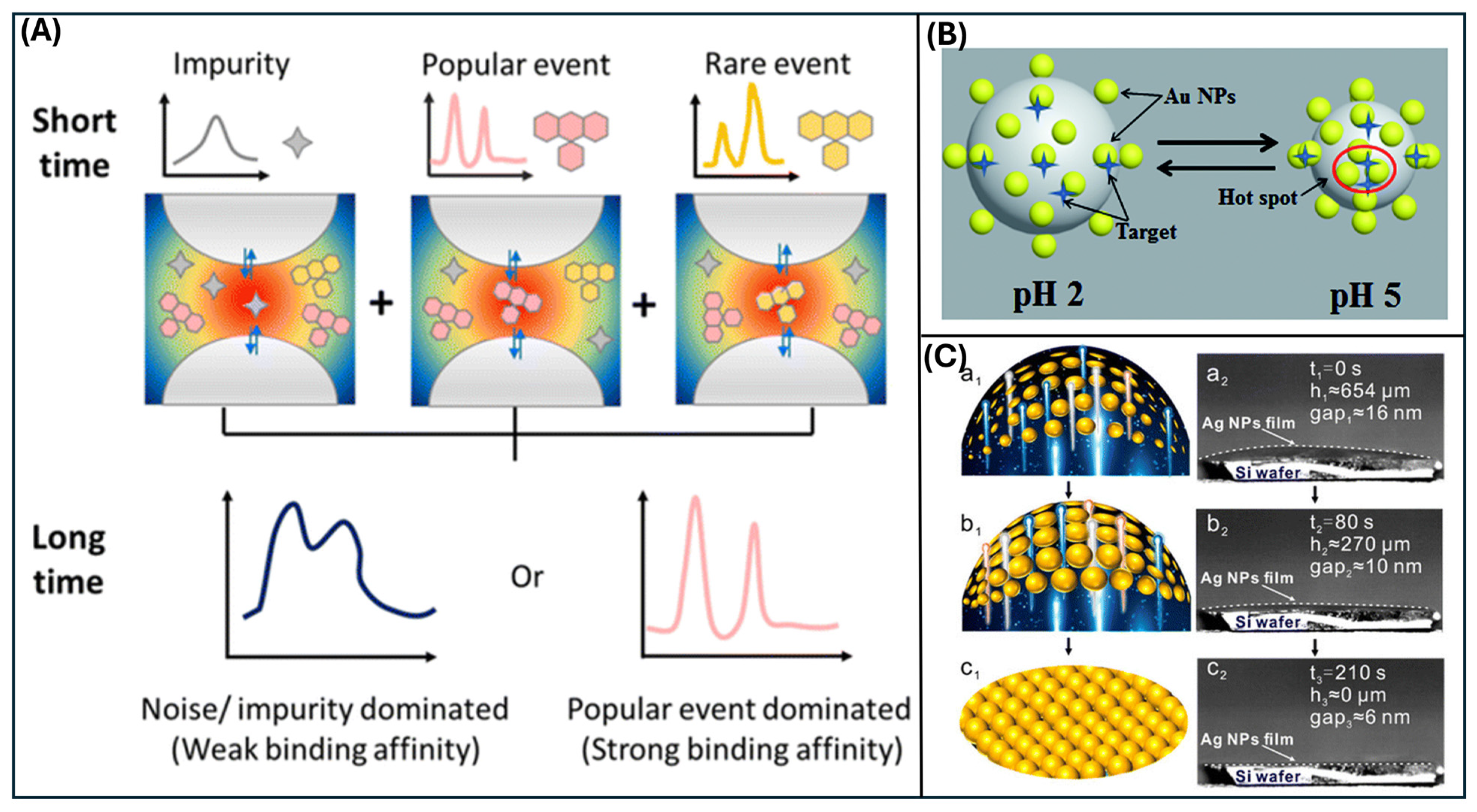
1.2. Spatial vs. Temporal Resolution
2. Fabrication and Microstructure Control of SERS Probes
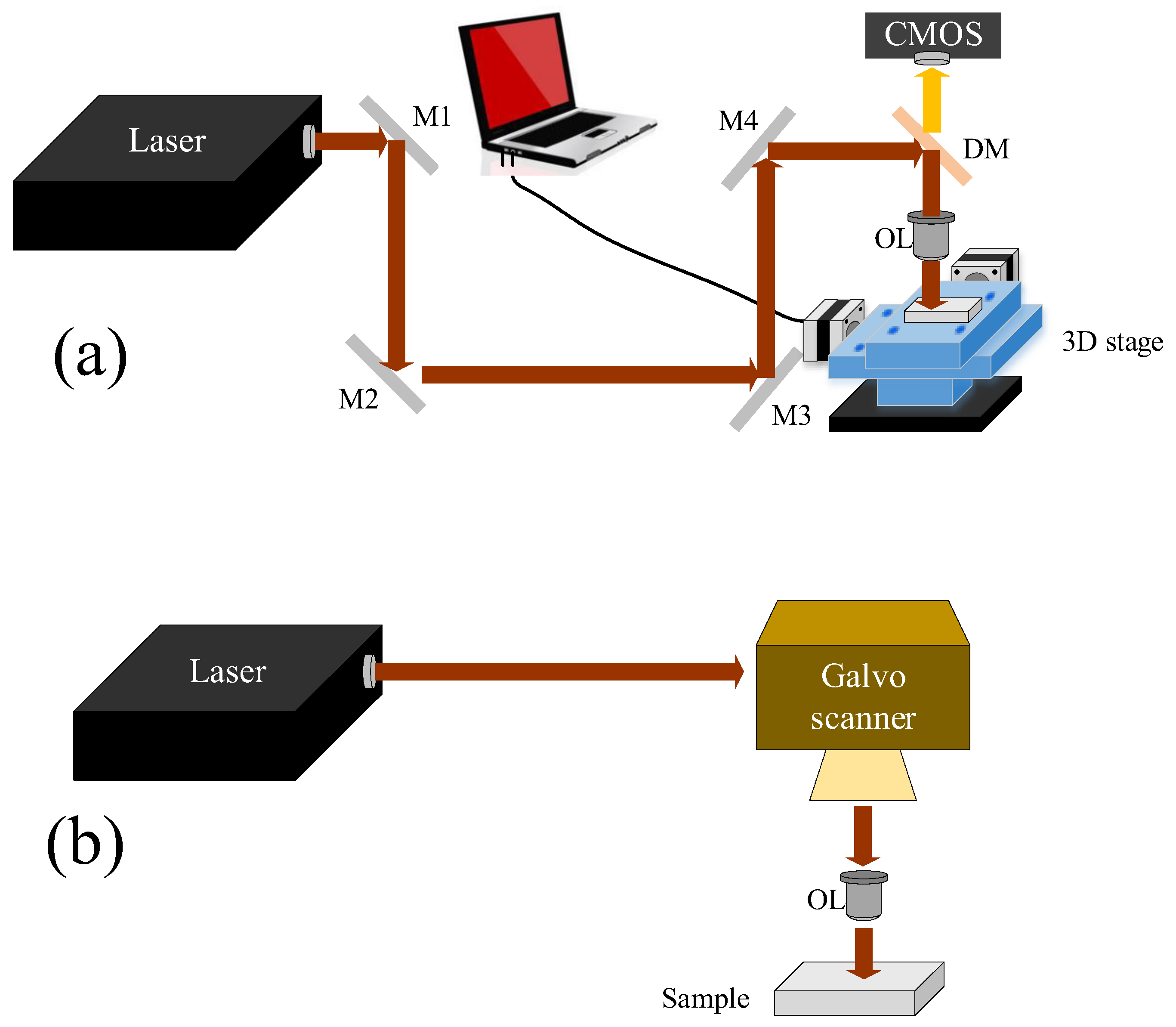
2.1. Laser Ablation in Liquids
2.1.1. Principles of Pulsed Laser Ablation in Liquids
- (1)
- Ablation of a second target immersed in colloidal suspension prepared by ablation of a first target;
- (2)
- Mixing of several colloidal solutions prepared by ablation of several targets, followed by further laser treatment;
- (3)
- Ablation of a multi-element target (e.g., metallic alloy);
- (4)
- Ablation of a solid target immersed in salt-containing (e.g., AgNO3; HAuCl4 etc.) solution.

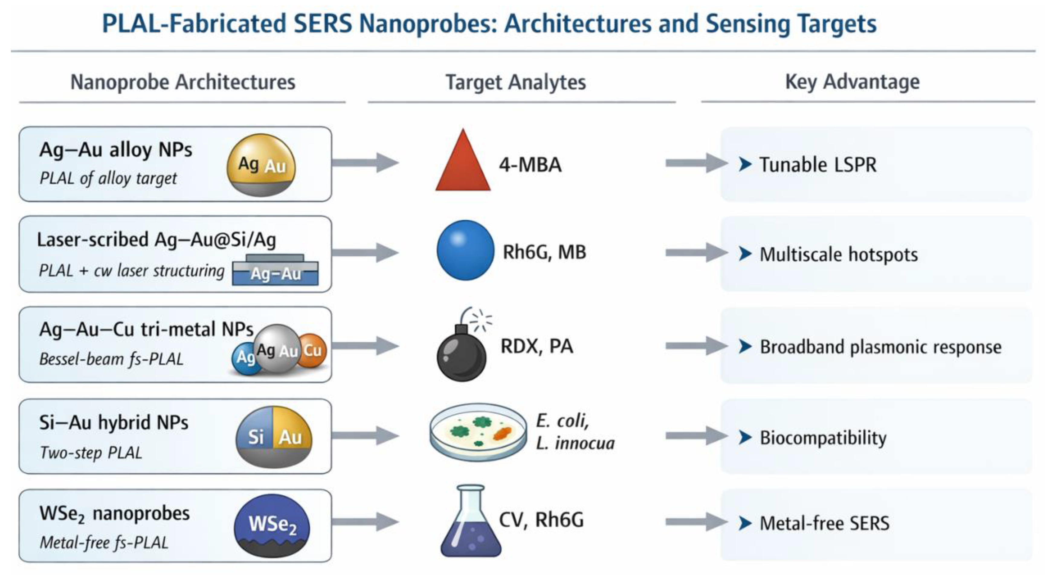
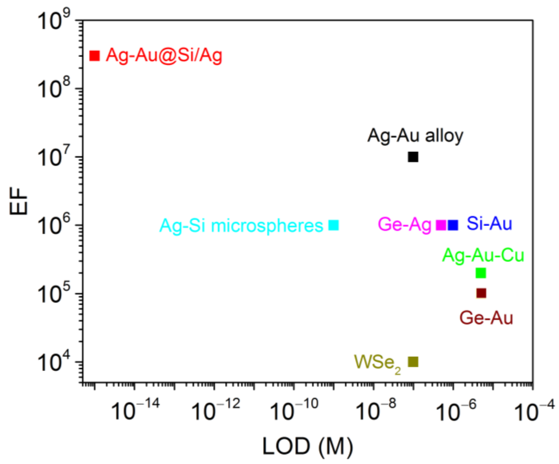
2.1.2. Multi-Metal SERS Nanoprobes
Alloy-Based bi-Metal SERS Nanoprobes (Ag–Au)
Laser-Scribed Bimetallic Ag–Au SERS Nanoplatforms
Tri-Metal Ag–Au–Cu SERS Nanoprobes Fabricated by Shaped-Beam PLAL
- -
- LOD: ~5 µM for RDX and ~0.5 µM for PA;
- -
- EF: ~1.7 × 10⁴ and ~2.4 × 10⁵, respectively [118].
Magneto-Plasmonic bi-Metal SERS Nanoprobes (Au–Fe)
2.1.3. Composite SERS Nanoprobes
Composite Si–Au Nanoprobes Synthesized by Two-Step PLAL
Ag-Decorated Si Microspheres Produced by Single-Step PLAL
Ge–Ag and Ge–Au Hybrid Nanoprobes Fabricated Using Bessel-Beam PLAL
Laser-Synthesized Tungsten Dichalcogenide Nanoprobes with Plasmon-like SERS Activity
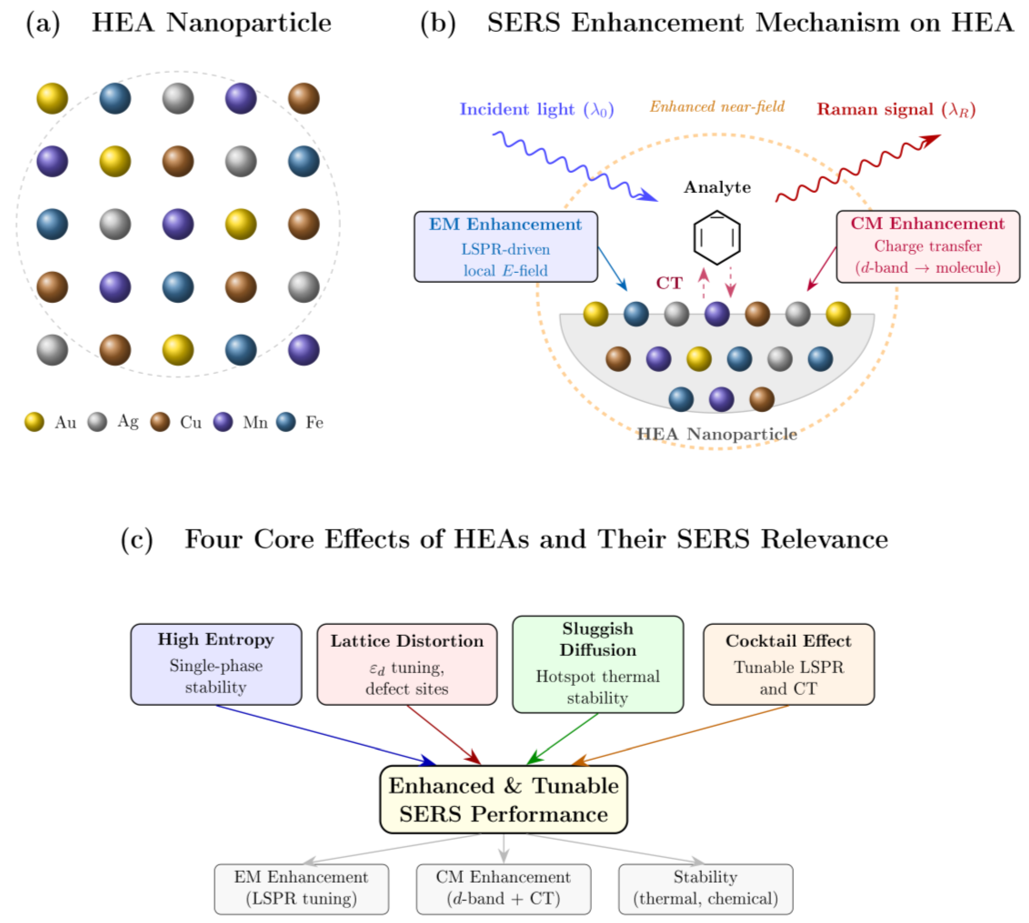
2.1.4. High Entropy Alloys (HEA) SERS Probes
From Conventional SERS Substrates to High Entropy Alloys
Thermodynamics, Structure, and Electronic Engineering of High Entropy Alloys
Synthesis and Microstructure Control of HEA-SERS Nanoprobes
2.2. SERS Fabricated Through Self-Assembly
2.3. Laser Writing and Laser Interference
2.3.1. Ultrafast Laser Direct Writing
2.3.2. Ultrafast Laser Interference Lithography
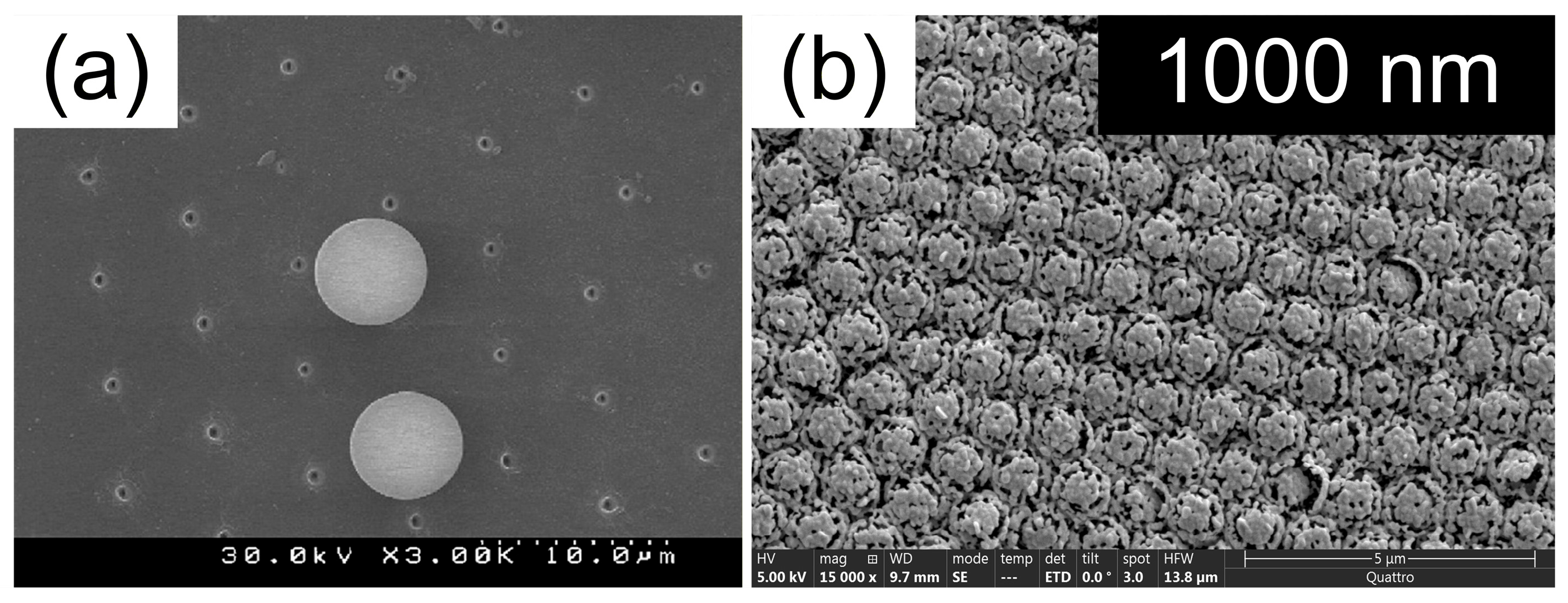
2.4. Nanosphere Lithography
2.5. Nanoimprinting
2.6. Lithography for Periodic Structures
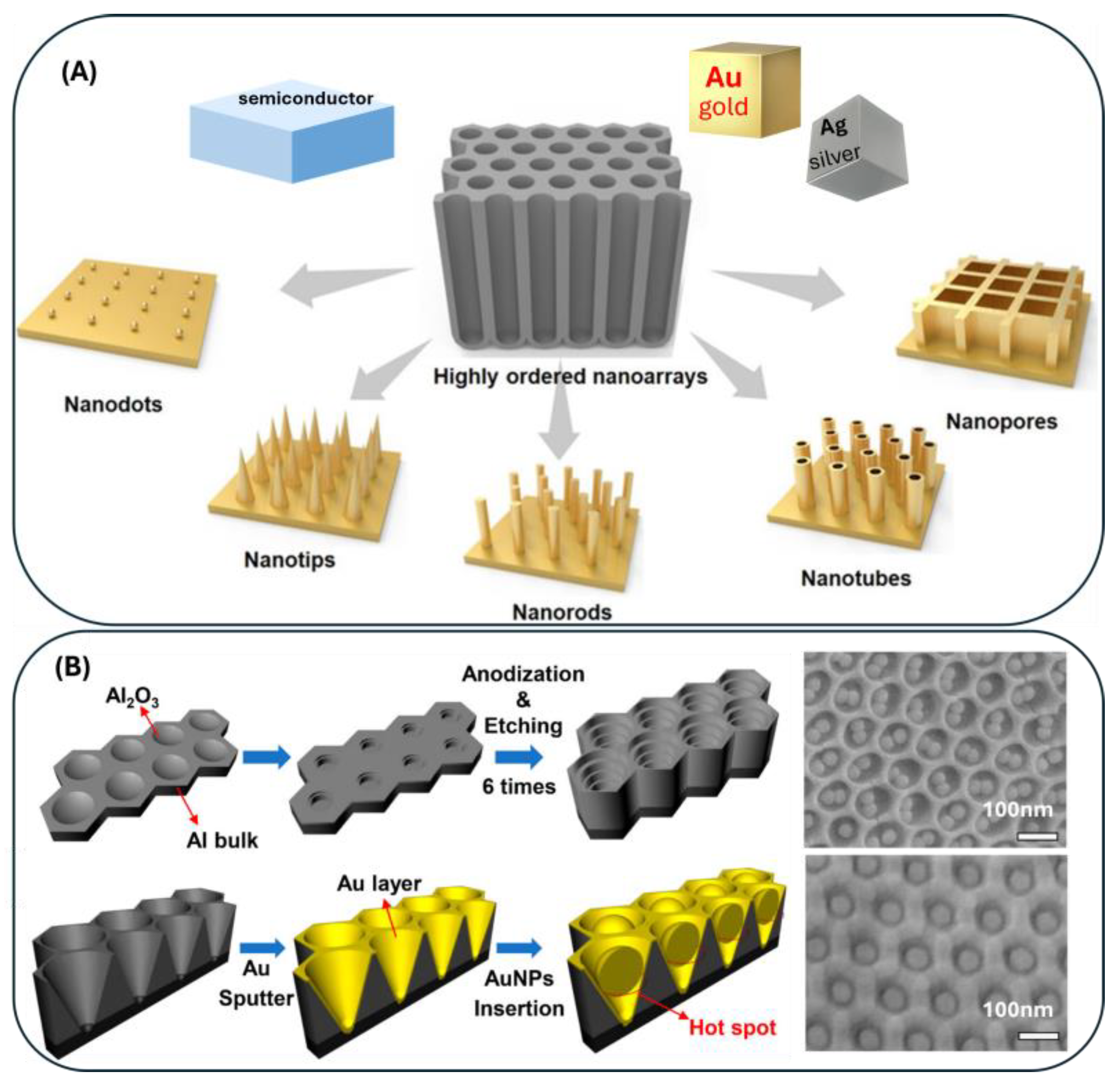
2.7. Anodic Aluminum Oxide (AAO) with Large Area Self-Assembled Structures

3. Engineering Optimization of SERS Probes
3.1. 2D Materials for Interfacial and Charger Transfer Optimization
3.2. Metal Oxide Substrates for Chemical Enhancement Dominated SERS Probes
3.3. Surface Chemistry and Functionalization
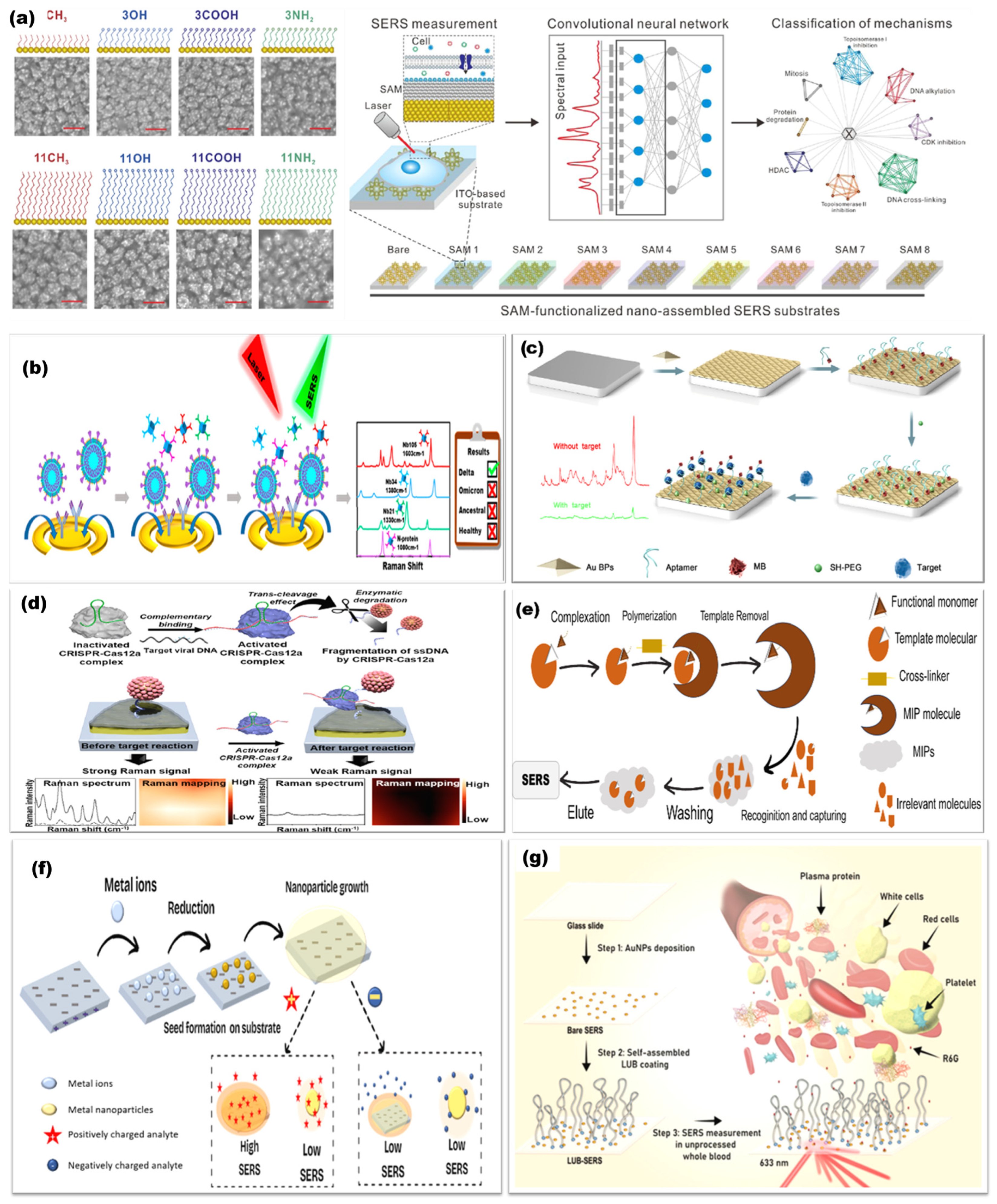
3.3.1. Self-Assembled Monolayers (SAMs)-Driven Hotspot Access
3.3.2. High-Affinity Biorecognition Interfaces
3.3.3. Molecular Escort Strategies
3.3.4. Electrostatic and Interfacial Regulation
3.3.5. Antifouling and Stability Engineering
3.3.6. Long-Term Performance Stability

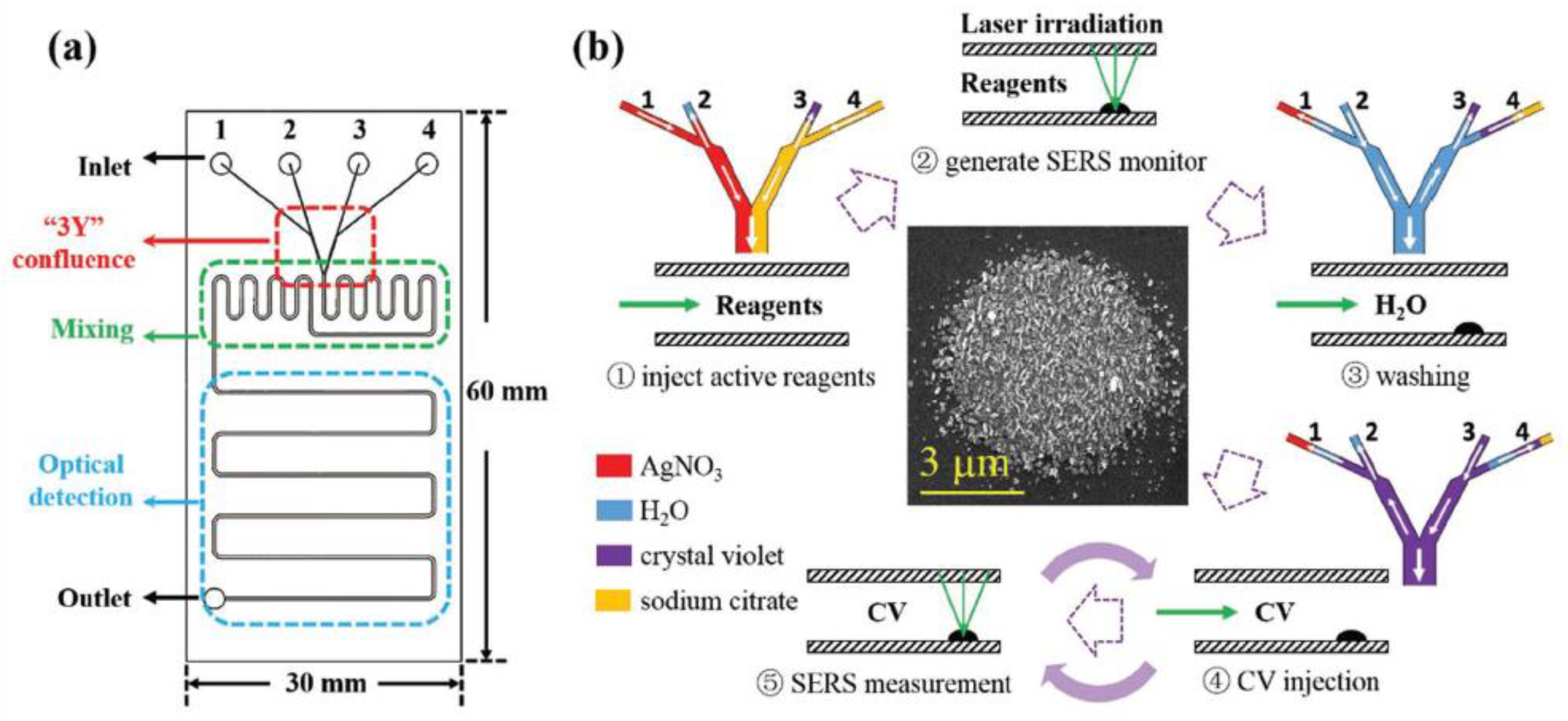
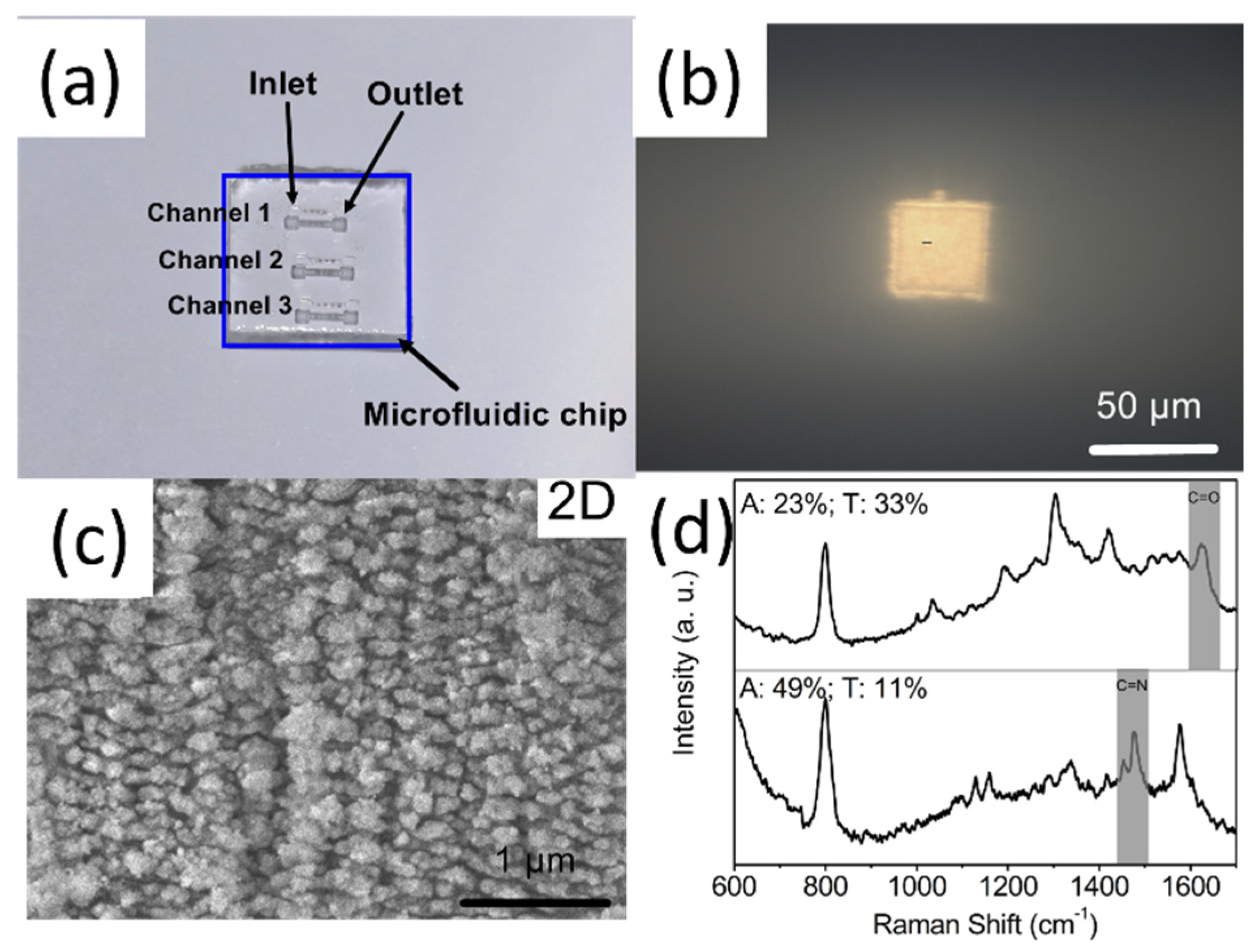
3.4. Dynamic Analysis with Microfluidics
4. Modeling and Machine Learning (ML) for SERS Analysis
4.1. DFT for Molecule Identification
4.1.1. First-Principles Modeling: Beyond Classical Electrodynamics
4.1.2. Validation and Structural Elucidation
4.2. ML for Spectrum Analysis
4.2.1. Theoretical Foundations of ML/DL for Spectral Data
4.2.2. Supervised Architectures: From Classical Chemometrics to Deep Learning
4.2.3. Unsupervised Learning: Clustering and Visualization
4.2.4. Transfer Learning and Validation
4.2.5. Limitations and Common Pitfalls
4.3. Integrated Workflows: DFT-Guided ML and ML-Accelerated DFT
4.3.1. DFT Spectra as ML Training Data
4.3.2. ML Surrogates for Accelerated DFT
4.3.3. Case Study: Environmental Monitoring Integration
4.4. Comparison of ML Methods for SERS
5. Emergent Applications
5.1. Environmental Applications
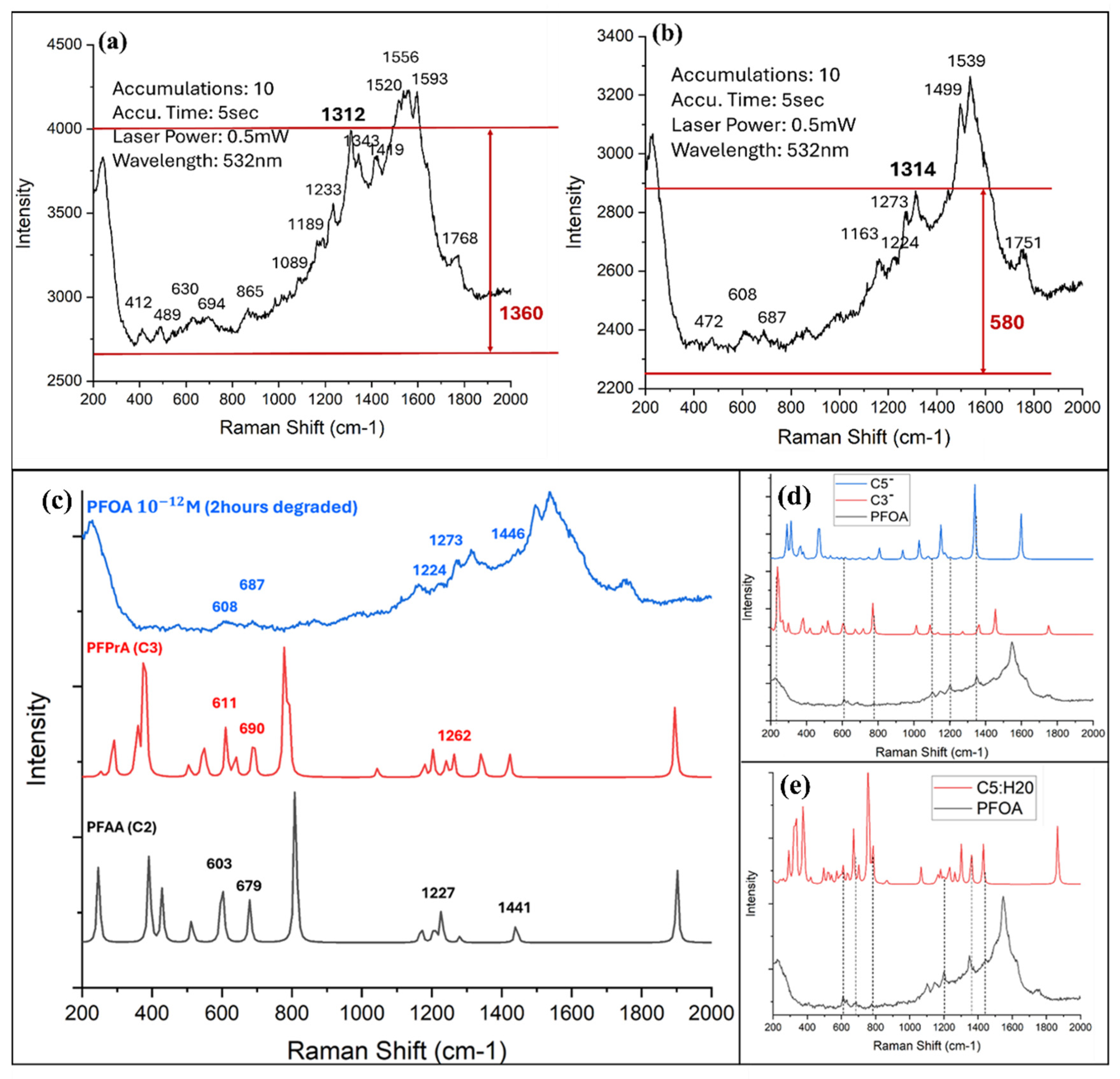
5.1.1. PFAS Detection
5.1.2. Micro- and Nanoplastic Detection
5.1.3. Crucial Mineral Detection
5.2. Biomedical Applications
5.3. National Security
5.3.1. Detection of Explosives
5.3.2. Chemical Warfare Agents
5.3.3. Detection of Illicit Drugs and Forensic Application
5.4. Automation Analysis with AI
6. Challenges and Future Perspectives
6.1. Detection Limitation and Hybrid Devices
6.2. Nanoparticles and Biomedical Applications
6.3. Perspectives of HEA-Based SERS
6.4. Challenges in Modeling
6.5. Field Deployability of SERS
7. Conclusions
Funding
Data Availability Statement
Acknowledgments
Conflicts of Interest
References
- Haynes, C.L.; McFarland, A.D.; Van Duyne, R.P. Surface-Enhanced Raman Spectroscopy. Anal. Chem. 2005, 77, 338 A–346 A. [Google Scholar] [CrossRef]
- Moskovits, M. Surface-enhanced spectroscopy. Rev. Mod. Phys. 1985, 57, 783–826. [Google Scholar] [CrossRef]
- Pérez-Jiménez, A.I.; Lyu, D.; Lu, Z.; Liu, G.; Ren, B. Surface-enhanced Raman spectroscopy: Benefits, trade-offs and future developments. Chem. Sci. 2020, 11, 4563–4577. [Google Scholar] [CrossRef]
- Sharma, B.; Frontiera, R.R.; Henry, A.-I.; Ringe, E.; Van Duyne, R.P. SERS: Materials, applications, and the future. Mater. Today 2012, 15, 16–25. [Google Scholar] [CrossRef]
- Jeon, T.Y.; Park, S.-G.; Lee, S.Y.; Jeon, H.C.; Yang, S.-M. Shape Control of Ag Nanostructures for Practical SERS Substrates. ACS Appl. Mater. Interfaces 2013, 5, 243–248. [Google Scholar] [CrossRef]
- Brown, R.J.C.; Milton, M.J.T. Nanostructures and nanostructured substrates for surface—Enhanced Raman scattering (SERS). J. Raman Spectrosc. 2008, 39, 1313–1326. [Google Scholar] [CrossRef]
- Ko, H.; Singamaneni, S.; Tsukruk, V.V. Nanostructured Surfaces and Assemblies as SERS Media. Small 2008, 4, 1576–1599. [Google Scholar] [CrossRef]
- Kleinman, S.L.; Frontiera, R.R.; Henry, A.-I.; Dieringer, J.A.; Van Duyne, R.P. Creating, characterizing, and controlling chemistry with SERS hot spots. Phys. Chem. Chem. Phys. 2013, 15, 21–36. [Google Scholar] [CrossRef] [PubMed]
- Willets, K.A. Super-resolution imaging of SERS hot spots. Chem. Soc. Rev. 2014, 43, 3854–3864. [Google Scholar] [CrossRef]
- Langer, J.; Jimenez de Aberasturi, D.; Aizpurua, J.; Alvarez-Puebla, R.A.; Auguié, B.; Baumberg, J.J.; Bazan, G.C.; Bell, S.E.J.; Boisen, A.; Brolo, A.G.; et al. Present and Future of Surface-Enhanced Raman Scattering. ACS Nano 2020, 14, 28–117. [Google Scholar] [CrossRef]
- Nie, S.; Emory, S.R. Probing Single Molecules and Single Nanoparticles by Surface-Enhanced Raman Scattering. Science 1997, 275, 1102–1106. [Google Scholar] [CrossRef]
- Kneipp, K.; Wang, Y.; Kneipp, H.; Perelman, L.T.; Itzkan, I.; Dasari, R.R.; Feld, M.S. Single Molecule Detection Using Surface-Enhanced Raman Scattering (SERS). Phys. Rev. Lett. 1997, 78, 1667–1670. [Google Scholar] [CrossRef]
- Huang, Z.; Zhang, A.; Zhang, Q.; Cui, D. Nanomaterial-based SERS sensing technology for biomedical application. J. Mater. Chem. B 2019, 7, 3755–3774. [Google Scholar] [CrossRef]
- Lada, Z.G.; Mathioudakis, G.N.; Soto Beobide, A.; Andrikopoulos, K.S.; Voyiatzis, G.A. Generic method for the detection of short & long chain PFAS extended to the lowest concentration levels of SERS capability. Chemosphere 2024, 363, 142916. [Google Scholar] [CrossRef]
- Guo, Y.; Li, Z.; Li, W.; Chen, X.; Cai, Z. Hydrophobic SERS substrate for PFOA sensing and cooperative adsorption. Talanta 2025, 294, 128244. [Google Scholar] [CrossRef]
- Lin, D.-Y.; Yu, C.-Y.; Ku, C.-A.; Chung, C.-K. Design, Fabrication, and Applications of SERS Substrates for Food Safety Detection: Review. Micromachines 2023, 14, 1343. [Google Scholar] [CrossRef]
- Duley, W.W.; Hu, A. Polyynes and interstellar carbon nanoparticles. Astrophys. J. 2009, 698, 808–811. [Google Scholar] [CrossRef]
- Hu, A.; Lu, Q.-B.; Duley, W.W.; Rybachuk, M. Spectroscopic characterization of carbon chains in nanostructured tetrahedral carbon films synthesized by femtosecond pulsed laser deposition. J. Chem. Phys. 2007, 126, 154705. [Google Scholar] [CrossRef] [PubMed]
- Fleischmann, M.; Hendra, P.J.; McQuillan, A.J. Raman spectra of pyridine adsorbed at a silver electrode. Chem. Phys. Lett. 1974, 26, 163–166. [Google Scholar] [CrossRef]
- Jeanmaire, D.L.; Van Duyne, R.P. Surface raman spectroelectrochemistry: Part I. Heterocyclic, aromatic, and aliphatic amines adsorbed on the anodized silver electrode. J. Electroanal. Chem. Interfacial Electrochem. 1977, 84, 1–20. [Google Scholar] [CrossRef]
- Albrecht, M.G.; Creighton, J.A. Anomalously intense Raman spectra of pyridine at a silver electrode. J. Am. Chem. Soc. 1977, 99, 5215–5217. [Google Scholar] [CrossRef]
- Otto, A. Surface-Enhanced Raman Scattering BT—Dynamics of Gas-Surface Interaction; Benedek, G., Valbusa, U., Eds.; Springer: Berlin/Heidelberg, Germany, 1982; pp. 186–195. [Google Scholar]
- Lombardi, J.R.; Birke, R.L.; Lu, T.; Xu, J. Charge-transfer theory of surface enhanced Raman spectroscopy: Herzberg–Teller contributions. J. Chem. Phys. 1986, 84, 4174–4180. [Google Scholar] [CrossRef]
- Kneipp, K.; Kneipp, H.; Manoharan, R.; Itzkan, I.; Dasari, R.R.; Feld, M.S. Surface-enhanced Raman scattering (SERS)—A new tool for single molecule detection and identification. Bioimaging 1998, 6, 104–110. [Google Scholar] [CrossRef]
- Kim, J.; Yoo, S.; Kim, J.; Choi, S.; Kim, J.; Park, S.; Park, S.; Park, D.; Nam, J.; Park, S.; et al. Synthesis and Single-Particle Surface-Enhanced Raman Scattering Study of Plasmonic Tripod Nanoframes with Y-Shaped Hot-Zones. Nano Lett. 2020, 20, 4362–4369. [Google Scholar] [CrossRef]
- Sibug-Torres, S.M.; Grys, D.; Kang, G.; Niihori, M.; Wyatt, E.; Spiesshofer, N.; Ruane, A.; De Nijs, B.; Baumberg, J.J. In situ electrochemical regeneration of nanogap hotspots for continuously reusable ultrathin SERS sensors. Nat. Commun. 2024, 15, 2022. [Google Scholar] [CrossRef]
- Sharma, B.; Cardinal, M.F.; Kleinman, S.L.; Greeneltch, N.G.; Frontiera, R.R.; Blaber, M.G.; Schatz, G.C.; Van Duyne, R.P. High-performance SERS substrates: Advances and challenges. MRS Bull. 2013, 38, 615–624. [Google Scholar] [CrossRef]
- Ouyang, L.; Ren, W.; Zhu, L.; Irudayaraj, J. Prosperity to challenges: Recent approaches in SERS substrate fabrication. Rev. Anal. Chem. 2017, 36, 2968–2979. [Google Scholar] [CrossRef]
- Tong, L.; Xu, H.; Käll, M. Nanogaps for SERS applications. MRS Bull. 2014, 39, 163–168. [Google Scholar] [CrossRef]
- Deriu, C.; Fabris, L. A surface chemistry perspective on SERS: Revisiting the basics to push the field forward. Chem. Soc. Rev. 2025, 54, 5224–5247. [Google Scholar] [CrossRef]
- Kim, G.-H.; Son, J.; Nam, J.-M. Advances, Challenges, and Opportunities in Plasmonic Nanogap-Enhanced Raman Scattering with Nanoparticles. ACS Nano 2025, 19, 2992–3007. [Google Scholar] [CrossRef]
- Ma, H.; Pan, S.; Wang, W.; Yue, X.; Xi, X.; Yan, S.; Wu, D.; Wang, X.; Liu, G.; Ren, B. Surface-Enhanced Raman Spectroscopy: Current Understanding, Challenges, and Opportunities. ACS Nano 2024, 18, 14000–14019. [Google Scholar] [CrossRef]
- Ruzi, M.; Celik, N.; Sahin, F.; Sakir, M.; Onses, M.S. Nanostructured Surfaces with Plasmonic Activity and Superhydrophobicity: Review of Fabrication Strategies and Applications. Small 2025, 21, 2408189. [Google Scholar] [CrossRef] [PubMed]
- Stiles, P.L.; Dieringer, J.A.; Shah, N.C.; Van Duyne, R.P. Surface-Enhanced Raman Spectroscopy. Annu. Rev. Anal. Chem. 2008, 1, 601–626. [Google Scholar] [CrossRef] [PubMed]
- Jensen, L.; Aikens, C.M.; Schatz, G.C. Electronic structure methods for studying surface-enhanced Raman scattering. Chem. Soc. Rev. 2008, 37, 1061–1073. [Google Scholar] [CrossRef]
- Liao, Y.; Wang, X.; Kang, H.; Tao, Y.; Chen, H. SERS for infectious disease diagnostics: An advanced platform for pathogen detection and antimicrobial resistance analysis. Int. J. Nanomed. 2026, 21, 589169. [Google Scholar] [CrossRef]
- Ding, S.; Yi, J.; Li, J.; Ren, B.; Wu, D.; Panneerselvam, R.; Tian, Z. Nanostructure-based plasmon-enhanced Raman spectroscopy for surface analysis of materials. Nat. Rev. Mater. 2016, 1, 16021. [Google Scholar] [CrossRef]
- Kerker, M.; Wang, D.-S.; Chew, H. Surface enhanced Raman scattering (SERS) by molecules adsorbed at spherical particles. Appl. Opt. 1980, 19, 3373–3388. [Google Scholar] [CrossRef]
- Itoh, T.; Yoshida, K.; Biju, V.; Kikkawa, Y.; Ishikawa, M.; Ozaki, Y. Second enhancement in surface-enhanced resonance Raman scattering revealed by an analysis of anti-Stokes and Stokes Raman spectra. Phys. Rev. B 2007, 76, 85405. [Google Scholar] [CrossRef]
- Yoshida, K.; Itoh, T.; Tamaru, H.; Biju, V.; Ishikawa, M.; Ozaki, Y. Quantitative evaluation of electromagnetic enhancement in surface-enhanced resonance Raman scattering from plasmonic properties and morphologies of individual Ag nanostructures. Phys. Rev. B 2010, 81, 115406. [Google Scholar] [CrossRef]
- Ding, S.-Y.; You, E.-M.; Tian, Z.-Q.; Moskovits, M. Electromagnetic theories of surface-enhanced Raman spectroscopy. Chem. Soc. Rev. 2017, 46, 4042–4076. [Google Scholar] [CrossRef]
- Yamamoto, Y.S.; Ozaki, Y.; Itoh, T. Recent progress and frontiers in the electromagnetic mechanism of surface-enhanced Raman scattering. J. Photochem. Photobiol. C Photochem. Rev. 2014, 21, 81–104. [Google Scholar] [CrossRef]
- Zhang, X.; Hu, A.; Zhang, T.; Lei, W.; Xue, X.; Zhou, Y.; Duley, W.W. Self-Assembly of Large-Scale and Ultrathin Silver Nanoplate Films with Tunable Plasmon Resonance Properties. ACS Nano 2011, 5, 9082–9092. [Google Scholar] [CrossRef]
- Hu, A.; Peng, P.; Alarifi, H.; Zhang, X.Y.; Guo, J.Y.; Zhou, Y.; Duley, W.W. Femtosecond laser welded nanostructures and plasmonic devices. J. Laser Appl. 2012, 24, 42001. [Google Scholar] [CrossRef]
- Starowicz, Z.; Wojnarowska-Nowak, R.; Ozga, P.; Sheregii, E.M. The tuning of the plasmon resonance of the metal nanoparticles in terms of the SERS effect. Colloid Polym. Sci. 2018, 296, 1029–1037. [Google Scholar] [CrossRef]
- Bell, S.E.J.; McCourt, M.R. SERS enhancement by aggregated Au colloids: Effect of particle size. Phys. Chem. Chem. Phys. 2009, 11, 7455–7462. [Google Scholar] [CrossRef]
- Zhang, Q.; Large, N.; Nordlander, P.; Wang, H. Porous Au Nanoparticles with Tunable Plasmon Resonances and Intense Field Enhancements for Single-Particle SERS. J. Phys. Chem. Lett. 2014, 5, 370–374. [Google Scholar] [CrossRef]
- Xu, J.; Kvasnička, P.; Idso, M.; Jordan, R.W.; Gong, H.; Homola, J.; Yu, Q. Understanding the effects of dielectric medium, substrate, and depth on electric fields and SERS of quasi-3D plasmonic nanostructures. Opt. Express 2011, 19, 20493–20505. [Google Scholar] [CrossRef]
- Tian, F.; Bonnier, F.; Casey, A.; Shanahan, A.E.; Byrne, H.J. Surface enhanced Raman scattering with gold nanoparticles: Effect of particle shape. Anal. Methods 2014, 6, 9116–9123. [Google Scholar] [CrossRef]
- Camargo, P.H.C.; Au, L.; Rycenga, M.; Li, W.; Xia, Y. Measuring the SERS enhancement factors of dimers with different structures constructed from silver nanocubes. Chem. Phys. Lett. 2010, 484, 304–308. [Google Scholar] [CrossRef] [PubMed]
- Kravets, V.G.; Kabashin, A.V.; Barnes, W.L.; Grigorenko, A.N. Plasmonic Surface Lattice Resonances: A Review of Properties and Applications. Chem. Rev. 2018, 118, 5912–5951. [Google Scholar] [CrossRef] [PubMed]
- Xu, H.; Bjerneld, E.J.; Käll, M.; Börjesson, L. Spectroscopy of Single Hemoglobin Molecules by Surface Enhanced Raman Scattering. Phys. Rev. Lett. 1999, 83, 4357–4360. [Google Scholar] [CrossRef]
- Prodan, E.; Radloff, C.; Halas, N.J.; Nordlander, P. A Hybridization Model for the Plasmon Response of Complex Nanostructures. Science 2003, 302, 419–422. [Google Scholar] [CrossRef]
- Zhu, W.; Crozier, K.B. Quantum mechanical limit to plasmonic enhancement as observed by surface-enhanced Raman scattering. Nat. Commun. 2014, 5, 5228. [Google Scholar] [CrossRef]
- Savage, K.J.; Hawkeye, M.M.; Esteban, R.; Borisov, A.G.; Aizpurua, J.; Baumberg, J.J. Revealing the quantum regime in tunnelling plasmonics. Nature 2012, 491, 574–577. [Google Scholar] [CrossRef]
- Le Ru, E.C.; Blackie, E.; Meyer, M.; Etchegoin, P.G. Surface Enhanced Raman Scattering Enhancement Factors: A Comprehensive Study. J. Phys. Chem. C 2007, 111, 13794–13803. [Google Scholar] [CrossRef]
- Schlücker, S. Surface-Enhanced Raman Spectroscopy: Concepts and Chemical Applications. Angew. Chem. Int. Ed. 2014, 53, 4756–4795. [Google Scholar] [CrossRef] [PubMed]
- Chaudhry, I.; Hu, G.; Ye, H.; Jensen, L. Toward Modeling the Complexity of the Chemical Mechanism in SERS. ACS Nano 2024, 18, 20835–20850. [Google Scholar] [CrossRef] [PubMed]
- Campion, A.; Kambhampati, P. Surface-enhanced Raman scattering. Chem. Soc. Rev. 1998, 27, 241–250. [Google Scholar] [CrossRef]
- Lombardi, J.R.; Birke, R.L. A Unified Approach to Surface-Enhanced Raman Spectroscopy. J. Phys. Chem. C 2008, 112, 5605–5617. [Google Scholar] [CrossRef]
- Persson, B.N.J.; Zhao, K.; Zhang, Z. Chemical Contribution to Surface-Enhanced Raman Scattering. Phys. Rev. Lett. 2006, 96, 207401. [Google Scholar] [CrossRef]
- Xia, L.; Chen, M.; Zhao, X.; Zhang, Z.; Xia, J.; Xu, H.; Sun, M. Visualized method of chemical enhancement mechanism on SERS and TERS. J. Raman Spectrosc. 2014, 45, 533–540. [Google Scholar] [CrossRef]
- Lombardi, J.R.; Birke, R.L. The theory of surface-enhanced Raman scattering. J. Chem. Phys. 2012, 136, 144704. [Google Scholar] [CrossRef]
- Campion, A.; Ivanecky, J.E.I.I.I.; Child, C.M.; Foster, M. On the Mechanism of Chemical Enhancement in Surface-Enhanced Raman Scattering. J. Am. Chem. Soc. 1995, 117, 11807–11808. [Google Scholar] [CrossRef]
- Moldovan, R.; Vereshchagina, E.; Milenko, K.; Iacob, B.; Bodoki, A.E.; Falamas, A.; Tosa, N.; Muntean, C.M.; Farcău, C.; Bodoki, E. Review on combining surface-enhanced Raman spectroscopy and electrochemistry for analytical applications. Anal. Chim. Acta 2022, 1209, 339250. [Google Scholar] [CrossRef]
- Lombardi, J.R.; Birke, R.L. Theory of Surface-Enhanced Raman Scattering in Semiconductors. J. Phys. Chem. C 2014, 118, 11120–11130. [Google Scholar] [CrossRef]
- Kim, J.; Jang, Y.; Kim, N.; Kim, H.; Yi, G.; Shin, Y.; Kim, M.H.; Yoon, S. Study of Chemical Enhancement Mechanism in Non-plasmonic Surface Enhanced Raman Spectroscopy (SERS). Front. Chem. 2019, 7, 582. [Google Scholar] [CrossRef]
- Zou, X.; Dong, S. Surface-Enhanced Raman Scattering Studies on Aggregated Silver Nanoplates in Aqueous Solution. J. Phys. Chem. B 2006, 110, 21545–21550. [Google Scholar] [CrossRef]
- He, S.; Chua, J.; Tan, E.K.M.; Kah, J.C.Y. Optimizing the SERS enhancement of a facile gold nanostar immobilized paper-based SERS substrate. RSC Adv. 2017, 7, 16264–16272. [Google Scholar] [CrossRef]
- Nalbant Esenturk, E.; Hight Walker, A.R. Surface-enhanced Raman scattering spectroscopy via gold nanostars. J. Raman Spectrosc. 2009, 40, 86–91. [Google Scholar] [CrossRef]
- Ghosal, S.; Nandi, S.; Giri, P.K. Recent advances in semiconductor nanostructure-based surface-enhanced Raman scattering sensors. Nanotechnology 2025, 36, 202002. [Google Scholar] [CrossRef]
- Zong, C.; Xu, M.; Xu, L.; Wei, T.; Ma, X.; Zheng, X.; Hu, R.; Ren, B. Surface-Enhanced Raman Spectroscopy for Bioanalysis: Reliability and Challenges. Chem. Rev. 2018, 118, 4946–4980. [Google Scholar] [CrossRef] [PubMed]
- Zou, Y.; Jin, H.; Ma, Q.; Zheng, Z.; Weng, S.; Kolataj, K.; Acuna, G.; Bald, I.; Garoli, D. Advances and applications of dynamic surface-enhanced Raman spectroscopy (SERS) for single molecule studies. Nanoscale 2025, 17, 3656–3670. [Google Scholar] [CrossRef] [PubMed]
- Li, G.; Zhao, X.; Tang, X.; Yao, L.; Li, W.; Wang, J.; Liu, X.; Han, B.; Fan, X.; Qiu, T.; et al. Wearable Hydrogel SERS Chip Utilizing Plasmonic Trimers for Uric Acid Analysis in Sweat. Nano Lett. 2024, 24, 13447–13454. [Google Scholar] [CrossRef]
- Zhou, W.; Hu, A.; Bai, S.; Ma, Y.; Bridges, D. Anisotropic optical properties of large-scale aligned silver nanowire films via controlled coffee ring effects. RSC Adv. 2015, 5, 39103–39109. [Google Scholar] [CrossRef]
- Lin, Y.; Zhang, X.; Bai, S.; Hu, A. Photo-reduction of metallic ions doped in patterned polymer films for the fabrication of plasmonic photonic crystals. J. Mater. Chem. C Mater. 2015, 3, 6046–6052. [Google Scholar] [CrossRef]
- Fortuni, B.; Ricci, M.; Vitale, R.; Inose, T.; Zhang, Q.; Hutchison, J.A.; Hirai, K.; Fujita, Y.; Toyouchi, S.; Krzyzowska, S.; et al. SERS Endoscopy for Monitoring Intracellular Drug Dynamics. ACS Sens. 2023, 8, 2340–2347. [Google Scholar] [CrossRef]
- Lindquist, N.C.; de Albuquerque, C.D.L.; Sobral-Filho, R.G.; Paci, I.; Brolo, A.G. High-speed imaging of surface-enhanced Raman scattering fluctuations from individual nanoparticles. Nat. Nanotechnol. 2019, 14, 981–987. [Google Scholar] [CrossRef]
- Zong, C.; Chen, C.; Wang, X.; Hu, P.; Liu, G.; Ren, B. Single-Molecule Level Rare Events Revealed by Dynamic Surface-Enhanced Raman Spectroscopy. Anal. Chem. 2020, 92, 15806–15810. [Google Scholar] [CrossRef]
- Scott, B.L.; Carron, K.T. Dynamic Surface Enhanced Raman Spectroscopy (SERS): Extracting SERS from Normal Raman Scattering. Anal. Chem. 2012, 84, 8448–8451. [Google Scholar] [CrossRef]
- Huang, J.; Mousavi, M.Z.; Zhao, Y.; Hubarevich, A.; Omeis, F.; Giovannini, G.; Schütte, M.; Garoli, D.; De Angelis, F. SERS discrimination of single DNA bases in single oligonucleotides by electro-plasmonic trapping. Nat. Commun. 2019, 10, 5321. [Google Scholar] [CrossRef] [PubMed]
- Zhan, C.; Wang, G.; Yi, J.; Wei, J.; Li, Z.; Chen, Z.; Shi, J.; Yang, Y.; Hong, W.; Tian, Z. Single-Molecule Plasmonic Optical Trapping. Matter 2020, 3, 1350–1360. [Google Scholar] [CrossRef]
- Fu, W.; Chi, H.; Dai, X.; Zhu, H.; St Dollente Mesias, V.; Liu, W.; Huang, J. Efficient optical plasmonic tweezer-controlled single-molecule SERS characterization of pH-dependent amylin species in aqueous milieus. Nat. Commun. 2023, 14, 6996. [Google Scholar] [CrossRef]
- Chen, H.; You, T.; Jiang, L.; Gao, Y.; Yin, P. Creating dynamic SERS hotspots on the surface of pH-responsive microgels for direct detection of crystal violet in solution. RSC Adv. 2017, 7, 32743–32748. [Google Scholar] [CrossRef]
- Ge, M.; Li, P.; Zhou, G.; Chen, S.; Han, W.; Qin, F.; Nie, Y.; Wang, Y.; Qin, M.; Huang, G.; et al. General Surface-Enhanced Raman Spectroscopy Method for Actively Capturing Target Molecules in Small Gaps. J. Am. Chem. Soc. 2021, 143, 7769–7776. [Google Scholar] [CrossRef]
- Chen, Z.; Xu, H.; Chen, H.; Zhu, S.; Huang, W.; He, Y.; Hafez, M.E.; Qian, R.; Li, D. AuNPs-COFs Core–Shell Reversible SERS Nanosensor for Monitoring Intracellular Redox Dynamics. Anal. Chem. 2022, 94, 14280–14289. [Google Scholar] [CrossRef]
- Huang, Q.; Guo, H.; Wang, W.; Kang, S.; Vikesland, P.J. Surface-Enhanced Raman Spectroscopy (SERS) Based Biological and Environmental 2D and 3D Imaging. ACS Environ. Au 2025, 5, 342–362. [Google Scholar] [CrossRef] [PubMed]
- Jimenez de Aberasturi, D.; Henriksen-Lacey, M.; Litti, L.; Langer, J.; Liz-Marzán, L.M. Using SERS Tags to Image the Three-Dimensional Structure of Complex Cell Models. Adv. Funct. Mater. 2020, 30, 1909655. [Google Scholar] [CrossRef]
- García-Astrain, C.; Henriksen-Lacey, M.; Lenzi, E.; Renero-Lecuna, C.; Langer, J.; Piñeiro, P.; Molina-Martínez, B.; Plou, J.; De Aberasturi, D.J.; Liz-Marzán, L.M. A Scaffold-Assisted 3D Cancer Cell Model for Surface-Enhanced Raman Scattering-Based Real-Time Sensing and Imaging. ACS Nano 2024, 18, 11257–11269. [Google Scholar] [CrossRef]
- Chang, H.; Hur, W.; Kang, H.; Jun, B.-H. In vivo surface-enhanced Raman scattering techniques: Nanoprobes, instrumentation, and applications. Light Sci. Appl. 2025, 14, 79. [Google Scholar] [CrossRef] [PubMed]
- Zhang, D.; Gökce, B.; Barcikowski, S. Laser Synthesis and Processing of Colloids: Fundamentals and Applications. Chem. Rev. 2017, 117, 3990–4103. [Google Scholar] [CrossRef]
- Amendola, V.; Meneghetti, M. Laser ablation synthesis in solution and size manipulation of noble metal nanoparticles. Phys. Chem. Chem. Phys. 2009, 11, 3805–3821. [Google Scholar] [CrossRef]
- Kanitz, A.; Kalus, M.; Gurevich, E.L.; Ostendorf, A.; Barcikowski, S.; Amans, D. Review on experimental and theoretical investigations of the early stage, femtoseconds to microseconds processes during laser ablation in liquid-phase for the synthesis of colloidal nanoparticles. Plasma Sources Sci. Technol. 2019, 28, 103001. [Google Scholar] [CrossRef]
- Barcikowski, S.; Compagnini, G. Advanced nanoparticle generation and excitation by lasers in liquids. Phys. Chem. Chem. Phys. 2013, 15, 3022–3026. [Google Scholar] [CrossRef]
- He, N.; Hu, T.; Kang, X.; Wei, S.; Wu, L. Laser Fragmentation Breaks the Limitation of Twin-Forming Ability of fcc Metals (Pt/Rh/Pd) with High Stacking Fault Energy. J. Phys. Chem. C 2025, 129, 12595–12606. [Google Scholar] [CrossRef]
- Ryabchikov, Y.V. Size Modification of Optically Active Contamination-Free Silicon Nanoparticles with Paramagnetic Defects by Their Fast Synthesis and Dissolution. Phys. Status Solidi A 2019, 216, 1800685. [Google Scholar] [CrossRef]
- Barcikowski, S.; Amendola, V.; Marzun, G.; Rehbock, C.; Reichenberger, S.; Zhang, D.; Gökce, B. Handbook of Laser Synthesis of Colloids; DuEPublico: Essen, Germany, 2016. [Google Scholar] [CrossRef]
- Zabotnov, S.V.; Skobelkina, A.V.; Sergeeva, E.A.; Kurakina, D.A.; Khilov, A.V.; Kashaev, F.V.; Kaminskaya, T.P.; Presnov, D.E.; Agrba, P.D.; Shuleiko, D.V.; et al. Nanoparticles Produced via Laser Ablation of Porous Silicon and Silicon Nanowires for Optical Bioimaging. Sensors 2020, 20, 4874. [Google Scholar] [CrossRef]
- Gurbatov, S.O.; Shevlyagin, A.V.; Zhizhchenko, A.Y.; Modin, E.B.; Kuchmizhak, A.A.; Kudryashov, S.I. Photothermal Conversion and Laser-Induced Transformations in Silicon–Germanium Alloy Nanoparticles. JETP Lett. 2024, 119, 910–916. [Google Scholar] [CrossRef]
- Ryabchikov, Y.V.; Alekseev, S.; Lysenko, V.; Bremond, G.; Bluet, J.-M. Photoluminescence thermometry with alkyl-terminated silicon nanoparticles dispersed in low-polar liquids. Phys. Status Solidi (RRL)—Rapid Res. Lett. 2013, 7, 414–417. [Google Scholar] [CrossRef]
- Ryabchikov, Y.V.; Belogorokhov, I.A.; Vorontsov, A.S.; Osminkina, L.A.; Timoshenko, V.Y.; Kashkarov, P.K. Dependence of the singlet oxygen photosensitization efficiency on morphology of porous silicon. Phys. Status Solidi A 2007, 204, 1271–1275. [Google Scholar] [CrossRef]
- Ryabchikov, Y.V.; Belogorokhov, I.A.; Gongalskii, M.B.; Osminkina, L.A.; Timoshenko, V.Y. Photosensitized generation of singlet oxygen in powders and aqueous suspensions of silicon nanocrystals. Semiconductors 2011, 45, 1059–1063. [Google Scholar] [CrossRef]
- Sokolovskaya, O.I.; Sergeeva, E.A.; Golovan, L.A.; Kashkarov, P.K.; Khilov, A.V.; Kurakina, D.A.; Orlinskaya, N.Y.; Zabotnov, S.V.; Kirillin, M.Y. Numerical Simulation of Enhancement of Superficial Tumor Laser Hyperthermia with Silicon Nanoparticles. Photonics 2021, 8, 580. [Google Scholar] [CrossRef]
- Belogorokhov, I.A.; Ryabchikov, Y.V.; Tikhonov, E.V.; Pushkarev, V.E.; Breusova, M.O.; Tomilova, L.G.; Khokhlov, D.R. Photoluminescence in semiconductor structures based on butyl-substituted erbium phthalocyanine complexes. Semiconductors 2008, 42, 321–324. [Google Scholar] [CrossRef]
- Gurbatov, S.O.; Zhizhchenko, A.Y.; Nesterov, V.Y.; Modin, E.B.; Zabotnov, S.V.; Kuchmizhak, A.A. Au–Si Nanocomposites with High Near-IR Light-to-Heat Conversion Efficiency via Single-Step Reactive Laser Ablation of Porous Silicon for Theranostic Applications. ACS Appl. Nano Mater. 2024, 7, 10779–10786. [Google Scholar] [CrossRef]
- Ryabchikov, Y.V.; Mirza, I.; Flimelová, M.; Kana, A.; Romanyuk, O. Merging of Bi-Modality of Ultrafast Laser Processing: Heating of Si/Au Nanocomposite Solutions with Controlled Chemical Content. Nanomaterials 2024, 14, 321. [Google Scholar] [CrossRef]
- Ryabchikov, Y.V.; Kana, A. Properties of Silicon-8-Metals Nanocomposite Colloidal Solutions Synthesized by Ultrafast Laser Processing. In Proceedings of the 2025 25th Anniversary International Conference on Transparent Optical Networks (ICTON), Barcelona, Spain, 6–10 July 2025. [Google Scholar] [CrossRef]
- Bissoli, M.; Negri, A.; Zerbato, A.; Badocco, D.; Pastore, P.; Filibian, M.; Brero, F.; Megalizzi, S.; Lascialfari, A.; Greco, N.; et al. Polymer Coated Iron–Boron and Gold–Iron Alloy Nanoparticles for Magnetic Resonance Imaging and Near Infrared Photothermal Applications. ACS Appl. Nano Mater. 2025, 8, 24512–24527. [Google Scholar] [CrossRef]
- Alrubaie, H.A.; Salam, R.; Abed, M.A. Optical and Structural Properties of Au@Fe2O3 Nanoparticles Synthesized by Nanosecond Laser Ablation in Liquid. Semiconductors 2025, 59, 436–440. [Google Scholar] [CrossRef]
- Ryabchikov, Y.V. Design of “green” plasmonic nanocomposites with multi-band blue emission for ultrafast laser hyperthermia. Nanoscale 2024, 16, 19453–19468. [Google Scholar] [CrossRef]
- Ryabchikov, Y.V.; Behrends, J. Expedient paramagnetic properties of surfactant-free plasmonic silicon-based nanoparticles. Opt. Quantum Electron. 2020, 52, 177. [Google Scholar] [CrossRef]
- Nesterov, V.Y.; Zabotnov, S.V.; Shuleiko, D.V.; Presnov, D.E.; Sokolovskaya, O.I.; Smirnov, N.A.; Golovan, L.A.; Kashkarov, P.K. Morphology and Stability of Composite Silicon–Silver Nanoparticles Produced by Laser Ablation in a Liquid. Nanobiotechnol. Rep. 2024, 19, S50–S58. [Google Scholar] [CrossRef]
- Ryabchikov, Y.V.; Zaderko, A. “Green” Fluorescent–Plasmonic Carbon-Based Nanocomposites with Controlled Performance for Mild Laser Hyperthermia. Photonics 2023, 10, 1229. [Google Scholar] [CrossRef]
- Ryabchikov, Y.V. Optical performance of silicon-metallic nanocomposites synthesized via doughnut shape laser processing. Proc. SPIE 2025, 13698, 136980W. [Google Scholar]
- Petrikaitė, V.; Talaikis, M.; Mikoliūnaitė, L.; Gkouzi, A.; Trusovas, R.; Skapas, M.; Niaura, G.; Stankevičius, E. Stability and SERS signal strength of laser-generated gold, silver, and bimetallic nanoparticles at different KCl concentrations. Heliyon 2024, 10, e34815. [Google Scholar] [CrossRef]
- Kadavath, J.K.; Krishnan, B.; Pelaes, R.F.C.; Guzman, S.S.; Garcia-Gomez, N.A.; Avellaneda, D.A.; Shaji, S. Ultrasensitive and stable SERS sensors by incorporating Noble metal/bimetallic nanoparticles produced by laser ablation in liquid. Microchem. J. 2025, 215, 114537. [Google Scholar] [CrossRef]
- Kadavath, J.K.; Pelaes, R.F.C.; Guzman, S.S.; Gomez, N.A.G.; Avellaneda, D.A.; Krishnan, B.; Shaji, S. Laser-Patterned Cu–Ag Alloy Micropillars Incorporated with Laser-Ablated AgAu Bimetallic Nanoparticles for Label-free Detection of Water Pollutants. ACS Appl. Nano Mater. 2025, 8, 7747–7759. [Google Scholar] [CrossRef]
- Banerjee, D.; Akkanaboina, M.; Soma, V.R. Plasmonic alloy nanocolloids fabricated via femtosecond Bessel beam for photonic and SERS-based sensing applications. Opt. Mater. 2024, 154, 115668. [Google Scholar] [CrossRef]
- Mikoliunaite, L.; Stankevičius, E.; Adomavičiūtė-Grabusovė, S.; Petrikaitė, V.; Trusovas, R.; Talaikis, M.; Skapas, M.; Zdaniauskienė, A.; Selskis, A.; Šablinskas, V.; et al. Magneto-Plasmonic Nanoparticles Generated by Laser Ablation of Layered Fe/Au and Fe/Au/Fe Composite Films for SERS Application. Coatings 2023, 13, 1523. [Google Scholar] [CrossRef]
- Kögler, M.; Ryabchikov, Y.V.; Uusitalo, S.; Popov, A.; Popov, A.; Tselikov, G.; Välimaa, A.; Al-Kattan, A.; Hiltunen, J.; Laitinen, R.; et al. Bare laser-synthesized Au-based nanoparticles as nondisturbing surface-enhanced Raman scattering probes for bacteria identification. J. Biophotonics 2018, 11, e201700225. [Google Scholar] [CrossRef]
- Ryabchikov, Y.V. Facile laser synthesis of multimodal composite silicon/gold nanoparticles with variable chemical composition. J. Nanoparticle Res. 2019, 21, 85. [Google Scholar] [CrossRef]
- Gurbatov, S.; Puzikov, V.; Modin, E.; Shevlyagin, A.; Gerasimenko, A.; Mitsai, E.; Kulinich, S.A.; Kuchmizhak, A. Ag-Decorated Si Microspheres Produced by Laser Ablation in Liquid: All-in-One Temperature-Feedback SERS-Based Platform for Nanosensing. Materials 2022, 15, 8091. [Google Scholar] [CrossRef]
- Rathod, J.; Moram, S.S.B.; Chandu, B.; Albrycht, P.; Soma, V.R. Single-step fabrication of hybrid germanium-gold/silver nanoentities by femtosecond laser ablation and applications in SERS-based sensing. Nanotechnology 2023, 34, 405301. [Google Scholar] [CrossRef]
- Ushkov, A.; Dyubo, D.; Belozerova, N.; Kazantsev, I.; Yakubovsky, D.; Syuy, A.; Tikhonowski, G.V.; Tselikov, D.; Martynov, I.; Ermolaev, G.; et al. Tungsten Diselenide Nanoparticles Produced via Femtosecond Ablation for SERS and Theranostics Applications. Nanomaterials 2025, 15, 4. [Google Scholar] [CrossRef]
- Hu, A.; Duley, W.W. Surface enhanced Raman spectroscopic characterization of molecular structures in diamond-like carbon films. Chem. Phys. Lett. 2008, 450, 375–378. [Google Scholar] [CrossRef]
- Hu, A. Interaction of Nanosecond and Femtosecond Laser Pulses with Carbon: Deposition of Carbon Films Having Novel Compositions. Ph.D. Thesis, University of Waterloo, Waterloo, ON, Canada, 2008. [Google Scholar]
- Hu, A.; Sanderson, J.; Zaidi, A.; Wang, C.; Zhang, T.; Zhou, Y.; Duley, W. Direct synthesis of polyyne molecules in acetone by dissociation using femtosecond laser irradiation. Carbon N. Y. 2008, 46, 1823–1825. [Google Scholar] [CrossRef]
- Hu, A.; Duley, W.W. Spectra of Carbon Nanoparticles: Laboratory Simulation of the Aromatic CH Emission Feature at 3.29 μm. Astrophys. J. 2008, 677, L153–L156. [Google Scholar] [CrossRef]
- Duley, W.W.; Hu, A. The 217.5 nm band, infrared absorption, and infrared emission features in hydrogenated amorphous carbon nanoparticles. Astrophys. J. 2012, 761, 115. [Google Scholar] [CrossRef]
- Nugroho, F.A.A.; Iandolo, B.; Wagner, J.B.; Langhammer, C. Bottom-Up Nanofabrication of Supported Noble Metal Alloy Nanoparticle Arrays for Plasmonics. ACS Nano 2016, 10, 2871–2879. [Google Scholar] [CrossRef]
- Tomboc, G.M.; Kwon, T.; Joo, J.; Lee, K. High Entropy Alloy Electrocatalysts: A Critical Assessment of Fabrication and Performance. J. Mater. Chem. A Mater. 2020, 8, 14844–14862. [Google Scholar] [CrossRef]
- Liu, D.; Duan, P.; Chen, J.; Mu, M.; Yang, Y.; Ma, F.; Zhao, B.; Song, W. Atomic Orbital-Driven SERS Enhancement Via D-Band Engineering in High-Entropy Alloy Aerogels. Laser Photon. Rev. 2026, 20, e01214. [Google Scholar] [CrossRef]
- Yeh, J.; Chen, S.; Lin, S.; Gan, J.; Chin, T.; Shun, T.; Tsau, C.; Chang, S. Nanostructured High-Entropy Alloys with Multiple Principal Elements: Novel Alloy Design Concepts and Outcomes. Adv. Eng. Mater. 2004, 6, 299–303. [Google Scholar] [CrossRef]
- Cantor, B.; Chang, I.T.H.; Knight, P.; Vincent, A.J.B. Microstructural Development in Equiatomic Multicomponent Alloys. Mater. Sci. Eng. A 2004, 375, 213–218. [Google Scholar] [CrossRef]
- Bridges, D.; Fieser, D.; Santiago, J.J.; Hu, A. Novel Frontiers in High-Entropy Alloys. Metals 2023, 13, 1193. [Google Scholar] [CrossRef]
- Kumar, S.; Kumar, M. Utilizations of High Entropy Alloy Nanoparticles for Gas Sensors. Trans. Indian Natl. Acad. Eng. 2024, 9, 679–687. [Google Scholar] [CrossRef]
- Huang, W.; Ren, Z.; Li, X.; Chen, R.; Fan, D.; Da, J.; Zha, Y.; Xu, Y. Ultrasmall High-Entropy Alloy-Nanolabels Based Immunochromatographic Test Strip for Rapid, Ultrasensitive, and Catalytic Detection of Staphylococcus aureus. Microchim. Acta 2025, 192, 408. [Google Scholar] [CrossRef]
- Zhang, B.; Mu, Q.; Pei, Y.; Hu, S.; Liu, S.; Sun, T.; Gao, G. Synthesis Strategies and Multi-field Applications of Nanoscale High-Entropy Alloys. Nanomicro Lett. 2025, 17, 283. [Google Scholar] [CrossRef] [PubMed]
- Wang, L.; Zhou, F.; Ying, P.; Chen, Y.; Liu, Y. Machine Learning-Accelerated Transition State Prediction for Strain-Engineered High-Entropy Alloy Catalysts. J. Mater. Inform. 2026, 6, 8. [Google Scholar] [CrossRef]
- Fieser, D.; Yin, K.; Shortt, H.; Dewanjee, U.; Steingrimsson, B.; Ivanov, I.N.; Burns, J.; Liaw, P.K.; Zuo, J.; Hu, A. Surface Nanostructure Control and Thermodynamic Stability Analysis of Femtosecond Laser-Ablated CuCoMn1.75NiFe0.25 Nanoparticles. Langmuir 2025, 41, 34173–34188. [Google Scholar] [CrossRef]
- Han, M.; Meghwal, A.; Ng, S.H.; Smith, D.; Mu, H.; Katkus, T.; Zhu, D.M.; Mukhlis, R.; Vongsvivut, J.; Berndt, C.C.; et al. Microparticles of High Entropy Alloys Made by Laser-Induced Forward Transfer. Materials 2022, 15, 8063. [Google Scholar] [CrossRef] [PubMed]
- Nishijima, Y.; Sudo, T.; Matsuo, Y.; Juodkazis, S. A Noble Metal High-Entropy Alloy for Mid-Infrared Metasurfaces. Engineering 2025, 49, 81–89. [Google Scholar] [CrossRef]
- Wu, C.-Y.; Wu, C.-Y.; Hsiao, Y.-C.; Yang, T.-H. Seed-Mediated Growth of High-Entropy Alloy Atomic Layers with Tunable Facets, Compositions, and Structures for Electrocatalysis. J. Electron. Mater. 2025, 54, 7401–7422. [Google Scholar] [CrossRef]
- Yang, L.; Li, Y.; Wang, Z.; Zhao, W.; Qin, C. Nanoporous Quasi-High-Entropy Alloy Microspheres. Metals 2019, 9, 345. [Google Scholar] [CrossRef]
- Tahghighi, M.; Mannelli, I.; Janner, D.; Ignés-Mullol, J. Tailoring plasmonic response by Langmuir–Blodgett gold nanoparticle templating for the fabrication of SERS substrates. Appl. Surf. Sci. 2018, 447, 416–422. [Google Scholar] [CrossRef]
- Swierczewski, M.; Burgi, T. Langmuir and Langmuir–Blodgett films of gold and silver nanoparticles. Langmuir 2023, 39, 2135–2151. [Google Scholar] [CrossRef]
- Mitra, C.K.; Ghosh, M.; Chowdhury, J. Fabrication of gold nano-particles embedded in the Langmuir Blodgett film of Poly (methyl methacrylate) for rapid screening of melamine adulterant in milk powder. Opt. Mater. 2025, 169, 117549. [Google Scholar] [CrossRef]
- Vieira, F.M.; Calisto, C.M.G.; Izumi, C. Construction of SERS substrates by gold nanoparticles assembly on polymeric matrices. Appl. Surf. Sci. 2023, 612, 155818. [Google Scholar] [CrossRef]
- Mitra, C.K.; Sharma, M.D.; Ghosh, M.; Pande, S.; Chowdhury, J. Fabrication of Au decorated MoS2 Langmuir Blodgett film as SERS sensing scaffold for detection of thiram in aqueous solution and in carrot peels. Mater. Lett. 2024, 372, 137033. [Google Scholar] [CrossRef]
- Garg, A.; Nam, W.; Zhou, W. Reusable Surface-Enhanced Raman Spectroscopy Membranes and Textiles via Template-Assisted Self-Assembly and Micro/Nanoimprinting. ACS Appl. Mater. Interfaces 2020, 12, 56290–56299. [Google Scholar] [CrossRef]
- Peng, R.; Zhang, T.; Yan, S.; Song, Y.; Liu, X.; Wang, J. Recent development and applications of stretchable SERS substrates. Nanomaterials 2023, 13, 2968. [Google Scholar] [CrossRef] [PubMed]
- Wijnhoven, J.E.; Vos, W.L. Preparation of photonic crystals made of air spheres in titania. Science 1998, 281, 802–804. [Google Scholar] [CrossRef]
- Bartlett, P.N.; Baumberg, J.J.; Birkin, P.R.; Ghanem, M.A.; Netti, M.C. Highly ordered macroporous gold and platinum films formed by electrochemical deposition through templates assembled from submicron diameter monodisperse polystyrene spheres. Chem. Mater. 2002, 14, 2199–2208. [Google Scholar] [CrossRef]
- Jiao, S.; Liu, J.; Sun, J.; Chang, Y.; Wang, S.; Dai, S.; Xu, R.; Dou, M.; Li, Q.; Wang, J.; et al. A highly sensitive and reproducible multiplex mycotoxin SERS array based on AuNPs-loaded inverse opal silica photonic crystal microsphere. Sens. Actuators B Chem. 2022, 355, 131245. [Google Scholar] [CrossRef]
- Wu, L.; Wang, W.; Zhang, W.; Su, H.; Liu, Q.; Gu, J.; Deng, T.; Zhang, D. Highly sensitive, reproducible and uniform SERS substrates with a high density of three-dimensionally distributed hotspots: Gyroid-structured Au periodic metallic materials. NPG Asia Mater. 2018, 10, e462. [Google Scholar] [CrossRef]
- Zhou, X.; Liu, S.; Xiang, H.; Li, X.; Wang, C.; Wu, Y.; Li, G. Three-dimensional SERS substrates: Architectures, hot spot engineering, and biosensing applications. Biosensors 2025, 15, 555. [Google Scholar] [CrossRef]
- Ren, F.; Campbell, J.; Rorrer, G.L.; Wang, A.X. Surface-enhanced Raman spectroscopy sensors from nanobiosilica with self-assembled plasmonic nanoparticles. IEEE J. Sel. Top. Quantum Electron. 2014, 20, 127–132. [Google Scholar]
- Wang, X.; Zhai, Y.; Chen, Y.; Zhang, W.; Liang, P.; Zhu, Q.; Zhang, H.; Kang, J.; Wang, L. Highly sensitive and reproducible SERS substrates based on a novel 3D waffle-like PMMA-CsPbBr3-Au ternary film. Sens. Actuators B Chem. 2025, 431, 137434. [Google Scholar] [CrossRef]
- Thacker, V.V.; Herrmann, L.O.; Sigle, D.O.; Zhang, T.; Liedl, T.; Baumberg, J.J.; Keyser, U.F. DNA origami based assembly of gold nanoparticle dimers for surface-enhanced Raman scattering. Nat. Commun. 2014, 5, 3448. [Google Scholar] [CrossRef]
- Schuknecht, F.; Kołątaj, K.; Steinberger, M.; Liedl, T.; Lohmueller, T. Accessible hotspots for single-protein SERS in DNA-origami assembled gold nanorod dimers with tip-to-tip alignment. Nat. Commun. 2023, 14, 7192. [Google Scholar] [CrossRef]
- Kanehira, Y.; Kogikoski, S.; Titov, E.; Tapio, K.; Mostafa, A.; Bald, I. Watching a Single Enzyme at Work Using Single-Molecule Surface-Enhanced Raman Scattering and DNA Origami-Based Plasmonic Antennas. ACS Nano 2024, 18, 20191–20200. [Google Scholar] [CrossRef] [PubMed]
- Fu, Q.; Zhan, Z.; Dou, J.; Zheng, X.; Xu, R.; Wu, M.; Lei, Y. Highly reproducible and sensitive SERS substrates with Ag inter-nanoparticle gaps of 5 nm fabricated by ultrathin aluminum mask technique. ACS Appl. Mater. Interfaces 2015, 7, 13322–13328. [Google Scholar] [CrossRef]
- Sloan-Dennison, S.; Wallace, G.Q.; Hassanain, W.A.; Laing, S.; Faulds, K.; Graham, D. Advancing SERS as a quantitative technique: Challenges, considerations, and correlative approaches to aid validation. Nano Converg. 2024, 11, 33. [Google Scholar] [CrossRef] [PubMed]
- Li, J.F.; Huang, Y.F.; Ding, Y.; Yang, Z.L.; Li, S.B.; Zhou, X.S.; Fan, F.R.; Zhang, W.; Zhou, Z.Y.; Wu, D.Y.; et al. Shell-isolated nanoparticle-enhanced Raman spectroscopy. Nature 2010, 464, 392–395. [Google Scholar] [CrossRef]
- Diebold, E.D.; Mack, N.H.; Doorn, S.K.; Mazur, E. Femtosecond Laser-Nanostructured Substrates for Surface-Enhanced Raman Scattering. Langmuir 2009, 25, 1790–1794. [Google Scholar] [CrossRef]
- Han, Y.; Lan, X.; Wei, T.; Tsai, H.-L.; Xiao, H. Surface enhanced Raman scattering silica substrate fast fabrication by femtosecond laser pulses. Appl. Phys. A 2009, 97, 721–724. [Google Scholar] [CrossRef]
- Zhou, Z.; Xu, J.; Cheng, Y.; Xu, Z.; Sugioka, K.; Midorikawa, K. Surface-Enhanced Raman Scattering Substrate Fabricated by Femtosecond Laser Direct Writing. Jpn. J. Appl. Phys. 2008, 47, 189. [Google Scholar] [CrossRef]
- Pavliuk, G.; Pavlov, D.; Mitsai, E.; Vitrik, O.; Mironenko, A.; Zakharenko, A.; Kulinich, S.A.; Juodkazis, S.; Bratskaya, S.; Zhizhchenko, A.; et al. Ultrasensitive SERS-Based Plasmonic Sensor with Analyte Enrichment System Produced by Direct Laser Writing. Nanomaterials 2020, 10, 49. [Google Scholar] [CrossRef]
- Yoshikawa, H.; Hironou, A.; Shen, Z.; Tamiya, E. Versatile Micropatterning of Plasmonic Nanostructures by Visible Light Induced Electroless Silver Plating on Gold Nanoseeds. ACS Appl. Mater. Interfaces 2016, 8, 23932–23940. [Google Scholar] [CrossRef]
- Ran, P.; Jiang, L.; Li, X.; Li, B.; Zuo, P.; Lu, Y. Femtosecond Photon-Mediated Plasma Enhances Photosynthesis of Plasmonic Nanostructures and Their SERS Applications. Small 2019, 15, 1804899. [Google Scholar] [CrossRef] [PubMed]
- Hamdorf, A.; Olson, M.; Lin, C.; Jiang, L.; Zhou, J.; Xiao, H.; Tsai, H. Femtosecond and nanosecond laser fabricated substrate for surface-enhanced Raman scattering. Opt. Lett. 2011, 36, 3353–3355. [Google Scholar] [CrossRef]
- Bai, S.; Li, Z.; Obata, K.; Kawabata, S.; Sugioka, K. λ/20 surface nanostructuring of ZnO by mask-less ultrafast laser processing. Nanophotonics 2023, 12, 1499–1510. [Google Scholar] [CrossRef] [PubMed]
- Mohammadi, L.; Ben Hundertmark, N.; Kullmann, F.; Fleissner, F.; Klotzbücher, T. Nano-structured surfaces by laser interference lithography and fs-laser direct writing as substrates for surface-enhanced Raman spectroscopy. Proc. SPIE 2012, 8244, 82440D. [Google Scholar]
- Bai, S.; Lin, Y.; Zhang, X.; Zhou, W.; Chen, T.; Ma, Y.; Hou, T.; Bridges, D.; Oakes, K.D.; Hu, A. Two-Step Photonic Reduction of Controlled Periodic Silver Nanostructures for Surface-Enhanced Raman Spectroscopy. Plasmonics 2015, 10, 1675–1685. [Google Scholar] [CrossRef]
- Zhou, Y.; Hong, M.; Fuh, J.; Lu, L.; Lukyanchuk, B.; Wang, Z. Near-field enhanced femtosecond laser nano-drilling of glass substrate. J. Alloys Compd. 2008, 449, 246–249. [Google Scholar] [CrossRef]
- Bai, S.; Hu, A.; Hu, Y.; Ma, Y.; Obata, K.; Sugioka, K. Plasmonic Superstructure Arrays Fabricated by Laser Near-Field Reduction for Wide-Range SERS Analysis of Fluorescent Materials. Nanomaterials 2022, 12, 970. [Google Scholar] [CrossRef]
- Barcelo, S.J.; Wu, W.; Li, X.; Li, Z.; Williams, R.S. Nanoimprint lithography of plasmonic platforms for SERS applications. Appl. Phys. A 2015, 121, 443–449. [Google Scholar] [CrossRef]
- Suresh, V.; Ding, L.; Chew, A.B.; Yap, F.L. Fabrication of Large-Area Flexible SERS Substrates by Nanoimprint Lithography. ACS Appl. Nano Mater. 2018, 1, 886–893. [Google Scholar] [CrossRef]
- Alvarez-Puebla, R.; Cui, B.; Bravo-Vasquez, J.-P.; Veres, T.; Fenniri, H. Nanoimprinted SERS-Active Substrates with Tunable Surface Plasmon Resonances. J. Phys. Chem. C 2007, 111, 6720–6723. [Google Scholar] [CrossRef]
- Chen, J.; Li, Y.; Huang, K.; Wang, P.; He, L.; Carter, K.R.; Nugen, S.R. Nanoimprinted Patterned Pillar Substrates for Surface-Enhanced Raman Scattering Applications. ACS Appl. Mater. Interfaces 2015, 7, 22106–22113. [Google Scholar] [CrossRef] [PubMed]
- Ferchichi, A.; Laariedh, F.; Sow, I.; Gourgon, C.; Boussey, J. Fabrication of disposable flexible SERS substrates by nanoimprint. Microelectron. Eng. 2015, 140, 52–55. [Google Scholar] [CrossRef]
- Colniță, A.; Marconi, D.; Dina, N.E.; Brezeștean, I.; Bogdan, D.; Turcu, I. 3D silver metallized nanotrenches fabricated by nanoimprint lithography as flexible SERS detection platform. Spectrochim. Acta A Mol. Biomol. Spectrosc. 2022, 276, 121232. [Google Scholar] [CrossRef] [PubMed]
- Badshah, M.A.; Kim, J.; Yeom, J.; Abbas, N.; Haq, M.R.; Kim, Y.; Lu, X.; Kim, S. Glass nanoimprinted plasmonic nanostructure for high power laser stable surface-enhanced Raman spectroscopy substrate. Appl. Surf. Sci. 2021, 542, 148587. [Google Scholar] [CrossRef]
- Hankus, M.E.; Stratis-Cullum, D.N.; Pellegrino, P.M. Characterization of next-generation commercial surface-enhanced Raman scattering (SERS) substrates. Proc. SPIE 2011, 8018, 80180P. [Google Scholar]
- Hankus, M.E.; Stratis-Cullum, D.N.; Pellegrino, P.M. Surface enhanced Raman scattering (SERS)-based next generation commercially available substrate: Physical characterization and biological application. Proc. SPIE 2011, 8099, 80990N. [Google Scholar]
- Radzol, A.R.M.; Lee, Y.K.; Mansor, W.; Yahaya, S.R. Surface-Enhanced Raman spectral analysis of substrates for salivary based disease detection. In Proceedings of the 2012 IEEE 8th International Colloquium on Signal Processing and its Applications, Malacca, Malaysia, 23–25 March 2012; pp. 505–509. [Google Scholar] [CrossRef]
- Tahir, M.A.; Zhang, X.; Cheng, H.; Xu, D.; Feng, Y.; Sui, G.; Fu, H.; Valev, V.K.; Zhang, L.; Chen, J. Klarite as a label-free SERS-based assay: A promising approach for atmospheric bioaerosol detection. Analyst 2020, 145, 277–285. [Google Scholar] [CrossRef]
- Du, F.; Cai, H.; Su, L.; Wang, W.; Zhang, L.; Sun, C.; Yan, B.; Shi, H. The missing small microplastics: Easily generated from weathered plastic pieces in labs but hardly detected in natural environments. Environ. Sci. Adv. 2024, 3, 227–238. [Google Scholar] [CrossRef]
- Li, A.P.; Müller, F.; Birner, A.; Nielsch, K.; Gösele, U. Hexagonal pore arrays with a 50–420 nm interpore distance formed by self-organization in anodic alumina. J. Appl. Phys. 1998, 84, 6023–6026. [Google Scholar] [CrossRef]
- Wu, Z.; Sha, M.; Ji, D.; Zhao, H.; Li, L.; Lei, Y. Ordered Anodic Aluminum Oxide-Based Nanostructures for Surface-Enhanced Raman Scattering: A Review. ACS Appl. Nano Mater. 2024, 7, 11–31. [Google Scholar] [CrossRef]
- Li, J.; Li, C.; Gao, X. Structural evolution of self-ordered alumina tapered nanopores with 100nm interpore distance. Appl. Surf. Sci. 2011, 257, 10390–10394. [Google Scholar] [CrossRef]
- Sun, K.; Meng, G.; Huang, Q.; Zhao, X.; Zhu, C.; Huang, Z.; Qian, Y.; Wang, X.; Hu, X. Gap-tunable Ag-nanorod arrays on alumina nanotip arrays as effective SERS substrates. J. Mater. Chem. C Mater. 2013, 1, 5015–5022. [Google Scholar] [CrossRef]
- Lee, S.M.; Lee, S.M.; Nam, D.H.; Lee, D.; Lim, S.H.; Son, S.J.; Lee, S.; Lee, S. Gold Nanoparticles Deposited on a Conical Anodic Aluminum Oxide Substrate for Improved Surface-Enhanced Raman Scattering. ACS Appl. Nano Mater. 2021, 4, 12905–12912. [Google Scholar] [CrossRef]
- Liu, Y.; Wei, Y.; Wang, G.; Jia, X.; Zhou, J.; Li, H.; Chang, H.; Li, Z. Investigating SERS performance of controllable fabricated Ag periodic nanostructures with morphologies of nanoclaws and nanocones in detecting SARS-CoV-2 S protein. Microchem. J. 2024, 207, 112078. [Google Scholar] [CrossRef]
- Zhang, Z.; Xu, Y.; Shi, J.; Liu, W.; Jiang, S.; Pan, J. Fabrication and SERS enhancement study of composite materials based on MoS2 and gold/silver nano particles membrane structure modified AAO. Opt. Laser Technol. 2025, 192, 113631. [Google Scholar] [CrossRef]
- Peng, P.; Hu, A.; Zhou, Y. Laser sintering of silver nanoparticle thin films: Microstructure and optical properties. Appl. Phys. A 2012, 108, 685–691. [Google Scholar] [CrossRef]
- Hu, A.; Guo, J.Y.; Alarifi, H.; Patane, G.; Zhou, Y.; Compagnini, G.; Xu, C.X. Low temperature sintering of Ag nanoparticles for flexible electronics packaging. Appl. Phys. Lett. 2010, 97, 153117. [Google Scholar] [CrossRef]
- Ma, Y.; Li, H.; Bridges, D.; Peng, P.; Lawrie, B.; Feng, Z.; Hu, A. Zero-dimensional to three-dimensional nanojoining: Current status and potential applications. RSC Adv. 2016, 6, 75916–75936. [Google Scholar] [CrossRef]
- Peng, P.; Hu, A.; Gerlich, A.P.; Zou, G.; Liu, L.; Zhou, Y.N. Joining of Silver Nanomaterials at Low Temperatures: Processes, Properties, and Applications. ACS Appl. Mater. Interfaces 2015, 7, 12597–12618. [Google Scholar] [CrossRef] [PubMed]
- Chen, M.; Liu, D.; Du, X.; Lo, K.H.; Wang, S.; Zhou, B.; Pan, H. 2D materials: Excellent substrates for surface-enhanced Raman scattering (SERS) in chemical sensing and biosensing. TrAC Trends Anal. Chem. 2020, 130, 115983. [Google Scholar] [CrossRef]
- Karthick Kannan, P.; Shankar, P.; Blackman, C.; Chung, C.-H. Recent Advances in 2D Inorganic Nanomaterials for SERS Sensing. Adv. Mater. 2019, 31, 1803432. [Google Scholar] [CrossRef]
- Tang, X.; Hao, Q.; Hou, X.; Lan, L.; Li, M.; Yao, L.; Zhao, X.; Ni, Z.; Fan, X.; Qiu, T. Exploring and Engineering 2D Transition Metal Dichalcogenides toward Ultimate SERS Performance. Adv. Mater. 2024, 36, 2312348. [Google Scholar] [CrossRef]
- Song, X.; Wang, Y.; Zhao, F.; Li, Q.; Ta, H.Q.; Rümmeli, M.H.; Tully, C.G.; Li, Z.; Yin, W.; Yang, L.; et al. Plasmon-Free Surface-Enhanced Raman Spectroscopy Using Metallic 2D Materials. ACS Nano 2019, 13, 8312–8319. [Google Scholar] [CrossRef]
- Jena, T.; Hossain, M.T.; Nath, U.; Sarma, M.; Sugimoto, H.; Fujii, M.; Giri, P.K. Evidence for intrinsic defects and nanopores as hotspots in 2D PdSe2 dendrites for plasmon-free SERS substrate with a high enhancement factor. npj 2D Mater. Appl. 2023, 7, 8. [Google Scholar] [CrossRef]
- Yao, Y.; Zhao, Y.; Zhang, H.; Pan, W.; Liang, W.; Jiang, Y.; Yan, X.; Yan, Y. Ultrafast Laser-Induced 1T′/2H-MoTe2 Nanopattern with Au-Nanoclusters for Raman Monitoring of Cellular Drug Metabolism. ACS Nano 2025, 19, 16732–16743. [Google Scholar] [CrossRef]
- Mamiyev, Z.; Balayeva, N.O.; Zahn, D.R.T.; Tegenkamp, C. Enhanced Light–Matter Interactions With a Single Sn Nanoantenna on Epitaxial Graphene. Adv. Opt. Mater. 2025, 13, e00979. [Google Scholar] [CrossRef]
- Gu, Y.; Li, Y.; Qiu, H.; Yang, Y.; Wu, Q.; Fan, X.; Ding, Y.; Yi, L.; Ge, K.; Shen, Y. Recent progress on noble-free substrates for surface-enhanced Raman spectroscopy analysis. Coord. Chem. Rev. 2023, 497, 215425. [Google Scholar] [CrossRef]
- Samriti Rajput, V.; Gupta, R.K.; Prakash, J. Engineering metal oxide semiconductor nanostructures for enhanced charge transfer: Fundamentals and emerging SERS applications. J. Mater. Chem. C Mater. 2022, 10, 73–95. [Google Scholar] [CrossRef]
- Li, W.; Liang, R.; Hu, A.; Huang, Z.; Zhou, Y.N. Generation of oxygen vacancies in visible light activated one-dimensional iodine TiO2 photocatalysts. RSC Adv. 2014, 4, 36959–36966. [Google Scholar] [CrossRef]
- Martinez Pancorbo, P.; Zhang, H.; Yu, X.; Xiao, T.-H.; Goda, K. Metal-free SERS: Where we are now, where we are heading. Europhys. Lett. 2021, 136, 34001. [Google Scholar] [CrossRef]
- Lakshmi, P.S.; Mohanarao, B.; Babuji, P.; Madduri, P.V.P.; Jyothi, L.; Podagatlapalli, G.K.; Chae, K.H.; Chang, G.S.; Saikiran, V. Semiconducting SnO2 nanoparticles for surface-enhanced Raman scattering applications. RSC Adv. 2026, 16, 21740–21755. [Google Scholar] [CrossRef] [PubMed]
- Li, X.; Yang, J.; Ye, F.; Xiao, L.; Li, X.; Weng, J.; Sun, L. Modulating the Energy-Band of Metal Oxide@Metal–Organic Framework Core–Shell Nanoparticles for Enhanced Raman Sensing. ACS Appl. Nano Mater. 2024, 7, 2291–2303. [Google Scholar] [CrossRef]
- Li, M.; Qiu, Y.; Fan, C.; Cui, K.; Zhang, Y.; Xiao, Z. Design of SERS nanoprobes for Raman imaging: Materials, critical factors and architectures. Acta Pharm. Sin. B 2018, 8, 381–389. [Google Scholar] [CrossRef]
- Huang, Q.; Xi, Z.; Wei, W. Controlling synthesis of Au@AgPd core–shell nanocubes and in situ monitoring SERS of their enhanced catalysis. J. Alloys Compd. 2020, 843, 155971. [Google Scholar] [CrossRef]
- Das, G.M.; Managò, S.; Mangini, M.; De Luca, A.C. Biosensing Using SERS Active Gold Nanostructures. Nanomaterials 2021, 11, 2679. [Google Scholar] [CrossRef]
- Singh, M.; Kaur, N.; Comini, E. The role of self-assembled monolayers in electronic devices. J. Mater. Chem. C Mater. 2020, 8, 3938–3955. [Google Scholar] [CrossRef]
- Xu, Y.; Zhang, Y.; Li, C.; Ye, Z.; Bell, S.E.J. SERS as a Probe of Surface Chemistry Enabled by Surface-Accessible Plasmonic Nanomaterials. Acc. Chem. Res. 2023, 56, 2072–2083. [Google Scholar] [CrossRef]
- De Carvalho Gomes, P.; Hardy, M.; Tagger, Y.; Rickard, J.J.S.; Mendes, P.; Oppenheimer, P.G. Optimization of Nanosubstrates toward Molecularly Surface-Functionalized Raman Spectroscopy. J. Phys. Chem. C 2022, 126, 13774–13784. [Google Scholar] [CrossRef]
- Ouhibi, A.; Raouafi, A.; Lorrain, N.; Guendouz, M.; Raouafi, N.; Moadhen, A. Functionalized SERS substrate based on silicon nanowires for rapid detection of prostate specific antigen. Sens. Actuators B Chem. 2021, 330, 129352. [Google Scholar] [CrossRef]
- Thirugnanasambandan, T. 3—Functionalization on Sensing Surfaces for Efficient Biomolecular Capturing. In Micro and Nano Technologies; Gopinath, S.C.B., Lakshmipriya, T.B.T.-N., Eds.; Elsevier: Amsterdam, The Netherlands, 2019; pp. 51–94. [Google Scholar] [CrossRef]
- Zhu, Y.; Zhang, L.; Yang, L. Designing of the functional paper-based surface-enhanced Raman spectroscopy substrates for colorants detection. Mater. Res. Bull. 2015, 63, 199–204. [Google Scholar] [CrossRef]
- Kim, N.; Thomas, M.R.; Bergholt, M.S.; Pence, I.J.; Seong, H.; Charchar, P.; Todorova, N.; Nagelkerke, A.; Belessiotis-Richards, A.; Payne, D.J.; et al. Surface enhanced Raman scattering artificial nose for high dimensionality fingerprinting. Nat. Commun. 2020, 11, 207. [Google Scholar] [CrossRef]
- Inkpen, M.S.; Liu, Z.; Li, H.; Campos, L.M.; Neaton, J.B.; Venkataraman, L. Non-chemisorbed gold-sulfur binding prevails in self-assembled monolayers. Nat. Chem. 2019, 11, 351–358. [Google Scholar] [CrossRef] [PubMed]
- Sun, J.; Lai, W.; Zhao, J.; Xue, J.; Zhu, T.; Xiao, M.; Man, T.; Wan, Y.; Pei, H.; Li, L. Rapid Identification of Drug Mechanisms with Deep Learning-Based Multichannel Surface-Enhanced Raman Spectroscopy. ACS Sens. 2024, 9, 4227–4235. [Google Scholar] [CrossRef]
- Kim, S.; Yoo, H. Self-Assembled Monolayers: Versatile Uses in Electronic Devices from Gate Dielectrics, Dopants, and Biosensing Linkers. Micromachines 2021, 12, 565. [Google Scholar] [CrossRef]
- Chen, N.; Li, S.; Zhao, P.; Liu, R.; Xie, Y.; Lin, J.; Nijhuis, C.A.; Xu, B.; Zhang, L.; Xu, H.; et al. Extreme long-lifetime self-assembled monolayer for air-stable molecular junctions. Sci. Adv. 2026, 9, eadh3412. [Google Scholar] [CrossRef]
- Vedelago, C.; Li, J.; Lowry, K.; Howard, C.; Wuethrich, A.; Trau, M. A Multiplexed SERS Microassay for Accurate Detection of SARS-CoV-2 and Variants of Concern. ACS Sens. 2023, 8, 1648–1657. [Google Scholar] [CrossRef]
- Thakare, S.; Shaikh, A.; Bodas, D.; Gajbhiye, V. Application of dendrimer-based nanosensors in immunodiagnosis. Colloids Surf. B Biointerfaces 2022, 209, 112174. [Google Scholar] [CrossRef] [PubMed]
- Lee, H.K.; Lee, Y.H.; Koh, C.S.L.; Phan-Quang, G.C.; Han, X.; Lay, C.L.; Sim, H.Y.F.; Kao, Y.; An, Q.; Ling, X.Y. Designing surface-enhanced Raman scattering (SERS) platforms beyond hotspot engineering: Emerging opportunities in analyte manipulations and hybrid materials. Chem. Soc. Rev. 2019, 48, 731–756. [Google Scholar] [CrossRef]
- Shatar, L.; Pillai, N.; Chee, H.Y.; Mustafa, F.H.; Nasir, M.N.M.; Zakaria, R.; Mustafa, M.K.; Zain, M.N.M.; Suhailin, F.H. Enhancement in surface-enhanced Raman spectroscopy (SERS) substrates and the potential for diseases detection. Appl. Spectrosc. Rev. 2025, 61, 401–448. [Google Scholar] [CrossRef]
- Zhang, R.; Xie, S.; Yang, X.; Liu, Y.; He, Y. Efficient SERS Substrate HOF@Au in Conjunction with DNAzyme-Mediated DNA Walker as a Signal Amplifier for the Ultrasensitive Detection of Lomefloxacin. Anal. Chem. 2025, 97, 3180–3189. [Google Scholar] [CrossRef]
- He, Y.; Zhang, R.; Xie, S.; Yang, X.; Liu, Y.; Wang, M. Efficient 2D HOF@Au SERS Substrates Synergized with the G-quadruplex Network-Based Catalytic Amplification Strategy for Low-Background Trimode Detection of Lomefloxacin. Anal. Chem. 2025, 97, 26971–26981. [Google Scholar] [CrossRef] [PubMed]
- Wang, J.; Guo, S.; Park, E.; Lee, S.; Park, Y.; Han, X.X.; Zhao, B.; Jung, Y.M. SERS-Based Aptamer Sensing Strategy for Diabetes Biomarker Detection. Anal. Chem. 2024, 96, 20082–20089. [Google Scholar] [CrossRef]
- Choi, J.; Choi, J.; Shin, M.; Yang, L.; Conley, B.; Yoon, J.; Lee, S.; Lee, K.; Choi, J.; Choi, J. Clustered Regularly Interspaced Short Palindromic Repeats-Mediated Amplification-Free Detection of Viral DNAs Using Surface-Enhanced Raman Spectroscopy-Active Nanoarray. ACS Nano 2021, 15, 13475–13485. [Google Scholar] [CrossRef]
- Lu, Y.; Zhong, J.; Yao, G.; Huang, Q. A label-free SERS approach to quantitative and selective detection of mercury (II) based on DNA aptamer-modified SiO2@Au core/shell nanoparticles. Sens. Actuators B Chem. 2018, 258, 365–372. [Google Scholar] [CrossRef]
- Cheshari, E.; Ren, X.; Li, X. Core–shell Ag-dual template molecularly imprinted composite for detection of carbamate pesticide residues. Chem. Pap. 2021, 75, 3679–3693. [Google Scholar] [CrossRef]
- Ren, X.; Yang, L.; Li, Y.; Li, X. Design and synthesis of a sandwiched silver microsphere/TiO2 nanoparticles/molecular imprinted polymers structure for suppressing background noise interference in high sensitivity surface-enhanced Raman scattering detection. Appl. Surf. Sci. 2021, 544, 148879. [Google Scholar] [CrossRef]
- Yu, C.-X.; Chang, C.-C.; Huang, K.-H.; Fu, L.-M. Electrokinetic Microfluidics at the Convergence Frontier: From Charge-Driven Transport to Intelligent Chemical Systems. Micromachines 2026, 17, 71. [Google Scholar] [CrossRef]
- Pei, Z.; Ji, C.; Shao, M.; Wu, Y.; Zhao, X.; Man, B.; Li, Z.; Yu, J.; Zhang, C. Enrichment strategies in surface-enhanced Raman scattering: Theoretical insights and optical design for enhanced light-matter interaction. Opto-Electron. Sci. 2025, 4, 250015-1–250015-29. [Google Scholar] [CrossRef]
- Zhang, X.; Yao, J.; Gong, X.; Sun, J.; Wang, R.; Wang, L.; Liu, L.; Huang, Y. Paper electrophoretic enrichment-assisted ultrasensitive SERS detection. Food Chem. 2024, 434, 137416. [Google Scholar] [CrossRef]
- Szymborski, T.R.; Czaplicka, M.; Nowicka, A.B.; Trzcińska-Danielewicz, J.; Girstun, A.; Kamińska, A. Dielectrophoresis-Based SERS Sensors for the Detection of Cancer Cells in Microfluidic Chips. Biosensors 2022, 12, 681. [Google Scholar] [CrossRef]
- Xu, X.; Zhao, S.; Xie, Y.; Zhang, L.; Shao, Y.; Lin, J.; Wu, A. Advances in SERS detection method combined with microfluidic technology for bio-analytical applications. Spectrochim. Acta A Mol. Biomol. Spectrosc. 2025, 332, 125797. [Google Scholar] [CrossRef] [PubMed]
- Cialla-May, D.; Bonifacio, A.; Markin, A.; Markina, N.; Fornasaro, S.; Dwivedi, A.; Dib, T.; Farnesi, E.; Liu, C.; Ghosh, A.; et al. Recent advances of surface enhanced Raman spectroscopy (SERS) in optical biosensing. TrAC Trends Anal. Chem. 2024, 181, 117990. [Google Scholar] [CrossRef]
- Zhang, H.; Fu, C.; Ding, J.; Xu, G.; Zhou, C.; He, Z.; Ou, Z.; Tong, D.; Qiu, M.; Zhou, X.; et al. An ultra-sensitive SERS approach by electrostatic enrichment of charged analytes on Ag/laponite nanocomposites. Appl. Clay Sci. 2025, 263, 107624. [Google Scholar] [CrossRef]
- Gong, H.B.; Hu, W.; Huang, Q.; Wang, W.; Pu, S.; Xie, Y.; Liu, G.; Tian, Z. Highly Sensitive Qualitative Analysis of Electronegative Molecules Based on Surface-enhanced Raman Spectroscopy. Chin. J. Anal. Chem. 2023, 51, 1213–1221. [Google Scholar]
- Gu, Y.; Wu, S.; Luo, Z.; Lin, L.L.; Ye, J. Oppositely-charged silver nanoparticles enable selective SERS molecular enhancement through electrostatic interactions. Spectrochim. Acta A Mol. Biomol. Spectrosc. 2024, 322, 124852. [Google Scholar] [CrossRef]
- Tu, Q.; Yang, T.; Qu, Y.; Gao, S.; Zhang, Z.; Zhang, Q.; Wang, Y.; Wang, J.; He, L. In situ colorimetric detection of glyphosate on plant tissues using cysteamine-modified gold nanoparticles. Analyst 2019, 144, 2017–2025. [Google Scholar] [CrossRef]
- Turino, M.; Pazos-Perez, N.; Guerrini, L.; Alvarez-Puebla, R.A. Positively-charged plasmonic nanostructures for SERS sensing applications. RSC Adv. 2022, 12, 845–859. [Google Scholar] [CrossRef]
- Dey, T. Microplastic pollutant detection by Surface Enhanced Raman Spectroscopy (SERS): A mini-review. Nanotechnol. Environ. Eng. 2022, 8, 41–48. [Google Scholar] [CrossRef]
- Peng, H.; Zheng, W.; Kim, G.-Y.; Lee, J.-W. Self-Assembled Monolayers as Hole-Selective Contacts in Inverted Perovskite Solar Cells: A Review. Korean J. Chem. Eng. 2024, 41, 3717–3735. [Google Scholar] [CrossRef]
- Freilinger, J.; Back, J.O.; Plangger, R.; Schottenberger, H.; Huck, C.W.; Rupprich, M.; Bakry, R. Development of a fluorophilic ion-exchange material with dual binding mechanism for solid-phase extraction of PFAS. J. Hazard. Mater. Lett. 2025, 6, 100158. [Google Scholar] [CrossRef]
- Ma, T.; Duan, H.; Zhang, W.; Shao, Y.; Hao, L.; Chen, X.; Leng, Y.; Huang, X.; Xiong, Y. An amphiphilic-ligand-modified gold nanoflower probe for enhancing the stability of lateral flow immunoassays in dried distillers grains. RSC Adv. 2019, 9, 36670–36679. [Google Scholar] [CrossRef]
- Sun, F.; Ella-Menye, J.; Galvan, D.D.; Bai, T.; Hung, H.; Chou, Y.; Zhang, P.; Jiang, S.; Yu, Q. Stealth surface modification of surface-enhanced Raman scattering substrates for sensitive and accurate detection in protein solutions. ACS Nano 2015, 9, 2668–2676. [Google Scholar] [CrossRef] [PubMed]
- Sun, F.; Hung, H.; Sinclair, A.; Zhang, P.; Bai, T.; Galvan, D.D.; Jain, P.; Li, B.; Jiang, S.; Yu, Q. Hierarchical zwitterionic modification of a SERS substrate enables real-time drug monitoring in blood plasma. Nat. Commun. 2016, 7, 13437. [Google Scholar] [CrossRef] [PubMed]
- Han, M.; Silva, S.M.; Russo, M.J.; Desroches, P.E.; Lei, W.; Quigley, A.F.; Kapsa, R.M.I.; Moulton, S.E.; Stoddart, P.R.; Greene, G.W. Lubricin (PRG-4) anti-fouling coating for surface-enhanced Raman spectroscopy biosensing: Towards a hierarchical separation system for analysis of biofluids. Analyst 2024, 149, 63–75. [Google Scholar] [CrossRef] [PubMed]
- Yu, W.; Li, Q.; Ren, J.; Feng, K.; Gong, J.; Li, Z.; Zhang, J.; Liu, X.; Xu, Z.; Yang, L. A sensor platform based on SERS detection/Janus textile for sweat glucose and lactate analysis toward portable monitoring of wellness status. Biosens. Bioelectron. 2024, 263, 116612. [Google Scholar] [CrossRef]
- Liu, B.; Blaszczyk, A.; Mayor, M.; Wandlowski, T. Redox-Switching in a Viologen-type Adlayer: An Electrochemical Shell-Isolated Nanoparticle Enhanced Raman Spectroscopy Study on Au(111)-(1×1) Single Crystal Electrodes. ACS Nano 2011, 5, 5662–5672. [Google Scholar] [CrossRef] [PubMed]
- Sevilhano Puglieri, T.; Madden, O.; Andrade, G. SHINERS in cultural heritage: Can SHINERS spectra always be compared with normal Raman spectra? A study of alizarin and its adsorption in the silicon dioxide shell. J. Raman Spectrosc. 2021, 52, 1406–1417. [Google Scholar] [CrossRef]
- Tan, H.-S.; Wang, T.; Han, J.-M.; Liu, M.; Li, S.-S. Dual-Signal SERS Biosensor Based on Spindle-Shaped Gold Array for Sensitive and Accurate Detection of miRNA 21. Sens. Actuators B Chem. 2024, 403, 135157. [Google Scholar] [CrossRef]
- Wang, W.; Wang, Y.; Wang, Z.; Lei, W.; Zhang, H. Impact of Structural Variations in Diverse High-Entropy Alloys Upon Electrochemical Behavior. Adv. Compos. Hybrid Mater. 2026, 9, 101. [Google Scholar] [CrossRef]
- Chandio, I.; Ai, Y.; Liang, Q. High-Entropy Alloy Nanosheets Engineered Aptasensor for Ultrasensitive Detection of Pro Gastrin Releasing Peptide in Small-Cell Lung Cancer. Nano Res. 2025, 19, 94908352. [Google Scholar] [CrossRef]
- Araujo, R.B.; Thyr, J.; Bayrak Pehlivan, I.; Edvinsson, T. Raman Activities of Nitrogen Reduction and Ammonia Oxidation Intermediates on the High-Entropy Alloy CoCuFeMoNi Catalytic Surface. J. Chem. Phys. 2024, 161, 174711. [Google Scholar] [CrossRef]
- Shan, W.; Liu, R.; Zhao, H.; He, Z.; Lai, Y.; Li, S.; He, G.; Liu, J. In Situ Surface-Enhanced Raman Spectroscopic Evidence on the Origin of Selectivity in CO2 Electrocatalytic Reduction. ACS Nano 2020, 14, 11363–11372. [Google Scholar] [CrossRef]
- Li, L.; Gao, Z.; Kang, W.; Feng, Y.; Hou, M.; Yin, P.; Liu, H.; Zhang, Z. Pt-Optimized AuAgCuPdPt High-Entropy Alloys for Selective CO2 Reduction and High-Performance Zn-CO2 Battery. Sci. China Mater. 2025, 69, 290–298. [Google Scholar] [CrossRef]
- Oketola, A.M.; Adegbola, T.A.; Jamiru, T.; Ogunbiyi, O.; Salifu, S. Advances in High-Entropy Alloy Research: Unraveling Fabrication Techniques, Microstructural Transformations, and Mechanical Properties. J. Bio Tribocorros. 2025, 11, 79. [Google Scholar] [CrossRef]
- Yan, W.; Yang, L.; Chen, J.; Wu, Y.; Wang, P.; Li, Z. In Situ Two-Step Photoreduced SERS Materials for On-Chip Single-Molecule Spectroscopy with High Reproducibility. Adv. Mater. 2017, 29, 1702893. [Google Scholar] [CrossRef]
- Abalde-Cela, S.; Abell, C.; Alvarez-Puebla, R.A.; Liz-Marzán, L.M. Real Time Dual-Channel Multiplex SERS Ultradetection. J. Phys. Chem. Lett. 2014, 5, 73–79. [Google Scholar] [CrossRef]
- Pallaoro, A.; Hoonejani, M.R.; Braun, G.B.; Meinhart, C.D.; Moskovits, M. Rapid Identification by Surface-Enhanced Raman Spectroscopy of Cancer Cells at Low Concentrations Flowing in a Microfluidic Channel. ACS Nano 2015, 9, 4328–4336. [Google Scholar] [CrossRef]
- Zhai, Z.; Zhang, F.; Chen, X.; Zhong, J.; Liu, G.; Tian, Y.; Huang, Q. Uptake of silver nanoparticles by DHA-treated cancer cells examined by surface-enhanced Raman spectroscopy in a microfluidic chip. Lab Chip 2017, 17, 1306–1313. [Google Scholar] [CrossRef]
- Bai, S.; Serien, D.; Hu, A.; Sugioka, K. 3D Microfluidic Surface-Enhanced Raman Spectroscopy (SERS) Chips Fabricated by All-Femtosecond-Laser-Processing for Real-Time Sensing of Toxic Substances. Adv. Funct. Mater. 2018, 28, 1706262. [Google Scholar] [CrossRef]
- Bai, S.; Ren, X.; Obata, K.; Ito, Y.; Sugioka, K. Label-free trace detection of bio-molecules by liquid-interface assisted surface-enhanced Raman scattering using a microfluidic chip. Opto-Electron. Adv. 2022, 5, 210110–210121. [Google Scholar] [CrossRef]
- Bai, S.; Ma, Y.; Obata, K.; Sugioka, K. Ultraminiaturized Microfluidic Electrochemical Surface-Enhanced Raman Scattering Chip for Analysis of Neurotransmitters Fabricated by Ship-in-a-Bottle Integration. Small Sci. 2023, 3, 2200093. [Google Scholar] [CrossRef]
- Dai, X.; Fu, W.; Chi, H.; St Dollente Mesias, V.; Zhu, H.; Leung, C.W.; Liu, W.; Huang, J. Optical tweezers-controlled hotspot for sensitive and reproducible surface-enhanced Raman spectroscopy characterization of native protein structures. Nat. Commun. 2021, 12, 1292. [Google Scholar] [CrossRef]
- Masson, J.-F.; Biggins, J.S.; Ringe, E. Machine learning for nanoplasmonics. Nat. Nanotechnol. 2023, 18, 111–123. [Google Scholar] [CrossRef] [PubMed]
- Srivastava, S.; Wang, W.; Zhou, W.; Jin, M.; Vikesland, P.J. Machine Learning-Assisted Surface-Enhanced Raman Spectroscopy Detection for Environmental Applications: A Review. Environ. Sci. Technol. 2024, 58, 20830–20848. [Google Scholar] [CrossRef]
- Zayak, A.T.; Hu, Y.S.; Choo, H.; Bokor, J.; Cabrini, S.; Schuck, P.J.; Neaton, J.B. Chemical Raman enhancement of organic adsorbates on metal surfaces. Phys. Rev. Lett. 2011, 106, 83003. [Google Scholar] [CrossRef] [PubMed]
- Dewanjee, U.; Deepak, G.; Bai, S.; Fieser, D.; Du, Y.; Fronzi, M.; He, Q.; Hu, A. Ultra-sensitive SERS analysis for PFOA degradation based on femtosecond laser-processed probes, DFT calculation and machine learning. Microchem. J. 2026, 224, 117787. [Google Scholar] [CrossRef]
- Sorrentino, S.; Gussoni, A.; Calcagno, F.; Pasotti, G.; Avagliano, D.; Rivalta, I.; Garavelli, M.; Polli, D. Mol2Raman: A graph neural network model for predicting Raman spectra from SMILES representations. Digit. Discov. 2026, 5, 161–176. [Google Scholar] [CrossRef] [PubMed]
- Lafiosca, P.; Nicoli, L.; Bonatti, L.; Giovannini, T.; Corni, S.; Cappelli, C. QM/Classical Modeling of Surface Enhanced Raman Scattering Based on Atomistic Electromagnetic Models. J. Chem. Theory Comput. 2023, 19, 3616–3633. [Google Scholar] [CrossRef] [PubMed]
- Le Ru, E.C.; Auguié, B. Enhancement Factors: A Central Concept during 50 Years of Surface-Enhanced Raman Spectroscopy. ACS Nano 2024, 18, 9773–9783. [Google Scholar] [CrossRef]
- Eric, C.; Ru, L.; Etchegoin, P.G. Copyright. In Principles of Surface-Enhanced Raman Spectroscopy, 4th ed.; Elsevier: Amsterdam, The Netherlands, 2009. [Google Scholar] [CrossRef]
- Yi, J.; You, E.; Hu, R.; Wu, D.; Liu, G.; Yang, Z.; Zhang, H.; Gu, Y.; Wang, Y.; Wang, X.; et al. Surface-enhanced Raman spectroscopy: A half-century historical perspective. Chem. Soc. Rev. 2025, 54, 1453–1551. [Google Scholar] [CrossRef]
- Merrick, J.P.; Moran, D.; Radom, L. An evaluation of harmonic vibrational frequency scale factors. J. Phys. Chem. A 2007, 111, 11683–11700. [Google Scholar] [CrossRef]
- Safir, F.; Vu, N.; Tadesse, L.F.; Firouzi, K.; Banaei, N.; Jeffrey, S.S.; Saleh, A.A.E.; Khuri-Yakub, B.T.; Dionne, J.A. Combining Acoustic Bioprinting with AI-Assisted Raman Spectroscopy for High-Throughput Identification of Bacteria in Blood. Nano Lett. 2023, 23, 2065–2073. [Google Scholar] [CrossRef]
- Ho, C.; Jean, N.; Hogan, C.A.; Blackmon, L.; Jeffrey, S.S.; Holodniy, M.; Banaei, N.; Saleh, A.A.E.; Ermon, S.; Dionne, J. Rapid identification of pathogenic bacteria using Raman spectroscopy and deep learning. Nat. Commun. 2019, 10, 4927. [Google Scholar] [CrossRef]
- Wu, S.; Zhang, Y.; He, C.; Luo, Z.; Chen, Z.; Ye, J. Self-Supervised Learning for Generic Raman Spectrum Denoising. Anal. Chem. 2024, 96, 17476–17485. [Google Scholar] [CrossRef]
- Walter, M.; Moseler, M. Ab Initio Wavelength-Dependent Raman Spectra: Placzek Approximation and Beyond. J. Chem. Theory Comput. 2020, 16, 576–586. [Google Scholar] [CrossRef] [PubMed]
- Pilot, R.; Signorini, R.; Durante, C.; Orian, L.; Bhamidipati, M.; Fabris, L. A Review on Surface-Enhanced Raman Scattering. Biosensors 2019, 9, 57. [Google Scholar] [CrossRef]
- Wei, Y.; Wang, S.; Zhang, Z.; Zang, Z.; Liang, L.; Huang, S.; Qian, Q. Van der Waals Sandwich Structures for Surface-Enhanced Raman Scattering. J. Phys. Chem. C 2025, 129, 6332–6339. [Google Scholar] [CrossRef]
- Muniz-Miranda, M.; Muniz-Miranda, F.; Menziani, M.C.; Pedone, A. Can DFT Calculations Provide Useful Information for SERS Applications? Molecules 2023, 28, 573. [Google Scholar] [CrossRef]
- Zozulya, A.; Zyubin, A.; Samusev, I. A review for DFT in chemical mechanism of SERS studies. R. Soc. Open Sci. 2025, 12, 242000. [Google Scholar] [CrossRef] [PubMed]
- Lombardi, J.R.; Birke, R.L. A Unified View of Surface-Enhanced Raman Scattering. Acc. Chem. Res. 2009, 42, 734–742. [Google Scholar] [CrossRef] [PubMed]
- Ly, N.; Nguyen, T.; Ngo, N.; Kim, Y.-H.; Joo, S.-W. Surface-Enhanced Raman Scattering Detection of Fipronil Pesticide Adsorbed on Silver Nanoparticles. Sensors 2019, 19, 1355. [Google Scholar] [CrossRef]
- Itoh, T.; Procházka, M.; Dong, Z.; Ji, W.; Yamamoto, Y.S.; Zhang, Y.; Ozaki, Y. Toward a New Era of SERS and TERS at the Nanometer Scale: From Fundamentals to Innovative Applications. Chem. Rev. 2023, 123, 1552–1634. [Google Scholar] [CrossRef]
- Guo, S.; Popp, J.; Bocklitz, T. Chemometric analysis in Raman spectroscopy from experimental design to machine learning–based modeling. Nat. Protoc. 2021, 16, 5426–5459. [Google Scholar] [CrossRef]
- Cialla-May, D.; Bonifacio, A.; Bocklitz, T.; Markin, A.; Markina, N.; Fornasaro, S.; Dwivedi, A.; Dib, T.; Farnesi, E.; Liu, C.; et al. Biomedical SERS—The current state and future trends. Chem. Soc. Rev. 2024, 53, 8957–8979. [Google Scholar] [CrossRef]
- Szaniawska, A.; Kudelski, A. Applications of Surface-Enhanced Raman Scattering in Biochemical and Medical Analysis. Front. Chem. 2021, 9, 2021. [Google Scholar] [CrossRef]
- Lin, L.L.; Alvarez-Puebla, R.; Liz-Marzán, L.M.; Trau, M.; Wang, J.; Fabris, L.; Wang, X.; Liu, G.; Xu, S.; Han, X.; et al. Surface-Enhanced Raman Spectroscopy for Biomedical Applications: Recent Advances and Future Challenges. ACS Appl. Mater. Interfaces 2025, 17, 16287–16379. [Google Scholar] [CrossRef] [PubMed]
- Hassan, M.; Zhao, Y.; Zughaier, S. Recent Advances in Bacterial Detection Using Surface-Enhanced Raman Scattering. Biosensors 2024, 14, 375. [Google Scholar] [CrossRef]
- Chen, J.R.T.; Tan, E.X.; Tang, J.; Leong, S.X.; Hue, S.K.X.; Pun, C.S.; Phang, I.Y.; Ling, X.Y. Machine Learning-Based SERS Chemical Space for Two-Way Prediction of Structures and Spectra of Untrained Molecules. J. Am. Chem. Soc. 2025, 147, 6654–6664. [Google Scholar] [CrossRef]
- Nyamdavaa, A.; Kaladharan, K.; Ganbold, E.; Jeong, S.; Paek, S.; Su, Y.; Tseng, F.; Ishdorj, T. DeepATsers: A deep learning framework for one-pot SERS biosensor to detect SARS-CoV-2 virus. Sci. Rep. 2025, 15, 12245. [Google Scholar] [CrossRef]
- Zaki, J.K.; Tomasik, J.; McCune, J.A.; Bahn, S.; Lió, P.; Scherman, O.A. Explainable Deep Learning Framework for SERS Bioquantification. ACS Sens. 2025, 10, 6597–6606. [Google Scholar] [CrossRef]
- Gautam, R.; Vanga, S.; Ariese, F.; Umapathy, S. Review of multidimensional data processing approaches for Raman and infrared spectroscopy. EPJ Tech. Instrum. 2015, 2, 8. [Google Scholar] [CrossRef]
- Tan, E.X.; Leong, S.X.; Liew, W.A.; Phang, I.Y.; Ng, J.Y.; Tan, N.S.; Lee, Y.H.; Ling, X.Y. Forward-predictive SERS-based chemical taxonomy for untargeted structural elucidation of epimeric cerebrosides. Nat. Commun. 2024, 15, 2582. [Google Scholar] [CrossRef]
- Tan, E.X.; Nguyen, L.B.T.; Jin, Y.; Lv, Y.; Phang, I.Y.; Ling, X.Y. SERS Cheminformatics: Opportunities for Data-Driven Discovery and Applications. ACS Cent. Sci. 2025, 11, 2034–2052. [Google Scholar] [CrossRef]
- Pang, S.; Yang, T.; He, L. Review of surface enhanced Raman spectroscopic (SERS) detection of synthetic chemical pesticides. TrAC Trends Anal. Chem. 2016, 85, 73–82. [Google Scholar] [CrossRef]
- Sahin, F.; Celik, N.; Camdal, A.; Sakir, M.; Ceylan, A.; Ruzi, M.; Onses, M.S. Machine Learning-Assisted Pesticide Detection on a Flexible Surface-Enhanced Raman Scattering Substrate Prepared by Silver Nanoparticles. ACS Appl. Nano Mater. 2022, 5, 13112–13122. [Google Scholar] [CrossRef]
- Hu, W.; Ye, S.; Zhang, Y.; Li, T.; Zhang, G.; Luo, Y.; Mukamel, S.; Jiang, J. Machine Learning Protocol for Surface-Enhanced Raman Spectroscopy. J. Phys. Chem. Lett. 2019, 10, 6026–6031. [Google Scholar] [CrossRef]
- Ding, Z.; Wang, C.; Song, X.; Li, N.; Zheng, X.; Wang, C.; Su, M.; Liu, H. Strong π-Metal Interaction Enables Liquid Interfacial Nanoarray–Molecule Co-assembly for Raman Sensing of Ultratrace Fentanyl Doped in Heroin, Ketamine, Morphine, and Real Urine. ACS Appl. Mater. Interfaces 2023, 15, 12570–12579. [Google Scholar] [CrossRef]
- Yi, J.; You, E.-M.; Liu, G.-K.; Tian, Z.-Q. AI–nano-driven surface-enhanced Raman spectroscopy for marketable technologies. Nat. Nanotechnol. 2024, 19, 1758–1762. [Google Scholar] [CrossRef] [PubMed]
- Li, C.; Fang, X.; Li, H.; Zhang, X. Direct and Rapid Sensing of Per- and Polyfluoroalkyl Substances Using SERS-Active Optical Fibers. ACS Appl. Opt. Mater. 2024, 2, 610–616. [Google Scholar] [CrossRef]
- Feng, Y.; Dai, J.; Wang, C.; Zhou, H.; Li, J.; Ni, G.; Zhang, M.; Huang, Y. Ag Nanoparticle/Au@Ag Nanorod Sandwich Structures for SERS-Based Detection of Perfluoroalkyl Substances. ACS Appl. Nano Mater. 2023, 6, 13974–13983. [Google Scholar] [CrossRef]
- Vo, T.S.; Vo, T.T.B.C.; Kim, K. SERS-enabled nanomaterials for PFAS detection: A review toward smart and sustainable micro/nano sensing systems. Micro Nano Syst. Lett. 2026, 14, 1. [Google Scholar] [CrossRef]
- Nam, D.H.; Kim, H.; Ban, H.; Durak, Ö.; Park, J.; Fortner, J.D.; Lee, S. Beyond persistence: SERS-driven strategies for PFAS detection and monitoring. Nano Converg. 2026, 13, 11. [Google Scholar] [CrossRef]
- Park, J.; Park, S. Recent ultratrace per- and polyfluoroalkyl substance (PFAS) detectors. Nanoscale 2026, 18, 5709–5726. [Google Scholar] [CrossRef]
- Mikac, L.; Rigó, I.; Himics, L.; Tolić, A.; Ivanda, M.; Veres, M. Surface-enhanced Raman spectroscopy for the detection of microplastics. Appl. Surf. Sci. 2023, 608, 155239. [Google Scholar] [CrossRef]
- Mogha, N.K.; Shin, D. Nanoplastic detection with surface enhanced Raman spectroscopy: Present and future. TrAC Trends Anal. Chem. 2023, 158, 116885. [Google Scholar] [CrossRef]
- Seo, J.; Shin, G.; Shin, D. Metal-free AAO membranes function as both filters and Raman enhancers for the analysis of nanoplastics. Water Res. 2025, 273, 123043. [Google Scholar] [CrossRef]
- Mercedi, A.; Gentili, G.; Poli, V.; Philipp, C.; Rosso, B.; Lavagnolo, M.C.; Hallanger, I.; Corami, F.; Meneghetti, M.; Litti, L. Selective Labeling of Small Microplastics with SERS-Tags Based on Gold Nanostars: Method Optimization Using Polystyrene Beads and Application in Environmental Samples. ACS Omega 2024, 9, 40821–40831. [Google Scholar] [CrossRef]
- Tang, H.; Zhu, C.; Meng, G.; Wu, N. Review—Surface-Enhanced Raman Scattering Sensors for Food Safety and Environmental Monitoring. J. Electrochem. Soc. 2018, 165, B3098–B3118. [Google Scholar] [CrossRef]
- Yang, D.; Youden, B.; Carrier, A.; Yu, N.; Oakes, K.; Servos, M.; Zhang, X. Nanomaterials for surface-enhanced Raman spectroscopy-based metal detection: A review. Environ. Chem. Lett. 2024, 22, 2425–2465. [Google Scholar] [CrossRef]
- Guo, Z.; Chen, P.; Yosri, N.; Chen, Q.; Elseedi, H.R.; Zou, X.; Yang, H. Detection of Heavy Metals in Food and Agricultural Products by Surface-enhanced Raman Spectroscopy. Food Rev. Int. 2023, 39, 1440–1461. [Google Scholar] [CrossRef]
- Zhou, X.; Hu, Z.; Yang, D.; Xie, S.; Jiang, Z.; Niessner, R.; Haisch, C.; Zhou, H.; Sun, P. Bacteria Detection: From Powerful SERS to Its Advanced Compatible Techniques. Adv. Sci. 2020, 7, 2001739. [Google Scholar] [CrossRef]
- Li, Z.; Luo, Y.; Song, Y.; Zhu, Q.; Xu, T.; Zhang, X. One-click investigation of shape influence of silver nanostructures on SERS performance for sensitive detection of COVID-19. Anal. Chim. Acta 2022, 1234, 340523. [Google Scholar] [CrossRef] [PubMed]
- Xiong, R.; Su, J.; Wang, S.; Tong, Z.; Zhu, R.; Liu, Y.; Xiao, R. Electronic structure engineered Fe-ZnO@au-DTNB nanotags for ultrasensitive SERS-based lateral flow immunoassay. Chem. Eng. J. 2026, 534, 175105. [Google Scholar] [CrossRef]
- Gao, Y.; Zhang, Y.; Han, X.; Fang, G.; Hasi, W.; Han, S. Diagnosis of uterine diseases by label-free serum SERS fingerprints with machine learning. Spectrochim. Acta A Mol. Biomol. Spectrosc. 2025, 342, 126482. [Google Scholar] [CrossRef] [PubMed]
- Ja’farawy, M.S.A.; Yang, J.; Mun, C.; Lee, S.; Cho, C.B.; Um, Y.H.; Lee, M.; Park, S.; Kim, D.; Yang, S.H.; et al. Label-Free SERS Fingerprinting of Neuroprotein Conformational Dynamics in Human Saliva. Adv. Mater. 2026, 38, e13500. [Google Scholar] [CrossRef]
- Liu, X.; Wei, X.; Zheng, X.; Xu, L.; Wu, G.; Feng, K. Surface-enhanced Raman spectroscopy of serum exosomes coupled with support vector machine for diagnosis of Parkinson’s disease. Spectrochim. Acta A Mol. Biomol. Spectrosc. 2026, 353, 127573. [Google Scholar] [CrossRef] [PubMed]
- Jung, G.B.; Bae, Y.M.; Lee, Y.J.; Ryu, S.H.; Park, H.-K. Nanoplasmonic Au nanodot arrays as an SERS substrate for biomedical applications. Appl. Surf. Sci. 2013, 282, 161–164. [Google Scholar] [CrossRef]
- Lyu, N.; Rajendran, V.K.; Diefenbach, R.J.; Charles, K.; Clarke, S.J.; Engel, A.; Sydney 1000 Colorectal Cancer Study Investigators; Rizos, H.; Molloy, M.P.; Wang, Y. Multiplex detection of ctDNA mutations in plasma of colorectal cancer patients by PCR/SERS assay. Nanotheranostics 2020, 4, 224–232. [Google Scholar] [CrossRef]
- O’Sullivan, O.; Zdilla, M. Properties and Promise of Catenated Nitrogen Systems As High-Energy-Density Materials. Chem. Rev. 2020, 120, 5682–5744. [Google Scholar] [CrossRef]
- Wang, Z.; Liu, W.; Dai, Y.; Liu, Z.; Ma, M.; Cui, S.; He, X.; Liu, Y. On-site trace detection of explosives: From ultra-sensitive SERS to integrated detection technology. Energetic Mater. Front. 2025, 6, 118–128. [Google Scholar] [CrossRef]
- Shi, L.; Liu, W.; He, X.; Wang, Z.; Xian, W.; Wang, J.; Cui, S. Highly sensitive fluorescent explosives detection via SERS: Based on fluorescence quenching of graphene oxide@Ag composite aerogels. Anal. Methods 2024, 16, 1489–1495. [Google Scholar] [CrossRef]
- He, X.; Liu, Y.; Liu, Y.; Cui, S.; Liu, W.; Li, Z. Controllable fabrication of Ag-NP-decorated porous ZnO nanosheet arrays with superhydrophobic properties for high performance SERS detection of explosives. CrystEngComm 2020, 22, 776–785. [Google Scholar] [CrossRef]
- Novotný, F.; Plutnar, J.; Pumera, M. Plasmonic Self-Propelled Nanomotors for Explosives Detection via Solution-Based Surface Enhanced Raman Scattering. Adv. Funct. Mater. 2019, 29, 1903041. [Google Scholar] [CrossRef]
- Zou, Q.; Chen, Y.; Wang, J.; Fan, X.; Zhang, A.; Liu, B.; Liu, Z. SERS and DFT studies of highly sensitive detection of trace nitro-explosives assisted by alkali ions. Anal. Methods 2025, 17, 3152–3163. [Google Scholar] [CrossRef]
- Liu, W.; Wang, Z.; Tang, X.; Liu, Z.; Xiong, Y.; Zhou, X.; Zhu, G.; Zhao, Z.; Yan, W.; Shi, L.; et al. Construction of Ultrasensitive Surface-Enhanced Raman Scattering Substates Based on TiO2 Aerogels. Adv. Opt. Mater. 2023, 11, 2300730. [Google Scholar] [CrossRef]
- Yan, H.; Gu, Z.; Zhang, Q.; Wang, Y.; Cui, X.; Liu, Y.; Yu, Z.; Ruan, R. Detoxification of copper and zinc from anaerobic digestate effluent by indigenous bacteria: Mechanisms, pathways and metagenomic analysis. J. Hazard. Mater. 2024, 469, 133993. [Google Scholar] [CrossRef]
- Gu, Z.; Yan, H.; Zhang, Q.; Wang, Y.; Liu, C.; Cui, X.; Liu, Y.; Yu, Z.; Wu, X.; Ruan, R. Elimination of copper obstacle factor in anaerobic digestion effluent for value-added utilization: Performance and resistance mechanisms of indigenous bacterial consortium. Water Res. 2024, 252, 121217. [Google Scholar] [CrossRef]
- Zhao, Q.; Liu, G.; Zhang, H.; Zhou, F.; Li, Y.; Cai, W. SERS-based ultrasensitive detection of organophosphorus nerve agents via substrate’s surface modification. J. Hazard. Mater. 2016, 324, 194–202. [Google Scholar] [CrossRef]
- Lafuente, M.; Sanz, D.; Urbiztondo, M.; Santamaría, J.; Pina, M.P.; Mallada, R. Gas phase detection of chemical warfare agents CWAs with portable Raman. J. Hazard. Mater. 2020, 384, 121279. [Google Scholar] [CrossRef] [PubMed]
- Hakonen, A.; Rindzevicius, T.; Asoli, D.; Engelbrektsson, J.; Bäckman, M.; Molander, N.; Andersson, P.O.; Boisen, A. Gas and vapor phase detection of chemical threats on cooled SERS substrates. Talanta 2025, 286, 127479. [Google Scholar] [CrossRef]
- Heleg-Shabtai, V.; Sharabi, H.; Zaltsman, A.; Ron, I.; Pevzner, A. Surface-enhanced Raman spectroscopy (SERS) for detection of VX and HD in the gas phase using a hand-held Raman spectrometer. Analyst 2020, 145, 6334–6341. [Google Scholar] [CrossRef] [PubMed]
- Quan, Y.; Weng, G.; Zhu, J.; Li, J.; Zhao, J. Metal–organic framework-based SERS probes with enrichment capability for trace detection: Applications in biomarkers and pollutants. Microchim. Acta 2025, 192, 200. [Google Scholar] [CrossRef]
- Sun, T.; Liu, L.; Zhuo, Z.; Jiang, S.; Zhang, T.; Li, Y. Surface-Enhanced Raman Spectroscopy for Quantitative Analysis of Anesthetic Agents: Toward Personalized Drug Monitoring in Anesthesia. ACS Mater. Lett. 2024, 6, 697–705. [Google Scholar] [CrossRef]
- Li, X.; Sun, L.; Xu, B.; Dai, L.; Xiao, Y.; Ding, Y.; Liu, Q.; Meng, M.; Xi, R.; Guo, L.; et al. MOF-gold core-satellite nanostructure based SERS platform for fentanyl detection in multiple complex samples. Sens. Actuators B Chem. 2023, 385, 133710. [Google Scholar] [CrossRef]
- Zhang, M.; Lu, Y.; Zhang, L.; Xu, X.; Li, B.; Zhao, X.; Yan, X.; Wang, C.; Sun, P.; Liu, X.; et al. Flexible and wearable glove-based SERS sensor for rapid sampling and sensitive detection of controlled drugs. Sens. Actuators B Chem. 2023, 386, 133738. [Google Scholar] [CrossRef]
- Zhang, B.; Hou, X.; Zhen, C.; Wang, A. Sub-Part-Per-Billion Level Sensing of Fentanyl Residues from Wastewater Using Portable Surface-Enhanced Raman Scattering Sensing. Biosensors 2021, 11, 370. [Google Scholar] [CrossRef]
- Mao, K.; Yang, Z.; Zhang, H.; Li, X.; Cooper, J.M. Paper-based nanosensors to evaluate community-wide illicit drug use for wastewater-based epidemiology. Water Res. 2021, 189, 116559. [Google Scholar] [CrossRef] [PubMed]
- Moram, S.S.B.; Shaik, A.K.; Byram, C.; Hamad, S.; Soma, V.R. Instantaneous trace detection of nitro-explosives and mixtures with nanotextured silicon decorated with Ag–Au alloy nanoparticles using the SERS technique. Anal. Chim. Acta 2020, 1101, 157–168. [Google Scholar] [CrossRef]
- Huang, C.; Li, A.; Chen, X.; Wang, T. Understanding the Role of Metal–Organic Frameworks in Surface-Enhanced Raman Scattering Application. Small 2020, 16, 2004802. [Google Scholar] [CrossRef]
- Zhang, Y.; Xue, C.; Xu, Y.; Cui, S.; Ganeev, A.A.; Kistenev, Y.V.; Gubal, A.; Chuchina, V.; Jin, H.; Cui, D. Metal-organic frameworks based surface-enhanced Raman spectroscopy technique for ultra-sensitive biomedical trace detection. Nano Res. 2022, 16, 2968–2979. [Google Scholar] [CrossRef]
- Juhlin, L.; Mikaelsson, T.; Hakonen, A.; Schmidt, M.S.; Rindzevicius, T.; Boisen, A.; Käll, M.; Andersson, P.O. Selective surface-enhanced Raman scattering detection of Tabun, VX and Cyclosarin nerve agents using 4-pyridine amide oxime functionalized gold nanopillars. Talanta 2020, 211, 120721. [Google Scholar] [CrossRef]
- Su, X.; Liu, X.; Xie, Y.; Chen, M.; Zhong, H.; Li, M. Quantitative Label-Free SERS Detection of Trace Fentanyl in Biofluids with a Freestanding Hydrophobic Plasmonic Paper Biosensor. Anal. Chem. 2023, 95, 3821–3829. [Google Scholar] [CrossRef]
- Ho, K.H.W.; Lai, H.; Zhang, R.; Chen, H.; Yin, W.; Yan, X.; Xiao, S.; Lam, C.Y.K.; Gu, Y.; Yan, J.; et al. SERS-Based Droplet Microfluidic Platform for Sensitive and High-Throughput Detection of Cancer Exosomes. ACS Sens. 2024, 9, 4860–4869. [Google Scholar] [CrossRef]
- Chen, M.; Liu, G.; Wang, L.; Zhang, A.; Yang, Z.; Li, X.; Zhang, Z.; Gu, S.; Cui, D.; Haick, H.; et al. Neural Network–Enhanced Electrochemical/SERS Dual-Mode Microfluidic Platform for Accurate Detection of Interleukin-6 in Diabetic Wound Exudates. Anal. Chem. 2025, 97, 4397–4406. [Google Scholar] [CrossRef]
- Chen, Z.; Wang, W.; Tian, H.; Yu, W.; Niu, Y.; Zheng, X.; Liu, S.; Wang, L.; Huang, Y. Wearable intelligent sweat platform for SERS-AI diagnosis of gout. Lab Chip 2024, 24, 1996–2004. [Google Scholar] [CrossRef]
- Homma, K.; Kaneko, S.; Tsukagoshi, K.; Nishino, T. Intermolecular and Electrode-Molecule Bonding in a Single Dimer Junction of Naphthalenethiol as Revealed by Surface-Enhanced Raman Scattering Combined with Transport Measurements. J. Am. Chem. Soc. 2023, 145, 15788–15795. [Google Scholar] [CrossRef] [PubMed]
- Wu, L.; Liu, C.; Han, Y.; Yu, Y.; Liu, Z.; Huang, Y. In Situ Spectroscopic Identification of the Electron-Transfer Intermediates of Photoelectrochemical Proton-Coupled Electron Transfer of Water Oxidation on Au. J. Am. Chem. Soc. 2023, 145, 2035–2039. [Google Scholar] [CrossRef] [PubMed]
- Zhang, X.-Y.; Zhang, T.; Hu, A.; Song, Y.-J.; Duley, W.W. Controllable plasmonic antennas with ultra narrow bandwidth based on silver nano-flags. Appl. Phys. Lett. 2012, 101, 153118. [Google Scholar] [CrossRef]
- Zhang, X.; Hu, A.; Wen, J.Z.; Zhang, T.; Xue, X.; Zhou, Y.; Duley, W.W. Numerical analysis of deep sub-wavelength integrated plasmonic devices based on Semiconductor-Insulator-Metal strip waveguides. Opt. Express 2010, 18, 18945–18959. [Google Scholar] [CrossRef]
- Guo, H.; Hu, Q.; Chen, Z.; Zhang, W.; Pan, S.; Zhang, C. TiO2-Based Hybrid Nanoscale Light Source. Laser Photon. Rev. 2023, 17, 2300375. [Google Scholar] [CrossRef]
- Xu, S.; Tian, M.; Li, C.; Liu, G.; Wang, J.; Yan, T.; Li, Z. Using a crumpled graphene field-effect transistor for ultrasensitive SARS-CoV-2 detection via its papain-like protease. Chem. Eng. J. 2025, 505, 159386. [Google Scholar] [CrossRef]
- Tian, M.; Wei, J.; Lv, E.; Li, C.; Liu, G.; Sun, Y.; Yang, W.; Wang, Q.; Shen, C.; Zhang, C.; et al. Drug evaluation platform based on non-destructive and real-time in situ organoid fate state monitoring by graphene field-effect transistor. Chem. Eng. J. 2024, 498, 155355. [Google Scholar] [CrossRef]
- Kidanemariam, A.; Cho, S. Design and Applications of MOF-Based SERS Sensors in Agriculture and Biomedicine. Sensors 2026, 26, 499. [Google Scholar] [CrossRef]
- Liu, D.; Yi, W.; Fu, Y.; Kong, Q.; Xi, G. In Situ Surface Restraint-Induced Synthesis of Transition-Metal Nitride Ultrathin Nanocrystals as Ultrasensitive SERS Substrate with Ultrahigh Durability. ACS Nano 2022, 16, 13123–13133. [Google Scholar] [CrossRef]
- Wang, S.; Jiang, J.; He, X.; Yang, S.; Wu, H.; Qin, Z.; Chu, M.; Zhang, Z.; Liao, J.; Wang, X. Research Progress of SERS on Uranyl Ions and Uranyl Compounds: A Review. J. Mater. Chem. C Mater. 2022, 10, 4006–4018. [Google Scholar] [CrossRef]
- Bolar, S.; Ito, Y.; Fujita, T. Future Prospects of High-Entropy Alloys as Next-Generation Industrial Electrode Materials. Chem. Sci. 2024, 15, 8664–8722. [Google Scholar] [CrossRef]
- Guselnikova, O.; Trelin, A.; Kang, Y.; Postnikov, P.; Kobashi, M.; Suzuki, A.; Shrestha, L.K.; Henzie, J.; Yamauchi, Y. Pretreatment-free SERS sensing of microplastics using a self-attention-based neural network on hierarchically porous Ag foams. Nat. Commun. 2024, 15, 4351. [Google Scholar] [CrossRef] [PubMed]
- Xu, Y.; Aljuhani, W.; Zhang, Y.; Ye, Z.; Li, C.; Bell, S.E.J. A practical approach to quantitative analytical surface-enhanced Raman spectroscopy. Chem. Soc. Rev. 2025, 54, 62–84. [Google Scholar] [CrossRef]
- Chang, C.-S.; Wang, J.-C.; Wu, K.-H. Flexible surface-enhanced Raman scattering substrate of cotton swab-Ag combination with smartphone app for chemical warfare agent simulants detection. Vib. Spectrosc. 2023, 127, 103566. [Google Scholar] [CrossRef]
- Wu, K.-H.; Huang, W.-C.; Wang, J.-C.; Li, C.-L. Flexible SERS Substrate of Cotton swab-AgMOF Combination with Smartphone app for Explosives Detection. J. Inorg. Organomet. Polym. Mater. 2024, 35, 3428–3438. [Google Scholar] [CrossRef]
- Singha, S.; Demir, M.M.; Morya, V.; Halvorsen, K.; Canbaz, M.A.; Chandrasekaran, A.R.; Yigit, M.V. Machine Learning-Assisted DNA Origami Shape Sorting Using Fingerprinting Nanosensors and Feature Engineering. Anal. Chem. 2026, 98, 1846–1853. [Google Scholar] [CrossRef]
- Arellano, C.; Govan, J. Machine Learning Techniques for Improving Nanosensors in Agroenvironmental Applications. Agronomy 2024, 14, 341. [Google Scholar] [CrossRef]
- Davidson, C.E.; Dixon, M.M.; Williams, B.R.; Kilper, G.K.; Lim, S.H.; Martino, R.A.; Rhodes, P.; Hulet, M.S.; Miles, R.W.; Samuels, A.C.; et al. Detection of Chemical Warfare Agents by Colorimetric Sensor Arrays. ACS Sens. 2020, 5, 1102–1109. [Google Scholar] [CrossRef]
- Yaqoob, U.; Younis, M.I. Chemical Gas Sensors: Recent Developments, Challenges, and the Potential of Machine Learning—A Review. Sensors 2021, 21, 2877. [Google Scholar] [CrossRef] [PubMed]
- Wang, J.; Zhou, Q.; Lowry, K.; Howard, C.; Trau, M. Development and Application of Multivalent Nanobody-functionalized Plasmonic Probes in SERS Sensing Platforms. Biosens. Bioelectron. 2025, 278, 117292. [Google Scholar] [CrossRef]


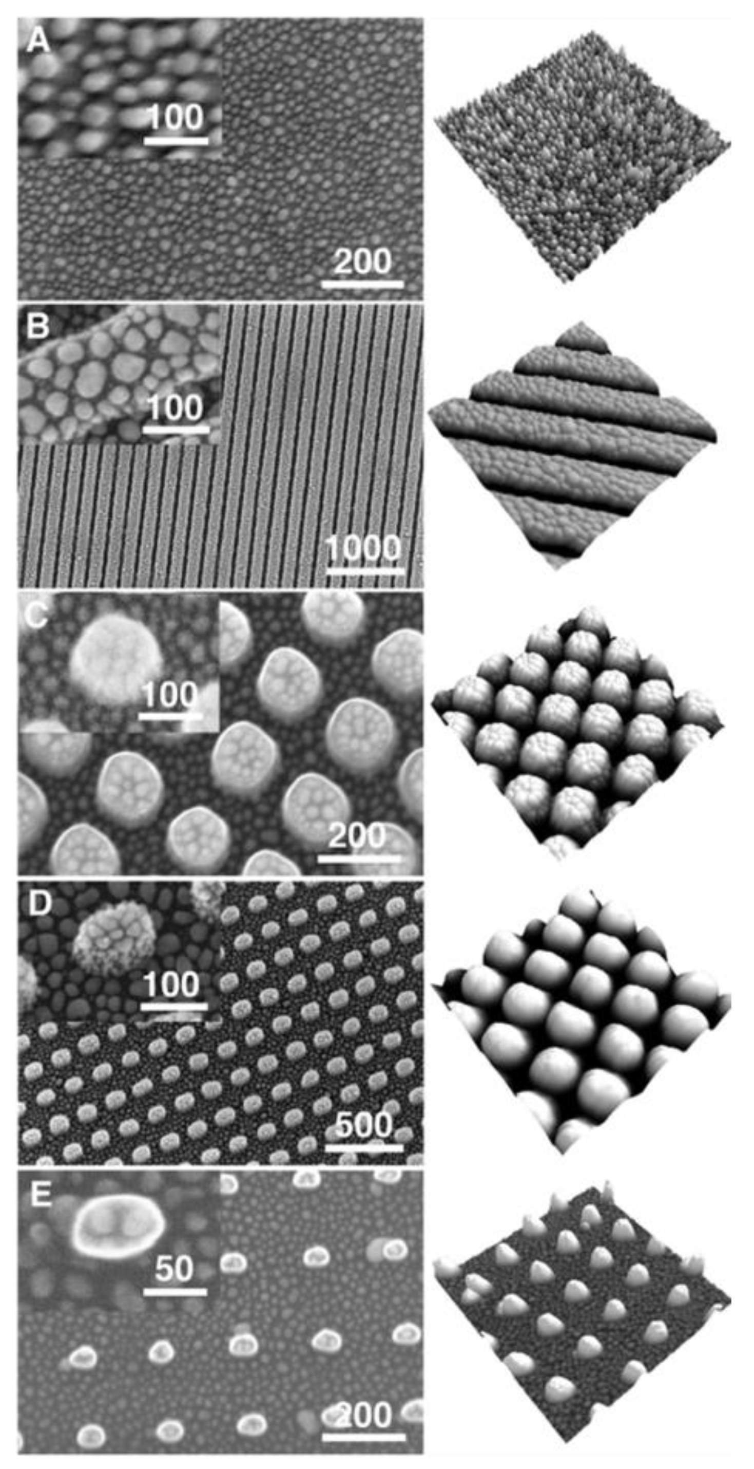


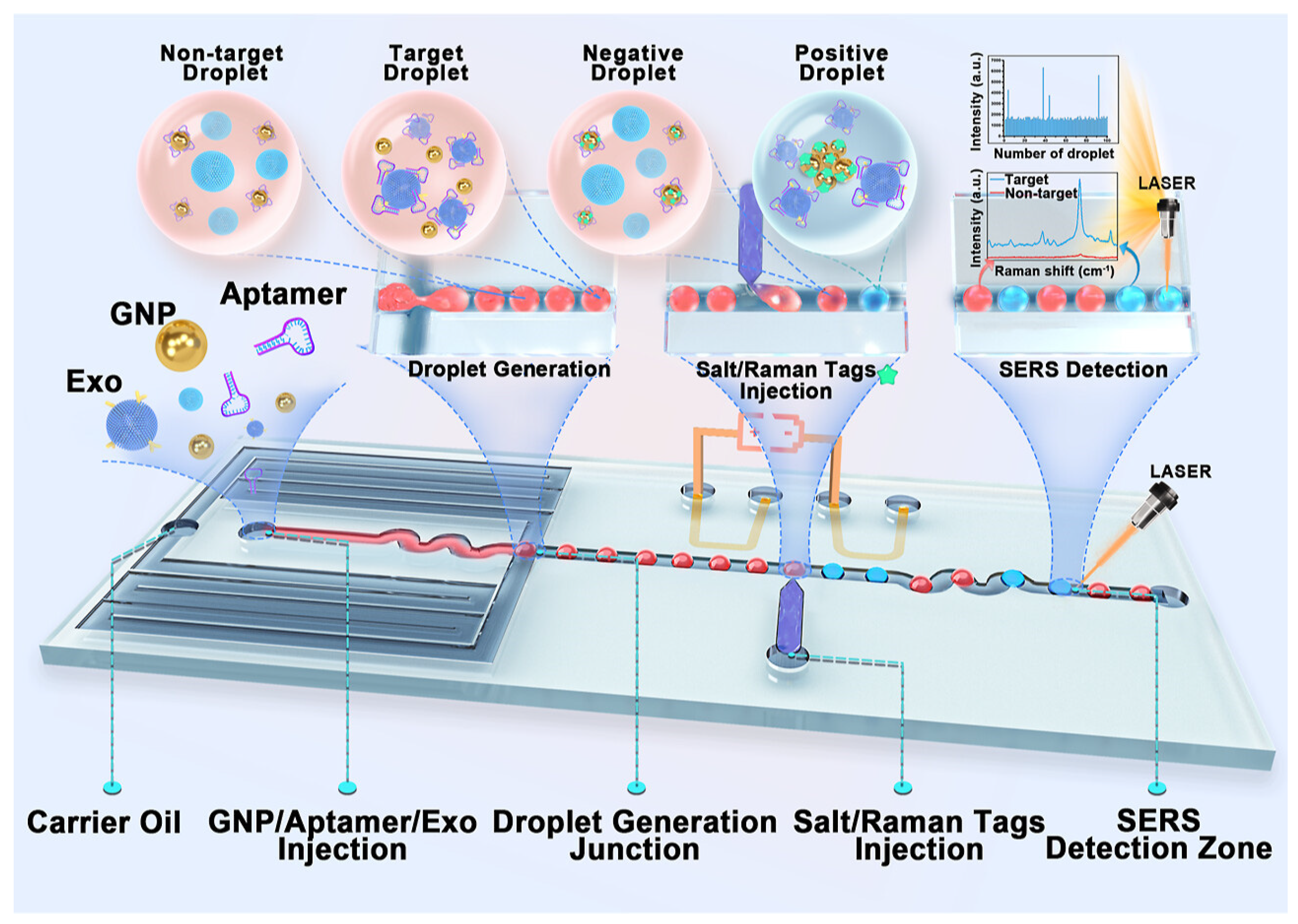

| Effect | Physical Description | Relevance to SERS |
|---|---|---|
| High Entropy | Stabilization of single-phase solid solutions over complex intermetallics. | Ensures structural homogeneity and prevents phase-dependent spectral artifacts. |
| Lattice Distortion | Atomic size mismatches between diverse elements create local strain fields. | Modulates electronic band structures and increases the density of surface defects/active sites. |
| Sluggish Diffusion | High potential barriers for atomic migration at elevated temperatures. | Improves thermal stability of nanostructures, preventing the collapse of hotspots. |
| Cocktail Effect | Cooperative combination of individual elemental properties yielding non-additive behavior. | Permits multi-elemental tuning of LSPR and d-band centers for particular molecular targets. |
| SERS Data Property | Algorithmic Inductive Bias |
|---|---|
| High-dimensional, strongly collinear wavenumber channels | Linear projection (PCA, PLS); ℓ1/ℓ2-regularised regression |
| Informative content concentrated in a sparse subset of bands | Tree ensembles with feature importance (RF, gradient-boosted trees); sparse linear models |
| Vibrational peaks are local 1-D patterns | Convolutional filters (1-D CNN); wavelet bases |
| Modest peak shifts from chemical or substrate variation | Translation-equivariant convolutions; data augmentation in ω |
| Long-range peak correlations (overtones, combination bands) | Self-attention/transformers; deep CNNs with large receptive fields |
| Strong fluorescence backgrounds and shot noise | Autoencoders; denoising autoencoders; physically-motivated baseline priors |
| Small labelled datasets (clinical, forensic) | Kernel SVM; Gaussian processes; Bayesian neural networks; transfer learning |
| Need for calibrated confidence (≥95% deployment thresholds) | Bayesian methods; deep ensembles; conformal prediction |
| Need for physically realistic data augmentation | GANs; VAEs; DFT-as-prior generative pipelines |
| Approach | Mathematical Core | Inductive Bias/SERS Rationale | Performance & Data | Interpretability | SERS Use Case |
|---|---|---|---|---|---|
| Supervised | |||||
| SVM (RBF kernel) | min ½‖w‖2 + C∑ξ; kernel k(x,x′) | Kernel similarity tolerates scarce data; no locality bias | ≈90%, N = 50–200 | Moderate | Bacteria classification |
| Random Forest | Ensemble of decorrelated trees; threshold splits | Feature importances localise discriminative bands; baseline-robust | ≈90%, N = 50–200 | High | Explosives detection |
| Gradient boosting | Boosted decision trees (XGBoost/LightGBM) | Stronger fitting on tabular spectral features than RF; preserves interpretability | 92–95%, N = 50–500 | Moderate–High | Quantitative classification |
| PLS Regression | Latent components ti maximising cov(t, y) | Handles channel collinearity; loadings interpretable as virtual spectra | Good, N = 30–100 | High | Concentration regression |
| Unsupervised | |||||
| PCA/LDA | Eigendecomposition of covariance | Linear denoising via dominant variance modes; fails under non-linear peak shifts | Good | High | Clustering, preprocessing |
| t-SNE/UMAP | KL/cross-entropy on neighbour probabilities | Non-linear manifolds absorb peak shifts; preserve local structure | Excellent visualisation | Moderate | Visualisation; biological subtypes |
| K-means/GMM | Centroid/likelihood partitioning | Anomaly detection via density modelling of normal spectra | Moderate | Moderate | Grouping; QC anomaly flagging |
| Deep learning | |||||
| 1-D CNN | Local equivariant convolutions | Locality + shift-equivariance match local, shift-prone vibrational bands | ≥95%, N > 103 | Moderate (CAM/Grad-CAM) | Virus/pathogen detection |
| 1-D Transformer | Self-attention over spectral channels | Captures long-range peak correlations (overtones, combination bands) | ≥95%, N > 103–104 | Moderate (attention maps) | Multiplexed identification |
| Autoencoder/VAE | Bottleneck reconstruction; KL-regularised latent | Learned non-linear baseline removal; latent likelihood for novelty | Good, N > 103 | Moderate | Denoising; novelty detection |
| GAN | Adversarial min–max | Synthetic augmentation for clinical data scarcity; mode-collapse risk | Good, N > 103 | Low | Data augmentation |
| GNN (Mol2Raman) | Message passing on molecular graph | Atomic permutation/bond locality match vibrational normal modes | High accuracy | Moderate | Inverse design; DFT surrogate |
| Probabilistic/hybrid | |||||
| Gaussian process | Posterior over functions | Calibrated uncertainty for low-data and safety-critical decisions | Good, N < 200 | High | Uncertainty-aware sensing |
| Bayesian NN/deep ensemble | Approximate posterior or ensemble averaging | Calibrated uncertainty in deep-learning regime | Good, N > 103 | Moderate | Clinical decision support |
| DFT + ML hybrid | Physics-grounded data augmentation | Principled training data; complements adversarial augmentation | Excellent, moderate N | High | Molecule ID for unseen analytes |
| Transfer learning | LT + λ Ω(θ − θS) | Low-level features instrument-invariant; high-level task-specific | Excellent, small target N | Moderate | Cross-instrument adaptation |
| Target Analyte | SERS Substrate/Material | Limit of Detection | Key Strategy/Feature | Reference |
|---|---|---|---|---|
| NTO, FOX-7, TNT | Ag-ZnO nanoflower substrate | 10−6–10−14 M | Electromagnetic hotspot enhancement | [334] |
| PA, RDX | Ag-Au nanoparticle micro square array | 3.6 × 10⁻⁸ M (PA), 4 × 10⁻⁷ M (RDX) | Mobile nanomotors enable real time SERS detection | [350] |
| CL-20, TNCB | Alkali ion assisted SERS on Ag surface | Sub-nanomolar | Electrostatic complex formation | [336] |
| NTO, TNB, PA | TiO2 aerogel semiconductor | 2.42 × 10⁻⁷ M | Oxygen vacancies enhance charge transfer Raman enhancement | [337] |
| DMMP (sarin simulant) | Ag nanoplatelets on graphite | 2.5 ppm | Gas phase SERS detection using portable Raman device | [341] |
| VX nerve agent | AuNP decorated quartz fibres | 0.008 µg/L | Fiber based plasmonic substrate enables vapor detection | [343] |
| Nito explosives, drugs | NH2-MIL-101(Fe)@AuNP core shell substrate, MIL-1-1(Cr), ZIF-8 | 0.46–2.31 ng/mL | MOF assisted adsorption enhances sensitivity | [309,351,352] |
| VX, Tabun, Cyclosarin | Au coated nanopillar substrate with oxime capture molecules | Sub-ppm | Functionalized biorecognition interface improves selectivity | [353] |
| Fentanyl (serum/urine) | Liquid–liquid interfacial AuNP plasmonic arrays | 1 ng/mL | Direct detection without pretreatment | [309] |
| Au nanostar decorated rGO | ~0.47 ng/mL | Hybrid plasmonic graphene substrate | [354] | |
| Tramadol (serum) | Au trisoctahedra on flexible tape | 69.19 ng/mL | Wearable sensing platform for forensic detection | [347] |
| Methamphetamine (wastewater) | Au@Ag modified fibrous paper substrate | Sub-ppb | Paper-based flexible SERS sensor | [349] |
Disclaimer/Publisher’s Note: The statements, opinions and data contained in all publications are solely those of the individual author(s) and contributor(s) and not of MDPI and/or the editor(s). MDPI and/or the editor(s) disclaim responsibility for any injury to people or property resulting from any ideas, methods, instructions or products referred to in the content. |
© 2026 by the authors. Licensee MDPI, Basel, Switzerland. This article is an open access article distributed under the terms and conditions of the Creative Commons Attribution (CC BY) license.
Share and Cite
Dewanjee, U.; Bai, S.; Ryabchikov, Y.V.; Fieser, D.; Pradakshina, S.; Wu, J.J.; Fronzi, M.; Hu, A. Next-Generation SERS Probes: Engineering Hotspots, Intelligent Molecular Targeting, and AI-Driven Spectral Analysis for Emerging Applications. Nanomaterials 2026, 16, 628. https://doi.org/10.3390/nano16100628
Dewanjee U, Bai S, Ryabchikov YV, Fieser D, Pradakshina S, Wu JJ, Fronzi M, Hu A. Next-Generation SERS Probes: Engineering Hotspots, Intelligent Molecular Targeting, and AI-Driven Spectral Analysis for Emerging Applications. Nanomaterials. 2026; 16(10):628. https://doi.org/10.3390/nano16100628
Chicago/Turabian StyleDewanjee, Unmanaa, Shi Bai, Yury V. Ryabchikov, David Fieser, Sharma Pradakshina, Jie Jayne Wu, Marco Fronzi, and Anming Hu. 2026. "Next-Generation SERS Probes: Engineering Hotspots, Intelligent Molecular Targeting, and AI-Driven Spectral Analysis for Emerging Applications" Nanomaterials 16, no. 10: 628. https://doi.org/10.3390/nano16100628
APA StyleDewanjee, U., Bai, S., Ryabchikov, Y. V., Fieser, D., Pradakshina, S., Wu, J. J., Fronzi, M., & Hu, A. (2026). Next-Generation SERS Probes: Engineering Hotspots, Intelligent Molecular Targeting, and AI-Driven Spectral Analysis for Emerging Applications. Nanomaterials, 16(10), 628. https://doi.org/10.3390/nano16100628