
Advancements in Bio-Nanotechnology: Green Synthesis and Emerging Applications of Bio-Nanoparticles

 ,
,  , , and
Abstract
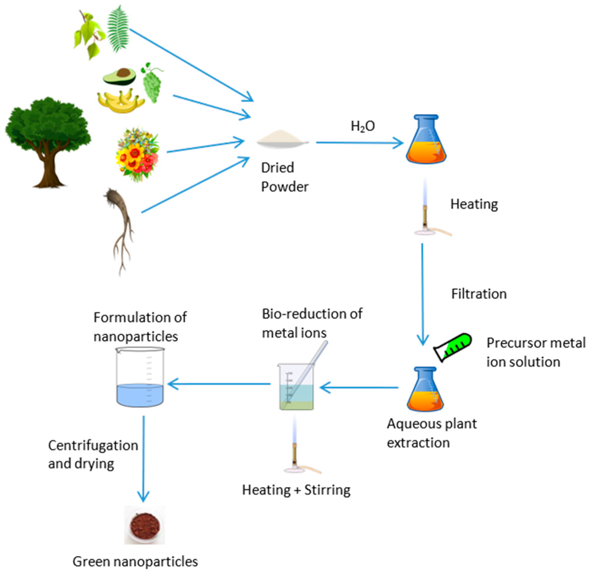
1. Introduction
2. Applications of Bio-Nanoparticles
2.1. Applications of Bio-Nanoparticles in Fuel-Cells
2.2. Applications of Bio-Nanoparticles in Therapeutics
2.3. Applications of Bio-Nanoparticles in Waste Water Treatment
2.4. Applications of Bio-Nanoparticles in the Energy Industry
3. Conclusions
Author Contributions
Funding
Data Availability Statement
Conflicts of Interest
Abbreviations
| ATR-FTIR | Attenuated total reflectance Fourier-transform infrared spectroscopy |
| BET | Brunauer–Emmett–Teller |
| DLS | Dynamic light scattering |
| EDAX | Energy-dispersive X-ray spectroscopy |
| EDS | Energy-dispersive X-ray spectroscopy |
| FESEM | Field emission scanning electron microscopy |
| FESEM-EDX | Field emission scanning electron microscopy with energy dispersive X-ray spectroscopy |
| FTIR | Fourier-transform infrared spectroscopy |
| HRSEM | High-resolution scanning electron microscopy |
| HRTEM | High-resolution transmission electron microscopy |
| SEM | Scanning electron microscopy |
| SEM-EDX | Scanning electron microscopy with energy dispersive X-ray spectroscopy |
| TEM | Transmission electron microscopy |
| TGA | Thermogravimetric analyzer |
| UV–vis | Ultraviolet–visible spectrophotometer |
References
- Galpaya, C.; Induranga, A.; Vithanage, V.; Mantilaka, P.; Koswattage, K.R. Comparative Study on the Thermal Properties of Engine Oils and Their Nanofluids Incorporating Fullerene-C60, TiO2 and Fe2O3 at Different Temperatures. Energies 2024, 17, 732. [Google Scholar] [CrossRef]
- Gunasena, M.D.K.M.; Alahakoon, A.M.P.D.; Polwaththa, K.P.G.D.M.; Galpaya, G.D.C.P.; Priyanjani, H.A.S.A.; Koswattage, K.R.; Senarath, W.T.P.S.K. Transforming Plant Tissue Culture with Nanoparticles: A Review of Current Applications. Plant Nano Biol. 2024, 10, 100102. [Google Scholar] [CrossRef]
- Induranga, A.; Galpaya, C.; Vithanage, V.; Koswattage, K.R. Thermal Properties of TiO2 Nanoparticle-Treated Transformer Oil and Coconut Oil. Energies 2024, 17, 49. [Google Scholar] [CrossRef]
- Shrivastava, V.; Chauhan, P.; Tomar, R.S. Bio-Fabrication of Metal Nanoparticles: A Review. Int. J. Curr. Res. Life Sci. 2019, 7, 1927–1932. [Google Scholar]
- Chen, W.; Cai, W.; Zhang, L.; Wang, G.; Zhang, L. Sonochemical Processes and Formation of Gold Nanoparticles within Pores of Mesoporous Silica. J. Colloid. Interface Sci. 2001, 238, 291–295. [Google Scholar] [CrossRef]
- Eustis, S.; Hsu, H.-Y.; El-Sayed, M.A. Gold Nanoparticle Formation from Photochemical Reduction of Au3+ by Continuous Excitation in Colloidal Solutions. A Proposed Molecular Mechanism. J. Phys. Chem. B 2005, 109, 4811–4815. [Google Scholar] [CrossRef]
- Frattini, A.; Pellegri, N.; Nicastro, D.; de Sanctis, O. Effect of Amine Groups in the Synthesis of Ag Nanoparticles Using Aminosilanes. Mater. Chem. Phys. 2005, 94, 148–152. [Google Scholar] [CrossRef]
- Rodríguez-Sánchez, L.; Blanco, M.C.; López-Quintela, M.A. Electrochemical Synthesis of Silver Nanoparticles. J. Phys. Chem. B 2000, 104, 9683–9688. [Google Scholar] [CrossRef]
- Starowicz, M.; Stypuła, B.; Banaś, J. Electrochemical Synthesis of Silver Nanoparticles. Electrochem. Commun. 2006, 8, 227–230. [Google Scholar] [CrossRef]
- Mafuné, F.; Kohno, J.; Takeda, Y.; Kondow, T. Dissociation and Aggregation of Gold Nanoparticles under Laser Irradiation. J. Phys. Chem. B 2001, 105, 9050–9056. [Google Scholar] [CrossRef]
- Mitrakos, D.; Jokiniemi, J.; Backman, U.; Housiadas, C. Aerosol Flow in a Tube Furnace Reactor of Gas-Phase Synthesised Silver Nanoparticles. J. Nanoparticle Res. 2008, 10, 153–161. [Google Scholar] [CrossRef]
- Zamiri, R.; Azmi, B.Z.; Ahangar, H.A.; Zamiri, G.; Husin, M.S.; Wahab, Z.A. Preparation and Characterization of Silver Nanoparticles in Natural Polymers Using Laser Ablation. Bull. Mater. Sci. 2012, 35, 727–731. [Google Scholar] [CrossRef]
- Zhang, G.; Wang, D. Fabrication of Heterogeneous Binary Arrays of Nanoparticles via Colloidal Lithography. J. Am. Chem. Soc. 2008, 130, 5616–5617. [Google Scholar] [CrossRef]
- Sarkar, T.; Kundu, S.; Ghorai, G.; Sahoo, P.; Reddy, V.; Bhattacharjee, A.C. Synthesis and Characterization of Zinc Ferrite Nanomaterials Vis-à-Vis Studies on Their Photocatalytic Application in Visible Light Dye Degradation. Appl. Phys. A 2025, 131, 266. [Google Scholar] [CrossRef]
- Mulay, M.; Patwardhan, S.; Martsinovich, N.D. Review of Bio-Inspired Green Synthesis of Titanium Dioxide for Photocatalytic Applications. Catalysts 2024, 14, 742. [Google Scholar] [CrossRef]
- Vijan, E.A.; Modan, E.M.; Moga, S.G.; Negrea, D.A.; Schiopu, A.-G.; Oproescu, M.; Istrate, D.E. Assisted Egg White Biogenic Synthesis for Elaboration of ZnO Nanoparticles. Crystals 2025, 15, 71. [Google Scholar] [CrossRef]
- Rajalakshmi, B.; Singh, N.; Madhavi, A.; Khan, I.; Hameed, A.A.; Singh, S.; Rao, A.V.L.F. Bio-Inspired Nanomaterial’s for Energy Harvesting and Storage: A Green Approach. E3S Web Conf. 2024, 552, 01122. [Google Scholar] [CrossRef]
- Akhtar, M.S.; Panwar, J.; Yun, Y.-S. Biogenic Synthesis of Metallic Nanoparticles by Plant Extracts. ACS Sustain. Chem. Eng. 2013, 1, 591–602. [Google Scholar] [CrossRef]
- Singh, H.; Desimone, M.F.; Pandya, S.; Jasani, S.; George, N.; Adnan, M.; Aldarhami, A.; Bazaid, A.S.; Alderhami, S.A. Revisiting the Green Synthesis of Nanoparticles: Uncovering Influences of Plant Extracts as Reducing Agents for Enhanced Synthesis Efficiency and Its Biomedical Applications. Int. J. Nanomed. 2023, 18, 4727–4750. [Google Scholar] [CrossRef]
- Dauthal, P.; Mukhopadhyay, M. Noble Metal Nanoparticles: Plant-Mediated Synthesis, Mechanistic Aspects of Synthesis, and Applications. Ind. Eng. Chem. Res. 2016, 55, 9557–9577. [Google Scholar] [CrossRef]
- Jadoun, S.; Arif, R.; Jangid, N.; Meena, R. Green Synthesis of Nanoparticles Using Plant Extracts: A Review. Environ. Chem. Lett. 2021, 19, 355–374. [Google Scholar] [CrossRef]
- Khatun, M.; Khatun, Z.; Karim, M.R.; Habib, M.R.; Rahman, M.H.; Aziz, M.A. Green Synthesis of Silver Nanoparticles Using Extracts of Mikania Cordata Leaves and Evaluation of Their Antioxidant, Antimicrobial and Cytotoxic Properties. Food Chem. Adv. 2023, 3, 100386. [Google Scholar] [CrossRef]
- Tesfaye, M.; Gonfa, Y.; Tadesse, G.; Temesgen, T.; Periyasamy, S. Green Synthesis of Silver Nanoparticles Using Vernonia Amygdalina Plant Extract and Its Antimicrobial Activities. Heliyon 2023, 9, e17356. [Google Scholar] [CrossRef]
- Tran, T.T.T.; Vu, T.T.H.; Nguyen, T.H. Biosynthesis of Silver Nanoparticles Using Tithonia Diversifolia Leaf Extract and Their Antimicrobial Activity. Mater. Lett. 2013, 105, 220–223. [Google Scholar] [CrossRef]
- Mohammadinejad, R.; Karimi, S.; Iravani, S.; Varma, R.S. Plant-Derived Nanostructures: Types and Applications. Green. Chem. 2015, 18, 20–52. [Google Scholar] [CrossRef]
- Dada, A.O.; Inyinbor, A.A.; Idu, E.I.; Bello, O.M.; Oluyori, A.P.; Adelani-Akande, T.A.; Okunola, A.A.; Dada, O. Effect of Operational Parameters, Characterization and Antibacterial Studies of Green Synthesis of Silver Nanoparticles Using Tithonia Diversifolia. PeerJ 2018, 6, e5865. [Google Scholar] [CrossRef] [PubMed]
- Kharissova, O.V.; Dias, H.V.R.; Kharisov, B.I.; Pérez, B.O.; Pérez, V.M.J. The Greener Synthesis of Nanoparticles. Trends Biotechnol. 2013, 31, 240–248. [Google Scholar] [CrossRef]
- Obayomi, K.; Oluwadiya, A.; Sie Yon, J.L.; Dada, A.O.; Akubuo, D.; Adelani-Akande, T.; Bari, A.S.M.F.; Temidayo, S.; Rahman, M.M. Biosynthesis of Tithonia Diversifolia Leaf Mediated Zinc Oxide Nanoparticles Loaded with Flamboyant Pods (Delonix Regia) for the Treatment of Methylene Blue Wastewater. Arab. J. Chem. 2021, 14, 103363. [Google Scholar] [CrossRef]
- Kaur, H.; Kaur, S.; Singh, M. Biosynthesis of Silver Nanoparticles by Natural Precursor from Clove and Their Antimicrobial Activity. Biologia 2013, 68, 1048–1053. [Google Scholar] [CrossRef]
- Dash, D.S.; Bag, B. Synthesis of Gold Nanoparticles Using Renewable Punica Granatum Juice and Study of Its Catalytic Activity. Appl. Nanosci. 2012, 4, 55–59. [Google Scholar] [CrossRef]
- Alsaiari, N.; Alzahrani, F.; Amari, A.; Osman, H.; Harharah, H.; Elboughdiri, N.; Tahoon, M.A. Plant and Microbial Approaches as Green Methods for the Synthesis of Nanomaterials: Synthesis, Applications, and Future Perspectives. Molecules 2023, 28, 463. [Google Scholar] [CrossRef] [PubMed]
- Arif, M.; Hoque, M.E.; Rahman, M.Z.; Shafoyat, M.U.B. Emerging Directions in Green Nanomaterials: Synthesis, Physicochemical Properties and Applications. Mater. Commun. 2024, 40, 109335. [Google Scholar] [CrossRef]
- Faheem, I.; Shahid, S.; Khan, S.; Ahmad, W.; Zaman, S. Green Synthesis of Copper Oxide Nanoparticles Using Abutilon Indicum Leaf Extract: Antimicrobial, Antioxidant and Photocatalytic Dye Degradation Activitie. Trop. J. Pharm. Res. 2017, 16, 743–753. [Google Scholar] [CrossRef]
- Ahmad, B.; Chang, L.; Satti, U.Q.; Rehman, S.u.; Arshad, H.; Mustafa, G.; Shaukat, U.; Wang, F.; Tong, C. Phyto-Synthesis, Characterization, and In Vitro Antibacterial Activity of Silver Nanoparticles Using Various Plant Extracts. Bioengineering 2022, 9, 779. [Google Scholar] [CrossRef] [PubMed]
- Dulta, K.; Koşarsoy Ağçeli, G.; Chauhan, P.; Jasrotia, R.; Chauhan, P.K.; Ighalo, J.O.I. Multifunctional CuO Nanoparticles with Enhanced Photocatalytic Dye Degradation and Antibacterial Activity. Sustain. Environ. Res. 2022, 32, 2. [Google Scholar] [CrossRef]
- Abou-Zeid, H.; Ismail, G. The Role of Priming with Biosynthesized Silver Nanoparticles in the Response of Triticum Aestivum L to Salt Stress. Egypt. J. Bot. 2018, 58, 73–85. [Google Scholar] [CrossRef]
- Mishra, D.; Chitara, M.K.; Negi, S.; Pal singh, J.; Kumar, R.; Chaturvedi, P. Biosynthesis of Zinc Oxide Nanoparticles via Leaf Extracts of Catharanthus Roseus (L.) G. Don and Their Application in Improving Seed Germination Potential and Seedling Vigor of Eleusine Coracana (L.) Gaertn. Adv. Agric. 2023, 2023, 7412714. [Google Scholar]
- Gonçalves, J.P.Z.; Seraglio, J.; Macuvele, D.L.P.; Padoin, N.; Soares, C.; Riella, H.G. Green Synthesis of Manganese Based Nanoparticles Mediated by Eucalyptus Robusta and Corymbia Citriodora for Agricultural Applications. Colloids Surf. A Physicochem. Eng. Asp. 2022, 636, 128180. [Google Scholar]
- Mathew, S.S.; Sunny, N.E.; Shanmugam, V. Green Synthesis of Anatase Titanium Dioxide Nanoparticles Using Cuminum cyminum Seed Extract; Effect on Mung Bean (Vigna radiata) Seed Germination. Inorg. Chem. Commun. 2021, 126, 108485. [Google Scholar]
- Golzarnezhad, F.; Allahdou, M.; Mehravaran, L.; Naderi, S. Green Synthesis of ZnO Nanoparticles from the Extract of Cymbopogon Olivieri and Investigation of Their Antimicrobial and Anticancer Effects. Discov. Appl. Sci. 2025, 7, 196. [Google Scholar] [CrossRef]
- Batool, S.U.; Javed, B.; Sohail; Zehra, S.S.; Mashwani, Z.-R.; Raja, N.I.; Khan, T.; ALHaithloul, H.A.S.; Alghanem, S.M.; Al-Mushhin, A.A. Exogenous Applications of Bio-Fabricated Silver Nanoparticles to Improve Biochemical, Antioxidant, Fatty Acid and Secondary Metabolite Contents of Sunflower. Nanomaterials 2021, 11, 1750. [Google Scholar] [CrossRef] [PubMed]
- Sackey, J.; Nwanya, A.; Bashir, A.K.H.; Matinise, N.; Ngilirabanga, J.B.; Ameh, A.E.; Coetsee, E.; Maaza, M. Electrochemical Properties of Euphorbia Pulcherrima Mediated Copper Oxide Nanoparticles. Mater. Chem. Phys. 2020, 244, 122714. [Google Scholar]
- Hemmati, S.; Ahmeda, A.; Salehabadi, Y.; Zangeneh, A.; Zangeneh, M.M. Synthesis, Characterization, and Evaluation of Cytotoxicity, Antioxidant, Antifungal, Antibacterial, and Cutaneous Wound Healing Effects of Copper Nanoparticles Using the Aqueous Extract of Strawberry Fruit and L-Ascorbic Acid. Polyhedron 2020, 180, 114425. [Google Scholar]
- Jafarirad, S.; Kosari-Nasab, M.; Tavana, R.M.; Mahjouri, S.; Ebadollahi, R. Impacts of Manganese Bio-Based Nanocomposites on Phytochemical Classification, Growth and Physiological Responses of Hypericum perforatum L. Shoot Cultures. Ecotoxicol. Environ. Saf. 2021, 209, 111841. [Google Scholar] [PubMed]
- Del Buono, D.; Di Michele, A.; Costantino, F.; Trevisan, M.; Lucini, L. Biogenic ZnO Nanoparticles Synthesized Using a Novel Plant Extract: Application to Enhance Physiological and Biochemical Traits in Maize. Nanomaterials 2021, 11, 1270. [Google Scholar] [CrossRef]
- Rafique, M.; Jahangir, J.; Amin, B.A.Z.; Bilal Tahir, M.; Nabi, G.; Isa Khan, M.; Khalid, N.R.; Gillani, S.S.A.; Sadaf, I. Investigation of Photocatalytic and Seed Germination Effects of TiO2 Nanoparticles Synthesized by Melia azedarach L. Leaf Extract. J. Inorg. Organomet. Polym. Mater. 2019, 29, 2133–2144. [Google Scholar]
- Raja, K.; Sowmya, R.; Sudhagar, R.; Moorthy, P.S.; Govindaraju, K.; Subramanian, K.S. Biogenic ZnO and Cu Nanoparticles to Improve Seed Germination Quality in Blackgram (Vigna Mungo). Mater. Lett. 2019, 235, 164–167. [Google Scholar]
- Ambedkar, A.K.; Gautam, D.; Vikal, S.; Singh, M.; Kumar, A.; Sanger, A.; Sharma, K.; Singh, B.P.; Gautam, Y.K. Ocimum sanctum Leaf Extract-Assisted Green Synthesis of Pd-Doped CuO Nanoparticles for Highly Sensitive and Selective NO2 Gas Sensors. ACS Omega 2023, 8, 29663–29673. [Google Scholar]
- Lima, A.K.O.; Vieira, Í.R.S.; Souza, L.M.d.S.; Florêncio, I.; da Silva, I.G.M.; Tavares Junior, A.G.; Machado, Y.A.A.; dos Santos, L.C.; Taube, P.S.; Nakazato, G.; et al. Green Synthesis of Silver Nanoparticles Using Paullinia Cupana Kunth Leaf Extract Collected in Different Seasons: Biological Studies and Catalytic Properties. Pharmaceutics 2025, 17, 356. [Google Scholar] [CrossRef]
- Abd El Aty, A.A. Hafr Al Batin Phoenix dactylifera L. Leaves Extract as Efficient Catalyst for Green Synthesis of New Silver Nanoparticles with Broad Spectrum Antimicrobial Activity: Characterization and Evaluation Compared to Fungi. Arab. J. Sci. Eng. 2025. [Google Scholar] [CrossRef]
- Fatima, E.; Arooj, I.; Javeed, M.; Yin, J.V. Green Synthesis, Characterization and Applications of Phyllanthus Emblica Fruit Extract Mediated Chromium Oxide Nanoparticles. Discov. Nano 2024, 19, 68. [Google Scholar] [CrossRef] [PubMed]
- Mary, A.A.; Ansari, A.T.; Subramanian, R. Sugarcane Juice Mediated Synthesis of Copper Oxide Nanoparticles, Characterization and Their Antibacterial Activity. J. King Saud. Univ. Sci. 2019, 31, 1103–1114. [Google Scholar]
- Buazar, F.; Sweidi, S.; Badri, M.; Kroushawi, F. Biofabrication of Highly Pure Copper Oxide Nanoparticles Using Wheat Seed Extract and Their Catalytic Activity: A Mechanistic Approach. Green. Process. Synth. 2019, 8, 691–702. [Google Scholar]
- Mann, S.; Frankel, R.B.; Blakemore, R.P. Structure, Morphology and Crystal Growth of Bacterial Magnetite. Nature 1984, 310, 405–407. [Google Scholar]
- Shivaji, S.; Madhu, S.; Singh, S. Extracellular Synthesis of Antibacterial Silver Nanoparticles Using Psychrophilic Bacteria. Process Biochem. 2011, 46, 1800–1807. [Google Scholar]
- Sunkar, S.; Nachiyar, C.V. Biogenesis of Antibacterial Silver Nanoparticles Using the Endophytic Bacterium Bacillus Cereus Isolated from Garcinia Xanthochymus. Asian Pac. J. Trop. Biomed. 2012, 2, 953–959. [Google Scholar]
- Wen, L.; Lin, Z.; Gu, P.; Zhou, J.; Yao, B.; Chen, G.; Fu, J. Extracellular Biosynthesis of Monodispersed Gold Nanoparticles by a SAM Capping Route. J. Nanoparticle Res. 2009, 11, 279–288. [Google Scholar]
- Southam, G.; Beveridge, T.J. The in Vitro Formation of Placer Gold by Bacteria. Geochim. Et Cosmochim. Acta 1994, 58, 4527–4530. [Google Scholar]
- Chen, Y.-L.; Tuan, H.-Y.; Tien, C.-W.; Lo, W.-H.; Liang, H.-C.; Hu, Y.-C. Augmented Biosynthesis of Cadmium Sulfide Nanoparticles by Genetically Engineered Escherichia Coli. Biotechnol. Prog. 2009, 25, 1260–1266. [Google Scholar]
- Du, L.; Jiang, H.; Liu, X.; Wang, E. Biosynthesis of Gold Nanoparticles Assisted by Escherichia Coli DH5α and Its Application on Direct Electrochemistry of Hemoglobin. Electrochem. Commun. 2007, 9, 1165–1170. [Google Scholar]
- Holmes, J.D.; Smith, P.R.; Evans-Gowing, R.; Richardson, D.J.; Russell, D.A.; Sodeau, J.R. Energy-Dispersive X-Ray Analysis of the Extracellular Cadmium Sulfide Crystallites of Klebsiella Aerogenes. Arch. Microbiol. 1995, 163, 143–147. [Google Scholar] [CrossRef] [PubMed]
- Korbekandi, H.; Iravani, S.; Abbasi, S. Optimization of Biological Synthesis of Silver Nanoparticles Using Lactobacillus Casei Subsp. Casei. J. Chem. Technol. Biotechnol. 2012, 87, 932–937. [Google Scholar] [CrossRef]
- Philipse, A.P.; Maas, D. Magnetic Colloids from Magnetotactic Bacteria: Chain Formation and Colloidal Stability. Langmuir 2002, 18, 9977–9984. [Google Scholar] [CrossRef]
- Lengke, M.F.; Fleet, M.E.; Southam, G. Morphology of Gold Nanoparticles Synthesized by Filamentous Cyanobacteria from Gold (I)−Thiosulfate and Gold (III)−Chloride Complexes. Langmuir 2006, 22, 2780–2787. [Google Scholar] [CrossRef]
- He, S.; Guo, Z.; Zhang, Y.; Zhang, S.; Wang, J.; Gu, N. Biosynthesis of Gold Nanoparticles Using the Bacteria Rhodopseudomonas Capsulata. Mater. Lett. 2007, 61, 3984–3987. [Google Scholar] [CrossRef]
- Singh, D.; Jain, D.; Rajpurohit, D.; Jat, G.; Kushwaha, H.S.; Singh, A.; Mohanty, S.R.; Al-Sadoon, M.K.; Zaman, W.; Upadhyay, S.K. Bacteria Assisted Green Synthesis of Copper Oxide Nanoparticles and Their Potential Applications as Antimicrobial Agents and Plant Growth Stimulants. Front. Chem. 2023, 11, 1154128. [Google Scholar] [CrossRef]
- Marshall, M.J.; Beliaev, A.S.; Dohnalkova, A.C.; Kennedy, D.W.; Shi, L.; Wang, Z.; Boyanov, M.I.; Lai, B.; Kemner, K.M.; McLean, J.S. C-Type Cytochrome-Dependent Formation of U (IV) Nanoparticles by Shewanella Oneidensis. PLoS Biol. 2006, 4, e268. [Google Scholar] [CrossRef]
- Konishi, Y.; Tsukiyama, T.; Tachimi, T.; Saitoh, N.; Nomura, T.; Nagamine, S. Microbial Deposition of Gold Nanoparticles by the Metal-Reducing Bacterium Shewanella Algae. Electrochim. Acta 2007, 53, 186–192. [Google Scholar] [CrossRef]
- Gajbhiye, M.; Kesharwani, J.; Ingle, A.; Gade, A.; Rai, M. Fungus-Mediated Synthesis of Silver Nanoparticles and Their Activity against Pathogenic Fungi in Combination with Fluconazole. Nanomed. Nanotechnol. Biol. Med. 2009, 5, 382–386. [Google Scholar] [CrossRef]
- Vigneshwaran, N.; Kathe, A.A.; Varadarajan, P.V.; Nachane, R.P.; Balasubramanya, R.H. Biomimetics of Silver Nanoparticles by White Rot Fungus, Phaenerochaete Chrysosporium. Colloids Surf. B Biointerfaces 2006, 53, 55–59. [Google Scholar] [CrossRef]
- Raliya, R.; Biswas, P.; Tarafdar, J.C. TiO2 Nanoparticle Biosynthesis and Its Physiological Effect on Mung Bean (Vigna radiata L.). Biotechnol. Rep. 2015, 5, 22–26. [Google Scholar] [CrossRef] [PubMed]
- Bhainsa, K.C.; D’souza, S.F. Extracellular Biosynthesis of Silver Nanoparticles Using the Fungus Aspergillus Fumigatus. Colloids Surf. B Biointerfaces 2006, 47, 160–164. [Google Scholar] [PubMed]
- Gade, A.K.; Bonde, P.; Ingle, A.P.; Marcato, P.D.; Duran, N.; Rai, M.K. Exploitation of Aspergillus Niger for Synthesis of Silver Nanoparticles. J. Biobased Mater. Bioenergy 2008, 2, 243–247. [Google Scholar]
- Raliya, R.; Tarafdar, J.C. Biosynthesis and Characterization of Zinc, Magnesium and Titanium Nanoparticles: An Eco-Friendly Approach. Int. Nano Lett. 2014, 4, 93. [Google Scholar] [CrossRef]
- Sanghi, R.; Verma, P. Biomimetic Synthesis and Characterisation of Protein Capped Silver Nanoparticles. Bioresour. Technol. 2009, 100, 501–504. [Google Scholar] [CrossRef]
- Balaji, D.S.; Basavaraja, S.; Deshpande, R.; Mahesh, D.B.; Prabhakar, B.K.; Venkataraman, A. Extracellular Biosynthesis of Functionalized Silver Nanoparticles by Strains of Cladosporium Cladosporioides Fungus. Colloids Surf. B Biointerfaces 2009, 68, 88–92. [Google Scholar] [CrossRef]
- Senapati, S.; Ahmad, A.; Khan, M.I.; Sastry, M.; Kumar, R. Extracellular Biosynthesis of Bimetallic Au–Ag Alloy Nanoparticles. Small 2005, 1, 517–520. [Google Scholar] [CrossRef]
- Basavaraja, S.; Balaji, S.D.; Lagashetty, A.; Rajasab, A.H.; Venkataraman, A. Extracellular Biosynthesis of Silver Nanoparticles Using the Fungus Fusarium Semitectum. Mater. Res. Bull. 2008, 43, 1164–1170. [Google Scholar]
- Ingle, A.; Rai, M.; Gade, A.; Bawaskar, M. Fusarium Solani: A Novel Biological Agent for the Extracellular Synthesis of Silver Nanoparticles. J. Nanoparticle Res. 2009, 11, 2079–2085. [Google Scholar] [CrossRef]
- Shaligram, N.S.; Bule, M.; Bhambure, R.; Singhal, R.S.; Singh, S.K.; Szakacs, G.; Pandey, A. Biosynthesis of Silver Nanoparticles Using Aqueous Extract from the Compactin Producing Fungal Strain. Process Biochem. 2009, 44, 939–943. [Google Scholar]
- Kathiresan, K.; Manivannan, S.; Nabeel, M.A.; Dhivya, B. Studies on Silver Nanoparticles Synthesized by a Marine Fungus, Penicillium Fellutanum Isolated from Coastal Mangrove Sediment. Colloids Surf. B Biointerfaces 2009, 71, 133–137. [Google Scholar]
- Vigneshwaran, N.; Ashtaputre, N.M.; Varadarajan, P.V.; Nachane, R.P.; Paralikar, K.M.; Balasubramanya, R.H. Biological Synthesis of Silver Nanoparticles Using the Fungus Aspergillus Flavus. Mater. Lett. 2007, 61, 1413–1418. [Google Scholar]
- Birla, S.S.; Tiwari, V.V.; Gade, A.K.; Ingle, A.P.; Yadav, A.P.; Rai, M.K. Fabrication of Silver Nanoparticles by Phoma Glomerata and Its Combined Effect against Escherichia Coli, Pseudomonas Aeruginosa and Staphylococcus Aureus. Lett. Appl. Microbiol. 2009, 48, 173–179. [Google Scholar]
- Ravindra, B.K.; Rajasab, A.H. A Comparative Study on Biosynthesis of Silver Nanoparticles Using Four Different Fungal Species. Int. J. Pharm. Pharm. Sci. 2014, 6, 372–376. [Google Scholar]
- Binupriya, A.R.; Sathishkumar, M.; Yun, S.-I. Biocrystallization of Silver and Gold Ions by Inactive Cell Filtrate of Rhizopus Stolonifer. Colloids Surf. B Biointerfaces 2010, 79, 531–534. [Google Scholar] [PubMed]
- Mourato, A.; Gadanho, M.; Lino, A.R.; Tenreiro, R. Biosynthesis of Crystalline Silver and Gold Nanoparticles by Extremophilic Yeasts. Bioinorg. Chem. Appl. 2011, 2011, 546074. [Google Scholar]
- Fayaz, A.M.; Balaji, K.; Girilal, M.; Yadav, R.; Kalaichelvan, P.T.; Venketesan, R. Biogenic Synthesis of Silver Nanoparticles and Their Synergistic Effect with Antibiotics: A Study against Gram-Positive and Gram-Negative Bacteria. Nanomed. Nanotechnol. Biol. Med. 2010, 6, 103–109. [Google Scholar]
- Ahmad, A.; Senapati, S.; Khan, M.I.; Kumar, R.; Sastry, M. Extra-/Intracellular Biosynthesis of Gold Nanoparticles by an Alkalotolerant Fungus, Trichothecium sp. J. Biomed. Nanotechnol. 2005, 1, 47–53. [Google Scholar]
- Mukherjee, P.; Ahmad, A.; Mandal, D.; Senapati, S.; Sainkar, S.R.; Khan, M.I.; Parishcha, R.; Ajaykumar, P.V.; Alam, M.; Kumar, R. Fungus-Mediated Synthesis of Silver Nanoparticles and Their Immobilization in the Mycelial Matrix: A Novel Biological Approach to Nanoparticle Synthesis. Nano Lett. 2001, 1, 515–519. [Google Scholar]
- Gericke, M.; Pinches, A. Microbial Production of Gold Nanoparticles. Gold. Bull. 2006, 39, 22–28. [Google Scholar]
- Abboud, Y.; Saffaj, T.; Chagraoui, A.; El Bouari, A.; Brouzi, K.; Tanane, O.; Ihssane, B. Biosynthesis, Characterization and Antimicrobial Activity of Copper Oxide Nanoparticles (CONPs) Produced Using Brown Alga Extract (Bifurcaria Bifurcata). Appl. Nanosci. 2014, 4, 571–576. [Google Scholar] [CrossRef]
- Kathiraven, T.; Sundaramanickam, A.; Shanmugam, N.; Balasubramanian, T. Green Synthesis of Silver Nanoparticles Using Marine Algae Caulerpa Racemosa and Their Antibacterial Activity against Some Human Pathogens. Appl. Nanosci. 2015, 5, 499–504. [Google Scholar] [CrossRef]
- Kannan, R.R.R.; Arumugam, R.; Ramya, D.; Manivannan, K.; Anantharaman, P. Green Synthesis of Silver Nanoparticles Using Marine Macroalga Chaetomorpha Linum. Appl. Nanosci. 2013, 3, 229–233. [Google Scholar] [CrossRef]
- Barwal, I.; Ranjan, P.; Kateriya, S.; Yadav, S.C. Cellular Oxido-Reductive Proteins of Chlamydomonas reinhardtii Control the Biosynthesis of Silver Nanoparticles. J. Nanobiotechnology 2011, 9, 56. [Google Scholar] [CrossRef] [PubMed]
- Annamalai, J.; Nallamuthu, T. Characterization of Biosynthesized Gold Nanoparticles from Aqueous Extract of Chlorella Vulgaris and Their Anti-Pathogenic Properties. Appl. Nanosci. 2015, 5, 603–607. [Google Scholar] [CrossRef]
- El-Rafie, H.M.; El-Rafie, M.; Zahran, M.K. Green Synthesis of Silver Nanoparticles Using Polysaccharides Extracted from Marine Macro Algae. Carbohydr. Polym. 2013, 96, 403–410. [Google Scholar] [CrossRef]
- Prasad, T.N.; Kambala, V.S.R.; Naidu, R. Phyconanotechnology: Synthesis of Silver Nanoparticles Using Brown Marine Algae Cystophora Moniliformis and Their Characterisation. J. Appl. Phycol. 2013, 25, 177–182. [Google Scholar] [CrossRef]
- Venkatesan, J.; Manivasagan, P.; Kim, S.-K.; Kirthi, A.V.; Marimuthu, S.; Rahuman, A.A. Marine Algae-Mediated Synthesis of Gold Nanoparticles Using a Novel Ecklonia Cava. Bioprocess. Biosyst. Eng. 2014, 37, 1591–1597. [Google Scholar] [CrossRef] [PubMed]
- Yousefzadi, M.; Rahimi, Z.; Ghafori, V. The Green Synthesis, Characterization and Antimicrobial Activities of Silver Nanoparticles Synthesized from Green Alga Enteromorpha flexuosa (wulfen) J. Agardh. Mater. Lett. 2014, 137, 1–4. [Google Scholar] [CrossRef]
- Francavilla, M.; Pineda, A.; Romero, A.A.; Colmenares, J.C.; Vargas, C.; Monteleone, M.; Luque, R. Efficient and Simple Reactive Milling Preparation of Photocatalytically Active Porous ZnO Nanostructures Using Biomass Derived Polysaccharides. Green Chem. 2014, 16, 2876–2885. [Google Scholar] [CrossRef]
- Sharma, B.; Purkayastha, D.D.; Hazra, S.; Thajamanbi, M.; Bhattacharjee, C.R.; Ghosh, N.N.; Rout, J. Biosynthesis of Fluorescent Gold Nanoparticles Using an Edible Freshwater Red Alga, Lemanea fluviatilis (L.) C. Ag. and Antioxidant Activity of Biomatrix Loaded Nanoparticles. Bioprocess. Biosyst. Eng. 2014, 37, 2559–2565. [Google Scholar] [CrossRef] [PubMed]
- Singh, M.; Kalaivani, R.; Manikandan, S.; Sangeetha, N.; Kumaraguru, A.K. Facile Green Synthesis of Variable Metallic Gold Nanoparticle Using Padina Gymnospora, a Brown Marine Macroalga. Appl. Nanosci. 2013, 3, 145–151. [Google Scholar]
- Sharma, B.; Purkayastha, D.D.; Hazra, S.; Gogoi, L.; Bhattacharjee, C.R.; Ghosh, N.N.; Rout, J. Biosynthesis of Gold Nanoparticles Using a Freshwater Green Alga, Prasiola Crispa. Mater. Lett. 2014, 116, 94–97. [Google Scholar]
- Mahdavi, M.; Namvar, F.; Ahmad, M.B.; Mohamad, R. Green Biosynthesis and Characterization of Magnetic Iron Oxide (Fe3O4) Nanoparticles Using Seaweed (Sargassum Muticum) Aqueous Extract. Molecules 2013, 18, 5954–5964. [Google Scholar] [CrossRef] [PubMed]
- Azizi, S.; Ahmad, M.B.; Namvar, F.; Mohamad, R. Green Biosynthesis and Characterization of Zinc Oxide Nanoparticles Using Brown Marine Macroalga Sargassum Muticum Aqueous Extract. Mater. Lett. 2014, 116, 275–277. [Google Scholar]
- Namvar, F.; Azizi, S.; Ahmad, M.B.; Shameli, K.; Mohamad, R.; Mahdavi, M.; Tahir, P.M. Green Synthesis and Characterization of Gold Nanoparticles Using the Marine Macroalgae Sargassum Muticum. Res. Chem. Intermed. 2015, 41, 5723–5730. [Google Scholar]
- Senapati, S.; Syed, A.; Moeez, S.; Kumar, A.; Ahmad, A. Intracellular Synthesis of Gold Nanoparticles Using Alga Tetraselmis Kochinensis. Mater. Lett. 2012, 79, 116–118. [Google Scholar]
- Fan, L.; Tu, Z.; Chan, S.H. Recent Development of Hydrogen and Fuel Cell Technologies: A Review. Energy Rep. 2021, 7, 8421–8446. [Google Scholar] [CrossRef]
- Sazali, N.; Wan Salleh, W.N.; Jamaludin, A.S.; Mhd Razali, M.N. New Perspectives on Fuel Cell Technology: A Brief Review. Membranes 2020, 10, 99. [Google Scholar] [CrossRef]
- Baroutaji, A.; Wilberforce, T.; Ramadan, M.; Olabi, A.G. Comprehensive Investigation on Hydrogen and Fuel Cell Technology in the Aviation and Aerospace Sectors. Renew. Sustain. Energy Rev. 2019, 106, 31–40. [Google Scholar] [CrossRef]
- Manoharan, Y.; Hosseini, S.E.; Butler, B.; Alzhahrani, H.; Senior, B.T.F.; Ashuri, T.; Krohn, J. Hydrogen Fuel Cell Vehicles; Current Status and Future Prospect. Appl. Sci. 2019, 9, 2296. [Google Scholar] [CrossRef]
- Chaturvedi, S.; Dave, P.N.; Shah, N.K. Applications of Nano-Catalyst in New Era. J. Saudi Chem. Soc. 2012, 16, 307–325. [Google Scholar] [CrossRef]
- Kamali, M.; Aminabhavi, T.M.; Abbassi, R.; Dewil, R.; Appels, L. Engineered Nanomaterials in Microbial Fuel Cells—Recent Developments, Sustainability Aspects, and Future Outlook. Fuel 2022, 310, 122347. [Google Scholar] [CrossRef]
- Macaskie, L.E.; Mikheenko, I.P.; Omajai, J.B.; Stephen, A.J.; Wood, J. Metallic Bionanocatalysts: Potential Applications as Green Catalysts and Energy Materials. Microb. Biotechnol. 2017, 10, 1171–1180. [Google Scholar] [CrossRef]
- Obileke, K.; Onyeaka, H.; Meyer, E.L.; Nwokolo, N. Microbial Fuel Cells, a Renewable Energy Technology for Bio-Electricity Generation: A Mini-Review. Electrochem. Commun. 2021, 125, 107003. [Google Scholar] [CrossRef]
- Stephen, A.J.; Rees, N.V.; Mikheenko, I.; Macaskie, L.E. Platinum and Palladium Bio-Synthesized Nanoparticles as Sustainable Fuel Cell Catalysts. Front. Energy Res. 2019, 7, 66. [Google Scholar]
- Yong, P.; Mikheenko, I.P.; Macaskie, L.E. A Novel Fuel Cell Catalyst for Clean Energy Production Based on a Bionanocatalyst. Adv. Mater. Res. 2007, 20, 655–658. [Google Scholar]
- Sekar, A.D.; Jayabalan, T.; Muthukumar, H.; Chandrasekaran, N.I.; Mohamed, S.N.; Matheswaran, M. Enhancing Power Generation and Treatment of Dairy Waste Water in Microbial Fuel Cell Using Cu-Doped Iron Oxide Nanoparticles Decorated Anode. Energy 2019, 172, 173–180. [Google Scholar]
- Matsena, M.T.; Tichapondwa, S.M.; Chirwa, E.M.N. Synthesis of Biogenic Palladium Nanoparticles Using Citrobacter Sp. for Application as Anode Electrocatalyst in a Microbial Fuel Cell. Catalysts 2020, 10, 838. [Google Scholar] [CrossRef]
- Lee, D.W.; Jang, J.-H.; Jang, I.; Kang, Y.S.; Jang, S.; Lee, K.Y.; Jang, J.H.; Kim, H.-J.; Yoo, S.J. Bio-derived Co2P Nanoparticles Supported on Nitrogen-doped Carbon as Promising Oxygen Reduction Reaction Electrocatalyst for Anion Exchange Membrane Fuel Cells. Small 2019, 15, 1902090. [Google Scholar]
- Fuku, X.; Modibedi, M.; Matinise, N.; Mokoena, P.; Xaba, N.; Mathe, M. Single Step Synthesis of Bio-Inspired NiO/C as Pd Support Catalyst for Dual Application: Alkaline Direct Ethanol Fuel Cell and CO2 Electro-Reduction. J. Colloid Interface Sci. 2019, 545, 138–152. [Google Scholar] [PubMed]
- Cui, Y.; Chen, X.; Pan, Z.; Wang, Y.; Xu, Q.; Bai, J.; Jia, H.; Zhou, J.; Yong, X.; Wu, X. Biosynthesized Iron Sulfide Nanoparticles by Mixed Consortia for Enhanced Extracellular Electron Transfer in a Microbial Fuel Cell. Bioresour. Technol. 2020, 318, 124095. [Google Scholar]
- Ishak, N.; Kamarudin, S.K.; Timmiati, S.N.; Karim, N.A.; Basri, S. Biogenic Platinum from Agricultural Wastes Extract for Improved Methanol Oxidation Reaction in Direct Methanol Fuel Cell. J. Adv. Res. 2021, 28, 63–75. [Google Scholar] [PubMed]
- Kasturi, P.R.; Arunchander, A.; Kalpana, D.; Selvan, R.K. Bio-Derived Carbon as an Efficient Supporting Electrocatalyst for the Oxygen Reduction Reaction. J. Phys. Chem. Solids 2019, 124, 305–311. [Google Scholar]
- Qian, C.; Guo, X.; Zhang, W.; Yang, H.; Qian, Y.; Xu, F.; Qian, S.; Lin, S.; Fan, T. Co3O4 Nanoparticles on Porous Bio-Carbon Substrate as Catalyst for Oxygen Reduction Reaction. Microporous Mesoporous Mater. 2019, 277, 45–51. [Google Scholar]
- Wang, A.Z.; Gu, F.; Zhang, L.; Chan, J.M.; Radovic-Moreno, A.; Shaikh, M.R.; Farokhzad, O.C. Biofunctionalized Targeted Nanoparticles for Therapeutic Applications. Expert. Opin. Biol. Ther. 2008, 8, 1063–1070. [Google Scholar] [CrossRef]
- Hu, B.; Liu, X.; Zhang, C.; Zeng, X. Food Macromolecule Based Nanodelivery Systems for Enhancing the Bioavailability of Polyphenols. J. Food Drug Anal. 2017, 25, 3–15. [Google Scholar] [CrossRef]
- Kumar, H.; Bhardwaj, K.; Nepovimova, E.; Kuča, K.; Singh Dhanjal, D.; Bhardwaj, S.; Bhatia, S.K.; Verma, R.; Kumar, D. Antioxidant Functionalized Nanoparticles: A Combat against Oxidative Stress. Nanomaterials 2020, 10, 1334. [Google Scholar] [CrossRef]
- Li, Y.; Zheng, X.; Chu, Q. Bio-Based Nanomaterials for Cancer Therapy. Nano Today 2021, 38, 101134. [Google Scholar] [CrossRef]
- Andleeb, A.; Andleeb, A.; Asghar, S.; Zaman, G.; Tariq, M.; Mehmood, A.; Nadeem, M.; Hano, C.; Lorenzo, J.M.; Abbasi, B.H. A Systematic Review of Biosynthesized Metallic Nanoparticles as a Promising Anti-Cancer-Strategy. Cancers 2021, 13, 2818. [Google Scholar] [CrossRef]
- Calhelha, R.C.; Haddad, H.; Ribeiro, L.; Heleno, S.A.; Carocho, M.; Barros, L. Inflammation: What’s There and What’s New? Appl. Sci. 2023, 13, 2312. [Google Scholar] [CrossRef]
- Agarwal, H.; Nakara, A.; Shanmugam, V.K. Anti-Inflammatory Mechanism of Various Metal and Metal Oxide Nanoparticles Synthesized Using Plant Extracts: A Review. Biomed. Pharmacother. 2019, 109, 2561–2572. [Google Scholar] [CrossRef] [PubMed]
- Saha, M.; Sarkar, A. Review on Multiple Facets of Drug Resistance: A Rising Challenge in the 21st Century. J. Xenobiotics 2021, 11, 197–214. [Google Scholar] [CrossRef]
- Madubuonu, N.; Aisida, S.O.; Ahmad, I.; Botha, S.; Zhao, T.; Maaza, M.; Ezema, F.I. Bio-Inspired Iron Oxide Nanoparticles Using Psidium Guajava Aqueous Extract for Antibacterial Activity. Appl. Phys. A 2020, 126, 72. [Google Scholar] [CrossRef]
- Xu, C.; Qiao, L.; Guo, Y.; Ma, L.; Cheng, Y. Preparation, Characteristics and Antioxidant Activity of Polysaccharides and Proteins-Capped Selenium Nanoparticles Synthesized by Lactobacillus Casei ATCC 393. Carbohydr. Polym. 2018, 195, 576–585. [Google Scholar] [CrossRef]
- Oladipo, I.C.; Lateef, A.; Elegbede, J.A.; Azeez, M.A.; Asafa, T.B.; Yekeen, T.A.; Akinboro, A.; Gueguim-Kana, E.B.; Beukes, L.S.; Oluyide, T.O. Enterococcus Species for the One-Pot Biofabrication of Gold Nanoparticles: Characterization and Nanobiotechnological Applications. J. Photochem. Photobiol. B Biol. 2017, 173, 250–257. [Google Scholar] [CrossRef]
- Netala, V.R.; Kotakadi, V.S.; Bobbu, P.; Gaddam, S.A.; Tartte, V. Endophytic Fungal Isolate Mediated Biosynthesis of Silver Nanoparticles and Their Free Radical Scavenging Activity and Anti Microbial Studies. 3 Biotech. 2016, 6, 132. [Google Scholar] [CrossRef]
- Joshi, C.G.; Danagoudar, A.; Poyya, J.; Kudva, A.K.; Dhananjaya, B.L. Biogenic Synthesis of Gold Nanoparticles by Marine Endophytic Fungus-Cladosporium Cladosporioides Isolated from Seaweed and Evaluation of Their Antioxidant and Antimicrobial Properties. Process Biochem. 2017, 63, 137–144. [Google Scholar]
- Firdhouse, J.; Lalitha, P. Apoptotic Efficacy of Biogenic Silver Nanoparticles on Human Breast Cancer MCF-7 Cell Lines. Progress. Biomater. 2015, 4, 113. [Google Scholar]
- Prabhu, D.; Arulvasu, C.; Babu, G.; Manikandan, R.; Srinivasan, P. Biologically Synthesized Green Silver Nanoparticles from Leaf Extract of Vitex Negundo L. Induce Growth-Inhibitory Effect on Human Colon Cancer Cell Line HCT15. Process Biochem. 2013, 48, 317–324. [Google Scholar] [CrossRef]
- KS, U.S.; Govindaraju, K.; Prabhu, D.; Arulvasu, C.; Karthick, V.; Changmai, N. Anti-Proliferative Effect of Biogenic Gold Nanoparticles against Breast Cancer Cell Lines (MDA-MB-231 & MCF-7). Appl. Surf. Sci. 2016, 371, 415–424. [Google Scholar]
- Sulaiman, G.M.; Tawfeeq, A.T.; Jaaffer, M.D. Biogenic Synthesis of Copper Oxide Nanoparticles Using Olea Europaea Leaf Extract and Evaluation of Their Toxicity Activities: An in Vivo and in Vitro Study. Biotechnol. Prog. 2018, 34, 218–230. [Google Scholar] [CrossRef]
- Noor, S.; Shah, Z.; Javed, A.; Ali, A.; Hussain, S.B.; Zafar, S.; Ali, H.; Muhammad, S.A. A Fungal Based Synthesis Method for Copper Nanoparticles with the Determination of Anticancer, Antidiabetic and Antibacterial Activities. J. Microbiol. Methods 2020, 174, 105966. [Google Scholar]
- David, L.; Moldovan, B.; Vulcu, A.; Olenic, L.; Perde-Schrepler, M.; Fischer-Fodor, E.; Florea, A.; Crisan, M.; Chiorean, I.; Clichici, S. Green Synthesis, Characterization and Anti-Inflammatory Activity of Silver Nanoparticles Using European Black Elderberry Fruits Extract. Colloids Surf. B Biointerfaces 2014, 122, 767–777. [Google Scholar]
- Moldovan, B.; David, L.; Vulcu, A.; Olenic, L.; Perde-Schrepler, M.; Fischer-Fodor, E.; Baldea, I.; Clichici, S.; Filip, G.A. In Vitro and in Vivo Anti-Inflammatory Properties of Green Synthesized Silver Nanoparticles Using Viburnum opulus L. Fruits Extract. Mater. Sci. Eng. C 2017, 79, 720–727. [Google Scholar]
- Muniyappan, N.; Nagarajan, N.S. Green Synthesis of Silver Nanoparticles with Dalbergia Spinosa Leaves and Their Applications in Biological and Catalytic Activities. Process Biochem. 2014, 49, 1054–1061. [Google Scholar] [CrossRef]
- Islam, N.U.; Amin, R.; Shahid, M.; Amin, M.; Zaib, S.; Iqbal, J. A Multi-Target Therapeutic Potential of Prunus Domestica Gum Stabilized Nanoparticles Exhibited Prospective Anticancer, Antibacterial, Urease-Inhibition, Anti-Inflammatory and Analgesic Properties. BMC Complement. Altern. Med. 2017, 17, 276. [Google Scholar] [CrossRef]
- Krithika, S.; Niraimathi, K.L.; Arun, K.P.; Narendran, R.; Balaji, K.; Brindha, P. In Vitro Anti-Inflammatory Studies on Silver Nanoparticles Synthesized from Centratherum Punctatum Cass. Int. J. Res. Ayurveda Pharm. 2016, 7, 61–66. [Google Scholar] [CrossRef]
- Aref, M.S.; Salem, S.S. Bio-Callus Synthesis of Silver Nanoparticles, Characterization, and Antibacterial Activities via Cinnamomum Camphora Callus Culture. Biocatal. Agric. Biotechnol. 2020, 27, 101689. [Google Scholar]
- Alsamhary, K.I. Eco-Friendly Synthesis of Silver Nanoparticles by Bacillus Subtilis and Their Antibacterial Activity. Saudi J. Biol. Sci. 2020, 27, 2185–2191. [Google Scholar]
- Coelho, N.; Jacinto, J.P.; Silva, R.; Soares, J.C.; Pereira, A.S.; Tavares, P. Green Synthesis and Antibacterial Activity of Silver Nanoparticles Obtained from Moringa Oleifera Seed Cake. Coatings 2023, 13, 1439. [Google Scholar] [CrossRef]
- Al Moudani, N.; Ouahidi, I.; Laaraj, S.; Aarab, L. Silver Bio-Nanoparticles Synthesis from Tetraclinis Articulata Leaves Extract and Their Anti-Inflammatory, Antioxidant, and Cytotoxicity Activities. ChemistrySelect 2025, 10, e202404860. [Google Scholar] [CrossRef]
- Kumar, B.; Smita, K.; Kumar, B. Phytochemical Functionalized Metal Nanocatalyst (Ag, Au, Fe, Zn and Pd) for Remediation of Organic Dyes. In Advances in Chemistry Research; Nova Science Publishers: Hauppauge, NY, USA, 2017; pp. 87–120. ISBN 978-1-5361-1054-8. [Google Scholar]
- Sharma, M.; Jain, T.; Singh, S.; Pandey, O.P. Photocatalytic Degradation of Organic Dyes under UV–Visible Light Using Capped ZnS Nanoparticles. Sol. Energy 2012, 86, 626–633. [Google Scholar] [CrossRef]
- Safavi, A.; Momeni, S. Highly Efficient Degradation of Azo Dyes by Palladium/Hydroxyapatite/Fe3O4 Nanocatalyst. J. Hazard. Mater. 2012, 201–202, 125–131. [Google Scholar] [CrossRef]
- Kumar, B. Green Synthesis of Gold, Silver, and Iron Nanoparticles for the Degradation of Organic Pollutants in Wastewater. J. Compos. Sci. 2021, 5, 219. [Google Scholar] [CrossRef]
- Ghosh, N.; Das, S.; Biswas, G.; Haldar, P.K. Review on Some Metal Oxide Nanoparticles as Effective Adsorbent in Wastewater Treatment. Water Sci. Technol. 2022, 85, 3370–3395. [Google Scholar] [CrossRef]
- Gupta, K.; Joshi, P.; Gusain, R.; Khatri, O.P. Recent Advances in Adsorptive Removal of Heavy Metal and Metalloid Ions by Metal Oxide-Based Nanomaterials. Coord. Chem. Rev. 2021, 445, 214100. [Google Scholar] [CrossRef]
- Eldoma, M.A.; Alaswad, S.O.; Mahmoud, M.A.; Qudsieh, I.Y.; Hassan, M.; Bakather, O.Y.; Elawadi, G.A.; Abouatiaa, A.F.F.; Alomar, M.S.; Elhassan, M.S.; et al. Enhancing Photocatalytic Performance of Co-TiO2 and Mo-TiO2-Based Catalysts through Defect Engineering and Doping: A Study on the Degradation of Organic Pollutants under UV Light. J. Photochem. Photobiol. A Chem. 2024, 446, 115164. [Google Scholar] [CrossRef]
- Balestri, A.; Cardellini, J.; Berti, D. Gold and Silver Nanoparticles as Tools to Combat Multidrug-Resistant Pathogens. Curr. Opin. Colloid. Interface Sci. 2023, 66, 101710. [Google Scholar] [CrossRef]
- Rafique, M.; Tahir, M.B.; Irshad, M.; Nabi, G.; Gillani, S.S.A.; Iqbal, T.; Mubeen, M. Novel Citrus Aurantifolia Leaves Based Biosynthesis of Copper Oxide Nanoparticles for Environmental and Wastewater Purification as an Efficient Photocatalyst and Antibacterial Agent. Optik 2020, 219, 165138. [Google Scholar]
- Arumugam, V.; Sriram, P.; Yen, T.-J.; Redhi, G.G.; Gengan, R.M. Nano-Material as an Excellent Catalyst for Reducing a Series of Nitroanilines and Dyes: Triphosphonated Ionic Liquid-CuFe2O4-Modified Boron Nitride. Appl. Catal. B Environ. 2018, 222, 99–114. [Google Scholar]
- Sebeia, N.; Jabli, M.; Ghith, A. Biological Synthesis of Copper Nanoparticles, Using Nerium Oleander Leaves Extract: Characterization and Study of Their Interaction with Organic Dyes. Inorg. Chem. Commun. 2019, 105, 36–46. [Google Scholar]
- Varshney, S.; Gupta, A. Forest Industrial Biomass Residue-Mediated Green Synthesized Multifunctional Copper Oxide Nanoparticles for Efficient Wastewater Treatment and Biomedical Applications. J. Clean. Prod. 2024, 434, 140109. [Google Scholar] [CrossRef]
- Eid, A.M.; Fouda, A.; Hassan, S.E.-D.; Hamza, M.F.; Alharbi, N.K.; Elkelish, A.; Alharthi, A.; Salem, W.M. Plant-Based Copper Oxide Nanoparticles; Biosynthesis, Characterization, Antibacterial Activity, Tanning Wastewater Treatment, and Heavy Metals Sorption. Catalysts 2023, 13, 348. [Google Scholar] [CrossRef]
- Fatima, B.; Siddiqui, S.; Ahmed, R.; Chaudhry, S.A. Preparation of Functionalized CuO Nanoparticles Using Brassica Rapa Leave Extract for Water Purification. Desalin. Water Treat. 2019, 164, 192–205. [Google Scholar]
- Vasantharaj, S.; Sathiyavimal, S.; Senthilkumar, P.; Kalpana, V.N.; Rajalakshmi, G.; Alsehli, M.; Elfasakhany, A.; Pugazhendhi, A. Enhanced Photocatalytic Degradation of Water Pollutants Using Bio-Green Synthesis of Zinc Oxide Nanoparticles (ZnO NPs). J. Environ. Chem. Eng. 2021, 9, 105772. [Google Scholar]
- Rambabu, K.; Bharath, G.; Banat, F.; Show, P.L. Green Synthesis of Zinc Oxide Nanoparticles Using Phoenix dactylifera Waste as Bioreductant for Effective Dye Degradation and Antibacterial Performance in Wastewater Treatment. J. Hazard. Mater. 2021, 402, 123560. [Google Scholar] [CrossRef] [PubMed]
- Chauhan, A.K.; Kataria, N.; Garg, V.K. Green Fabrication of ZnO Nanoparticles Using Eucalyptus Spp. Leaves Extract and Their Application in Wastewater Remediation. Chemosphere 2020, 247, 125803. [Google Scholar] [CrossRef] [PubMed]
- Kumar, B.; Smita, K.; Debut, A.; Cumbal, L. Utilization of Persea Americana (Avocado) Oil for the Synthesis of Gold Nanoparticles in Sunlight and Evaluation of Antioxidant and Photocatalytic Activities. Environ. Nanotechnol. Monit. Manag. 2018, 10, 231–237. [Google Scholar]
- Baruah, D.; Goswami, M.; Yadav, R.N.S.; Yadav, A.; Das, A.M. Biogenic Synthesis of Gold Nanoparticles and Their Application in Photocatalytic Degradation of Toxic Dyes. J. Photochem. Photobiol. B Biol. 2018, 186, 51–58. [Google Scholar]
- Sharma, P.; Pant, S.; Rai, S.; Yadav, R.B.; Dave, V. Green Synthesis of Silver Nanoparticle Capped with Allium Cepa and Their Catalytic Reduction of Textile Dyes: An Ecofriendly Approach. J. Polym. Environ. 2018, 26, 1795–1803. [Google Scholar]
- Rather, M.Y.; Sundarapandian, S. Magnetic Iron Oxide Nanorod Synthesis by Wedelia Urticifolia (Blume) DC. Leaf Extract for Methylene Blue Dye Degradation. Appl. Nanosci. 2020, 10, 2219–2227. [Google Scholar]
- Lohrasbi, S.; Kouhbanani, M.A.J.; Beheshtkhoo, N.; Ghasemi, Y.; Amani, A.M.; Taghizadeh, S. Green Synthesis of Iron Nanoparticles Using Plantago Major Leaf Extract and Their Application as a Catalyst for the Decolorization of Azo Dye. BioNanoScience 2019, 9, 317–322. [Google Scholar]
- El Golli, A.; Contreras, S.; Dridi, C. Bio-Synthesized ZnO Nanoparticles and Sunlight-Driven Photocatalysis for Environmentally-Friendly and Sustainable Route of Synthetic Petroleum Refinery Wastewater Treatment. Sci. Rep. 2023, 13, 20809. [Google Scholar] [CrossRef]
- Yusuf, M.; Abdullah, B. Fossil Fuels, Rising Population, and Global Warming: The Interlinked Phenomena. Orient. J. Phys. Sci. 2020, 5, 49–52. [Google Scholar] [CrossRef]
- Fang, H. Analysis of the Causes and Crisis of Global Warming. MATEC Web Conf. 2023, 386, 03018. [Google Scholar] [CrossRef]
- Ghosh, S.; Ghosh, B. 160-Fossil Fuel Consumption Trend and Global Warming Scenario: Energy Overview. Glob. J. Eng. Sci. 2020, 5, 1–5. [Google Scholar] [CrossRef]
- Akram, F.; Saleem, B.; Irfan, M.; Shakir, H.A.; Khan, M.; Ali, S.; Saeed, S.; Mehmood, T.; Franco, M. Recent Trends for Production of Biofuels Using Algal Biomass. In Basic Research Advancement for Algal Biofuels Production; Srivastava, N., Mishra, P.K., Eds.; Springer: Singapore, 2023; pp. 27–58. ISBN 978-981-19-6810-5. [Google Scholar]
- Sanju, S.; Thakur, A.; Misra, P.; Shukla, P.K. Algal Biomass and Biofuel Production. In Bioprospecting of Microorganism-Based Industrial Molecules; John Wiley & Sons, Ltd.: Hoboken, NJ, USA, 2021; pp. 357–376. ISBN 978-1-119-71731-7. [Google Scholar]
- Dabirian, E.; Hajipour, A.; Mehrizi, A.A.; Karaman, C.; Karimi, F.; Loke-Show, P.; Karaman, O. Nanoparticles Application on Fuel Production from Biological Resources: A Review. Fuel 2023, 331, 125682. [Google Scholar] [CrossRef]
- Jayabal, R.; Soundararajan, G.; Kumar, R.A.; Choubey, G.; Devarajan, Y.; Raja, T.; Kaliappan, N. Study of the Effects of Bio-Silica Nanoparticle Additives on the Performance, Combustion, and Emission Characteristics of Biodiesel Produced from Waste Fat. Sci. Rep. 2023, 13, 18907. [Google Scholar] [CrossRef]
- Karpagam, R.; Rani, K.; Ashokkumar, B.; Ganesh Moorthy, I.; Dhakshinamoorthy, A.; Varalakshmi, P. Green Energy from Coelastrella sp. M-60: Bio-Nanoparticles Mediated Whole Biomass Transesterification for Biodiesel Production. Fuel 2020, 279, 118490. [Google Scholar] [CrossRef]
- Lahiri, D.; Nag, M.; Ghosh, S.; Ray, R.R. Chapter 8—Green Synthesis of Nanoparticles and Their Applications in the Area of Bioenergy and Biofuel Production. In Nanomaterials; Kumar, R.P., Bharathiraja, B., Eds.; Academic Press: Cambridge, MA, USA, 2021; pp. 195–219. ISBN 978-0-12-822401-4. [Google Scholar]
- Ettefaghi, E.; Ghobadian, B.; Rashidi, A.; Najafi, G.; Khoshtaghaza, M.H.; Rashtchi, M.; Sadeghian, S. A Novel Bio-Nano Emulsion Fuel Based on Biodegradable Nanoparticles to Improve Diesel Engines Performance and Reduce Exhaust Emissions. Renew. Energy 2018, 125, 64–72. [Google Scholar] [CrossRef]
- Duman, F.; Sahin, U.; Atabani, A.E. Harvesting of Blooming Microalgae Using Green Synthetized Magnetic Maghemite (γ-Fe2O3) Nanoparticles for Biofuel Production. Fuel 2019, 256, 115935. [Google Scholar] [CrossRef]
- Sawaira; Alsaiari, M.; Ahmad, M.; Munir, M.; Zafar, M.; Sultana, S.; Dawood, S.; Almohana, A.I.; Hassan, M.H.A.-M.; Alharbi, A.F.; et al. Efficient Application of Newly Synthesized Green Bi2O3 Nanoparticles for Sustainable Biodiesel Production via Membrane Reactor. Chemosphere 2023, 310, 136838. [Google Scholar] [CrossRef]
- Hazmi, B.; Rashid, U.; Taufiq-Yap, Y.H.; Ibrahim, M.L.; Nehdi, I.A. Supermagnetic Nano-Bifunctional Catalyst from Rice Husk: Synthesis, Characterization and Application for Conversion of Used Cooking Oil to Biodiesel. Catalysts 2020, 10, 225. [Google Scholar] [CrossRef]
- Pandit, P.R.; Fulekar, M.H. Egg Shell Waste as Heterogeneous Nanocatalyst for Biodiesel Production: Optimized by Response Surface Methodology. J. Environ. Manag. 2017, 198, 319–329. [Google Scholar] [CrossRef]
- Umeagukwu, O.E.; Onukwuli, D.O.; Ude, C.N.; Esonye, C.; Ekwueme, B.N.; Asadu, C.O.; Okey-Onyesolu, F.C.; Ikenna, M.U.; Chukwudi, E.I.; Makhkamov, T.; et al. Transesterification of Persea Americana Seed Oil to Methyl Ester Using Bio-Based Heterogeneous Catalyst: Optimization and Techno-Economic Analysis. Green. Technol. Sustain. 2024, 2, 100086. [Google Scholar] [CrossRef]

| Biological Material | Name | Morphology | Nanoparticle Size (nm) | Nanoparticle | Reference |
|---|---|---|---|---|---|
| Plant | Abutilon indicum leaves | Hexagonal | 16 | CuO | [33] |
| Aloe vera leaves | Spherical | 15–50 | Ag | [34] | |
| Bergenia ciliata Rhizome | Spherical | 20 | CuO | [35] | |
| Capparis spinosa tissues | Spherical and semispherical | 15–30 | Ag | [36] | |
| Catharanthus roseus leaves | Hexagonal | 35 | ZnO | [37] | |
| Coriandrum sativum leaves | Spherical | 15–50 | Ag | [34] | |
| Corymbia citriodora leaves | Needle | 21–28 | Mn | [38] | |
| Cuminum cyminum seeds | Crystalline | 15 | TiO2 | [39] | |
| Cymbopogon citratus leaves | Spherical | 15–50 | Ag | [34] | |
| Cymbopogon olivieri | Spherical | 28 | ZnO | [40] | |
| Eucalyptus robusta leaves | Spherical | 16–23 | Mn | [38] | |
| Euphorbia helioscopia leaves | Crystalline | 30–100 | Ag | [41] | |
| Euphorbia pulcherrima flowers | Cubical | 16–54 | CuO | [42] | |
| Fragaria ananassa fruits | Spherical | 10–30 | Cu | [43] | |
| Hypericum perforatum leaves | Spherical | 20–50 | MnO2 | [44] | |
| Lemna minor tissues | Spherical | 10–20 | ZnO | [45] | |
| Melia azedarach leaves | Crystalline and spherical | 50–71 | TiO2 | [46] | |
| Mentha arvensis leaves | Spherical | 15–50 | Ag | [34] | |
| Nerium oleander leaves | Spherical | 26 | Cu | [47] | |
| Ocimum sanctum leaf | Granular | - | CuO | [48] | |
| Paullinia cupana Kunth leaf extract | Spherical morphology | 39–126 | Ag | [49] | |
| Phoenix dactylifera L leaves | Cubic to spherical | 12–97 | Ag | [50] | |
| Phyllanthus emblica fruit | Large, irregularly shaped flakes | - | Cr2O3 | [51] | |
| Saccharum officinarum stem | Spherical, square, cube, plate, rectangular | 29–60 | CuO | [52] | |
| Triticum aestivum seed | Spherical | 21–42 | CuO | [53] | |
| Bacteria | Aquaspirillum magnetotacticum | Octahedral prism | 40–50 | Fe2O3 | [54] |
| Arthrobacter gangotriensis | Spherical | 5–6 | Ag | [55] | |
| Arthrobacter kerguelensis | Spherical | 5 | Ag | [55] | |
| Bacillus cecembensis | Spherical | 7 | Ag | [55] | |
| Bacillus cereus | Spherical | 20–40 | Ag | [56] | |
| Bacillus indicus | - | 4–6 | Ag | [55] | |
| Bacillus megaterium D01 | Spherical | 2.5 | Au | [57] | |
| Bacillus subtilis 168 | Hexagonal-octahedral | 5–50 | Au | [58] | |
| Escherichia coli | Wurtzite structure | 2–5 | CdS | [59] | |
| Escherichia coli DH 5α | Spherical | 8–25 | Au | [60] | |
| Klebsiella aerogenes | - | 20–200 | CdS | [61] | |
| Lactobacillus casei | Spherical | 20–50 | Ag | [62] | |
| Magnetospirillum magnetotacticum | Chain | 47 | Fe3O4 | [63] | |
| Plectonemaboryanum UTEX 485 | Cubic, octahedral | 10–25 | Au | [64] | |
| Pseudomonas antarctica | Spherical | 11–12 | Ag | [55] | |
| Pseudomonas meridiana | Spherical | 5–6 | Ag | [55] | |
| Pseudomonas proteolytica | Spherical | 7 | Ag | [55] | |
| Rhodopseudomonas capsulate | Spherical | 10–20 | Au | [65] | |
| Serratia sp. (ZTB29) | Polydisperse, spherical | 20–40 | CuO | [66] | |
| Shewanella oneidensis | - | 1–5 | UO2 | [67] | |
| Shewanella alga | Triangular | 10–20 | Au | [68] | |
| Fungi | Alternata alternate | Spherical | 20–60 | Ag | [69] |
| Aspergillus flavus | - | 1–8 | Ag | [70] | |
| Aspergillus flavus TFR7 | Spherical | 12–15 | TiO2 | [71] | |
| Aspergillus fumigates | Spherical | 5–25 | Ag | [72] | |
| Aspergillus niger | Spherical | 20 | Ag | [73] | |
| Aspergillus terreus | Spherical | 8 | ZnO | [74] | |
| Cariolus versicolor | Spherical | 25–75 | Ag | [75] | |
| Cladosporium cladosporioides | Spherical | 10–100 | Ag | [76] | |
| Fusarium oxysporum | Spherical | 8–14 | Au-Ag alloy | [77] | |
| Fusarium semitectum | Crystalline spherical | 10–60 | Ag | [78] | |
| Fusarium solani | Spherical | 5–35 | Ag | [79] | |
| Penicillium brecompactum | Crystalline spherical | 23–105 | Ag | [80] | |
| Penicillium fellutanum | Spherical | 5–25 | Ag | [81] | |
| Phanerochaete chrysosporium | Pyramidal | 50–200 | Ag | [82] | |
| Phoma glomerata | Spherical | 60–80 | Ag | [83] | |
| Rhizopus nigricans | Round | 35–40 | Ag | [84] | |
| Rhizopus stolonifer | Spherical | 25–30, 1–5 | Ag Au | [85] | |
| Saccharimyces cerevisae broth | Spherical | 4–15 | Ag, Au | [86] | |
| Trichoderma viride | Spherical | 5–40 | Ag | [87] | |
| Trichothecium sp. | Spherical, rod-like, triangular | 10–25 | Au | [88] | |
| Verticillium | Spherical | 21–25 | Ag | [89] | |
| Verticillium luteoalbum | Triangular, hexagonal | 10 | Au | [90] | |
| Algae | Bifurcaria bifurcate | Crystalline | 5–45 | CuO | [91] |
| Caulerpa racemosa | Spherical and triangular | 5–25 | Ag | [92] | |
| Chaetomorpha linum | Nano-clusters | 3–44 | Ag | [93] | |
| Chlamydomonas reinhardtii | Round/rectangular | 5–35 | Ag | [94] | |
| Chlorella vulgaris | Crystalline | 2–10 | Au | [95] | |
| Colpmenia sinusa | Spherical | 20 | Ag | [96] | |
| Cystophora moniliformis | Spherical | 50–100 | Ag | [97] | |
| Ecklonia cava | Spherical and triangular | 30 | Au | [98] | |
| Enteromorpha flexuosa | Spherical | 2–32 | Ag | [99] | |
| Enteromorpha flexuosa | Spherical | 2–32 | Ag | [99] | |
| Gracilaria gracilis | Crystalline | 25–50 | ZnO | [100] | |
| Jania rubins | Spherical | 12 | Ag | [96] | |
| Lemanea fluviatilis | Spherical | 5–15 | Au | [101] | |
| Padina gymnospora | Spherical | 53–67 | Au | [102] | |
| Prasiola crispa | Spherical | 5–25 | Au | [103] | |
| Pterocladia capillacae | Spherical | 7 | Ag | [96] | |
| Sargassum muticum | Cubic | 18 | Fe3O4 | [104] | |
| Sargassum muticum | Hexagonal wurtzite | 30–57 | ZnO | [105] | |
| Sargassum muticum | Spherical | 5.4 | Au | [106] | |
| Tetraselmis kochinensis | Spherical and triangular | 5–35 | Au | [107] | |
| Ulva faciata | Spherical | 7 | Ag | [96] |
| Biological Material | Synthesized NP | Characterization Technique | Nanoparticle Size and Morphology | Application | Method/ Measurement | Results | Ref. |
|---|---|---|---|---|---|---|---|
| Escherichia coli MC4100 | E. coli-Pt/Pd (10%: 10%), E-coil-Pt (10%), and E-coil-Pd (10%) alloyed catalysts | Transmission electron microscope (TEM) X-ray diffraction (XRD) | 5.2 nm | Fuel cell catalysts in polymer electrolyte fuel cell catalysts | The nanoparticles were synthesized by initially forming Pd nanoparticles on the E. coli cells, followed by Pt synthesis mediated by the Pd nanoparticles reducing Pt (IV) using K2PtCl6 and Na2PdCl4. | E. coli-Pt/Pd (10%:10%) showed better ECSA (electrochemical loaded area) compared to the other two samples. | [116] |
| Escherichia coli MC4100 | Bio-Pd (desulfurized) nanoparticles Bio-Pd (E-coil) nanoparticles | TEM | 30 nm | Fuel cell catalysts in proton exchange fuel cell catalysts | Four electrodes were manufactured: 1—Commercial Pt nanoparticles; 2—Commercial Pd nanoparticles; 3—Desulfurized Bio-Pd nanoparticles; 4—E-coil bio-nanoparticles. | Maximum power generated by each electrode was 0.13, 0.10, 0.11, and 0.04 watts. | [117] |
| Dairy wastewater | Cu-doped FeO | XRD Scanning electron microscope (SEM) | 70–200 nm | Anode catalysts in a microbial fuel cell | Copper-doped iron oxide nanoparticles (Cu-doped FeO) were synthesized using phyto-compounds of the A. blitum plant. | 161.5 W/m2 peak power density was delivered at 270 A/m2 current density. | [118] |
| Citrobacter | Bio-Pd nanoparticles | SEM XRD Energy-dispersive X-ray spectroscopy (EDS) | 15.65–11.37 nm | Electrocatalysts for anion exchange membrane fuel cells | Bio-Pd was extracted from Pd (II) solution in the basal mineral medium using Citrobacter; 4 mg/cm2 and 2 mg/cm2 Bio-Pd nanoparticles were applied as anode catalysts. | 4 mg/cm2 solution achieved 539.3 mW/cm2 maximum power density, which is 31.1% and 59.6% higher than that of 2 mg/cm2 solution and carbon rod. | [119] |
| Bean sprout | Bio-derived Co2P nanoparticles | SEM TEM X-ray photoelectron spectroscopy (XPS) XRD | 10–100 nm | Electrocatalysts for anion exchange membrane fuel cells | Co2P nanoparticles were synthesized using the NH3 heat treatment. | Maximum power density of 172.2 mW/cm2 was achieved. | [120] |
| Pomegranate peel | Pd-NiO/C nanocatalyst | XPS XRD High-resolution scanning electron microscopy (HRSEM) SEM | 5 nm | Pd support catalyst for alkaline direct ethanol fuel cell and CO2 electro-reduction | NiO nanoparticles were extracted from pomegranate, and Pd was added through the Pd (II) solution. | Cell output was reported as 117 mW. | [121] |
| Anaerobic digester sludge | Biosynthesized FeS nanoparticles | SEM XPS Field emission scanning Electron microscopy with energy dispersive X-Ray spectroscopy (FESEM-EDX) XRD | 29.97 ± 7.1 nm | Anode of a microbial fuel cell | FeS was extracted from FeCl3 and Na2S2O3 using a biofilm | A maximum power density of 519 W/m2 was obtained | [122] |
| Banana, pineapple peels, and sugarcane bagasse | Biogenic platinum nanoparticles | UV–visible spectrophotometer Fourier-transform infrared spectroscopy (FTIR) XRD FESEM | Spherical shape 2–17 nm | For the improved methanol oxidation reaction in direct methanol fuel cell | Biosynthesis from banana peel, pineapple peel, and sugarcane bagasse. | ECSA values were reported for Pt extracted from sugarcane bagasse, banana peels, and pineapple peels as 94.58, 9.91, and 1.69 m2/g, respectively. | [123] |
| Jackfruit seed | Pt ornamented N-doped porous carbon | XPS TEM | 5.12 nm | A catalyst for the oxygen reduction reaction | Carbon nanoparticles were derived from jackfruit seed. | ECSA of 68.5 m2/g and current density of 59.7 mA/cm2. | [124] |
| Butterfly wings | Bio-carbon substrate (porous carbon) | SEM TEM XRD | 2.4–10 nm | A catalyst for the oxygen reduction reaction | Synthesized porous carbon from the black forewing of the butterfly Troides aeacus and synthesized Co3O4/CW. | Current density of 4.59 mA/cm2. | [125] |
| Biological Material | Synthesized NP | Characterization Technique | Characteristics of NP (Size and Morphology) | Application | Method/Measurement | Results | References |
|---|---|---|---|---|---|---|---|
| Lactobacillus casei 393 culture | Se | TEM SEM XPS EDX FTIR | 50–80 nm Spherical | Antioxidant | H2O2-induced cell oxidative damage model and diquat-induced oxidative damage model | Inhibition of H2O2-induced oxidative damage and apoptosis and diquat-caused cytotoxicity in intestinal epithelial cells | [135] |
| Cell-free extracts of four strains of non-pathogenic Enterococcus sp. | Au | UV–vis FTIR TEM EDX | 8–50 nm Spherical | Antioxidant | DPPH free radical scavenging assay | Significant antioxidant activity of 33.24–51.47% | [136] |
| Aspergillus versicolor ENT7 | Ag | UV–vis FTIR TEM XRD | 3–40 nm Spherical | Antioxidant | DPPH free radical scavenging assay | Antioxidant potential with IC50 value of 60.64 lg/mL | [137] |
| Marine endophytic fungi Cladosporium cladosporioides | Au | UV–vis FE-SEM XRD FTIR DLS EDX | 30–60 nm Rough surface | Antioxidant | DPPH free radical scavenging assay, ferric reducing ability of plasma (FRAP) assay | Dose-dependent DPPH scavenging activity and moderate activity on FRAP-1.51 ± 0.03 mg of AAE/g sample | [138] |
| Red alga, Lemanea fluviatilis (L.) | Au | UV–vis XRD TEM FT-IR DLS | 5–15 nm Nearly spherical, poly-dispersed, with the tendency to assemble together to form a chain-like structure | Antioxidant | DPPH free radical scavenging assay | Dose-dependent DPPH scavenging activity | [101] |
| Aqueous extract of aerial parts of Alternanthera sessilis | Ag | UV–vis TEM | 10–30 nm Spherical | Anticancer | MTT assay against breast cancer MCF-7 cell line | Prominent anticancer activity, complete cell inhibition (99%) of MCF-7 cell line with 25 μg/mL, IC50 = 3.04 μg/mL | [139] |
| Vitex negundo L leaf extract | Ag | UV–vis FESEM TEM FTIR XRD EDX | 5 to 47 nm Spherical and well dispersed | Anticancer | MTT assay against human colon HCT15 cancer cell line | High anticancer effects with IC50 of 20 μg/mL | [140] |
| Mimosa pudica leaf extract | Au | UV–vis FTIR XRD HR-TEM | 12.5 nm Predominantly spherical and well dispersed | Anticancer | MTT assay against breast cancer cell lines (MDA-MB-231 and MCF-7) | Anticancer activity with IC50 of 4 µg/mL for MDA-MB-231 and IC50 of 6 µg/mL for MCF-7 | [141] |
| Leaf extracts of Olea europaea | CuO | XRD FTIR SEM TEM | 20–50 nm Spherical, smooth surfaces | Anticancer | MTT assay against AMJ-13 and SKOV-3 cancer cell lines | Cytotoxicity of IC50 for Brest cancer-AMJ-13—1.47 μg/mL and Ovarian cancer-SKOV-3—2.27 μg/mL | [142] |
| Aspergillus niger strain STA9 | Cu | UV–vis FTIR DLS TEM SEM | 5 to 100 nm Spherical, poly-dispersed | Anticancer | MTT assay against human hepatocellular carcinoma cell lines (Huh-7) | Significant cytotoxic effect against Huh-7 with IC50 3.09 μg/mL value | [143] |
| Fruit extract of Sambucus nigra | Ag | UV–vis FTIR XRD TEM | 20–80 nm Spherical | Anti-inflammatory | HaCaT cells exposed to UVB radiation, acute inflammation model | Significant anti-inflammatory activity with a decrease in cytokine production and reduction in edema formation | [144] |
| European cranberry bush (Viburnum opulus) fruit extract | Ag | UV–vis FTIR XRD TEM | 10–50 nm Spherical | Anti-inflammatory | HaCaT cell line, exposed to UVB radiation, acute inflammation model | Significant anti-inflammatory activity with a decrease in cytokine production and reduction in edema formation | [145] |
| Dalbergiaspinosa leaf extract | Ag | UV–vis FTIR HR-TEM | 18 8 ±4 nm Spherical | Anti-inflammatory | Human RBC membrane stabilization assay | Moderate anti-inflammatory effects with red blood cell membrane stabilization | [146] |
| Prunus domestica gum extract | Au | UV–vis FTIR SEM EDX | 7–30 nm Spherical | Anti-inflammatory | Carrageenan-induced paw edema model | Significant anti-inflammatory effects by reducing paw edema | [147] |
| Centratherum punctatum Cass. leaf extract | Ag | UV–vis FTIR XRD SEM TEM XPS | 50–100 nm Spherical | Anti-inflammatory | In vitro protein denaturation inhibition assay, human RBC membrane stabilization assay, and proteinase inhibitory assay | Significant anti-inflammatory effects via protein denaturation inhibition, RBC membrane stabilization, and proteinase inhibition | [148] |
| Callus extract of Cinnamonum camphora | Ag | UV–vis TEM SEM-EDX DLS FT-IR XRD | 5.47–9.48 nm Spherical, homogenous distribution | Antibacterial | Minimum inhibitory effect (MIC) via well diffusion method against E. coli, P. aeruginosa, S. aureus, and B. subtilis | MIC = 10 µg/mL for S. aureus and B. subtilis; MIC = 20 µg/mL for E. coli and P. aeruginosa | [149] |
| Aspergillus niger strain STA9 | Cu | UV–vis FTIR SEM TEM DLS | 5 to 100 nm Spherical, poly-distributed | Antibacterial | In vitro agar well diffusion assay against E. coli, S. aureus, K. pneumoniae, Micrococcus luteus, and B. subtilis. | Inhibition zone of 19, 21, 16, 20, and 17 mm against E. coli, S. aureus, K. pneumoniae, Micrococcus luteus, and B. subtilis, respectively | [143] |
| Bacillus subtilis culture | Ag | UV–vis TEM FT-IR | 3–20 nm Spherical or roughly spherical | Antibacterial | Minimum inhibitory effect (MIC) via agar disc diffusion assay against MRSA, S. epidermidis, K. pneumoniae, E. coli, and C. albicans | Significant antimicrobial efficacy; MIC of 230, 180, 200, 100, and 0.300 mgmL−1 for MRSA, S. epidermidis, E. coli, C. albicans, and K. pneumonia, respectively. | [150] |
| Psidium guajava leaf extract | FeO | XRD SEM HR-TEM UV–vis | 1–6 nm Morphology: ND | Antibacterial | Minimum inhibitory effect (MIC) via well diffusion method against S. aureus, E. coli, P. aeruginosa, Shigella, S. typhi, and Pasteurella | Strong antibacterial activity chiefly against E. coli and S. aureus at low concentration | [134] |
| Ethanolic extract from Moringa oleifera seed residue | Ag | SEM XRD DLS | 90–180 nm Spherical | Antibacterial | Growth inhibition of E. coli BL21(DE3) | Significant inhibition of bacterial growth, elongating the lag phase in a dose-dependent manner | [151] |
| Tetraclinis articulata leaf extract | Ag | UV–vis SEM FTIR | Spherical 80 nm | Anti-inflammatory Antioxidant Cytotoxicity | Cell proliferation tests | Significant anti-inflammatory and antioxidant capacity, with an activity level similar to the control but without causing harm to cells | [152] |
| Biological Material | Synthesized NP | Characterization Technique | Characteristics of NP (Size and Morphology) | Application | Method/Measurement | Results | Ref. |
|---|---|---|---|---|---|---|---|
| Citrus aurantifolia (keylime) | CuO | XRD UV–vis SEM FTIR | Size of ~22 nm and 3.48– 3.51 eV band gap | Degradation of organic pollutants | Photocatalytic activity antibacterial activity | 91% dye removal; exhibited good antibacterial activity | [161] |
| Cupressus sempervirens (Mediterranean cypress) | CuFe2O4 | XPS AFM SEM TEM | Nanosheet thickness ∼2.5 nm Size 20–30 nm | Degradation of organic pollutants | Catalytic activity measurements | Observed greater catalytic performances, reusability, and recovery | [162] |
| Nerium oleander | CuO | FTIR SEM EDX XRD | Size 21 nm | Degradation of organic pollutants | Adsorbent measurements | Effective and eco-friendly nano-adsorbent treatment ability shown for the colored water | [163] |
| Sal seed de-oiled cake | CuO | UV–vis | Degradation of organic pollutants | Adsorbent measurements | Removed three azo dyes, namely Erichrome black T (EBT), Congo red (CR), and reactive violet 1 (RV1). Performed 80% dye removal efficiency, with re-usability | [164] | |
| Portulaca oleracea | CuO | UV–vis FTIR XRD TEM EDX DLS Zeta potential | Spherical and crystalline Size 5–30 nm Surface plasmon resonance 275 nm | Degradation of organic pollutants | Antimicrobial activity and tanning wastewater treatment | The catalytic activity of nanoparticles in darkness recorded 70.3% decolorization, while sunlight irradiation improved the catalytic activity of nanoparticles to 88.6%; reduced the heavy metal percentage in wastewater | [165] |
| Brassica leaf | CuO | EDX FTIR SEM XRD UV–vis TEM EDAX | Size 50 nm | Degradation of organic pollutants | Adsorbent measurements Determination of pH (point of zero charge) | The percentage of dye adsorbent increased up to 99%; the dye removal efficiency decreased with increasing the amaranth dye concentration, with point of zero charge at pH 7.7 | [166] |
| Ruellia tuberosa | ZnO | UV–vis FTIR TEM EDAX | Rod-shaped nanoparticles Size 40–50 nm | Degradation of organic pollutants | Photocatalytic property Degradation of synthetic dyes | Maximum dye removal percentages were 94% for methylene blue and 92% for malachite green | [167] |
| Phoenix dactylifera waste | ZnO | UV–vis EDX XPS FTIR XRD | Spherical shape nanoparticles Size 30 nm | Degradation of organic pollutants Disinfection | Dye degradation and antibacterial performance (disc-diffusion method) | Degradation efficiency was 90% for methylene blue and eosin yellow dyes; demonstrated significant antibacterial effects on Gram-positive and Gram-negative bacterial strains | [168] |
| Eucalyptus spp. Fresh, green leaves | ZnO | FESEM XRD BET TGA HRTEM EDX FTIR | Irregular in shape Size 40 nm Nanoparticles contained 76.6% zinc and 23.3% oxygen | Degradation of organic pollutants | Dye adsorption measurements (Langmuir and Temkin isotherm models) pH measurements | Maximum adsorption capacities were 48.3 mg/g for Congo red dye and 169.5 mg/g for malachite green dye; maximum removal was achieved at pH 6.0 and pH 8.0 for Congo red and malachite green dyes, respectively. | [169] |
| Persea americana (Avocado) oil | Au | UV–vis TEM FTIR DLS XRD | Spherical, decahedron, and triangular 48.8 ± 24.8 nm | Degradation of organic pollutants Removal of heavy metals | Antioxidant activity Dye adsorption measurements Photocatalytic activity | Enhanced antioxidant 30%, 40 μL photocatalytic decomposition of the methylene blue > 84%, 10 mg/L, 0.0057664 min | [170] |
| Alpinia nigra leaf | Au | UV–vis FTIR XRD TEM | Spherical 21.52 nm | Degradation of organic pollutants; Disinfection | Antioxidant activity Antimicrobial activity Photocatalytic activity | Antioxidant activity with IC50 value of 52.16 µg/mL; resistance to the growth of both Gram-positive and Gram-negative bacteria | [171] |
| Allium cepa | Ag | SEM TEM XRD ATR-FTIR | Spherical 50–100 nm | Degradation of organic pollutants | Photocatalytic activity Antimicrobial activity | Photocatalytic decomposition of the methylene blue > 80% | [172] |
| Cynara cardunculus Leaf | Fe3O4 | UV–vis SEM XRD | Semi-spherical, aggregated 13.5 nm | Degradation of organic pollutants (kinetic adsorption model) | Photocatalytic activity | Photocatalytic decomposition of the methylene blue > 90% | [173] |
| Plantago major leaf | FeO | UV–vis TEM XRD FTIR | Spherical 4.6–30.6 nm | Degradation of organic pollutants | Photocatalytic activity | Methyl orange dye removal efficiency of 83.33% after a 6 h process | [174] |
| Moringa oleifera leaf | ZnO NP | UV–vis XRD FE-SEM TEM | Spherical 14 nm | Effectively breaking down the organic compounds present in synthetic petroleum wastewater | Photocatalytic activity | Degradation efficiency of green-ZnO, which, within 180 min of irradiation, achieved removal rates of 51%, 52%, 88%, and 93% for phenol and O-Cresol | [175] |
| Biological Material | Synthesized NP | Characterization Technique | Characters of NP (Size and Morphology) | Application | Method/Measurement | Results | Ref. |
|---|---|---|---|---|---|---|---|
| Orange peel | Carbon quantum dots | DLS XRD TEM FTIR | - | Bio-nano emulsion fuel; Fuel was prepared with diesel, biodiesel, nanoparticles, and distilled water. The study was performed to observe the performance and emission of the bio-nano emulsion fuel using a four-stroke engine. | Fuel samples were prepared using three steps. Water was used as an intermediate fuel, as carbon quantum dots are highly stable in water. Fatty acids and neutral salt were used to stabilize water in diesel. Engine power, fuel consumption, and torque were measured. | The optimum concentration ratio of water 5 vol%/nanoparticle 60 ppm resulted in a 21% power increase at 2700 rpm | [185] |
| Pomegranate peel | Magnetic Fe2O3 | XRD DLS Zeta potential analysis SEM EDX | 28–80 nm Hexagonal/round-shaped | Biodiesel production; the study was performed to produce biodiesel from hazardous algae in water using bio-synthesized magnetic nanomaterials. | The optimum microalgae harvest conditions were determined using RSM (response surface methodology). Experimental data were obtained for the amount of γ-Fe2O3, stirring speed, mixing time, and temperature. | The optimal microalgae harvest conditions were identified as 56 mg L−1, 310 rpm, 48 s, and 22.5 °C, respectively. The biodiesel produced satisfied the ASTM D6751 standard, the specification for biodiesel fuel, excluding acid levels. | [186] |
| Chicken-egg shell | Calcium oxide (CaO) | FTIR TEM XRD SEM BET | 75 nm Heterogeneous | Biodiesel production; the study was performed to produce biodiesel from microalgae dry biomass using bio-calcium oxide (CaO) as a nanocatalyst. | The transesterification process was used to produce biodiesel with chicken-egg shell waste-synthesized calcium oxide (CaO) nanocatalysts. Reaction parameters such as catalyst ratio, reaction time, and interactions with stirring rate were studied with RSM (response surface methodology). | The 1.7% (w/w) nanocatalysts ratio provided the optimum reaction performance with 86.41% biodiesel yield. | [189] |
| Euphorbia royleana plant | Bi2O3 (bismuth oxide) | XRD SEM EDX FTIR | - | Biodiesel production; the study was performed to produce biodiesel from the Cannabis sativa plant, and bio-synthesized Bi2O3 (bismuth oxide) nanoparticles were used as a nanocatalysts. | The seed oil of Cannabis sativa was used as the biomass for the synthesis of biodiesel. Reaction parameters of the transesterification reaction, such as catalyst concentration, reaction time, molar ratio, and temperature, were analyzed. | The 1.5 w/w% Bi2O3 (bismuth oxide) catalyst sample provided the optimum reaction conditions with 92% methyl ester yield at 12:1 methanol/oil, 92 °C, 210 min reaction duration. | [187] |
| Rice husk | Nano-bifunctional super magnetic RHC/K2O/Fe catalysts | XRD FTIR BET TGA VSM | - | Biodiesel production; the study was performed to study the effect of RHC/K2O/Fe catalyst for the transesterification of used cooking oil to produce biodiesel. | The reaction parameters such as temperature, reaction duration, methanol/oil molar ratio, and catalyst concentration were analyzed. | The RHC/K2O-20%/Fe-5% catalyst 4 wt% sample provided the optimum reaction conditions with a yield of 98.6% at 75 °C, 4 h reaction time, and methanol/oil 12:1. | [188] |
| Madhuca indica oil | Reusable magnetic multimetal nano-catalyst (Fe3O4·Cs2O) | XRD FTIR FE-SEM | Used for esterification and transesterification of Madhuca indica oil to produce biodiesel. | Variables involved in the process include catalyst concentration, the molar ratio of methanol to oil, reaction temperature, and duration of the reaction. | A peak conversion of 97.4% was achieved under the specified conditions of an 18:1 methanol-to-oil ratio, 7 wt% catalyst loading, a reaction temperature of 65 °C, and a reaction duration of 300 min. | [190] |
Disclaimer/Publisher’s Note: The statements, opinions and data contained in all publications are solely those of the individual author(s) and contributor(s) and not of MDPI and/or the editor(s). MDPI and/or the editor(s) disclaim responsibility for any injury to people or property resulting from any ideas, methods, instructions or products referred to in the content. |
© 2025 by the authors. Licensee MDPI, Basel, Switzerland. This article is an open access article distributed under the terms and conditions of the Creative Commons Attribution (CC BY) license (https://creativecommons.org/licenses/by/4.0/).
Share and Cite
Gunasena, M.D.K.M.; Galpaya, G.D.C.P.; Abeygunawardena, C.J.; Induranga, D.K.A.; Priyadarshana, H.V.V.; Millavithanachchi, S.S.; Bandara, P.K.G.S.S.; Koswattage, K.R. Advancements in Bio-Nanotechnology: Green Synthesis and Emerging Applications of Bio-Nanoparticles. Nanomaterials 2025, 15, 528. https://doi.org/10.3390/nano15070528
Gunasena MDKM, Galpaya GDCP, Abeygunawardena CJ, Induranga DKA, Priyadarshana HVV, Millavithanachchi SS, Bandara PKGSS, Koswattage KR. Advancements in Bio-Nanotechnology: Green Synthesis and Emerging Applications of Bio-Nanoparticles. Nanomaterials. 2025; 15(7):528. https://doi.org/10.3390/nano15070528
Chicago/Turabian StyleGunasena, M. D. K. M., G. D. C. P. Galpaya, C. J. Abeygunawardena, D. K. A. Induranga, H. V. V. Priyadarshana, S. S. Millavithanachchi, P. K. G. S. S. Bandara, and K. R. Koswattage. 2025. "Advancements in Bio-Nanotechnology: Green Synthesis and Emerging Applications of Bio-Nanoparticles" Nanomaterials 15, no. 7: 528. https://doi.org/10.3390/nano15070528
APA StyleGunasena, M. D. K. M., Galpaya, G. D. C. P., Abeygunawardena, C. J., Induranga, D. K. A., Priyadarshana, H. V. V., Millavithanachchi, S. S., Bandara, P. K. G. S. S., & Koswattage, K. R. (2025). Advancements in Bio-Nanotechnology: Green Synthesis and Emerging Applications of Bio-Nanoparticles. Nanomaterials, 15(7), 528. https://doi.org/10.3390/nano15070528