Review

A Comprehensive Review of Niobium Nanoparticles: Synthesis, Characterization, Applications in Health Sciences, and Future Challenges

by
Muhammad Usman Khalid
1,
Austeja Rudokaite
1,
Alessandro Marcio Hakme da Silva
2,
Monika Kirsnyte-Snioke
1,
Arunas Stirke
1 and
Wanessa C. M. A. Melo
1,*
1
Department of Functional Materials and Electronics, State Research Institute Centre for Physical Sciences and Technology (FTMC), 10257 Vilnius, Lithuania
2
Bioengineering Graduate Program, Scientific and Technological Institute, Brazil University, São Paulo 05508-220, Brazil
*
Author to whom correspondence should be addressed.
Nanomaterials 2025, 15(2), 106; https://doi.org/10.3390/nano15020106
Submission received: 5 December 2024 / Revised: 3 January 2025 / Accepted: 10 January 2025 / Published: 12 January 2025
(This article belongs to the Section Biology and Medicines)

Abstract

:
Niobium nanoparticles (NbNPs) have gained attention as promising materials in biomedical applications due to their exceptional biocompatibility, corrosion resistance, and versatility. These nanoparticles offer potential in drug delivery, imaging, and tissue engineering, where their nanoscale properties allow precise interactions with biological systems. Among niobium-based nanomaterials, niobium pentoxide (Nb2O5) is the most extensively studied due to its chemical stability, bioactivity, and optical properties. Nb2O5 nanoparticles have shown significant potential in catalysis, biosensing, and photodynamic therapy, as their stability and reactivity make them ideal for functionalization in advanced biomedical applications. Despite these advantages, challenges remain regarding the biodegradability and long-term retention of NbNPs in biological systems. Their accumulation in tissues can lead to risks such as chronic inflammation or toxicity, emphasizing the importance of designing nanoparticles with controlled clearance and biodegradability. Surface modifications, such as coatings with biocompatible polymers, have demonstrated the ability to mitigate these risks while enhancing therapeutic efficacy. This review provides a comprehensive overview of NbNPs, with a focus on Nb2O5, highlighting their unique properties, current biomedical applications, and limitations. By addressing the remaining challenges, this work aims to guide the development of safer and more effective niobium-based nanomaterials for future medical innovations.

Graphical Abstract

1. Introduction

Nanoparticles have revolutionized the field of biomaterials by offering unprecedented control over material properties, enabling advances in medical applications such as drug delivery, imaging, and tissue engineering. Their small size, large surface area-to-volume ratio, and tunable surface chemistry allow precise interactions with biological systems. Among the diverse materials used in nanoparticle formulations, metallic nanoparticles stand out due to their mechanical strength, thermal stability, and versatility in surface modifications. Niobium nanoparticles (NbNPs) have recently emerged as a promising class of metallic nanomaterials, owing to niobium’s inherent biocompatibility, corrosion resistance, and low allergenicity. These properties make niobium nanoparticles highly attractive for biomedical applications, where long-term stability and safety are critical considerations [1,2].
Niobium-based nanoparticles are absorbed because of their potential to integrate seamlessly into various biomedical systems. Niobium is already well established in applications such as orthopedic implants and dental prosthetics, where its corrosion resistance and biocompatibility are widely recognized. Translating these properties to the nanoscale has opened new avenues for using niobium nanoparticles in targeted drug delivery, bioimaging, and tissue scaffolding. The ability of niobium nanoparticles to support surface functionalization also allows them to be tailored for specific applications, such as enhanced cell adhesion, antibacterial coatings, or controlled drug release. Despite their promising characteristics, the field of niobium nanoparticles remains relatively underexplored compared to other metallic nanoparticles like gold or silver, underscoring the need for focused research [3].
A critical area of interest in niobium nanoparticle research is understanding the role of niobium pentoxide (Nb2O5), a stable oxide form of niobium. Niobium pentoxide is definitely the most studied form of niobium in nanotechnology and biomaterials due to its unique combination of chemical stability, bioactivity, and optical properties. Nb2O5 nanoparticles have demonstrated potential in catalysis, photonics, and biomedicine, with notable applications in biosensing and drug delivery. Their ability to form a stable oxide layer not only enhances biocompatibility but also provides a reactive surface for functionalization, allowing for diverse biomedical applications. This has made niobium pentoxide a focal point in the broader exploration of niobium-based nanomaterials [4,5].
The potential biomedical applications of niobium pentoxide nanoparticles are further amplified by their photocatalytic and electrical properties. These features make them suitable for advanced applications such as photodynamic therapy and bioelectronics, where their responsiveness to light and electrical stimuli can be leveraged. Research has shown that Nb2O5 can support cell growth while minimizing adverse effects, making it an ideal candidate for next-generation tissue engineering scaffolds and implantable devices. Furthermore, their stability in biological environments addresses one of the critical challenges faced by metallic nanoparticles: the risk of degradation or toxicity over time. These characteristics position niobium pentoxide nanoparticles as a key player in the development of multifunctional biomaterials [6,7].
Despite these advances, significant knowledge gaps remain in the understanding of niobium pentoxide nanoparticles’ biodegradability, clearance, and long-term biological interactions. While initial studies have demonstrated their safety and efficacy in controlled settings, more research is needed to fully explore their potential and address challenges such as nanoparticle accumulation and long-term toxicity [8]. This review aims to provide a comprehensive overview of niobium nanoparticles, with a particular focus on niobium pentoxide, in the period of 2017 to 2024, highlighting their unique properties, current applications, and future prospective in the field of biomaterials. By consolidating existing knowledge and identifying areas for further investigation, this work seeks to advance the development of niobium-based nanoparticles as versatile and safe tools for biomedical innovation.

2. Synthesis and Characterization of Niobium Nanoparticles

2.1. Synthesis Methods

The sol–gel method is a versatile bottom-up synthesis process that has proven useful in producing various metal oxides, including Nb2O5 nanoparticles. It is characterized as a “wet chemical” process since it requires a solution phase precursor, in which metal alkoxides are dissolved in an organic solvent to form a stable colloidal suspension, or “sol”. The sol then undergoes hydrolysis and polycondensation processes, resulting in the creation of a “gel” network—a continuous solid structure that contains both solid and liquid phases. This gel can then be dried and calcined to form the required oxide nanoparticles, as shown in Figure 1. The sol–gel method enables the synthesis of nanoparticles with a high degree of purity, fine particle size, controlled porosity, and a large specific surface area, all of which are critical for catalytic and biomedical applications [5,9]. The sol–gel process has a significant advantage in that it can function at very low temperatures, making it energy-efficient and perfect for synthesizing thermally sensitive compounds. Conventional oxide production processes, such as solid-state reactions or chemical vapor deposition, sometimes require temperatures ranging from 400 °C to 3600 °C. In contrast, the sol–gel technique can provide comparable results at temperatures ranging from 70 °C to 320 °C, making it an appealing option for applications with limited thermal budgets [10].
The sol–gel method involves dissolving the molecular precursor in alcohol or water, heating it, and stirring it to cause hydrolysis to turn it into a gel [11]. The acquired damp gel needs to be dried (for example, the alcoholic solution drying process is alcohol burning), powdered, and calcined [12]. Niobium pentoxide (Nb2O5) can be prepared using metal alkoxide precursors, e.g., niobium (V) ethoxide (Nb (OC2H5)5) with absolute ethanol and ammonia [5,11] or niobium chloride (NbCl5) precursor with citric acid (C6H8O7·H2O), ethylene glycol (C2H6O2), deionized water (H2O), and hydrogen peroxide (H2O2) [12] (Table 1).
Another method for Nb2O5 nanoparticle production is hydrothermal synthesis, as shown in Figure 2. It is one of the most effective liquid-phase synthesis methods for producing crystallographically and morphologically controlled ceramic oxide nanoparticles [27]. This is because high temperatures and high pressures in an autoclave speed up the hydrolysis of starting materials and the crystallization of ceramic nanoparticles [27]. The advantages of this technique include low-temperature (up to 300 °C) usage, low energy consumption, and inexpensive equipment and precursors—a straight path to fine, homogenous oxide powders [28,29]. Hydrothermal synthesis starts with dissolving niobium oxide in water or other solvents to create a precursor solution and placing it in a vessel under controlled high pressure and heat, creating an environment where niobium ions react to form nanoparticles. After a certain period, the autoclave is cooled and the reaction mixture is washed and dried to remove any residual products, obtaining Nb2O5 nanoparticle powder [30] (Table 2).

2.2. Characterization Techniques

Niobium characterization employs a range of advanced techniques to investigate its structural, morphological, and chemical properties. Transmission Electron Microscopy (TEM) provides high-resolution imaging to analyze crystalline defects and grain boundaries. Scanning Electron Microscopy (SEM) complements this by offering detailed surface morphology and microstructure analysis. X-ray Diffraction (XRD) is utilized to determine phase composition, lattice parameters, and crystallographic orientations. Fourier Transform Infrared Spectroscopy (FTIR) aids in identifying chemical bonding and surface functional groups. Together, these methods offer a comprehensive understanding of niobium’s physical and chemical characteristics, essential for its applications in superconducting materials, alloys, and catalytic systems [55]. Numerous studies have utilized these characterization techniques to reveal critical insights into niobium’s properties and applications. For instance, TEM analysis of niobium films has demonstrated the presence of nanocrystalline structures, which play a pivotal role in enhancing its mechanical and superconducting properties [56]. Biodegradable magnesium (Mg) alloys, such as AZ31B, benefit from enhanced corrosion resistance and biocompatibility when coated with niobium (Nb) and niobium oxide (Nb2O5) thin films using sputtering techniques. X-ray Diffraction (XRD) confirmed the crystallinity of the coatings, revealing well-defined peaks corresponding to Nb and Nb2O5, critical for structural integrity. Scanning Electron Microscopy (SEM) showed a uniform, defect-free surface morphology with tightly packed grains, ensuring improved corrosion resistance. These findings underline the importance of XRD and SEM in characterizing and optimizing Nb coatings for biomedical applications, enabling superior performance in simulated body fluid and promoting cell attachment and growth [57].
FTIR spectroscopy has proven particularly valuable in surface modification studies. For example, researchers used FTIR to confirm the successful production of Nb2O5 thin films, enabling enhanced corrosion resistance and biocompatibility for biomedical implants [58]. These examples demonstrate the utility of combining these techniques to achieve a multidimensional understanding of niobium’s structural, compositional, and functional attributes, guiding its tailored application in diverse fields such as superconductors, medical devices, and advanced alloys.

2.3. Properties of Niobium Nanoparticles (Physicochemical)

Niobium pentoxide is a significant research material due to its structural isotropy and unusual physicochemical properties. It has numerous applications in domains like catalysis, energy storage, drug delivery, and optoelectronics. Nb2O5 has been recognized for its polymorphism, with over 15 different structural phases. The orthorhombic (T-Nb2O5), pseudo-hexagonal (TT-Nb2O5), and monoclinic (H-Nb2O5) phases are commonly investigated and can be synthesized using several processes, including sol–gel processing, chemical vapor deposition, and hydrothermal synthesis [59]. Physical aspects of NbO5, especially its electrical features, such as the band gap, are influenced by its structural phase.
The usefulness of NbO5 in particular applications can be strongly impacted by changes in the band gap. In their investigation of the photoconductivity of NbO5 polymorphs, for instance, Soares et al. found that polycrystalline H-NbO5 displays a constant band gap, which helps to maintain stable electronic behavior. Polycrystalline T-NbO5, on the other hand, showed two separate photoconductivity peaks at 3.4 eV and 4.7 eV. These peaks, which highlight the special electrical characteristics of T-Nb2O5 and may be useful for applications needing multi-level excitation, correspond to the basic absorption edge and a secondary transition from the conduction band to a higher energy state, respectively [60].
Niobium pentoxide has impressive optical characteristics that have been widely explored using spectroscopic ellipsometry and spectrophotometry. These experiments show that Nb2O5 films have a refractive index ranging from 2.0 to 2.3, indicating a strong interaction with light. Furthermore, the thermal treatment and crystallinity of the film have a major influence on its refractive index. As Nb2O5 films are annealed from room temperature to 700 °C, their crystallinity increases, affecting the refractive index, which drops from around 2.30 to 2.20 as the material’s structure changes [59,61].
Nb2O5 films have a distinct electrochromic reaction, particularly when doped or intercalated with ions like H⁺ and Li⁺. This intercalation allows for quick and reversible color changes in Nb2O5, making it an attractive option for smart window technology and display applications. The degree of this coloration effect is extremely adjustable across the UV, visible, and near-IR bands, allowing for fine control over optical transmission. In its quasi-transparent state, Nb2O5 may attain transmission rates of over 85%. However, with ion intercalation, the transmission rate drops substantially to less than 10%, essentially blocking light. Furthermore, the optical characteristics of Nb2O5 can cause distinct color appearances based on the film’s crystallinity. Amorphous or partially crystalline films may be blue, whereas more crystalline layers can be brown. Nb2O5 has tremendous potential in applications such as optical coatings, smart electronics, and energy-efficient window technologies, which require controlled light transmission and color adaption [62,63].
Nb2O5 has improved thermal diffusivity and conductivity at high temperatures, making it a suitable choice for heat control in microelectronic devices. Nb2O5’s ability to sustain enhanced thermal characteristics at elevated temperatures makes it well suited for use in high-performance electronics. It contributes to device longevity and efficiency by alleviating thermal stresses and minimizing overheating. Nb2O5’s thermal characteristics make it a suitable material for applications requiring efficient heat management in tiny electronic devices [63,64].
In today’s electronics, when circuit miniaturization and power increases generate significant heat, efficient thermal management is critical. These systems’ components must have high thermal conductivity to effectively dissipate heat and a low coefficient of thermal expansion to sustain repeated heating and cooling cycles without mechanical deterioration. Thin-film materials show promise in meeting these requirements. A study employing the time domain thermal reflectance (TDTR) method evaluated the thermal characteristics of Nb2O5 thin films throughout a temperature range of 25 °C to 500 °C. In this technology, a brief heating laser pulse causes a thermal response in the material, with the following cooling behavior caught in the picosecond range, allowing for exact measurement of thermal properties. The thermal diffusivity of Nb2O5 thin films increased significantly from 0.43 mm2/s at ambient temperature to 0.74 mm2/s at increasing temperatures. In addition, when the temperature climbed, the films’ thermal conductivity increased from 1.0 W/mK to 2.3 W/mK [64].
Mechanical characteristics are critical for the growth of flexible electronic systems, which require components to withstand repetitive bending and stretching without performance loss. For flexible electronics, a bending or collapsing radius test is obligatory to verify the robustness of materials under continuous flexing and to notice any potential impact on device output. These tests ensure that each layer of a device, particularly thin films and functional layers, can resist mechanical stress without detaching or experiencing material fatigue. Hota et al. conducted a collapsing radius test on a metal–insulator–metal (MIM) capacitor construction with a ~50 nm Nb2O5 thin film as the insulating layer generated via sputtering. This test indicated that Nb2O5 films possess exceptional mechanical flexibility and outstanding electrical stability under bending loads, which are linked to the inherent plasticity of niobium and the low-temperature manufacturing of Nb2O5 [65].
Thus, to demonstrate the efficiency of Nb2O5 nanoparticles’ properties, Table 3 provides a comparative summary of their properties and applications in relation to other commonly used nanoparticles.

2.4. Biocompatibility and Toxicity

2.4.1. In Vitro Studies

When a biomaterial is inserted into the body, its surface comes into contact with the living tissue first. Consequently, the surface characteristics determine how the live tissue will react to the biomaterial at first. Therefore, there has been a rapid expansion of surface modifications. One of the critical surface properties for orthopedic or dental implants, which is capable of being modified, is corrosion resistance, which determines the product’s service life and prevents harmful corrosion products from being released into living organisms. One of the refractory metals, niobium, has been studied, used in implants, and shown good biocompatibility, viable to be used as a biomaterial. Olivares-Navarrete et al. performed in vitro tests (cellular adhesion, proliferation, and viability) of niobium thin-film coatings using human alveolar bone-derived cells in comparison with samples of medical-grade stainless steel and tissue culture plastics. The results showed that niobium-coated surfaces had better biocompatibility than stainless steel [73]. Another example of an in vitro biocompatibility test of a titanium–niobium alloy (S_TiNb) versus commercially pure smooth titanium (S_TiCp), which are both used in dental implants, evaluated cell adhesion, growth morphology, and viability using human osteoblast-like SaOs-2 and monocyte THP-1 cell lines [74].
The results concluded that S_TiCp and S_TiNb alloys had no significant effect on the proliferation and viability of the SaOs-2 cell model; however, the presence of S_TiNb discs significantly reduced the proliferation and viability of THP-1 cells. This suggests a potentially safe and trouble-free use of smooth titanium–niobium alloys in dental implants and a further need for subsequent experiments and in vivo studies for the biocompatibility of titanium alloys [74].
It is also very important to perform toxicity tests for biomedical applications because isolated elements or their compounds could be released from the implant surface due to corrosion or wear of the device. In a recent article, scientists measured not only the biocompatibility and antibacterial effects but also the cytotoxicity of niobium oxynitride coatings [75]. Metallic oxynitride thin films have become more technologically well known, with known increased biocompatibility due to the presence of oxygen in their structure. In this study, the cytotoxic effects of the coatings were analyzed using human fibroblast cells, confirming them as non-toxic and supporting cell attachment and proliferation [76].
In another example, a cytotoxicity test of niobium (V) oxide nanoparticles in Chinese hamster ovary (CHO-K1) cells showed reduced cell toxicity and harmful effects on cell division [76]. Lastly, the micro- and nanoparticle cytotoxicity of calcium silicate-based cement combined with niobium oxide (Nb2O5) was evaluated using four cell lines—primary cultures (human dental pulp cells (hDPCs) and human dental follicle cells (hDFCs)) and immortalized cultures (human osteoblast-like cells (Saos-2) and mouse periodontal ligament cells (mPDL)) [77]. MTT and Trypan Blue tests were used for cell viability assessment, demonstrating viability in all cell lines [77].
Additionally, studies have explored the functionalization and coating of niobium nanoparticles for various biomedical applications, particularly in drug delivery and tissue engineering. Niobium, due to its excellent biocompatibility, low toxicity, and ability to form stable oxide layers, has become a promising candidate for use in the development of biomaterials. In one study, researchers functionalized niobium nanoparticles with polyethylene glycol (PEG) to enhance their colloidal stability and improve their interaction with biological tissues. This functionalization not only facilitated the controlled release of drugs but also ensured that the nanoparticles remained stable in physiological environments. The PEG coating also reduced potential immunogenic responses, making these nanoparticles suitable for long-term use in drug delivery systems [78].
Another area of investigation has been the use of niobium for enhancing bone regeneration in tissue engineering. A study demonstrated that niobium nanoparticles, when coated with hydroxyapatite (HA), significantly improved osteoconductivity and promoted osteoblast differentiation. The HA coating mimicked the mineral phase of bone, which enhanced the biocompatibility of niobium nanoparticles and encouraged bone tissue formation. The functionalized niobium–HA composite exhibited high stability and bioactivity, showing great promise for bone regeneration applications. This coating also enabled the controlled release of bioactive ions, further supporting the healing process and the growth of new bone tissue [79].
Additionally, niobium-based nanoparticles have been functionalized for use in cancer therapy. In one study, niobium oxide nanoparticles were coated with a targeting ligand—such as folic acid—to bind to cancer cells expressing folate receptors specifically. This functionalization enabled the nanoparticles to selectively deliver chemotherapeutic agents to tumor cells, minimizing side effects to healthy tissues. Furthermore, the surface modification of niobium nanoparticles with targeting molecules, along with their ability to undergo photothermal effects under laser irradiation, has been explored for use in photothermal therapy for cancer. The functionalized niobium nanoparticles effectively absorbed light and converted it into heat, selectively performing ablation of cancer cells. This dual approach—drug delivery and photothermal treatment—demonstrated the versatility of functionalized niobium nanoparticles in cancer therapy [80].

2.4.2. In Vivo Studies

In biomedical applications, in vivo studies are crucial for developing procedures, medical devices, or surgical instruments and addressing shortcomings from in vitro studies, such as evaluating safety, toxicity, and compatibility in complex models. Matsuno et al. evaluated the biocompatibility of niobium, among other refractory metals, when implanted in rats‘ femoral bone marrow and the abdominal region‘s subcutaneous tissue for 2 or 4 weeks [81]. There was no inflammatory response or dissolution. There was also visible healing around the implant insertion position (surrounded by thin fibrous connective tissue), indicating good biocompatibility [81].
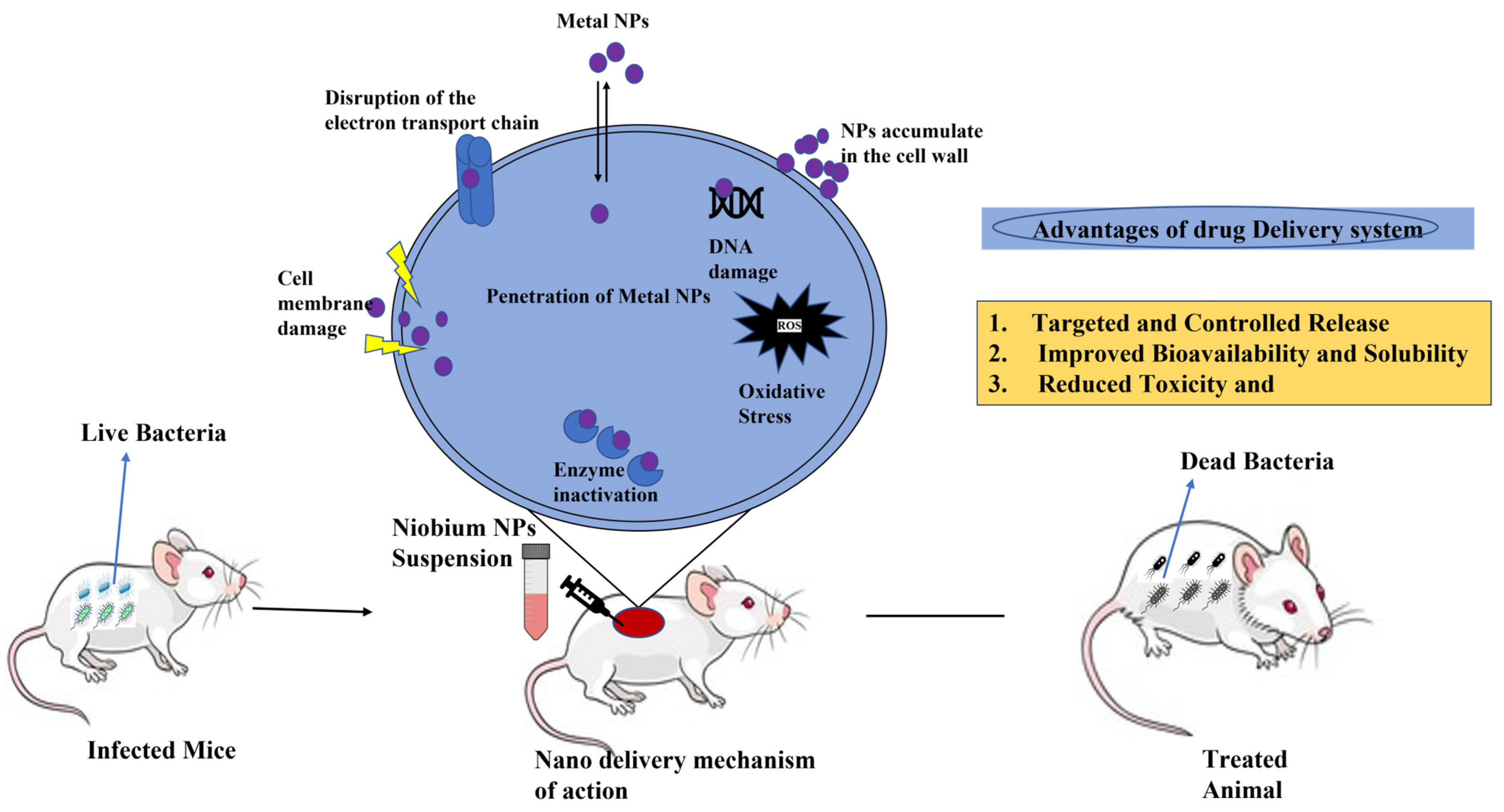
An in vivo cytotoxicity test was performed on Swiss mice, as shown in Figure 3, after treating them with a single dose of niobium oxide (Nb2O5) for 3, 7, and 12 days [7]. Data showed cell count increase in the peritoneal area and liver cell regeneration, which indicate cytotoxicity as not being progressive [7].
Recently, researchers administered NbNPs to rats to assess their biodistribution, clearance mechanisms, and potential toxicity [82]. The results revealed that the nanoparticles primarily accumulated in the liver, spleen, and lungs, with minimal retention in other organs. Histopathological analysis indicated mild inflammation and tissue damage in these organs, raising concerns about the long-term retention of NbNPs in vivo. While there was no severe toxicity observed, the study suggested that prolonged exposure could lead to organ-specific stress, particularly oxidative damage in the liver and potential renal tubule degeneration. These findings emphasize the need for careful consideration of dosing regimens and long-term effects when evaluating NbNPs for biomedical use.
Another in vivo study investigated the immunotoxicity of NbNPs by administering them to mice at varying doses [83]. The research focused on the immune responses triggered by the nanoparticles, including cytokine production and immune cell activation. At higher doses, NbNPs induced a significant increase in pro-inflammatory cytokines, suggesting an activation of the immune system. However, lower doses of NbNPs showed minimal immune response, indicating a potential safety margin for controlled exposure. The study concluded that while NbNPs could have immunomodulatory effects, their safety profile could be optimized through careful dose management, highlighting the importance of understanding their impact on immune system function in preclinical evaluations. These in vivo findings provide valuable insight into both the therapeutic potential and risks associated with NbNPs.
A study conducted by Li et al. (2001) [84] explored the long-term effects of NbNPs on liver function in rats, showing that chronic exposure to NbNPs could lead to oxidative stress, which in turn caused cellular damage in liver tissues. The researchers observed an increase in reactive oxygen species (ROS) levels and lipid peroxidation markers, signifying oxidative damage. Histopathological analysis revealed signs of hepatic cell degeneration, though the damage was not as severe as that induced by other types of nanoparticles. The study emphasizes the need for extended studies to assess the long-term liver toxicity of NbNPs, which is critical for evaluating their safety for clinical applications.
Another in vivo study examined the effects of NbNPs on the renal system in rats after both acute and chronic exposure [85]. The results showed that while acute exposure to NbNPs did not induce significant renal damage, prolonged exposure led to renal tubule degeneration and inflammatory cell infiltration in the kidneys. The study suggested that NbNPs may have a dose-dependent nephrotoxic effect, with higher concentrations leading to greater renal impairment. These findings underscore the necessity of considering organ-specific effects, especially the kidneys, when evaluating the safety of NbNPs for medical use.
A separate study focused on the cardiovascular impact of NbNPs in vivo, administering them to rats over several weeks to assess potential vascular toxicity [86]. The results indicated that while there was no significant change in heart function, there were signs of endothelial cell dysfunction in the blood vessels, along with mild arterial thickening. This vascular response suggests that NbNPs could have subtle but important effects on cardiovascular health, particularly with chronic exposure. These findings further emphasize the importance of understanding the full range of potential adverse effects of NbNPs before they can be used in clinical settings, particularly for long-term treatments.
It is crucial to continue in vivo studies, as there is a lack of them, to further elucidate the biocompatibility and cytotoxicity of niobium in living organisms to develop medical solutions.

2.5. Biodegradability and Clearance Mechanisms

The biodegradability and clearance mechanisms of NbNPs in the body are essential topics in understanding their biomedical safety and effectiveness. Niobium, a biocompatible metal known for its corrosion resistance, is increasingly being explored for use in nanoparticle form in medical applications like imaging, drug delivery, and tissue engineering. However, the biodegradability of niobium nanoparticles is limited, given niobium’s metallic nature, which raises concerns about long-term retention and possible toxicity if they accumulate in tissues. Studies suggest that surface modifications, such as coating niobium with biodegradable polymers, may enhance their clearance and reduce the potential for persistence in organs, providing a more controlled release and safer profile for in vivo use [87].
The primary clearance mechanisms for niobium nanoparticles largely depend on their size, surface charge, and the specific characteristics of the biological environment. Small niobium nanoparticles, generally below 10 nm, are more likely to pass through renal filtration and be cleared via the urinary system, while larger particles are more likely to be taken up by the mononuclear phagocyte system (MPS), primarily in the liver and spleen [88]. Niobium nanoparticles that evade immediate clearance mechanisms might remain in the body longer, with studies showing that certain surface functionalization can alter the rate of phagocytosis and cellular uptake, particularly by macrophages in the liver and spleen. This accumulation can pose a risk for chronic inflammation or other toxic effects over time, underscoring the importance of designing nanoparticles with a view toward efficient clearance from the body [89].

3. Applications of Niobium in Health Sciences

3.1. Niobium in Drug Delivery Systems and Therapeutic Applications

Because of its unique physical, optical, and biological properties, such as antibiofilm and antimicrobial ability, niobium can be applied to develop innovative drug delivery systems or act as a part of therapeutic agents. In a recent study, scientists created a delivery platform containing ultrathin niobium carbide (Nb2C) nanosheets, which exhibit a large specific surface area, offering plenty of anchoring sites for proteins, drugs, or macromolecules, and berberine (BBR), the plant-derived chemical, showing antibacterial, antioxidative, and anticancer activities, especially metastasis-related protein regulation in the tumor, was integrated into the nanosheets. These nanocomposites were used in combined chemotherapy and photothermal therapy on breast cancer cells, resulting in the efficient elimination of cancer cells and successful metastasis inhibition by regulating the expression of proteins linked with extracellular matrix and epithelial–mesenchymal transition [90]. Studies have also been based on the thermo-plasmonic characteristics of rod-shaped, core–shell, and spherical niobium nanoparticles to evaluate if and which type of niobium nanoparticles can potentially target cancer cells. To investigate these properties, scientists simulated synchrotron radiation emission as a function of the beam energy and niobium nanoparticles and discovered that the laser light stimulates the resonance of surface plasmon of the nanoparticles, converting the absorbed energy into heat which destroys tumor tissue near to nanoparticles without hurting sound tissues [91]. This makes niobium nanoparticles appropriate for tissue, tumor, and optothermal human cancer cell treatment. Another experiment was performed with niobium as a part of a hybrid compound called Wells–Dawson polyoxometalate (POM) covalently bound with folic acid, (Bu4N)5H4[P2W15Nb3O62]–folic acid.
In this compound, niobium replaces the tungsten addenda atom, which can increase the oxygen atom’s tendency to form esters when Tris molecules are present, increasing the atom nucleophilicity in the POM structure. The bioactivity of the POM was tested against human fibroblast (HFB), cervical (HeLa), and human breast cancer (MCF-7) cells, showing it effectively, more than 2- to 3-fold more biocompatible and safer than the positive control of methotrexate [92]. The compound successfully knocked down cancer cells with folic acid‘s cytotoxicity due to the overexpression of folate receptors on the cancer cell surface.
A recently published study examined the thermo-plasmonic properties of niobium nanorods for optothermal treatment of human cancer cells, tissues, and tumors under synchrotron radiation. The research demonstrated that niobium nanorods effectively convert absorbed light energy into heat, enabling targeted destruction of cancerous tissues while minimizing damage to surrounding healthy cells. This approach highlights the promising role of niobium nanoparticles in enhancing the efficacy of cancer therapies [93].
The use of niobium carbide (NbC) nanoparticles for photothermal therapy in infected wound treatment has also been reported. The research found that NbC nanoparticles possess excellent near-infrared absorption properties, allowing them to convert laser irradiation into localized heat. This heat effectively eradicates bacteria such as Staphylococcus aureus and Escherichia coli, promoting faster wound healing. The study underscores the potential of niobium-based nanoparticles in developing advanced antimicrobial therapies [94].
In addition to the studies on cancer therapy and antimicrobial applications, niobium-based materials have also been investigated for their potential in enhancing the bioavailability and controlled release of therapeutic agents. A study reported the use of niobium oxide (Nb2O5) nanoparticles for the delivery of anticancer drugs. The research highlighted that niobium oxide nanoparticles have high surface area and biocompatibility, which makes them suitable carriers for drug molecules. These nanoparticles were found to release the anticancer drug doxorubicin in a controlled manner, improving the therapeutic index and reducing side effects typically associated with conventional chemotherapy. The study also demonstrated that niobium oxide nanoparticles were able to target tumor cells, leading to an enhanced therapeutic effect compared to traditional drug delivery methods [95].
Moreover, a study focused on the use of niobium as a catalyst for enhancing the therapeutic properties of certain drugs through its effect on cellular uptake mechanisms. The research revealed that niobium-based materials could facilitate the incorporation of therapeutic compounds into cells, promoting better drug penetration. This ability to improve cellular uptake has vast implications for increasing the efficacy of a range of treatments, including those for chronic diseases such as diabetes and cardiovascular conditions. Additionally, the study found that niobium particles exhibited low toxicity and stable biocompatibility, making them ideal candidates for long-term drug delivery applications [96].
In conclusion, niobium is a promising material for applications in the medical field for drug delivery and therapeutics.

3.2. Niobium as an Imaging Agent

Mass spectrometry imaging (MSI) is a technology to analyze the spatial distribution of proteins, peptides, metabolites, etc., without chemical labeling, and when applied with enhanced laser desorption/ionization (LDI) performance substrates, it allows the visualization of multiple classes of molecular species. This method can be applied in surgical procedures for precise identification of specific biomarkers, helping to diagnose various diseases, injuries, or other clinical issues. Surface-assisted laser desorption/ionization mass spectrometry imaging (SALDI MSI) using noble-metal nanoparticle matrices is an exceptional biochemical analysis tool [97].
In this particular article, niobium nanoparticles were used as a matrix for SALDI MSI, seeded onto mouse brain slices, leading to enhanced biomolecule fragmentation, making transition-metal (such as niobium) nanoparticles promising linker- and residue-free, cost-effective matrices [97]. Novel MRI-compatible material development is crucial, as MRI has become an essential non-invasive diagnostic tool without X-ray irradiation in modern medicine. Therefore, scientists have developed novel Zr–Mo–Nb alloys for biomedical applications, incorporating niobium because of its high biocompatibility, potential to enhance corrosion resistance, and mechanical performance [5]. Also, mixing niobium (Nb), zirconium (Zr), and molybdenum (Mo) causes an uneven mix of materials, creating a new kind of crystal, called the β-Zr phase, which makes the grain size of the material much smaller. This results in a fine and layered structure of two crystal types (α and β) in the alloy. This is an option for biocompatible, long-lasting, and MRI-compatible materials that can raise the standard of medical care and improve patients’ general well-being [98].
A study explored the use of niobium-based nanoparticles as agents for positron emission tomography (PET) imaging. The research demonstrated that niobium nanoparticles, when labeled with a suitable radioisotope, could effectively target tumor cells, offering high-resolution images for early cancer detection. The study highlighted the excellent biocompatibility of niobium and its promising application for non-invasive diagnostic imaging, suggesting that it could offer a new avenue for visualizing malignant tissue with minimal side effects. Additionally, the use of niobium in PET imaging allowed for the development of tracers with enhanced tissue penetration, which is critical for detecting smaller or deep-seated tumors [99].
In another investigation, niobium nanoparticles were assessed for their potential in magnetic resonance imaging (MRI) as contrast agents. The study revealed that niobium oxide nanoparticles demonstrated significant potential to improve MRI sensitivity due to their unique magnetic properties. These nanoparticles were found to provide a stronger contrast effect in MRI scans compared to conventional agents, particularly in the imaging of vascular tissues and organs like the liver. Niobium’s ability to tune its magnetic properties through surface modification also enables the fine-tuning of imaging contrast, further advancing its utility in precise and high-quality diagnostics. This body of research points toward niobium’s growing role in enhancing the effectiveness of medical imaging techniques, opening doors to more accurate and early detection of various diseases [100].
Additionally, niobium-based materials have been investigated for their role in enhancing the imaging capabilities of other diagnostic techniques, such as X-ray imaging. A study showed the integration of niobium into composite materials for use in X-ray contrast agents. The study found that niobium’s high atomic number contributed to significant attenuation of X-ray beams, providing clearer and sharper images when used as a contrast agent. The niobium-based composite demonstrated improved biocompatibility and stability compared to traditional iodine-based contrast agents. This novel approach to X-ray imaging holds promise for reducing the dose of contrast agents required, potentially minimizing the associated risks while maintaining high-quality diagnostic images. Researchers suggest that niobium’s versatility in medical imaging could extend beyond PET and MRI, providing a broader range of applications in clinical settings [101].

3.3. Biosensing

The synthesis of Nb2O5 nanorod array films and their electrochemical properties are explored for biosensing applications. The unique characteristics of Nb2O5 nanorods, including high surface area and excellent conductivity, make them ideal for improving the performance of biosensors. Their ability to interact efficiently with biological molecules enhances the sensitivity and accuracy of detection, offering significant potential for the development of advanced biosensors in medical diagnostics, pathogen detection, and real-time monitoring of biological processes [35]. The impact of deposition geometry on the structural, morphological, and optical properties of Nb2O5 nanostructures prepared via hydrothermal methods was investigated. The study suggests that by carefully controlling deposition parameters, the surface characteristics of Nb2O5 can be optimized for applications such as biosensing. The enhanced surface area, crystallinity, and optical properties of the nanostructures can improve the efficiency and sensitivity of biosensors, making them more effective for detecting biological molecules and biomarkers [51].
Recently, a study explored the use of niobium-based nanomaterials in electrochemical biosensors for detecting glucose levels in diabetic patients. The researchers modified the niobium surfaces with functionalized nanostructures, enhancing their sensitivity and selectivity towards glucose. The electrochemical response was shown to be significantly improved compared to conventional sensors, providing a more efficient and reliable platform for continuous glucose monitoring. This study highlighted the advantages of niobium’s stable oxide layer, which acts as an ideal surface for immobilizing biomolecules such as enzymes, ensuring high performance even in complex biological environments [102].
In another study, niobium was explored as a key component in the development of a sensor for detecting cancer biomarkers. The research focused on creating a niobium-based field-effect transistor (FET) biosensor, where the surface of the niobium electrode was functionalized with antibodies specific to the biomarker protein. The FET biosensor exhibited a high degree of sensitivity and specificity, capable of detecting trace amounts of the biomarker in serum samples. The study demonstrated niobium’s potential in precision medicine, where early detection of cancer through non-invasive biosensors could lead to improved diagnostic outcomes. Niobium’s robust performance in diverse biological conditions further establishes its promising role in advancing biosensor technologies [103].
Another study focused on the development of a niobium-based surface-enhanced Raman scattering (SERS) biosensor, utilizing niobium’s unique surface properties to amplify Raman signals for highly sensitive biomolecular detection. The researchers incorporated niobium nanoparticles as substrates for the SERS biosensor, demonstrating their ability to enhance the Raman scattering of targeted biomolecules, such as DNA and proteins. The enhanced signal allowed for the detection of low-abundance biomarkers with high specificity, making it a promising approach for early disease diagnosis and environmental monitoring. The study emphasized niobium’s ability to provide stable and reproducible SERS signals, even in complex biological matrices, underscoring its potential in the field of biosensing where high sensitivity and selectivity are crucial [104].
In addition to electrochemical and optical sensing, niobium has also been investigated for its potential in piezoelectric biosensors. A study developed a Nb2O5 thin-film-based quartz crystal microbalance (QCM) biosensor for the detection of protein interactions. The researchers utilized the high surface area and stability of niobium oxide films to immobilize specific antibodies, enabling the sensitive detection of target proteins in complex biological fluids. The results demonstrated that niobium oxide-based QCM biosensors exhibited excellent sensitivity, with the ability to detect femtomolar concentrations of target proteins. This application highlights niobium’s versatility in biosensing, offering a promising platform for real-time monitoring of protein–protein interactions, which could be crucial for diagnostic and therapeutic purposes in the field of personalized medicine [105].

4. Challenges and Future Perspectives

Niobium oxide nanoparticles (Nb2O5 NPs) have potential in health sciences for medication delivery, imaging, and antibacterial therapies, but confront hurdles in clinical recognition. Among these is the need for a better understanding of their biocompatibility and toxicity; while niobium oxide generally shows positive biocompatibility, nanoparticles can behave unpredictably depending on their size, shape, and surface modifications, occasionally causing oxidative stress or inflammation [106]. Additionally, Nb2O5 NPs face stability issues, often aggregating in physiological environments, which limits their effectiveness and bioavailability. Complex surface functionalization strategies are necessary to enhance their targeting and reduce toxicity, add to production costs, and raise scalability concerns. Furthermore, limited pharmacokinetic data and a lack of standardized testing protocols complicate regulatory approval, with inconsistencies in synthesis and characterization making it difficult to establish safety benchmarks. Addressing these challenges will require focused research on nanoparticle behavior in biological systems, the development of stable surface modifications, and clear regulatory guidelines, all of which are essential to unlock the full potential of niobium oxide nanoparticles in healthcare.
Niobium oxide nanoparticles have emerged as promising materials in health sciences, with potential applications spanning drug delivery, diagnostic imaging, antimicrobial treatments, and cancer therapy. Future research is anticipated to focus on developing eco-friendly synthesis methods, optimizing surface modifications for targeted delivery, and establishing standardized testing protocols, paving the way for safer and more effective medical applications. As these advancements unfold, niobium-based nanomaterials could become integral components of next-generation therapeutic and diagnostic tools in precision medicine. In this review article, metal oxide niobium was explained with its synthesis methods, characterization techniques, and applications in health sciences (in vitro, in vivo, biocompatibility). In addition, challenges and future perspectives were discussed.

5. Author Perspectives

From a biomedical standpoint, NbNPs, particularly Nb2O5, present a compelling opportunity for advancing therapeutic and diagnostic technologies. The unique properties of NbNPs, such as their excellent biocompatibility, corrosion resistance, and ability to be functionalized for specific applications, make them an attractive choice for a wide range of medical uses, including drug delivery, imaging, and tissue engineering. However, the path to clinical translation requires overcoming critical challenges related to their in vivo behavior. The limited biodegradability of NbNPs and their potential for long-term accumulation necessitate focused research on optimizing surface modifications and bio-coating strategies that can promote faster clearance from the body. Moreover, further investigation into their interaction with immune cells and potential long-term toxicological effects is essential to ensure their safe use in humans.
Looking ahead, the future of niobium-based nanoparticles in nanomedicine lies in their strategic integration with other emerging technologies, such as gene therapy, photothermal treatment, or even regenerative medicine. The potential for NbNPs to target specific tissues or cross biological barriers like the blood–brain barrier could unlock new possibilities for treating complex diseases such as cancer and neurodegenerative disorders. However, a more thorough understanding of their in vivo fate, coupled with advances in manufacturing techniques for producing consistent and scalable niobium nanoparticles, will be key to realizing their full therapeutic potential. The development of standardized synthesis methods and comprehensive safety evaluations will be pivotal in advancing NbNPs from the laboratory to clinical applications, ultimately making them an integral part of the next generation of biomedical nanomaterials.

Author Contributions

M.U.K.: Writing—original draft, writing—review and editing. A.R.: Writing—original draft, writing—review and editing. A.M.H.d.S.: Writing—original draft, writing—review and editing. M.K.-S.: Writing—original draft, writing—review and editing. A.S.: Writing—original draft, writing—review and editing. W.C.M.A.M.: Writing—original draft, writing—review and editing. All authors have read and agreed to the published version of the manuscript.

Funding

The authors declare that financial support was received for this article’s research, authorship, and/or publication. The authors thanks for FTMC support. Research Lithuanian Council—European Union Funds Investment Operational Program “4th competition of 11th call for the implementation of researcher groups projects”—(P-MIP-22-201).

Acknowledgments

The authors give thanks to FTMC and the Research Lithuanian Council—European Union Funds Investment Operational Program for their support.

Conflicts of Interest

The authors declare that this research was conducted in the absence of any commercial or financial relationships that could be construed as a potential conflict of interest.

References

  1. Goldberg, M.; Langer, R.; Jia, X. Nanostructured Materials for Applications in Drug Delivery and Tissue Engineering. J. Biomater. Sci. Polym. Ed. 2007, 18, 241–268. [Google Scholar] [CrossRef] [PubMed]
  2. Rizvi, S.A.; Saleh, A.M. Applications of Nanoparticle Systems in Drug Delivery Technology. Saudi Pharm. J. 2018, 26, 64–70. [Google Scholar] [CrossRef] [PubMed]
  3. Mobarak, M.H.; Islam, M.A.; Hossain, N.; Al Mahmud, M.Z.; Rayhan, M.T.; Nishi, N.J.; Chowdhury, M.A. Recent Advances of Additive Manufacturing in Implant Fabrication—A Review. Appl. Surf. Sci. Adv. 2023, 18, 100462. [Google Scholar] [CrossRef]
  4. Nico, C.; Rino, L.; Matos, M.; Monteiro, R.; Costa, F.M.; Monteiro, T.; Graça, M.P.F. NbO/Nb2O5 Core–Shells by Thermal Oxidation. J. Eur. Ceram. Soc 2013, 33, 3077–3083. [Google Scholar] [CrossRef]
  5. Raba, A.M.; Bautista-Ruíz, J.; Joya, M.R. Synthesis and Structural Properties of Niobium Pentoxide Powders: A Comparative Study of the Growth Process. Mater. Res. 2016, 19, 1381–1387. [Google Scholar] [CrossRef]
  6. Borowski, T.; Zielińska, K.; Spychalski, M.; Adamczyk-Cieślak, B.; Żrodowski, Ł. Effect of Oxidation Temperature on the Properties of Niobium in View of Its Biomedical Applications. Surf. Coat. Technol. 2023, 473, 129911. [Google Scholar] [CrossRef]
  7. Dsouki, N.A.; de Lima, M.P.; Corazzini, R.; Gáscon, T.M.; Azzalis, L.A.; Junqueira, V.B.C.; Feder, D.; Fonseca, F.L.A. Cytotoxic, Hematologic and Histologic Effects of Niobium Pentoxide in Swiss Mice. J. Mater. Sci. Mater. Med. 2014, 25, 1301–1305. [Google Scholar] [CrossRef]
  8. Chehelgerdi, M.; Chehelgerdi, M.; Allela, O.Q.B.; Pecho, R.D.C.; Jayasankar, N.; Rao, D.P.; Thamaraikani, T.; Vasanthan, M.; Viktor, P.; Lakshmaiya, N.; et al. Progressing Nanotechnology to Improve Targeted Cancer Treatment: Overcoming Hurdles in Its Clinical Implementation. Mol. Cancer 2023, 22, 169. [Google Scholar] [CrossRef]
  9. Lima, L.F.d.S.; Coelho, C.R.; Gomes, G.H.M.; Mohallem, N.D.S. Nb2O5/SiO2 Mesoporous Monoliths Synthetized by Sol–Gel Process Using Ammonium Niobate Oxalate Hydrate as Porogenic Agent. J. Sol-Gel Sci. Technol. 2020, 93, 168–174. [Google Scholar] [CrossRef]
  10. Bokov, D.; Turki Jalil, A.; Chupradit, S.; Suksatan, W.; Javed Ansari, M.; Shewael, I.H.; Valiev, G.H.; Kianfar, E. Nanomaterial by Sol-Gel Method: Synthesis and Application. Adv. Mater. Sci. Eng. 2021, 2021, 5102014. [Google Scholar] [CrossRef]
  11. Graça, M.P.F.; Meireles, A.; Nico, C.; Valente, M.A. Nb2O5 Nanosize Powders Prepared by Sol–Gel—Structure, Morphology and Dielectric Properties. J. Alloys Compd. 2013, 553, 177–182. [Google Scholar] [CrossRef]
  12. Ristić, M.; Popović, S.; Musić, S. Sol–Gel Synthesis and Characterization of Nb2O5 Powders. Mater. Lett. 2004, 58, 2658–2663. [Google Scholar] [CrossRef]
  13. Griesmar, P.; Papin, G.; Sanchez, C.; Livage, J. Sol-gel route to niobium pentoxide. Chem. Mater. 1991, 3, 335–339. [Google Scholar] [CrossRef]
  14. Ohtani, B.; Iwai, K.; Nishimoto, S.I.; Inui, T. Electrochromism of Niobium Oxide Thin Films Prepared by the Sol-Gel Process. J. Electrochem. Soc. 1994, 141, 2439. [Google Scholar] [CrossRef]
  15. Maček, M.; Orel, B. Electrochromism of Sol–Gel Derived Niobium Oxide Films. Sol. Energy Mater. Sol. Cells 1998, 54, 121–130. [Google Scholar] [CrossRef]
  16. Özer, N.; Rubin, M.D.; Lampert, C.M. Optical and Electrochemical Characteristics of Niobium Oxide Films Prepared by Sol-Gel Process and Magnetron Sputtering A Comparison. Sol. Energy Mater. Sol. Cells 1996, 40, 285–296. [Google Scholar] [CrossRef]
  17. Francisco, M.S.P.; Gushikem, Y. Synthesis and Characterization of SiO2–Nb2O5 Systems Prepared by the Sol–Gel Method: Structural Stability Studies. J. Mater. Chem. 2002, 12, 2552–2558. [Google Scholar] [CrossRef]
  18. Aronne, A.; Marenna, E.; Califano, V.; Fanelli, E.; Pernice, P.; Trifuoggi, M.; Vergara, A. Sol–Gel Synthesis and Structural Characterization of Niobium-Silicon Mixed-Oxide Nanocomposites. J. Sol-Gel Sci. Technol. 2007, 43, 193–204. [Google Scholar] [CrossRef]
  19. Joshi, B.N.; Yoon, H.; Hest, M.F.; Yoon, S.S. Niobium-doped titania photocatalyst film prepared via a nonaqueous sol-gel method. J. Am. Ceram. Soc. 2013, 96, 2623–2627. [Google Scholar] [CrossRef]
  20. Mohammadi, S.; Golobostanfard, M.R.; Abdizadeh, H. Synthesis and Characterization of Indium Niobium Oxide Thin Films via Sol–Gel Spin Coating Method. J. Mater. Sci. Technol. 2013, 29, 923–928. [Google Scholar] [CrossRef]
  21. Duta, M.; Predoana, L.; Calderon-Moreno, J.M.; Preda, S.; Anastasescu, M.; Marin, A.; Gartner, M. Nb-Doped TiO2 Sol–Gel Films for CO Sensing Applications. Mater. Sci. Semicond. Process. 2016, 42, 397–404. [Google Scholar] [CrossRef]
  22. Joya, M.R.; Barba Ortega, J.J.; Raba Paez, A.M.; Silva Filho, J.G.; Cavalcante Freire, P.D.T. Synthesis and Characterization of Nano-Particles of Niobium Pentoxide with Orthorhombic Symmetry. Metals 2017, 7, 142. [Google Scholar] [CrossRef]
  23. Jafari, H.; Sadeghzadeh, S.; Rabbani, M.; Rahimi, R. Effect of Nb on the Structural, Optical and Photocatalytic Properties of Al-Doped ZnO Thin Films Fabricated by the Sol-Gel Method. Ceram. Int. 2018, 44, 20170–20177. [Google Scholar] [CrossRef]
  24. Lucas, J.M.F.; Soreto Teixeira, S.; Gavinho, S.R.; Prezas, P.R.; Silva, C.C.; Sales, A.J.M.; Graça, M.P.F. Niobium Oxide Prepared by Sol–Gel Using Powder Coconut Water. J. Mater. Sci. Mater. Electron. 2019, 30, 11346–11353. [Google Scholar] [CrossRef]
  25. Baek, H.; Lee, C.; Choi, J.; Cho, J. Nonvolatile Memory Devices Prepared from Sol–Gel Derived Niobium Pentoxide Films. Langmuir 2013, 29, 380–386. [Google Scholar] [CrossRef] [PubMed]
  26. Zhou, Y.; Qiu, Z.; Lü, M.; Zhang, A.; Ma, Q. Preparation and Characterization of Porous Nb2O5 Nanoparticles. Mater. Res. Bull. 2008, 43, 1363–1368. [Google Scholar] [CrossRef]
  27. Fuchigami, T.; Kakimoto, K. Spiky Niobium Oxide Nanoparticles through Hydrothermal Synthesis. J. Mater. Res. 2017, 32, 3326–3332. [Google Scholar] [CrossRef]
  28. Santos, I.; Loureiro, L.H.; Silva, M.F.P.; Cavaleiro, A.M. Studies on the Hydrothermal Synthesis of Niobium Oxides. Polyhedron 2002, 21, 2009–2015. [Google Scholar] [CrossRef]
  29. Vega, M.R.O.; Consul, V.; Cadorin, M.; Arenas, L.T.; Aguzzoli, C.; Hübler, R.; Takimi, A.; de Fraga Malfatti, C. Niobium Oxide Nanorods Obtained by Hydrothermal Synthesis—Structure, Morphology, and Electrochemical Detection of Oxygen via Oxygen Reduction Reaction. Coatings 2023, 13, 1786. [Google Scholar] [CrossRef]
  30. Silva, R.M.; Noremberg, B.S.; Marins, N.H.; Milne, J.; Zhitomirsky, I.; Carreño, N.L. Microwave-Assisted Hydrothermal Synthesis and Electrochemical Characterization of Niobium Pentoxide/Carbon Nanotubes Composites. J. Mater. Res. 2019, 34, 592–599. [Google Scholar] [CrossRef]
  31. Zhang, H.; Wang, Y.; Yang, D.; Li, Y.; Liu, H.; Liu, P.; Zhao, H. Directly Hydrothermal Growth of Single Crystal Nb3O7(OH) Nanorod Film for High Performance Dye-Sensitized Solar Cells. Adv. Mater. 2012, 24, 1598–1603. [Google Scholar] [CrossRef] [PubMed]
  32. Li, G.; Wang, X.; Ma, X. Nb2O5-Carbon Core-Shell Nanocomposite as Anode Material for Lithium Ion Battery. J. Energy Chem. 2013, 22, 357–362. [Google Scholar] [CrossRef]
  33. Liu, M.; Xue, D. Large-Scale Fabrication of H2(H2O)Nb2O6 and Nb2O5 Hollow Microspheres. Mater. Res. Bull. 2010, 45, 333–338. [Google Scholar] [CrossRef]
  34. Lopes, O.F.; Paris, E.C.; Ribeiro, C. Synthesis of Nb2O5 Nanoparticles through the Oxidant Peroxide Method Applied to Organic Pollutant Photodegradation: A Mechanistic Study. Appl. Catal. B Environ. 2014, 144, 800–808. [Google Scholar] [CrossRef]
  35. Wen, H.; Liu, Z.; Wang, J.; Yang, Q.; Li, Y.; Yu, J. Facile Synthesis of Nb2O5 Nanorod Array Films and Their Electrochemical Properties. Appl. Surf. Sci. 2011, 257, 10084–10088. [Google Scholar] [CrossRef]
  36. Liu, F.; Xue, D. Fabrication of Nb2O5 Nanotrees with Controlled Branching Degrees. Phys. Scr. 2010, 014074. [Google Scholar] [CrossRef]
  37. Zhao, Y.; Eley, C.; Hu, J.; Foord, J.S.; Ye, L.; He, H.; Tsang, S.C.E. Shape-Dependent Acidity and Photocatalytic Activity of Nb2O5 Nanocrystals with an Active TT (001) Surface. Angew. Chem.-Int. Ed. 2012, 51, 3846. [Google Scholar] [CrossRef] [PubMed]
  38. Li, C.C.; Dou, J.; Chen, L.; Lin, J.; Zeng, H.C. Antisolvent Precipitation for the Synthesis of Monodisperse Mesoporous Niobium Oxide Spheres as Highly Effective Solid Acid Catalysts. ChemCatChem 2012, 4, 1675–1682. [Google Scholar] [CrossRef]
  39. Ayudhya, S.K.N.; Soottitantawat, A.; Praserthdam, P.; Satayaprasert, C. Effect of Aging on the Properties of Mesoporous Niobium Oxide. Mater. Chem. Phys. 2008, 110, 387–392. [Google Scholar] [CrossRef]
  40. Luo, H.; Wei, M.; Wei, K. Synthesis of Nb2O5 Nanosheets and Its Electrochemical Measurements. Mater. Chem. Phys. 2010, 120, 6–9. [Google Scholar] [CrossRef]
  41. Luo, H.; Wei, M.; Wei, K. Synthesis of Nb2O5 Nanorods by a Soft Chemical Process. J. Nanomater. 2009, 2009, 758353. [Google Scholar] [CrossRef]
  42. Hu, W.; Zhao, Y.; Liu, Z.; Zhu, Y. NbS2/Nb2O5 nanocables. Nanotechnology 2007, 18, 095605. [Google Scholar] [CrossRef]
  43. Wei, M.; Qi, Z.M.; Ichihara, M.; Zhou, H. Synthesis of single-crystal niobium pentoxide nanobelts. Acta Mater. 2008, 56, 2488–2494. [Google Scholar] [CrossRef]
  44. Kominami, H.; Oki, K.; Kohno, M.; Onoue, S.I.; Kera, Y.; Ohtani, B. Novel Solvothermal Synthesis of Niobium (V) Oxide Powders and Their Photocatalytic Activity in Aqueous Suspensions. J. Mater. Chem. 2001, 11, 604–609. [Google Scholar] [CrossRef]
  45. Buha, J.; Arčon, D.; Niederberger, M.; Djerdj, I. Solvothermal and Surfactant-Free Synthesis of Crystalline Nb2O5, Ta2O5, HfO2, and Co-Doped HfO2 Nanoparticles. Phys. Chem. Chem. Phys. 2010, 12, 15537–15543. [Google Scholar] [CrossRef] [PubMed]
  46. Hu, W.; Liu, Z.; Tian, D.; Zhang, S.; Zhao, Y.; Yao, K. Morphological Evolution of Nb2O5 in a Solvothermal Reaction: From Nb2O5 Grains to Nb2O5 Nanorods and Hexagonal Nb2O5 Nanoplatelets. J. Wuhan Univ. Technol.-Mater. Sci. Ed. 2009, 24, 245–248. [Google Scholar] [CrossRef]
  47. Prado, N.T.; Oliveira, L.C. Nanostructured Niobium Oxide Synthetized by a New Route Using Hydrothermal Treatment: High Efficiency in Oxidation Reactions. Appl. Catal. B Environ. 2017, 205, 481–488. [Google Scholar] [CrossRef]
  48. Prado, N.T.; Souza, T.E.; Machado, A.R.T.; Souza, P.P.; Monteiro, R.S.; Oliveira, L.C. Enhanced Catalytic Activity for Fructose Conversion on Nanostructured Niobium Oxide after Hydrothermal Treatment: Effect of Morphology and Porous Structure. J. Mol. Catal. A Chem. 2016, 422, 23–34. [Google Scholar] [CrossRef]
  49. Chen, J.; Wang, H.; Huang, G.; Zhang, Z.; Han, L.; Song, W.; Zhang, Y. Facile Synthesis of Urchin-like Hierarchical Nb2O5 Nanospheres with Enhanced Visible Light Photocatalytic Activity. J. Alloys Compd. 2017, 728, 19–28. [Google Scholar] [CrossRef]
  50. Liu, X.; Liu, G.; Chen, H.; Ma, J.; Zhang, R. Facile Synthesis of Nb2O5 Nanobelts Assembled from Nanorods and Their Applications in Lithium Ion Batteries. J. Phys. Chem. Solids 2017, 111, 8–11. [Google Scholar] [CrossRef]
  51. Salim, E.T.; Ismail, R.A.; Halbos, H.T. Deposition Geometry Effect on Structural, Morphological and Optical Properties of Nb2O5 Nanostructure Prepared by Hydrothermal Technique. Appl. Phys. A 2020, 126, 891. [Google Scholar] [CrossRef]
  52. Zu, D.; Song, H.; Wang, Y.; Chao, Z.; Li, Z.; Wang, G.; Ma, J. One-Pot In-Situ Hydrothermal Synthesis of CdS/Nb2O5/Nb2C Heterojunction for Enhanced Visible-Light-Driven Photodegradation. Appl. Catal. B Environ. 2020, 277, 119140. [Google Scholar] [CrossRef]
  53. Salim, E.T.; Ismail, R.A.; Halbos, H.T. Growth of Nb2O5 Film Using Hydrothermal Method: Effect of Nb Concentration on Physical Properties. Mater. Res. Express 2019, 6, 116429. [Google Scholar] [CrossRef]
  54. Tayyab, M.; Liu, Y.; Liu, Z.; Pan, L.; Xu, Z.; Yue, W.; Zhang, J. One-Pot in-Situ Hydrothermal Synthesis of Ternary In2S3/Nb2O5/Nb2C Schottky/S-Scheme Integrated Heterojunction for Efficient Photocatalytic Hydrogen Production. J. Colloid Interface Sci. 2022, 628, 500–512. [Google Scholar] [CrossRef] [PubMed]
  55. Santos, V.; Brandalise, R.N.; Savaris, M. Biomaterials: Characteristics and Properties. Eng. Biomater. 2017, 5–15. [Google Scholar] [CrossRef]
  56. Nivedita, L.R.; Haubert, A.; Battu, A.K.; Ramana, C.V. Correlation between Crystal Structure, Surface/Interface Microstructure, and Electrical Properties of Nanocrystalline Niobium Thin Films. Nanomaterials 2020, 10, 1287. [Google Scholar] [CrossRef]
  57. Ücker, C.L.; Gularte, L.T.; Fernandes, C.D.; Goetzke, V.; Moreira, E.C.; Raubach, C.W.; Moreira, M.L.; Cava, S.S. Investigation of the Properties of Niobium Pentoxide for Use in Dye-sensitized Solar Cells. J. Am. Ceram. Soc. 2019, 102, 1884–1892. [Google Scholar] [CrossRef]
  58. Moreto, J.A.; Gelamo, R.V.; Da Silva, M.V.; Steffen, T.T.; De Oliveira, C.J.F.; De Almeida Buranello, P.A.; Pinto, M.R. New Insights of Nb2O5-Based Coatings on the 316L SS Surfaces: Enhanced Biological Responses. J. Mater. Sci. Mater. Med. 2021, 32, 25. [Google Scholar] [CrossRef]
  59. Rani, R.A.; Zoolfakar, A.S.; O’Mullane, A.P.; Austin, M.W.; Kalantar-Zadeh, K. Thin Films and Nanostructures of Niobium Pentoxide: Fundamental Properties, Synthesis Methods and Applications. J. Mater. Chem. A 2016, 2, 15683–15703. [Google Scholar] [CrossRef]
  60. Soares, M.; Leite, S.; Nico, C.; Peres, M.; Fernandes, A.; Graça, M.; Matos, M.; Monteiro, R.; Monteiro, T.; Costa, F. Effect of Processing Method on Physical Properties of Nb2O5. J. Eur. Ceram. Soc. 2011, 31, 501–506. [Google Scholar] [CrossRef]
  61. Çetinörgü-Goldenberg, E.; Klemberg-Sapieha, J.; Martinu, L. Effect of Postdeposition Annealing on the Structure, Composition, and the Mechanical and Optical Characteristics of Niobium and Tantalum Oxide Films. Appl. Opt. 2012, 51, 6498. [Google Scholar] [CrossRef]
  62. Yao, D.D.; Rani, R.A.; O’Mullane, A.P.; Kalantar-Zadeh, K.; Ou, J.Z. High Performance Electrochromic Devices Based on Anodized Nanoporous Nb2O5. J. Phys. Chem. C 2013, 118, 476–481. [Google Scholar] [CrossRef]
  63. Pawlicka, A.; Atik, M.; Aegerter, M. Synthesis of Multicolor Nb2O5 Coatings for Electrochromic Devices. Thin Solid Film. 1997, 301, 236–241. [Google Scholar] [CrossRef]
  64. Mitterhuber, L.; Kraker, E.; Defregger, S. Structure Function Analysis of Temperature-Dependent Thermal Properties of NM-Thin Nb2O5. Energies 2019, 12, 610. [Google Scholar] [CrossRef]
  65. Hota, M.K.; Bera, M.K.; Maiti, C.K. Flexible Metal–Insulator–Metal Capacitors on Polyethylene Terephthalate Plastic Substrates. Semicond. Sci. Technol. 2012, 27, 105001. [Google Scholar] [CrossRef]
  66. Joudeh, N.; Linke, D. Nanoparticle Classification, Physicochemical Properties, Characterization, and Applications: A Comprehensive Review for Biologists. J. Nanobiotechnol. 2022, 20, 262. [Google Scholar] [CrossRef] [PubMed]
  67. De, M.; Ghosh, P.S.; Rotello, V.M. Applications of Nanoparticles in Biology. Adv. Mater. 2008, 20, 4225–4241. [Google Scholar] [CrossRef]
  68. Huynh, K.-H.; Pham, X.-H.; Kim, J.; Lee, S.H.; Chang, H.; Rho, W.-Y.; Jun, B.-H. Synthesis, Properties, and Biological Applications of Metallic Alloy Nanoparticles. Int. J. Mol. Sci. 2020, 21, 5174. [Google Scholar] [CrossRef] [PubMed]
  69. Buzea, C.; Pacheco, I.I.; Robbie, K. Nanomaterials and Nanoparticles: Sources and Toxicity. Biointerphases 2007, 2, MR17–MR71. [Google Scholar] [CrossRef] [PubMed]
  70. Ealia, S.A.M.; Saravanakumar, M.P. A Review on the Classification, Characterisation, Synthesis of Nanoparticles and Their Application. IOP Conf. Ser. Mater. Sci. Eng. 2017, 263, 032019. [Google Scholar] [CrossRef]
  71. Yagublu, V.; Karimova, A.; Hajibabazadeh, J.; Reissfelder, C.; Muradov, M.; Bellucci, S.; Allahverdiyev, A. Overview of Physicochemical Properties of Nanoparticles as Drug Carriers for Targeted Cancer Therapy. J. Funct. Biomater. 2022, 13, 196. [Google Scholar] [CrossRef] [PubMed]
  72. Lu, Y.; Ozcan, S. Green Nanomaterials: On Track for a Sustainable Future. Nano Today 2015, 10, 417–420. [Google Scholar] [CrossRef]
  73. Olivares-Navarrete, R.; Olaya, J.J.; Ramírez, C.; Rodil, S.E. Biocompatibility of niobium coatings. Coatings 2011, 1, 72–87. [Google Scholar] [CrossRef]
  74. Chézeau, L.; Tchinda, A.; Pierson, G.; Bravetti, P.; Ferrari, L.; Joubert, O.; Zaiou, M.; Rihn, B.H. In vitro Molecular study of Titanium-Niobium Alloy biocompatibility. Biomedicines 2022, 10, 1898. [Google Scholar] [CrossRef] [PubMed]
  75. Senocak, T.C.; Ezirmik, K.V.; Aysin, F.; Ozek, N.S.; Cengiz, S. Niobium-Oxynitride Coatings for Biomedical Applications: Its Antibacterial Effects and in-Vitro Cytotoxicity. Mater. Sci. Eng. C Biomim. Mater. Sens. Syst. 2021, 120, 111662. [Google Scholar] [CrossRef] [PubMed]
  76. Carli Schardosim, R.F.; Cardozo, T.R.; Souza, A.P.; Seeber, A.; Flores, W.H.; Lehmann, M.; Dihl, R.R. Cyto—Genotoxicity of Crystalline and Amorphous Niobium (V) Oxide Nanoparticles in CHO-K1 Cells. Toxicol. Res. 2022, 11, 765–773. [Google Scholar] [CrossRef] [PubMed]
  77. Mestieri, L.B.; Gomes-Cornélio, A.L.; Rodrigues, E.M.; Faria, G.; Guerreiro-Tanomaru, J.M.; Tanomaru-Filho, M. Cytotoxicity and Bioactivity of Calcium Silicate Cements Combined with Niobium Oxide in Different Cell Lines. Braz. Dent. J. 2017, 28, 65–71. [Google Scholar] [CrossRef] [PubMed]
  78. Ferreira, M.O.A.; Mariani, F.E.; Leite, N.B.; Gelamo, R.V.; Aoki, I.V.; de Siervo, A.; Pinto, H.C.; Moreto, J.A. Niobium and Carbon Nanostructured Coatings for Corrosion Protection of the 316 L Stainless Steel. Mater. Chem. Phys. 2024, 312, 128610. [Google Scholar] [CrossRef]
  79. Capanema, N.S.; Mansur, A.A.; Carvalho, S.M.; Silva, A.R.; Ciminelli, V.S.; Mansur, H.S. Niobium-Doped Hydroxyapatite Bioceramics: Synthesis, Characterization and In Vitro Cytocompatibility. Materials 2015, 8, 4191–4209. [Google Scholar] [CrossRef]
  80. Yassin, M.T.; Al-Otibi, F.O.; Al-Sahli, S.A.; El-Wetidy, M.S.; Mohamed, S. Metal Oxide Nanoparticles as Efficient Nanocarriers for Targeted Cancer Therapy: Addressing Chemotherapy-Induced Disabilities. Cancers 2024, 16, 4234. [Google Scholar] [CrossRef] [PubMed]
  81. Matsuno, H. Biocompatibility and osteogenesis of refractory metal implants, titanium, hafnium, niobium, tantalum and rhenium. Biomaterials 2001, 22, 1253–1262. [Google Scholar] [CrossRef]
  82. Li, J.; Xie, Q.; Ma, R.; Li, Y.; Yuan, J.; Ren, M.; Li, H.; Wang, J.; Lu, D.; Xu, Z. Recent Progress on the Synergistic Antitumor Effect of a Borneol-Modified Nanocarrier Drug Delivery System. Front. Med. 2021, 8, 750170. [Google Scholar] [CrossRef]
  83. Yuan, R.; Huang, Y.; Chan, L.; He, D.; Chen, T. Engineering EHD1-Targeted Natural Borneol Nanoemulsion Potentiates Therapeutic Efficacy of Gefitinib Against Nonsmall Lung Cancer. ACS Appl. Mater. Interfaces 2020, 12, 45714–45727. [Google Scholar] [CrossRef] [PubMed]
  84. Lei, D.-X.; Peng, C.-H.; Peng, S.-Y.; Jiang, X.-C.; Wu, Y.-L.; Shen, H.-W. Safe Upper Limit of Intermittent Hepatic Inflow Occlusion for Liver Resection in Cirrhotic Rats. World J. Gastroenterol. 2001, 7, 713. [Google Scholar] [CrossRef] [PubMed]
  85. Iavicoli, I.; Fontana, L.; Nordberg, G. The Effects of Nanoparticles on the Renal System. Crit. Rev. Toxicol. 2016, 46, 490–560. [Google Scholar] [CrossRef] [PubMed]
  86. Rana, K.; Verma, Y.; Rani, V.; Rana, S.V.S. Renal Toxicity of Nanoparticles of Cadmium Sulphide in Rat. Chemosphere 2018, 193, 142–150. [Google Scholar] [CrossRef] [PubMed]
  87. Xie, H.; Geng, S.; Shao, J.; Luo, G.; Liu, Q.; Wang, J.; Chen, Y.; Chu, P.K.; Li, Z.; Yu, X. Niobium Diselenide Nanosheets: An Emerging Biodegradable Nanoplatform for Efficient Cancer Phototheranostics in the NIR-II Window. Adv. Healthc. Mater. 2022, 11, 2202126. [Google Scholar] [CrossRef] [PubMed]
  88. Wang, X.; Zhong, X.; Li, J.; Liu, Z.; Cheng, L. Inorganic Nanomaterials with Rapid Clearance for Biomedical Applications. Chem. Soc. Rev. 2021, 50, 8669–8742. [Google Scholar] [CrossRef] [PubMed]
  89. Havelikar, U.; Ghorpade, K.B.; Kumar, A.; Patel, A.; Singh, M.; Banjare, N.; Gupta, P.N. Comprehensive Insights into Mechanism of Nanotoxicity, Assessment Methods and Regulatory Challenges of Nanomedicines. Discov. Nano 2024, 19, 165. [Google Scholar] [CrossRef]
  90. Lin, X.; Li, Z.; Du, S.; Wang, Q.; Guan, Y.; Cheng, G.; Hong, H.; Li, J.; Chen, X.; Chen, T. Occam’s Razor-Inspired Nb2C Delivery Platform Potentiates Breast Cancer Therapy and Inhibits Lung Metastasis. Chem. Eng. J. 2023, 464, 142732. [Google Scholar] [CrossRef]
  91. Heidari, A.; Schmitt, K.; Henderson, M.; Besana, E. Classification of Drug Delivery System of Niobium Nanoparticles in Human Gum Cancer Gum Cells, Tissues and Tumors Treatment under Synchrotron Radiation. Dent. Oral Maxillofac. Res. 2020, 6, 1–17. [Google Scholar] [CrossRef]
  92. Alizadeh, M.; Yadollahi, B. A Niobium Polyoxometalate–Folic Acid Conjugate as a Hybrid Drug for Cancer Therapeutics. New J. Chem. 2022, 46, 18199–18206. [Google Scholar] [CrossRef]
  93. Baffou, G.; Cichos, F.; Quidant, R. Applications and Challenges of Thermoplasmonics. Nat. Mater. 2020, 19, 946–958. [Google Scholar] [CrossRef] [PubMed]
  94. Chen, J.; Liu, Y.; Cheng, G.; Guo, J.; Du, S.; Qiu, J.; Wang, C.; Li, C.; Yang, X.; Chen, T.; et al. Tailored Hydrogel Delivering Niobium Carbide Boosts ROS-Scavenging and Antimicrobial Activities for Diabetic Wound Healing. Small 2022, 18, 2201300. [Google Scholar] [CrossRef] [PubMed]
  95. Cao, J.; Song, Z.; Du, T.; Du, X. Antimicrobial Materials Based on Photothermal Action and Their Application in Wound Treatment. Burn. Trauma 2024, 12, tkae046. [Google Scholar] [CrossRef]
  96. Ziolek, M.; Sobczak, I. The Role of Niobium Component in Heterogeneous Catalysts. Catal. Today 2017, 285, 211–225. [Google Scholar] [CrossRef]
  97. Pleskunov, P.; Prysiazhnyi, V.; Nikitin, D.; Košutová, T.; Cieslar, M.; Gordeev, I.; Krtouš, Z.; Ali-Ogly, S.; Šomvársky, J.; Protsak, M.; et al. Magnetron-Sputtered Niobium Nanoparticles for Molecular Imaging of Brain Tissues through Surface-Assisted Laser Desorption/Ionization Mass Spectrometry. ACS Appl. Nano Mater. 2022, 5, 12865–12875. [Google Scholar] [CrossRef]
  98. Yan, J.; Wang, X.; Wang, Q.; Sun, X. Novel MRI-Compatible Zr–Mo–Nb Alloys with Superior Mechanical Performance, High Corrosion Resistance and Good Cytocompatibility for Biomedical Applications. J. Mater. Res. Technol. 2024, 30, 794–806. [Google Scholar] [CrossRef]
  99. Radchenko, V.; Hauser, H.; Eisenhut, M.; Vugts, D.J.; Van Dongen, G.A.M.S.; Roesch, F. 90Nb—A Potential PET Nuclide: Production and Labeling of Monoclonal Antibodies. Radiochim. Acta 2012, 100, 857–864. [Google Scholar] [CrossRef]
  100. Nassar, E.J.; Moscardini, S.B.; Lechevallier, S.; Vereslt, M.; Borak, B.; Rocha, L.A. Niobium Oxide Doped with Tm3+ and Gd3+ Ions for Multimodal Imaging in Biology. J. Sol-Gel Sci. Technol. 2020, 93, 546–553. [Google Scholar] [CrossRef]
  101. Ravel, B.; Kropf, A.J.; Yang, D.; Wang, M.; Topsakal, M.; Lu, D.; Stennett, M.C.; Hyatt, N.C. Nonresonant Valence-to-Core X-Ray Emission Spectroscopy of Niobium. Phys. Rev. B 2018, 97, 125139. [Google Scholar] [CrossRef]
  102. Waheed, A.; Zaid, M.H.M.; Shahid, F.; Razipwee, M.F.M.; Shahidan, M.A. Mediator-Free Electrochemical Biosensor Based on Carboxyl-Functionalized Niobium Carbide for Sensitive Ammonium Determination. Microchem. J. 2025, 208, 112408. [Google Scholar] [CrossRef]
  103. Barzegar, M. Niobium Incorporation in 2D MoSe2 for Lung Cancer Biomarkers Detection: The First-Principle Study of Sensitivity Improvement. Comput. Theor. Chem. 2023, 1225, 114169. [Google Scholar] [CrossRef]
  104. Peng, Y.; Lin, C.; Tang, M.; Yang, L.; Yang, Y.; Liu, J.; Huang, Z.; Li, Z. Niobium Pentoxide Ultra-Thin Nanosheets: A Photocatalytic Degradation and Recyclable Surface-Enhanced Raman Scattering Substrate. Appl. Surf. Sci. 2020, 509, 145376. [Google Scholar] [CrossRef]
  105. Heydari, S.; Haghayegh, G.H. Application of Nanoparticles in Quartz Crystal Microbalance Biosensors. J. Sens. Technol. 2014, 4, 47365. [Google Scholar] [CrossRef]
  106. Egbuna, C.; Parmar, V.K.; Jeevanandam, J.; Ezzat, S.M.; Patrick-Iwuanyanwu, K.C.; Adetunji, C.O.; Khan, J.; Onyeike, E.N.; Uche, C.Z.; Akram, M.; et al. Toxicity of Nanoparticles in Biomedical Application: Nanotoxicology. J. Toxicol. 2021, 2021, 9954443. [Google Scholar] [CrossRef] [PubMed]
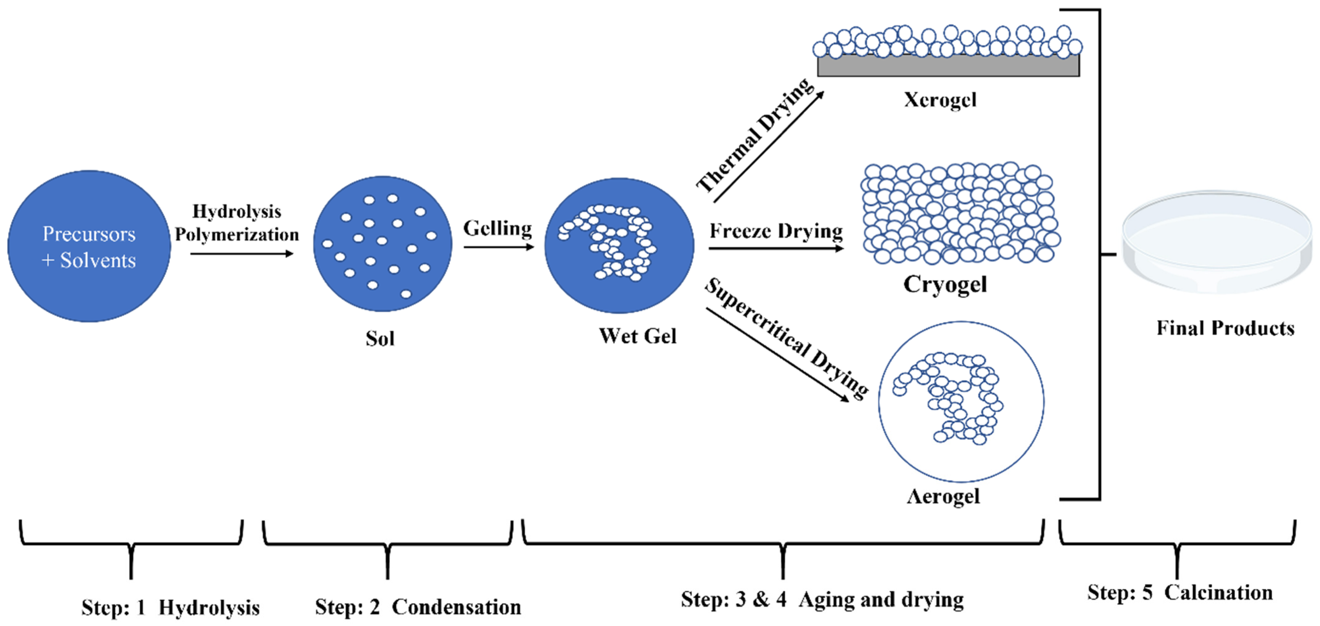
Figure 1. Sol–gel synthesis method.
Nanomaterials 15 00106 g001
Figure 2. Hydrothermal synthesis method.
Nanomaterials 15 00106 g002
Figure 3. Drug delivery to infected mice.
Nanomaterials 15 00106 g003
Table 1. A summary of citations followed by sol–gel methods.
SolutionTime (h)Temp. °CMorphologyCrystal PhaseDimensions (nm)Applications
Niobium alkoxides (Nb(OR)5) with acetic acid [13]480Xerogels and powdersMonolithic, tetragonal when heated above 500 °CN/ANb2O5 powders were synthesized
Niobium oxide (Nbx) with ethanol and HCL [14]2425Xerogels and powdersAmorphousN/AElectrochromic material
NbCl5 with propanol and acetic acid [15]1300–500Xerogel filmsAmorphous at 300 °C and pseudo-hexagonal at 500 °CN/AElectrochemical reversibility and dip coating
NbCl5 with ethanol and acetic acid [16]2450Thin FilmsAmorphousT: 83–178Counter electrode, particularly in nickel oxide devices.
NbCl5 with ethanol [17]2425Silica aerogelAmorphousN/AN/A
Niobium chloride, NbCl5, with tetraethoxysilane [18]4825Dried gelsAmorphousN/AN/A
NbCl5 with citric acid, ethylene glycol, deionized water, hydrogen peroxide [11]1225Transparent and yellow gelBelow 600 °C, amorphous Orthorhombic when calcined at 900 °CT: 50, when heated T: >200 nmCapacitors
Niobium ethoxide with acetic acid [19]24300Thin FilmsTetragonalN/APhotocatalytic activity
Anhydrous NbCl5 with InCl3 and acetylacetone [20]185Thin FilmsCubic bixbyiteT: 15–25Highest optical transmittance, conductive films
Niobium ethoxide with tetra-ethyl-ortho-titanate and ethanol [21]1450Thin FilmsAmorphous to crystallineT: 100–370CO sensing applications
Niobium ethoxide with NH4OH [22]225NanoparticlesOrthorhombic and hexagonalD: 1–100N/A
Niobium chloride (NbCl5) with aluminum chloride hexahydrate [23]170Nanorods and thin filmsHexagonalN/APhotocatalytic Activities
Niobium chloride (NbCl5) with hydrogen peroxide and coconut water powder [24]13625PowderOrthorhombic and monoclinic when heated at 1000 °CN/ADielectric constant
Niobium ethoxide with isopropyl alcohol [25]2180Thin FilmsPolycrystalline and amorphous structureN/AFabrication of unipolarswitching memory devices
Nb2O5 powder with hydrofluoric acid [26]2100NanoparticlesPolycrystallineN/AN/A
Table 2. A summary of citations followed by hydrothermal synthesis methods.
SolutionTime (h) and Tem. °CMorphologyCrystal PhaseSize (nm)Application
NbCl5 in HCl [31]24 and 210NanorodsMonoclinic after being calcined at 450 °CD: 22
L: 230
Lithium-ion batteries and dye-sensitized solar cells
Ammonium niobate oxalate hydrate, sucrose and HCl in deionized water [32]12 and 180NanocompositesPseudo-hexagonalD: 25–29Highly active and stable catalyst for electrochemical reactions
Nb2O5, LiOH and NH3.H2O in H2O2 [33]24 and 240Hollow microspheresPseudo-hexagonal after being calcined at 500 °CD: 1000–2000Catalysis, drug carriers, and gas sensors
Ammonium niobate oxalate and hydrogen peroxide distilled water [34]2–24 and 100–175NanoparticleOrthorhombicD: 9–35Photoactive degradation of pollutants
Niobium foil with ammonium fluoride [35]24–144 and 150NanorodsOrthorhombic for duration > 48 hD: 50–100Nano-scaled sensors, optoelectronic devices
Lithium hydroxide in HF acid [36]20–40 and 150–200Nano-treesPseudo-hexagonalD: 30–500UV sensors
Ammonium niobate oxalate hydrate and oleic acid in triethylamine [37]2–6 and 180NanorodsPseudo-hexagonalD: 5–20
L: 100–500
Photoactive degradation of pollutants
Ammonium niobate oxalate hydrate in distilled water [37]1 and 580NanospheresPseudo-hexagonalD: 20–50Photoactive degradation of pollutants
Niobium ethoxide, diethylene glycol and acetone in water [38]4–12 and 180Mesoporous spheresPseudo-hexagonalD: 400–500Highly effective solid acid catalysts
NbCl5 in ethanol mixed with triblock copolymer dissolved in distilled water [39]24 and 110MesoporousOrthorhombic after being calcined at 600 °CN/AN/A
NbO2 powder in distilled water and ethanol containing 1 M urea [40]72–720 and 130NanosheetsOrthorhombic and monoclinicT: 3–5High reversible charge/discharge capacity and cycling stability
Nb powder in distilled water [41]72–720 and 200NanorodsOrthorhombicD: 50An efficient material synthesized without catalyst
NbCl5 and ethanol in cyclohexanol [42]8–90 and 200–240Nanocables and nanorodsOrthorhombicD: 50–80
L: >1000
High-performance optoelectrical devices
Nb powder in urea [43]24–336 and 170–200NanobeltsN/AW: ~60 T ~15Lithium-ion batteries and dye-sensitized solar cells
Niobium Penta butoxide in toluene [44]2 and 300Nano powdersPseudo-hexagonalL: <80Photocatalytic dehydro-genation of methanol in an aqueous solution under deaerated conditions
NbCl5 in anhydrous benzyl alcohol [45] 72 and 250NanoparticlesPseudo-hexagonalD: 18–35High-rate-performance supercapacitor
NbCl5 and ethanol in cyclohexanol [46]8–90 and 200–240Nanograins and nanoparticlesOrthorhombic for T > 225 °CD: 50–80
L: >1000
Catalysis and its structure–activity relationships
Oxalic acid and hydrogen peroxide [47]4–12 and 150–220Nanorods and nanospheresOctahedralL: 5Used as nano catalyst
Oxalic acid and hydrogen peroxide [48]4–12 and 150–220Nanospheres and mesoporousMonoclinicD: 6Used as nano catalyst
Aluminum niobium oxalate with deionized water and ethanol [49]24 and 180Nanorods and nanospheresOrthorhombic after being calcined at 600 °CD: 300–500
D: 5–10
L: 100
Higher photocatalytic activities (organic pollutants, dye sensitized and solar cells)
Nb2O5 with isopropanol [50]48 and 180NanobeltsPseudo-hexagonalD: 100–200Energy storage electrodes
Nb powder with distilled water [51]72 and 180Flake, Nanorods, SpringsPolycrystallineL: 20–100Nano-scaled sensor, solar cells
Ammonium oxalate with Nb2AlC [52]21 and 180NanorodsHexagonal and orthorhombicD: 20–50
L: 70–150
Catalyst and water treatments
Nb with distilled water [53]72 and 150NanoflakesOrthorhombicD: 46–53High-performance optoelectrical devices
Nb2AlC with Hydrofluoric acid [54]20 and 190NanorodsMonoclinicL: 60–180Photocatalyst
Niobium with distilled water [29]20–40 and 130–150NanorodsOctahedralD: 20–50Biomedicine and environmental monitoring
Table 3. A summary of Nb2O5 properties and applications versus other commonly used nanoparticles.
PropertyNiobium Pentoxide Nanoparticles (Nb2O5NPs) [11]Gold Nanoparticles (AuNPs)
[66,67]
Silica Nanoparticles (SiO2NPs) [68]Silver Nanoparticles (AgNPs)
[69]
Zinc Oxide Nanoparticles (ZnONPs) [70]Carbon-Based Nanoparticles [69,71]Titanium Dioxide Nanoparticles (TiO2) [68,72]
Chemical CompositionNiobium and oxygenPure goldSilicon dioxidePure silverZinc and oxygenCarbon (e.g., fullerenes, graphene)Titanium and oxygen
Size Range (nm)10–200 nm1–100 nm5–500 nm1–100 nm10–200 nmVaries (e.g., graphene sheets, carbon dots)1–100 nm
MorphologyCrystalline or amorphousSpherical, rods, othersSpherical, mesoporous, othersSpherical, triangular, othersSpherical, rod-shaped, othersSpherical, tubular (e.g., nanotubes), sheetsSpherical, rods, others
Surface ModificationFunctionalized with organic groups, metals, or polymersThiol groups, PEGylation, antibodiesFunctionalized with silanes, organic groups, or biomoleculesCitrate, PEGylation, antibodiesOrganic molecules, polymersFunctionalized with various chemical groupsCoatings, doping with metals or non-metals
Optical PropertiesHigh refractive index, photoluminescentSurface plasmon resonance (SPR)Transparent, adjustable refractive indexStrong SPR, antimicrobialUV absorption, photoluminescentFluorescence (e.g., carbon dots), high surface areaUV absorption, photo
catalytic activity
Electrical PropertiesHigh dielectric constant, resistive switchingConductive, plasmonic effectsInsulatorConductiveSemi-conductorConductive (graphene), semi-conducting (carbon dots)Semi-conductor
Thermal StabilityHighModerateHighModerateHighHighHigh
BiocompatibilityModerate; dependent on surface functionalizationHigh; excellent for biological applicationsHigh; generally inertVariable; size- and concentration-dependentGenerally good; antimicrobial propertiesHigh (e.g., graphene oxide), variable for othersGenerally good; dependent on surface properties
ToxicityLow; surface-dependentLow; size- and concentration-dependentGenerally lowPotential cytotoxicity; dose-dependentLow; antimicrobial propertiesLow; dependent on form and functionalizationLow; dependent on crystal structure and surface properties
Key ApplicationsCatalysis, electrochromic devices, gas sensors, photocatalysis, batteriesBiomedical imaging, drug delivery, photothermal therapy, sensorsDrug delivery, imaging, coatings, catalysis, chromatographyAntimicrobial agents, biosensing, medical imagingSunscreens, antibacterial agents, photocatalysis, sensorsDrug delivery, imaging, electronics (e.g., graphene)Photocatalysis, UV blockers, sensors, biomedical applications
Unique FeaturesHigh photocatalytic activity, ion storage capabilities, high refractive indexStrong SPR for optical applications, high specificity with functionalizationHigh surface area (e.g., mesoporous), high chemical stabilityPotent antimicrobial activity, strong SPRAntimicrobial, UV-blocking properties, semi-conductor behaviorExceptional mechanical strength (graphene), tunable electronic propertiesPhotocatalytic efficiency, UV absorption, chemical stability
Synthesis MethodsSol–gel, hydro-
thermal, precipitation
Chemical reduction, seed-mediated growthStöber process, sol–gel methodsChemical reduction, photo-chemical methodsSol–gel, hydro-thermal, precipitationChemical vapor deposition (CVD), arc discharge, laser ablationSol–gel, hydro-thermal, chemical vapor deposition
ChallengesLimited commercial availability, complex surface functionalizationCost of gold, stability in solutionsAggregation in some environments, limited thermal conductivityPotential cytotoxicity, stability under various conditionsPhotocatalytic activity leading to ROS generation, potential cytotoxicityProduction scalability (e.g., graphene), potential cytotoxicityControl over crystal phase, potential environmental impact
CostModerateHighLow to moderateModerateLowVariable (e.g., graphene can be expensive)Low
Disclaimer/Publisher’s Note: The statements, opinions and data contained in all publications are solely those of the individual author(s) and contributor(s) and not of MDPI and/or the editor(s). MDPI and/or the editor(s) disclaim responsibility for any injury to people or property resulting from any ideas, methods, instructions or products referred to in the content.