Article

Nanovesicles and Human Skin Interaction: A Comparative Ex-Vivo Study

1
Department of Chemical, Pharmaceutical and Agricultural Sciences, University of Ferrara, I-44121 Ferrara, Italy
2
Laboratory for Technologies of Advanced Therapies “LTTA”-Electron Microscopy Center, University of Ferrara, I-44121 Ferrara, Italy
3
Department of Translational Medicine and for Romagna, University of Ferrara, I-44121 Ferrara, Italy
4
Department of Life and Environmental Sciences, Marche Polytechnic University, I-60131 Ancona, Italy
5
SOLARIS, National Synchrotron Radiation Centre, Jagiellonian University, 30-392 Kraków, Poland
6
Department of Neurosciences, Biomedicine and Movement Sciences, University of Verona, I-37134 Verona, Italy
*
Author to whom correspondence should be addressed.
Nanomaterials 2025, 15(12), 937; https://doi.org/10.3390/nano15120937
Submission received: 18 April 2025 / Revised: 28 May 2025 / Accepted: 13 June 2025 / Published: 16 June 2025
(This article belongs to the Special Issue Green Nanoparticles for Topical Administration of Drugs)

Abstract

:
The topical administration of drugs on the skin by nanovesicular systems can represent a tool to treat skin pathologies. The study of nanovesicle biodistribution after skin administration is crucial to understanding their transdermal potential. A formative study enabled us to investigate the influence of some methods in the production of nanovesicles based on phosphatidylcholine, differing in their ethanol amount. Particularly, both liposomes and ethosomes produced by different methods, i.e., microfluidics and solvent injection, were considered. The evaluation of size distribution, shape and internal morphology was performed using photon correlation spectroscopy, cryogenic electron microscopy, hyperspectral dark-field microscopy and small-angle X-ray scattering. Transmission electron microscopy was then used to observe and compare the transdermal passage of selected liposomes and ethosomes applied to human skin explants in a bioreactor. The mean diameters of nanovesicles prepared by the ethanol injection method were smaller with respect to those obtained by microfluidics, measuring roughly 140 and 230 nm, respectively. The uni- or multilamellar ultrastructure of the vesicles was influenced by the solvent injection procedure. Ultrastructural analysis of skin penetration revealed (i) the ability of intact vesicles to cross the different skin layers, with ethosomes produced by the water injection method showing greater transdermal potential and (ii) the role of ethanol as a penetration enhancer.

1. Introduction

Skin’s primary function is to protect the body against exogenous stimuli, such as insults, infections, as well as exposure to environmental stressors [1]. The protective effect is mainly exerted by the stratum corneum, acting as the major barrier. Although the skin offers a potential route for drug administration, especially topically for skin conditions, the stratum corneum presence hinders drug passage. Consequently, in recent years, significant research has been focused on developing effective transdermal drug delivery systems to overcome this barrier [2,3,4]. Particularly, liposomes (LIPO) and ethosomes (ETHO) are colloidal delivery systems made of biocompatible and biodegradable phospholipids organized as bi-layered vesicles dispersed in water [4,5,6,7,8,9]. Typically, the main component of LIPO and ETHO is represented by phosphatidylcholine (PC), a zwitterionic surfactant that spontaneously self-aggregates in water, giving rise to lamellar structures [10]. The ultrastructural organization of LIPO and ETHO allows the solubilization of both hydrophilic and lipophilic compounds, promoting their transdermal delivery. Indeed, due to their affinity with the skin architecture and composition, LIPO and ETHO endorse the active compound passage through the upper skin strata towards the dermis [4,5,6,11]. LIPO and ETHO have proven effective in biomedical applications, improving the delivery of therapeutics for various skin disorders. ETHO formulations show promise for treating viral (e.g., acyclovir for herpes) [12], fungal (e.g., fluconazole) [13], and inflammatory (e.g., colchicine for psoriasis) skin conditions [14]. These carriers facilitate drug penetration into deeper skin layers, enhancing treatment outcomes for conditions like acne, psoriasis, and skin cancers. For instance, tretinoin ETHO offers improved efficacy and reduced irritation compared to conventional acne treatments [15]. Initially utilized in dermatology for their moisturizing and regenerative effects, the focus of LIPO and ETHO research has shifted towards their ability to encapsulate and deliver biological materials to epidermal cells and deeper skin layers [16,17].
The main difference between LIPO and ETHO lies in the ethanol presence, potentially reaching 45% w/w in ETHO. Ethanol plays diverse pivotal roles; indeed, on the one hand, it makes the vesicles more flexible; on the other, it interacts with skin and vesicle lipids, helping the drug associated with the vesicles to pass through the stratum corneum [18]. Additionally, ethanol noticeably improves vesicle stability and its ability to hold lipophilic drugs [19,20,21,22]. Some authors demonstrated that LIPO exhibited insufficient flexibility for skin penetration, resulting in the formation of a drug depot within the stratum corneum, from which encapsulated drugs diffused slowly [23]. Consequently, deformable LIPO was engineered through the incorporation of edge activators (e.g., surfactants), thereby destabilizing the lipid bilayers composed of PC and water and, in turn, increasing vesicle flexibility [24,25]. Analogously, the addition of edge activators to ETHO resulted in transethosomes in an attempt to further increase the vesicle penetration potential [22,26].
Various techniques have been created over time to yield lipid-based nanocarriers. Most of these “top-down” methods rely on physically breaking down larger, pre-formed structures. Sonication is a key example, though its scalability is limited. Conversely, “bottom-up” approaches offer an alternative by promoting the self-assembly of lipids in solution into lipid-based nanocarriers, thus avoiding the need for size reduction [27].
Different “bottom-up” methodologies can be employed for LIPO and ETHO preparation [28]: (i) the classic thin-film hydration method, based on the hydration of a thin lipid film obtained under evaporation of an organic solvent from a lipid solution; (ii) ethanol injection, involving the injection of a lipid ethanol solution into water; (iii) reverse-phase evaporation, forming LIPO by creating a water-in-oil emulsion from which the organic solvent is evaporated, leading to LIPO formation; and (iv) microfluidics, a more modern approach based on the use of microchannels to mix lipid solutions and aqueous phase [28].
The choice of method depends on the desired LIPO size, lamellarity (number of lipid bilayers), and the type of drug to be encapsulated. Post-processing steps, such as size reduction and purification, may be necessary to obtain the desired vesicle features [29]. Proper characterization of nanovesicles for skin application is essential for a complete understanding of their behaviors on the skin and their biological effects [30]. Key physical attributes that significantly impact skin interactions, such as size distribution, shape, and inner morphology, should be thoroughly investigated [31].
A crucial point in the potential of a transdermal delivery system is related to the maintenance of its structural integrity throughout the skin strata to carry the associated drug and release it deeply toward the dermis.
Despite several in vitro and in vivo studies have examined the transdermal effects of LIPO and ETHO, focusing on drug delivery and therapeutic potential [22,26,32,33], a limited number of studies have been dedicated to investigating the effective cutaneous penetration of LIPO and ETHO when applied onto the skin [34,35].
Recently, we investigated ETHO and transethosomes applied on healthy human skin explants maintained in a bioreactor, an in vitro model that reliably simulates in vivo physiological conditions and preserves skin structure for extended periods [36]. Visualization of vesicles within skin strata was accomplished through transmission electron microscopy (TEM) experiments, enabling them to unequivocally determine their penetration potential and their fate in the different skin layers. The mean size, morphology, and deformability of these vesicles were shown to be affected by polysorbate 80, used as an edge activator in transethosome production, and by the concentration of PC [36].
In the present work, a comparative study was carried out, comparing the LIPO and ETHO penetration capability throughout healthy human skin.
LIPO and ETHO produced by different methodologies, such as microfluidics, water injection, and ethanol injection, were characterized and compared for their physical-chemical features, such as size distribution, determined by photon correlation spectroscopy (PCS), and morphology, evaluated by cryogenic electron microscopy (Cryo-EM), Small Angle X-Ray Scattering (SAXS) and hyperspectral dark-field microscopy [37].
In the second part of the study, the transdermal passage of selected LIPO and ETHO applied on human skin explants maintained in a bioreactor was monitored and compared by TEM [38]. TEM allows for direct visualization of nanovesicles within the skin, shedding light on the impact of methodology on nanovesicle physicochemical features and cutaneous fate [39].

2. Materials and Methods

2.1. Materials

Phosphatidylcholine (PC) (purity 94%) from soybean was acquired by A.C.E.F. Spa. (Milan, Italy). The solvents used were HPLC grade, while all other chemicals were analytical grade.

2.2. Preparation of Nanovesicular Systems

LIPO and ETHO production was made alternatively by a microfluidic approach or by two bulk methods, specifically “water injection” and “ethanol injection”.

2.2.1. Microfluidic Approach

For the microfluidic approach, the preparation of LIPOMF and ETHOMF was performed using a cross-junction microfluidic chip based on a Large Droplet Junction Chip (Dolomite, Alfatest, Rome, Italy) with a hydrophilic quartz channel etched to a depth of 100 µm, mounted on a Chip interface H equipped with two Linear Connector 4-way (Dolomite, Alfatest, Rome, Italy).
The inner lipid phase (LP) consisted of a PC ethanol solution (30 mg/mL), while bi-distilled water served as the aqueous outer phase (AP). The AP and LP flow rates (FAP and FLP, respectively) were regulated using two syringe pumps (IPS-14 syringe series, Inovenso Inc., Başakşehir/Istanbul, Turkey). Visualization was performed with an optical microscope (Leica DM LS2, Leica Microsystems Srl, Buccinasco (MI), Italy). The Flow Rate Ratio (FRR) between FLP and FAP was set to 10:1 and 2:1 v/v, respectively for LIPOMF or ETHOMF. The Total Flow Rates (TFR) were 55 and 90 µL min−1.
After stabilization of the focused stream, each sample (3 mL) was collected into glass vials and stored at 4 °C; sample preparation was performed at 25 °C.

2.2.2. Bulk Approach

In the case of the bulk approach, LIPO and ETHO preparation was performed by a cold method alternatively based on the dropwise addition of bidistilled water to PC ethanol solution (“water injection”) or by the dropwise addition of PC ethanol solution to bidistilled water (“ethanol injection”).
Particularly, in the case of the water injection method, LIPOWI and ETHOWI were prepared dripping bidistilled water at 1500 μL min−1 (IPS-14 syringe series, Inovenso Inc., Başakşehir/Istanbul, Turkey) in the PC ethanol solution (30 mg/mL) kept under magnetic stirring at 750 rpm (IKA RCT basic, IKA®-Werke GmbH and Co. KG, Staufen, Germany). The final water/ethanol ratios were 90:10 (v/v) for LIPOWI and 70:30 for ETHOWI.
For the ethanol injection method, LIPOEI and ETHOEI were prepared by adding the PC ethanol solution (30 mg/mL) to bidistilled water dropwise, maintaining the same flow, stirring rates and time requirements as for the water injection method. The final water/ethanol ratio was 90:10 or 70:30 (v/v) for LIPOEI and ETHOEI, respectively.
In both the water injection and ethanol injection methods, stirring was carried out at 22–25 °C for 30 min after the water or ethanol phases had been added.

2.3. Vesicle Characterization

2.3.1. Photon Correlation Spectroscopy (PCS)

Vesicle size distribution was determined using a Zetasizer Nano-S90 (Malvern Instruments, Malvern, UK) equipped with a 5 mW helium-neon laser operating at a wavelength of 633 nm. Measurements were conducted at 25 °C, using a detection angle of 173°, following a 120-s equilibration period. Prior to analysis, samples were diluted 1:10 (v/v) with bidistilled water. The size distribution was calculated using the CONTIN algorithm [40]. All measurements were performed in triplicate, and the results are reported as mean values.
Moreover, the size distribution of samples stored at 22–25 °C for 3 months was measured to gain information on vesicle size stability.

2.3.2. Cryogenic Electron Microscopy (Cryo-EM)

Cryo-EM data were acquired at the National Cryo-EM Centre SOLARIS in Kraków, Poland, using a Titan Krios G3i transmission electron microscope (Thermo Fisher Scientific, Brainport Eindhoven, The Netherlands) operated at 300 kV. Images were recorded at a nominal magnification of ×105,000, corresponding to a calibrated pixel size of 1.7 Å, using EPU 2.10 software. A K3 direct electron detector (Gatan, Pleasanton, CA, USA), operated in counting mode and integrated with a BioQuantum Imaging Filter (20 eV slit width) (Gatan, Pleasanton, CA, USA), was employed for data collection. The total electron dose applied was 41.09 e2, with a defocus setting of −2.0 µm.
Prior to imaging, samples were plunge-frozen in liquid ethane using a Vitrobot Mark IV (Thermo Fisher Scientific, Brainport Eindhoven, The Netherlands). For each grid, 3 µL of the sample suspension was applied, and excess liquid was blotted from both sides. Vitrification was performed under controlled conditions: 95% humidity, 4 °C temperature, 3 s blot time, and blot force setting of 3.

2.3.3. Small Angle X-Ray Scattering (SAXS)

SAXS measurements were performed at the Austro-SAXS beamline of the Elettra Synchrotron facility in Trieste, Italy [41]. The samples were enclosed in quartz capillaries with an inner diameter of 1.5 mm and maintained at a constant temperature of 32 °C using a temperature-controlled sample holder equipped with Peltier heating/cooling (KPR, Anton Paar, Graz, Austria).
For each measurement, the sample was illuminated with an X-ray beam for 10 s per frame, and a total of 18 frames were acquired. The raw two-dimensional scattering images were processed to correct for background noise, detector sensitivity, and transmission effects. Following these corrections, the data were azimuthally averaged to obtain the one-dimensional scattering profile, expressed as the differential scattering cross-section d Σ d Ω or the intensity I(Q), where Q represents the magnitude of the scattering vector and is calculated as Q = 4π sin(θ)/λ (with 2θ being the scattering angle and λ the wavelength of the incident X-rays).
The sample-to-detector distance was set to a Q-range of approximately 0.1 to 5 nm¹. Scattering patterns were recorded using a two-dimensional Pilatus3 1M detector, equipped with a pixel size of 172 × 172 μm2. The detector operated at a photon energy of 8 keV, corresponding to an X-ray wavelength of 0.154 nm.
The uncertainties on all fitting parameters were estimated using the GENFIT software, version 2024.11 (available at https://sites.google.com/site/genfitweb/download) by repeating the fitting procedure 20 times. In each repetition, the experimental SAXS data points were randomly perturbed by sampling from a normal distribution with a standard deviation equal to the experimental error associated with each q-point. The mean value and standard deviation of each parameter were then calculated from the 20 values obtained across the repeated fits.

2.3.4. Hyperspectral Dark-Field Microscopy

Five microliters of LIPOEI, ETHOWI, or ETHO EI dispersion was placed on a glass slide (Epredia Glass Slides 76 × 26 mm, provided by Menzel Gläser, Geelong, Australia) and let dry until the solvent fully evaporation to prevent light scattering phenomena. Before imaging, a coverslip was placed on the sample. Hyperspectral imaging was performed using a CytoViva® dark-field hyperspectral microscopy system (CytoViva Inc., Auburn, AL, USA), which included a halogen light source, a BX-43 research-grade optical microscope, and a hyperspectral imaging setup. The system was equipped with a sCMOS spectral camera coupled to a VNIR imaging spectrograph (ImSpector V10E, Specim, Spectral Imaging Ltd. Oulu, Finland), enabling data acquisition across the visible to near-infrared (VNIR) range of 400–1000 nm.
Acquisition parameters were: (i) light intensity of 80% of maximum, (ii) 100× immersion oil objective (Olympus UPlan FL N, 100×/1.30), (iii) spatial resolution (x;y) of 1024 × 1024, (iv) spectral resolution (λ) of 1.2 nm, (v) exposure time 0.2 s.
Post-process imaging was performed with ENVI 4.0 (CytoViva® proprietary software): a minimum of 3 regions of interest (ROIs) or 15 single pixels, located in different areas of the image and characterized by their light intensity value 10% below the maximum level, were selected to obtain specific and representative spectra.

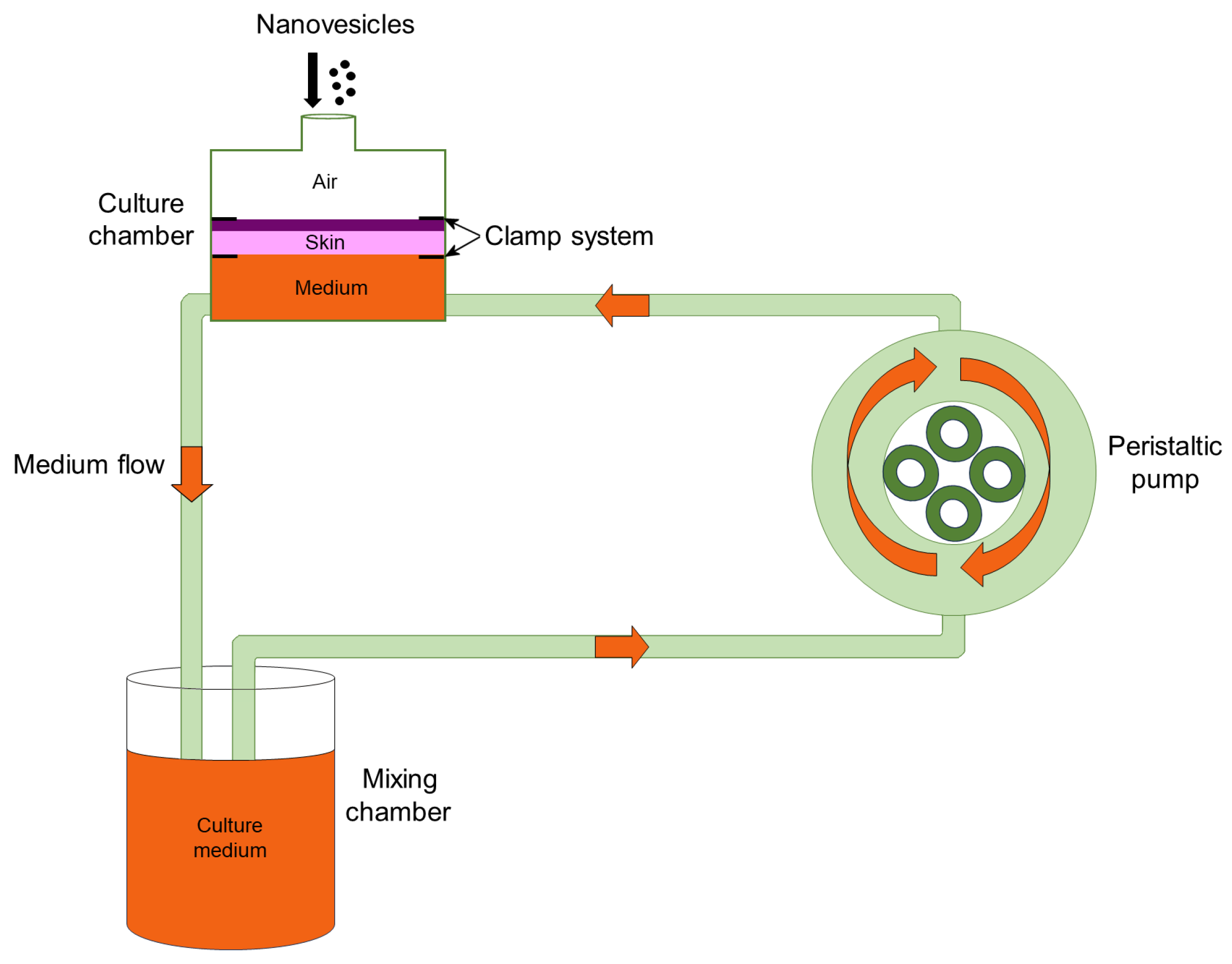
2.4. Human Skin Sampling

Skin samples were surgically removed from the abdominal region of four healthy women undergoing abdominoplasty at the Verona University Hospital. The patients signed an informed consent. The study was conducted in accordance with the Declaration of Helsinki and authorized by the Institutional Ethics Committee of the Verona University Hospital (protocol code 3449CESC, 21 April 2021). The excised skin samples were immediately washed in physiological solution (NaCl 0.9% w/v), placed in Dulbecco’s Modified Eagle’s Medium (DMEM), supplemented with 4.5 g/L D-glucose, 10% FBS, 2% penicillin-streptomycin, 200 mM L-glutamine, and 0.3 μg/mL Amphotericin B (Gibco, Waltham, MA, USA) at 37 °C for a few minutes, and then processed for treatment in the bioreactor.

2.5. Skin Treatment and Transmission Electron Microscopy (TEM) Analysis

The bioreactor used in this study (IV-Tech, Massarosa, LU, Italy) is schematically represented in Figure 1 and has been extensively described in [38].
Round samples with a diameter of 1.5 cm were placed in culture chambers modified to house flat organs (LiveBox2; IV-Tech): the epidermis was placed at the top, in contact with air, while the dermis was placed at the bottom, in contact with the flowing (500 μL/min) culture medium [38]. A 300 µL drop of LIPOEI, ETHOWI, or ETHOEI dispersion (PC 0.9% w/w) was applied to the stratum corneum. The bioreactor was maintained in an incubator at 37 °C in a 5% CO2 humidified atmosphere for 1 h, 3 h, 6 h, and 24 h. As a control, some skin samples were treated with the dispersion medium devoid of nanovesicles. After incubation, skin samples were aldehyde-fixed (2% (v/v) paraformaldehyde and 2.5% (v/v) glutaraldehyde in 0.1 M phosphate buffer) for 3 h at 4 °C, treated with 1% (v/v) OsO4 and 1.5% (v/v) C6FeK4N6 for 1.5 h, dehydrated with acetone and embedded in epoxy resin (Epon-Araldite, Electron Microscopy Sciences, Hatfield, PA, USA). Sections of 70–90 nm in thickness were placed on copper grids (Electron Microscopy Sciences) and stained with Reynold’s lead citrate. Observations were made with a Philips Morgagni transmission electron microscope (FEI Company Italia Srl, Milan, Italy) equipped with a Megaview II camera for digital image acquisition.
LIPOEI, ETHOWI, or ETHOEI diameter was measured in TEM micrographs (×22,000) with the ImageJ software 1.10z (NIH), and the mean and standard deviation (s.d.) values were obtained (n = 20 for LIPOEI, ETHOWI, or ETHOEI). In the case of flattened nanovesicles, the major axis was considered.

3. Results

3.1. Preparation of Nanovesicles

Nanovesicles based on PC were designed as biocompatible nanocarriers for dermal and transdermal delivery of drugs. Both LIPO and ETHO were investigated, embodying in their composition different amounts of ethanol, respectively 10 or 30%, w/w. The presence of ethanol, acting as a penetration enhancer in conjunction with PC, allows the vesicles to reach deeper skin levels. A previous study on ETHO, conducted by our research group, led to the selection of a 0.9% w/w PC, resulting in stable vesicles with a mean diameter of around 200 nm and a homogeneous size distribution [42,43,44]. Moreover, PC concentration equal to 9 mg/mL is an intermediate between the typical lipid concentration range of 5 to 15 mg/mL in liposomal nanomedicines [45,46]. This concentration was achieved using initial PC solutions 90 and 30 mg/mL for LIPO and ETHO, respectively.
In the present investigation, LIPO and ETHO produced by different procedures were compared.
As the first approach, a microfluidic method was employed using a device equipped with two syringe pumps connected to a chip containing two intersecting microchannels [47]. Control was maintained using two flow regulators and an optical microscope. Within the microfluidic chip, the LP and AP flows converged at the intersection of the two channels. The subsequent diffusion of water and ethanol at their interface triggered the self-assembly of the PC, reproducibly generating vesicles of precise and controlled size [48].
As a second approach, two bulk methods were employed, namely “water injection,” (extensively studied by our group) based on adding water by a syringe pump to a PC solution in ethanol under magnetic stirring, and “ethanol injection” method, where PC ethanol solution is gradually added to bidistilled water, under stirring [49].
Table 1 summarizes the parameters and compositions employed for nanovesicle preparation. The TFR and FRR parameters were selected based on a previous study conducted by our research group [48] aimed at obtaining vesicles with homogeneous size distribution and mean diameter around 200 nm. Indeed, PCS’s previous experiments let us demonstrate that lower TFR resulted in smaller monodispersed vesicles (lower D.I.), while higher TFR led to larger and polydisperse vesicles. Also, a high TFR (around 90 µL/min) led to phase separation of the LIPO dispersions. This was attributed to the potential development of unstable flow patterns within the microchip, negatively affecting the mixing of AP and LP.
In the case of vesicles prepared by the bulk method, in both LIPO and ETHO, FAP and FLP were 1500 µL/min, thus extremely greater with respect to those employed for the microfluidics method.
In the case of vesicles prepared by microfluidics, TFR and FRR were adjusted to obtain different amounts of ethanol, namely 3-fold higher in the case of ETHOMF compared to LIPOMF. In both LIPO and ETHO, ethanol enables to maintain the vesicle stability. Typically, the presence of 20–45% w/w ethanol differentiates ETHO from LIPO, resulting in more stable and softer vesicles while improving their penetration potential.
Nanovesicular systems appeared milky, translucent, and homogeneous, apart from LIPOWI that, showed phase separation 1 day after preparation (Table 2).

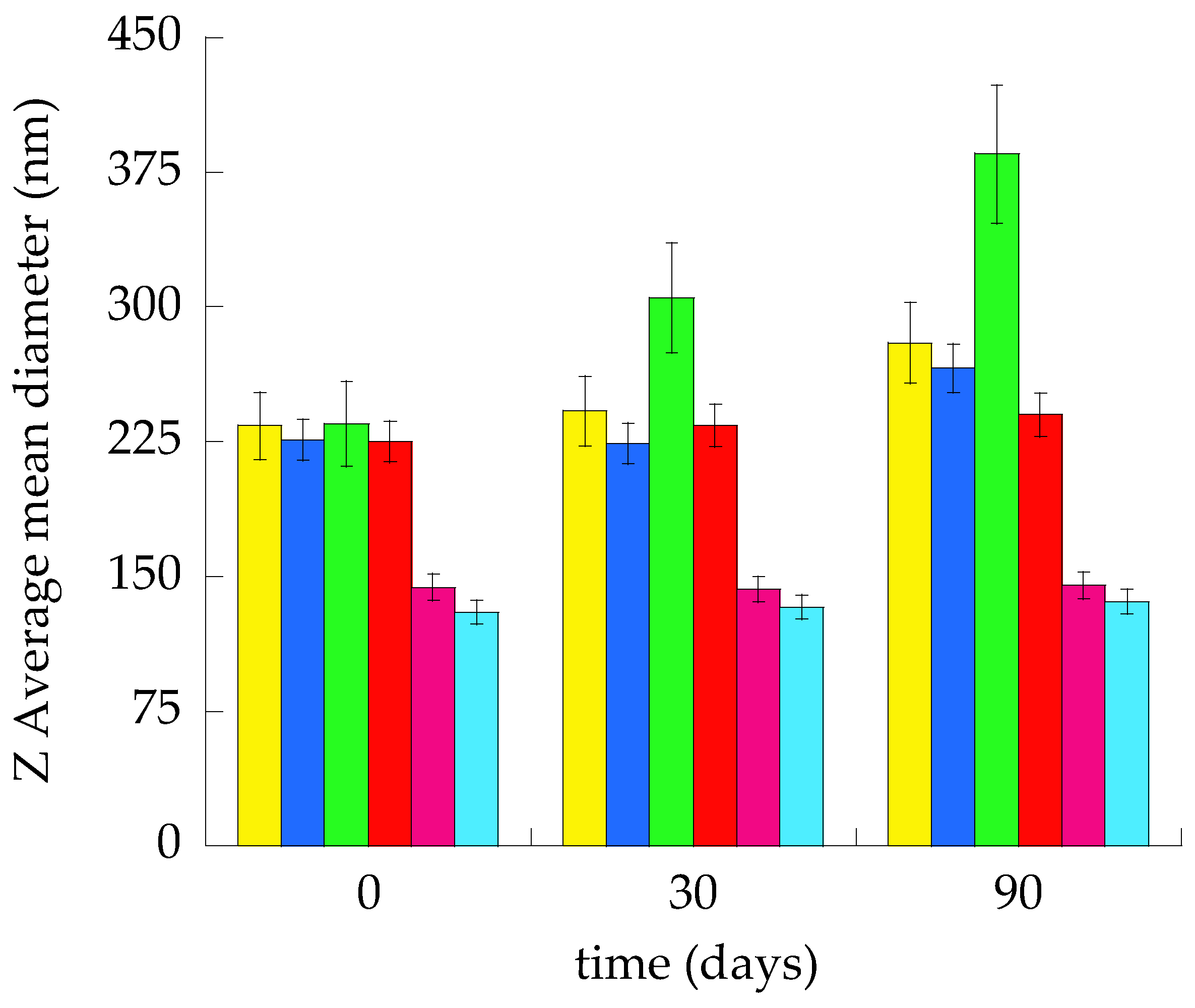
3.2. Size Distribution

PCS enabled the measurement of the size distribution of vesicles one day after their preparation. Table 2 reports the main size distribution parameters as well as information about the macroscopic aspect of the different nanovesicular systems evaluating by visual inspection possible phase separation phenomena.
The average hydrodynamic diameters ranged between 130 and 235 nm, maintaining dispersity indexes always below or equal to 0.27. The smallest vesicles were obtained by the bulk ethanol injection method. Indeed, mean diameters were 70–100 nm smaller than the corresponding vesicles produced by the water injection method. In general, ETHO vesicles displayed slightly smaller mean diameters with respect to LIPO. It was found that nanovesicular systems produced by the ethanol injection method were characterized by Z-Average mean diameters smaller with respect to the ones obtained by the water injection method. Conversely, this latest method resulted in vesicles whose mean diameter value was near the vesicle’s mean diameter prepared by microfluidics. Since LIPOWI displayed a macroscopic aspect different from other nanovesicular systems, a size stability study was performed, measuring Z Average mean diameters up to 3 months from the vesicle preparation (Figure 2).
As clearly detectable, the more stable nanovesicular systems were ETHOEI, ETHOWI, and LIPOEI, maintaining almost unvaried Z-Average mean diameters with respect to the initial values (1–6%). Conversely, ETHOMF, LIPOMF, and LIPOWI underwent a Z-average increase of 18, 20 and 64%, respectively.
The largest mean diameter and dispersity index detected after LIPOWI preparation is responsible for vesicle aggregation and phase separation phenomena.
Because of size distribution, stability, and macroscopic aspect, LIPOEI, ETHOWI and ETHOEI were selected for further studies.

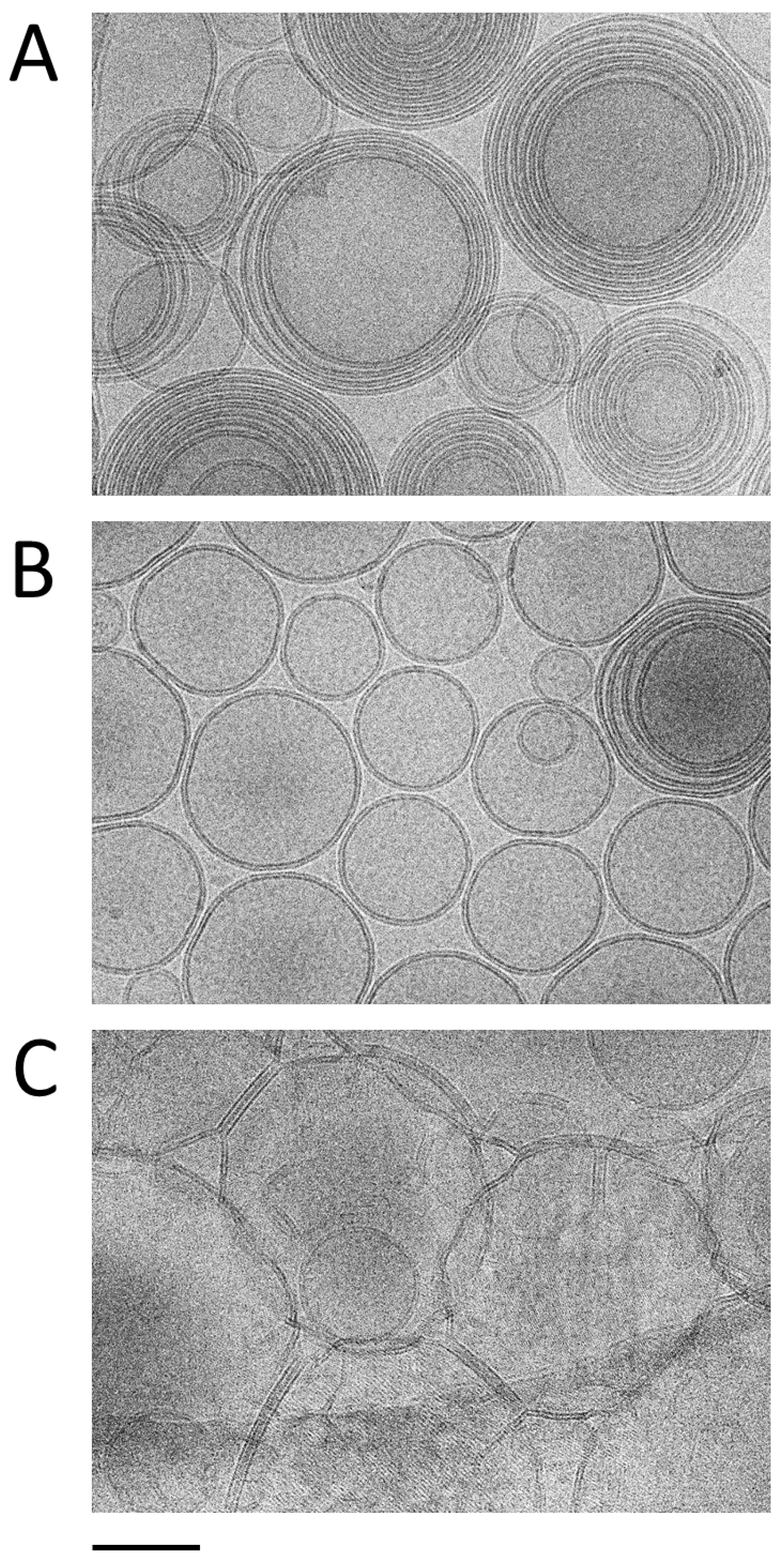
3.3. Morphological Characterization
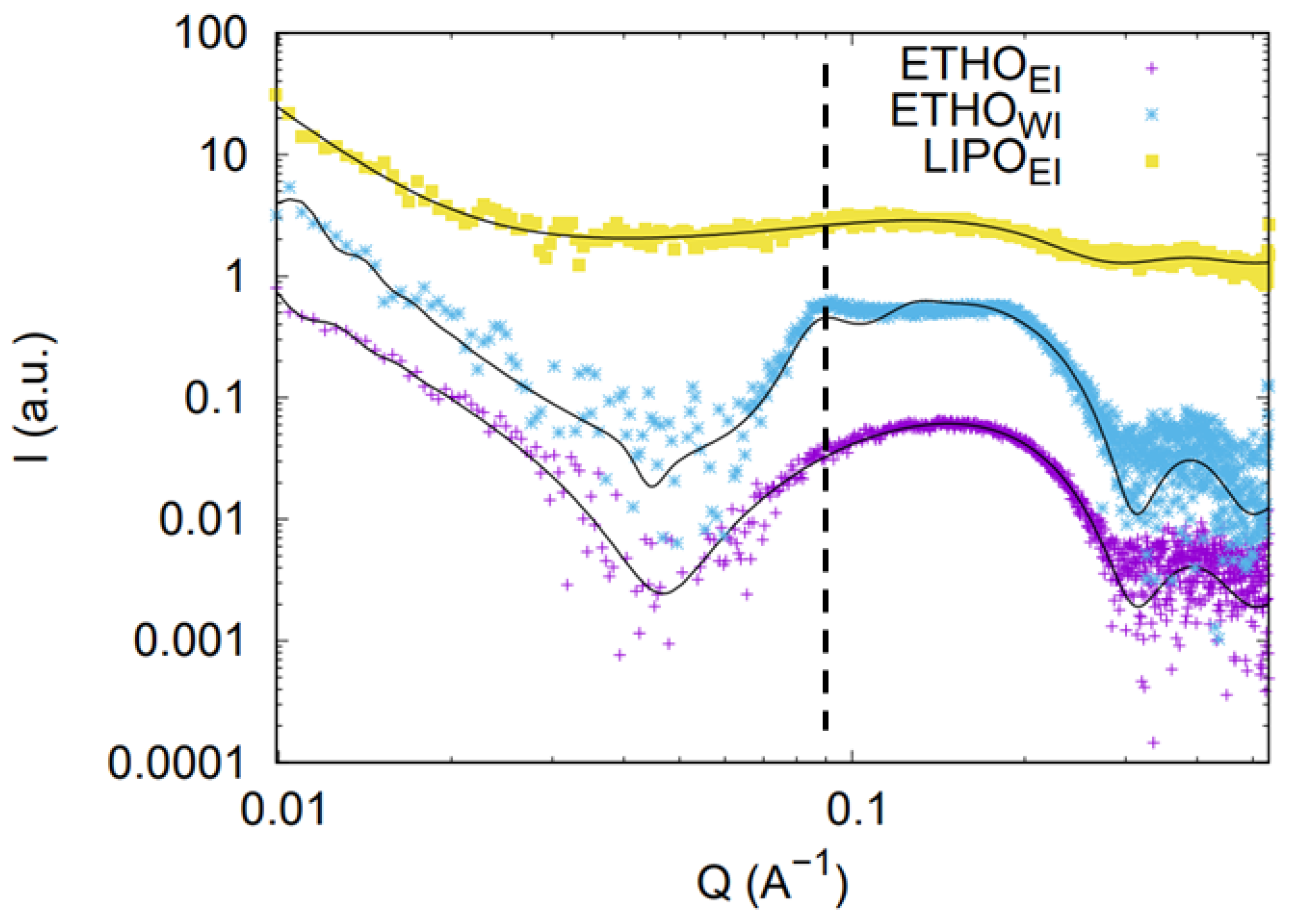
The morphology of nanovesicles obtained via the ethanol and water injection methods was evaluated using Cryo-EM. The resulting micrographs corresponding to LIPOEI, ETHOWI and ETHOEI (Figure 3A–C) show spherical vesicles, whose size distribution is in agreement with PCS analysis, with a very well-defined double layer. Notably, the water injection method (Figure 3A) resulted in multilamellar vesicles, whilst the ethanol injection method (Figure 3B,C) yielded a predominance of unilamellar and oligolamellar vesicles, as previously observed in the case of gossypin-loaded ETHO [49]. In the case of LIPOEI (Figure 3C), the vesicles appear more irregular and have a tendency to aggregate. These observations underscore the impact of the preparation technique on nanovesicle morphology. It can be hypothesized that, in the water injection method, the gradual addition of water to the PC-ethanol solution, on the one hand, facilitates a more controlled vesiculation process, leading to larger structures; on the other, it promotes the assembly of multiple lipid bilayers, resulting in the observed prevalence of multilamellar vesicles. Conversely, the rapid removal of the organic solvent during ethanol injection could accelerate vesiculation, thereby impeding the formation of complex multilamellar structures [49].

3.4. SAXS Study

SAXS was employed to investigate the structural characteristics of LIPO and ETHO produced via ethanol injection and water injection methodologies. Representative SAXS profiles, acquired at 32 °C, are presented in Figure 4. Although the data for LIPO show rather weak intensity, all curves exhibit a broad scattering band in the range of 0.6 to 3 nm−1, confirming the presence of the lipid bilayer in the ETHOWI, ETHOEI, and LIPOEI vesicles, in good agreement with Cryo-EM results. A main distinction between the formulations is the appearance of a clear Bragg reflection at Q = 0.087 Å−1 in the ETHOWI profile. This observation is interpreted as evidence of a prevalent multilamellar vesicle structure in this sample, thus validating the results obtained through Cryo-EM. Furthermore, the Bragg peak corresponds to an interlamellar distance of 7.22 nm, which aligns with previously reported data for comparable drug-delivery systems [43]. A final point to note is that the overall SAXS intensity for LIPOEI is very low, likely due to a low scattering contrast between the components, which may be attributed to the higher water content inside the LIPO nanovesicles (90:10 compared to 70:30 in ETHO).
To derive quantitative information, SAXS profiles have been then fitted considering the nanovesicles as a polydisperse system of spheres with core radius R0 and a dispersion ξRo described by a Schultz distribution [34]. The nanovesicle core is filled with a water/ethanol solution. Surrounding the core, there is a symmetric bilayer composed of three concentric shells with constant electron densities: the polar head group (thickness R1 and electron density ρ1), the central aliphatic chain region (thickness R2 and electron density ρ2), and the terminal methyl group (thickness R3 and electron density ρ3). Additional bilayers, identical in composition and structure to the one enveloping the nanovesicles and spaced by layers of water with electron density ρ0 can also be included in the model: each bilayer is defined by the same parameters Ri and ρi (i = 1, 2, 3). These N repeating bilayers are distributed with an average inter-bilayer spacing c, and their positional disorder is accounted for using paracrystalline theory with distortion parameter gc.
The fitting curves and corresponding best-fit parameters are shown in Figure 4 and summarized in Table 3.
Fitting data confirm the qualitative observations: in all cases, the fit indicates the presence of vesicles with dimensions of a few hundred nanometers and with lipid membranes of similar thickness (around 3.6 nm). However, nanovesicle structural characteristics depend both on composition and preparation method: ETHOWI vesicles (with a low polydispersity in size) show a multilamellar structure with a low degree of cumulative positional disorder, whilst ETHOEI vesicles (rather monodisperse in size) are predominantly unilamellar or oligolamellar. On the contrary, LIPOEI vesicles show a strong degree of disorder with regard to the interplanar distances between the lamellae. Moreover, the fitting results are fully consistent with the suggested variation in scattering contrast between components due to the increased water content inside the LIPO nanovesicles.

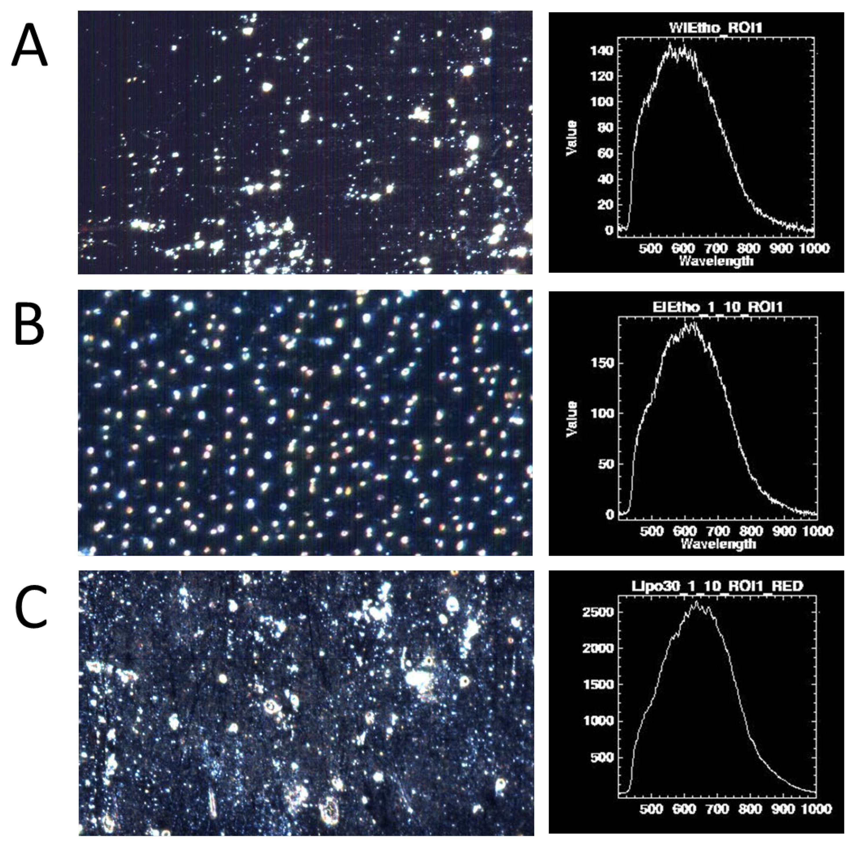
3.5. Hyperspectral Dark-Field Microscopy Study

Hyperspectral Light Microscopy is a potent analytical technique for nanoparticulate and nanovesicular systems that integrates spectral data acquisition with optical microscopy methods like fluorescence, brightfield, or darkfield. This synergy enables the extraction of richer information about the sample being studied. The technique utilizes hyperspectral imaging sensors to capture UV/Vis or infrared spectra from closely spaced points, organizing the data into three-dimensional cubes. In these cubes, the x and y axes represent spatial information, while the z-axis corresponds to a specific range of wavelengths. The unique spectrum associated with each pixel allows for the accurate identification of observed objects, enabling precise analysis and mapping [50,51,52]. ROIs can also be visually defined to aggregate spectral data from numerous pixels, yielding an average spectrum for the selected area [52]. This average spectrum is constructed from the underlying spectra of the individual pixels within the ROIs. The combination of dark-field microscopy and Hyperspectral Imaging provides a powerful approach for spectrally characterizing materials with a far superior resolution than standard brightfield microscopy. Given that dark-field mode can image nanoparticles down to 5 nm, these particles can subsequently be analyzed using hyperspectral data acquisition. Specifically, hyperspectral analysis proves useful for identifying whether nanoparticles/nanovesicles are isolated or aggregated in aqueous environments and inside living cells.
In Figure 5, hyperspectral images of the different nanovesicular systems are compared. In the case of ETHOWI (Figure 5A) and ETHOEI (Figure 5B), nanovesicles are individually dispersed, while LIPOEI shows some aggregates (Figure 5C). This is in agreement with cryo-EM findings. In all cases, light scattering spectra show similar maximum intensities, indicating a uniform size distribution of the lipid vesicles.

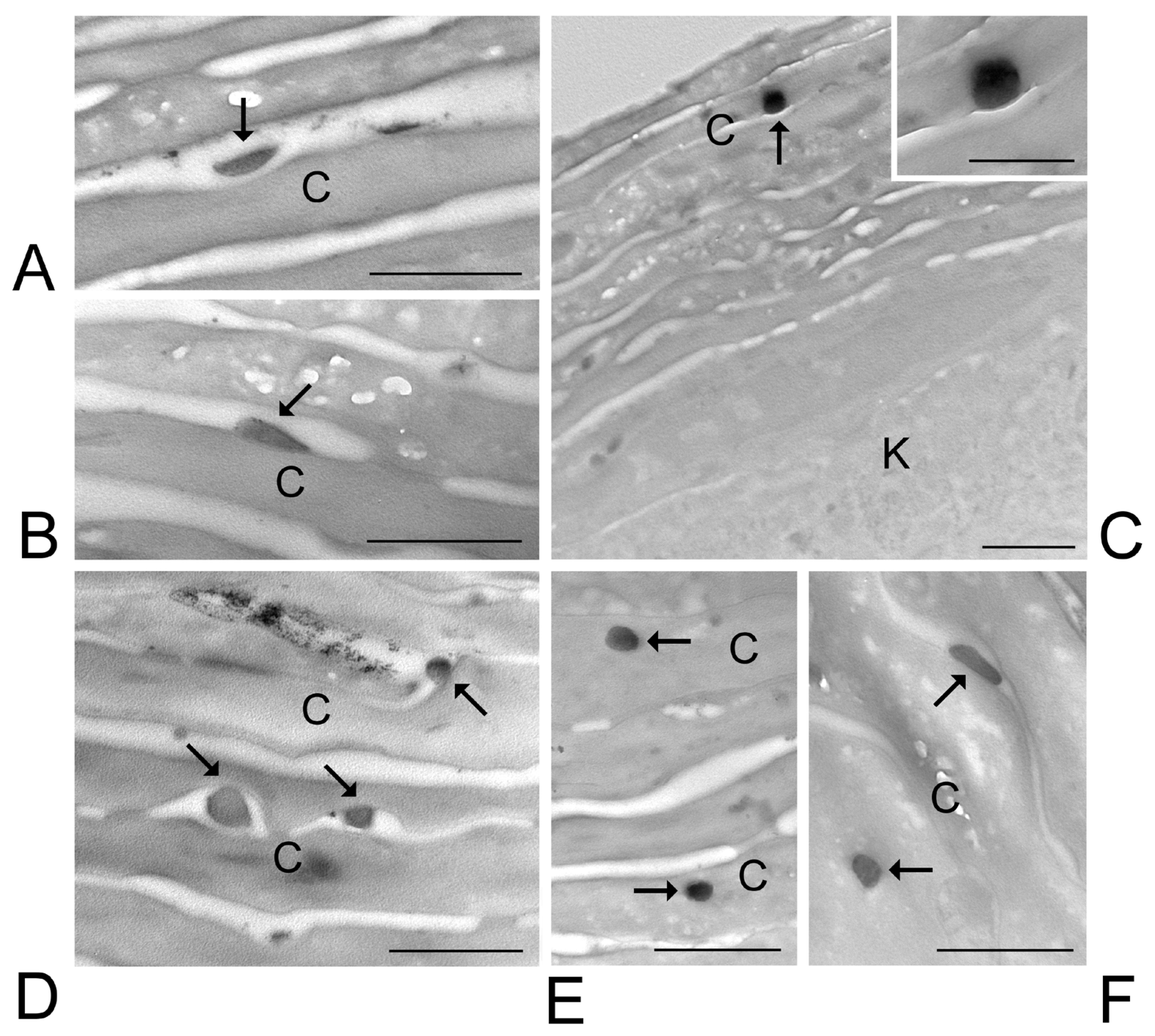
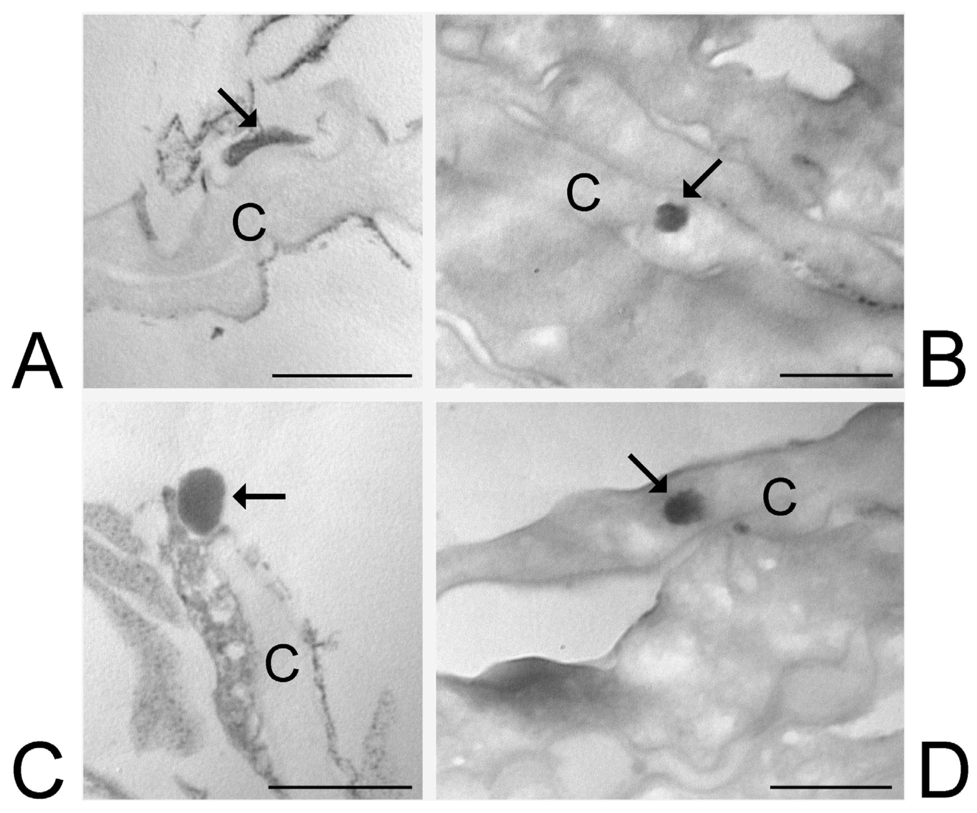
3.6. Transmission Electron Microscopy Analysis of Skin Treated with ETHO and LIPO

ETHOWI, ETHOEI and LIPOEI were applied on healthy human skin explants maintained in a bioreactor able to improve structural and functional preservation [38]. This experimental procedure allowed us to track nanovesicles in the different skin strata at increasing incubation times, namely 1 h, 3 h, 6 h and 24 h. At each incubation time, treated and control skin samples were processed for ultrastructural analysis.
At TEM, ETHOWI, ETHOEI and LIPOEI were detectable in the skin components as round or ovoid electron-dense structures (Figure 6, Figure 7 and Figure 8).
After 1 h and 3 h incubation, numerous ETHOWI were found among or inside corneocytes of the stratum corneum. (Figure 6A–C). Some ETHOWI were also found inside keratinocytes, from the stratum spinosum to the stratum basale, although in smaller amounts compared to corneocytes (Figure 6C,D). After 6 h incubation, rare ETHOWI were observed in the upper papillary dermis underlying the epidermis; in particular, ETHOWI were found to be internalized into macrophages (Figure 6D). After 24 h incubation, no ETHOWI was visible in any skin layer. The mean diameter of ETHOWI as measured at TEM was 207.46 ± 29.07 nm.
Many ETHOEI were observed after 1 h in the stratum corneum, both in the extracellular interstices and adhering to the surface of corneocytes (Figure 7A,B), while their occurrence inside the corneocytes was less frequent (Figure 7C). After 3 h, ETHOEI were numerous in the extracellular space and inside corneocytes of the stratum corneum (Figure 7D,E). After 6 h, a few ETHOEI were still visible at the corneocyte surface or inside them (Figure 7F). Notably, many ETHOEI occurring between corneocytes showed a flattened shape. ETHOEI was never observed inside keratinocytes or in the dermis. After 24 h incubation, no ETHOEI was found in any skin layer. The mean diameter of ETHOEI as measured at TEM was 128.92 ± 19.31 nm.
LIPOEI were observed in a limited number in the stratum corneum, either into corneocytes or adhering to their surface, after 1 h and 3 h incubation (Figure 8). LIPOEI occurring in the intercellular interstices frequently appeared as flattened (Figure 8A). LIPOEI was never observed in keratinocytes or the dermis. No LIPOEI was found in any skin layer after 6 h incubation. The mean diameter of LIPOEI as measured at TEM was 148.15 ± 22.98 nm.
No structural alteration of cellular or extracellular skin components was observed after treatment with any nanovesicles compared to controls.

4. Discussion

The formulation section of this study enabled us to compare different kinds of methods for nanovesicle preparation, namely microfluidics and solvent injections. Both LIPO and ETHO were constituted of PC, ethanol, and water. Beyond its self-assembling characteristics, PC is particularly well-suited for skin administration due to its affinity with stratum corneum lipid composition. While PC concentration was the same in LIPO and ETHO, ethanol amount was tripled in the case of ETHO with respect to LIPO. Some authors report the need to remove alcohol from vesicles produced by microfluidics or ethanol injection since its incomplete removal could influence the stability of vesicles, leading to aggregation phenomena [53,54]. Nevertheless, in the present study, apart from LIPOWI showing separation phenomena and mean size increase, ETHOEI, ETHOWI, and LIPOEI displayed strong size stability, while ETHOMF and LIPOMF underwent a moderate size increase after 3 months. Ethanol function is crucial for PC solubilization and maintenance of nanovesicle stability while promoting their penetration through the skin. Moreover, as a general trend, the higher amount of ethanol in ETHO led to a slight reduction of mean diameter with respect to LIPO. Importantly, as previously demonstrated, the 30% w/w amount of ethanol in ETHO is considered safe for skin administration, resulting in no irritation on the human skin.
Overall, in the present research, the bulk solvent injection method was determined to be more advantageous than microfluidics, primarily due to its higher yield. Indeed, both in the case of water injection and ethanol injection, the bulk method led to 5 mL of LIPO or ETHO in 30 min. Achieving comparable nanovesicular systems by microfluidics requires a TFR, leading to a significantly smaller volume. Namely, the production volume of LIPOMF and ETHOMF is 3 or 1.85-fold smaller with respect to the bulk solvent injection (i.e., 1650 or 2700 µL in 30 min for LIPOMF and ETHOMF). However, microfluidic devices can be employed in parallel by operating many microfluidic channels, achieving scalability and reproducibility in view of the industrial production of LIPO and ETHO [55].
The differences in size distribution and morphology between the nanovesicular systems produced by the different methods can be justified by examining the events during the mixing of the two phases. In the case of microfluidics, the interaction between LP and AP hydrodynamically focuses the lipid-containing stream into a narrow, rectangular sheet [54]. The vesicle formation is a result of reciprocal diffusion occurring between the lipid and water phases. This diffusion process establishes a controlled gradient of ethanol and water at their shared liquid interface. As ethanol (carrying the dissolved lipids) diffuses into the water and water concurrently enters the lipid phase, the ethanol concentration decreases until it falls below the solubility limit of the lipids. This reciprocal diffusion subsequently initiates vesicle formation through a self-assembly mechanism [56]. Consequently, the dimensional and structural attributes of the resulting LIPOMF or ETHOMF are strongly influenced and regulated by the concentration gradients present at the liquid interface between the solvent (ethanol) and non-solvent (water) phases, which are, in turn, determined by the focused stream’s geometric properties. Importantly, the size of this focused stream can be adjusted by modifying TFR and FRR [57].
In the case of the bulk solvent injection methods, the key mechanism is the mutual diffusion of ethanol and water at the interface, which compels PC units to associate into bilayers that subsequently fold into vesicles. The spontaneous formation of vesicles through PC self-assembly at the ethanol-water interface is a well-established phenomenon driven by the mixing process.
Regarding the bulk solvent injection method, a significant difference (approximately 100 nm) is observed in the Z-average values obtained for ETHO prepared using the different methods. Precisely, as the formulations are identical in composition, dimensional variations must be attributed to the preparation method. It can, therefore, be deduced that simply reversing the way the two phases are mixed induces significant changes in terms of size. Indeed, in the water injection method, the addition of AP into the LP promotes a thicker diffusion layer and a more gradual vesiculation process, which favors the formation of larger multilamellar structures. Conversely, in the case of the ethanol injection method, the rapid disappearance of the organic solvent leads to a thinner diffusion layer [46] since the swift solvent depletion accelerates the closure of the bilayer, resulting in smaller unilamellar vesicles. Noteworthily, the vesicle mean diameter measured by PCS agreed with Cryo-EM and hyperspectral dark microscopy observations, while the ultrastructures of nanovesicles measured by SAXS are in good agreement with Cryo-EM findings.
Since both size and lamellarity of the vesicles can strongly influence their delivery process and their passage through the skin [28,58], we decided to select the unilamellar nanovesicular systems obtained by ethanol injection method (ETHOEI and LIPOEI), characterized by the smallest size, and to compare their behavior under skin administration with the larger multilamellar ETHOWI, previously investigated by our research group [36].
Thus, to shed light on the vesicle transdermal fate investigating the impact of size and lamellarity, ex-vivo distribution studies were conducted on ETHOWI, ETHOEI and LIPOEI.
ETHOWI were previously demonstrated to penetrate the skin crossing the epidermis in 3 h, while their presence in the dermis was quite rare [36]. In the present work, we provide original evidence that ETHOWI reaching the papillary dermis undergoes phagocytosis by the resident macrophages. The administered ETHOWI -or at least many of them- are therefore able to preserve their morphological integrity during the passage through the epidermis strata, being detectable until they reach the dermis, where they disappear because they are removed by macrophages. This is consistent with a previous in vitro study [39] where ETHOWI internalized in keratinocytes was demonstrated to be only partially degraded by intracellular phospholipases, remaining easily recognizable at TEM until 24 h from administration. It is evident that their degradation is more rapid in the epidermis than in cultured cells: ETHOWI penetrates the stratum corneum by sliding into the extracellular space and then enters corneocytes and keratinocytes, undergoing lytic degradation. Notably, keratinocytes are linked by numerous desmosomes, whose integrity is guaranteed using the bioreactor [38], thus making the extracellular passage of ETHOWI. The transcellular way is, therefore, the most probable route followed by ETHOWI to penetrate the epidermis, according to the already proven ability of these nanovesicles to easily cross the cell membrane [39]. The multilamellar structure of ETHOWI could be responsible for their fate. Indeed, they probably possess a higher affinity with cell membranes with respect to ETHOEI and LIPOEI, characterized by unilamellar vesicles. Indeed, vesicle lamellarity, a variable determined by the preparation method, profoundly influences a range of vesicle behaviors, including encapsulation capacity, the rate at which encapsulated substances escape, the regulation of drug release, and penetration capabilities. Additionally, lamellarity significantly affects the intracellular handling of drugs carried by vesicle post-cellular uptake [59].
ETHOEI exhibited a lower capability to penetrate the skin as intact nanovesicles compared to ETHOWI. In fact, ETHOEI has never been observed in keratinocytes, even in the upper layer (stratum spinosum). Their presence remained confined to the stratum corneum, where they occur both in the intercellular interstices and inside corneocytes. It may be, therefore, hypothesized that their capability to cross cell membranes is limited in comparison to ETHOWI. On the other hand, ETHOEI demonstrated resistance to lytic degradation like ETHOWI since they were morphologically recognizable until 6 h from their administration. Notably, ETHOEI occurring in the interstices between corneocytes often showed a flattened shape, suggesting that their unilamellar structure could confer a higher flexibility to the vesicles with respect to the multilamellar ETHOWI. Thus, it can be assumed that the ETHOEI can deform during passage through the skin while maintaining its structure intact.
Moreover, LIPOEI demonstrated scarce penetration capability and high degradability. In fact, they were found in low numbers only in the stratum corneum (both inside corneocytes and in the interstices between them) and only until 3 h from administration. As observed for ETHOEI, many LIPOEI showed flattened shapes in the intercellular spaces, suggesting that their unilamellar structure can confer flexibility to the vesicles. Notably, the poor penetration of LIPOEI can be attributed to the lower amount of ethanol in their composition, being 3-fold lower with respect to ETHOWI and ETHOEI. In this respect, the role of ethanol as a penetration enhancer appears to be crucial in promoting the vesicle passage through the skin.

5. Conclusions

The presented findings confirm that both the modality of vesicle preparation and the amount of ethanol affect the mean size and inner structural organization of LIPO and ETHO. Particularly, PCS demonstrated that the ethanol injection method enabled the obtain of nanovesicles with Z-Average mean diameters of 130–145 nm, while the water injection and microfluidic methods resulted in vesicles measuring 225–235 nm mean diameters. LIPOWI underwent vesicle aggregation and phase separation phenomena. The stability study evidenced that ETHOEI, ETHOWI, and LIPOEI maintained almost unvaried Z-Average mean diameters over 3 months. Cryo-EM, SAXS and hyperspectral dark-field microscopy shed light on the nanocarrier ultrastructure, characterized by unilamellar bi-layered vesicles in the case of ETHOEI and LIPOEI and multilamellar bi-layered vesicles in the case of ETHOWI.
Remarkably, the lamellarity of the vesicles seems to influence their fate, resulting in different degradation times and a deeper skin interaction in the case of multilamellar vesicles. Particularly, the ETHOWI uptake in keratinocytes suggests the vesicle potential to deliver the loaded drug directly into the cells. In addition, the deeper permeation of ETHO with respect to LIPO corroborates the penetration enhancer role of ethanol, confirming the transdermal potential of both ETHOWI and ETHOEI. Further studies will be performed to better investigate the skin penetration mechanisms of the vesicles. Moreover, investigations conducted on inflamed skin biopsies will shed light on the possibility of ETHO loaded with anti-inflammatory agents to counteract the inflammatory status induced by exogenous stimuli.

Author Contributions

Conceptualization, E.E. and L.C.; Data curation, C.C. and M.M.; Formal analysis, V.D., F.S. (Federico Santamaria), C.B., F.S. (Francesco Spinozzi), A.P., P.M. and M.M.; Funding acquisition, E.E.; Investigation, A.G., C.B., L.M.N., F.S. (Francesco Spinozzi), A.P., M.R., M.G.O., P.M., L.C. and M.M.; Methodology, A.G., V.D., F.S. (Federico Santamaria), C.C. and M.M.; Project administration, E.E.; Supervision, L.M.N. and P.S.; Validation, M.R. and M.G.O.; Writing—original draft, E.E., M.G.O., P.M., L.C. and M.M.; Writing—review and editing, V.D., F.S. (Federico Santamaria), C.C., L.M.N., P.S., M.G.O., P.M., L.C. and M.M. All authors have read and agreed to the published version of the manuscript.

Funding

This research was funded by the European Union, Next Generation EU, PRIN 2022MC2SKF Italian Ministry of University and Research, and PNRR M6C2—PNRR-MAD-2022-12376878.

Data Availability Statement

Data will be made available on request.

Acknowledgments

The authors acknowledge the CERIC-ERIC Consortium for access to experimental facilities (proposal number 20242026) and Heinz Amenitsch for beamline set-up and stimulating discussions. The work is supported under the Polish Ministry and Higher Education project: “Support for research and development with the use of research infrastructure of the National Synchrotron Radiation Centre SOLARIS” under contract nr 1/SOL/2021/2.

Conflicts of Interest

The authors declare no conflicts of interest.

Abbreviations

The following abbreviations are used in this manuscript:
LIPOLiposomes
ETHOEthosomes
WIWater injection
EIEthanol injection
PCPhosphatidylcholine
PCSPhoton Correlation Spectroscopy
Cryo-EMCryogenic Electron Microscopy
SAXSSmall Angle X-Ray Scattering
TEMTransmission Electron Microscopy
APAqueous Phase
LPLipid Phase
TFRTotal Flow Rate
FRRFlow Rate Ratio

References

  1. Valacchi, G.; Sticozzi, C.; Pecorelli, A.; Cervellati, F.; Cervellati, C.; Maioli, E. Cutaneous Responses to Environmental Stressors. Ann. N. Y. Acad. Sci. 2012, 1271, 75–81. [Google Scholar] [CrossRef] [PubMed]
  2. Gravitz, L. Skin. Nature 2018, 563, S83. [Google Scholar] [CrossRef] [PubMed]
  3. Diaz, A.; Guttman-Yassky, E. Topical Agents for the Treatment of Atopic Dermatitis. Expert. Rev. Clin. Immunol. 2019, 15, 369–382. [Google Scholar] [CrossRef]
  4. Tambunlertchai, S.; Geary, S.M.; Salem, A.K. Skin Penetration Enhancement Strategies Used in the Development of Melanoma Topical Treatments. AAPS J. 2021, 23, 19. [Google Scholar] [CrossRef] [PubMed]
  5. El-Zaafarany, G.M.; Nasr, M. Insightful Exploring of Advanced Nanocarriers for the Topical/Transdermal Treatment of Skin Diseases. Pharm. Dev. Technol. 2021, 26, 1136–1157. [Google Scholar] [CrossRef]
  6. Chacko, I.A.; Ghate, V.M.; Dsouza, L.; Lewis, S.A. Lipid Vesicles: A Versatile Drug Delivery Platform for Dermal and Transdermal Applications. Colloids Surf. B Biointerfaces 2020, 195, 111262. [Google Scholar] [CrossRef]
  7. Wadhwa, S.; Garg, V.; Gulati, M.; Kapoor, B.; Singh, S.K.; Mittal, N. Nanovesicles for Nanomedicine: Theory and Practices. In Pharmaceutical Nanotechnology; Methods in Molecular Biology Volume 2000; Humana: New York, NY, USA, 2019; pp. 1–17. [Google Scholar] [CrossRef]
  8. Sharma, A.; Kuhad, A.; Bhandari, R. Novel Nanotechnological Approaches for Treatment of Skin-Aging. J. Tissue Viability 2022, 31, 374–386. [Google Scholar] [CrossRef]
  9. Garg, U.; Jain, K. Dermal and Transdermal Drug Delivery through Vesicles and Particles: Preparation and Applications. Adv. Pharm. Bull. 2022, 12, 45–57. [Google Scholar] [CrossRef]
  10. Arana-Linares, A.C.; Barrera-Ocampo, A.; Salamanca, C.H. Determination of the Critical Aggregation Concentration of Phospholipids Widely Used in Nanoliposomal Development from Different Experimental Methodologies. J. Mol. Liq. 2025, 417, 126677. [Google Scholar] [CrossRef]
  11. Touitou, E.; Dayan, N.; Bergelson, L.; Godin, B.; Eliaz, M. Ethosomes—Novel Vesicular Carriers for Enhanced Delivery: Characterization and Skin Penetration Properties. J. Control. Release 2000, 65, 403–418. [Google Scholar] [CrossRef]
  12. Hussain, A.; Altamimi, M.A.; Afzal, O.; Altamimi, A.S.A.; Ramzan, M.; Khuroo, T. Mechanistic of Vesicular Ethosomes and Elastic Liposomes on Permeation Profiles of Acyclovir across Artificial Membrane, Human Cultured EpiDerm, and Rat Skin: In Vitro-Ex Vivo Study. Pharmaceutics 2023, 15, 2189. [Google Scholar] [CrossRef] [PubMed]
  13. Asadi, P.; Mehravaran, A.; Soltanloo, N.; Abastabar, M.; Akhtari, J. Nanoliposome-Loaded Antifungal Drugs for Dermal Administration: A Review. Curr. Med. Mycol. 2021, 7, 71–78. [Google Scholar] [CrossRef]
  14. Wei, Y.Y.; Zhang, Y.; Lu, X.; Zhou, J.; Cheng, H.W.; Liu, Y.Y.; Zhang, H.; Chen, W. Novel Colchicine Ethosomes Cataplasm for the Treatment of Acute Gouty Arthritis. Colloids Surf. B Biointerfaces 2025, 254, 114776. [Google Scholar] [CrossRef] [PubMed]
  15. Dragicevic, N.; Maibach, H.I. Liposomes and Other Nanocarriers for the Treatment of Acne Vulgaris: Improved Therapeutic Efficacy and Skin Tolerability. Pharmaceutics 2024, 16, 309. [Google Scholar] [CrossRef]
  16. Musielak, E.; Krajka-Kuźniak, V. Liposomes and Ethosomes: Comparative Potential in Enhancing Skin Permeability for Therapeutic and Cosmetic Applications. Cosmetics 2024, 11, 191. [Google Scholar] [CrossRef]
  17. Leong, M.Y.; Kong, Y.L.; Burgess, K.; Wong, W.F.; Sethi, G.; Looi, C.Y. Recent Development of Nanomaterials for Transdermal Drug Delivery. Biomedicines 2023, 11, 1124. [Google Scholar] [CrossRef] [PubMed]
  18. Jafari, A.; Daneshamouz, S.; Ghasemiyeh, P.; Mohammadi-Samani, S. Ethosomes as Dermal/Transdermal Drug Delivery Systems: Applications, Preparation and Characterization. J. Liposome Res. 2023, 33, 34–52. [Google Scholar] [CrossRef]
  19. Natsheh, H.; Vettorato, E.; Touitou, E. Ethosomes for Dermal Administration of Natural Active Molecules. Curr. Pharm. Des. 2019, 25, 2338–2348. [Google Scholar] [CrossRef]
  20. Godin, B.; Touitou, E. Ethosomes: New Prospects in Transdermal Delivery. Crit. Rev. Ther. Drug Carrier Syst. 2003, 20, 63–102. [Google Scholar] [CrossRef]
  21. Paiva-Santos, A.C.; Silva, A.L.; Guerra, C.; Peixoto, D.; Pereira-Silva, M.; Zeinali, M.; Mascarenhas-Melo, F.; Castro, R.; Veiga, F. Ethosomes as Nanocarriers for the Development of Skin Delivery Formulations. Pharm. Res. 2021, 38, 947–970. [Google Scholar] [CrossRef]
  22. Nainwal, N.; Jawla, S.; Singh, R.; Saharan, V.A. Transdermal Applications of Ethosomes—A Detailed Review. J. Liposome Res. 2019, 29, 103–113. [Google Scholar] [CrossRef] [PubMed]
  23. Almeida, B.; Nag, O.K.; Rogers, K.E.; Delehanty, J.B. Recent Progress in Bioconjugation Strategies for Liposome-Mediated Drug Delivery. Molecules 2020, 25, 5672. [Google Scholar] [CrossRef] [PubMed]
  24. Oyarzún, P.; Gallardo-Toledo, E.; Morales, J.; Arriagada, F. Transfersomes as Alternative Topical Nanodosage Forms for The Treatment of Skin Disorders. Nanomedicine 2021, 16, 2465–2489. [Google Scholar] [CrossRef]
  25. Souto, E.B.; Macedo, A.S.; Dias-Ferreira, J.; Cano, A.; Zielińska, A.; Matos, C.M. Elastic and Ultradeformable Liposomes for Transdermal Delivery of Active Pharmaceutical Ingredients (APIs). Int. J. Mol. Sci. 2021, 22, 9743. [Google Scholar] [CrossRef]
  26. El Zaafarany, G.M.; Awad, G.A.S.; Holayel, S.M.; Mortada, N.D. Role of Edge Activators and Surface Charge in Developing Ultradeformable Vesicles with Enhanced Skin Delivery. Int. J. Pharm. 2010, 397, 164–172. [Google Scholar] [CrossRef]
  27. Zhigaltsev, I.V.; Tam, Y.K.; Leung, A.K.K.; Cullis, P.R. Production of Limit Size Nanoliposomal Systems with Potential Utility as Ultra-Small Drug Delivery Agents. J. Liposome Res. 2016, 26, 96–102. [Google Scholar] [CrossRef] [PubMed]
  28. Lombardo, D.; Kiselev, M.A. Methods of Liposomes Preparation: Formation and Control Factors of Versatile Nanocarriers for Biomedical and Nanomedicine Application. Pharmaceutics 2022, 14, 543. [Google Scholar] [CrossRef]
  29. Large, D.E.; Abdelmessih, R.G.; Fink, E.A.; Auguste, D.T. Liposome Composition in Drug Delivery Design, Synthesis, Characterization, and Clinical Application. Adv. Drug Deliv. Rev. 2021, 176, 113851. [Google Scholar] [CrossRef]
  30. Fan, Y.; Marioli, M.; Zhang, K. Analytical Characterization of Liposomes and Other Lipid Nanoparticles for Drug Delivery. J. Pharm. Biomed. Anal. 2021, 192, 113642. [Google Scholar] [CrossRef]
  31. Dzyhovskyi, V.; Romani, A.; Pula, W.; Bondi, A.; Ferrara, F.; Melloni, E.; Gonelli, A.; Pozza, E.; Voltan, R.; Sguizzato, M.; et al. Characterization Methods for Nanoparticle-Skin Interactions: An Overview. Life 2024, 14, 599. [Google Scholar] [CrossRef]
  32. Bendas, E.R.; Tadros, M.I. Enhanced Transdermal Delivery of Salbutamol Sulfate via Ethosomes. AAPS PharmSciTech 2007, 8, 107. [Google Scholar] [CrossRef] [PubMed]
  33. Soni, K.; Mujtaba, A.; Akhter, M.H.; Zafar, A.; Kohli, K. Optimisation of Ethosomal Nanogel for Topical Nano-CUR and Sulphoraphane Delivery in Effective Skin Cancer Therapy. J. Microencapsul. 2020, 37, 91–108. [Google Scholar] [CrossRef] [PubMed]
  34. Jain, S.; Tiwary, A.K.; Sapra, B.; Jain, N.K. Formulation and Evaluation of Ethosomes for Transdermal Delivery of Lamivudine. AAPS PharmSciTech 2007, 8, 111. [Google Scholar] [CrossRef] [PubMed]
  35. Dubey, V.; Mishra, D.; Jain, N.K. Melatonin Loaded Ethanolic Liposomes: Physicochemical Characterization and Enhanced Transdermal Delivery. Eur. J. Pharm. Biopharm. 2007, 67, 398–405. [Google Scholar] [CrossRef]
  36. Esposito, E.; Calderan, L.; Galvan, A.; Cappellozza, E.; Drechsler, M.; Mariani, P.; Pepe, A.; Sguizzato, M.; Vigato, E.; Dalla Pozza, E.; et al. Ex Vivo Evaluation of Ethosomes and Transethosomes Applied on Human Skin: A Comparative Study. Int. J. Mol. Sci. 2022, 23, 15112. [Google Scholar] [CrossRef]
  37. Abdulbaqi, I.M.; Darwis, Y.; Khan, N.A.K.; Assi, R.A.; Khan, A.A. Ethosomal Nanocarriers: The Impact of Constituents and Formulation Techniques on Ethosomal Properties, in Vivo Studies, and Clinical Trials. Int. J. Nanomed. 2016, 11, 2279–2304. [Google Scholar] [CrossRef]
  38. Cappellozza, E.; Zanzoni, S.; Malatesta, M.; Calderan, L. Integrated Microscopy and Metabolomics to Test an Innovative Fluid Dynamic System for Skin Explants In Vitro. Microsc. Microanal. 2021, 27, 923–934. [Google Scholar] [CrossRef]
  39. Costanzo, M.; Esposito, E.; Sguizzato, M.; Lacavalla, M.A.; Drechsler, M.; Valacchi, G.; Zancanaro, C.; Malatesta, M. Formulative Study and Intracellular Fate Evaluation of Ethosomes and Transethosomes for Vitamin D3 Delivery. Int. J. Mol. Sci. 2021, 22, 5341. [Google Scholar] [CrossRef]
  40. Pecora, R. Dynamic Light Scattering Measurement of Nanometer Particles in Liquids. J. Nanoparticle Res. 2000, 2, 123–131. [Google Scholar] [CrossRef]
  41. Amenitsch, H.; Bernstorff, S.; Kriechbaum, M.; Lombardo, D.; Mio, H.; Rappolt, M.; Laggner, P. Performance and First Results of the ELETTRA High-Flux Beamline for Small-Angle X-Ray Scattering. J. Appl. Crystallogr. 1997, 30, 872–876. [Google Scholar] [CrossRef]
  42. Hallan, S.S.; Sguizzato, M.; Mariani, P.; Cortesi, R.; Huang, N.; Simelière, F.; Marchetti, N.; Drechsler, M.; Ruzgas, T.; Esposito, E. Design and Characterization of Ethosomes for Transdermal Delivery of Caffeic Acid. Pharmaceutics 2020, 12, 740. [Google Scholar] [CrossRef] [PubMed]
  43. Sguizzato, M.; Mariani, P.; Spinozzi, F.; Benedusi, M.; Cervellati, F.; Cortesi, R.; Drechsler, M.; Prieux, R.; Valacchi, G.; Esposito, E. Ethosomes for Coenzyme Q10 Cutaneous Administration: From Design to 3D Skin Tissue Evaluation. Antioxidants 2020, 9, 485. [Google Scholar] [CrossRef] [PubMed]
  44. Sguizzato, M.; Ferrara, F.; Hallan, S.S.; Baldisserotto, A.; Drechsler, M.; Malatesta, M.; Costanzo, M.; Cortesi, R.; Puglia, C.; Valacchi, G.; et al. Ethosomes and Transethosomes for Mangiferin Transdermal Delivery. Antioxidants 2021, 10, 768. [Google Scholar] [CrossRef]
  45. Nsairat, H.; Khater, D.; Sayed, U.; Odeh, F.; Al Bawab, A.; Alshaer, W. Liposomes: Structure, Composition, Types, and Clinical Applications. Heliyon 2022, 8, e09394. [Google Scholar] [CrossRef]
  46. Choi, S.; Kang, B.; Yang, E.; Kim, K.; Kwak, M.K.; Chang, P.S.; Jung, H.S. Precise Control of Liposome Size Using Characteristic Time Depends on Solvent Type and Membrane Properties. Sci. Rep. 2023, 13, 4728. [Google Scholar] [CrossRef]
  47. Ward, K.; Fan, Z.H. Mixing in Microfluidic Devices and Enhancement Methods. J. Micromech. Microeng. 2015, 25, 094001. [Google Scholar] [CrossRef] [PubMed]
  48. Esposito, E.; Pozza, E.; Contado, C.; Pula, W.; Bortolini, O.; Ragno, D.; Toldo, S.; Casciano, F.; Bondi, A.; Zauli, E.; et al. Microfluidic Fabricated Liposomes for Nutlin-3a Ocular Delivery as Potential Candidate for Proliferative Vitreoretinal Diseases Treatment. Int. J. Nanomed. 2024, 19, 3513–3536. [Google Scholar] [CrossRef]
  49. Bondi, A.; Pula, W.; Benedusi, M.; Trinchera, G.; Baldisserotto, A.; Manfredini, S.; Ortore, M.G.; Pepe, A.; Mariani, P.; Stuart, M.C.A.; et al. Gossypin-Loaded Ethosome Gel for Cutaneous Administration: A Preliminary Study on Melanoma Cells. Antioxidants 2025, 14, 186. [Google Scholar] [CrossRef]
  50. Păvăloiu, R.D.; Sha’at, F.; Bubueanu, C.; Deaconu, M.; Neagu, G.; Sha’at, M.; Anastasescu, M.; Mihailescu, M.; Matei, C.; Nechifor, G.; et al. Polyphenolic Extract from Sambucus ebulus L. Leaves Free and Loaded into Lipid Vesicles. Nanomaterials 2019, 10, 56. [Google Scholar] [CrossRef]
  51. Friedrich, R.P.; Kappes, M.; Cicha, I.; Tietze, R.; Braun, C.; Schneider-Stock, R.; Nagy, R.; Alexiou, C.; Janko, C. Optical Microscopy Systems for the Detection of Unlabeled Nanoparticles. Int. J. Nanomed. 2022, 17, 2139–2163. [Google Scholar] [CrossRef]
  52. Fakhrullin, R.; Nigamatzyanova, L.; Fakhrullina, G. Dark-Field/Hyperspectral Microscopy for Detecting Nanoscale Particles in Environmental Nanotoxicology Research. Sci. Total Environ. 2021, 772, 145478. [Google Scholar] [CrossRef] [PubMed]
  53. Pittiu, A.; Pannuzzo, M.; Casula, L.; Pireddu, R.; Valenti, D.; Cardia, M.C.; Lai, F.; Rosa, A.; Sinico, C.; Schlich, M. Production of Liposomes by Microfluidics: The Impact of Post-Manufacturing Dilution on Drug Encapsulation and Lipid Loss. Int. J. Pharm. 2024, 664, 124641. [Google Scholar] [CrossRef] [PubMed]
  54. Carugo, D.; Bottaro, E.; Owen, J.; Stride, E.; Nastruzzi, C. Liposome Production by Microfluidics: Potential and Limiting Factors. Sci. Rep. 2016, 6, 25876. [Google Scholar] [CrossRef] [PubMed]
  55. Lopes, C.; Cristóvão, J.; Silvério, V.; Lino, P.R.; Fonte, P. Microfluidic Production of MRNA-Loaded Lipid Nanoparticles for Vaccine Applications. Expert. Opin. Drug Deliv. 2022, 19, 1381–1395. [Google Scholar] [CrossRef] [PubMed]
  56. Rebollo, R.; Oyoun, F.; Corvis, Y.; El-Hammadi, M.M.; Saubamea, B.; Andrieux, K.; Mignet, N.; Alhareth, K. Microfluidic Manufacturing of Liposomes: Development and Optimization by Design of Experiment and Machine Learning. ACS Appl. Mater. Interfaces 2022, 14, 39736–39745. [Google Scholar] [CrossRef]
  57. Hamdallah, S.I.; Zoqlam, R.; Erfle, P.; Blyth, M.; Alkilany, A.M.; Dietzel, A.; Qi, S. Microfluidics for Pharmaceutical Nanoparticle Fabrication: The Truth and the Myth. Int. J. Pharm. 2020, 584, 119408. [Google Scholar] [CrossRef]
  58. Elizondo, E.; Moreno, E.; Cabrera, I.; Córdoba, A.; Sala, S.; Veciana, J.; Ventosa, N. Liposomes and Other Vesicular Systems: Structural Characteristics, Methods of Preparation, and Use in Nanomedicine. Prog. Mol. Biol. Transl. Sci. 2011, 104, 1–52. [Google Scholar] [CrossRef]
  59. Giuliano, C.B.; Cvjetan, N.; Ayache, J.; Walde, P. Multivesicular Vesicles: Preparation and Applications. ChemSystemsChem 2021, 3, e2000049. [Google Scholar] [CrossRef]
Figure 1. Schematic representation of the bioreactor used in this study.
Nanomaterials 15 00937 g001
Figure 2. Variation of Z Average mean diameters of LIPOMF (yellow), ETHOMF (blue), LIPOWI (green), ETHOWI (red), LIPOEI (violet), and ETHOEI (light blue).
Nanomaterials 15 00937 g002
Figure 3. Cryo-EM images of ETHOWI (A), ETHOEI (B), and LIPOEI (C). The bar corresponds to 100 nm.
Nanomaterials 15 00937 g003
Figure 4. Comparison among the SAXS profiles of ETHOWI (blue), ETHOEI (purple), and LIPOEI (yellow). The black vertical line indicates the position of the Bragg peak at Q = 0.087 Å−1 observed in the ETHOWI profile. Model fitting curves are also reported.
Nanomaterials 15 00937 g004
Figure 5. Hyperspectral images of ETHOWI (A), ETHOEI (B) and LIPOEI (C) were acquired by enhanced dark-field microscopy using Cytoviva. In the right column, the corresponding spectra collected in isolated points are reported.
Nanomaterials 15 00937 g005
Figure 6. ETHOWI. TEM images of ETHOWI in the skin. (A,B) After 1-h incubation, some ETHOWI (arrows) are present inside corneocytes of the stratum corneum, while another ETHOWI (arrowhead) occurs in the extracellular interstices. (C) After 3-h incubation, ETHOWI (arrows) occur both inside corneocytes and keratinocytes of the stratum spinosum (C) and stratum basale (D). (E) After 6-h incubation, an ETHOWI (open arrow) has been phagocyted by a macrophage in the upper papillary dermis. Corneocytes, C; keratinocyte, K; macrophage, M; collagen bundles, CB. Bars: 500 nm.
Nanomaterials 15 00937 g006
Figure 7. ETHOEI. TEM images of ETHOEI in the skin. (AC) After 1-h incubation, ETHOEI (arrows) occurs (A) in the extracellular interstices of the stratum corneum, (B) adhering to the surface of corneocytes or (C and inset) into corneocytes. (D,E) After 3-h incubation, ETHOEI (arrows) occur both (D) in the intracellular space of the stratum corneum, also adhering to the surface of corneocytes, and (E) inside corneocytes. (F) After 6-h incubation, ETHOEI (arrows) occurs in the stratum corneum, both in the intracellular space and inside corneocytes. Corneocytes, C; keratinocyte, K. Bars: 500 nm; inset in C, 250 nm.
Nanomaterials 15 00937 g007
Figure 8. LIPOEI. TEM micrographs of LIPOEI in the skin. Both after 1-h (A,B) and 3-h (C,D) incubation, LIPOEI (arrows) were found in the stratum corneum, (A,C) adhering to the surface of corneocytes or (B,D) internalized into corneocytes. Corneocytes, C. Bars: 500 nm.
Nanomaterials 15 00937 g008
Table 1. Preparation parameters and compositions of nanovesicular systems.
Nanovesicular SystemTFR a
(μL/min)
FRR bFAP c
(μL/min)
FLP d
(μL/min)
PC e
(%, w/w)
Ethanol
(%, w/w)
Water
(%, w/w)
LIPOMF5510:125 + 2550.99.190
ETHOMF902:130 + 30300.929.170
LIPOWI--1500-0.99.190
ETHOWI--1500-0.929.170
LIPOEI---15000.99.190
ETHOEI---15000.929.170
a: Total Flow Rate (FAP + FLP); b: Flow Rate Ratio (FAP/FLP) volumetric aqueous phase/lipid phase ratio; c: Aqueous Phase Flow; d: Lipid Phase Flow; e: soy phosphatidylcholine.
Table 2. Size distribution parameters of nanovesicular systems.
Nanovesicular
System
Z-Average (nm) ± s.d.Dispersity Index ± s.d.Typical Intensity
Distribution (nm)
Macroscopic
Aspect
LIPOMF234 ± 100.24 ± 0.01233.02 (91%)milky, homogeneous
ETHOMF226 ± 70.26 ± 0.03238.21 (90%)milky, homogeneous
LIPOWI235 ± 140.27 ± 0.02226.61 (97%)phase separation
ETHOWI225 ± 90.20 ± 0.01225.11 (95%)milky, homogeneous
LIPOEI144 ± 30.17 ± 0.01143.21 (98%)milky, homogeneous
ETHOEI130 ± 130.15 ± 0.02138.40 (100%)milky, homogeneous
Table 3. Main parameters resulting from SAXS data analysis. Fixed parameters were ρ1 = 370 e/nm3, ρ2 = 320 e/nm3, ρ3 = 300 e/nm3,ρ0 = 330 e/nm3 and ρEtH = 285 e/nm3.
LIPOEIETHOWIETHOEI
R1 (nm)0.49 ± 0.070.66 ± 0.040.64 ± 0.02
R2 (nm)1.19 ± 0.021.03 ± 0.061.04 ± 0.02
R3 (nm)0.10 ± 0.020.16 ± 0.050.14 ± 0.02
N6 ± 25 ± 22 ± 4
c (nm)17 ± 1014 ± 135 ± 9
gc0.9 ± 0.10.14 ± 0.010.5 ± 0.2
R0 (nm)500 ± 100488 ± 85500 ± 90
ξRo0.1 ± 0:20.01 ± 0.060.0 ± 0.2
Disclaimer/Publisher’s Note: The statements, opinions and data contained in all publications are solely those of the individual author(s) and contributor(s) and not of MDPI and/or the editor(s). MDPI and/or the editor(s) disclaim responsibility for any injury to people or property resulting from any ideas, methods, instructions or products referred to in the content.

