
Micro- and Nanoengineered Devices for Rapid Chemotaxonomic Profiling of Medicinal Plants


Abstract
1. Introduction
2. Target Metabolites and Analytical Needs
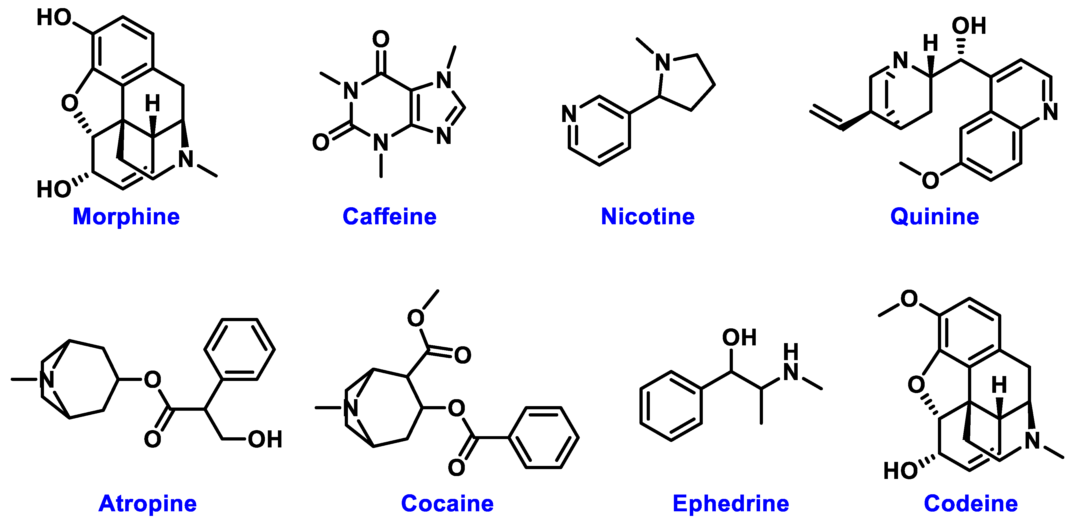
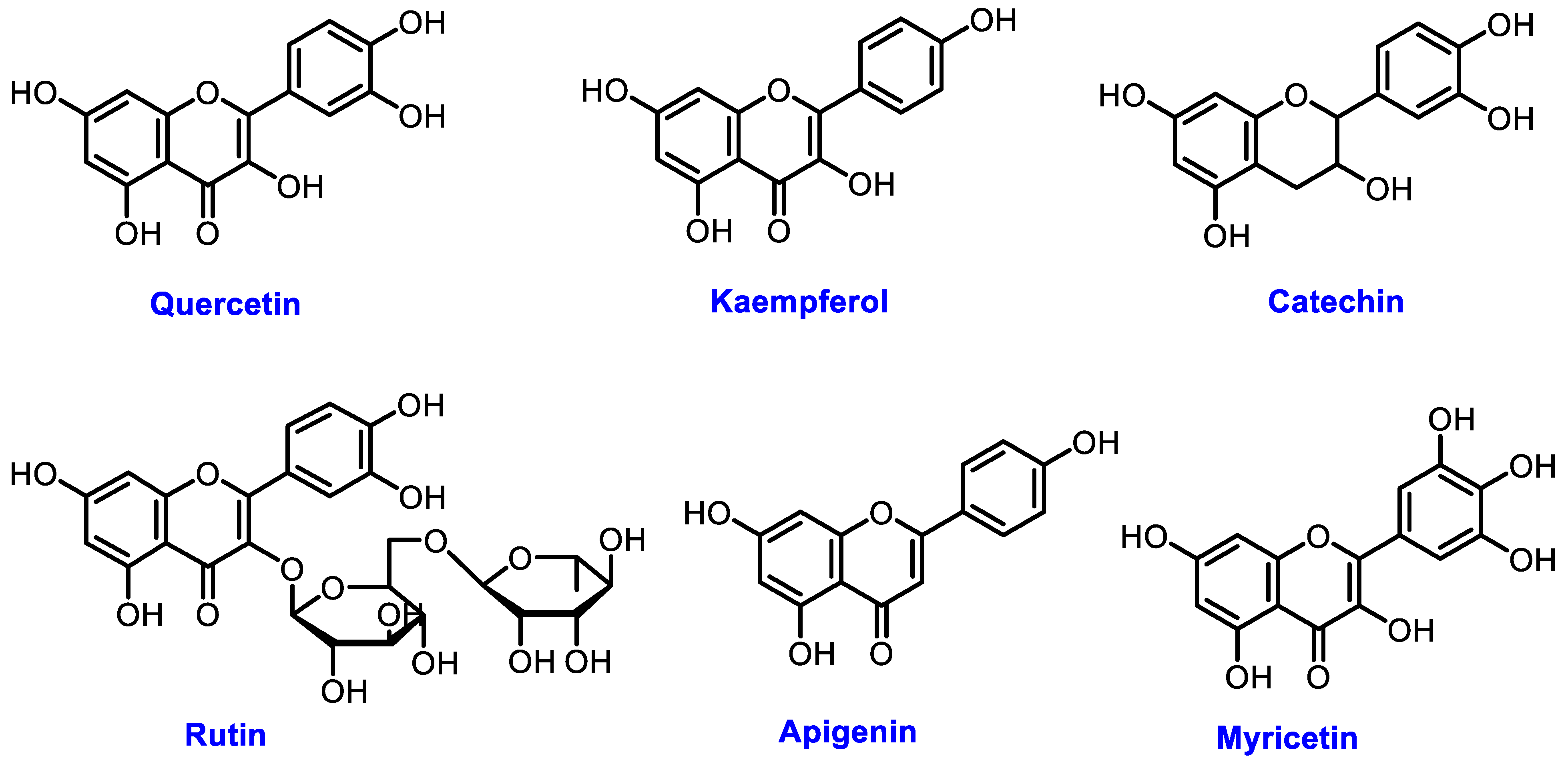
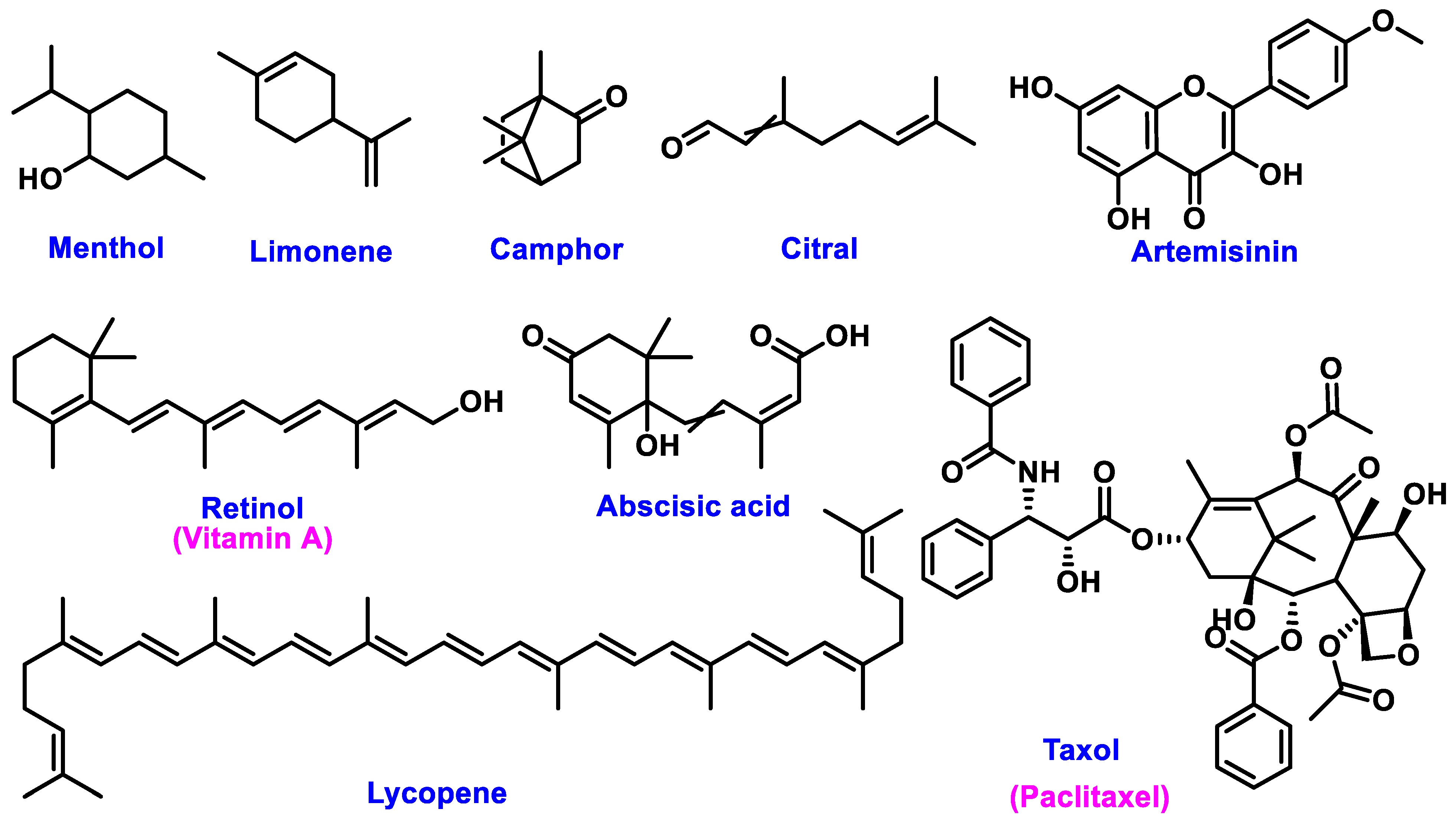
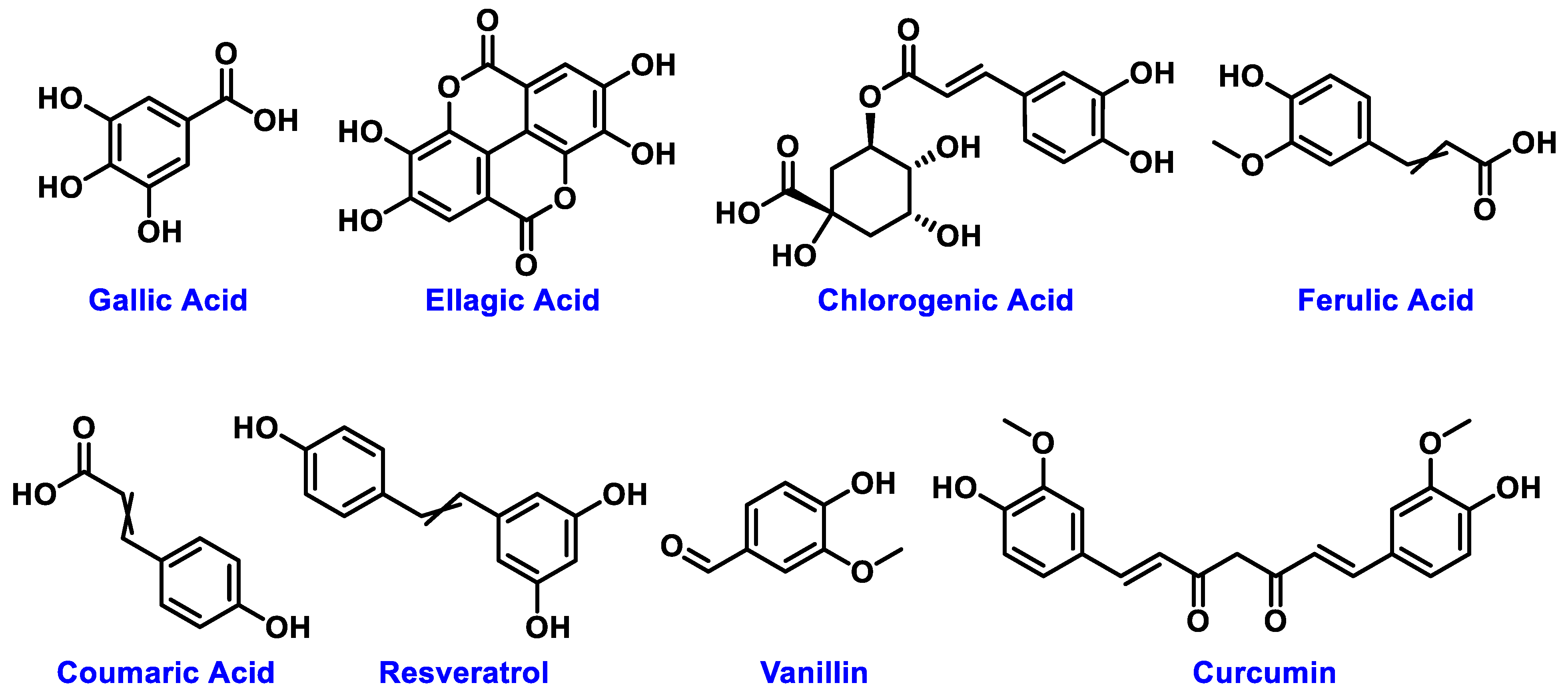
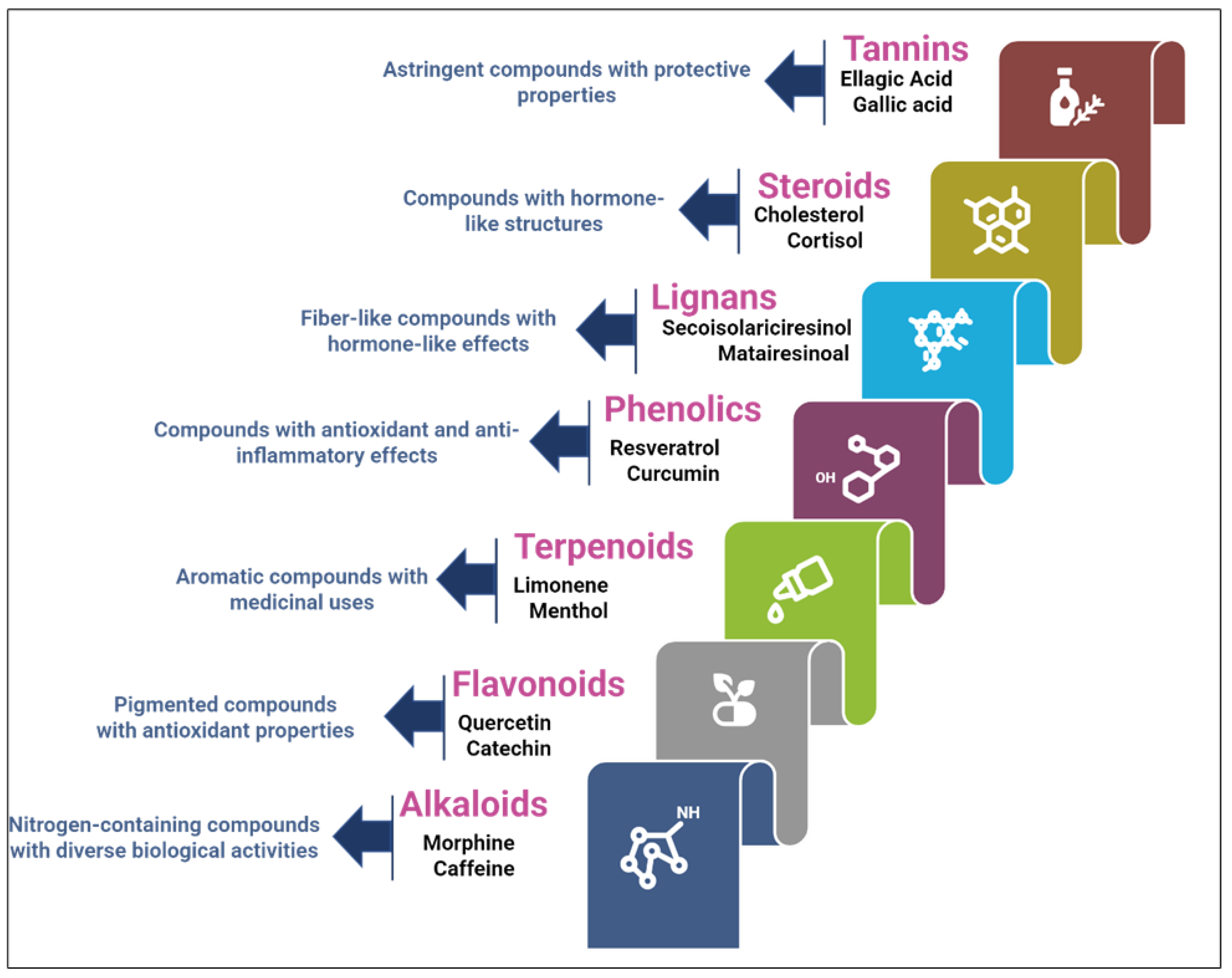
2.1. Overview of Major Phytochemicals in Medicinal Plants
2.2. Challenges in Traditional Analytical Techniques
3. Microfluidics and Lab-on-a-Chip Platforms
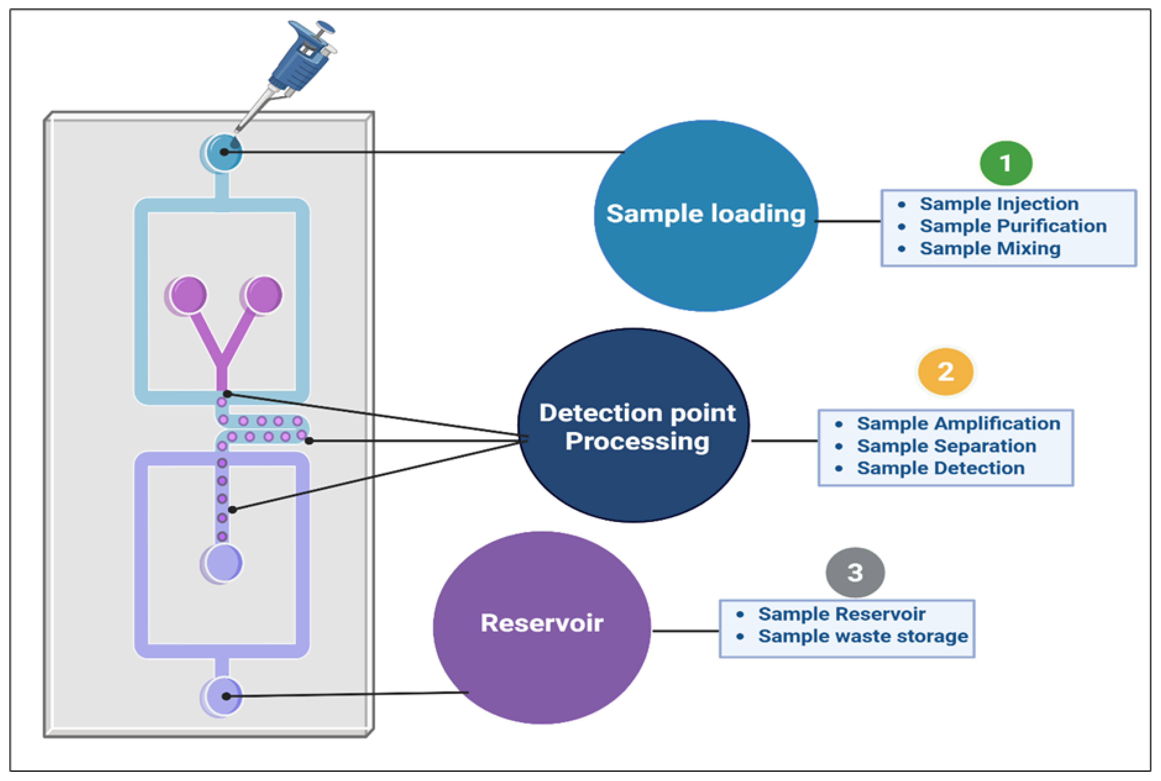
3.1. Introduction to Microfluidic Systems
3.2. Fabrication Techniques for Lab-on-a-Chip Devices
3.3. Integrating Detection Methods in Lab-on-a-Chip Systems

| Detection Technique | Advantages | Limitations | Real-World Case Study | Examples of Plant Metabolites Detected |
|---|---|---|---|---|
| Fluorescence detection | High sensitivity for low-abundance metabolites. Non-invasive, rapid, and real-time detection. Easily integrated with microfluidic systems. | Requires fluorescent tagging or natural fluorescence. May not be applicable to all plant metabolites. | Fluorescence-based lab-on-a-chip devices are employed to detect and quantify flavonoids in plant species, such as Citrus and Ginkgo biloba, aiding in the identifying secondary metabolites in medicinal plants [68]. | Flavonoids, phenolic acids, and anthocyanins |
| Absorbance detection | Simple, cost-effective, and widely used. Applicable to UV or visible light-absorbing compounds. | Lower sensitivity than that of fluorescence. May require extensive sample preparation for complex mixtures. | Commonly used to analyze phenolics and flavonoids, especially in agricultural and food safety applications. For example, used for polyphenol analysis in tea and grape samples [69]. | Phenolic compounds, flavonoids, and tannins |
| Electrochemical detection | High selectivity and sensitivity for low concentrations. Suitable for real-time monitoring. Highly specific for certain compounds (such as alkaloids and terpenoids). | Requires specialized electrodes and systems. Limited to metabolites that can undergo redox reactions. Potential interference from other electroactive substances. | Electrochemical sensors integrated into lab-on-a-chip devices have been employed to detect alkaloids in Cinchona bark (for quinine) and terpenoids in aromatic plants such as lavender and peppermint. These sensors are particularly useful in herbal medicine research and conservation [70,71] | Alkaloids, terpenoids, cinchonine, and quinine |
| SPR | Provides real-time detection without labeling. Sensitive to changes in refractive index near the sensor surface. Non-destructive to samples. | Sensitive to surface conditions and requires highly specialized equipment. | Used in lab-on-a-chip devices to detect polyphenols and flavonoids by measuring refractive index changes at the sensor surface, often used for profiling complex plant mixtures [72]. | Polyphenols, flavonoids, and antioxidants |
| CE | High resolution, fast, and effective for separating various metabolites. Can be combined with detection methods (UV, fluorescence, and electrochemical). | Requires more complex sample preparation and sophisticated equipment. May not be suitable for large-scale screening. | Used for separating and quantifying carotenoids and fatty acids in various plant extracts, especially in food quality control and metabolite profiling [73,74]. | Carotenoids, fatty acids, and lipids |
4. Nano-Enabled Optical and Electrochemical Sensors
4.1. Introducing Nano-Enabled Sensors
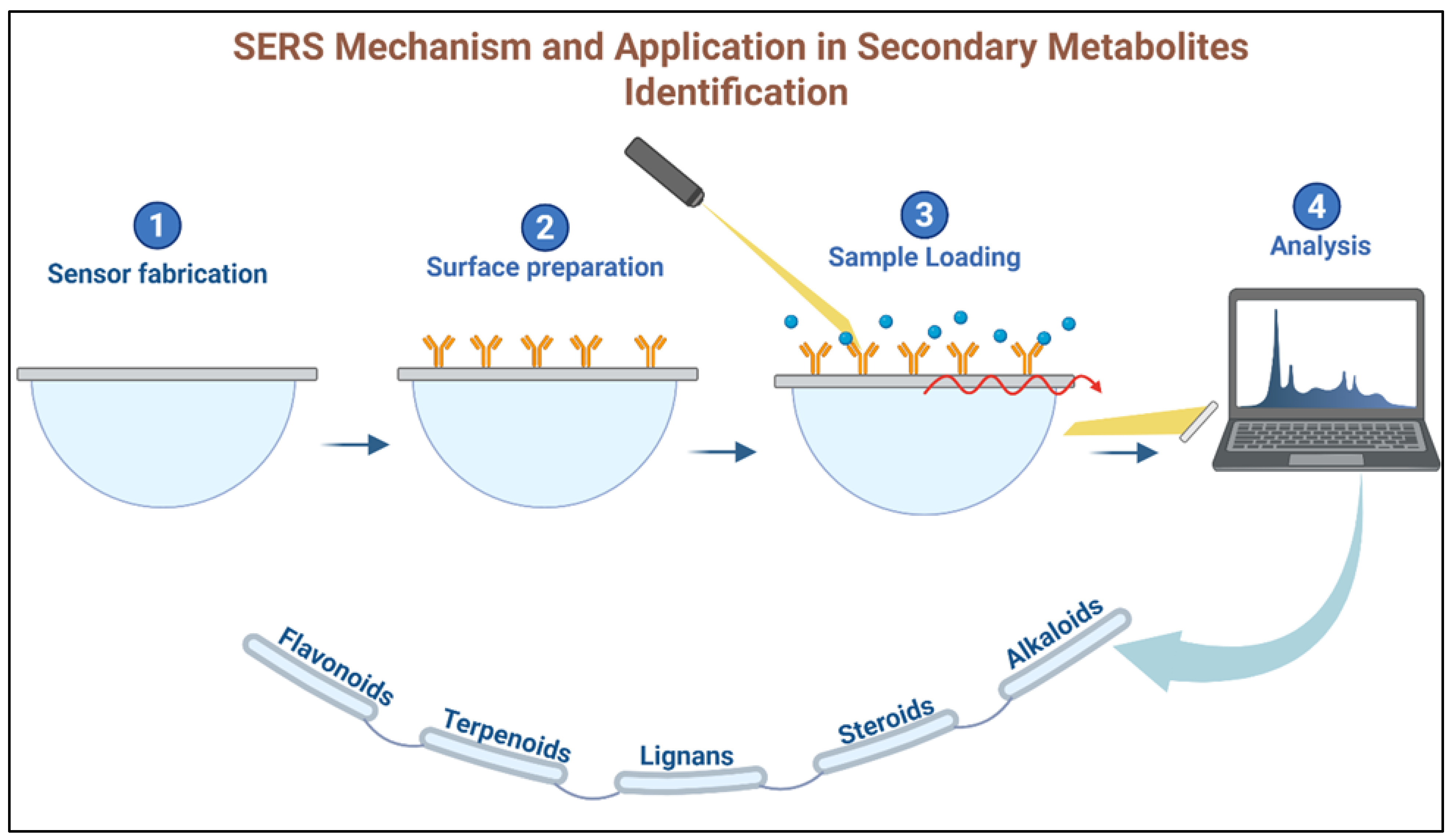
4.2. Surface-Enhanced Raman Spectroscopy for Plant Metabolites
4.3. Field-Effect Transistor FET-Based Sensors for Phytochemical Detection
5. Integration with Artificial Intelligence and Data Processing
5.1. Role of Machine Learning in Chemotaxonomy
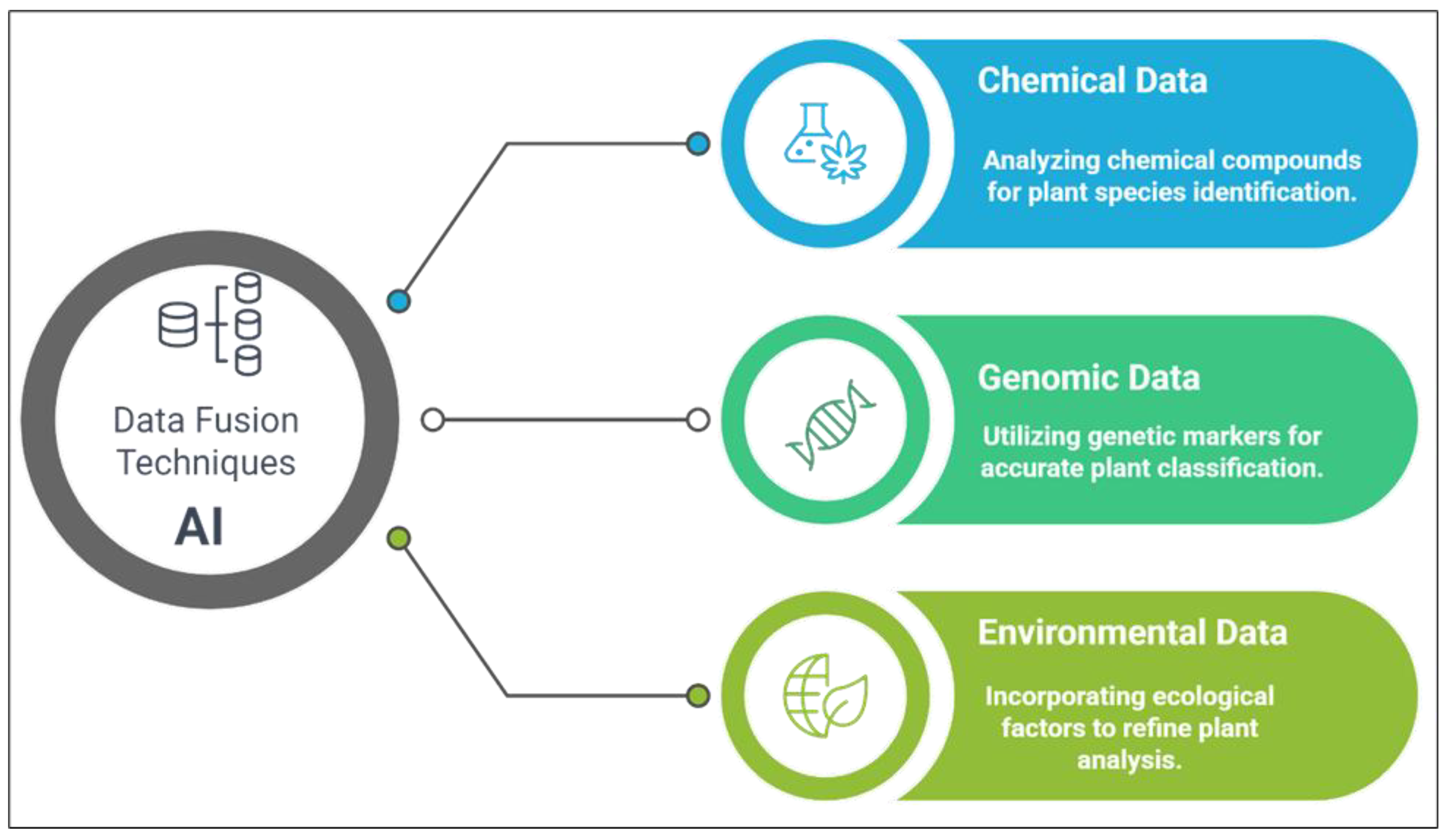
5.2. Data Fusion Techniques: Integrating Metabolomics and Genomics
5.3. Real-Time Data Processing for Field-Based Applications
6. Applications and Case Studies
6.1. Applications in Herbal Medicine
6.2. Medicinal Plant Authentication
6.3. Biodiversity and Conservation Studies
7. Challenges and Future Outlook
7.1. Challenges in Current Micro- and Nanoengineered Devices
7.2. Innovations in Nanoengineering for Field-Based Chemotaxonomy
7.3. Regulatory and Standardization Issues
8. Conclusions
Author Contributions
Funding
Data Availability Statement
Acknowledgments
Conflicts of Interest
References
- Marne, P.A.; Pawar, A.T.; Tagalpallewar, A.A.; Baheti, A.M. Comparative Phytochemistry and Chemotaxonomy. In Pharmacognosy and Phytochemistry: Principles, Techniques, and Clinical Applications; Wiley: Hoboken, NJ, USA, 2025; pp. 333–346. [Google Scholar]
- Arceusz, A.; Radecka, I.; Wesołowski, M. Identification of diversity in elements content in medicinal plants belonging to different plant families. Food Chem. 2010, 120, 52–58. [Google Scholar] [CrossRef]
- Singh, R. Chemotaxonomy of medicinal plants: Possibilities and limitations. In Natural Products and Drug Discovery; Elsevier: Amsterdam, The Netherlands, 2018; pp. 119–136. [Google Scholar]
- Wang, M.; Lin, H.; Lin, H.; Du, P.; Zhang, S. From species to varieties: How modern sequencing technologies are shaping Medicinal Plant Identification. Genes 2024, 16, 16. [Google Scholar] [CrossRef] [PubMed]
- Song, W.; Qiao, X.; Chen, K.; Wang, Y.; Ji, S.; Feng, J.; Li, K.; Lin, Y.; Ye, M. Biosynthesis-Based Quantitative Analysis of 151 Secondary Metabolites of Licorice To Differentiate Medicinal Glycyrrhiza Species and Their Hybrids. Anal. Chem. 2017, 89, 3146–3153. [Google Scholar] [CrossRef] [PubMed]
- Roaa, M.H. A review article: The importance of the major groups of plants secondary metabolism phenols, alkaloids, and terpenes. Int. J. Res. Appl. Sci. Biotechnol. (IJRASB) 2020, 7, 354–358. [Google Scholar]
- Hao, D.-C.; Gu, X.-J.; Xiao, P. Chemotaxonomy: A Phylogeny-Based Approach; Woodhead Publishing: Cambridge, UK, 2015; pp. 1–48. [Google Scholar]
- Salam, U.; Ullah, S.; Tang, Z.-H.; Elateeq, A.A.; Khan, Y.; Khan, J.; Khan, A.; Ali, S. Plant metabolomics: An overview of the role of primary and secondary metabolites against different environmental stress factors. Life 2023, 13, 706. [Google Scholar] [CrossRef]
- Farag, M.A.; Porzel, A.; Wessjohann, L.A. Comparative metabolite profiling and fingerprinting of medicinal licorice roots using a multiplex approach of GC–MS, LC–MS and 1D NMR techniques. Phytochemistry 2012, 76, 60–72. [Google Scholar] [CrossRef]
- Gashaw, A.D.; Desta, M.A.; Yaya, E.E. A comprehensive review-current development in spectroscopic and chromatographic techniques for natural product analysis. Results Chem. 2025, 16, 102341. [Google Scholar] [CrossRef]
- Porzel, A.; Farag, M.A.; Mülbradt, J.; Wessjohann, L.A. Metabolite profiling and fingerprinting of Hypericum species: A comparison of MS and NMR metabolomics. Metabolomics 2014, 10, 574–588. [Google Scholar] [CrossRef]
- Farag, M.A.; Porzel, A.; Schmidt, J.; Wessjohann, L.A. Metabolite profiling and fingerprinting of commercial cultivars of Humulus lupulus L. (hop): A comparison of MS and NMR methods in metabolomics. Metabolomics 2012, 8, 492–507. [Google Scholar] [CrossRef]
- Crevillen, A.; Pumera, M.; González, M.C.; Escarpa, A. Towards lab-on-a-chip approaches in real analytical domains based on microfluidic chips/electrochemical multi-walled carbon nanotube platforms. Lab A Chip 2009, 9, 346–353. [Google Scholar] [CrossRef]
- Maisch, J.; Kreppenhofer, K.; Büchler, S.; Merle, C.; Sobich, S.; Görling, B.; Luy, B.; Ahrens, R.; Guber, A.E.; Nick, P. Time-resolved NMR metabolomics of plant cells based on a microfluidic chip. J. Plant Physiol. 2016, 200, 28–34. [Google Scholar] [CrossRef] [PubMed]
- Lu, Z.; Yuan, Y.; Han, Q.; Wang, Y.; Liang, Q. Lab-on-a-chip: An advanced technology for the modernization of traditional Chinese medicine. Chin. Med. 2024, 19, 80. [Google Scholar] [CrossRef] [PubMed]
- Aboulthana, W.M.K.; Refaat, E.; Khaled, S.E.; Ibrahim, N.E.-S.; Youssef, A.M. Metabolite profiling and biological activity assessment of Casuarina equisetifolia bark after incorporating gold nanoparticles. Asian Pac. J. Cancer Prev. APJCP 2022, 23, 3457. [Google Scholar] [CrossRef] [PubMed]
- Kruszka, D.; Selvakesavan, R.K.; Kachlicki, P.; Franklin, G. Untargeted metabolomics analysis reveals the elicitation of important secondary metabolites upon treatment with various metal and metal oxide nanoparticles in Hypericum perforatum L. cell suspension cultures. Ind. Crops Prod. 2022, 178, 114561. [Google Scholar] [CrossRef]
- Cembrowska-Lech, D.; Krzemińska, A.; Miller, T.; Nowakowska, A.; Adamski, C.; Radaczyńska, M.; Mikiciuk, G.; Mikiciuk, M. An integrated multi-omics and artificial intelligence framework for advance plant phenotyping in horticulture. Biology 2023, 12, 1298. [Google Scholar] [CrossRef]
- Varghese, R.; Shringi, H.; Efferth, T.; Ramamoorthy, S. Artificial intelligence driven approaches in phytochemical research: Trends and prospects. Phytochem. Rev. 2025. [Google Scholar] [CrossRef]
- Labroo, P.; Cui, Y. Graphene nano-ink biosensor arrays on a microfluidic paper for multiplexed detection of metabolites. Anal. Chim. Acta 2014, 813, 90–96. [Google Scholar] [CrossRef]
- Yang, C.; Zhao, Y.; Jiang, S.; Sun, X.; Wang, X.; Wang, Z.; Wu, Y.; Wu, J.; Li, Y. A breakthrough in phytochemical profiling: Ultra-sensitive surface-enhanced Raman spectroscopy platform for detecting bioactive components in medicinal and edible plants. Mikrochim. Acta 2024, 191, 286. [Google Scholar] [CrossRef]
- Babamiri, B.; Sadri, R.; Farrokhnia, M.; Hassani, M.; Kaur, M.; Roberts, E.; Ashani, M.M.; Nezhad, A.S. Molecularly Imprinted Polymer Biosensor Based on Nitrogen-Doped Electrochemically Exfoliated Graphene/Ti3 CNTX MXene Nanocomposite for Metabolites Detection. ACS Appl. Mater. Interfaces 2024, 16, 27714–27727. [Google Scholar] [CrossRef]
- Kumar, A.; Jain, D.; Bahuguna, J.; Bhaiyya, M.; Dubey, S.; Javed, A.; Goel, S. Machine learning assisted and smartphone integrated homogeneous electrochemiluminescence biosensor platform for sample to answer detection of various human metabolites. Biosens. Bioelectron. 2023, 238, 115582. [Google Scholar] [CrossRef]
- Palacio, E.-D.; Iaz-Navarro, C.; Algieri, F.; Iguez-Cabezas, E.R.; Genilloud, O.; Vicente, F. Metabolomic analysis of Lavandula dentata L. and Lavandula stoechas L. extracts by LC-QTOF/MS experiments and multivariate analysis techniques as a chemotaxonomical tool. Plant Biosyst. Int. J. Deal. All Asp. Plant Biol. 2020, 154, 231–240. [Google Scholar]
- Zhao, S.-Y.; Liu, Z.-L.; Shu, Y.; Wang, M.; He, D.; Song, Z.-Q.; Zeng, H.; Ning, Z.; Lu, C.; Lu, A.; et al. Chemotaxonomic Classification Applied to the Identification of Two Closely-Related Citrus TCMs Using UPLC-Q-TOF-MS-Based Metabolomics. Mol. A J. Synth. Chem. Nat. Prod. Chem. 2017, 22, 1721. [Google Scholar] [CrossRef] [PubMed]
- Mannochio-Russo, H.; De Almeida, R.; Nunes, W.; Bueno, P.; Caraballo-Rodríguez, A.; Bauermeister, A.; Dorrestein, P.; Bolzani, V. Untargeted Metabolomics Sheds Light on the Diversity of Major Classes of Secondary Metabolites in the Malpighiaceae Botanical Family. Front. Plant Sci. 2022, 13, 854842. [Google Scholar] [CrossRef]
- Qiu, F.; Fine, D.; Wherritt, D.; Lei, Z.; Sumner, L. PlantMAT: A Metabolomics Tool for Predicting the Specialized Metabolic Potential of a System and for Large-Scale Metabolite Identifications. Anal. Chem. 2016, 88, 11373–11383. [Google Scholar] [CrossRef]
- Gupta, N. Therapeutic Efficacy of the Plant Bioactive Phytochemicals with Special Reference to Alkaloids, Terpenoids, Phenolics and Cardiac Glycosides. Int. J. Plant Environ. 2024, 10, 22–30. [Google Scholar] [CrossRef]
- Fatima, M.; Dar, M.; Dhanavade, M.; Abbas, S.Z.; Bukhari, M.N.; Arsalan, A.; Liao, Y.; Wan, J.; Bukhari, J.S.S.; Zhen, O. Biosynthesis and Pharmacological Activities of the Bioactive Compounds of White Mulberry (Morus alba): Current Paradigms and Future Challenges. Biology 2024, 13, 506. [Google Scholar] [CrossRef]
- Kumaravel, S.; Muthukumaran, P.; Thomas, N. Phytochemical, GC-MS and FT-IR analysis of Papver somniferum L. J. Pharm. Biol. Sci. 2019, 7, 1–8. [Google Scholar]
- Gupcsó, K.; Kókai, Z.; Bálint, M.; Tavaszi-Sárosi, S.; Németh, É.Z. Studies on Sensory and Phytochemical Characteristics of Poppy (Papaver somniferum L.) Varieties for Their Oil Utilisation. Foods 2023, 12, 3165. [Google Scholar] [CrossRef]
- Sundowo, A.; Artanti, N.; Hanafi, M.; Minarti, M.; Primahana, G. Phytochemical screening, total phenolic, total flavonoids contents and antioxidant activity of Cinchona ledgeriana leaves ethanol extract. In Proceedings of the 3rd International Symposium on Applied Chemistry, Jakarta, Indonesia, 23–24 October 2017. [Google Scholar]
- Yadav, R.; Sahu, M.; Yadav, P.K.; Thakur, S.S.; Rathi, J. Phytochemical Estimation and Antioxidant Potential of Cinchona officinalis L. Stem Bark Extracts. Int. J. Med. Sci. Pharma Res. 2023, 9, 32–35. [Google Scholar] [CrossRef]
- Almatroodi, S.; Alsahli, M.; Almatroudi, A.; Verma, A.; Aloliqi, A.; Allemailem, K.; Khan, A.; Rahmani, A. Potential Therapeutic Targets of Quercetin, a Plant Flavonol, and Its Role in the Therapy of Various Types of Cancer through the Modulation of Various Cell Signaling Pathways. Molecules 2021, 26, 1315. [Google Scholar] [CrossRef]
- Ullah, A.; Munir, S.; Badshah, S.; Khan, N.; Ghani, L.; Poulson, B.; Emwas, A.; Jaremko, M. Important Flavonoids and Their Role as a Therapeutic Agent. Molecules 2020, 25, 5243. [Google Scholar] [CrossRef] [PubMed]
- Cox-Georgian, D.; Ramadoss, N.; Dona, C.; Basu, C. Therapeutic and Medicinal Uses of Terpenes. In Medicinal Plants; Springer: Berlin/Heidelberg, Germany, 2019; pp. 333–359. [Google Scholar]
- Borges, A.; Mandim, F.; Heleno, S.; Calhelha, R. Application and Medicinal of Terpenoids. Austin J. Anal. Pharm. Chem. 2024, 11, 1167. [Google Scholar] [CrossRef]
- Shrivastava, A.K.; Keshari, M.; Neupane, M.; Chaudhary, S.; Dhakal, P.K.; Shrestha, L.; Palikhey, A.; Yadav, C.; Lamichhane, G.; Shekh, M.U.; et al. Evaluation of Antioxidant and Anti-Inflammatory Activities, and Metabolite Profiling of Selected Medicinal Plants of Nepal. J. Trop. Med. 2023, 2023, 6641018. [Google Scholar] [CrossRef]
- Osmakov, D.; Kalinovskii, A.; Belozerova, O.; Andreev, Y.; Kozlov, S. Lignans as Pharmacological Agents in Disorders Related to Oxidative Stress and Inflammation: Chemical Synthesis Approaches and Biological Activities. Int. J. Mol. Sci. 2022, 23, 6031. [Google Scholar] [CrossRef]
- Gadéa, A.; Khazem, M.; Gaslonde, T. Current knowledge on chemistry of Proteaceae family, and biological activities of their bis-5-alkylresorcinol derivatives. Phytochem. Rev. 2022, 21, 1969–2005. [Google Scholar] [CrossRef]
- Tanaka, N.; Kashiwada, Y. Characteristic metabolites of Hypericum plants: Their chemical structures and biological activities. J. Nat. Med. 2021, 75, 423–433. [Google Scholar] [CrossRef]
- Zager, J.; Lange, I.; Srividya, N.; Smith, A.; Lange, B. Gene Networks Underlying Cannabinoid and Terpenoid Accumulation in Cannabis1[OPEN]. Plant Physiol. 2019, 180, 1877–1897. [Google Scholar] [CrossRef]
- Ahmed, E.; Arshad, M.; Khan, M.Z.; Amjad, M.; Sadaf, H.; Riaz, I.; Sabir, S.; Ahmad, N. Secondary metabolites and their multidimensional prospective in plant life. J. Pharmacogn. Phytochem. 2017, 6, 205–214. [Google Scholar]
- Heblinski, M.; Santiago, M.; Fletcher, C.; Stuart, J.; Connor, M.; McGregor, I.; Arnold, J. Terpenoids Commonly Found in Cannabis sativa Do Not Modulate the Actions of Phytocannabinoids or Endocannabinoids on TRPA1 and TRPV1 Channels. Cannabis Cannabinoid Res. 2020, 5, 305–317. [Google Scholar] [CrossRef]
- Gathungu, R.M.; Kautz, R.; Kristal, B.S.; Bird, S.S.; Vouros, P. The integration of LC-MS and NMR for the analysis of low molecular weight trace analytes in complex matrices. Mass Spectrom. Rev. 2020, 39, 35–54. [Google Scholar] [CrossRef]
- Seger, C.; Sturm, S.; Stuppner, H. Mass spectrometry and NMR spectroscopy: Modern high-end detectors for high resolution separation techniques–state of the art in natural product HPLC-MS, HPLC-NMR, and CE-MS hyphenations. Nat. Prod. Rep. 2013, 30, 970–987. [Google Scholar] [CrossRef] [PubMed]
- Gika, H.G.; Wilson, I.D.; Theodoridis, G.A. LC–MS-based holistic metabolic profiling. Problems, limitations, advantages, and future perspectives. J. Chromatogr. B 2014, 966, 1–6. [Google Scholar] [CrossRef] [PubMed]
- Mlynek, F.; Himmelsbach, M.; Buchberger, W.; Klampfl, C. A new analytical workflow using HPLC with drift-tube ion-mobility quadrupole time-of-flight/mass spectrometry for the detection of drug-related metabolites in plants. Anal. Bioanal. Chem. 2020, 412, 1817–1824. [Google Scholar] [CrossRef]
- Wojtanowski, K.; Mroczek, T. Study of a complex secondary metabolites with potent anti-radical activity by two dimensional TLC/HPLC coupled to electrospray ionization time-of-flight mass spectrometry and bioautography. Anal. Chim. Acta 2018, 1029, 104–115. [Google Scholar] [CrossRef] [PubMed]
- Lu, X.; Zhao, X.; Bai, C.; Zhao, C.; Lu, G.; Xu, G. LC–MS-based metabonomics analysis. J. Chromatogr. B 2008, 866, 64–76. [Google Scholar] [CrossRef]
- Pires, N.M.M.; Dong, T.; Hanke, U.; Hoivik, N. Recent developments in optical detection technologies in lab-on-a-chip devices for biosensing applications. Sensors 2014, 14, 15458–15479. [Google Scholar] [CrossRef]
- Iyer, V.; Issadore, D.A.; Aflatouni, F. The next generation of hybrid microfluidic/integrated circuit chips: Recent and upcoming advances in high-speed, high-throughput, and multifunctional lab-on-IC systems. Lab A Chip 2023, 23, 2553–2576. [Google Scholar] [CrossRef]
- Sarwar, H.; Rahman, M. A Systematic Short Review of Machine Learning and Artificial Intelligence Integration in Current Project Management Techniques. In Proceedings of the 2024 IEEE 4th International Conference on Software Engineering and Artificial Intelligence (SEAI), Xiamen, China, 21–23 June 2024; pp. 262–270. [Google Scholar]
- Kumar, K.; Kumar, V. Integration of Artificial Intelligence and Machine Learning for Internet of Things. In Proceedings of the 2023 International Conference on Sustainable Communication Networks and Application (ICSCNA), Theni, India, 15–17 November 2023; pp. 491–497. [Google Scholar]
- Islam, S. Future Trends in SQL Databases and Big Data Analytics: Impact of Machine Learning and Artificial Intelligence. Int. J. Sci. Eng. 2024. [Google Scholar] [CrossRef]
- Beaton, A.D.; Cardwell, C.L.; Thomas, R.S.; Sieben, V.J.; Legiret, F.-E.; Waugh, E.M.; Statham, P.J.; Mowlem, M.C.; Morgan, H. Lab-on-chip measurement of nitrate and nitrite for in situ analysis of natural waters. Environ. Sci. Technol. 2012, 46, 9548–9556. [Google Scholar] [CrossRef]
- Roshan, U.; Dai, Y.; Yadav, A.S.; Hettiarachchi, S.; Mudugamuwa, A.; Zhang, J.; Nguyen, N.-T. Flexible droplet microfluidic devices for tuneable droplet generation. Sens. Actuators B Chem. 2025, 422, 136617. [Google Scholar] [CrossRef]
- Das, A.; Prajapati, P. Navigating pharmaceuticals: Microfluidic devices in analytical and formulation sciences. Discov. Chem. 2025, 2, 49. [Google Scholar] [CrossRef]
- Zhao, X.; Zhai, L.; Chen, J.; Zhou, Y.; Gao, J.; Xu, W.; Li, X.; Liu, K.; Zhong, T.; Xiao, Y. Recent advances in microfluidics for the early detection of plant diseases in vegetables, fruits, and grains caused by bacteria, fungi, and viruses. J. Agric. Food Chem. 2024, 72, 15401–15415. [Google Scholar] [CrossRef] [PubMed]
- Bhagat, A.A.S.; Jothimuthu, P.; Papautsky, I. Photodefinable polydimethylsiloxane (PDMS) for rapid lab-on-a-chip prototyping. Lab A Chip 2007, 7, 1192–1197. [Google Scholar] [CrossRef] [PubMed]
- Dkhar, D.S.; Kumari, R.; Malode, S.J.; Shetti, N.P.; Chandra, P. Integrated lab-on-a-chip devices: Fabrication methodologies, transduction system for sensing purposes. J. Pharm. Biomed. Anal. 2023, 223, 115120. [Google Scholar] [CrossRef]
- Whitesides, G.M.; Ostuni, E.; Takayama, S.; Jiang, X.; Ingber, D.E. Soft lithography in biology and biochemistry. Annu. Rev. Biomed. Eng. 2001, 3, 335–373. [Google Scholar] [CrossRef]
- Huang, H.; Liu, H.; Ma, W.; Qin, L.; Chen, L.; Guo, H.; Xu, H.; Li, J.; Yang, C.; Hu, H.; et al. High-throughput MALDI-MSI metabolite analysis of plant tissue microarrays. Plant Biotechnol. J. 2023, 21, 2574–2584. [Google Scholar] [CrossRef]
- Schumacher, S.; Nestler, J.; Otto, T.; Wegener, M.; Ehrentreich-Förster, E.; Michel, D.; Wunderlich, K.; Palzer, S.; Sohn, K.; Weber, A. Highly-integrated lab-on-chip system for point-of-care multiparameter analysis. Lab A Chip 2012, 12, 464–473. [Google Scholar] [CrossRef]
- Dervisevic, E.; Tuck, K.; Voelcker, N.; Cadarso, V. Recent Progress in Lab-On-a-Chip Systems for the Monitoring of Metabolites for Mammalian and Microbial Cell Research. Sensors 2019, 19, 5027. [Google Scholar] [CrossRef]
- Alghannam, F.; Alayed, M.; Alfihed, S.; Sakr, M.A.; Almutairi, D.; Alshamrani, N.; Al Fayez, N. Recent Progress in PDMS-Based Microfluidics Toward Integrated Organ-on-a-Chip Biosensors and Personalized Medicine. Biosensors 2025, 15, 76. [Google Scholar] [CrossRef]
- Wang, X.; Zeng, S. Stereoselective metabolic and pharmacokinetic analysis of the chiral active components from herbal medicines. Curr. Pharm. Anal. 2010, 6, 39–52. [Google Scholar] [CrossRef]
- Wu, S.-Y.; Wen, Y.; Serre, N.B.C.; Laursen, C.C.H.; Dietz, A.G.; Taylor, B.R.; Drobizhev, M.; Molina, R.S.; Aggarwal, A.; Rancic, V. A sensitive and specific genetically-encoded potassium ion biosensor for in vivo applications across the tree of life. PLoS Biol. 2022, 20, e3001772. [Google Scholar] [CrossRef] [PubMed]
- Rafiq, M.T.; Sajid, Z.A.; Khilji, S.A. Graphene Oxide Nanoparticle-Assisted Promotion of Stevioside, Rebaudioside A, and Selected Biochemical Attributes in Stevia rebaudiana Bertoni. Scientifica 2024, 2024, 6693085. [Google Scholar] [CrossRef] [PubMed]
- Vaishampayan, V.; Dahake, R.; Athira, G.K.; Tyagi, M.; Kapoor, A.; Gumfekar, S. Current and emerging techniques for the detection of environmental contaminants. In Molecularly Imprinted Polymers for Environmental Monitoring: Fundamentals and Applications; IOP Publishing Ltd.: Bristol, UK, 2023; pp. 5-1–5-17. [Google Scholar]
- Kapoor, A.; Ramamoorthy, S.; Sundaramurthy, A.; Vaishampayan, V.; Sridhar, A.; Balasubramanian, S.; Ponnuchamy, M. Based lab-on-a-chip devices for detection of agri-food contamination. Trends Food Sci. Technol. 2024, 147, 104476. [Google Scholar] [CrossRef]
- Abdel-Rahman, M.A.; Alshallash, K.S.; Eid, A.M.; Hassan, S.E.-D.; Salih, M.; Hamza, M.F.; Fouda, A. Exploring the Antimicrobial, Antioxidant, and Antiviral Potential of Eco-Friendly Synthesized Silver Nanoparticles Using Leaf Aqueous Extract of Portulaca oleracea L. Pharmaceuticals 2024, 17, 317. [Google Scholar] [CrossRef]
- Singh, G.; Kumar, P.; Mandal, P.; Aziz, D.; Rafiq, S.; Saini, P.; Gudi, S.; Saini, D.K. Plant metabolomic profiling to better understand biotic and abiotic stress resistance in cereals. In Omics and System Biology Approaches for Delivering Better Cereals; CRC Press: Boca Raton, FL, USA, 2024; pp. 268–292. [Google Scholar]
- Helmeczi, E.; Kroezen, Z.; Shanmuganathan, M.; Stanciu, A.R.; Martinez, V.; Kurysko, N.; Normando, P.; Castro, I.s.R.R.d.; Schincaglia, R.M.; Kac, G. A Software Tool for Rapid and Automated Preprocessing of Large-Scale Serum Metabolomic Data by Multisegment Injection-Capillary Electrophoresis-Mass Spectrometry. Anal. Chem. 2024, 97, 175–184. [Google Scholar] [CrossRef]
- Singh, A.P.; Palani, H.; Kumari, A.; Kushwaha, J.K.; Das, U. Nano (Bio) Sensor Technologies: Fostering the Renaissance of Horticulture. In Contemporary Suitability of Nanobionics in Agriculture: Nanotechnology in Horticultural Crops; Springer: Berlin/Heidelberg, Germany, 2025; pp. 255–273. [Google Scholar]
- Bharti, A.; Jain, U.; Chauhan, N. From lab to field: Nano-biosensors for real-time plant nutrient tracking. Plant Nano Biol. 2024, 9, 100079. [Google Scholar] [CrossRef]
- Goel, R.; Chakraborty, S.; Awasthi, V.; Bhardwaj, V.; Dubey, S.K. Exploring the various aspects of Surface enhanced Raman spectroscopy (SERS) with focus on the recent progress: SERS-active substrate, SERS-instrumentation, SERS-application. Sens. Actuators A Phys. 2024, 376, 115555. [Google Scholar] [CrossRef]
- Lee, H.K.; Lee, Y.H.; Koh, C.S.L.; Phan-Quang, G.C.; Han, X.; Lay, C.L.; Sim, H.Y.F.; Kao, Y.-C.; An, Q.; Ling, X.Y. Designing surface-enhanced Raman scattering (SERS) platforms beyond hotspot engineering: Emerging opportunities in analyte manipulations and hybrid materials. Chem. Soc. Rev. 2019, 48, 731–756. [Google Scholar] [CrossRef]
- Chugh, V.; Basu, A.; Kaushik, A.; Bhansali, S.; Basu, A.K. Employing nano-enabled artificial intelligence (AI)-based smart technologies for prediction, screening, and detection of cancer. Nanoscale 2024, 16, 5458–5486. [Google Scholar] [CrossRef]
- Khondakar, K.R.; Kaushik, A.K. Next-Generation Smart Biosensing: Nano-Platforms, Nano-Microfluidics Interfaces, and Emerging Applications of Quantum Sensing; Elsevier: Amsterdam, The Netherlands, 2024. [Google Scholar]
- Hatami, M.; Naghdi Badi, H.; Ghorbanpour, M. Nano-elicitation of secondary pharmaceutical metabolites in plant cells: A review. J. Med. Plants 2019, 18, 6–36. [Google Scholar] [CrossRef]
- Dzhagan, V.; Smirnov, O.; Kovalenko, M.; Mazur, N.; Hreshchuk, O.; Taran, N.; Plokhovska, S.; Pirko, Y.; Yemets, A.; Yukhymchuk, V. Spectroscopic study of phytosynthesized Ag nanoparticles and their activity as SERS substrate. Chemosensors 2022, 10, 129. [Google Scholar] [CrossRef]
- Zhao, L.; Hu, J.; Huang, Y.; Wang, H.; Adeleye, A.; Ortiz, C.; Keller, A.A. 1H NMR and GC–MS based metabolomics reveal nano-Cu altered cucumber (Cucumis sativus) fruit nutritional supply. Plant Physiol. Biochem. 2017, 110, 138–146. [Google Scholar] [CrossRef] [PubMed]
- Si, Y.; Lee, H.J. Carbon nanomaterials and metallic nanoparticles-incorporated electrochemical sensors for small metabolites: Detection methodologies and applications. Curr. Opin. Electrochem. 2020, 22, 234–243. [Google Scholar] [CrossRef]
- Carrara, S.; Bolomey, L.; Boero, C.; Cavallini, A.; Meurville, E.; De Micheli, G.; Rezzonico, T.; Proietti, M.; Grassi, F. Single-metabolite bio-nano-sensors and system for remote monitoring in animal models. In Proceedings of the SENSORS, 2011 IEEE, Limerick, Ireland, 28–31 October 2011; pp. 716–719. [Google Scholar]
- Javed, R.; Yucesan, B.; Zia, M.; Gurel, E. Elicitation of secondary metabolites in callus cultures of Stevia rebaudiana Bertoni grown under ZnO and CuO nanoparticles stress. Sugar Tech 2018, 20, 194–201. [Google Scholar] [CrossRef]
- Dong, B.-R.; Jiang, R.; Chen, J.-F.; Xiao, Y.; Lv, Z.-Y.; Chen, W.-S. Strategic nanoparticle-mediated plant disease resistance. Crit. Rev. Biotechnol. 2023, 43, 22–37. [Google Scholar] [CrossRef]
- Bag, B.G.; Garai, C.; Majumdar, R.; Laguerre, M. Natural triterpenoids as renewable nanos. Struct. Chem. 2012, 23, 393–398. [Google Scholar] [CrossRef]
- Wu, H.; Li, Z. Recent advances in nano-enabled agriculture for improving plant performance. Crop J. 2022, 10, 1–12. [Google Scholar] [CrossRef]
- Segneanu, A.-E.; Bradu, I.A.; Calinescu, M.S.; Vlase, G.; Vlase, T.; Herea, D.-D.; Buema, G.; Mihailescu, M.; Grozescu, I. Novel Nanocomposites and Biopolymer-Based Nanocomposites for Hexavalent Chromium Removal from Aqueous Media. Polymers 2024, 16, 3469. [Google Scholar] [CrossRef]
- Xu, Y.; Hassan, M.M.; Sharma, A.S.; Li, H.; Chen, Q. Recent advancement in nano-optical strategies for detection of pathogenic bacteria and their metabolites in food safety. Crit. Rev. Food Sci. Nutr. 2023, 63, 486–504. [Google Scholar] [CrossRef]
- Son, W.K.; Choi, Y.S.; Han, Y.W.; Shin, D.W.; Min, K.; Shin, J.; Lee, M.J.; Son, H.; Jeong, D.H.; Kwak, S.-Y. In vivo surface-enhanced Raman scattering nanosensor for the real-time monitoring of multiple stress signalling molecules in plants. Nat. Nanotechnol. 2023, 18, 205–216. [Google Scholar] [CrossRef]
- Weng, S.; Hu, X.; Wang, J.; Tang, L.; Li, P.; Zheng, S.; Zheng, L.; Huang, L.; Xin, Z. Advanced application of Raman spectroscopy and surface-enhanced Raman spectroscopy in plant disease diagnostics: A review. J. Agric. Food Chem. 2021, 69, 2950–2964. [Google Scholar] [CrossRef] [PubMed]
- Park, M.; Somborn, A.; Schlehuber, D.; Keuter, V.; Deerberg, G. Raman spectroscopy in crop quality assessment: Focusing on sensing secondary metabolites: A review. Hortic. Res. 2023, 10, uhad074. [Google Scholar] [CrossRef] [PubMed]
- Elli, G.; Hamed, S.; Petrelli, M.; Ibba, P.; Ciocca, M.; Lugli, P.; Petti, L. Field-effect transistor-based biosensors for environmental and agricultural monitoring. Sensors 2022, 22, 4178. [Google Scholar] [CrossRef]
- Dullius, A.; Buffon, G.; Junior, M.F.; Giuliatti, S. Artificial Intelligence in Phycochemicals Recognition. In Value-Added Products from Algae: Phycochemical Production and Applications; Springer: Berlin/Heidelberg, Germany, 2023; pp. 97–122. [Google Scholar]
- Kalita, I.; Bhattacharjee, S.; Saharia, M. Advancements in Medicinal Plant Research: Harnessing Artificial Intelligence, Machine Learning, Deep Learning, and Bioinformatics. In Biotechnology, Multiple Omics, and Precision Breeding in Medicinal Plants; CRC Press: Boca Raton, FL, USA, 2025; pp. 135–145. [Google Scholar]
- Zhong, C.; Li, L.; Wang, Y.-Z. Applications of chemical fingerprints and machine learning in plant ecology: Recent progress and future perspectives. Microchem. J. 2024, 206, 111447. [Google Scholar] [CrossRef]
- Chen, J.; Yang, W.; Tan, G.; Tian, C.; Wang, H.; Zhou, J.; Liao, H. Prediction of the taxonomical classification of the Ranunculaceae family using a machine learning method. New J. Chem. 2022, 46, 5150–5161. [Google Scholar] [CrossRef]
- Abdullah-Zawawi, M.-R.; Govender, N.; Karim, M.B.; Altaf-Ul-Amin, M.; Kanaya, S.; Mohamed-Hussein, Z.-A. Chemoinformatics-driven classification of angiosperms using sulfur-containing compounds and machine learning algorithm. Plant Methods 2022, 18, 118. [Google Scholar] [CrossRef]
- Julier, A.C.M.; Jardine, P.E.; Coe, A.L.; Gosling, W.D.; Lomax, B.H.; Fraser, W.T. Chemotaxonomy as a tool for interpreting the cryptic diversity of Poaceae pollen. Rev. Palaeobot. Palynol. 2016, 235, 140–147. [Google Scholar] [CrossRef]
- Kadam, Y.; Yache, A.; Solunke, P.; Tati, R.; Kachhoria, R.; Buchade, A. Automated Medicinal Plant Identification through Image Processing and Machine Learning. In Proceedings of the 2024 8th International Conference on Computing, Communication, Control and Automation (ICCUBEA), Pune, India, 23–24 August 2024; pp. 1–5. [Google Scholar]
- Ramesh, S.; Hebbar, R.; Niveditha, M.; Pooja, R.; Shashank, N.; Vinod, P.V. Plant disease detection using machine learning. In Proceedings of the 2018 International Conference on Design Innovations for 3Cs Compute Communicate Control (ICDI3C), Bangalore, India, 25–28 April 2018; pp. 41–45. [Google Scholar]
- Zhang, X.; Yang, L.-E.; Hu, Y.; Wu, X.; Wang, Z.; Miao, Y.; Sun, H.; Nie, Z.; Tan, N. Integrating morphology, molecular phylogeny and chemotaxonomy for the most effective authentication in Chinese Rubia with insights into origin and distribution of characteristic Rubiaceae-type cyclopeptides. Ind. Crops Prod. 2023, 191, 115775. [Google Scholar] [CrossRef]
- Mara, M.; Pop, P.; Barata, J. A Comparative Study of Machine Learning Models for Plant Disease Identification. In Proceedings of the 19th International Conference on Soft Computing Models in Industrial and Environmental Applications SOCO, Salamanca, Spain, 9–11 October 2024; pp. 107–116. [Google Scholar]
- Slynko, N.M.; Burmakina, N.V.; Potseluyev, O.M.; Kapustyanchik, S.Y.; Galitsin, G.Y.; Goryachkovskaya, T.N.; Kuybida, L.V.; Shekhovtsov, S.V.; Peltek, S.E.; Shumny, V.K. Gas chromatography-mass spectrometry in the taxonomy of Miscanthus. Вавилoвский Журнал Генетики и Селекции 2019, 23, 1076–1081. [Google Scholar]
- Varshney, D.; Babukhanwala, B.; Khan, J.; Saxena, D.; Singh, A.K. Plant disease detection using machine learning techniques. In Proceedings of the 2022 3rd International Conference for Emerging Technology (INCET), Belgaum, India, 27–29 May 2022; pp. 1–5. [Google Scholar]
- Pargaien, A.V.; Singh, D.; Chauhan, M.; Negi, H.; Chilwal, B.; Pargaien, N. Identification of plant leaves having anti-diabetic property using machine learning. In Proceedings of the 2023 2nd International Conference on Applied Artificial Intelligence and Computing (ICAAIC), Salem, India, 4–6 May 2023; pp. 195–200. [Google Scholar]
- Prajapati, S.; Qureshi, S.; Rao, Y.; Nadkarni, S.; Retharekar, M.; Avhad, A. Plant disease identification using deep learning. In Proceedings of the 2023 4th International Conference for Emerging Technology (INCET), Belgaum, India, 26–28 May 2023; pp. 1–5. [Google Scholar]
- Kalpana, P.; Anandan, R. A Capsule Attention Network for Plant Disease Classification. Trait. Signal 2023, 40, 2051. [Google Scholar] [CrossRef]
- de Azevedo, M.O.; Paucar, V.L. Performance analysis of algorithms based on intelligence of plants. In Proceedings of the IEEE CACIDI 2016-IEEE Conference on Computer Sciences, Buenos Aires, Argentina, 30 November–2 December 2016; pp. 1–6. [Google Scholar]
- Nanda, S.J.; Panda, G.; Majhi, B.; Tah, P. Improved identification of nonlinear MIMO plants using new hybrid FLANN-AIS model. In Proceedings of the 2009 IEEE International Advance Computing Conference, Patiala, India, 6–7 March 2009; pp. 141–146. [Google Scholar]
- Venkateswara Reddy, E.; Naveen Kumar, G.S.; Swathi, B.; Siva Naga Dhipti, G. Deep learning approach for image-based plant species classification. In Proceedings of the International Conference on Soft Computing and Signal Processing, Shanghai, China, 24–26 September 2021; pp. 405–412. [Google Scholar]
- Grapov, D.; Fahrmann, J.; Wanichthanarak, K.; Khoomrung, S. Rise of deep learning for genomic, proteomic, and metabolomic data integration in precision medicine. Omics A J. Integr. Biol. 2018, 22, 630–636. [Google Scholar] [CrossRef] [PubMed]
- Cambiaghi, A.; Ferrario, M.; Masseroli, M. Analysis of metabolomic data: Tools, current strategies and future challenges for omics data integration. Brief. Bioinform. 2017, 18, 498–510. [Google Scholar] [CrossRef] [PubMed]
- Maurya, R.; Mahapatra, S.; Rajput, L. A Lightweight Meta-Ensemble Approach for Plant Disease Detection Suitable for IoT-Based Environments. IEEE Access 2024, 12, 28096–28108. [Google Scholar] [CrossRef]
- Walsh, J.; Mangina, E.; Negrão, S. Advancements in Imaging Sensors and AI for Plant Stress Detection: A Systematic Literature Review. Plant Phenomics 2024, 6, 0153. [Google Scholar] [CrossRef]
- Cardoso, R.; Pereira, T.; Facure, M.; Santos, D.D.; Mercante, L.; Mattoso, L.; Correa, D. Current Progress in Plant Pathogen Detection Enabled by Nanomaterials-based (Bio)Sensors. Sens. Actuators Rep. 2022, 4, 100068. [Google Scholar] [CrossRef]
- Soria, N.C.; Bisson, M.; Atilla-Gokcumen, G.; Aga, D. High-resolution mass spectrometry-based metabolomics reveal the disruption of jasmonic pathway in Arabidopsis thaliana upon copper oxide nanoparticle exposure. Sci. Total Environ. 2019, 693, 133443. [Google Scholar] [CrossRef]
- Patabadige, D.; Millet, L.; Aufrecht, J.; Shankles, P.; Standaert, R.; Retterer, S.; Doktycz, M. Label-free time- and space-resolved exometabolite sampling of growing plant roots through nanoporous interfaces. Sci. Rep. 2019, 9, 10272. [Google Scholar] [CrossRef]
- Jalili, A.; Bagherifar, R.; Nokhodchi, A.; Conway, B.; Javadzadeh, Y. Current Advances in Nanotechnology-Mediated Delivery of Herbal and Plant-Derived Medicines. Adv. Pharm. Bull. 2023, 13, 712–722. [Google Scholar] [CrossRef]
- Dewi, M.; Chaerunisaa, A.; Muhaimin, M.; Joni, I. Improved Activity of Herbal Medicines through Nanotechnology. Nanomaterials 2022, 12, 4073. [Google Scholar] [CrossRef]
- Prabhakar, P.; Anand, K.; Bala, I.; Shakya, R.; Massaon, H.K.; Suwalka, A.; Cp, B. Revolutionizing Herbal Medicine: Exploring Nano Drug Delivery Systems. Sumat. Med. J. 2023, 6, 143–154. [Google Scholar] [CrossRef]
- Bayer, I.S. Controlled drug release from nanoengineered polysaccharides. Pharmaceutics 2023, 15, 1364. [Google Scholar] [CrossRef] [PubMed]
- Ramalingam, S.; Singh, S.; Ramamurthy, P.C.; Dhanjal, D.S.; Subramanian, J.; Singh, J.; Singh, A. Plant Secondary Metabolites: A Biosensing Approach. In Advances in Agricultural and Industrial Microbiology: Volume 1: Microbial Diversity and Application in Agroindustry; Springer: Berlin/Heidelberg, Germany, 2022; pp. 249–268. [Google Scholar]
- Geetha, P.; Sudha, K.; Praveena, H.D. Nano Engineering Concepts, Principles and Applications in Food Technology. In Nanoelectronics Devices: Design, Materials, and Applications-Part II; Bentham Science Publishers: Sharjah, United Arab Emirates, 2023; pp. 246–279. [Google Scholar] [CrossRef]
- Pandey, Y.; Ambwani, S. Nano Metal based Herbal theranostics for Cancer management: Coalescing nature’s boon with nanotechnological advancement. Curr. Pharm. Biotechnol. 2021, 23, 30–46. [Google Scholar] [CrossRef] [PubMed]
- Hezekiah, O. Business Modelling for the Quality Control and Commercialisation of Engineered Nano-Materials. Ph.D. Thesis, Durban University of Technology, Durban, South Africa, 2021. [Google Scholar]
- Munir, S.; Ahmed, S.; Ibrahim, M.; Khalid, M.; Ojha, S. A Spellbinding Interplay Between Biological Barcoding and Nanotechnology. Front. Bioeng. Biotechnol. 2020, 8, 556291. [Google Scholar] [CrossRef] [PubMed]
- Thiruvengadam, M.; Kim, J.-T.; Kim, W.-R.; Kim, J.-Y.; Jung, B.-S.; Choi, H.-J.; Chi, H.-Y.; Govindasamy, R.; Kim, S.-H. Safeguarding public health: Advanced detection of food adulteration using nanoparticle-based sensors. Crit. Rev. Anal. Chem. 2024, 1–21. [Google Scholar] [CrossRef]
- Kumar, R. Nanotechnology in herbal medicine: Challenges and future perspectives. In Nanotechnology in Herbal Medicine; Elsevier: Amsterdam, The Netherlands, 2023; pp. 515–548. [Google Scholar]
- Singh, N.; Yadav, S.S. Nanotechnological advancement in spices adulteration detection and authenticity validation. Food Control 2024, 167, 110806. [Google Scholar] [CrossRef]
- Gasmi, A.; Shanaida, M.; Oleshchuk, O.; Semenova, Y.; Mujawdiya, P.K.; Ivankiv, Y.; Pokryshko, O.; Noor, S.; Piscopo, S.; Adamiv, S. Natural ingredients to improve immunity. Pharmaceuticals 2023, 16, 528. [Google Scholar] [CrossRef]
- Benedetti, R.; Bajardi, F.; Capozziello, S.; Carafa, V.; Conte, M.; Del Sorbo, M.R.; Nebbioso, A.; Singh, M.; Stunnenberg, H.G.; Valadan, M. Different approaches to unveil biomolecule configurations and their mutual interactions. Anal. Lett. 2021, 54, 40–56. [Google Scholar] [CrossRef]
- Sun, L.; Wang, S.; Zhu, T. 1, 3, 5-Triethynylbenzene and melamine as monomers to synthesize three-dimensional network porous aromatic frameworks based silica/florisil for determination of carbendazim and thiabendazole in spinach. J. Sep. Sci. 2020, 43, 2842–2849. [Google Scholar] [CrossRef]
- Tavakoli, Z.; Ranjbar, F.; Tackallou, S.H.; Ranjbar, B. Nanostructures for the Prevention, Diagnosis, and Treatment of COVID-19: A Review. Part. Part. Syst. Charact. 2025, 42, 2400083. [Google Scholar] [CrossRef]
- Zhang, T.; Chen, Y.; Huang, W.; Wang, Y.; Hu, X. A novel AuNPs-doped COFs composite as electrochemical probe for chlorogenic acid detection with enhanced sensitivity and stability. Sens. Actuators B Chem. 2018, 276, 362–369. [Google Scholar] [CrossRef]
- Zhang, Y.; Luo, D.; Zhou, S.-K.; Yang, L.; Yao, W.-F.; Cheng, F.-F.; Zhu, J.-J.; Zhang, L. Analytical and biomedical applications of nanomaterials in Chinese herbal medicines research. TrAC Trends Anal. Chem. 2022, 156, 116690. [Google Scholar] [CrossRef]
- Bernela, M.; Seth, M.; Kaur, N.; Sharma, S.; Pati, P. Harnessing the potential of nanobiotechnology in medicinal plants. Ind. Crops Prod. 2023, 194, 116266. [Google Scholar] [CrossRef]
- Sharma, B.; Yadav, D. Metabolomics and Network Pharmacology in the Exploration of the Multi-Targeted Therapeutic Approach of Traditional Medicinal Plants. Plants 2022, 11, 3243. [Google Scholar] [CrossRef] [PubMed]
- Shivalkar, S.; Chowdhary, P.; Afshan, T.; Chaudhary, S.; Roy, A.; Samanta, S.; Sahoo, A. Nanoengineering of biohybrid micro/nanobots for programmed biomedical applications. Colloids Surf. B Biointerfaces 2022, 222, 113054. [Google Scholar] [CrossRef]
- Wang, S.; Liu, X.; Wang, Y.; Xu, D.; Liang, C.; Guo, J.; Ma, X. Biocompatibility of artificial micro/nanomotors for use in biomedicine. Nanoscale 2019, 11, 14099–14112. [Google Scholar] [CrossRef]
- Li, J.; Kang, L.; Yu, Y.; Long, Y.; Jeffery, J.; Cai, W.; Wang, X. Study of Long-Term Biocompatibility and Bio-Safety of Implantable Nanogenerators. Nano Energy 2018, 51, 728–735. [Google Scholar] [CrossRef]
- Omanović-Mikličanin, E.; Stambolić, A. 5.1 Application of nanospectroscopy in food science and agriculture. Opt. Nanospectroscopy 2022, 311. [Google Scholar] [CrossRef]
- Fu, L.; Zheng, Y.; Zhang, P.; Zhu, J.; Zhang, H.; Zhang, L.; Su, W. Embedding leaf tissue in graphene ink to improve signals in electrochemistry-based chemotaxonomy. Electrochem. Commun. 2018, 92, 39–42. [Google Scholar] [CrossRef]
- Yien, R.M.K.; Matos, A.P.d.S.; Gomes, A.C.C.; Garófalo, D.d.A.; Santos-Oliveira, R.; Simas, N.K.; Ricci-Júnior, E. Nanotechnology promoting the development of products from the biodiversity of the Asteraceae family. Nutrients 2023, 15, 1610. [Google Scholar] [CrossRef]
- Pandey, K.; Chudasama, D. Hybrid Authentication Scheme Based on Cyber-Physical System use in Agriculture. J. Artif. Intell. Res. Adv. 2022, 9, 24–35. [Google Scholar]
- Ali, F.; Neha, K.; Parveen, S. Current regulatory landscape of nanomaterials and nanomedicines: A global perspective. J. Drug Deliv. Sci. Technol. 2022, 80, 104118. [Google Scholar] [CrossRef]
- Rodríguez-Gómez, F.D.; Penon, O.; Monferrer, D.; Rivera-Gil, P. Classification system for nanotechnology-enabled health products with both scientific and regulatory application. Front. Med. 2023, 10, 1212949. [Google Scholar] [CrossRef]
- Azahar, N.I.; Arifin, M.A.; Mahmood, S. Natural Product-Based Nanomedicine. In Nanoengineered Materials for Medical and Healthcare Applications; Wiley: Hoboken, NJ, USA, 2025; pp. 53–91. [Google Scholar]
- Ghosh, M.; Kumar, R. Regulatory issues in nanotechnology. In Nanotechnology Theranostics in Livestock Diseases and Management; Springer: Berlin/Heidelberg, Germany, 2024; pp. 765–788. [Google Scholar]

| Sensor Type | Nanomaterials Used | Principle | Specific Applications | References |
|---|---|---|---|---|
| Optical sensors | Gold nanoparticles, silver nanoparticles, and quantum dots | They operate by amplifying light absorption or fluorescence with nanomaterials to enhance the signal. | Used in SERS to detect trace plant metabolites such as terpenoids and flavonoids. | [81,82] |
| Fluorescence sensors | Quantum dots, gold nanoparticles | Nanomaterials enhance fluorescence, enabling the detection of low metabolite concentrations. | Used to detect metabolites such as polyphenols and alkaloids in medicinal plants. | [83] |
| Electrochemical sensors | Carbon nanotubes and gold nanowires | These sensors detect electrical changes (current/voltage) caused by redox reactions when metabolites interact with the sensor surface. | Used for monitoring plant metabolites such as amino acids, vitamins, and neurotransmitters based on their electrochemical properties. | [84] |
| Electrochemical biosensors | Metallic nanoparticles and carbon nanomaterials | Integrate electrochemical sensors with biosensors to detect small biomolecules through surface interactions. | Applied to detect metabolites such as ATP, lactate, and glutamate, aiding in plant stress response and metabolite profiling. | [85] |
| Optical detection (SERS) | Gold nanoparticles and silver nanoparticles | SERS amplifies Raman scattering, facilitating the detection of plant metabolites at low concentrations. | Enhances the detection of secondary metabolites such as phenolics, terpenoids, and alkaloids, often used in chemotaxonomy for medicinal plant identification. | [86] |
| Nano-electrochemical sensors | Carbon nanotubes and silver nanoparticles | Detects changes in electrical properties induced by metabolite interactions with electrodes. | Provide rapid, on-site detection of plant metabolites with high sensitivity, such as polyphenols in medicinal plants. | [87] |
| Quantum dot sensors | Quantum dots and nanostructured carbon | Utilizes photoluminescence properties of quantum dots to detect specific plant metabolites. | Profiles secondary metabolites in plants, especially for identifying medicinal plant varieties. | [88] |
| Nanobiocompatible sensors | Chitosan nanoparticles and gold nanoparticles | Combines nanomaterials with biological molecules to improve selectivity and sensitivity. | Used for metabolite detection and profiling secondary metabolites in response to environmental stressors. | [89] |
| Biocomposite sensors | Silver nanoparticles and graphene oxide | Leverages the unique properties of carbon-based materials and silver nanoparticles to enhance metabolite detection. | Used to detect bioactive compounds, particularly for agricultural biotechnology and stress tolerance. | [90] |
| Multi-platform sensors | Graphene and silver nanoparticles | Combines various nanomaterials to create highly sensitive, multi-platform detection methods. | Applied in food safety for pathogen detection and in plant metabolite analysis. | [91] |
| Machine Learning Algorithm | Application in Chemotaxonomy | Strengths | Specific Use Cases | References |
|---|---|---|---|---|
| NN | Pattern recognition in large-scale plant metabolite datasets. | Capable of modeling complex datasets and recognizing nonlinear relationships. | Applied in plant classification based on their metabolic profiles, such as distinguishing species with overlapping chemical signatures. | [102] |
| SVM | Classification of plant species using biochemical data. | Well-suited for high-dimensional datasets and classification tasks. | Utilized in species classification, such as sweet oranges or Miscanthus, based on secondary metabolite profiles. | [103] |
| DT | Classifying plants based on metabolic markers. | Transparent and interpretable decision-making process. | Employed for identifying plant species, particularly effective for novel or rare species using chemical profile data. | [104] |
| RF | Improving classification accuracy in large and noisy datasets. | Ensemble approach that mitigates overfitting and improves model generalizability. | Classify and cluster plant species based on multifaceted metabolite data and environmental factors. | [105] |
| PCA | Dimensionality reduction for large chemotaxonomic datasets. | Simplifies complex data while preserving key variance. | Applied to simplify plant metabolite profile analysis and clustering of plant species. | [106] |
| KNN | Plant classification based on metabolite similarities. | Simple and effective for small to medium-sized datasets. | Applied in species classification by comparing chemical profiles and visual attributes. | [107] |
| LR | Binary classification for identifying specific plant traits. | Suitable for probability-based classification in binary scenarios. | Identifies specific plant diseases or traits based on metabolite data. | [108] |
| CNN | Image-based plant species and disease identification. | Well-suited for image recognition tasks and extracting spatial features from plant images. | Applied in real-time identification of plant disease and species from leaf images. | [109] |
| Capsule networks | Image-based classification, particularly for plant diseases. | Effectively captures spatial hierarchies and addresses CNN limitations. | Enhances accuracy and efficiency in plant disease classification and reduces computational overhead. | [110] |
| FPA | Optimizing plant phenotypic data and classification tasks. | Solves complex optimization problems through nature-inspired strategies. | Applied to optimize plant classification models and environmental data analysis. | [111] |
| AIS | Adaptive identification and optimization of plant data. | Mimics immune system for anomaly detection and pattern recognition. | Enhances species classification accuracy in complex environments through adaptive algorithms. | [112] |
| RF | Classification using complex plant traits and metabolite profiles. | Robust ensemble learning that mitigates overfitting. | Applied for classifying and clustering plant species from metabolic and environmental datasets. | [113] |
| No. | Title | Year | Medicinal Plant | Technology/Technique | Outcome |
|---|---|---|---|---|---|
| 1 | Pandey and Ambwani [127] | 2022 | Ginseng | Nanoparticle-based sensors | Detected adulteration in ginseng products by identifying non-authentic species, supporting quality control |
| 2 | Hezekiah [128] | 2021 | Artemisia annua | Nano-fingerprinting techniques | Developed a nano-enabled framework to prevent misidentification of Artemisia annua |
| 3 | Munir et al. [129] | 2020 | Artemisia annua | Chemotaxonomic profiling and nanotechnology | Accurately distinguished Artemisia annua from similar species |
| 4 | Geetha, Sudha, and Praveena [126] | 2023 | Ginseng | Nano-biosensors | Enhanced accuracy in identifying Panax species, minimizing the risk of species substitution |
| 5 | Thiruvengadam, et al. [130] | 2024 | Various herbs | Nano-based biosensors and molecular analysis | Enabled detection of adulterated and counterfeit herbal products, ensuring consumer safety |
| 6 | Kumar [131] | 2023 | Various herbs | Nanotechnology for plant species identification | Demonstrated the efficacy of nanotechnology for rapid and accurate plant species identification |
| 7 | Singh and Yadav [132] | 2024 | Various herbs | Nano-based spectrometry and chemical analysis | Identified counterfeit herbs and quantified adulterants with high precision |
| 8 | Gasmi et al. [133] | 2023 | Ginseng | Nanoengineered delivery systems | Advanced understanding of the therapeutic properties of Ginseng through nanoengineered systems |
Disclaimer/Publisher’s Note: The statements, opinions and data contained in all publications are solely those of the individual author(s) and contributor(s) and not of MDPI and/or the editor(s). MDPI and/or the editor(s) disclaim responsibility for any injury to people or property resulting from any ideas, methods, instructions or products referred to in the content. |
© 2025 by the authors. Licensee MDPI, Basel, Switzerland. This article is an open access article distributed under the terms and conditions of the Creative Commons Attribution (CC BY) license (https://creativecommons.org/licenses/by/4.0/).
Share and Cite
Ali, S.; Amin, A.; Akhtar, M.S.; Zaman, W. Micro- and Nanoengineered Devices for Rapid Chemotaxonomic Profiling of Medicinal Plants. Nanomaterials 2025, 15, 899. https://doi.org/10.3390/nano15120899
Ali S, Amin A, Akhtar MS, Zaman W. Micro- and Nanoengineered Devices for Rapid Chemotaxonomic Profiling of Medicinal Plants. Nanomaterials. 2025; 15(12):899. https://doi.org/10.3390/nano15120899
Chicago/Turabian StyleAli, Sajid, Adnan Amin, Muhammad Saeed Akhtar, and Wajid Zaman. 2025. "Micro- and Nanoengineered Devices for Rapid Chemotaxonomic Profiling of Medicinal Plants" Nanomaterials 15, no. 12: 899. https://doi.org/10.3390/nano15120899
APA StyleAli, S., Amin, A., Akhtar, M. S., & Zaman, W. (2025). Micro- and Nanoengineered Devices for Rapid Chemotaxonomic Profiling of Medicinal Plants. Nanomaterials, 15(12), 899. https://doi.org/10.3390/nano15120899