Article

Defect-Mediated Energy Transfer Mechanism by Modulating Lattice Occupancy of Alkali Ions for the Optimization of Upconversion Luminescence

by
Rongyao Gao
1,
Yuqian Li
1,
Yuhang Zhang
1,
Limin Fu
1,* and
Luoyuan Li
2,*
1
Laboratory of Advanced Light Conversion Materials and Biophotonics, School of Chemistry and Life Resource, Renmin University of China, Beijing 100872, China
2
The Eighth Affiliated Hospital, Sun Yat-sen University, Shenzhen 518033, China
*
Authors to whom correspondence should be addressed.
Nanomaterials 2024, 14(23), 1969; https://doi.org/10.3390/nano14231969
Submission received: 4 November 2024 / Revised: 28 November 2024 / Accepted: 5 December 2024 / Published: 7 December 2024
(This article belongs to the Section Synthesis, Interfaces and Nanostructures)

Abstract

:
The performance optimization of photoluminescent (PL) materials is a hot topic in the field of applied materials research. There are many different crystal defects in photoluminescent materials, which can have a significant impact on their optical properties. The luminescent properties and chemical stability of materials can be effectively improved by adjusting lattice defects in crystals. We systematically studied the effect of doping ions on the energy transfer upconversion mechanism in different defect crystals by changing the matrix alkali metal ions. Meanwhile, the influence mechanism of crystal defect distribution on luminescence performance is explored by adjusting the ratio of Na–Li. The PL spectra indicate that changing the alkaline ions significantly affects the luminescence performance and efficiency of UCNPs. The change in ion radius leads to substitution or gap changes in the main lattice, which may alter the symmetry and strength of the crystal field around doped RE ions, thereby altering the UCL performance. Additionally, we demonstrated the imaging capabilities of the synthesized upconversion nanoparticles (UCNPs) in cellular environments using fluorescence microscopy. The results revealed that Na0.9Li0.1LuF4–Yb, Er nanoparticles exhibited significantly enhanced fluorescence intensity in cell imaging compared to other compositions. We further investigated the mechanism by which structural defects formed by doping ions in UCNPs with different alkali metals affect energy transfer upconversion (ETU). This work emphasizes the importance of defect regulation in the ETU mechanism to improve the limitations of crystal structure on the luminescence performance and promote the future application of upconversion nanomaterials, which will provide important theoretical references for the exploration of high-performance luminescent materials in the future.

1. Introduction

Luminescent materials are an important component of the preparation of photonic devices, optical sensors, and detectors, widely used in high-tech fields such as display lighting, biotechnology, optical anti-counterfeiting, and optical sensing [1,2,3,4,5,6,7,8]. The performance optimization of luminescent materials has always been a hot topic in the field of applied materials research. In the process of crystal synthesis, the irregular arrangement of crystal cell atoms may cause the generation of crystal defects. Photoluminescent materials are mainly applied in the form of polycrystalline materials, in which crystal defects may occur at grain boundaries, phase boundaries, and surfaces, and are prone to dislocations, vacancies, etc. [9,10]. These defects will have a certain impact on the optical properties of optical crystals, such as photorefractive properties, light damage resistance, and refractive index [11,12,13]. Especially, point defects have a significant impact on their luminescent properties, making them the research focus of photoluminescent materials. It can effectively improve the luminescence performance and chemical stability of materials by introducing lattice defects into crystals. The distribution of defects could be regulated by adjusting the element ratio, co-doping sensitizers, and substituting with ions of the same main group or near radius. It is a remarkably attractive research field because effectively regulates defects for designing materials with the required performance, which will provide important theoretical references for the subsequent exploration of high-performance luminescent materials [14,15,16,17,18,19,20,21,22].
Rare earth (RE)-doped upconversion nanoparticles have attracted widespread attention due to their superior physicochemical features such as large anti-Stokes shifts, sharp-band emissions, tunable emission, long lifetimes, as well as low auto-fluorescence background from biological samples. Traditional molecular probes are always excited in the ultraviolet or visible spectral region, so there are limitations in biological imaging due to auto-fluorescence, photo-bleaching, and photo-damage. RE-doped nanoparticles show unique upconversion luminescence (UCL) properties under near-infrared (NIR) laser irradiation, such as a large anti-Stokes shift, long lifetime, and sharp emission line [23,24,25,26]. Attributed to the complicated energy levels and multiple transitions of RE ions, it is considered to be important for applications such as biosensors, optoelectronic probes, and display devices to manipulate pure color output [1,2,3,7,27,28]. However, the luminescence intensities and quantum efficiency of RE-doped nanomaterials are limited to some extent in promoting future applications. Recently, lots of approaches have been employed to enhance the UCL properties through internal or external adjustments, such as changing crystal phase, doping-assisted sensitizer, and deactivating surface defects by core/shell structure [16,17,18,19,20,21,29,30]. However, it is still a formidable challenge to further increase the luminescence properties of RE-doped nanomaterials.
It is well known that the UCL intensities of RE-doped nanomaterials depend on their 4f transition probabilities, which are affected significantly by the crystal field population of sensitizer and luminescence ions. It should be noted that the narrow excitation band covers at most 100 nm for the 4f−4f inner-shell transitions considering crystal field splitting [1]. Numerous efforts have been attempted to overcome the limitation of crystalline structures. Despite these ways to some extent improving the UCL properties, the mechanism of energy transfer in intra-crystal structures is affected by different alkali ions in RE-doped nanomaterials [31,32,33,34]. Therefore, there is a lack of systematic research on the impact of alkali ions on the energy transfer upconversion mechanism in rare earth nanomaterials.
In this work, the influence of doping ions on the energy transfer upconversion mechanism in different defect crystals was systematically studied by changing the matrix alkali metal ions. Meanwhile, the ratio of Na–Li was regulated to explore the mechanism of crystal defect distribution on luminescence performance. We also measured the steady-state photoluminescence (PL) and nanosecond time-resolved PL spectra of these different UCNPs at room temperature. The PL spectrum shows that changing the alkali ions (Li+ to Cs+) can have a significant impact on the luminescence performance and efficiency of UCNPs. The change in ion radius causes substitution or interstitial changes in the host lattice, which could alter the symmetry of the crystal field around RE-doped ions, thereby altering the UCL performance. We further explored the influence mechanism of structural defects formed by doped ions in the UCNPs of different alkali metals on energy transfer upconversion. This work emphasizes the importance of defect regulation on the ETU mechanism to improve the limitations of crystal structure and promote the future application of upconversion nanomaterials.

2. Materials and Methods

2.1. Materials

Rare-earth oxides, Lu2O3 (99.99%), Yb2O3 (99.99%), Tm2O3(99.99%), and Er2O3 (99.99%), were purchased from Foshan Lansu Chemical Industry Company (Foshan, China). NH4F (98%) and NaOH (98.5%) were purchased from J&K Chemical (J&K Chemical, Hong Kong, China). LiOH (98%), KOH (90%), and CsOH·H2O (99.95%) were purchased from Sigma-Aldrich, St. Louis, MO, USA. Oleic acid (>90%), HCl (37%), and 1-octadecene (>90%) were purchased from Alfa Aesar (Waltham, MA, USA). Cyclohexane and ethanol were supplied by Beijing Chemical Plants (Beijing, China). Hoechst 33,258 (98%) were purchased from Sigma-Aldrich. LysoTracker was purchased from Aladdin (Dubai, United Arab Emirates). RE chlorides (LuCl3, YbCl3, TmCl3, and ErCl3) were obtained by dissolving the metal oxides in 10% hydrochloric solutions and then evaporating the water completely. In a traditional procedure, the doping concentration of Yb3+ and Er3+/Tm3+ was fixed at 20% and 2%, and the upconversion nanoparticles (UCNP) with different alkali ions (M: Li, Na, K, Cs) were synthesized by a solvothermal method. Firstly, LuCl3 (0.78 mmol), YbCl3 (0.20 mmol), and ErCl3/TmCl3 (0.02 mmol/0.01 mmol) were added to four 250 mL three-necked flasks, respectively; then, 1-octadecene (10 mL) and oleic acid (6 mL) were added followed by heating to 140 °C under the atmosphere of argon to form a transparent solution. After cooling down to room temperature, 2.5 mmol of different alkali hydroxides (LiOH, NaOH, KOH, and CsOH) and 4 mmol NH4F were added to these flasks and stirred for 30 min under vacuum, respectively. Subsequently, the solutions were heated to 295 ± 3 °C for 1 h under an Ar atmosphere. At 25 °C, the mixed solution was treated with 2 mL of ethanol and 2 mL of cyclohexane, followed by centrifugation at 12,000 rpm for three cycles to obtain the final precipitated product. Finally, the obtained four kinds of upconversion nanoparticles with different matrices that doping with different alkali ions were dried under 60 °C for 6 h. A549 cells (non-small-cell lung cancer cells, NSCLCs) were grown in RPMI 1640 medium supplemented with 10% fetal bovine serum (FBS, Gibco, Waltham, MA, USA). Cultures were maintained at 37 °C under a humidified atmosphere containing 5% CO2.

2.2. Experimental Methods

The size and morphology of the nanoparticles were determined at 120 kV using a Hitachi H-7650B transmission electron microscope (TEM) (Hitachi, Tokyo, Japan) and a low- to high-resolution transmission electron microscope. The distribution histograms of particle sizes were derived from the TEM images of more than 200 nanoparticles. The EDX spectra of the samples were acquired using a field emission scanning electron microscope coupled with an energy-dispersive X-ray spectrometer operating at 10.0 kV. The crystal form of the nanomaterial was analyzed by Shimadzu XRD-7000 X-ray diffraction (XRD, Shimadzu Corporation, Kyoto, Japan). The radiation source was the Cu Kα line (λ = 0.1540), the scanning speed was 2°/min, and the scanning range was 10°~70°. The reference data come from the Joint Committee on Powder Diffraction Standards (JCPDS). All the nanomaterials were re-dispersed in ethanol for further spectral testing. The steady-state upconversion luminescence spectra were measured using an FLS980 fluorescence spectrometer (Edinburgh Instruments, Edinburgh, UK), which was excited by an external continuous-wave 980 nm diode laser (Changchun New Industries Optoelectronics Tech Co., Changchun, China, MDL-H-980-5W) scanning from the range of 500 nm to 700 nm. In this experiment, the sample is a dispersion of 1 mg/mL UCNP in cyclohexane, with an excitation power density of 500 mW/cm2, and the experimental conditions for each sample are the same. The time-resolved luminescence spectra were collected by fluorescence lifetime spectrofluorometer in multi-channel scanning mode combined with a signal synchronous modulator (974 nm pulse) that was generated by a YAG laser (355 nm, 7 ns, 10 MHz); pumped optical parametric oscillator sets were used as the pump source. For fluorescence microscopy imaging, A549 cells were incubated with the medium containing upconversion nanoparticles (200 μg⋅mL−1) for 24 h. The fluorescence signal was collected at 540 ± 30 nm exposed by a 980 nm (200 mW/cm2) continuous wave laser and measured by obtained digitally on a Nikon CCD camera (Nikon, Tokyo, Japan). Cell nucleus and lysosome were stained with Hoechst-33258 (Sigma Aldrich, St. Louis, MO, USA) and LysoTracker (Thermo Fisher Scientific, Waltham, MA, USA), respectively. All the tests were recorded at room temperature.

3. Results and Discussion

In this work, the UCNPs with different alkali ions (Li, Na, K, Cs) were obtained through the conventional solvothermal method. Firstly, the doping concentration of Yb3+ and Er3+ was fixed at 20% and 2%, and the UCNPs with different alkali ions were identified as MLuF4 (M: Li, Na) and MLu2F7 (M: K, Cs). Their morphology and particle size were measured through a transmission electron microscope (TEM). The samples showed different shapes (Figure 1a), diamond-shaped for LiLuF4 nanoparticles, sphere-shaped for NaLuF4 nanoparticles, and hexagon-shaped for KLu2F7 and CsLu2F7 nanoparticles. The particle size distributions (Figure 1c) showed LiLuF4 of ~25 nm, NaLuF4 of ~16 nm, KLu2F7 of ~19 nm, and CsLu2F7 of ~23 nm. Energy-dispersive X-ray spectra (EDX) displayed the elemental components of the corresponding nanocrystals (Figure 1c), and the data showed there were differences in the alkali elements of these samples. The result indicated that different alkali ions induced various crystalline forms. To further indicate the crystal phase data, the samples were characterized by X-ray diffraction. Figure 2 shows that the patterns of these samples correspond to the standard cards (JCPDS card No. 27-1251, 27-0726, 27-0459, 43-0504) of the pure hexagonal (β-) phase, respectively. The β-phase nanoparticles had been proven to perform stronger UCL properties than α-phase UCNPs.
To demonstrate diversity caused by different alkali ions, the upconversion luminescence (UCL) properties of these samples were measured by steady-state photoluminescence (PL) spectroscopy under a 980 nm continuous-wave laser irradiation. The MLuF4 nanomaterials co-doped with Er3+ and upon excitation of Yb3+ in the near-infrared absorption band at 980 nm. These emissions can be attributed to 2H11/24I15/2 (520 nm), 4S3/24I15/2 (539 nm), and 4F9/24I15/2 (654 nm) transitions of Er3+, respectively [35,36,37,38]. The PL spectra of these nanoparticles doped with 20% Yb3+, 2% Er3+ and 20% Yb3+, 20% Er3+ with different alkali ions (Li, Na, K, and Cs) showed the UCL intensities of 540 nm emission significantly stronger than the UCL intensities of 654 nm emission (Figure 3), so that the UCL color mainly displayed green. And these spectra of the samples showed there were significant differences in the ratio of red emission (654 nm) to green emission (540 nm), whereas the UCL intensities decreased gradually with alkali ions of host materials changed from Li+ to Cs+. The splitting degree of characteristic emissions also changed significantly, especially at 540 nm emissions. These results indicated that the remarkable changes in UCL intensity and the splitting degree of characteristic emissions could be attributed to the host materials with different alkali ions (Li, Na, K, and Cs). And the two kinds of Er3+-doped nanoparticles were used to demonstrate that the defect-mediated energy transfer mechanism could be adjusted by the lattice occupancy of different alkali ions, especially for the impact of red light (4F9/24I15/2, 654 nm). As well, the steady-state PL spectra of UCNPs by doping with Yb3+ (20%) and Tm3+ (1%) were measured according to the same method (Figures S1–S4, ESI). To study these changes induced by different alkali ions, we carried out a more in-depth analysis. Firstly, the important parameters of the ion-radii ratio are 0.414 (20.5–1) and 0.732 (30.5–1). If the radius ratio of positive and negative ions (r+/r) is not less than 0.732 (r+/r ≥ 0.732), the coordination number of metal ions is 8 coordination; when 0.732 > r+/r ≥ 0.414, then the coordination number is 6. In these samples, their ion radius was approximately 0.076 nm (Li+), 0.102 nm (Na+), 0.133 nm (K+), and 0.169 nm (Cs+), respectively. According to these data,
It could be considered that the coordination forms of Li+ and Na+ in the UCNP matrix (MLuF4) are obviously different from that of K+ and Cs+ in the UCNP matrix (MLu2F7), thereby affecting the distribution of the electronic cloud. In the hexagonal UCNPs, the lattice structure consists of one type of anion site occupied by F ions, and two types of cation sites selectively occupied by alkali ions and RE3+ ions, resulting in partial electron cloud distortion to accompany the lattice changes. With the ionic radii increasing (Li+ → Cs+), the electron cloud exhibited dramatic change due to the dipole polarizability increased. It can be reasoned that doping of alkali ions with an obvious difference in ion radii with Lu3+ (0.076 nm) in the host lattices should influence the structure formation of upconversion nanocrystals as show in Scheme 1, which can further alter the UCL properties. The kinetic curves and fitting lines of the 1G4 (477 nm) and 3H4 (800 nm) levels of Tm3+ were also explored (Figures S5–S8, ESI).
To further investigate the ETU population and UCL process in these samples with different alkali ions (Li+, Na+, K+, and Cs+), their lifetime decay was measured. The kinetic curves of the 4S3/2 (540 nm) and 4F9/2 (654 nm) levels of Er3+ have been explored (Figure 4). And the fitting results for the rising component are shown in Table S1. These kinetic curves were fitted with the bi-exponential equation as the following Equation (1):
It = A1exp (−t1) + A2exp (−t2)
where τ1 and τ2 represent the rising and the decay time constant describing the time scales of population and depopulation for corresponding energy levels, respectively. A1 and A2 represent the amplitude of first exponential decay and second exponential decay, respectively. t is the independent variable, indicating the time at which the process occurs. From the dynamic results of these samples (Table 1), the decay time (τ2) of the 4S3/2 (540 nm) and 4F9/2 (654 nm) levels decreased significantly with the radii of alkali ions increasing, e.g., τ2 of 2H11/2 level decreased approximately by an order of magnitude, from 264.58 μs (Li+) into 27.07 μs (Cs+).
In the four samples, the kinetics was obviously changed; a comparatively short lifetime from CsLu2F7–Yb, Er nanoparticles suggests the distinct luminescence indicated that the increase in ions radii from Li+ (60 pm) to Cs+ (169 pm) ion altered the host lattice substitutionally or interstitially, which led to the difference of ETU population, owing to the symmetry of crystal field surrounding RE3+ ions changed. By comparison, the lifetime of Er3+ emission from Li/NaLuF4 nanoparticles was significantly longer, indicating the effective suppression of luminescence quenching. It could be attributed that the luminescence quenching caused by cross-relaxation would not predominate in these nanocrystals, owing to the recovery in the excitation energy.
Furthermore, the emission spectra were measured to examine the quenching effects induced by the concentration variation of Li+/Na+ co-doping nanoparticles. The data showed that an interesting UCL variation can be observed by increasing the doping ratio of Li+/Na+ concentration rather than considering the concentration quenching effect (Figure 5). As replaced by ions with larger ionic radii, the bond length between M–F (M = Li, Na) in the crystal structure would increase, which makes the crystal field intensity decrease and thus the emission peak splitting of Er3+ disappears. Notably, the population process of green emission is significantly suppressed as the ratio of Li+/Na+ is approximated [1].
This phenomenon could be attributed to the significant changes in the crystal structure caused by the co-doping of two kinds of alkali metals (Figure 6), thereby resulting in a series of crystal defects and then the suppression of the three-photon green emission. By comprehensive consideration, these results explicitly indicate that the defect-mediated energy transfer is responsible for the regulation of luminescence color and, thus, expands their optical applications. The ETU process of Tm3+ was also investigated (Figures S9 and S10, ESI). As shown in Figure 7, the luminescence performance of UCNPs in cells was detected by fluorescence microscopy imaging under near-infrared light irradiation. It has been demonstrated that UCNPs doped with four different ratios of alkali ions (Na+–Li+) exhibit significantly distinct imaging effects in cells. Amongst these, the signal intensity of the Na0.9Li0.1LuF4–Yb, Er nanoparticles was notably stronger than that of the other UCNPs. This phenomenon offers an effective means of obtaining fluorescent molecular probes with enhanced fluorescence emission. Therefore, the luminescence performance of rare-earth doped nanoprobes could be improved by optimizing the defect-mediated energy transfer by adjusting the lattice occupancy of alkali ions.
A similar ETU mechanistic investigation was corroborated by theoretical calculations, showing that different coordination forms could result in remarkable changes in the energy transfer between sensitizer ions and luminescence ions [39,40,41,42]. The site occupancy of different alkali ions could bring about different energy transfer paths, as shown in Figure 8. In the lattice structure of hexagonal UCNPs, there were cation sites selectively occupied by alkali ions and RE3+ ions, resulting in partial electron cloud distortion to accompany the lattice changes. When the ionic radii of alkali ions exhibit more differences from that of RE3+ ions, there will be more defects in the nanocrystals, which could shorten the lifetimes of the population and depopulation for corresponding energy levels; therefore, the UCL.

4. Conclusions

In this work, the doping concentration of Yb3+ and Er3+ fixed at 20% and 2%, the UCL properties were studied systematically for their influence on the energy transfer upconversion mechanism. The MLuF4-host (M: Li, Na) and MLu2F7-host (M: K, Cs) materials are synthesized through the solvothermal method. The steady-state PL showed a significant difference in visible-NIR range among the UCNPs with different alkali ions. The mechanism of energy transfer upconversion of these UCNPs was further explored to elucidate the influence of different alkali ions on crystalline structure through using the nanosecond time-resolved PL spectra. The change in ion radius from Li+ to Cs+ ion made the host lattice substitutionally or interstitially, which could alter the symmetry of the crystal field around RE-doped ions, leading to the improvement of UCL properties. The UCL properties should be attributed to the distortion of local asymmetry surrounding Er3+ ions. We also studied the luminescent properties of UCNPs containing different luminescent centers Er3+ and Tm3+. Our study highlights the influence of alkali ions on the ETU mechanism to improve the limitation of site occupancy in the upconversion nanomaterials for promoting their future applications.

Supplementary Materials

The following supporting information can be downloaded at https://www.mdpi.com/article/10.3390/nano14231969/s1, Figure S1: The steady-state PL luminescence spectra of LiLuF4: Yb, Tm nanoparticles; Figure S2: The steady-state PL luminescence spectra of NaLuF4: Yb, Tm nanoparticles; Figure S3: The steady-state PL luminescence spectra of KLu2F7: Yb, Tm nanoparticles; Figure S4: The steady-state PL luminescence spectra of CsLu2F7: Yb, Tm nanoparticles; Figure S5: The kinetic curves and fitting lines of LiLuF4: Yb, Tm nanoparticles; Figure S6: The kinetic curves and fitting lines of NaLuF4: Yb, Tm nanoparticles; Figure S7: The kinetic curves and fitting lines of KLu2F7: Yb, Tm nanoparticles; Figure S8: The s kinetic curves and fitting lines of CsLu2F7: Yb, Tm nanoparticles; Figure S9: Energy transfer mechanisms of MLuF4:Yb3+, Tm3+ (M: Li, Na) nanoparticles.; Figure S10: Energy transfer mechanisms of MLu2F7:Yb3+, Tm3+ (M: K, Cs) nanoparticles.

Author Contributions

Conceptualization, R.G., L.F. and L.L.; funding acquisition, L.F. and L.L.; methodology, R.G. and L.F.; investigation, R.G., Y.Z. and Y.L.; writing—original draft, R.G. and L.F.; writing—review and editing, R.G., L.F. and L.L. All authors have read and agreed to the published version of the manuscript.

Funding

This work was supported by the funding of National Natural Science Foundation of China (No. 21373268, 21301121, 21227803, and 61905130), the open funding of Renmin University of China (No. 15XNLQ04, 10XNI007), Shenzhen Science and Technology Program (Grant No. JCYJ20220530144205012, JCYJ20230807111308017), High-level talent introduction program at The Eighth Affiliated Hospital of Sun Yatsen University (Grant No. GCCRCYJ039), and China Postdoctoral Science Foundation (Grant Nos. 2018M640145 and 2019T120106).

Data Availability Statement

Data are contained within the article and Supplementary Materials.

Conflicts of Interest

The authors declare no competing financial interests.

References

  1. Auzel, F. Upconversion and Anti-Stokes Processes with f and d Ions in Solids. Chem. Rev. 2004, 104, 139–173. [Google Scholar] [CrossRef] [PubMed]
  2. Hong, A.R.; Kyhm, J.H.; Kang, G.; Jang, H.S. Orthogonal R/G/B Upconversion Luminescence-based Full-Color Tunable Upconversion Nanophosphors for Transparent Displays. Nano Lett. 2021, 21, 4838–4844. [Google Scholar] [CrossRef] [PubMed]
  3. Deng, R.; Qin, F.; Chen, R.; Huang, W.; Hong, M.; Liu, X. Temporal full-colour tuning through non-steady-state upconversion highly cited paper. Nat. Nanotechnol. 2015, 10, 237–242. [Google Scholar] [CrossRef] [PubMed]
  4. Li, R.; Li, Z.; Sun, X.; Ji, J.; Liu, L.; Gu, Z.; Wang, G.F. Graphene quantum dot-rare earth upconversion nanocages with extremely high efficiency of upconversion luminescence, stability and drug loading towards controlled delivery and cancer theranostics. Chem. Eng. J. 2020, 382, 122992. [Google Scholar]
  5. Zhang, Y.; Zhu, X.; Zhang, Y. Exploring Heterostructured Upconversion Nanoparticles: From Rational Engineering to Diverse Applications. ACS Nano 2021, 15, 3709–3735. [Google Scholar] [CrossRef]
  6. Tong, N.B.; Van Si, L.; Dung, C.T.M.; Hanh, T.T.K.; Lam, T.T.N.; Thang, P.B.; Binh, D.H.; Uyen, N.T.N.; Van, T.T.T. Intense green upconversion in core-shell structured NaYF4: Er, Yb@SiO2 microparticles for anti-counterfeiting printing. Ceram. Int. 2023, 49, 28484–28491. [Google Scholar] [CrossRef]
  7. Saidi, K.; Hernández-Álvarez, C.; Runowski, M.; Dammak, M.; Rafael Martín Benenzuela, I. Temperature and Pressure Sensing Using an Optical Platform Based on Upconversion Luminescence in NaSrY(MoO4)3 Codoped with Er3+ and Yb3+ Nanophosphors. ACS Appl. Nano Mater. 2023, 6, 19431–19442. [Google Scholar] [CrossRef]
  8. Liu, X.; Liu, M.; Chen, J.; Li, Z.; Yuan, Q. Rational design and biomedical applications of DNA-functionalized upconversion nanoparticles. Chin. Chem. Lett. 2018, 29, 1321–1332. [Google Scholar] [CrossRef]
  9. Chen, J.; Liang, L.; Tan, S.; Xi, S.; Lin, C.H.; Wu, T.; He, Q.; Liu, X. Volumetric Nanocrystal Lattice Reconstruction through Dynamic Metal Complex Docking. Nano Lett. 2023, 23, 7221–7227. [Google Scholar] [CrossRef]
  10. Huang, J.; Li, J.; Zhang, X.; Zhang, W.; Yu, Z.; Ling, B.; Yang, X.; Zhang, Y. Artificial Atomic Vacancies Tailor Near-Infrared II Excited Multiplexing Upconversion in Core-Shell Lanthanide Nanoparticles. Nano Lett. 2020, 20, 5236–5242. [Google Scholar] [CrossRef]
  11. Xu, J.; Yang, D.; Han, W.; Dong, S.; Jia, T.; He, F.; Bi, H.; Gai, S.; Li, L.; Yang, P. A novel strategy for markedly enhancing the red upconversion emission in Er3+/Tm3+ cooperated nanoparticles. J. Mater. Chem. C 2018, 6, 7533–7540. [Google Scholar] [CrossRef]
  12. Qin, X.; Liu, X. First-principles calculations of strain engineering in NaYF4-based nanocrystals with hydroxyl impurities. Nanoscale 2021, 13, 19561–19567. [Google Scholar] [CrossRef] [PubMed]
  13. Wang, Q.; Zhang, Q.; Zhao, X.; Luo, X.; Wong, C.P.Y.; Wang, J.; Wan, D.; Venkatesan, T.; Pennycook, S.J.; Loh, K.P.; et al. Photoluminescence Upconversion by Defects in Hexagonal Boron Nitride. Nano Lett. 2018, 18, 6898–6905. [Google Scholar] [CrossRef] [PubMed]
  14. Wang, Y.; Low, J.; Bai, Y.; Bai, Y.; Jiang, Y.; Wang, H.; Liu, W.; Ma, Y.; Chen, Y.; Long, R.; et al. Multilayer core-shell nanostructures for enhanced 808 nm responsive upconversion. Chin. Chem. Lett. 2022, 33, 1087–1090. [Google Scholar] [CrossRef]
  15. Fan, S.; Gao, G.; Sun, S.; Fan, S.; Sun, H.; Hu, L. Absolute up-conversion quantum efficiency reaching 4% in -NaYF4:Yb3+, Er3+ micro-cylinders achieved by Li+/Na+ ion-exchange. J. Mater. Chem. C 2018, 6, 5453–5461. [Google Scholar] [CrossRef]
  16. Lai, J.; Zhang, Y.; Pasquale, N.; Lee, K.B. An Upconversion Nanoparticle with Orthogonal Emissions Using Dual NIR Excitations for Controlled Two-Way Photoswitching. Angew. Chem. Int. Ed. 2014, 53, 14419–14423. [Google Scholar] [CrossRef]
  17. Wang, Q.Q.; Hu, R.; Fang, Z.Q.; Shi, G.; Zhang, S.; Zhang, M. A multifunctional upconversion nanoparticles probe for Cu2+ sensing and pattern recognition of biothiols. Chin. Chem. Lett. 2022, 33, 3782–3786. [Google Scholar] [CrossRef]
  18. Ramasamy, P.; Chandra, P.; Rhee, S.W.; Kim, J. Enhanced upconversion luminescence in NaGdF4: Yb,Er nanocrystals by Fe3+ doping and their application in bioimaging. Nanoscale 2013, 5, 8711–8717. [Google Scholar] [CrossRef]
  19. Cheng, X.; Zhou, J.; Yue, J.; Wei, Y.; Gao, C.; Xie, X.; Huang, L. Recent Development in Sensitizers for Lanthanide-Doped Upconversion Luminescence. Chem. Rev. 2022, 122, 15998–16050. [Google Scholar] [CrossRef]
  20. Dong, H.; Sun, L.D.; Feng, W.; Gu, Y.; Li, F.; Yan, C.H. Versatile Spectral and Lifetime Multiplexing Nanoplatform with Excitation Orthogonalized Upconversion Luminescence. ACS Nano 2017, 11, 3289–3297. [Google Scholar] [CrossRef]
  21. Wang, Y.F.; Liu, G.Y.; Sun, L.D.; Xiao, J.W.; Zhou, J.C.; Yan, C.H. Nd3+-Sensitized Upconversion Nanophosphors: Efficient In Vivo Bioimaging Probes with Minimized Heating Effect. ACS Nano 2013, 7, 7200–7206. [Google Scholar] [CrossRef] [PubMed]
  22. Dou, Q.; Zhang, Y. Tuning of the Structure and Emission Spectra of Upconversion Nanocrystals by Alkali Ion Doping. Langmuir 2011, 27, 13236–13241. [Google Scholar] [CrossRef] [PubMed]
  23. Liu, X.; Qiu, J. Recent advances in energy transfer in bulk and nanoscale luminescent materials: From spectroscopy to applications. Chem. Soc. Rev. 2015, 44, 8714–8746. [Google Scholar] [CrossRef]
  24. Zhou, J.; Chizhik, A.I.; Chu, S.; Jin, D. Single-particle spectroscopy for functional nanomaterials. Nature 2020, 579, 41–50. [Google Scholar] [CrossRef]
  25. Lin, H.; Chen, Y.; Shi, J. Nanoparticle-triggered in situ catalytic chemical reactions for tumour-specific therapy. Chem. Soc. Rev. 2018, 47, 1938–1958. [Google Scholar] [CrossRef]
  26. Zhang, G.; Yang, X.; Wang, X.; Dong, H.; Sun, L.; Yan, C. Research Progress on Upconversion Emission Modulation of Rare Earth Nanocrystals. Chin. J. Lumin. 2023, 44, 1149–1166. [Google Scholar] [CrossRef]
  27. Chen, Y.; Zhang, T.; Gao, X.; Pan, W.; Li, N.; Tang, B. A highly selective and instantaneous upconversion fluorescent nanoprobe for ascorbic acid detection in biological samples. Chin. Chem. Lett. 2017, 28, 1983–1986. [Google Scholar] [CrossRef]
  28. Matulionyte, M.; Skripka, A.; Guerra, A.R.; Benayas, A.; Vetrone, F. The Coming of Age of Neodymium: Redefining Its Role in Rare Earth Doped Nanoparticles. Chem. Rev. 2023, 123, 515–554. [Google Scholar] [CrossRef]
  29. Han, S.; Deng, R.; Xie, X.; Liu, X. Enhancing Luminescence in Lanthanide-Doped Upconversion Nanoparticles highly cited paper. Angew. Chem. Int. Ed. 2014, 53, 11702–11715. [Google Scholar] [CrossRef]
  30. Wang, J.; Wang, F.; Wang, C.; Liu, Z.; Liu, X. Single-Band Upconversion Emission in Lanthanide-Doped KMnF3 Nanocrystals. Angew. Chem. Int. Ed. 2011, 123, 10553–10556. [Google Scholar] [CrossRef]
  31. Thakur, H.; Singh, S.; Gathania, A.K.; Singh, S.K.; Singh, R.K. Effect of Li+/K+/Zn2+doping on the optical and temperature sensing properties of Tm3+and Yb3+doped YVO4 phosphor. Ceram. Int. 2023, 49, 25935–25944. [Google Scholar] [CrossRef]
  32. Jiang, H.; Jia, H.; Zhang, Y.; Zhang, X.; Feng, Z.; Yuan, Y.; Chen, Z.; Hu, Y.; Peng, F.; Liu, X.; et al. High-performance dual-mode self-calibrating optical thermometry for Er3+, Li+ co-doped oxides. J. Mater. Chem. C 2022, 10, 17917–17924. [Google Scholar] [CrossRef]
  33. Liu, L.; Wang, Y.; Zhang, X.; Yang, K.; Liu, L.; Song, Y. Optical temperature sensor through infrared excited blue upconversion emission in Tm3+/Yb3+ codoped Y2O3. Opt. Commun. 2012, 285, 1925–1928. [Google Scholar] [CrossRef]
  34. Shi, Y.; Yuan, M.; Li, J.; Li, F.; Cui, W.; Jiao, X.; Peng, Y.; Huang, Y.; Chen, L. Upconversion Properties and Temperature-Sensing Behaviors of Alkaline-Earth-Metal Scandate Nanocrystals Doped with Er3+/Yb3+Ions in the Presence of Alkali Ions (Li+, Na+, and K+). Inorg. Chem. 2022, 61, 5309–5317. [Google Scholar] [CrossRef]
  35. Zeng, J.H.; Su, J.; Li, Z.H.; Yan, R.X.; Li, Y.D. Synthesis and upconversion luminescence of hexagonal-phase NaYF4: Yb, Er3+, phosphors of controlled size and morphology. Adv. Mater. 2005, 17, 2119–2123. [Google Scholar] [CrossRef]
  36. Anderson, R.B.; Smith, S.J.; May, P.S.; Berry, M.T. Revisiting the NIR-to-Visible Upconversion Mechanism in β-NaYF4:Yb3+, Er3+. J. Phys. Chem. Lett. 2014, 5, 36–42. [Google Scholar] [CrossRef]
  37. Su, Y.; Liu, X.; Lei, P.; Xu, X.; Dong, L.; Guo, X.; Yan, X.; Wang, P.; Song, S.; Feng, J.; et al. Core-shell-shell heterostructures of α-NaLuF4: Yb/Er@NaLuF4: Yb@MF2 (M = Ca, Sr, Ba) with remarkably enhanced upconversion luminescence. Dalton Trans. 2016, 45, 11129–11136. [Google Scholar] [CrossRef]
  38. Yang, Z.; Yang, J.; Qiu, J.; Song, Z. Comprehensive investigations of near infrared downshift and upconversion luminescence mechanisms in Yb3+ single-doped and Er3+, Yb3+ co-doped SiO2 inverse opals. Phys. Chem. Chem. Phys. 2017, 19, 31997–32006. [Google Scholar] [CrossRef]
  39. Liang, H.; Chen, G.; Liu, H.; Zhang, Z. Ultraviolet upconversion luminescence enhancement in Yb3+/Er3+-codoped Y2O3 nanocrystals induced by tridoping with Li+ ions. J. Lumin. 2009, 129, 197–202. [Google Scholar] [CrossRef]
  40. Guo, Y.; Zeng, H.; Jiang, Y.; Qi, G.; Chen, G.; Chen, J.; Sun, L. Tunable upconversion emission in Er3+/Yb3+ co-doped oxyfluoride glass ceramics containing NaYF4 nanocrystals by the incorporation of Li+ ions. J. Lumin. 2019, 214, 116524. [Google Scholar] [CrossRef]
  41. Fujii, M.; Nakano, T.; Imakita, K.; Hayashi, S. Upconversion Luminescence of Er and Yb Codoped NaYF4 Nanoparticles with Metal Shells. J. Phys. Chem. C 2013, 117, 1113–1120. [Google Scholar] [CrossRef]
  42. Qin, X.; Shen, L.; Liang, L.; Han, S.; Yi, Z.; Liu, X. Suppression of Defect-Induced Quenching via Chemical Potential Tuning: A Theoretical Solution for Enhancing Lanthanide Luminescence. J. Phys. Chem. C 2019, 123, 11151–11161. [Google Scholar] [CrossRef]
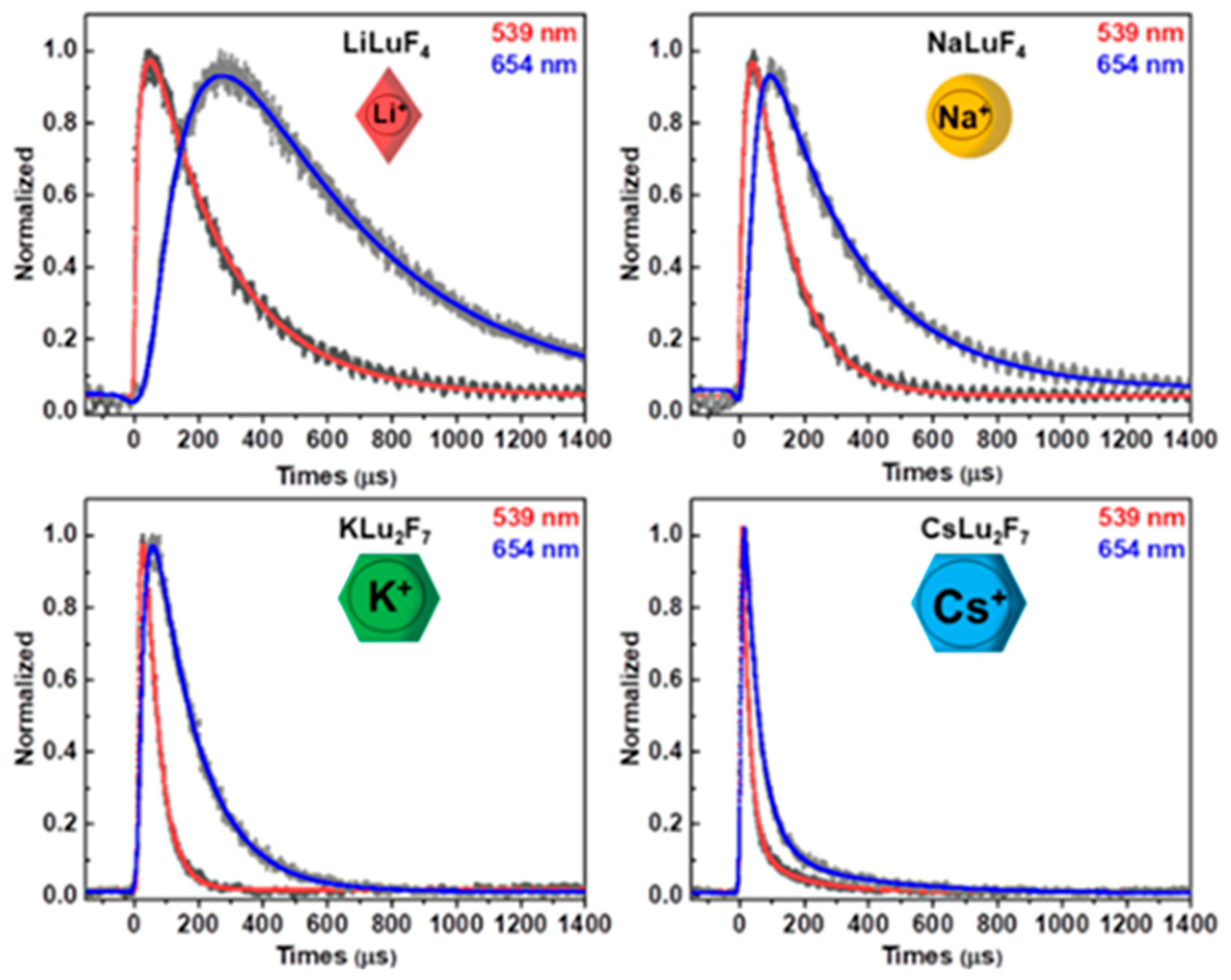
Figure 1. The characterization of the UCNPs with different alkali ions (Li, Na, K, and Cs): the TEM images (a), EDX spectra (b), and the size distribution data (c).
Nanomaterials 14 01969 g001
Figure 2. The XRD patterns of the UCNPs with different alkali ions, M: Li, Na, K, and Cs, respectively.
Nanomaterials 14 01969 g002
Scheme 1. Schematic illustration of the possible changes in the crystal lattice and upconversion luminescence of UCNPs doped with different alkali ions. (a) UCNPs lattice doped with different alkali ions. (b) Fluorescence of UCNPs doped with different alkali ions and their luminescence photographs.
Nanomaterials 14 01969 sch001
Figure 3. The steady-state PL luminescence spectra in vis-NIR regions of UCNPs doped with (a) 20% Yb3+, 2% Er3+and (b) 20% Yb3+, 20% Er3+ with different alkali ions (Li, Na, K, and Cs) under 980 nm laser irradiation, respectively.
Nanomaterials 14 01969 g003
Figure 4. The kinetic curves and fitting lines of the 4S3/2 (539 nm, black dots) and 4F9/2 (654 nm, gray dots) levels of Er3+ in the UCNPs doped with 20% Yb3+, 2% Er3+with different alkali ions (Li, Na, K, and Cs), respectively.
Nanomaterials 14 01969 g004
Figure 5. The emission spectra of the MLuF4–20% Yb3+, 2% Er3+ nanoparticles doped with different Na+ concentrations (0~100%), showing the green emission with a first decrease and then the increase trends, the red emission with a first increase and then the decrease trends.
Nanomaterials 14 01969 g005
Figure 6. The XRD patterns of the MLuF4–20% Yb3+, 2% Er3+ nanoparticles doped with different Na+ concentration (0~100%).
Nanomaterials 14 01969 g006
Figure 7. Fluorescence microscopy images of cells incubated with medium containing (a) NaLuF4–Yb, Er, (b) Na0.9Li0.1LuF4–Yb, Er, (c) Na0.5Li0.5LuF4–Yb, Er, and (d) Na0.1Li0.9LuF4–Yb, Er nanoparticles under NIR laser radiation and the corresponding intracellular UCNPs fluorescence intensity. * represents that the UCNP cell imaging performance is relatively good, and *** represents that the UCNP cell imaging performance is the best.
Nanomaterials 14 01969 g007
Figure 8. Proposed energy transfer mechanism in the (a) MLuF4–Yb3+, Er3+, (b) MLu2F7–Yb3+, Er3+, and (c) the MLuF4–Yb3+, Er3+ nanoparticles doped with different Na+ concentration (0~100%) properties showed better in MLuF4-host nanoparticles than in the other nanoparticles. Meanwhile, there is significant splitting in the emission spectrum as the decrease in ions radii from alkali cation, the host lattice altered substitutionally or interstitially, which led to the difference of ETU population, owing to the symmetry of crystal field surrounding RE3+ ions changed. As the Li+ is replaced by Na+, the bond length between M and F (M = Li, Na) in the crystal structure would increase, which makes the crystal field intensity decrease and thus, the emission peak splitting of Er3+ disappears.
Nanomaterials 14 01969 g008
Table 1. Lifetimes of the 2H11/2, 4S3/2, and 4F9/2 states of Er3+ ions for UCNPs with different alkali ions (Li, Na, K, and Cs).
Matrixτ (μs)2H11/24S3/24F9/2
LiLuF4τ117.9722.99121.74
τ2264.59246.95463.43
NaLuF4τ118.8920.1529.07
τ2125.90121.96283.73
KLu2F7τ16.2110.578.59
τ239.6447.8587.33
CsLu2Fτ11.611.050.95
τ227.0745.5361.23
Disclaimer/Publisher’s Note: The statements, opinions and data contained in all publications are solely those of the individual author(s) and contributor(s) and not of MDPI and/or the editor(s). MDPI and/or the editor(s) disclaim responsibility for any injury to people or property resulting from any ideas, methods, instructions or products referred to in the content.

