Review

Recent Advances on Electrospun Nanofibers for Periodontal Regeneration

by
Mafalda S. Santos
1,2,
Marta S. Carvalho
1,2,*,† and
João C. Silva
1,2,*,†
1
Department of Bioengineering, iBB-Institute for Bioengineering and Biosciences, Instituto Superior Técnico, Universidade de Lisboa, Av. Rovisco Pais, 1049-001 Lisboa, Portugal
2
Associate Laboratory i4HB-Institute for Health and Bioeconomy, Instituto Superior Técnico, Universidade de Lisboa, Av. Rovisco Pais, 1049-001 Lisboa, Portugal
*
Authors to whom correspondence should be addressed.
These authors jointly supervised this work.
Nanomaterials 2023, 13(8), 1307; https://doi.org/10.3390/nano13081307
Submission received: 5 March 2023 / Revised: 29 March 2023 / Accepted: 4 April 2023 / Published: 7 April 2023
(This article belongs to the Special Issue New Insights in Nanomaterials for Dental Diseases Management)

Abstract

:
Periodontitis is an inflammatory infection caused by bacterial plaque accumulation that affects the periodontal tissues. Current treatments lack bioactive signals to induce tissue repair and coordinated regeneration of the periodontium, thus alternative strategies are needed to improve clinical outcomes. Electrospun nanofibers present high porosity and surface area and are able to mimic the natural extracellular matrix, which modulates cell attachment, migration, proliferation, and differentiation. Recently, several electrospun nanofibrous membranes have been fabricated with antibacterial, anti-inflammatory, and osteogenic properties, showing promising results for periodontal regeneration. Thus, this review aims to provide an overview of the current state of the art of these nanofibrous scaffolds in periodontal regeneration strategies. First, we describe the periodontal tissues and periodontitis, as well as the currently available treatments. Next, periodontal tissue engineering (TE) strategies, as promising alternatives to the current treatments, are addressed. Electrospinning is briefly explained, the characteristics of electrospun nanofibrous scaffolds are highlighted, and a detailed overview of electrospun nanofibers applied to periodontal TE is provided. Finally, current limitations and possible future developments of electrospun nanofibrous scaffolds for periodontitis treatment are also discussed.

1. Introduction

Periodontitis is a chronic inflammatory infection of the periodontium, the structure responsible for ensuring tooth attachment and stability [1]. Bacterial dental plaque accumulation causes this infection, which can lead to the inflammation and destruction of the periodontium and, ultimately, tooth loss [2]. Advanced stages of the disease require regenerative procedures to restore the lost tissues, which include membranes for guided tissue regeneration (GTR) and bone grafts. However, these current treatments lack effective strategies to induce tissue repair and coordinated regeneration of all periodontal tissues, increasing the demand for alternative solutions to improve clinical outcomes [3].
Nanofibrous scaffolds have been widely explored in tissue engineering (TE) strategies due to their unique properties such as high porosity, surface area, and interconnectivity. Electrospinning allows the efficient production of nanofibrous scaffolds that can mimic the morphology and scale of native extracellular matrix (ECM) proteins, thus promoting cell attachment, proliferation, and differentiation [4]. Through the optimization of electrospinning parameters and selection of the most adequate polymers and additives, it is possible to tailor the characteristics of electrospun polymeric nanofibrous scaffolds for a desired purpose. Nanofibrous scaffolds with high porosity and small pore size are highly suitable for the development of novel periodontal barrier membranes [5]. Specific additives incorporated in scaffolds, such as antibiotics and bioceramics, can provide antibacterial activity against oral pathogens and osteogenic properties to promote tissue regeneration, which are advantageous features for periodontitis treatment [6]. Furthermore, the biomimetic characteristics of nanofibers can be exploited for the development of more suitable constructs to insert in periodontal defects as alternatives to bone grafts.
Recently, substantial research has been carried out regarding the development of electrospun nanofibrous scaffolds for periodontal regeneration. Nanofibers produced from a wide variety of polymers and composites, with diverse biochemical factors incorporated, have been studied and have shown promising results in in vitro and in vivo settings. This review covers the principal features of periodontal tissues, describes the main causes and symptoms of periodontal disease, and summarizes the currently available treatments. Alternative strategies proposed by periodontal TE are explored. The electrospinning technique is detailed and the features of electrospun nanofibrous scaffolds are outlined. The recent research on electrospun nanofibrous scaffolds for periodontal regeneration is summarized and discussed. Finally, shortcomings and future perspectives of using electrospun nanofibrous scaffolds for periodontitis treatment are debated.

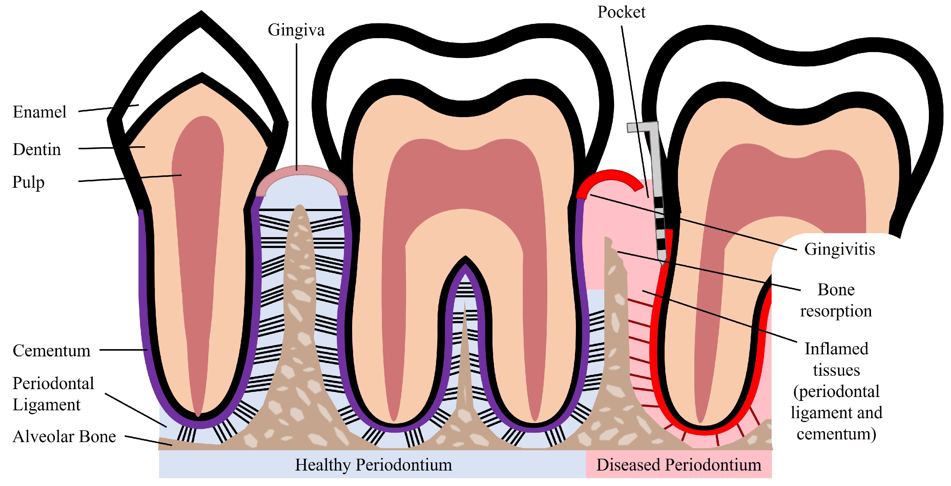
2. Periodontal Tissue Features and Periodontal Disease

The periodontium is a complex structure composed of hard and soft tissues that support the tooth. It has an important role of ensuring tooth attachment to the bone of the jaw and allowing the teeth to withstand the forces of mastication. The periodontium consists of alveolar bone, root cementum, and periodontal ligament (PDL) [7]. The anatomy of the periodontium is illustrated in Figure 1.
The alveolar bone is the part of the maxilla or mandible that contains the sockets that surround and anchor the teeth. The alveolar bone is a highly mineralized, hard tissue composed of 60% (w/w) inorganic material, 25% (w/w) organic material, and 15% water [8]. In the root of the teeth, the alveolar bone is connected to the root cementum through the PDL, as can be observed in Figure 1. The alveolar bone is perforated by channels, which allow the passage of blood vessels and nerve fibers that extend to within the pulp of the teeth [9]. Similar to what occurs in other types of bones, alveolar bone is maintained through constant bone remodeling. Because the teeth are continuously making minor movements and there is a functional demand due to the forces of mastication, the alveolar bone undergoes constant remodeling. Bone remodeling relies on a balance between bone resorption and bone deposition, which is maintained by progenitor cells that can differentiate into osteoclasts (bone resorption) and osteoblasts (bone deposition) [10,11].
The cementum is a hard, avascular connective tissue that covers the roots of teeth. It is located between the dentin and the PDL, as can be seen in Figure 1. The primary function of the cementum is to anchor the PDL fibers. The cementum’s composition is very similar to that of the alveolar bone, namely 65% (w/w) inorganic material, 23% (w/w) organic material, and 12% water [8]. The organic material is constituted by up to 90% of collagen type I. Interestingly, the majority of non-collagenous matrix proteins present in the cementum are also found in bone, namely fibronectin, osteocalcin, osteonectin, and osteopontin [12]. The cementum is produced as repair tissue to fill root fractures and resorptive defects [9,12]. In periodontal regeneration, new cementum is formed from cementoblasts. Reports suggest that the PDL serves as a source of progenitor cells for cementoblasts involved in cementum formation and also for osteoclasts and osteoblasts involved in bone remodeling [10,11].
The PDL is a complex, highly cellular, fibrous connective tissue located between the alveolar bone and the cementum, as can be seen in Figure 1. The width of the PDL ranges between 100 and 400 µm; however, it progressively decreases in thickness with age [10,13]. The extracellular compartment of the PDL is composed of highly aligned and organized collagen fiber bundles and non-collagenous matrix constituents, such as glycoproteins and proteoglycans [14]. The collagen fiber bundles provide the structural strength of the PDL and are mainly composed of collagen type I [8]. The fibers and fibrils present in the PDL are in the scale of nanometers to micrometers [9,13]. The extremities of the collagen fiber bundles are embedded in cementum or alveolar bone. The PDL is primarily responsible for providing support and mechanical stability to the teeth. It connects the cementum covering the tooth to the alveolar bone, ensuring the attachment of the tooth to the bone, while absorbing the shock from the considerable forces associated with mastication [3,14]. When characterized through tensile testing under loads between 1 and 5 N, the PDL demonstrated values of elastic modulus in the range between 0.607 and 4.274 MPa. Its elastic behavior is influenced by the loading rate, type of tooth, root level, and individual variability [15]. Furthermore, the PDL is innervated and can act as a sensory receptor for regulating pressure on the teeth and proper positioning of the jaw during mastication.
The PDL possesses an extensive blood supply and a diversity of cell populations, which include osteoblasts, osteoclasts, cementoblasts, fibroblasts, and progenitor cells [16] Another cell population that is present in the PDL are periodontal ligament stem/stromal cells (PDLSCs), which serve as a source for renewable progenitor cells, which can differentiate into osteoblasts, cementoblasts, and fibroblasts. Due to the presence of these heterogeneous cell populations, the PDL serves as a cell reservoir for tissue homeostasis, repair, and regeneration [3,14]. Blood vessels present in the PDL provide nutrients necessary for the maintenance of the ligament and the hard tissues. The PDL connects the root cementum to the alveolar bone and sustains a balance between formation and maintenance of the hard and soft tissues. The unique structure and composition of the PDL is essential for the physiological functionalities of periodontal tissues [3].
Periodontal disease is characterized by an inflammatory infection of the periodontium. This infection is caused and sustained by bacteria from dental plaque accumulation. In early stages of the disease, there is inflammation only of the gingiva, known as gingivitis, which is reversible with effective oral hygiene [1]. However, if left untreated, gingivitis can progress to periodontitis. Periodontitis in its advanced form is characterized by the loss and destruction of the periodontal tissues, including PDL, root cementum, and alveolar bone, as can be seen in Figure 1 [2,17]. This results in the loss of the tooth attachment to its supporting structures of the periodontium and in the formation of pockets surrounding the tooth. The symptoms of severe periodontitis include pain and discomfort during mastication, drifting and mobility of teeth, and tooth loss [18]. Periodontitis is the main cause of tooth loss, which is a global health problem representing a burden to society and the economy, particularly affecting older people [2]. The economic burden of periodontal disease was estimated to be USD 154.06B in the United States and EUR 158.64B in Europe, in 2018 [19]. Periodontitis is prevalent in adults and elderly populations and can also occur in children and adolescents. The prevalence of periodontal disease, which includes gingivitis and periodontitis, is estimated to range from 20% to 50% worldwide [1,17]. This large range of estimated prevalence is due to the absence of a unique and consensual case definition among different countries and populations [18]. Periodontitis can be characterized by the number of affected teeth, the magnitude of the pocket depth, the loss of tooth attachment capacity, and the loss of alveolar bone. More severe forms of periodontitis are estimated to affect 10% of the population [17].
Although bacterial plaque accumulation is the initiator of gingivitis, the host’s susceptibility to disease progression plays an important role. In patients not susceptible to periodontitis, the primary defense mechanisms are able to control the infection, and the inflammation of the gingiva may persist indefinitely without progressing to periodontitis. On the other hand, the primary defenses of patients susceptible to periodontitis cannot contain the infection of the gingiva, and the infection spreads to the periodontium [17]. The destruction of the periodontal tissues is in fact caused by host-derived mediators and enzymes from inflammatory cells in response to the bacterial infection of the periodontium. The inability to control the infection allows it to further progress into the tooth root, deepening the pockets and resulting in tooth attachment loss and alveolar bone loss [1].
Patient susceptibility is significantly affected by risk factors that increase the probability of periodontitis development. The risk factors can be genetic or environmental. Genetic risk factors that increase patient susceptibility to disease include defects of phagocytosis, which leads to an insufficient response to the bacterial infection, and enhanced enzyme production for a bacterial challenge, resulting in an excessive response with increased tissue damage [1]. Environmental or acquired risk factors include smoking, which is associated with decreased wound healing and reduced bacterial killing. Studies show that smokers are more likely to have severe periodontitis, present increased loss of alveolar bone, and have higher prevalence of tooth loss when compared to non-smokers [1,2]. Poor oral hygiene is another risk factor, as it allows accumulation of dental plaque and is linked to increased severity of periodontitis [2].
Furthermore, there is a link between systemic diseases and periodontitis. Periodontitis poses a risk of systemic complications associated with cardiovascular disease, cancer, lung diseases, and diabetes [20]. In fact, diabetes has a bi-directional relationship with periodontitis. Diabetic patients show higher concentrations of inflammatory mediators compared to non-diabetic individuals. The severity and extent of periodontitis is directly influenced by the metabolic control of diabetic individuals [17,21]. Age is a potential risk factor, as the risk of periodontitis increases with the advancing age, with a higher prevalence of the disease in elderly populations [2]. Poor oral health has been shown to be associated with disability and poor physical function in older populations [22]. In addition to motor disability, intellectual or developmental disability can also have an effect on the state of periodontal health [23]. For example, patients suffering from autism have difficulties in correctly applying oral hygiene rules and undergoing dental visits and therapies, which expose these patients to a greater risk in developing periodontitis [24].

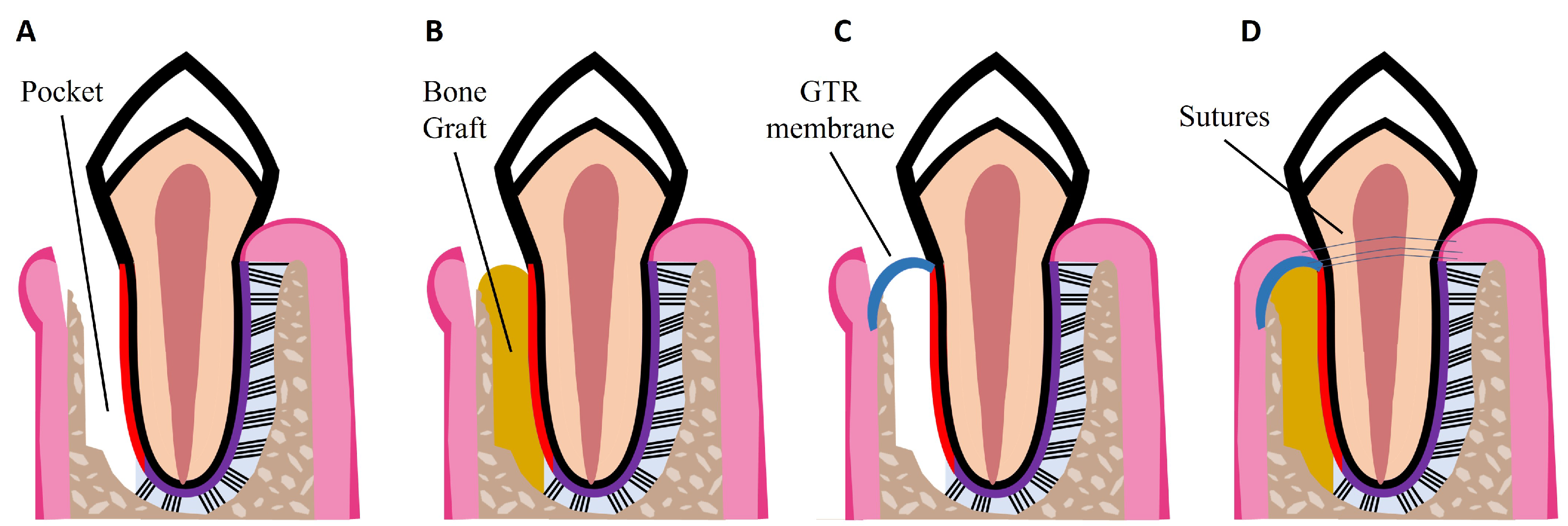
3. Currently Available Treatments for Periodontal Disease

Initial stages of periodontitis can be treated with non-surgical procedures such as dental plaque and tartar removal with scaling and root planing [1,18]. The main goal of these treatments is to control and reduce bacterial plaque accumulation. After the clinical removal of the dental plaque, the patient should practice adequate oral hygiene to achieve a good clinical outcome [18]. Non-surgical treatments can be combined with adjunctive therapies, such as local drug delivery, systemic antibiotics, and systemic host response modulation to improve treatment outcomes. Adjunctive drugs include antibiotics and antimicrobials that are directly administered to the periodontal pocket via a gel or fiber delivery system. Examples of systemic antibiotics are amoxicillin and metronidazole, which in combination result in pronounced clinical improvements [18]. Host response modulation can be particularly beneficial for patients susceptible to disease development. Host modulatory therapies influence the destructive components of the host response to reduce periodontal tissue destruction. These therapies include non-steroidal anti-inflammatory drugs: doxycycline that downregulates collagenases in inflamed periodontal tissues; and bisphosphonates, which reduce osteoclast activity and bone resorption [1].
Non-surgical treatments have been shown to reduce pocket depth and allow formation of new tooth attachment, which can be sufficient for early to moderate stages of periodontitis. However, in some cases and in advanced stages of the disease, surgical therapy is necessary to access sites deeper in the tooth root to control the inflammation, to fully eliminate bacterial plaque, and to stimulate the regeneration of lost periodontal tissues [18]. Pocket reduction surgery is a procedure that involves resecting soft and hard necrotic tissues. Regenerative surgery includes GTR membranes and bone grafts, which are illustrated in Figure 2. An innovative adjuvant is the use of laser treatment in non-surgical or surgical procedures [25]. The surgical and non-surgical currently available methods used to treat periodontitis are summarized in Table 1.
GTR is based on the use of a mechanical barrier membrane that prevents epithelial cells and fibroblasts from migrating into the defect site while maintaining sufficient space for the regeneration of all the periodontal tissues, namely alveolar bone, cementum, and PDL [34]. There are two types of membranes already commercially available that can be used for periodontal regeneration: non-degradable and degradable membranes. Currently available non-degradable membranes include polytetrafluoroethylene membranes, such as CytoplastTM TXT-200; however, a second surgery is required for their removal. To avoid additional surgeries, there are degradable membranes on the market, which are composed of synthetic polymers such as polycaprolactone (PCL), polylactic acid (PLA), and polyglycolic acid (PGA), and of natural polymers such as collagen, for example from porcine collagen, which is used in the Bio-Gide® commercially available membrane. However, current GTR membranes have limitations such as low attachment to the adjacent tissues, which can lead to an early exposure of the defect site and allow bacteria infiltration; lack of antibacterial properties; and poor ability to enhance the regeneration of all the periodontal tissues [3,34]. Thus, new improved membranes need to be developed aiming to meet all the criteria for an ideal GTR membrane, namely: biocompatibility; non-immunogenicity as to not trigger adverse reactions; biodegradability without release of toxic byproducts; cell-occlusivity to exclude specific cell types; and ease of use in a clinical setting [35]. They should also possess appropriate surface area and high porosity for cell attachment, proliferation, and differentiation, as well as mechanical strength to stay in place for at least 4–6 weeks and to maintain space for the slow regenerating periodontium. Finally, GTR membranes should present bioactivity to accelerate tissue repair and induce a coordinated regeneration of all the periodontal tissues [35,36]. Considering the slowly regenerating alveolar bone, bone grafts can be used to fill the defect site. GTR membranes can be combined with bone grafts to prevent membrane collapse, as illustrated in Figure 2D.
Bone grafts are transplanted into bone defects, where they promote bone healing either alone or in combination with other materials. Their main functions are to provide mechanical support and enhance bone regeneration [33]. Bone grafts need to have four essential properties for achieving successful bone regeneration: osseointegration, which refers to the graft’s capability to bind to the bone’s surface; osteogenesis, which is the formation of new bone through osteoblasts present in the graft; osteoconductivity, which is the graft’s potential to generate a scaffold on which host cells can grow; and osteoinductivity, which translates to the graft’s ability to recruit host stem cells into it and induce their differentiation into osteoblasts through local proteins and growth factors [37]. Unfortunately, current bone grafts mainly fulfill only the osteoconductivity property by serving as a structure for regeneration processes to occur [38]. Although some bone grafts might present almost all four essential properties for successful bone regeneration, their success is also influenced by the grafts biocompatibility, biodegradability, structural integrity, and porosity [39]. Moreover, the grafts are envisaged for bone formation, thus neglecting PDL regeneration. It is important to note that not only is osseointegration important, but so also is the attachment of newly formed bone to a regenerated PDL, which in turn connects the newly formed bone to the cementum of the tooth. In addition to bone grafts, GTR membranes also fail to achieve PDL regeneration and integration of soft (PDL) and hard tissues (alveolar bone, cementum) [3]. If the PDL is not regenerated, there is no connection between cementum and alveolar bone, and the tooth will eventually be lost due to the lack of attachment to the bone. These regenerative procedures are still exposed to clinical failures and do not effectively promote periodontal regeneration. Therefore, innovative strategies that promote the regeneration of the entire hierarchical structure of the periodontium are needed to improve clinical outcomes.

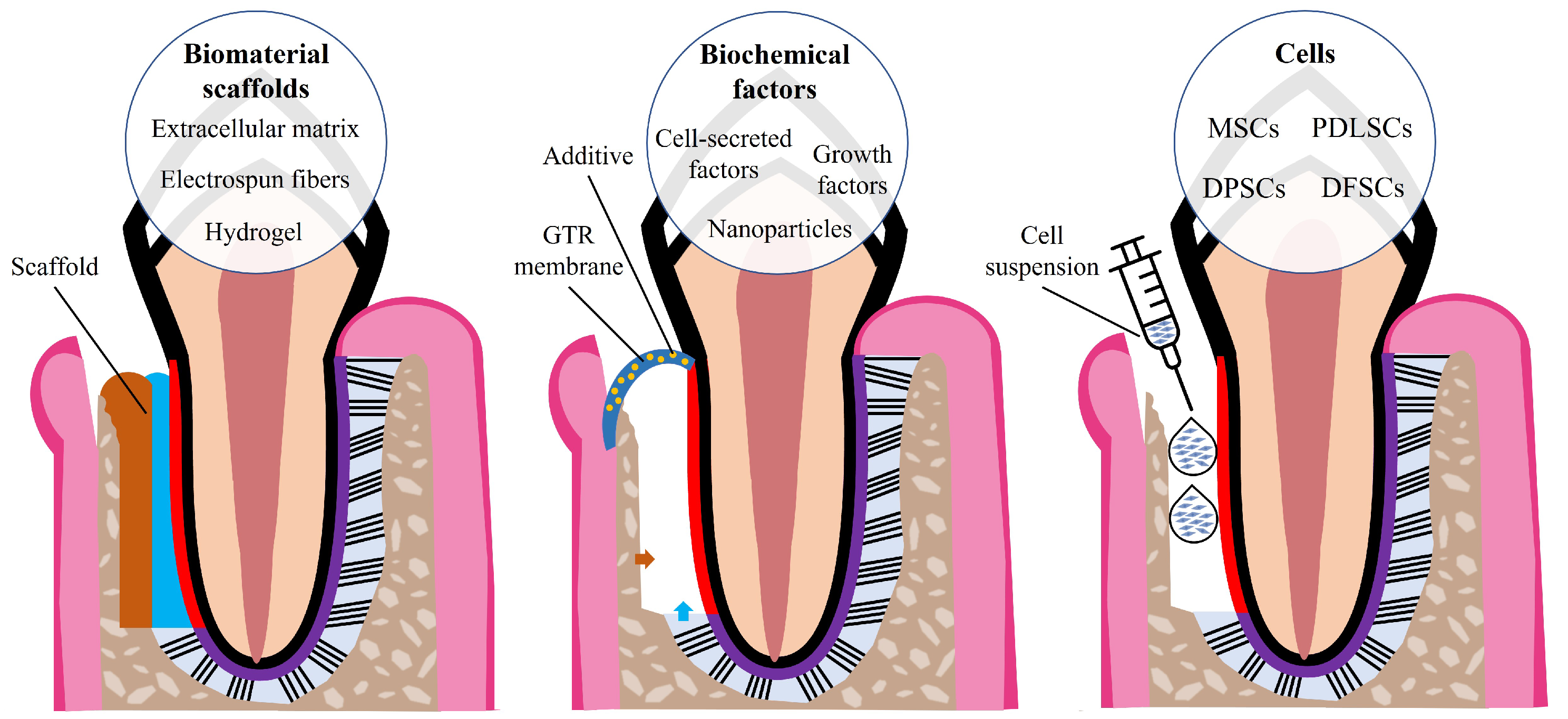
4. Periodontal Tissue Engineering

TE relies on the use or combination of cells, scaffolds, and biochemical factors to facilitate tissue regeneration (Figure 3). TE strategies use or manipulate one or more of these mediators with the aim of promoting the regeneration of diseased or damaged tissues. Various TE strategies for periodontal regeneration have been reported in the literature, proposing alternatives to the current regenerative treatments of bone grafts and GTR membranes used in the treatment of periodontitis.
Mesenchymal stem/stromal cells (MSCs) are commonly used in periodontal TE strategies. MSCs present hypoimmunogenicity and immunomodulatory properties, making them promising candidates for TE applications [40]. MSCs derived from adult tissues (e.g., bone marrow, adipose tissue, and synovial tissue) present no ethical or legal concerns, can be expanded in vitro, and used in TE strategies [41,42]. PDLSCs show similar characteristics to MSCs, such as fibroblast-like morphology, multilineage differentiation capacity, and expression of MSC-related surface markers [43,44,45]. PDLSCs are present in the PDL and serve as a source for renewable progenitor cells, which differentiate into osteoblasts, cementoblasts, and fibroblasts, responsible for bone, cementum, and PDL formation [14]. Stem cells have also been isolated from the dental pulp and the dental follicle. The dental follicle is a loose connective tissue that surrounds the enamel and the dental pulp of the developing tooth germ before tooth eruption, hence dental follicle stem cells (DFSCs) also give rise to progenitors of osteoblasts, cementoblasts, and PDL cells [46].
Current stem cell-based therapies rely mainly on the delivery of cells that were expanded in vitro to the periodontal defect site with the goal of promoting regeneration [47]. This delivery can be performed using single-cell suspensions injected into the defect site, which represents a simple and minimally invasive procedure [48]. Bone marrow-derived MSCs (BMMSCs) have been injected into rat periodontal defect models and showed the capacity to exert anti-inflammatory and immunomodulatory effects and promote periodontal regeneration, as MSCs can differentiate into the osteogenic lineage [47,48,49]. However, injection of single-cell suspensions have drawbacks, including poor engraftment, significant decrease in cell number after implantation, spreading to surrounding tissues, and loss of cell fate control [48]. Because PDLSCs were shown to be highly proliferative and capable of regenerating cementum/PDL-like tissues in vivo [43], interest was raised regarding their potential for use in periodontal tissue regeneration as a stem cell-based therapy to treat periodontal defects. Their regenerative capacity was studied in dental defects using several animal models (e.g., rat, miniature pig, and beagle dog defect models), and results showed that PDLSCs had the potential to form soft and hard periodontal-like structures and to promote periodontal regeneration [43,49,50,51,52]. In addition to single-cell suspensions, another possible stem-cell based approach is the delivery of monolayer or stacked cell sheets. Cell sheets remain intact as a whole due to cellular junctions and ECM. This technique is based on harvesting confluent cultured cells without any enzyme, which is easier to implement than cell suspensions and results in minimized cell loss and higher cell viability [47,48]. Interestingly, a study compared cell injection and cell sheet transplantation of human dental pulp stem cells (DPSCs) in swine periodontal bone defect models. The results showed that both approaches were able to significantly regenerate alveolar bone; however, the cell sheet transplantation exhibited higher bone regeneration capacity [53]. Nevertheless, cell sheets require a longer culture period, are fragile if cells are not confluent enough, attach weakly to hard tissues, and cell sheets that are too thick present necrotic cells. These limitations may be addressed with the use of biomaterial scaffolds. Examples of different sources from which stem cells can be isolated and used for periodontal regeneration are summarized in Table 2.

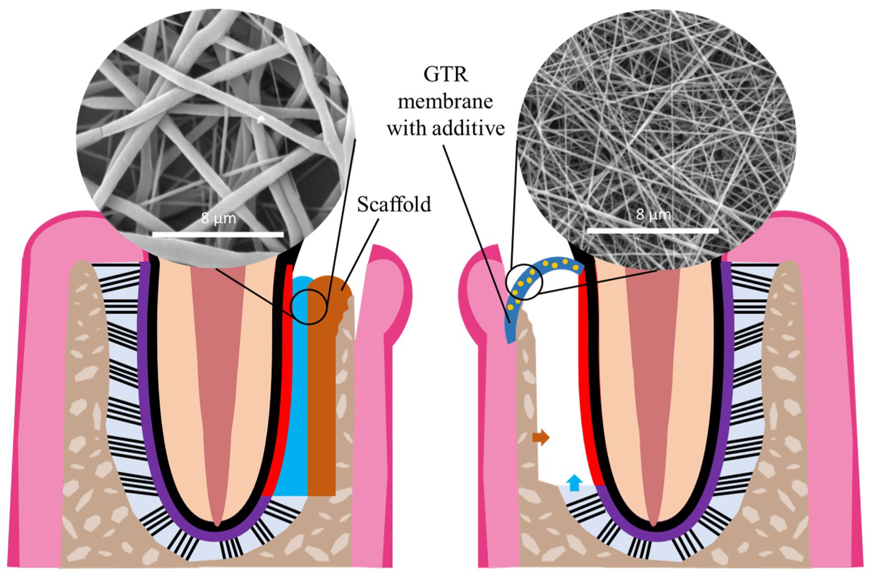
5. Electrospun Nanofibers for Periodontal Tissue Engineering

Tissue-engineered scaffolds made of synthetic polymers, such as PCL, PLA, poly(lactic-co-glycolic acid) (PLGA), and polyethylene glycol (PEG), or of natural polymers, such as chitosan, gelatin, and collagen, have been employed for periodontal regeneration. Synthetic polymers offer tailorable and reproducible structural properties, which allows mass production. Although they present good mechanical properties, they have slow degradation rates and poor biological activity [57]. Natural polymers have high biocompatibility and advantageous bioactivity, associated with enhanced cell adhesion, proliferation, and matrix production [58]. Synthetic and natural polymers can be combined to address their limitations and obtain scaffolds with the advantageous features of both polymer types [59].
Nanofibrous scaffolds possess unique properties, such as high surface area to volume ratio, porosity, and interconnectivity which favor cell attachment and proliferation and also enable nutrient and waste exchange. Electrospinning is a technique for fabricating continuous fibers with an average diameter ranging from few nanometers to micrometers [60]. Cumulative fibers form non-woven fibrous membranes that mimic the morphology of ECM proteins, therefore facilitating cell attachment, proliferation, and differentiation. Electrospinning allows the production of fibrous scaffolds with controllable fiber diameter, fiber orientation, porosity, and surface characteristics [61].
The electrospinning technique is simple, cost-effective, and requires four main components: a syringe containing a polymeric solution, a spinneret with a metallic needle, a high-voltage power supply, and a grounded metal collector. The syringe with the polymeric solution is placed in a pump, which ejects the solution and controls its flow rate. The solution is ejected through the metallic needle that is connected to the high voltage power supply. The power supply is also connected to the metal collector, and an electrostatic field is formed between the needle and the collector. The high voltage makes the droplets formed at the needle tip by electrically charging the polymeric solution. The droplets are stretched with electrostatic forces that counteract the solution’s surface tension into an elongated shape, known as the Taylor cone, from which a jet of charged fluid is pulled towards the grounded collector. The electrospun polymeric fibers deposit and solidify in the collector, and the solvent evaporates during the electrospinning process, resulting in dry fibers on the collector, which accumulate overtime to form fibrous scaffolds [4].
Essentially, electrospinning uses a polymeric solution to fabricate fibers in a high electrostatic field. There are various factors that influence the characteristics of the collected electrospun fibers, and these factors can be divided in three categories: solution parameters, process parameters, and environmental parameters. The solution parameters include solution concentration, solution viscosity, solution surface tension, polymer solubility, and polymer molecular weight. The process parameters consist of the voltage, solution flow rate, needle inner diameter, needle-to-collector distance, and type of collector. The environmental parameters comprise temperature and humidity [62].
By using the most adequate polymers and through the optimization and consideration of all the parameters that influence the features of electrospun fibers, it is possible to produce electrospun polymeric fibrous scaffolds with the most appropriate properties for a specific application. Electrospinning has been used in various TE strategies, including periodontal TE, as electrospun fibrous scaffolds are highly suitable for the development of periodontal GTR barrier membranes and biomimetic scaffolds, as illustrated in Figure 4. The high porosity with small pore size can prevent the migration of fibroblasts across the nanofibrous scaffolds, which is a vital feature of a GTR membrane [5]. By providing a closer mimicry of the native ECM, electrospun nanofibrous scaffolds can be part of a TE construct that facilitates tissue regeneration as an alternative to bone grafts. Through the use of optimized electrospinning parameters and the combination of carefully selected synthetic and natural polymers, nanofibrous scaffolds can meet the criteria of an ideal GTR membrane: biodegradable, biocompatible, osteoinductive, and having good mechanical properties [36]. The properties of electrospun fibers are summarized in Table 3.
In addition to their resemblance to natural ECM, the scaffolds can be functionalized with additives, such as ceramics and growth factors, to enhance their biological effects through surface coatings or biomolecule incorporation [63]. Hydroxyapatite, a major component of natural bone, can be incorporated into the fibers to emulate the native inorganic bone component and increase the scaffolds bioactivity and osteoconductivity [64,65]. Through the increase in bioactivity and close mimicry of the native ECM, these nanofibrous scaffolds might have the capacity to recruit host stem and progenitor cells and promote their proliferation and differentiation into fibroblasts, osteoblasts, and cementoblasts, possibly regenerating all the periodontal tissues. Other additives that have been incorporated into the scaffolds include antibiotics [66,67] that confer antibacterial activity, anti-inflammatory drugs [68,69], small molecules [70,71], and gene delivery vectors [72,73], which can enhance the osteogenic and angiogenic properties of the scaffolds.
The development of electrospun scaffolds for periodontal regeneration envisaging novel GTR membranes or scaffolds to insert in the defect site has been widely researched. Table 4 and Table 5 provide an overview of the recent research studies on electrospun nanofibrous scaffolds along with the specific features and main results from each study. The tables summarize the findings of the literature research carried out as represented in Figure 5. The keywords used were: periodontal regeneration, electrospinning, fibers, and synonyms or alternative words. Papers were excluded if they were published before 2015, did not include in vitro human cell culture on the electrospun scaffolds (for Table 4), or in vivo animal studies were not performed (for Table 5).
Table 4 and Table 5 demonstrate that various strategies have used additives, including growth factors, ceramics, metal oxides, antibiotics, and small molecules. Considering the inflammatory nature of periodontitis, some studies used specific additives to control and reduce inflammation, with clear results from in vitro and in vivo tests [68,69,70]. Because periodontal disease treatment requires removal of bacteria and dental plaque, several studies incorporated metal oxides [78,85,96] or antibiotics, such as metronidazole [66,81,108], tetracycline hydrochloride [67,97,98,99], and amoxicillin [66,104]. The use of metal oxides and antibiotics conferred antibacterial activity to the nanofibrous scaffolds; however, high quantities of the additive often resulted in a decrease in cell viability [81,85,97,98,99,105]. Metal oxides also were shown to enhance the osteogenic effects of the scaffolds, confirmed by increased cell mineralization, upregulated osteogenic gene expression, and elevated ALP activity in vitro [78,79], as well as accelerated new bone formation in vivo [79,109].
The incorporation of osteoinductive or osteoconductive additives, such as bone morphogenetic protein 2, dexamethasone, hydroxyapatite, β-tricalcium phosphate, bioactive glass, and silicate nanoparticles, into nanofibrous scaffolds resulted in augmented osteogenic potential and bioactivity. These scaffolds showed upregulated osteogenic gene expression [71,72,88] and increased cell viability [72,76,103] and promoted bone regeneration in vivo, as reported in various studies [71,88,108,110,111].
Instead of focusing on bone regeneration, one study shifted the focus to cementum regeneration. Chen et al. incorporated recombinant cementum protein 1 (rCMP1) in nanofibrous scaffolds, which downregulated osteogenic gene expression (osteocalcin and osteopontin) and upregulated cementoblastic markers (CMP1 and cementum attachment protein). The scaffolds showed less new bone formation and more cementum-like tissue in rat calvarial defects in vivo [90]. It should be noted that some studies only study bone formation in vivo using calvarial bone defects [75,79,90,110]. It is also relevant to study ligament regeneration, as it plays the essential role of connecting alveolar bone to cementum. For example, rat periodontal defects [69,71,77,106,108,109] or canine periodontal defects [107,112] can be used as more accurate models to study the possible effects of nanofibrous scaffolds in periodontal treatment. Some studies also presented in vivo tests limited to subcutaneous implantation, which were used only to assess the biocompatibility and cell barrier effects of the nanofibrous scaffolds [66,94,95].
The frequent use of natural polymers, such as chitosan, alginate, gelatin, and collagen, to produce nanofibers for periodontal regeneration is evident when analyzing Table 4 and Table 5. Natural polymers enhance the biological effects of the scaffolds, as confirmed by increased cell viability [75,87,89,91], and also improve the scaffolds’ osteogenic potential, as demonstrated by increased cell mineralization [86], ALP activity [91], and new bone formation in vivo [75]. When selecting the polymers to prepare electrospun scaffolds, certain characteristics, such as molecular weight, may significantly impact the scaffolds properties. In one study, low molecular weight chitosan conferred antibacterial activity to the scaffolds, whereas medium molecular weight chitosan did not [101].
Another strategy to improve the bioactivity of scaffolds is the use of proteins to stimulate the differentiation of stem cells. Lam et al. produced core-shell nanofibers loaded with enamel matrix proteins in the core. The use of the enamel matrix derivative Emdogain® resulted in upregulated osteogenic gene expression by PDLSCs. [93] This enamel matrix protein-based gel relies on biological mimicry to stimulate periodontal regeneration. Through the use of native tissue components, such as hydroxyapatite, growth factors, and certain proteins, it is possible to more closely mimic the natural microenvironment, thus facilitating regeneration.
One strategy that has not yet been extensively researched in periodontal regeneration is the use of cell-derived ECM. Cell-derived ECM is a reservoir of proteins and growth factors that influence cell proliferation and differentiation. It consists of secreted ECM by cells cultured in vitro, thus mimicking the composition of native ECM [114,115,116]. Cell-derived ECM has been used in combination with scaffolds in TE, as it mimics the in vivo microenvironment and enhances the scaffolds bioactivity [117,118,119]. Jiang et al. placed PDLSC-sheets on top of PCL/gelatin nanofibers and then decellularized the constructs. Decellularized cell sheets, with and without nanofibers, showed periodontium regeneration potential in rat periodontal defects, confirmed by formation of new bone, cementum, and PDL in vivo [120]. Farag and colleagues transferred PDLSC-sheets onto melt electrospun PCL membranes and then decellularized the constructs [121,122]. Decellularized cell sheets were shown to maintain the ECM intact, retain growth factors, and support recellularization by allogenic PDLSCs in vitro [121]. The decellularized cell-sheets also demonstrated enhanced expression of osteogenic genes by PDLSCs compared to PCL scaffolds alone. Decellularized cell-sheets were biocompatible in vivo and supported periodontal attachment in a rat periodontal defect model [122].
Interestingly, in the majority of the studies, non-aligned nanofibers are produced. From the few studies that fabricated aligned nanofibers, two stand out with in vivo tests. Jiang et al. combined various layers of PCL-PEG nanofibers, either aligned or non-aligned, with a CTS solution and lyophilized the assembly to obtain multilayer scaffolds. Their in vivo performance was evaluated in a rat periodontal defect model. The multilayer scaffolds were placed in contact with the tooth root surface and the defect was filled with the bone graft Bio-Oss® to immobilize the scaffolds. The aligned PCL-PEG nanofibers resulted in a higher expression of periostin, more mature collagen fibers, and significant formation of tooth-supporting mineralized tissue, as well as oriented PDL-like fibers in the regenerated periodontium [106]. Yang et al. produced aligned and non-aligned PCL nanofibers, which were stacked and then immersed in a gelatin solution, creating aligned and non-aligned constructs that were then lyophilized. The aligned construct facilitated collagen formation and maturation, significantly enhanced the angulation of new-born PDL-like tissue, and showed higher periostin expression at periodontal fenestration defects [77]. The regeneration of functional and organized PDL is important, as its unique structure is essential for the physiological functionalities of periodontal tissues.
In addition to the in vivo studies on animals, two clinical trials were recently carried out. Chen et al. studied PLA electrospun microfibrous scaffolds with β-tricalcium phosphate (β-TCP) incorporated in four LanYu pigs and fifteen human periodontal patients. The commercially available PLA dental membrane Epi-guide® was used as a control. PLA/β-TCP scaffolds showed no cytotoxicity in vitro and similar regeneration in vivo compared to the control. The electrospun scaffolds and the control membranes blocked the migration of fast-growing connective tissue into the defect site, created space for new tissue regeneration, and showed increased new cementum and bone formation compared to the blank control. The results from the clinical trials demonstrated significantly more tooth attachment gain, shallower probing depths, and improvement of periodontal inflammation in patients with electrospun scaffolds and the control membranes. In contrast with the patients with electrospun scaffolds, few patients with the control membrane showed soreness at the surgical sites [123]. The study revealed the suitability of electrospun PLA/β-TCP scaffold as an alternative GTR membrane for clinical applications. In another clinical trial, polyvinyl acetate/Ocimum sanctum electrospun fibrous scaffolds were placed in patients following scaling and root planing. Patients belonging to the control groups only underwent scaling and root planing without placement of the electrospun scaffolds. Results from this clinical trial showed significant clinical tooth attachment gain in the group treated with electrospun scaffolds. There was significant reduction in the pro-inflammatory cytokine interleukin 1-β(IL-1β) levels between before and after the treatment with scaling and root planing and electrospun scaffold placement. The post-treatment results did not differ significantly from the group undergoing only scaling and root planing [124]. This clinical trial (ID CTRI/2018/07/014961) demonstrated additional benefit when using electrospun fibrous scaffolds together with a non-surgical periodontitis treatment.

6. Current Challenges and Future Perspectives

Considering the complexity of the periodontium’s structure and the presence of various cell types, it is important that periodontal treatments promote a coordinated, organized, and hierarchical tissue regeneration. Although many studies report quantitative results related to new alveolar bone formation, this is not necessarily accompanied by PDL and cementum regeneration [125]. The regeneration of functional PDL is a key requirement for periodontal regeneration. However, more accurate periodontal disease models are needed to study PDL formation. Currently available in vitro models cannot recreate the complexity of the PDL, thus limiting a more detailed investigation of the tissue, leading to a dependence on animal studies [126]. More advanced in vitro periodontal disease models are needed and are currently being explored [126].
In order to stimulate a hierarchical tissue regeneration, biomimetic multilayered scaffolds can be produced through the combination of various techniques (e.g., 3D printing, electrospinning, solution casting) [127]. By developing hierarchical scaffolds that emulate the various periodontal tissues, a more synchronized tissue regeneration may occur. Another concern when designing novel biomimetic scaffolds is the spatiotemporal organization of the several cell types present in the periodontium. The PDL provides a cell reservoir necessary to maintain a balance between formation and maintenance of the hard and soft periodontal tissues, therefore scaffolds should support cell migration, proliferation, and differentiation of various cell types. One aspect that should also be taken into consideration is the mechanical stimulation that the PDL is physiologically subjected to. On one hand, advanced in vitro models should mimic the periodontium’s structural organization and reproduce the mechanical loading experienced in vivo. On the other hand, scaffolds should be stimulated mechanically not only to study their features but also to analyze how the stimulation affects cell behavior [128].
Electrospun nanofibrous scaffolds present suitable characteristics to be used as GTR membranes or to be part of multilayered biomimetic constructs for periodontal regeneration, showing promising results both in vitro and in vivo. These features result from a laborious optimization process due to the number of electrospinning parameters to control, which can pose some challenges. Although there are many synthetic and natural polymers, only a number of polymers can satisfy biocompatibility, non-toxicity, degradability, and mechanical properties. The polymers used in the process dictate the basic characteristics of the electrospun scaffolds [36]. It is important to carefully select the most appropriate polymer or combination of polymers to achieve the desired scaffolds features. In addition to polymer selection and dissolution, the electrospinning process and environmental parameters need to be optimized to obtain homogeneous fibers with the desired diameter and alignment and fibrous scaffolds made of cummulative fibers with coveted porosity, mechanical properties, and biodegradability [4]. Additionally, if the electrospinning solution flow rate is slow, the process may take several hours to produce fibrous scaffolds with enough thickness. The morphology of the fibers can be tailored; nevertheless, it may be difficult to control and tune considering the multiple parameters involved, not only from the process but also from the polymeric solution itself. Furthermore, fabricating electrospun fibers with an additive incorporated may result in additional complications, especially when one wishes to control the additive release or homogeneous distribution on the fibers without aggregation [4].
Even though electrospinning may include a laborious optimization process, once all the parameters are set it is crucial to maintain them, with the aim of obtaining reproducible results. For biological and TE applications, it is essential to properly sterilize the scaffolds while avoiding damages to the nanofibers [129]. In the case of scaffolds that require crosslinking, the toxicity of the agent must be taken into consideration and the non-toxicity of the crosslinked scaffolds must be verified. It is important to address the aforementioned limitations and have a good understanding of the scaffolds properties before translating the electrospun scaffolds from the laboratory to clinical applications [129].
This review presented a detailed overview of the use of electrospun nanofibers in periodontal tissue engineering applications, focusing on studies in vitro combining human stem cell cultures with electrospun nanofibers and also in in vivo animal studies, in which the nanofibrous scaffolds were implanted to promote periodontal regeneration. As a result of the chosen exclusion criteria, this review does not include studies with in vitro animal cell culture and papers published before 2015. The exclusion of these papers, which may contain valuable information, represents a limitation of this review.
Taking into account the promising results from the presented in vitro studies and in vivo tests in animal models and the publication of clinical trials using electrospun fibrous scaffolds, more studies on these scaffolds with in vivo tests and possibly more clinical trials will emerge in the near future. However, it is important to note that, although promising results were obtained in vitro with human stem cells and in vivo animal models, the translation into clinical practice is still a difficult concern. Future studies should aim to achieve scaffolds with anti-inflammatory and antibacterial properties, as well as hierarchical regeneration of functional bone-PDL-cementum complexes. The use of appropriate periodontal defect animal models for in vivo testing is necessary, as well as the study of the scaffolds effects on PDL regeneration and new fiber angulation, as it connects alveolar bone to cementum and is vital for a functional periodontium. In the future, the research on nanofibers for periodontal regeneration will continue, either with the development of novel additive-loaded GTR membranes with anti-inflammatory, antibacterial, and bioactive effects, or with the development of multilayer biomimetic scaffolds, in which nanofibers can represent the layer in contact with the root cementum and responsible for cementum and aligned PDL fibers regeneration. Considering its high potential, one strategy that will presumably be further investigated is the use of cell-derived ECM as an additive to provide a closer mimicry of the native tissue composition, thus accelerating tissue regeneration. Additionally, it would be interesting to test nanofibers in combination with other compounds, such as probiotics and other natural-derived molecules. In fact, probiotics have been used in adjunctive treatments to treat periodontitis and demonstrated an influence on plaque and other periodontal parameters [130]. These compounds should be evaluated in combination with nanofibers in future in vivo studies and clinical trials.

7. Conclusions

The periodontium is a highly complex structure, consisted of alveolar bone, PDL, and cementum. The PDL is composed of highly organized and aligned collagen fibers, whose organization is essential for the physiology and functionality of the periodontium. Advanced stages of periodontitis lead to inflammation and loss of all periodontal tissues. Possible treatment candidates for periodontal regeneration should promote an hierarchical regeneration of bone-PDL-cementum complexes to restore functional periodontium.
Electrospun nanofibers have high porosity and a high surface area to volume ratio, which, together with the mimicry of the scale and morphology of native ECM, favor cell adhesion, proliferation, and differentiation. Electrospun nanofibers form scaffolds with features suitable for GTR membranes and hierarchical scaffolds. This review provides an overview of the recent studies focused on the use of electrospun nanofibrous scaffolds for periodontal regeneration. These scaffolds can be complemented with various additives to enhance biological characteristics or provide antibacterial, anti-inflammatory, or osteogenic effects. Recent research on electrospun nanofibrous scaffolds shows promising results from in vivo tests, such as enhanced biocompatibility, increased bone formation, formation of newly and well oriented PDL fibers similar to the native tissue, decreased inflammation, and antibacterial activity. However, a biomimetic scaffold able to achieve all these regenerative outcomes is yet to be developed, and the difficult translation from in vitro/in vivo tests into clinical practice must be taken into account.
Future research should focus on the use of appropriate animal models to study the effects of electrospun nanofibrous scaffolds on periodontal regeneration, especially on the formation and alignment of newly formed PDL in addition to bone formation. The production efficiency and reproducibility of these scaffolds still needs to be explored in order to obtain more standardized membranes that can become alternative effective treatment options for periodontitis. The development of highly biomimetic hierarchical scaffolds using electrospun nanofibers and cell-derived ECM to facilitate periodontal regeneration holds great promise for the near future.

Author Contributions

Conceptualization: M.S.S., M.S.C. and J.C.S.; investigation: M.S.S.; writing—original draft preparation: M.S.S., M.S.C. and J.C.S.; writing—review and editing: M.S.S., M.S.C. and J.C.S.; supervision: M.S.C. and J.C.S.; funding acquisition: M.S.C. and J.C.S. All authors have read and agreed to the published version of the manuscript.

Funding

The authors acknowledge funding from Fundação para a Ciência e Tecnologia (FCT, Portugal) with dedicated funding from the FCT project “DentalBioMatrix” (PTDC/BTM- BTM/3538/2020). This work was also supported through FCT funding through the project InSilico4OCReg (PTDC/EME-SIS/0838/2021) and institutional funds to iBB (UIDB/04565/2020 and UIDP/04565/2020) and Associate Laboratory i4HB (LA/P/0140/2020).

Institutional Review Board Statement

Not applicable.

Informed Consent Statement

Not applicable.

Data Availability Statement

Not applicable.

Conflicts of Interest

The authors declare no conflict of interest.

References

  1. Preshaw, P.M.; Seymour, R.A.; Heasman, P.A. Current Concepts in Periodontal Pathogenesis. Dent. Update 2004, 31, 570–578. [Google Scholar] [CrossRef]
  2. Nazir, M.A. Prevalence of periodontal disease, its association with systemic diseases and prevention. Int. J. Health Sci. 2017, 11, 72–80. [Google Scholar]
  3. De Jong, T.; Bakker, A.D.; Everts, V.; Smit, T.H. The intricate anatomy of the periodontal ligament and its development: Lessons for periodontal regeneration. J. Periodontal Res. 2017, 52, 965–974. [Google Scholar] [CrossRef] [PubMed]
  4. Xue, J.; Wu, T.; Dai, Y.; Xia, Y. Electrospinning and Electrospun Nanofibers: Methods, Materials, and Applications. Chem. Rev. 2019, 119, 5298–5415. [Google Scholar] [CrossRef] [PubMed]
  5. Zhao, P.; Chen, W.; Feng, Z.; Liu, Y.; Liu, P.; Xie, Y.; Yu, D.G. Electrospun Nanofibers for Periodontal Treatment: A Recent Progress. Int. J. Nanomed. 2022, 17, 4137–4162. [Google Scholar] [CrossRef] [PubMed]
  6. Zhuang, Y.; Lin, K.; Yu, H. Advance of Nano-Composite Electrospun Fibers in Periodontal Regeneration. Front. Chem. 2019, 7, 495. [Google Scholar] [CrossRef]
  7. Newman, M.G.; Takei, H.H.; Klokkevold, P.R.; Carranza, F.A. Newman and Carranza’s Clinical Periodontology; Elsevier: Amsterdam, The Netherlands, 2019. [Google Scholar]
  8. Fehrenbach, M.J.; Popowics, T. Illustrated Dental Embryology, Histology, and Anatomy; Saunders: Maryland Heights, MO, USA, 2015. [Google Scholar]
  9. Goudouri, O.M.; Kontonasaki, E.; Boccaccini, A.R. Layered scaffolds for periodontal regeneration. In Biomaterials for Oral and Dental Tissue Engineering; Woodhead Publishing: Sawston, UK, 2017; pp. 279–295. [Google Scholar] [CrossRef]
  10. Kumar, G.S. Orban’s Oral Histology and Embryology; Elsevier: Haryana, India, 2019. [Google Scholar]
  11. Cho, M.I.; Garant, P.R. Development and general structure of the periodontium. Periodontology 2000, 24, 9–27. [Google Scholar] [CrossRef]
  12. Bosshardt, D. Are Cementoblasts a Subpopulation of Osteoblasts or a Unique Phenotype? J. Dent. Res. 2005, 84, 390–406. [Google Scholar] [CrossRef]
  13. Jiang, N.; Guo, W.; Chen, M.; Zheng, Y.; Zhou, J.; Kim, S.G.; Embree, M.C.; Song, K.S.; Marao, H.F.; Mao, J.J. Periodontal Ligament and Alveolar Bone in Health and Adaptation: Tooth Movement. Front. Oral Biol. 2015, 18, 1–8. [Google Scholar] [CrossRef] [Green Version]
  14. Nanci, A.; Bosshardt, D.D. Structure of periodontal tissues in health and disease. Periodontology 2006, 40, 11–28. [Google Scholar] [CrossRef]
  15. Wu, B.; Fu, Y.; Shi, H.; Yan, B.; Lu, R.; Ma, S.; Markert, B. Tensile testing of the mechanical behavior of the human periodontal ligament. BioMed. Eng. Online 2018, 17, 172. [Google Scholar] [CrossRef] [Green Version]
  16. Jia, L.N.; Zhang, X.; Xu, H.Y.; Hua, F.; Hu, X.G.; Xie, Q.; Wang, W.; Jia, J. Development of a Doxycycline Hydrochloride-Loaded Electrospun Nanofibrous Membrane for GTR/GBR Applications. J. Nanomater. 2016, 2016, 6507459. [Google Scholar] [CrossRef] [Green Version]
  17. Sanz, M.; D’aiuto, F.; Deanfield, J.; Fernandez-Avilés, F. European workshop in periodontal health and cardiovascular disease—Scientific evidence on the association between periodontal and cardiovascular diseases: A review of the literature. Eur. Heart J. Suppl. 2010, 12, B3–B12. [Google Scholar] [CrossRef]
  18. Kinane, D.F.; Stathopoulou, P.G.; Papapanou, P.N. Periodontal diseases. Nat. Rev. Dis. Prim. 2017, 3, 17038. [Google Scholar] [CrossRef] [PubMed]
  19. Botelho, J.; Machado, V.; Leira, Y.; Proença, L.; Chambrone, L.; Mendes, J.J. Economic burden of periodontitis in the United States and Europe: An updated estimation. J. Periodontol. 2022, 93, 373–379. [Google Scholar] [CrossRef] [PubMed]
  20. Bui, F.Q.; da Silva, C.L.C.A.; Huynh, B.; Trinh, A.; Liu, J.; Woodward, J.; Asadi, H.; Ojcius, D.M. Association between periodontal pathogens and systemic disease. Biomed. J. 2019, 42, 27–35. [Google Scholar] [CrossRef]
  21. Bhuyan, R.; Bhuyan, S.K.; Mohanty, J.N.; Das, S.; Juliana, N.; Juliana, I.F. Periodontitis and Its Inflammatory Changes Linked to Various Systemic Diseases: A Review of Its Underlying Mechanisms. Biomedicines 2022, 10, 2659. [Google Scholar] [CrossRef]
  22. Kotronia, E.; Wannamethee, S.G.; Papacosta, A.O.; Whincup, P.H.; Lennon, L.T.; Visser, M.; Weyant, R.J.; Harris, T.B.; Ramsay, S.E. Oral Health, Disability and Physical Function: Results From Studies of Older People in the United Kingdom and United States of America. J. Am. Med. Dir. Assoc. 2019, 20, 1654.e1–1654.e9. [Google Scholar] [CrossRef] [PubMed]
  23. Wilson, N.J.; Lin, Z.; Villarosa, A.; Lewis, P.; Philip, P.; Sumar, B.; George, A. Countering the poor oral health of people with intellectual and developmental disability: A scoping literature review. BMC Public Health 2019, 19, 1530. [Google Scholar] [CrossRef]
  24. Carli, E.; Pasini, M.; Pardossi, F.; Capotosti, I.; Narzisi, A.; Lardani, L. Oral Health Preventive Program in Patients with Autism Spectrum Disorder. Children 2022, 9, 535. [Google Scholar] [CrossRef]
  25. Kwon, T.; Lamster, I.B.; Levin, L. Current Concepts in the Management of Periodontitis. Int. Dent. J. 2021, 71, 462–476. [Google Scholar] [CrossRef] [PubMed]
  26. Slot, D.E.; Valkenburg, C.; Van der Weijden, G.F. Mechanical plaque removal of periodontal maintenance patients: A systematic review and network meta-analysis. J. Clin. Periodontol. 2020, 47, 107–124. [Google Scholar] [CrossRef]
  27. Sanz-Sánchez, I.; Montero, E.; Citterio, F.; Romano, F.; Molina, A.; Aimetti, M. Efficacy of access flap procedures compared to subgingival debridement in the treatment of periodontitis. A systematic review and meta-analysis. J. Clin. Periodontol. 2020, 47, 282–302. [Google Scholar] [CrossRef] [Green Version]
  28. Geisinger, M.L.; Kaur, M.; Basma, H. Nonsurgical Periodontal Therapy: A Review of Current Standards of Care and Innovations to Improve Gingival and Periodontal Health. Curr. Oral Health Rep. 2019, 6, 177–187. [Google Scholar] [CrossRef]
  29. Polak, D.; Wilensky, A.; Antonoglou, G.N.; Shapira, L.; Goldstein, M.; Martin, C. The efficacy of pocket elimination/reduction compared to access flap surgery: A systematic review and meta-analysis. J. Clin. Periodontol. 2020, 47, 303–319. [Google Scholar] [CrossRef] [PubMed] [Green Version]
  30. Liaw, A.; Miller, C.; Nimmo, A. Comparing the periodontal tissue response to non-surgical scaling and root planing alone, adjunctive azithromycin, or adjunctive amoxicillin plus metronidazole in generalized chronic moderate-to-severe periodontitis: A preliminary randomized controlled trial. Aust. Dent. J. 2019, 64, 145–152. [Google Scholar] [CrossRef] [PubMed]
  31. Liu, Y.; Guo, L.; Li, X.; Liu, S.; Du, J.; Xu, J.; Hu, J.; Liu, Y. Challenges and Tissue Engineering Strategies of Periodontal-Guided Tissue Regeneration. Tissue Eng. Part C Methods 2022, 28, 405–419. [Google Scholar] [CrossRef]
  32. Valenti, C.; Pagano, S.; Bozza, S.; Ciurnella, E.; Lomurno, G.; Capobianco, B.; Coniglio, M.; Cianetti, S.; Marinucci, L. Use of the Er:YAG Laser in Conservative Dentistry: Evaluation of the Microbial Population in Carious Lesions. Materials 2021, 14, 2387. [Google Scholar] [CrossRef]
  33. Baldwin, P.; Li, D.J.; Auston, D.A.; Mir, H.S.; Yoon, R.S.; Koval, K.J. Autograft, Allograft, and Bone Graft Substitutes: Clinical Evidence and Indications for Use in the Setting of Orthopaedic Trauma Surgery. J. Orthop. Trauma 2019, 33. [Google Scholar] [CrossRef]
  34. Ren, Y.; Fan, L.; Alkildani, S.; Liu, L.; Emmert, S.; Najman, S.; Rimashevskiy, D.; Schnettler, R.; Jung, O.; Xiong, X.; et al. Barrier Membranes for Guided Bone Regeneration (GBR): A Focus on Recent Advances in Collagen Membranes. Int. J. Mol. Sci. 2022, 23, 14987. [Google Scholar] [CrossRef]
  35. Mancini, L.; Fratini, A.; Marchetti, E. Periodontal Regeneration. Encyclopedia 2021, 1, 87–98. [Google Scholar] [CrossRef]
  36. Xu, X.; Ren, S.; Li, L.; Zhou, Y.; Peng, W.; Xu, Y. Biodegradable engineered fiber scaffolds fabricated by electrospinning for periodontal tissue regeneration. J. Biomater. Appl. 2021, 36, 55–75. [Google Scholar] [CrossRef] [PubMed]
  37. Bhatt, R.A.; Rozental, T.D. Bone Graft Substitutes. Hand Clin. 2012, 28, 457–468. [Google Scholar] [CrossRef]
  38. Zhao, R.; Yang, R.; Cooper, P.R.; Khurshid, Z.; Shavandi, A.; Ratnayake, J. Bone grafts and substitutes in dentistry: A review of current trends and developments. Molecules 2021, 26, 3007. [Google Scholar] [CrossRef]
  39. Kolk, A.; Handschel, J.; Drescher, W.; Rothamel, D.; Kloss, F.; Blessmann, M.; Heiland, M.; Wolff, K.D.; Smeets, R. Current trends and future perspectives of bone substitute materials—From space holders to innovative biomaterials. J. Cranio-Maxillofac. Surg. 2012, 40, 706–718. [Google Scholar] [CrossRef]
  40. Li, Q.; Yang, G.; Li, J.; Ding, M.; Zhou, N.; Dong, H.; Mou, Y. Stem cell therapies for periodontal tissue regeneration: A network meta-Analysis of preclinical studies. Stem Cell Res. Ther. 2020, 11, 427. [Google Scholar] [CrossRef] [PubMed]
  41. Silva, J.C.; Moura, C.S.; Borrecho, G.; de Matos, A.P.A.; Cabral, J.M.S.; Linhardt, R.J.; Ferreira, F.C. Effects of glycosaminoglycan supplementation in the chondrogenic differentiation of bone marrow- and synovial- derived mesenchymal stem/stromal cells on 3D-extruded poly (ε-caprolactone) scaffolds. Int. J. Polym. Mater. Polym. Biomater. 2021, 70, 207–222. [Google Scholar] [CrossRef]
  42. Carvalho, M.S.; Poundarik, A.A.; Cabral, J.M.S.; da Silva, C.L.; Vashishth, D. Biomimetic matrices for rapidly forming mineralized bone tissue based on stem cell-mediated osteogenesis. Sci. Rep. 2018, 8, 14388. [Google Scholar] [CrossRef] [Green Version]
  43. Seo, B.M.; Miura, M.; Gronthos, S.; Bartold, P.M.; Batouli, S.; Brahim, J.; Young, M.; Robey, P.G.; Wang, C.Y.; Shi, S. Investigation of multipotent postnatal stem cells from human periodontal ligament. Lancet 2004, 364, 149–155. [Google Scholar] [CrossRef]
  44. Couto de Carvalho, L.A.; Tosta dos Santos, S.L.; Sacramento, L.V.; de Almeida, V.R.; de Aquino Xavier, F.C.; dos Santos, J.N.; Águida Cristina Gomes Henriques Leitão. Mesenchymal stem cell markers in periodontal tissues and periapical lesions. Acta Histochem. 2020, 122, 151636. [Google Scholar] [CrossRef]
  45. Kadkhoda, Z.; Rafiei, S.C.; Azizi, B.; Khoshzaban, A. Assessment of Surface Markers Derived from Human Periodontal Ligament Stem Cells: An In Vitro Study. J. Dent. 2016, 13, 325–332. [Google Scholar]
  46. Zhou, T.; Pan, J.; Wu, P.; Huang, R.; Du, W.; Zhou, Y.; Wan, M.; Fan, Y.; Xu, X.; Zhou, X.; et al. Dental Follicle Cells: Roles in Development and beyond. Stem Cells Int. 2019, 2019, 9159605. [Google Scholar] [CrossRef] [PubMed] [Green Version]
  47. Xu, X.Y.; Li, X.; Wang, J.; He, X.T.; Sun, H.H.; Chen, F.M. Concise Review: Periodontal Tissue Regeneration Using Stem Cells: Strategies and Translational Considerations. Stem Cells Transl. Med. 2018, 8, 392–403. [Google Scholar] [CrossRef] [PubMed] [Green Version]
  48. Zeng, W.Y.; Ning, Y.; Huang, X. Advanced technologies in periodontal tissue regeneration based on stem cells: Current status and future perspectives. J. Dent. Sci. 2021, 16, 501–507. [Google Scholar] [CrossRef]
  49. Takewaki, M.; Kajiya, M.; Takeda, K.; Sasaki, S.; Motoike, S.; Komatsu, N.; Matsuda, S.; Ouhara, K.; Mizuno, N.; Fujita, T.; et al. MSC/ECM Cellular Complexes Induce Periodontal Tissue Regeneration. J. Dent. Res. 2017, 96, 984–991. [Google Scholar] [CrossRef]
  50. Qiu, J.; Wang, X.; Zhou, H.; Zhang, C.; Wang, Y.; Huang, J.; Liu, M.; Yang, P.; Song, A. Enhancement of periodontal tissue regeneration by conditioned media from gingiva-derived or periodontal ligament-derived mesenchymal stem cells: A comparative study in rats. Stem Cell Res. Ther. 2020, 11, 42. [Google Scholar] [CrossRef] [Green Version]
  51. Flores, M.G.; Yashiro, R.; Washio, K.; Yamato, M.; Okano, T.; Ishikawa, I. Periodontal ligament cell sheet promotes periodontal regeneration in athymic rats. J. Clin. Periodontol. 2008, 35, 1066–1072. [Google Scholar] [CrossRef]
  52. Liu, Y.; Zheng, Y.; Ding, G.; Fang, D.; Zhang, C.; Bartold, P.M.; Gronthos, S.; Shi, S.; Wang, S. Periodontal Ligament Stem Cell-Mediated Treatment for Periodontitis in Miniature Swine. Stem Cells 2008, 26, 1065–1073. [Google Scholar] [CrossRef] [Green Version]
  53. Hu, J.; Cao, Y.; Xie, Y.; Wang, H.; Fan, Z.; Wang, J.; Zhang, C.; Wang, J.; Wu, C.T.; Wang, S. Periodontal regeneration in swine after cell injection and cell sheet transplantation of human dental pulp stem cells following good manufacturing practice. Stem Cell Res. Ther. 2016, 7, 130. [Google Scholar] [CrossRef] [Green Version]
  54. Gao, X.; Shen, Z.; Guan, M.; Huang, Q.; Chen, L.; Qin, W.; Ge, X.; Chen, H.; Xiao, Y.; Lin, Z. Immunomodulatory Role of Stem Cells from Human Exfoliated Deciduous Teeth on Periodontal Regeneration. Tissue Eng. Part A 2018, 24, 1341–1353. [Google Scholar] [CrossRef]
  55. Yang, H.; Li, J.; Hu, Y.; Sun, J.; Guo, W.; Li, H.; Chen, J.; Huo, F.; Tian, W.; Li, S. Treated dentin matrix particles combined with dental follicle cell sheet stimulate periodontal regeneration. Dent. Mater. 2019, 35, 1238–1253. [Google Scholar] [CrossRef] [PubMed]
  56. Wu, P.H.; Chung, H.Y.; Wang, J.H.; Shih, J.C.; Kuo, M.Y.P.; Chang, P.C.; Huang, Y.D.; Wang, P.C.; Chang, C.C. Amniotic membrane and adipose-derived stem cell co-culture system enhances bone regeneration in a rat periodontal defect model. J. Formos. Med. Assoc. 2016, 115, 186–194. [Google Scholar] [CrossRef] [PubMed] [Green Version]
  57. Al-Abduljabbar, A.; Farooq, I. Electrospun Polymer Nanofibers: Processing, Properties, and Applications. Polymers 2023, 15, 65. [Google Scholar] [CrossRef]
  58. Sell, S.A.; Wolfe, P.S.; Garg, K.; McCool, J.M.; Rodriguez, I.A.; Bowlin, G.L. The Use of Natural Polymers in Tissue Engineering: A Focus on Electrospun Extracellular Matrix Analogues. Polymers 2010, 2, 522–553. [Google Scholar] [CrossRef]
  59. Keshvardoostchokami, M.; Majidi, S.S.; Huo, P.; Ramachandran, R.; Chen, M.; Liu, B. Electrospun Nanofibers of Natural and Synthetic Polymers as Artificial Extracellular Matrix for Tissue Engineering. Nanomaterials 2021, 11, 21. [Google Scholar] [CrossRef]
  60. Thenmozhi, S.; Dharmaraj, N.; Kadirvelu, K.; Kim, H.Y. Electrospun nanofibers: New generation materials for advanced applications. Mater. Sci. Eng. B 2017, 217, 36–48. [Google Scholar] [CrossRef]
  61. Chinnappan, B.A.; Krishnaswamy, M.; Xu, H.; Hoque, M.E. Electrospinning of Biomedical Nanofibers/Nanomembranes: Effects of Process Parameters. Polymers 2022, 14, 3719. [Google Scholar] [CrossRef]
  62. Haider, A.; Haider, S.; Kang, I.K. A comprehensive review summarizing the effect of electrospinning parameters and potential applications of nanofibers in biomedical and biotechnology. Arab. J. Chem. 2018, 11, 1165–1188. [Google Scholar] [CrossRef]
  63. Stojanov, S.; Berlec, A. Electrospun Nanofibers as Carriers of Microorganisms, Stem Cells, Proteins, and Nucleic Acids in Therapeutic and Other Applications. Front. Bioeng. Biotechnol. 2020, 8, 130. [Google Scholar] [CrossRef]
  64. Wu, X.; Miao, L.; Yao, Y.; Wu, W.; Liu, Y.; Chen, X.; Sun, W. Electrospun fibrous scaffolds combined with nanoscale hydroxyapatite induce osteogenic differentiation of human periodontal ligament cells. Int. J. Nanomed. 2014, 9, 4135–4143. [Google Scholar] [CrossRef] [Green Version]
  65. Samadian, H.; Mobasheri, H.; Azami, M.; Faridi-Majidi, R. Osteoconductive and electroactive carbon nanofibers/hydroxyapatite nanocomposite tailored for bone tissue engineering: In vitro and in vivo studies. Sci. Rep. 2020, 10, 14853. [Google Scholar] [CrossRef]
  66. Mirzaeei, S.; Mansurian, M.; Asare-Addo, K.; Nokhodchi, A. Metronidazole-and amoxicillin-loaded plga and pcl nanofibers as potential drug delivery systems for the treatment of periodontitis: In vitro and in vivo evaluations. Biomedicines 2021, 9, 975. [Google Scholar] [CrossRef]
  67. Dos Santos, D.M.; Chagas, P.A.; Leite, I.S.; Inada, N.M.; de Annunzio, S.R.; Fontana, C.R.; Campana-Filho, S.P.; Correa, D.S. Core-sheath nanostructured chitosan-based nonwovens as a potential drug delivery system for periodontitis treatment. Int. J. Biol. Macromol. 2020, 142, 521–534. [Google Scholar] [CrossRef]
  68. Cristo, F.D.; Valentino, A.; Luca, I.D.; Peluso, G.; Bonadies, I.; Calarco, A.; Salle, A.D. PLA Nanofibers for Microenvironmental-Responsive Quercetin Release in Local Periodontal Treatment. Molecules 2022, 27, 2205. [Google Scholar] [CrossRef]
  69. Sun, M.; Liu, Y.; Jiao, K.; Jia, W.; Jiang, K.; Cheng, Z.; Liu, G.; Luo, Y. A periodontal tissue regeneration strategy via biphasic release of zeolitic imidazolate framework-8 and FK506 using a uniaxial electrospun Janus nanofiber. J. Mater. Chem. B 2022, 10, 765–778. [Google Scholar] [CrossRef]
  70. He, Z.; Liu, S.; Li, Z.; Xu, J.; Liu, Y.; Luo, E. Coaxial TP/APR electrospun nanofibers for programmed controlling inflammation and promoting bone regeneration in periodontitis-related alveolar bone defect models. Mater. Today Bio 2022, 16, 100438. [Google Scholar] [CrossRef]
  71. Shang, L.; Liu, Z.; Ma, B.; Shao, J.; Wang, B.; Ma, C.; Ge, S. Dimethyloxallyl glycine/nanosilicates-loaded osteogenic/angiogenic difunctional fibrous structure for functional periodontal tissue regeneration. Bioact. Mater. 2021, 6, 1175–1188. [Google Scholar] [CrossRef]
  72. Xie, Q.; Jia, L.N.; Xu, H.Y.; Hu, X.G.; Wang, W.; Jia, J. Fabrication of Core-Shell PEI/pBMP2-PLGA Electrospun Scaffold for Gene Delivery to Periodontal Ligament Stem Cells. Stem Cells Int. 2016, 2016, 5385137. [Google Scholar] [CrossRef] [PubMed] [Green Version]
  73. Jiang, L.; Ding, Z.; Xia, S.; Liu, Y.; Lei, S.; Zhong, M.; Chen, X. Poly lactic-co-glycolic acid scaffold loaded with plasmid DNA encoding fibroblast growth factor-2 promotes periodontal ligament regeneration of replanted teeth. J. Periodontal Res. 2020, 55, 488–495. [Google Scholar] [CrossRef] [PubMed]
  74. Qasim, S.B.; Najeeb, S.; Delaine-Smith, R.M.; Rawlinson, A.; Rehman, I.U. Potential of electrospun chitosan fibers as a surface layer in functionally graded GTR membrane for periodontal regeneration. Dent. Mater. 2017, 33, 71–83. [Google Scholar] [CrossRef] [PubMed] [Green Version]
  75. Guo, S.; He, L.; Yang, R.; Chen, B.; Xie, X.; Jiang, B.; Weidong, T.; Ding, Y. Enhanced effects of electrospun collagen-chitosan nanofiber membranes on guided bone regeneration. J. Biomater. Sci. Polym. Ed. 2020, 31, 155–168. [Google Scholar] [CrossRef]
  76. Sousa, F.B.D.; Barud, H.S.; Delgado, A.; Dieterle, M.P.; Steinberg, T.; Tomakidi, P.; Nohava, J.; Vach, K.; Schulz, S.D.; Hellwig, E.; et al. Novel In Situ-Cross-Linked Electrospun Gelatin/Hydroxyapatite Nonwoven Scaffolds Prove Suitable for Periodontal Tissue Engineering. Pharmaceutics 2022, 14, 1286. [Google Scholar] [CrossRef]
  77. Yang, M.; Gao, X.; Shen, Z.; Shi, X.; Lin, Z. Gelatin-assisted conglutination of aligned polycaprolactone nanofilms into a multilayered fibre-guiding scaffold for periodontal ligament regeneration. RSC Adv. 2019, 9, 507–518. [Google Scholar] [CrossRef] [Green Version]
  78. Peng, W.; Ren, S.; Zhang, Y.; Fan, R.; Zhou, Y.; Li, L.; Xu, X.; Xu, Y. MgO Nanoparticles-Incorporated PCL/Gelatin-Derived Coaxial Electrospinning Nanocellulose Membranes for Periodontal Tissue Regeneration. Front. Bioeng. Biotechnol. 2021, 9. [Google Scholar] [CrossRef]
  79. Ren, S.; Zhou, Y.; Zheng, K.; Xu, X.; Yang, J.; Wang, X.; Miao, L.; Wei, H.; Xu, Y. Cerium oxide nanoparticles loaded nanofibrous membranes promote bone regeneration for periodontal tissue engineering. Bioact. Mater. 2022, 7, 242–253. [Google Scholar] [CrossRef]
  80. Huang, T.Y.; Shahrousvand, M.; Hsu, Y.T.; Su, W.T. Polycaprolactone/Polyethylene glycol blended with dipsacus asper wall extract nanofibers promote osteogenic differentiation of periodontal ligament stem cells. Polymers 2021, 13, 2245. [Google Scholar] [CrossRef]
  81. Tondnevis, F.; Ketabi, M.A.; Fekrazad, R.; Sadeghi, A.; Keshvari, H.; Abolhasani, M.M. In Vitro Characterization of Polyurethane-Carbon Nanotube Drug Eluting Composite Scaffold for Dental Tissue Engineering Application. J. Biomimetics Biomater. Biomed. Eng. 2020, 47, 13–24. [Google Scholar] [CrossRef]
  82. Serôdio, R.; Schickert, S.L.; Costa-Pinto, A.R.; Dias, J.R.; Granja, P.L.; Yang, F.; Oliveira, A.L. Ultrasound sonication prior to electrospinning tailors silk fibroin/PEO membranes for periodontal regeneration. Mater. Sci. Eng. C 2019, 98, 969–981. [Google Scholar] [CrossRef]
  83. Zhang, H.; Ma, H.; Zhang, R.; Wang, K.; Liu, J. Construction and characterization of antibacterial PLGA/wool keratin/ornidazole composite membranes for periodontal guided tissue regeneration. J. Biomater. Appl. 2020, 34, 1267–1281. [Google Scholar] [CrossRef]
  84. Zhang, H.; Wang, K.; Gao, T.; Zhang, R.; Cai, Z.; Liu, J.; Ma, H.; Zhang, W. Controlled release of bFGF loaded into electrospun core-shell fibrous membranes for use in guided tissue regeneration. Biomed. Mater. 2020, 15, 35021. [Google Scholar] [CrossRef]
  85. Münchow, E.A.; Albuquerque, M.T.P.; Zero, B.; Kamocki, K.; Piva, E.; Gregory, R.L.; Bottino, M.C. Development and characterization of novel ZnO-loaded electrospun membranes for periodontal regeneration. Dent. Mater. 2015, 31, 1038–1051. [Google Scholar] [CrossRef] [PubMed] [Green Version]
  86. Shen, R.; Xu, W.; Xue, Y.; Chen, L.; Ye, H.; Zhong, E.; Ye, Z.; Gao, J.; Yan, Y. The use of chitosan/PLA nano-fibers by emulsion eletrospinning for periodontal tissue engineering. Artif. Cells Nanomed. Biotechnol. 2018, 46, 419–430. [Google Scholar] [CrossRef] [PubMed] [Green Version]
  87. Ye, Z.; Xu, W.; Shen, R.; Yan, Y. Emulsion electrospun PLA/calcium alginate nanofibers for periodontal tissue engineering. J. Biomater. Appl. 2020, 34, 763–777. [Google Scholar] [CrossRef] [PubMed]
  88. Zhou, T.; Liu, X.; Sui, B.; Liu, C.; Mo, X.; Sun, J. Development of fish collagen/bioactive glass/chitosan composite nanofibers as a GTR/GBR membrane for inducing periodontal tissue regeneration. Biomed. Mater. 2017, 12, 55004. [Google Scholar] [CrossRef] [PubMed]
  89. Song, J.; Klymov, A.; Shao, J.; Zhang, Y.; Ji, W.; Kolwijck, E.; Jansen, J.A.; Leeuwenburgh, S.C.; Yang, F. Electrospun Nanofibrous Silk Fibroin Membranes Containing Gelatin Nanospheres for Controlled Delivery of Biomolecules. Adv. Healthc. Mater. 2017, 6, 1700014. [Google Scholar] [CrossRef]
  90. Chen, X.; Liu, Y.; Miao, L.; Wang, Y.; Ren, S.; Yang, X.; Hu, Y.; Sun, W. Controlled release of recombinant human cementum protein 1 from electrospun multiphasic scaffold for cementum regeneration. Int. J. Nanomed. 2016, 11, 3145–3158. [Google Scholar] [CrossRef] [Green Version]
  91. Yang, F.; Miao, Y.; Wang, Y.; Zhang, L.M.; Lin, X. Electrospun zein/gelatin scaffold-enhanced cell attachment and growth of human periodontal ligament stem cells. Materials 2017, 10, 1168. [Google Scholar] [CrossRef] [Green Version]
  92. Sundaram, M.N.; Sowmya, S.; Deepthi, S.; Bumgardener, J.D.; Jayakumar, R. Bilayered construct for simultaneous regeneration of alveolar bone and periodontal ligament. J. Biomed. Mater. Res. Part B Appl. Biomater. 2016, 104, 761–770. [Google Scholar] [CrossRef]
  93. Lam, L.R.W.; Schilling, K.; Romas, S.; Misra, R.; Zhou, Z.; Caton, J.G.; Zhang, X. Electrospun core-shell nanofibers with encapsulated enamel matrix derivative for guided periodontal tissue regeneration. Dent. Mater. J. 2021, 40, 1208–1216. [Google Scholar] [CrossRef]
  94. Zhang, L.; Dong, Y.; Zhang, N.; Shi, J.; Zhang, X.; Qi, C.; Midgley, A.C.; Wang, S. Potentials of sandwich-like chitosan/polycaprolactone/gelatin scaffolds for guided tissue regeneration membrane. Mater. Sci. Eng. C 2020, 109, 110618. [Google Scholar] [CrossRef]
  95. Chen, G.; Chen, J.; Yang, B.; Li, L.; Luo, X.; Zhang, X.; Feng, L.; Jiang, Z.; Yu, M.; Guo, W.; et al. Combination of aligned PLGA/Gelatin electrospun sheets, native dental pulp extracellular matrix and treated dentin matrix as substrates for tooth root regeneration. Biomaterials 2015, 52, 56–70. [Google Scholar] [CrossRef]
  96. Dos Santos, D.M.; Dias, L.M.; Surur, A.K.; de Moraes, D.A.; Pavarina, A.C.; Fontana, C.R.; Correa, D.S. Electrospun Composite Bead-on-String Nanofibers Containing CaO2 Nanoparticles and MnO2 Nanosheets as Oxygen-Release Systems for Biomedical Applications. ACS Appl. Nano Mater. 2022, 5, 14425–14436. [Google Scholar] [CrossRef]
  97. Dos Santos, D.M.; de Annunzio, S.R.; Carmello, J.C.; Pavarina, A.C.; Fontana, C.R.; Correa, D.S. Combining Coaxial Electrospinning and 3D Printing: Design of Biodegradable Bilayered Membranes with Dual Drug Delivery Capability for Periodontitis Treatment. ACS Appl. Bio Mater. 2022, 5, 146–159. [Google Scholar] [CrossRef]
  98. Jenvoraphot, T.; Thapsukhon, B.; Daranarong, D.; Molloy, R.; Supanchart, C.; Krisanaprakornkit, S.; Topham, P.D.; Tighe, B.; Mahomed, A.; Punyodom, W. Tetracycline-Loaded Electrospun Poly(l-lactide-co-ε-caprolactone) Membranes for One-Step Periodontal Treatment. ACS Appl. Polym. Mater. 2022, 4, 2459–2469. [Google Scholar] [CrossRef]
  99. Ranjbar-Mohammadi, M.; Zamani, M.; Prabhakaran, M.; Bahrami, S.H.; Ramakrishna, S. Electrospinning of PLGA/gum tragacanth nanofibers containing tetracycline hydrochloride for periodontal regeneration. Mater. Sci. Eng. C 2016, 58, 521–531. [Google Scholar] [CrossRef]
  100. Dikici, B.A.; Dikici, S.; Reilly, G.C.; MacNeil, S.; Claeyssens, F. A novel bilayer polycaprolactone membrane for guided bone regeneration: Combining electrospinning and emulsion templating. Materials 2019, 12, 2643. [Google Scholar] [CrossRef] [Green Version]
  101. Budai-szűcs, M.; Ruggeri, M.; Faccendini, A.; Léber, A.; Rossi, S.; Varga, G.; Bonferoni, M.C.; Vályi, P.; Burián, K.; Csányi, E.; et al. Electrospun scaffolds in periodontal wound healing. Polymers 2021, 13, 307. [Google Scholar] [CrossRef]
  102. Wang, Y.; Li, H.; Feng, Y.; Jiang, P.; Su, J.; Huang, C. Dual micelles-loaded gelatin nanofibers and their application in lipopolysaccharide-induced periodontal disease. Int. J. Nanomed. 2019, 14, 963–976. [Google Scholar] [CrossRef] [Green Version]
  103. Shoba, E.; Lakra, R.; Kiran, M.S.; Korrapati, P.S. 3 D nano bilayered spatially and functionally graded scaffold impregnated bromelain conjugated magnesium doped hydroxyapatite nanoparticle for periodontal regeneration. J. Mech. Behav. Biomed. Mater. 2020, 109, 103822. [Google Scholar] [CrossRef]
  104. Ho, M.H.; Claudia, J.C.; Tai, W.C.; Huang, K.Y.; Lai, C.H.; Chang, C.H.; Chang, Y.C.; Wu, Y.C.; Kuo, M.Y.P.; Chang, P.C. The treatment response of barrier membrane with amoxicillin-loaded nanofibers in experimental periodontitis. J. Periodontol. 2021, 92, 886–895. [Google Scholar] [CrossRef]
  105. Hakimi, F.; Hashemikia, S.; Sadighian, S.; Ramazani, A. Nanofibrous chitosan/polyethylene oxide silver/hydroxyapatite/silica composite as a potential biomaterial for local treatment of periodontal disease. Polym. Bull. 2022. [Google Scholar] [CrossRef]
  106. Jiang, W.; Li, L.; Zhang, D.; Huang, S.; Jing, Z.; Wu, Y.; Zhao, Z.; Zhao, L.; Zhou, S. Incorporation of aligned PCL-PEG nanofibers into porous chitosan scaffolds improved the orientation of collagen fibers in regenerated periodontium. Acta Biomater. 2015, 25, 240–252. [Google Scholar] [CrossRef] [PubMed]
  107. Zhang, H.; Wang, J.; Wang, K.; Xu, L. A bilayered PLGA/multiwall carbon nanotubes/bacterial cellulose composite membrane for tissue regeneration of maxillary canine periodontal bone defects. Mater. Lett. 2018, 212, 118–121. [Google Scholar] [CrossRef]
  108. Deepak, A.; Goyal, A.K.; Rath, G. Development and Characterization of Novel Medicated Nanofiber for the Treatment of Periodontitis. AAPS PharmSciTech 2018, 19, 3687–3697. [Google Scholar] [CrossRef]
  109. Liu, X.; He, X.; Jin, D.; Wu, S.; Wang, H.; Yin, M.; Aldalbahi, A.; El-Newehy, M.; Mo, X.; Wu, J. A biodegradable multifunctional nanofibrous membrane for periodontal tissue regeneration. Acta Biomater. 2020, 108, 207–222. [Google Scholar] [CrossRef]
  110. El-Fiqi, A.; Kim, J.H.; Kim, H.W. Osteoinductive Fibrous Scaffolds of Biopolymer/Mesoporous Bioactive Glass Nanocarriers with Excellent Bioactivity and Long-Term Delivery of Osteogenic Drug. ACS Appl. Mater. Interfaces 2015, 7, 1140–1152. [Google Scholar] [CrossRef]
  111. Xu, M.; Zhang, X.; Meng, S.; Dai, X.; Han, B.; Deng, X. Enhanced Critical Size Defect Repair in Rabbit Mandible by Electrospun Gelatin/β-TCP Composite Nanofibrous Membranes. J. Nanomater. 2015, 2015, 396916. [Google Scholar] [CrossRef] [Green Version]
  112. Zhang, H.; Wang, J.; Ma, H.; Zhou, Y.; Ma, X.; Liu, J.; Huang, J.; Yu, N. Bilayered PLGA/Wool Keratin Composite Membranes Support Periodontal Regeneration in Beagle Dogs. ACS Biomater. Sci. Eng. 2016, 2, 2162–2175. [Google Scholar] [CrossRef]
  113. Lotfi, G.; Shokrgozar, M.A.; Mofid, R.; Abbas, F.M.; Ghanavati, F.; Baghban, A.A.; Yavari, S.K.; Pajoumshariati, S. Biological Evaluation (In Vitro and In Vivo) of Bilayered Collagenous Coated (Nano Electrospun and Solid Wall) Chitosan Membrane for Periodontal Guided Bone Regeneration. Ann. Biomed. Eng. 2016, 44, 2132–2144. [Google Scholar] [CrossRef]
  114. Assunção, M.; Dehghan-Baniani, D.; Yiu, C.H.K.; Später, T.; Beyer, S.; Blocki, A. Cell-Derived Extracellular Matrix for Tissue Engineering and Regenerative Medicine. Front. Bioeng. Biotechnol. 2020, 8, 602009. [Google Scholar] [CrossRef]
  115. Guan, Y.; Yang, B.; Xu, W.; Li, D.; Wang, S.; Ren, Z.; Zhang, J.; Zhang, T.; Liu, X.; Li, J.; et al. Cell-Derived Extracellular Matrix Materials for Tissue Engineering. Tissue Eng. Part B Rev. 2022, 28, 1007–1021. [Google Scholar] [CrossRef]
  116. Xing, H.; Lee, H.; Luo, L.; Kyriakides, T.R. Extracellular matrix-derived biomaterials in engineering cell function. Biotechnol. Adv. 2020, 42, 107421. [Google Scholar] [CrossRef]
  117. Kang, Y.; Kim, S.; Bishop, J.; Khademhosseini, A.; Yang, Y. The osteogenic differentiation of human bone marrow MSCs on HUVEC-derived ECM and β-TCP scaffold. Biomaterials 2012, 33, 6998–7007. [Google Scholar] [CrossRef] [Green Version]
  118. Carvalho, M.S.; Silva, J.C.; Udangawa, R.N.; Cabral, J.M.; Ferreira, F.C.; da Silva, C.L.; Linhardt, R.J.; Vashishth, D. Co-culture cell-derived extracellular matrix loaded electrospun microfibrous scaffolds for bone tissue engineering. Mater. Sci. Eng. C 2019, 99, 479–490. [Google Scholar] [CrossRef]
  119. Silva, J.C.; Carvalho, M.S.; Udangawa, R.N.; Moura, C.S.; Cabral, J.M.S.; da Silva, C.L.; Ferreira, F.C.; Vashishth, D.; Linhardt, R.J. Extracellular matrix decorated polycaprolactone scaffolds for improved mesenchymal stem/stromal cell osteogenesis towards a patient-tailored bone tissue engineering approach. J. Biomed. Mater. Res. Part Appl. Biomater. 2020, 108, 2153–2166. [Google Scholar] [CrossRef]
  120. Jiang, Y.; Liu, J.M.; Huang, J.P.; Lu, K.X.; Sun, W.L.; Tan, J.Y.; Li, B.X.; Chen, L.L.; Wu, Y.M. Regeneration potential of decellularized periodontal ligament cell sheets combined with 15-deoxy-Δ 12,14-prostaglandin J2 nanoparticles in a rat periodontal defect. Biomed. Mater. 2021, 16, 45008. [Google Scholar] [CrossRef]
  121. Farag, A.; Vaquette, C.; Theodoropoulos, C.; Hamlet, S.M.; Hutmacher, D.W.; Ivanovski, S. Decellularized periodontal ligament cell sheets with recellularization potential. J. Dent. Res. 2014, 93, 1313–1319. [Google Scholar] [CrossRef] [Green Version]
  122. Farag, A.; Hashimi, S.M.; Vaquette, C.; Bartold, P.M.; Hutmacher, D.W.; Ivanovski, S. The effect of decellularized tissue engineered constructs on periodontal regeneration. J. Clin. Periodontol. 2018, 45, 586–596. [Google Scholar] [CrossRef] [Green Version]
  123. Chen, C.C.; Lee, S.Y.; Teng, N.C.; Hu, H.T.; Huang, P.C.; Yang, J.C. In Vitro and In Vivo Studies of Hydrophilic Electrospun PLA95/β-TCP Membranes for Guided Tissue Regeneration (GTR) Applications. Nanomaterials 2019, 9, 599. [Google Scholar] [CrossRef] [Green Version]
  124. George, P.; Jayakumar, N.; Kaarthikeyan, G. Effectiveness of electrospun Ocimum sanctum nanofibers as an adjunct to scaling and root planning in the management of chronic periodontitis: A randomized controlled clinical trial. J. Int. Oral Health 2021, 13, 115–121. [Google Scholar] [CrossRef]
  125. Fraser, D.; Caton, J.; Benoit, D.S.W. Periodontal Wound Healing and Regeneration: Insights for Engineering New Therapeutic Approaches. Front. Dent. Med. 2022, 3, 815810. [Google Scholar] [CrossRef]
  126. Aveic, S.; Craveiro, R.B.; Wolf, M.; Fischer, H. Current Trends in In Vitro Modeling to Mimic Cellular Crosstalk in Periodontal Tissue. Adv. Healthc. Mater. 2021, 10, 2001269. [Google Scholar] [CrossRef] [PubMed]
  127. Liu, J.; Ruan, J.; Weir, M.D.; Ren, K.; Schneider, A.; Wang, P.; Oates, T.W.; Chang, X.; Xu, H.H.K. Periodontal Bone-Ligament-Cementum Regeneration via Scaffolds and Stem Cells. Cells 2019, 8, 537. [Google Scholar] [CrossRef] [PubMed] [Green Version]
  128. Roato, I.; Masante, B.; Putame, G.; Massai, D.; Mussano, F. Challenges of Periodontal Tissue Engineering: Increasing Biomimicry through 3D Printing and Controlled Dynamic Environment. Nanomaterials 2022, 12, 3878. [Google Scholar] [CrossRef] [PubMed]
  129. Zafar, M.; Najeeb, S.; Khurshid, Z.; Vazirzadeh, M.; Zohaib, S.; Najeeb, B.; Sefat, F. Potential of Electrospun Nanofibers for Biomedical and Dental Applications. Materials 2016, 9, 73. [Google Scholar] [CrossRef]
  130. Butera, A.; Pascadopoli, M.; Pellegrini, M.; Gallo, S.; Zampetti, P.; Cuggia, G.; Scribante, A. Domiciliary Use of Chlorhexidine vs. Postbiotic Gels in Patients with Peri-Implant Mucositis: A Split-Mouth Randomized Clinical Trial. Appl. Sci. 2022, 12, 2800. [Google Scholar] [CrossRef]
Figure 1. Schematic illustration of a healthy and diseased periodontium.
Nanomaterials 13 01307 g001
Figure 2. Schematic illustrations of procedures for periodontal regeneration. (A) Loss of PDL and alveolar bone, resulting in periodontal pocket formation. (B) Bone graft placed in the defect site. (C) GTR membrane placed over the defect site. (D) Combination of GTR membrane and bone graft. Wound closure with sutures.
Nanomaterials 13 01307 g002
Figure 3. The three key components of TE and examples of periodontal TE strategies (bilayered scaffold, GTR membrane with biochemical additives, and cell suspension).
Nanomaterials 13 01307 g003
Figure 4. Examples of possible applications of nanofibers in periodontal TE strategies: as scaffold (left image) or as GTR membrane (right image).
Nanomaterials 13 01307 g004
Figure 5. PRISMA flow diagram for the studies retrieved from the search and selection criteria.
Nanomaterials 13 01307 g005
Table 1. Currently available non-surgical and surgical treatments for periodontal disease.
Non-SurgicalSurgical
Plaque control [26]Open flap surgery [27]
Scaling and root planing [28]Pocket reduction [29]
Adjunctive therapies [30]Guided tissue regeneration [31]
Laser treatment [32]Bone grafting [33]
Table 2. Sources of stem cells for periodontal regeneration.
Dental TissuesOther Tissues
Periodontal Ligament [51]Dental Pulp [53]Gingiva [50]Bone Marrow [49]
Exfoliated Deciduous Teeth [54]Dental Follicle [55] Adipose Tissue [56]
Table 3. Physical, mechanical, and chemical properties of electrospun nanofibers.
PhysicalMechanicalChemical
Fiber diameterUltimate tensile stressComposition  Hydrophilicity
Fiber alignmentElastic modulus  ElongationFunctionalization (e.g., additives)
Table 4. Examples of in vitro studies using electrospun nanofibers seeded with human cells for periodontal regeneration.
Polymers + AdditivesMFD (nm)AlignmentCellsMain Outcomes from In Vitro Culture StudiesYear; Ref
CTS, PEG410 ± 163
288 ± 107
aligned
non-aligned
ES-MPsNon-aligned fibers: ↑ calcium deposition
by ES-MPs; Aligned fibers: ↑ cell viability
2017; [74]
COL, CTS, PCL239 ± 26non-alignedPDLCsMembranes with CTS → ↑ cell viability2020; [75]
GEL, PEG
+ HAP
528 ± 17non-alignedBMMSCs
PDLCs
↑ porosity → ↑ cell viability; ↑ POSTN
and OPN expression in cocultures
2022; [76]
PCL, GEL599 ± 95
590 ± 167
aligned
non-aligned
PDLSCsBoth types non-cytotoxic; cells elongated
along fiber alignment on aligned fibers
2019; [77]
PCL//GEL
+ MgO
≈400non-alignedPDLSCsMgO NPs in the core → antibacterial +
↑ mineralization + ↑ ALP and RUNX2
2021; [78]
PCL, GEL
+ CeO2
378 ± 204
355 ± 181
non-alignedPDLSCs↑ cell proliferation and ALP activity in
membranes with CeO2 NPs
2022; [79]
PCL, PEG522 ± 159non-alignedPDLSCs↑ ALP, RUNX2, OC gene expression2021; [80]
PU + MET200–300non-alignedDPSCs↑ metronidazole content→ ↓ cell viability2020; [81]
PLGA
+ pFGF-2
165 ± 60non-alignedPDLCspFGF-2 → ↑ cell viability; ↑ COL I
and scleraxis gene expression
2020; [73]
SF, PEG300–400non-alignedPDLCsSonication+↓ PEG→ ↑ cell proliferation2019; [82]
PLGA, WK
+ ORN or bFGF
600–700non-alignedPDLCsORN or bFGF→ ↑ proliferation; ↑ ALP;
cytocompatible but ↓ with ↑ ORN/bFGF
2020; [83]
[84]
PCL, GEL
+ ZnO
250–650non-alignedDPSCsAntibacterial due to ZnO NPs;
cytocompatible (but ↓ with ↑ ZnO)
2015; [85]
PLA, CTS≈200non-alignedPDLCs;
BMMSCs
↑ pro-inflammatory mediators expression
by PDLCs; ↑ CTS NPs → ↑ hydrophilic,
↑ OPG and mineralization by BMMSCs
2018; [86]
PLA,
Calcium
alginate
250 ± 90non-alignedPDLCs;
BMMSCs
↑ COL I, RUNX2 and OPG by BMMSCs and
↑ pro-inflammatory levels by PDLCs;
Alginate → ↑ hydrophilic, ↑ cell viability
2020; [87]
COL, CTS
+ BG
159 ± 59non-alignedPDLCs↑ cell viability; ↑ RUNX2, OPN, OC, ALP
gene expression; antibacterial activity
2017; [88]
SF, PEG
+ vancomycin
≈500non-alignedPDLCsAntibacterial due to vancomycin loaded
GEL NSs; ↑ GEL NSs → ↑ cell proliferation
2017; [89]
PCL + DOX150–300non-alignedPDLCs↑ DOX → antibacterial, but ↓ cell viability2016; [16]
PCL, COL, PEG
+ rCMP1
≈200non-alignedPDLCsrCMP1 → ↑ CMP1, CAP; ↓ OC, OPN2016; [90]
Zein, GEL407 ± 140non-alignedPDLSCsGEL → ↑ cell proliferation; ↑ ALP activity2017; [91]
PCL377 ± 3non-alignedDFSCsFibroblastic differentiation2016; [92]
PLGA
+ DMOG, nSi
1069
922
non-alignedPDLSCsDMOG→ ↑ VEGF; nSi→ ↑ ALP, OC, OPN
RUNX2; induced angio- and osteogenesis
2021; [71]
PCL//PEG
+ EMD
500–1000non-alignedPDLSCsEnamel Matrix Derivative in the core →
↑ OC, RUNX2, ALP and OPN expression
2021; [93]
PCL, GEL500–600non-alignedGFs↑ cell viability; ↑ COL secretion/deposition2020; [94]
PLGA, GEL322 ± 58alignedDFSCsCell proliferation along fiber direction;
↓ ALP, RUNX2; ↑ VEGF gene expression
2015; [95]
PLGA or PCL
+ MET, AMX
240 ± 48
282 ± 68
non-alignedGFsBoth: > 80 % cell viability with different
drug concentrations; PCL → ↑ cell viability
2021; [66]
PLA
+ CaO2, MnO2
≈300non-alignedNOKSustained oxygen release; 7.5% CaO2
+ MnO2 → antibacterial and ↑ cell viability
2022; [96]
PCL, Zein
+ β-GP//PEG
+ Curc, TH
150–300non-alignedNOKCoaxial fibers on the surface of 3D-printed
layer; TH → antibacterial; Biocompatible
but ↑ Curc/TH release → ↓ cell viability
2022; [97]
PLCL + TH≈600non-alignedOK; OFOF > OK viability; ↑ TH → ↓ cell viability2022; [98]
CTS//PVA + TH150–300non-alignedDFCytocompatible; TH → antibacterial2020; [67]
PLGA//GT
+ TH
200–400non-alignedDFCoaxial → ↑ prolonged TH release than
blend; cytocompatible; TH → ↓ cell viability
2016; [99]
PCL300–800non-alignedDFBiocompatible; cell barrier properties2019; [100]
GEL, CTS
+ CA
350–500non-alignedDF
SAOS-2
Biocompatible; only scaffolds with low
molecular weight CTS → antibacterial
2021; [101]
GEL
+ SP600125,
SB203580
≈350non-alignedPDLCsCytocompatible: similar cell proliferation
on loaded fibers, pure GEL fibers and
TCP; ↓ MMP-2 and MMP-13 expression
2019; [102]
PVA
+ BR, Mg-HAP
200 ± 15
300 ± 14
non-alignedGFSurface of a porous scaffold; BR/Mg-HAP
→↑ cell adhesion, proliferation, migration
2020; [103]
PLA
+ AMX
737 ± 128
775 ± 174
non-alignedPDLCsCell viability similar to TCP; ↑ cyclin D and
ROCK II expression; AMX → antibacterial
2021; [104]
PLGA//PEI
+ pBMP-2
300–500non-alignedPDLSCsBlend and coaxial show similar cell viability;
Coaxial → ↑ BMP-2, RUNX2 expression,
↑ calcium levels, ↑ transfection efficiency
2016; [72]
PLA
+ QUE
≈500non-alignedGFQUE release in acidic conditions; QUE →
↓ pro-inflammatory mediator expression
2022; [68]
CTS, PEG + Ag,
HAP, Si, DOXH
150–200non-alignedGFBiocompatible; DOXH → ↓ cell viability;
antibacterial due to DOXH and Ag/HAP/Si
2022; [105]
Table 5. Examples of in vivo studies on electrospun nanofibers for periodontal regeneration.
Polymers + AdditivesMFD (nm)AlignmentModelMain Outcomes from In Vivo StudiesYear; Ref.
PCL, COL,
PEG + rCMP1
≈200non-alignedratrCMP1 → ↑ cementum-like tissue and
↓ new bone formation in defect
2016; [90]
COL,
CTS, PCL
239 ± 26non-alignedratMembranes with CTS → ↑ new bone
formed; ↑ bone ALP and OC expression
2020; [75]
PCL, GEL599 ± 95
590 ± 167
aligned
non-aligned
ratAligned Fibers → ↑ new oriented PDL
fibers similar to natural PDL, ↑ POSTN
2019; [77]
COL, CTS
+ BG
159 ± 59non-aligneddog↑ new bone formation; ↓ inflammation
compared to the control (no membrane)
2017; [88]
PCL, GEL
+ CeO2
378 ± 204
355 ± 181
non-alignedratMembranes with CeO2 NPs
accelerated new bone formation
2022; [79]
PLGA
+ pFGF-2
165 ± 60non-aligneddogpFGF-2 → ↓ root surface resorption;
more regular PDL-like tissues formed
2020; [73]
PLGA +
DMOG, nSi
1069
922
non-alignedratDMOG/nSi→ functional cementum-
PDL-bone complex; ↑ new bone formed
2021; [71]
PCL, PEG616 ± 213
574 ± 218
aligned
non-aligned
ratAligned fibers: ↑ new PDL-like oriented
fibers; ↑ COL I, ↓ COL III; ↑ POSTN
2015; [106]
PCL, GEL500–600non-alignedratBiodegradable; strong cell barrier effects2020; [94]
PLGA, GEL322 ± 58alignedswineLayer of a p-DFSCs seeded construct →
cementum and PDL-like tissues formed
2015; [95]
PLGA
+ MWNTs
<1000non-aligneddog↑ new cementum, PDL and bone formed
compared to the control (no membrane)
2018; [107]
PVA
+ HAP, MET
200–300non-alignedratMET → antibacterial, disease control;
HAP → significant ↓ in pocket depth
2018; [108]
PLA + MgO≈500non-alignedratMgO NPs → ↑ new bone formation2020; [109]
PCL, GEL
+ BG, DEX
<1000non-alignedratBiocompatible with and without DEX;
DEX→ ↑ bone volume and surface density
2015; [110]
GEL + β-TCP≈400non-alignedrabbitβ-TCP → ↑ bone volume; ↑ OC2015; [111]
PLGA, WK200–500non-aligneddogResults similar to COL membrane: ↑ new
cementum, PDL and bone; ↑ bone density
than with periodontal flap surgery
2016; [112]
PCL, PVP
+ ZIF-8, FK506
797 ± 138
654 ± 332
non-alignedratZIF-8/FK506 → ↑ bone volume; ↑ OC;
antibacterial + anti-inflammatory effects
2022; [69]
PLGA, GEL
+ TP, APR
200–400non-alignedratTP/APR → ↑ bone volume; ↑ dense PDL
with ↑ COL fibers; anti-inflammatory
2022; [70]
COL50–300non-alignedrabbitLayer of COL nanofibers on CTS film →
↑ new bone formed (similar to Bio-Gide)
2016; [113]
PLGA or PCL
+ MET, AMX
240 ± 48
282 ± 68
non-alignedrat
rabbit
Prolonged drug release; Biocompatible;
↓ inflammation in rabbits than in rats
2021; [66]
GEL + rBMP-2,
SP600125,
SB203580
≈350non-aligneddogBoth inhibitors → ↑ bone volume;
larger angulation of the regenerated
PDL fibers, closer to natural PDL
2019; [102]
PVA
+ BR, Mg-HAP
200 ± 15
300 ± 14
non-alignedratBR/Mg-HAP → ↑ oral wound healing
rate; more regular arrangement of COL
fibers; BR → antibacterial agent
2020; [103]
PLA
+ AMX
737 ± 128
775 ± 174
non-alignedratAMX → ↑ dense-packed and well aligned
regenerated COL fibers; ↓ inflammation
2021; [104]
Abbreviations in Table 4 and Table 5 in order of appearance: Polymers: CTS: chitosan, PEG: polyethylene glycol, COL: collagen, PCL: polycaprolactone, GEL: gelatin, shell//core: coaxial fibers, PU: polyurethane, PLGA: poly(lacticco- glycolic acid), SF: silk fibroin, WK: wool keratin, PLA: polylactic acid, PLCL: Poly(l-lactide-co-ε-caprolactone), PVA: polyvinyl alcohol, GT: gum tragacanth, PEI: polyethylenimine, PVP: Polyvinylpyrrolidone; Additives: HAP: hydroxyapatite, MgO: magnesium oxide, CeO2: cerium oxide, pFGF-2: plasmid DNA encoding fibroblast growth factor-2, ORN: ornidazole, bFGF: basic fibroblast growth factor, ZnO: zinc oxide, BG: bioactive glass, DOX: doxycycline hydrochloride, rCMP1: recombinant cementum protein 1, DMOG: dimethyloxalylglycine, nSi: nanosilicate, EMD: enamel matrix derivative, MET: metronidazole, AMX: amoxicillin, CaO2: calcium peroxide, MnO2: manganese dioxide, β-GP: β-glycerophosphate disodium salt hydrate, Curc: curcumin, TH: tetracycline hydrochloride, CA: citric acid, SP600125: a JNK inhibitor, SB203580: a p38 inhibitor, BR: bromelain, pBMP-2: Bone Morphogenic Protein 2 plasmid, QUE: quercetin, Ag: silver, Si: silica, DOXH: doxycycline hyclate, MWNTs: multi-walled carbon nanotubes, DEX: dexamethasone, β-TCP: β-tricalcium phosphate, ZIF-8: zeolitic imidazolate framework-8, FK506: tacrolimus, TP: tea polyphenols, APR: adipoRon; Cells: ES-MPs: embryonic stem cellderived mesenchymal progenitor cells, PDL(S)Cs: PDL (stem) cells, BMMSCs: bone marrow-derived MSCs, GF: gingival fibroblasts, (N)OK: (normal) oral keratinocytes, OF: oral fibroblasts, DF: dermal fibroblasts, SAOS-2: a human primary osteogenic sarcoma cell line; Results: POSTN: periostin, OPN: osteopontin, NPs: nanoparticles, ALP: alkaline phosphatase, RUNX2: runt-related transcription factor 2, OC: osteocalcin, OPG: osteoprotegerin, NSs: nanospheres, CAP: cementum attachment protein, VEGF: vascular endothelial growth factor, TCP: tissue culture polystyrene, MMPs: matrix metalloproteinases, ROCK II: Rho-associated protein kinase II.
Disclaimer/Publisher’s Note: The statements, opinions and data contained in all publications are solely those of the individual author(s) and contributor(s) and not of MDPI and/or the editor(s). MDPI and/or the editor(s) disclaim responsibility for any injury to people or property resulting from any ideas, methods, instructions or products referred to in the content.

