Article

Fabrication of Bi2MoO6 Nanosheets/TiO2 Nanorod Arrays Heterostructures for Enhanced Photocatalytic Performance under Visible-Light Irradiation

1
School of Optoelectronic Materials and Technologies, Jianghan University, Wuhan 430056, China
2
Laboratory of Low-Dimension Functional Nanostructures and Devices, Hubei University of Science and Technology, Xianning 437100, China
*
Authors to whom correspondence should be addressed.
Nanomaterials 2022, 12(3), 574; https://doi.org/10.3390/nano12030574
Submission received: 5 January 2022 / Revised: 28 January 2022 / Accepted: 29 January 2022 / Published: 8 February 2022

Abstract

:
Bi2MoO6/TiO2 heterostructures (HSs) were synthesized in the present study by growing Bi2MoO6 nanosheets on vertically aligned TiO2 nanorod arrays using a two-step solvothermal method. Their morphology and structure were characterized by scanning electron microscopy (SEM) and X-ray diffraction (XRD), respectively. Excellent visible-light absorption was observed by UV–Vis absorption spectroscopy, which was attributed to the presence of the Bi2MoO6 nanosheets with a narrow-band-gap. The specific surface area and pore volume of the photocatalysts were significantly increased due to the hierarchical structure composed of Bi2MoO6 nanosheets and TiO2 nanorods. The photoluminescence and photoelectrochemical characterizations showed improved separation and collection efficiency of the Bi2MoO6/TiO2 HSs towards the interface charge carrier. The photocatalytic analysis of the Bi2MoO6/TiO2 HSs demonstrated a significantly better methylene blue (MB) degradation efficiency of 95% within 3 h than pristine TiO2 nanorod arrays under visible-light irradiation. After three photocatalytic cycles, the degradation rate remained at ~90%. The improved performance of the Bi2MoO6/TiO2 HSs was attributed to the synergy among the extended absorption of visible light; the large, specific surface area of the hierarchical structure; and the enhanced separation efficiency of the photogenerated electron-hole pairs. Finally, we also established the Bi2MoO6/TiO2 HSs band structure and described the photocatalytic dye degradation mechanism. The related electrochemical analysis and free-radical trapping experiments indicated that h+, ·O2 and ·OH have significant effects on the degradation process.

Graphical Abstract

1. Introduction

Light- and catalysis-assisted removal of pollutants and water electrolysis are popular processes because of their environmental friendliness. Titanium dioxide (TiO2) is a popular photocatalyst because of its favorable electron mobility, resistance to photocorrosion, low cost, and low toxicity [1,2,3,4]. Nevertheless, its poor solar-light utilization, due to its band-gap value (3.0–3.2 eV) and high photo-charge-carrier recombination rate [5,6], limit the extensive application of anatase and rutile. These drawbacks were somewhat successfully overcome by TiO2 dye-sensitization, doping, and coupling with other metals and their oxides [7,8,9,10]. The most promising method is the combination of TiO2 with materials possessing narrow band-gaps. The resulting heterostructures (HSs) demonstrate an extended (to the visible-light spectrum) optical absorption and increased separation of charge carriers [11,12,13,14].
Bismuth (III)-containing oxides, such as Bi2O3, BiOI, BiOCl, BiVO4, Bi4Ti3O12, and Bi2WO6, have attracted much interest due to their superior photocatalytic and physicochemical performances [15,16,17,18]. Importantly, Bi2MoO6 is a layered oxide with an Aurivillius structure that possesses visible-light-driven photocatalytic activity for water and organic electrolysis and decomposition [19,20]. Interestingly, some studies reported that coupling Bi2MoO6 with TiO2 yielded HSs with enhanced photocatalytic performance. For instance, the flake-like Bi2MoO6 grown on TiO2 films demonstrated excellent significant visible-light self-cleaning properties [21], which were attributed to the synergy between the individual components of these HSs, including superhydrophilicity and effective charge-carrier separation [21]. Pan et al. [22] reported that Bi2MoO6/TiO2 HS microspheres exhibited excellent photocatalytic activity towards phenol and nitrobenzene decomposition under visible light. Zhang et al. [23] prepared Bi2MoO6/TiO2 HSs with two different morphologies using Bi2MoO6 nanoparticles and nanosheets. Both morphologies efficiently degraded organic pollutants because of the extended visible-light-absorption capability of Bi2MoO6 and excellent separation of charge carriers driven by the photo-induced potential differential of the Bi2MoO6/TiO2 heterojunction [21,22,23,24]. A summary of Bi2MoO6-based photocatalysts and their photocatalytic performance is illustrated in Table S1 in the Supplementary Materials.
As is widely accepted, enhancement in the surface area could contribute to improving the photocatalytic performance because photodegradation is typically a surface-based process [25]. In this respect, one-dimensional (1D) nanomaterials, including nanowires, nanotubes, and nanorods, have attracted extensive attention because of their large aspect ratio, chemical stability, and unique geometrical morphologies, offering direct pathways for charge transport. On the one hand, nanomaterials with aligned 1D morphologies possess a short diffusion and transport path for holes, along with their radial directions. Simultaneously, their long axes are the preferred channels for electron transfer as well as optical scattering and adsorption [13,14]. Lindquist et al. [26] used Fe2O3 nanorod arrays as anodes in a photoelectrochemical (PEC) cell to address issues of the PEC system and improve its efficiency. Recent studies also demonstrated superior photocatalytic, photovoltaic, and PEC properties of the aligned 1D nanostructures in addition to their recyclable and reusable characteristics, unlike their bulk or randomly shaped (not aligned) counterparts [27,28,29,30]. On the other hand, nanomaterials with nanosheet morphologies could favor the adsorption of pollutants during photodegradation [31]. However, dispersed Bi2MoO6/TiO2 nanoparticles have downsides owing to their tendency to agglomerate during the reaction and to the difficulty in separating and fully recovering them from the reaction mixture. Few studies have been published on HSs-containing Bi2MoO6 and 1D TiO2 (Bi2MoO6/TiO2 nanobelts, nanotubes, and nanorods) and their photocatalytic performance. Therefore, to further address and explore this topic, a strategy was developed to embed the photocatalytic species on a high surface area material [32,33]. In this context, we developed a simple hydrothermal/solvothermal method to synthesize HSs containing Bi2MoO6 nanosheets and TiO2 nanorod arrays grown on an FTO surface in advance. The crystallinity, structure, morphology, band structure, and optical properties of these HSs were thoroughly analyzed. Our Bi2MoO6/TiO2 HSs exhibited excellent photocatalytic activity in the visible-light region due to the combination of the Bi2MoO6 light-absorption and charge-separation efficiency of the Bi2MoO6/TiO2 heterojunction.

2. Materials and Methods

2.1. Materials

All chemicals used in this study were of analytical grade, purchased from Sinopharm Chemical Reagent Co., Tianjing, China and used as received. FTO (SnO2:F conducting glass), used as a substrate, was acquired from Kejing Materials Technology Co., Hefei, China. Deionized (DI) water, which was used throughout all experiments, was prepared in our laboratory using the water purifying system RC–K2 (Ruicheng Technology Co., Ltd., Beijing, China).

2.2. Preparation of TiO2 Nanorod Arrays

TiO2 nanorod arrays were grown on an FTO substrate hydrothermally [13,14]. First, a 1:1 (by volume) mixture of hydrochloric acid (HCl, AR, 36.0 ~ 38.0%) and DI water with a specific Ti(OC4H9)4 (AR, 98.0%) content was prepared. Then, the above solution was transferred to a Teflon pot, in which a rectangular piece of FTO, with the conducting layer facing down, was placed against the wall of the Teflon pot. Prior to the synthesis, the FTO was ultrasonicated in DI water, then in acetone, and finally in ethanol. The hydrothermal reaction was performed in a Teflon-lined, stainless-steel autoclave for 6 h at 453 K. The resulting product was TiO2 nanorod arrays grown on the FTO pieces.

2.3. Synthesis of the Bi2MoO6/TiO2 Composites

Bi2MoO6/TiO2 composites were synthesized solvothermally [34,35,36]. The stepwise synthesis protocol is shown in Figure S1 in the Supplementary Materials. First, Bi(NO3)3·5H2O (AR, 99.0%) and Na2MoO4·2H2O (AR, 99.0%) (at 2:1 mole ratio) were dissolved in a mixture of ethylene glycol (EG, AR, 99.5%) and ethanol (C2H5OH, AR, 99.7%) (at 1:1 volume ratio) under constant stirring. Then, the resulting clear mixtures were placed into a 50 mL Teflon-lined, stainless-steel autoclave containing an FTO substrate coated with TiO2 nanorod arrays. The reaction was conducted at 433 K for 14 h. Under these conditions, several Bi2MoO6/TiO2 composites were prepared with different amounts of Bi2MoO6 by varying the mass of the raw materials, as shown in Table S2. The corresponding samples were marked as BMT-1, BMT-2, BMT-3, and BMT-4. In addition, pure Bi2MoO6 nanosheets (without TiO2) were also synthesized under the same conditions.

2.4. Characterization

The phase structures of the as-prepared products were analyzed by X-ray diffraction (XRD) performed using a D8 Advance (Bruker Corp., Karlsruhe, Germany) instrument equipped with Cu Kα radiation as an X-ray source. The sample morphologies were inspected using scanning electron microscopy (SEM) performed with a JSM-7100F (Hitachi Corp., Tokyo, Japan) instrument. The elemental composition and valence-band potential (EVB) were obtained using X-ray photoelectron spectroscopy (XPS) performed with an ESCALAB 250 (Thermo Scientific (Shanghai) Corp., China) instrument equipped with Al Kα radiation as an X-ray source. The ultraviolet-visible (UV–Vis) absorption spectra were recorded by a UV2600 (Shimadzu (China) Corp., Shanghai, China) spectrophotometer. The Brunauer–Emmett–Teller (BET) specific surface areas (SBET) and pore volume of the as-prepared samples were estimated from the nitrogen adsorption−desorption isotherms that were recorded by a nitrogen adsorption apparatus (ASAP 2020, Micromeritics Instruments Corp., Atlanta, GA, USA) at 77 K. Electron spin resonance (ESR) signals of the radicals’ spin were recorded by a E500 spectrometer (Bruker Corp., Karlsruhe, Germany). The 5,5-dimethyl-1-pyrroline N-oxide (DMPO) was selected as a free radical scavenger to capture ·OH and ·O2 species. Photoluminescence (PL) emission spectra were measured at room temperature using a FluoTime-300 spectrophotometer (PicoQuant Co., Berlin, Germany) under a 325 nm laser as an excitation source.

2.5. Photocatalytic Activity

Methylene blue (MB) was used as a compound representing organic pollutants and other organic dyes. MB is chemically stable and difficult to decompose. The photocatalytic degradation was tested in a MB aqueous solution (1.0 × 10⁻⁵ mol·L−1, 50 mL) using a 1 × 1 cm2 FTO substrate containing the Bi2MoO6/TiO2 HSs. A blank sample (without any catalysts) and TiO2/FTO nanorod arrays were also used for comparison for the photocatalytic MB degradation experiments. AM 1.5 G simulated solar light (with 100 mW/cm2 fluence) was provided by a 300 W Xe PLS-SXE300D (Perfectlight Technology Co., Beijing, China) lamp. Prior to the photocatalytic experiments, the aqueous solutions containing MB and photocatalysts were kept in the dark for 30 min to establish an adsorption/desorption equilibrium. Absorption spectra of MB aqueous solutions, at a wavelength of 664 nm, were collected using a UV–Vis spectrophotometer at specific intervals, which revealed the changes in MB content after irradiation. The dye degradation efficiency was calculated by the formula below [37]:
X = C C 0 × 100 %
where C0 and C are MB contents at times 0 and t, respectively.

2.6. Assessment of PEC Performance

The PEC performance of our samples was tested using a CHI660E (Chenhua Instruments Inc., Shanghai, China) electrochemical workstation equipped with a three-electrode (Pt foil as a counter, calomel as a reference, and working electrodes) configuration. In addition, 0.5 M Na2SO4 served as an electrolyte. The effective area of the working electrodes was 1.5 cm2. Electrochemical-impedance-spectroscopy (EIS) was performed with a 5 mV amplitude AC voltage in the 10 Hz–1 MHz range. Mott–Schottky plots of pure TiO2 and Bi2MoO6 were recorded at 1000 Hz. The light source was the same as in the photocatalytic experiment.

3. Results and Discussion

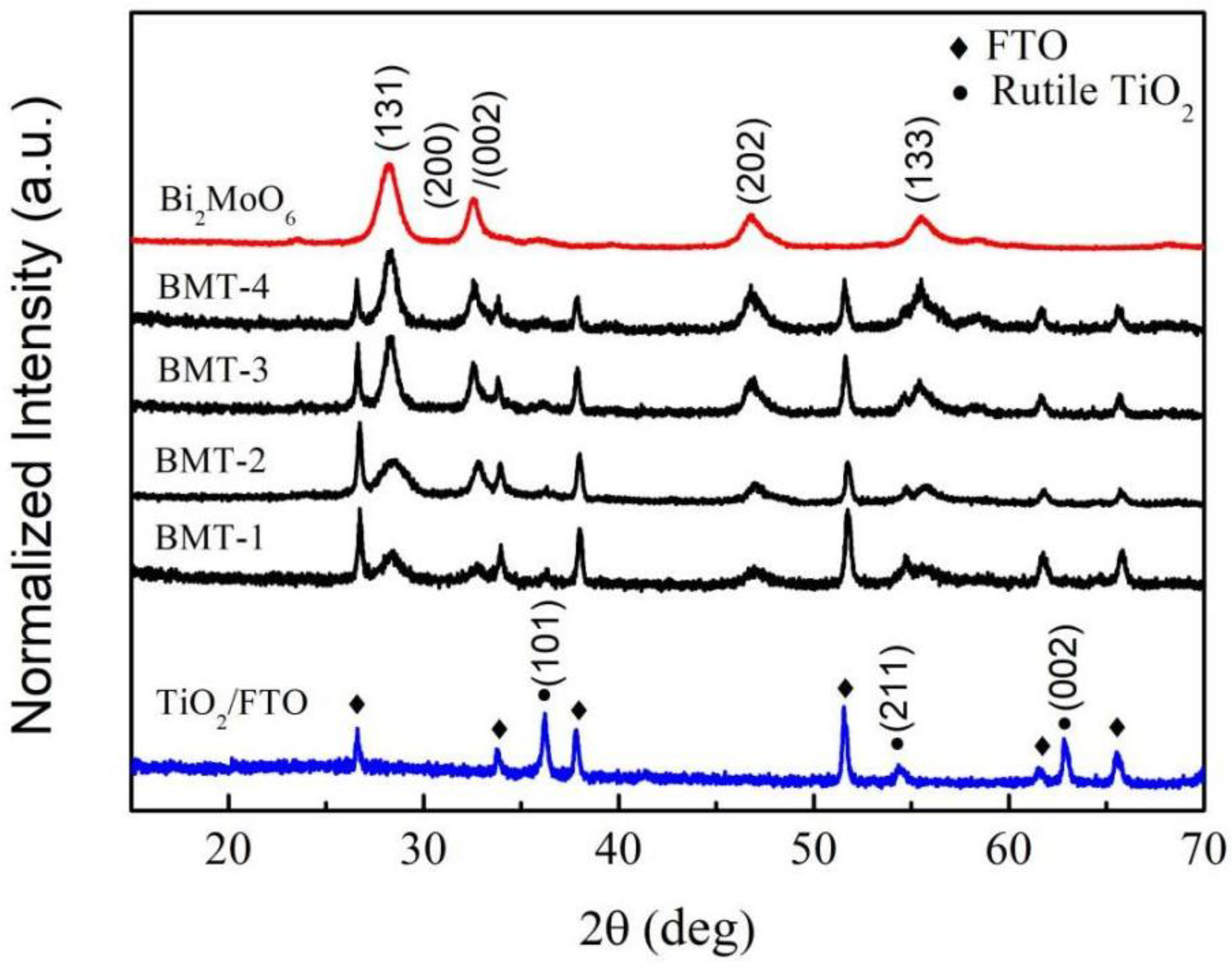
3.1. X-ray Diffraction (XRD)

As shown in Figure 1, synthesized TiO2, Bi2MoO6, and Bi2MoO6/TiO2 composite films possessed good crystallinity according to the XRD results. The XRD pattern revealed diffraction peaks at 36.2°, 54.3°, and 62.8°, attributed to (101), (211), and (002) crystal planes of rutile TiO2, respectively (PDF card number 21-1276). The XRD patterns of Bi2MoO6/TiO2 revealed TiO2 and FTO peaks, as well as peaks at 28.2°, 32.5°, 46.7°, and 55.5°, that were attributed to (131), (200)/(002), (202), and (133) planes of the orthorhombic koechlinite phase of Bi2MoO6, according to PDF card number 76-2388. These results confirmed the formation of Bi2MoO6/TiO2 composites. XRD peaks of Bi2MoO6 observed in the Bi2MoO6/TiO2 spectra became sharper and narrower as the initial Bi2MoO6 amount was increased, while the XRD peaks of TiO2 became less intense. No other changes in the crystalloid structure were observed as the initial materials used were changed.

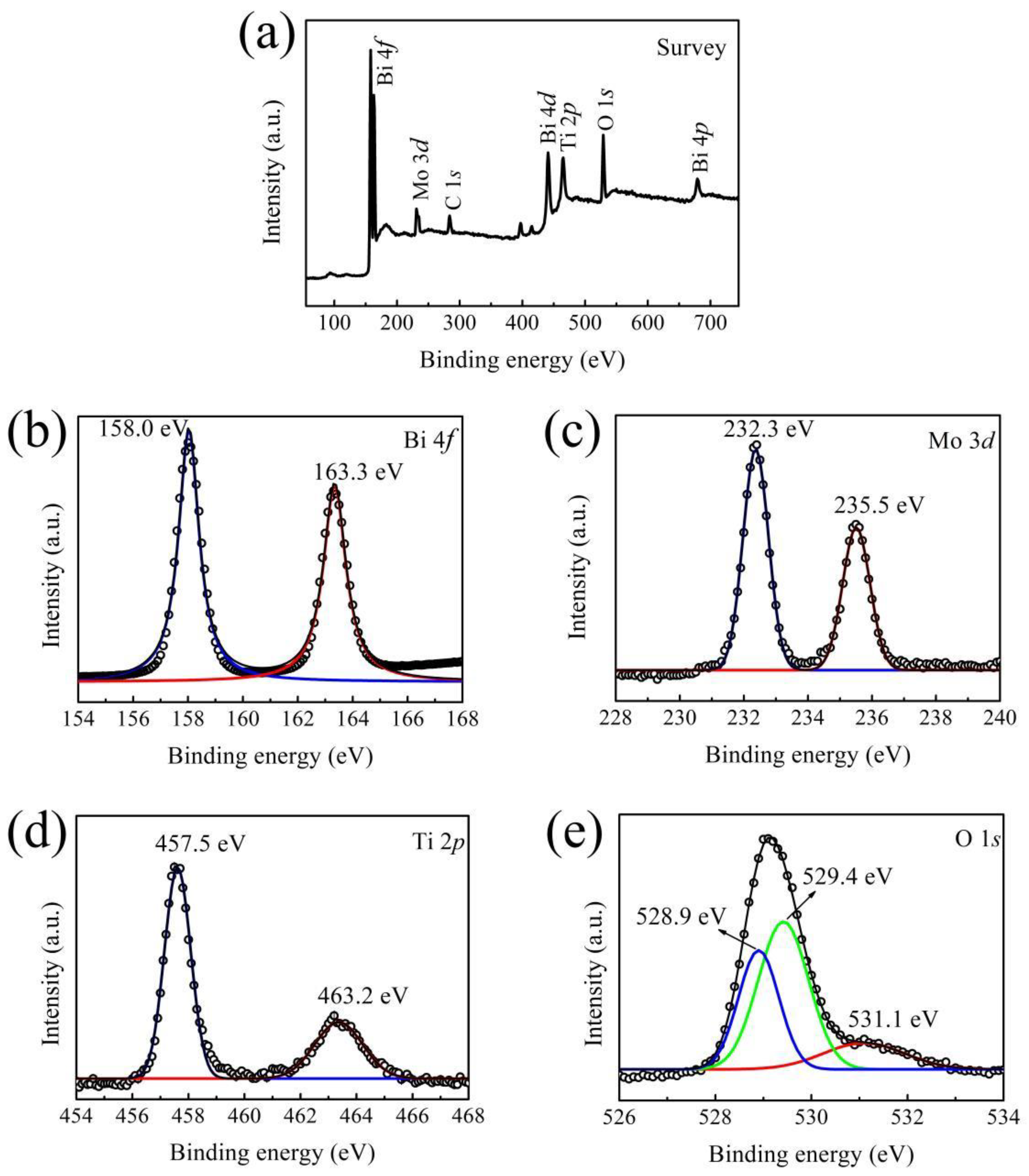
3.2. X-ray Photoelectron Spectroscopy (XPS)
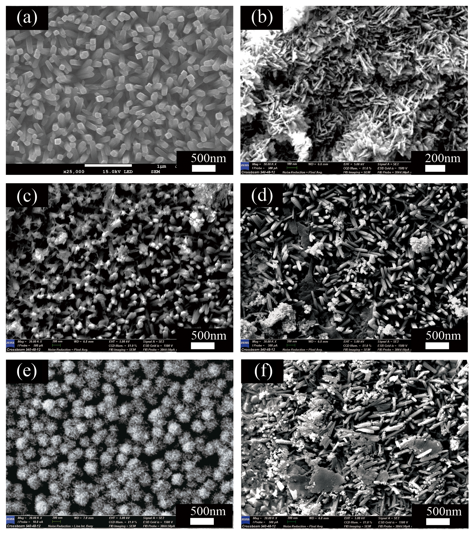
XPS of the BMT-3 composites revealed the presence of Bi, Mo, Ti, and O elements (see Figure 2a). Some carbon was also observed due to environmental and instrument contamination. The high-resolution Bi 4f XPS spectrum showed two peaks at 158.0 and 163.3 eV (see Figure 2b), which were assigned to the spin-orbit splitting peaks of Bi 4f7/2 and Bi 4f5/2, respectively [38]. Thus, Bi in Bi2MoO6/TiO2 existed as Bi3+. Two strong peaks at 232.3 and 235.5 eV, corresponding to Mo 3d5/2 and Mo 3d3/2 spin-orbit components of Mo6+, respectively [39] (see Figure 2c), were observed in the high-resolution Mo 3d XPS spectrum. The peaks at 457.5 and 463.2 eV matched the binding energies of Ti 2p3/2 and Ti 2p1/2 states, respectively (see Figure 2d). Thus, Ti was present as Ti4+ in the Bi2MoO6/TiO2 composites. The fitting of the high-resolution O 1s XPS spectrum revealed three peaks at 528.9, 529.4, and 531.1 eV (see Figure 2e), which corresponded to Bi-O, Mo-O, and Ti-O bonds, respectively [22,38]. Thus, these results confirmed the coexistence of Bi2MoO6 and TiO2 in the synthesized HSs.

3.3. Morphologies

SEM showed that the diameters of the bare TiO2 nanorods were 200 nm (see Figure 3a). Moreover, the nanorods were vertically aligned and uniformly distributed on the FTO substrate. Pure Bi2MoO6 nanosheets exhibited laminar and irregular, sheet-like morphology (see Figure 3b). SEM of the Bi2MoO6/TiO2 composites showed that some Bi2MoO6 nanosheets adhered to the TiO2 nanorods (see Figure 3c–f). Additionally, as the initial Bi/Mo content was increased, the Bi2MoO6 nanosheets increased in number and began to aggregate. The sample (BMT-3, shown in Figure 3e) with appropriate initial Bi/Mo content showed the growth of dispersed Bi2MoO6 nanosheets around TiO2 nanorods. Instead, a serious agglomeration of Bi2MoO6 was observed, as shown in Figure 3f. These findings suggest that the initial concentration of the materials used significantly affected the Bi2MoO6/TiO2 composite morphology.
It is widely known that the photocatalytic activity of catalysts is significantly influenced by the specific surface area and pore volume of photocatalysts due to the presence of surface-reactive sites [40]. The nitrogen adsorption−desorption isotherms of the TiO2, Bi2MoO6, BMT-3, and BMT-4 samples are shown in Figure S2 in the Supplementary Materials. The similar adsorption−desorption curves revealed strong N2 adsorption−desorption performance, which indicated the existence of capillary condensation in the large mesopores in the samples. The SBET and pore volume of TiO2, Bi2MoO6, BMT-3, and BMT-4 HSs are listed in Table S3. Interestingly, the coupling of TiO2 and Bi2MoO6 significantly increased the SBET and pore volume (the maximum value for BMT-3 are 88.2 m2/g and 0.18 cm3/g, respectively) compared to the pure Bi2MoO6 (26.0 m2/g and 0.05 cm3/g, respectively). Combined with the photocatalytic experiment results we can deduce that the high SBET and pore volume of BMT-3 played significant roles in enhancing photocatalytic performance.

3.4. Optical Properties

The UV–Vis spectra of TiO2 nanorod arrays showed that they were adsorbed, as expected, in the ultraviolet region at 400 nm (see Figure 4a), which is mainly accounted for by absorption within the rutile band-gap [41]. The absorption intensity of Bi2MoO6 was relatively mild and peaked at ~450 nm. Thus, a slight extension to the visible-light region was observed in comparison to TiO2. The absorption edges of the Bi2MoO6/TiO2 composites exhibited a certain degree of red-shift, which indicates the extension of the material’s absorption towards visible light. The band-gap energies of the TiO2, Bi2MoO6, and Bi2MoO6/TiO2 HSs were calculated from the Kubelka–Munk function, plotted against the photon energy (see Figure 4b). The band-gap values of TiO2 and Bi2MoO6 were equal to 3.1 and 2.77 eV, respectively. Moreover, the band-gap values of the BMT-1, BMT-2, BMT-3, and BMT-4 composites were 2.88, 2.85, 2.81, and 2.79 eV, respectively. Furthermore, the photogenerated electron-hole pairs were excited due to the extension of absorption into the visible-light region, favoring enhanced PEC and photocatalytic performance.
Photoluminescence (PL) emission spectra have been widely employed to reveal the separation efficiency of the photo-induced electrons and holes in the composite semiconductors [42]. The PL spectra of the as-prepared TiO2, BMT-3, and BMT-4 samples in the present study are shown in Figure 4c. The strongest emission intensity peak observed in pure TiO2 nanorods corresponded to the band-gap transition. A significant decrease in fluorescence of Bi2MoO6/TiO2 (BMT-3 and BMT-4) was observed, indicating a lower photoelectron-hole recombination in Bi2MoO6/TiO2 than in TiO2. Moreover, the similar fluorescence intensity of BMT-3 and BMT-4 indicated similar separation performance of the photo-induced electrons and holes in the BMT-3 and BMT-4 samples. The migration and recombination efficiencies of the photo-induced carriers in all samples were further revealed by EIS characterization.

3.5. Photocatalytic Properties
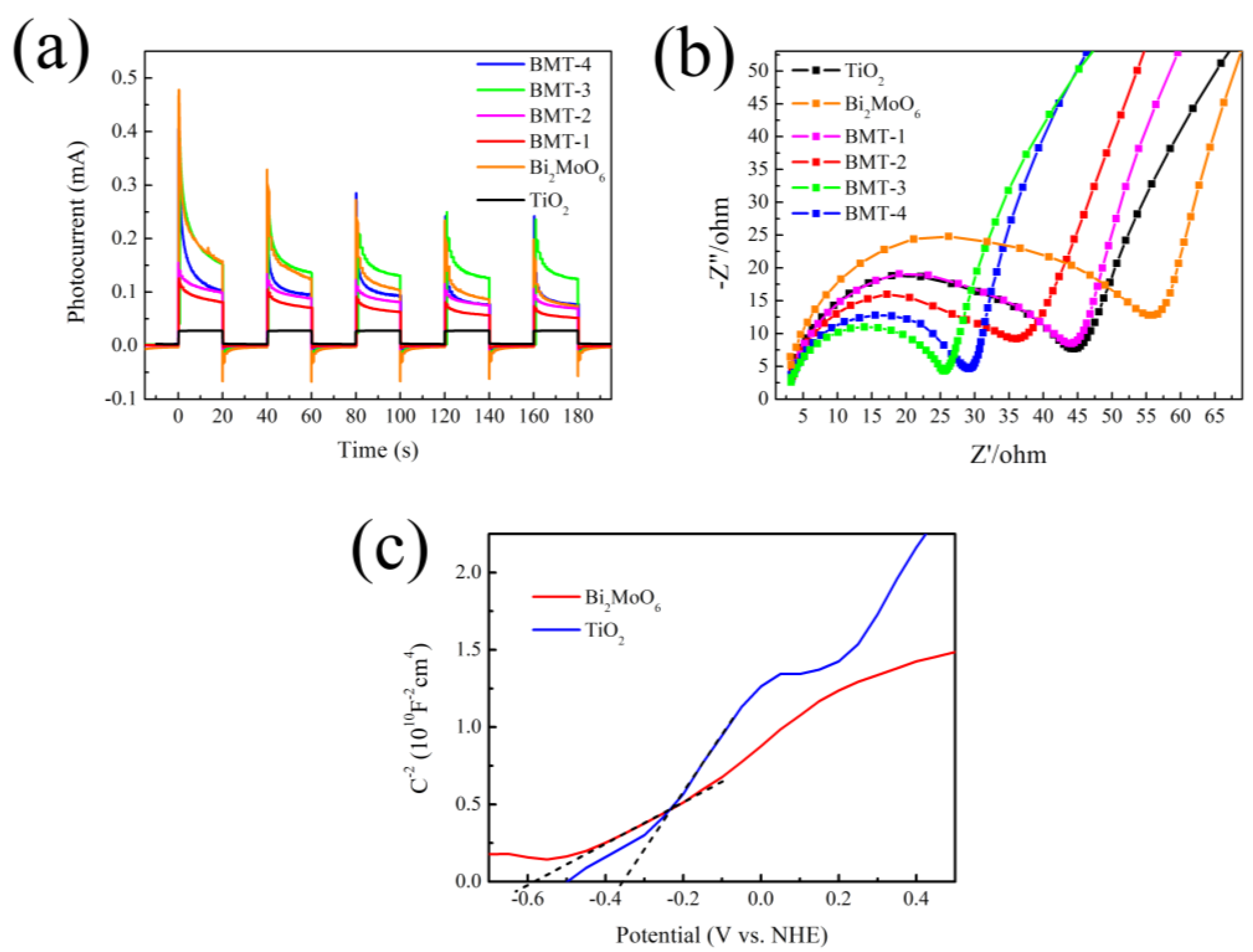
The relationship between residual MB contents and irradiation time, with and without photocatalyst, is shown in Figure 5a. Figure 5b shows the fitting curves of the kinetics of MB photodegradation. The degradation of MB in the presence of TiO2 and Bi2MoO6/TiO2 obeyed the pseudo-first-order kinetics and could be expressed as ln(C/C0) = k(tt0), where k is the reaction rate constant [43,44]. The rate constants and the corresponding correlation coefficients (R-Square) of different photocatalysts and the blank sample are given in Table S4, which were calculated by linear fitting −ln(C/C0) to irradiation time (t). The values of R-Square are close to 1, which reveal a good correlation to the pseudo-first-order reaction kinetics. MB degradation without catalysts was negligible under visible light, with a k value of 0.000538 min−1 (see Figure 5b). In contrast, the reaction rate constant of MB decomposition in the presence of TiO2 nanorods was 0.00113 min−1, which was attributed to the photon-trapping effect caused by the morphology of the TiO2 nanostructure [13,14]. However, the photocatalytic performances of the Bi2MoO6/TiO2 HSs were significantly better. Thus, the presence of Bi2MoO6 in the composite played a significant role. The photocatalytic ability of Bi2MoO6/TiO2 (BMT-4) was lower for samples with higher Bi2MoO6 content because of the recombination of the photogenerated electron-hole pairs inside the numerous Bi2MoO6 nanosheets [44]. From the viewpoint of practical application, it is important to evaluate the stability of the as-prepared catalyst. Comparison of the XRD pattern of the Bi2MoO6/TiO2 (BMT-3) before and after the photocatalytic reaction (performed three times with the same catalyst) showed that, even after three cycles, the positions and intensities of the XRD peaks remained almost the same (Figure 5c), which confirmed the excellent stability of the Bi2MoO6/TiO2 composite catalysts. The stable activity was further validated by repeating the photocatalytic degradation processes thrice, as shown in Figure 5d. The three degradation curves showed a similar trend in each running cycle, which indicated that the Bi2MoO6/TiO2 photocatalyst exhibited high and stable activity for degradation.
To investigate the mechanisms underlying the photo-oxidation ability, ESR technology was used to detect active free radicals. The ESR signals of BMT-3 dispersed in the DMPO solution are shown in Figure S3. No ·OH ESR signals were generated in darkness; however, a set of four feature peaks with an intensity ratio of 1:2:2:1 were observed after light illumination (see Figure S3a), attributed to DMPO-·OH adducts. Similarly, no ·O2 signals were generated in the dark, and six peaks of DMPO-·O2 were observed in the ESR spectra (see Figure S3b) with illumination. These results demonstrated that ·O2 and ·OH are both active species, and their synergistic effect significantly promoted the photocatalytic degradation of MB.

3.6. PEC Analysis

The PEC performance of the Bi2MoO6/TiO2 HSs was compared to that of pure TiO2 nanorod arrays and Bi2MoO6. For this purpose, we recorded the photocurrent as a function of time (It curves) by alternating exposure to darkness and visible light (see Figure 6a). All samples exhibited similar photocurrent responses. No photocurrent was observed in the dark, which confirmed the absence of any electrochemical processes. A minimal photocurrent response was observed for the TiO2 nanorod array, and a significant one for the Bi2MoO6/TiO2 HSs. The BMT-3 sample exhibited optimal stability and reproducibility of the photocurrent response since the Bi2MoO6/TiO2 HSs, and the amount of Bi2MoO6 was the most favorable out of all the BMT samples. The separation and collection efficiency of interface charges were further studied by EIS (see Figure 6b). Typically, the semicircles in the corresponding Nyquist plots corresponded to Faradic reactions. It has been established that the semicircle radius is negatively correlated with the charge transfer efficiency [45,46]. In our study, the semicircle diameters for the Bi2MoO6/TiO2 HSs were significantly smaller than those obtained for Bi2MoO6 and TiO2. This data confirms the lower interfacial charge-transfer resistance and fast charge-transfer process of our composite Bi2MoO6/TiO2 HSs due to the presence of Bi2MoO6 and its interfacial interaction with TiO2, which enhanced the separation and transfer efficiency of the electron-hole pairs photogenerated in the Bi2MoO6/TiO2 HSs. The heterojunction of the Bi2MoO6/TiO2 interface suppressed the charge recombination, thereby producing more (re)active species, which resulted in a high photocurrent response and photocatalytic activity.
The Mott–Schottky plots of TiO2 and Bi2MoO6 are shown in Figure 6c. The positive slopes of both compounds imply that they are n-type semiconductors. The flat-band potential (Vfb) can be calculated by the Mott–Schottky equation [42]:
1 C 2 = 2 e ε ε 0 N d [ ( V V fb ) k T e ]
where C is the capacitance at the interface with the electrolyte, e is the electronic charge, ε 0 is the vacuum permittivity, ε is the sample dielectric constant, Nd is the charge-carrier concentration, V and Vfb are the applied and flat-band potentials, k is the Boltzmann’s constant, and T is the temperature [47]. The Vfb could be calculated from the intercept of the 1/C2 curve (plotted as a V function) with the x-axis [48]. The Vfb values for the pure TiO2 and Bi2MoO6 were equal to −0.35 and −0.58 V (vs. a normal hydrogen electrode, NHE), respectively.

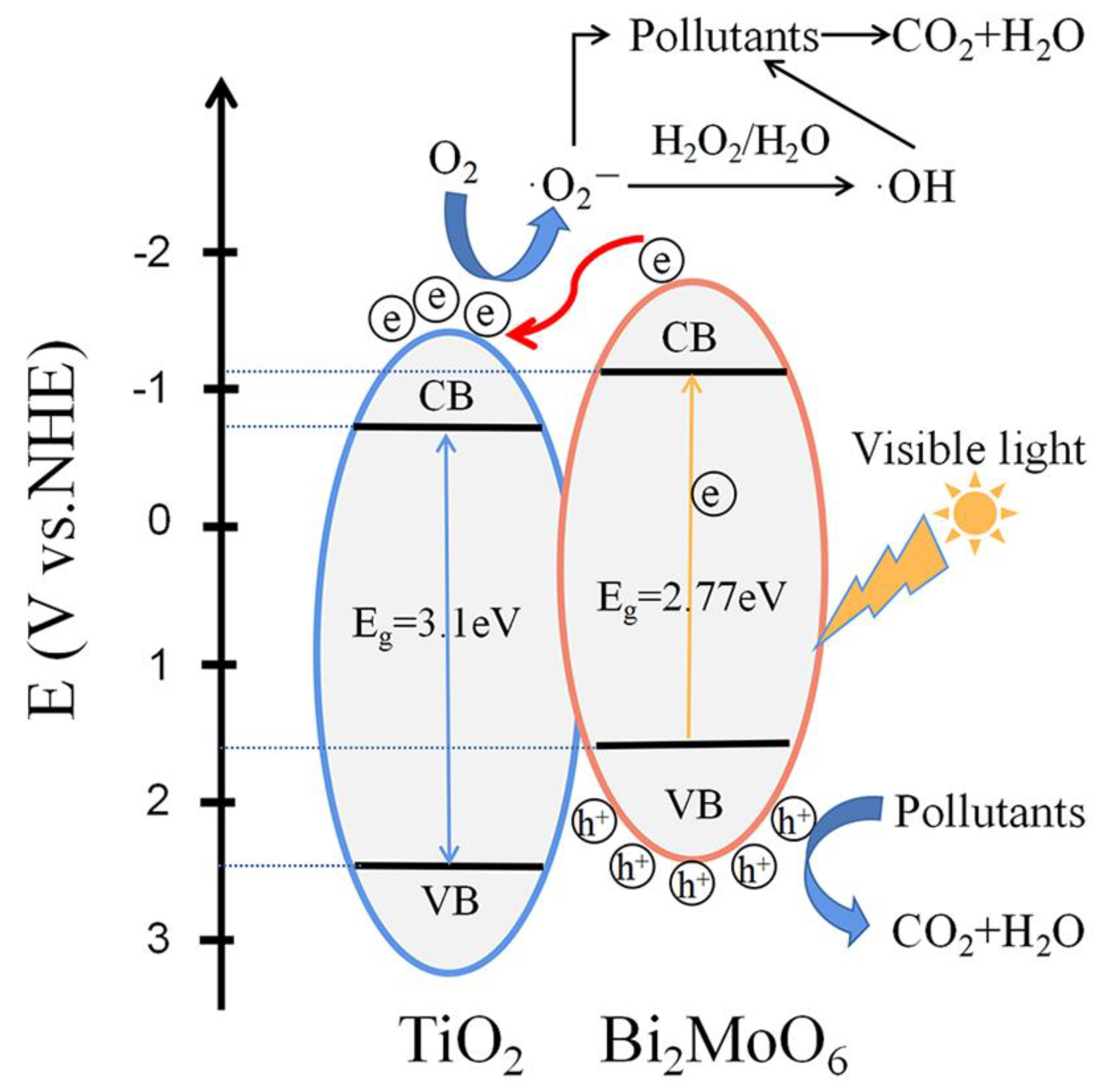
3.7. Energy Band Alignment and Photocatalytic Mechanism

In order to explain the photocatalytic process, the energy band alignment of the Bi2MoO6/TiO2 HSs was investigated. Mott–Schottky plots revealed the flat-band potential of TiO2 and Bi2MoO6 (Figure 6c). A gap between EVB and flat-band potential can be inferred from the XPS–valance band (XPS–VB) plots, as shown in Figure S4. Thus, the calculated EVB values of TiO2 and Bi2MoO6 were 2.4 and 1.68 V, respectively. Moreover, the conduction band potential (ECB) of TiO2 and Bi2MoO6 were calculated to be −0.7 and −1.09 V, respectively. Based on these results, the band alignment of the Bi2MoO6/TiO2 was established, as shown in Figure 7. The ECB and EVB of TiO2 were more positive than those of Bi2MoO6. Accordingly, it is highly likely that Bi2MoO6/TiO2 HSs possess staggered band alignment. Visible-light irradiation excites Bi2MoO6 molecules, generating electron-hole pairs. At the same time, a large band-gap prevents TiO2 molecules from being excited by visible-light irradiation. In this case, the electrons travel from the conduction band of the Bi2MoO6 to TiO2, which suppresses electron-hole pair recombination by the internal field of the Bi2MoO6/TiO2 heterojunction since the ECB of Bi2MoO6 is relatively more negative. The separated electrons can react with O2 molecules, forming ·O2 radicals since the corresponding redox potential is equal to −0.046 V and more positive than the ECB of TiO2. Subsequently, the H2O molecules are transformed into ·OH radicals after trapping an electron. Both photogenerated holes, together with ·O2 and ·OH radicals, can react with MB molecules, damaging their structures. We believe that this process, based on the interfacial charge transfer, is indeed feasible. The reaction mechanism and chemical equations of the above processes are proposed as follows and can be found in the previous literature [23,38,49,50].
Bi 2 MoO 6 + h ν Bi 2 MoO 6   ( e + h + )
Bi 2 MoO 6   ( e + h + ) + TiO 2 Bi 2 MoO 6   ( h + ) + TiO 2   ( e )
TiO 2   ( e ) + O 2 TiO 2 + · O 2
· O 2 + H 2 O · HO 2 + OH
· HO 2 + H 2 O H 2 O 2 + · OH
H 2 O 2 2 · OH
Bi 2 MoO 6   ( h + ) + MB degraded   products
· O 2 + MB degraded   products
· OH + MB degraded   products

4. Conclusions

In summary, the Bi2MoO6/TiO2 HSs were synthesized through a simple two-step solvothermal process by growing Bi2MoO6 nanosheets on TiO2 nanorod arrays. The Bi2MoO6/TiO2 HSs exhibited enhanced photocatalytic activity for MB degradation under visible-light irradiation, and BMT-3 achieved the highest degradation rate of k = 0.015 min−1 among all samples. The enhancement was attributed to the heterojunction structures established by the close contact between the Bi2MoO6 nanosheets and the TiO2 nanorods. The results of the UV–Vis absorption spectra show an extended absorption to visible light after coupling Bi2MoO6 with TiO2. The results of N2 adsorption−desorption isotherms show a significantly increased SBET and pore volume compared to the pure Bi2MoO6 due to the hierarchical structure of the Bi2MoO6/TiO2 HSs. The results of PL and PEC characterization of the Bi2MoO6/TiO2 HSs reveal improved separation efficiency and enhanced migration rate of photogenerated electron-hole pairs. Thus, the synergy between Bi2MoO6 and TiO2 was identified as the crucial factor leading to the improved photocatalytic performance. Furthermore, the reusability and chemical stability of Bi2MoO6/TiO2 HSs is demonstrated by photocatalytic cycle test. Finally, the mechanism and process of the dye photodegradation were discussed. The h+, ·O2, and ·OH radicals were validated as being active species that react with MB dye molecules. The results of this work suggest that Bi2MoO6/TiO2 HSs are promising candidate materials for wastewater treatment. Collectively, our current strategy can help in the synthesis and photocatalytic application of other heterostructures in the future.

Supplementary Materials

The following supporting information can be downloaded at: https://www.mdpi.com/article/10.3390/nano12030574/s1. Table S1: Bi2MoO6-based photocatalysts and their photocatalytic performance; Table S2: The abbreviations of products with different amounts of Bi2MoO6 by varying the mass of raw materials in precursor solution; Table S3: SBET, Pore Volume, and Mean Pore Diameter of TiO2, Bi2MoO6, BMT-3, and BMT-4 samples; Table S4: Pseudo-first-order rate constants and corresponding R-Square values of different samples; Figure S1: Stepwise synthesis protocol of TiO2 nanorod arrays and Bi2MoO6/TiO2 HSs; Figure S2: N2 adsorption−desorption isotherms (a) and the corresponding pore-size distribution curves (b) of TiO2, Bi2MoO6, BMT-3, and BMT-4 samples; Figure S3: The ESR signals of ·OH (a) and ·O2 radicals (b) of BMT-3 photocatalysts; Figure S4: XPS valence-band spectra of TiO2 and Bi2MoO6.

Author Contributions

Conceptualization, D.Z. and Z.H.; Data curation, Y.T., Y.F., and Y.Z.; Formal analysis, Y.T., Y.F., and Y.Z.; Funding acquisition, G.Z.; Investigation, R.D. and S.G.; Methodology, R.D.; Resources, G.Z.; Supervision, D.Z.; Visualization, S.G.; Writing–original draft, D.Z.; Writing–review and editing, D.Z. and Z.H. All authors have read and agreed to the published version of the manuscript.

Funding

This work is supported by the National Natural Science Foundation of China (Grant Numbers: 61575085 and 61240056) and guidance project of the science and technology research of the Education Department of Hubei Province (Grant Number: B2019235).

Data Availability Statement

All data generated or analyzed during this study are contained within the article and supplementary materials.

Conflicts of Interest

The authors declare no conflict of interest.

References

  1. Fujishima, A.; Honda, K. Electrochemical photolysis of water at a semiconductor electrode. Nature 1972, 238, 37–38. [Google Scholar] [CrossRef] [PubMed]
  2. Schrauzer, G.N.; Guth, T.D. Photolysis of water and photoreduction of nitrogen on titanium dioxide. J. Am. Chem. Soc. 1977, 99, 7189–7193. [Google Scholar] [CrossRef]
  3. Kraeutler, B.; Bard, A.J. Heterogeneous photocatalytic preparation of supported catalysts. Photodeposition of platinum on titanium dioxide powder and other substrates. J. Am. Chem. Soc. 1978, 100, 4317–4318. [Google Scholar] [CrossRef]
  4. Fujishima, A.; Hashimoto, K.; Watanabe, T. TiO2 Photocatalysis: Fundamentals and Applications; BKC, Inc.: Tokyo, Japan, 1999. [Google Scholar]
  5. Tang, H.; Prasad, K.; Sanjinbs, R.; Schmid, P.E.; Levy, F. Electrical and optical properties of TiO2 anatase thin films. J. Appl. Phys. 1994, 75, 2042–2047. [Google Scholar] [CrossRef]
  6. Yadav, H.M.; Kim, J.S.; Pawar, S.H. Developments in photocatalytic antibacterial activity of nano TiO2: A review. Korean J. Chem. Eng. 2016, 33, 1989–1998. [Google Scholar] [CrossRef]
  7. Sahare, S.; Veldurthi, N.; Singh, R.; Swarnkar, A.K.; Bhave, T. Enhancing the efficiency of flexible dye-sensitized solar cells utilizing natural dye extracted from Azadirachta indica. Mater. Res. Express 2015, 2, 105903. [Google Scholar] [CrossRef]
  8. Grabowska, E.; Reszczyńska, J.; Zaleska, A. Mechanism of phenol photodegradation in the presence of pure and modified-TiO2: A review. Water Res. 2012, 46, 5453–5471. [Google Scholar] [CrossRef]
  9. Mohamed, M.A.; Jaafar, J.; Zain, M.F.M.; Minggu, L.J.; Kassim, M.B.; Salehmin, M.N.I.; Rosmie, M.S.; Salleh, W.N.W.; Othman, M.H.D. Concurrent growth, structural and photocatalytic properties of hybridized C, N co-doped TiO2 mixed phase over g-C3N4 nanostructured. Scr. Mater. 2018, 142, 143–147. [Google Scholar] [CrossRef]
  10. Saber, N.B.; Mezni, A.; Alrooqi, A.; Altalhi, T. Fabrication of efficient Au@TiO2/rGO heterojunction nanocomposite: Boosted photocatalytic activity under ultraviolet and visible light irradiation. J. Mater. Res. Technol. 2021, 12, 2238–2246. [Google Scholar] [CrossRef]
  11. Ismael, M. A review and recent advances in solar-hydrogen energy conversion based on photocatalytic water splitting over doped-TiO2 nanoparticles. Sol. Energy 2020, 211, 522–546. [Google Scholar] [CrossRef]
  12. Pham, V.V.; Phat, B.D.; Huy, T.H.; Cao, M.T.; Le, V.H. Photoreduction route for Cu2O/TiO2 nanotubes junction for enhanced photocatalytic activity. RSC Adv. 2018, 8, 12420–12427. [Google Scholar] [CrossRef] [Green Version]
  13. Fu, Y.; Mao, Z.P.; Zhou, D.; Hu, Z.L.; Tu, Y.F.; Tian, Y.; Zheng, G. Fabrication of Ni-doped PbTiO3-coated TiO2 nanorod arrays for improved photoelectrochemical performance. J. Nanomater. 2019, 11, 2019. [Google Scholar] [CrossRef]
  14. Fu, Y.; Mao, Z.P.; Zhou, D.; Hu, Z.L.; Zheng, G. Preparation of BiFeO3-overcoated TiO2 nanorod arrays for the enhanced visible-light activity. Mater. Res. Express 2019, 6, 1050c6. [Google Scholar] [CrossRef]
  15. Liu, X.; Gu, S.; Zhao, Y.; Zhou, G.; Li, W. BiVO4, Bi2WO6 and Bi2MoO6 photocatalysis: A brief review. J. Mater. Sci. Technol. 2020, 56, 45–68. [Google Scholar] [CrossRef]
  16. Chai, S.Y.; Kim, Y.J.; Jung, M.H.; Chakraborty, A.K.; Jung, D.; Lee, W.I. Heterojunctioned BiOCl/Bi2O3, a new visible light photocatalyst. J. Catal. 2009, 262, 144–149. [Google Scholar] [CrossRef]
  17. Xiang, Z.; Wang, Y.; Zhang, D.; Ju, P. BiOI/BiVO4 p-n heterojunction with enhanced photocatalytic activity under visible-light irradiation. J. Ind. Eng. Chem. 2016, 40, 83–92. [Google Scholar] [CrossRef]
  18. Wei, W.; Dai, Y.; Huang, B. First-principles characterization of Bi-based photocatalysts: Bi12TiO20, Bi2Ti2O7, and Bi4Ti3O12. J. Phys. Chem. C 2009, 113, 5658–5663. [Google Scholar] [CrossRef]
  19. Yu, H.; Jiang, L.; Wang, H.; Huang, B.; Zeng, G. Photocatalysis: Modulation of Bi2MoO6-based materials for photocatalytic water splitting and environmental application: A critical review. Small 2019, 15, 1901008. [Google Scholar] [CrossRef] [PubMed]
  20. Peng, Y.; Liu, Q.; Zhang, J.; Zhang, Y.; Geng, M.; Yu, J. Enhanced visible-light-driven photocatalytic activity by 0D/2D phase heterojunction of quantum dots/nanosheets on bismuth molybdates. J. Phys. Chem. C. 2018, 122, 3738–3747. [Google Scholar] [CrossRef]
  21. Tian, G.; Chen, Y.; Zhai, R.; Zhou, J.; Zhou, W.; Wang, R.; Pan, K.; Tiana, C.; Fu, H. Hierarchical flake-like Bi2MoO6/TiO2 bilayer films for visible-light-induced self-cleaning applications. J. Mater. Chem. A 2013, 1, 6961–6968. [Google Scholar] [CrossRef]
  22. Li, J.; Liu, X.; Sun, Z.; Pan, L. Novel Bi2MoO6/TiO2 heterostructure microspheres for degradation of benzene series compound under visible light irradiation. J. Colloid Interface Sci. 2016, 463, 145–153. [Google Scholar] [CrossRef] [PubMed]
  23. Zhang, M.; Shao, C.; Mu, J.; Zhang, Z.; Guo, Z.; Zhang, P.; Liu, Y. One-dimensional Bi2MoO6/TiO2 hierarchical heterostructures with enhanced photocatalytic activity. CrystEngComm 2012, 14, 605–612. [Google Scholar] [CrossRef]
  24. Li, L.; Salvador, P.A.; Rohrer, G.S. Photocatalysts with internal electric fields. Nanoscale 2014, 6, 24–42. [Google Scholar] [CrossRef] [PubMed] [Green Version]
  25. Sharma, S.; Basu, S. Highly reusable visible light active hierarchical porous WO3/SiO2 monolith in centimeter length scale for enhanced photocatalytic degradation of toxic pollutants. Sep. Purif. Technol. 2020, 231, 115916. [Google Scholar] [CrossRef]
  26. Lindgren, T.; Wang, H.; Beermann, N.; Vayssieres, L.; Hagfeldt, A.; Lindquist, S.-E. Aqueous photoelectrochemistry of hematite nanorod array. Sol. Energy Mater. Sol. Cells 2002, 71, 231–243. [Google Scholar] [CrossRef]
  27. Zhang, D.S.; Downing, J.A.; Knorr, F.J.; McHale, J.L. Nanostructured semiconductor composites for solar cells. J. Chem. B 2006, 110, 32–36. [Google Scholar]
  28. Ge, M.; Cao, C.; Huang, J.; Li, S.; Lai, Y. A review of one-dimensional TiO2 nanostructured materials for environmental and energy applications. J. Mater. Chem. A 2016, 4, 6772–6801. [Google Scholar] [CrossRef]
  29. Shuang, S.; Zheng, X.; Zhang, Z. Review: Nanostructured TiO2 for enhanced photocatalytic property by glancing angle deposition method. J. Harbin Inst. Technol. 2017, 24, 1–11. [Google Scholar]
  30. Cheng, X.; Zhang, Y.; Bi, Y. Spatial dual-electric fields for highly enhanced the solar water splitting of TiO2 nanotube arrays. Nano Energy 2019, 57, 542–548. [Google Scholar] [CrossRef]
  31. Nazim, M.; Khan, A.A.P.; Asiri, A.M.; Kim, J.H. Exploring rapid photocatalytic degradation of organic pollutants with porous CuO nanosheets: Synthesis, dye removal, and kinetic studies at room temperature. ACS Omega 2021, 6, 2601–2612. [Google Scholar] [CrossRef]
  32. Cani, D.; Waal, J.C.; Pescarmona, P.P. Highly-accessible, doped TiO2 nanoparticles embedded at the surface of SiO2 as photocatalysts for the degradation of pollutants under visible and UV radiation. Appl. Catal. A Gen. 2021, 621, 118179. [Google Scholar] [CrossRef]
  33. D’Angelo, D.; Filice, S.; Scarangella, A.; Iannazzo, D.; Compagnini, G.; Scalese, S. Bi2O3/Nexar® polymer nanocomposite membranes for azo dyes removal by UV–vis or visible light irradiation. Catal. Today 2019, 321–322, 158–163. [Google Scholar] [CrossRef]
  34. Li, Z.Q.; Chen, X.T.; Xue, Z.L. Bi2MoO6 microstructures: Controllable synthesis, growth mechanism, and visible-light-driven photocatalytic activities. CrystEngComm 2013, 15, 498–508. [Google Scholar] [CrossRef]
  35. Zhang, L.; Xu, T.; Zhao, X.; Zhu, Y. Controllable synthesis of Bi2MoO6 and effect of morphology and variation in local structure on photocatalytic activities. Appl. Catal. B Environ. 2010, 98, 138–146. [Google Scholar] [CrossRef]
  36. Li, N.; Gao, H.; Wang, X.; Zhao, S.; Da, L.; Yang, G.; Gao, X.; Fan, H.; Gao, Y.; Ge, L. Novel indirect Z-scheme g-C3N4/Bi2MoO6/Bi hollow microsphere heterojunctions with SPR-promoted visible absorption and highly enhanced photocatalytic performance. Chin. J. Catal. 2020, 41, 426–434. [Google Scholar] [CrossRef]
  37. Mia, M.S.; Yao, P.; Zhu, X.; Lei, X.; Chen, G. Degradation of textile dyes from aqueous solution using tea-polyphenol/Fe loaded waste silk fabrics as Fenton-like catalysts. RSC Adv. 2021, 11, 8290–8305. [Google Scholar] [CrossRef]
  38. Li, H.; Zhang, T.; Pan, C.; Pu, C.; Hu, Y.; Hu, X.; Liu, E.; Fan, J. Self-assembled Bi2MoO6/TiO2 nanofiber heterojunction film with enhanced photocatalytic activities. Appl. Surf. Sci. 2017, 391, 303–310. [Google Scholar] [CrossRef]
  39. Cai, K.; Lv, S.; Song, L.; Chen, L.; He, J.; Chen, P.; Au, C.; Yin, S. Facile preparation of ultrathin Bi2MoO6 nanosheets for photocatalytic oxidation of toluene to benzaldehyde under visible light irradiation. J. Solid State Chem. 2019, 269, 145–150. [Google Scholar] [CrossRef]
  40. Yunarti, R.T.; Safitri, T.N.; Dimonti, L.C.C.; Aulia, G.; Khalil, M.; Ridwan, M. Facile synthesis of composite between titania nanoparticles with highly exposed (001) facet and coconut shell-derived graphene oxide for photodegradation of methylene blue. J. Phys. Chem. Solids 2022, 160, 110357. [Google Scholar] [CrossRef]
  41. Dinh, C.T.; Yen, H.; Kleitz, F.; Do, T.O. Three-dimensional ordered assembly of thin-shell Au/TiO2 hollow nanospheres for enhanced visible-light-driven photocatalysis. Angew. Chem. Int. Ed. Engl. 2014, 53, 6618–6623. [Google Scholar] [CrossRef]
  42. Li, Q.; Li, L.; Long, X.; Tu, Y.; Ling, L.; Gu, J.; Hou, L.; Xu, Y.; Liu, N.; Li, Z. Rational design of MIL-88A(Fe)/Bi2WO6 heterojunctions as an efficient photocatalyst for organic pollutant degradation under visible light irradiation. Opt. Mater. 2021, 118, 111260. [Google Scholar] [CrossRef]
  43. Ali, H.; Guler, A.C.; Masar, M.; Urbanek, P.; Urbanek, M.; Skoda, D.; Suly, P.; Machovsky, M.; Galusek, D.; Kuritka, I. Solid-state synthesis of direct Z-scheme Cu2O/WO3 nanocomposites with enhanced visible-light photocatalytic performance. Catalysts 2021, 11, 293. [Google Scholar] [CrossRef]
  44. Tian, J.; Hao, P.; Wei, N.; Cui, H.; Liu, H. 3D Bi2MoO6 nanosheet/TiO2 nanobelt heterostructure: Enhanced photocatalytic activities and photoelectochemistry performance. ACS Catal. 2015, 5, 4530–4536. [Google Scholar] [CrossRef]
  45. Yus, J.; Ferrari, B.; Sanchez-Herencia, A.J.; Gonzalez, Z. Understanding the effects of different microstructural contributions in the electrochemical response of Nickel-based semiconductor electrodes with 3D hierarchical networks shapes. Electrochim. Acta 2020, 335, 135629. [Google Scholar] [CrossRef]
  46. Zhao, K.; Zhao, S.; Gao, C.; Qi, J.; Yin, H.; Wei, D.; Mideksa, M.F.; Wang, X.; Gao, Y.; Tang, Z.; et al. Metallic Cobalt–Carbon Composite as Recyclable and Robust Magnetic Photocatalyst for Efficient CO2 Reduction. Small 2018, 14, 1800762. [Google Scholar] [CrossRef]
  47. Singh, S.; Sangle, A.L.; Wu, T.; Khare, N.; Macmanus-Driscoll, J.L. Growth of doped SrTiO3 ferroelectric nanoporous thin films and tuning of photoelectrochemical properties with switchable ferroelectric polarization. ACS Appl. Mater. Interfaces 2019, 11, 45683–45691. [Google Scholar] [CrossRef]
  48. Jiang, S.; Liu, J.; Zhao, K.; Cui, D.; Liu, P.; Yin, H.; Al-Mamun, M.; Lowe, S.E.; Zhang, W.; Zhong, Y.L.; et al. Ru(bpy)32+-sensitized {001} facets LiCoO2 nanosheets catalyzed CO2 reduction reaction with 100% carbonaceous products. Nano Res. 2021, 15, 1061–1068. [Google Scholar] [CrossRef]
  49. Zhou, T.; Zhang, H.; Zhang, X.; Yang, W.; Cao, Y.; Yang, P. BiOI/Bi2O2CO3 two-dimensional heteronanostructures with boosting charge carrier separation behavior and enhanced visiblelight photocatalytic performance. J. Phys. Chem. C 2020, 124, 20294–20308. [Google Scholar] [CrossRef]
  50. Hu, J.R.T.; Gong, Q.; Wang, Q.; Sun, B.; Gao, T.; Cao, P.; Zhou, G. Spherical Bi2WO6/Bi2S3/MoS2 n-p heterojunction with excellent visible-light photocatalytic reduction Cr(VI) activity. Nanomaterials 2020, 10, 1813. [Google Scholar]
Figure 1. XRD patterns of pristine TiO2/FTO, Bi2MoO6, and BMT-1, BMT-2, BMT-3, and BMT-4 samples.
Nanomaterials 12 00574 g001
Figure 2. (a) XPS fully scanned spectrum of the BMT-3 composite, and high-resolution XPS of (b) Bi 4f, (c) Mo 3d, (d) Ti 2p, and (e) O 1s. The circle symbol represent the experimental data, the black lines represent the fitting curves, and the colored lines represent the multimodal fitting curves.
Nanomaterials 12 00574 g002
Figure 3. SEM images of (a) pristine TiO2 nanorod arrays, (b) pure Bi2MoO6 nanosheets, and Bi2MoO6/TiO2 composites denoted as (c) BMT-1, (d) BMT-2, (e) BMT-3, and (f) BMT-4.
Nanomaterials 12 00574 g003
Figure 4. (a) UV–Vis absorption spectra: (b) Kubelka–Munk plots of pristine TiO2 nanorod arrays, pure Bi2MoO6, and Bi2MoO6/TiO2 composites; and (c) PL spectra of TiO2 and Bi2MoO6/TiO2 HSs photocatalysts.
Nanomaterials 12 00574 g004
Figure 5. (a) Normalized MB concentration versus the irradiation time; (b) Kinetic curves of MB photodegradation; (c) XRD patterns of original BMT-3 sample and after three recycles photodegradation; (d) Repeated photocatalytic activity of BMT-3 under visible-light irradiation for MB degradation.
Nanomaterials 12 00574 g005
Figure 6. (a) The transient photocurrent response; (b) Nyquist plots of pristine TiO2, Bi2MoO6, and Bi2MoO6/TiO2; (c). Mott–Schottky plots of pristine TiO2 and Bi2MoO6.
Nanomaterials 12 00574 g006
Figure 7. The band structures of the Bi2MoO6/TiO2 HSs and postulated photocatalytic mechanism of dye degradation.
Nanomaterials 12 00574 g007
Publisher’s Note: MDPI stays neutral with regard to jurisdictional claims in published maps and institutional affiliations.

