Article

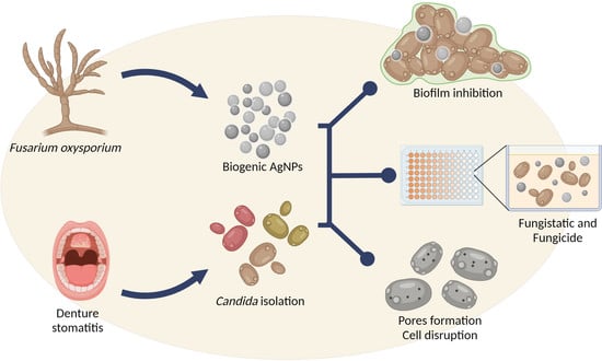
Activity of Fusarium oxysporum-Based Silver Nanoparticles on Candida spp. Oral Isolates

by
Maísa Santos Fonseca
1,
Daniela Méria Rodrigues
1,
Ana Rita Sokolonski
2,
Danijela Stanisic
3,
Luiz Marcelo Tomé
4,
Aristóteles Góes-Neto
4,
Vasco Azevedo
5,
Roberto Meyer
1,
Danilo Barral Araújo
2,
Ljubica Tasic
3 and
Ricardo Dias Portela
1,*
1
Laboratório de Imunologia e Biologia Molecular, Instituto de Ciências da Saúde, Universidade Federal da Bahia (UFBA), Salvador 40110-100, BA, Brazil
2
Laboratório de Bioquímica Oral, Instituto de Ciências da Saúde, Universidade Federal da Bahia (UFBA), Salvador 40110-100, BA, Brazil
3
Laboratório de Química Biológica, Instituto de Química, Universidade Estadual de Campinas (UNICAMP), Campinas 13083-970, SP, Brazil
4
Laboratório de Biologia Molecular e Computacional de Fungos, Instituto de Ciências Biológicas, Universidade Federal de Minas Gerais (UFMG), Belo Horizonte 31270-901, MG, Brazil
5
Laboratório de Genética Celular e Molecular, Instituto de Ciências Biológicas, Universidade Federal de Minas Gerais (UFMG), Belo Horizonte 31270-901, MG, Brazil
*
Author to whom correspondence should be addressed.
Nanomaterials 2022, 12(3), 501; https://doi.org/10.3390/nano12030501
Submission received: 21 November 2021 / Revised: 18 January 2022 / Accepted: 19 January 2022 / Published: 31 January 2022
(This article belongs to the Special Issue Nanomaterials in Oral Science)

Abstract

:
Candida spp. resistant to commercially available antifungals are often isolated from patients with oral candidiasis, a situation that points to the need for the development of new therapies. Thus, we evaluated the activity of Fusarium oxysporum-based silver nanoparticles (AgNPs) on Candida spp. isolated from denture stomatitis lesions. Candida isolates were molecularly identified and submitted to susceptibility assays using AgNPs and commercial fungicides. The interference on biofilm formation and the mechanisms of action of AgNPs on Candida spp. were also investigated. Scanning electron microscopy was used to evaluate the morphology of AgNP-treated Candida. Candida albicans was the most frequent species isolated from denture stomatitis cases. All Candida spp. were susceptible to AgNPs at low concentrations, except Candida parapsilosis. AgNPs caused surface damage, cell disruption, and biofilm formation inhibition. The ergosterol supplementation protected C. albicans against the AgNP action. AgNPs are effective against Candida spp. and can be faced as a promising new therapeutic agent against oral candidiasis.

Graphical Abstract

1. Introduction

Denture stomatitis is an infection of the oral cavity characterized by inflammation and erythema, and predisposing conditions increase the ability of opportunistic Candida fungi to cause oral candidiasis [1]. This infection is caused by Candida albicans and by other non-Candida albicans (NCA) species, such as Candida parapsilosis, Candida tropicalis and Candida glabrata [2].
Infections caused by Candida spp. are extensively recurrent [3], being this recurrence, as well as the development of chronic infections, associated with the formation of biofilm [4]. Candida species are competent biofilm-forming microorganisms, and these structures are correlated with an enhanced resistance to antifungals [5]. The inappropriate use of antifungals has resulted in the emergence of multidrug-resistant fungi [6]. Moreover, less attention has been given to the development of new antifungals [7]. The use of alternative technologies, such as metallic nanoparticles, has been considered in the last few years since they present significant antimicrobial and antiviral properties [8]. Among these nanoparticles, silver nanoparticles (AgNPs) are the most studied nanocompounds as a consequence of their several applications [9].
AgNPs consist of nanostructures originated from silver that can be synthesized by chemical or biogenic methods [10,11]. The chemical synthesis of metallic nanoparticles is used for large-scale production; however, a significant amount of energy is needed, and some toxic reagents that can negatively affect human health and the environment are used in this synthesis protocol [10]. In contrast, biogenic synthesis uses microorganisms [12], and does not require toxic reagents, being considered as an ecofriendly alternative [11]. In dentistry, silver nanoparticles synthesized by several chemical processes have been used as antimicrobials and in dentistry material development [13]. These nanoparticles have been successfully synthetized on a poly (methyl methacrylate) resin (acrylic glass), which led to a significant reduction on the adhesion and viability of C. albicans [14]. The diversity of AgNPs studies on dentistry can be applied to the clinical field if rigorous criteria are included on their evaluation [15].
AgNPs exhibit a broad-spectrum antimicrobial activity against bacteria and fungi [16]. Biogenic AgNPs are a good option among nanoparticles with antimicrobial activities since they present low production cost and good efficacy, stability, and durability [17,18]. Green synthesis using spruce bark extracts generated biogenic AgNPs that inhibited the growth and biofilm formation by several Candida species [19]. Furthermore, AgNPs synthetized using Terminalia catappa leaf extract exhibited an inhibition of the biofilm formation by multidrug-resistant Pseudomonas aeruginosa, methicillin-resistant Staphylococcus aureus and Candida albicans [20]. Additional AgNP mechanisms of action include an increase in the production of reactive oxygen species and changes in the general cellular metabolism and fluidity of the target cell plasma membrane [21,22].
Considering the potential of biogenic AgNPs as antimicrobial agents, we aimed to evaluate their antifungal activity against Candida spp. isolated from patients with prosthetic stomatitis. A comparative evaluation of the efficiency of commercial antifungals and AgNPs against planktonic Candida spp. and their associated biofilm was also performed. Additionally, an evaluation of the effects of AgNP on Candida specimens was performed.

2. Materials and Methods

2.1. Fungal Strains, Media and Commercial Antifungals

The Candida spp. reference strains used in this study were kindly provided by Fundação Oswaldo Cruz (FIOCRUZ, Rio de Janeiro, Brazil) and included four C. albicans (IOC 2508, IOC 2517, IOC 3703, and IOC 3704), C. parapsilosis (CP310), C. tropicalis (CT 309), and C. glabrata (CG74) strains. Sabouraud dextrose agar (SDA) (HIMEDIA, Mumbai, India) was used for fungal growth. RPMI 1640 medium supplemented with sodium bicarbonate and glutamine (Thermo Fisher, Waltham, MA, USA), 2% dextrose (Merck, Darmstadt, Germany), 0.165 mol/L 3-(N-morpholino) propanesulfonic acid, 4-morpholine-propanesulfonic acid (MOPS) (Thermo Fisher, Waltham, MA, USA), and 0.2% chloramphenicol was used in the susceptibility tests. The commercial antifungals used in this study were fluconazole, ketoconazole, nystatin (Infinity Pharma, Campinas, Brazil), and amphotericin B (Cristália, São Paulo, Brazil).

2.2. Synthesis of Silver Nanoparticles

The synthesis of the AgNPs was performed following the procedure described by Ballottin et al. [11]. Briefly, the biobased synthesis consisted of two steps: (1) production of the fungal secreted protein extract, and (2) production of the AgNPs. In the first part, the fungus Fusarium oxysporum was grown on a solid culture medium consisting of 0.5% yeast extract and 2% agar and kept at 28 °C for 1 week. Then, approximately 1 cm2 of the solid medium containing the fungus was removed and transferred to a sterile liquid medium consisting of 0.5% yeast extract and 2% malt extract. The 500 mL culture medium was then incubated under 150 rpm at 28 °C for 6 days. The biomass obtained was then filtered using filter paper and washed with deionized water. Approximately 10 g of the biomass was resuspended in 100 mL of deionized water. This material was then stirred at 150 rpm at 28 °C for 72 h. The biomass showed a pinkish color and was filtered using a Buchner funnel and filter paper, and the filtrate was used for the synthesis of the silver nanoparticles. In the second step, the secreted protein extract, with a concentration of 0.1 g/mL, was mixed with 0.01 mol/L of AgNO3. The solution was maintained at 28 °C in flasks sealed with aluminum foil until the formation of the nanoparticles. The characterization of the Fusarium oxysporum-AgNPs was performed using UV-Vis, showing a characteristic maximum absorbance at 440 nm (Supplementary Figure S1), dynamic light scattering and transmission electron microscopy, as previously described [11,23] and showed in Table 1 and Supplementary Figure S2. The AgNP were spherical, showing a size of 28.0 ± 13.1 nm, a polydispersity of 0.231, and a zeta potential of −31.7 ± 2.8 mV (Figure S1) [11,23].

2.3. Fungal Samples and Ethical Aspects

Clinical fungal isolates were obtained from patients screened by dentistry professionals at the Dentistry Ambulatory of the UNIME University, Salvador, Brazil. The samples from palate dentures presenting stomatitis lesions suggestive of oral candidiasis were collected using sterile swabs, inoculated on SDA supplemented with 0.2% chloramphenicol and incubated at 37 °C for 48 h [24]. The colonies were then isolated and maintained by weekly reinoculations. The Committee of Ethics on Research of the Institute of Health Sciences of the Federal University of Bahia approved this research under the protocol number 2.118.563, and an informed consent was obtained from all patients.
The genomic DNA of the fungal isolates was obtained using the FastDNA Spin Kit (MP Biomedicals, Solon, OH, USA). Polymerase chain reactions (PCR) were performed using the primers ITS4 and ITS5 for the amplification of the complete internal transcribed spacer (ITS) region [25], and LROR and LR7 primers for the amplification of the fungal large subunit (LSU) rDNA. For the PCR, it was used 1.25 U DNA Polymerase, 50 mM MgCl2, 10 mM DNTPs, 10 pmol of each primer, 0.5% ultrapure DMSO, 0.75% BSA (stock at 1 μg/μL), 5 M betaine, and 50 ng DNA template, in a final volume of 50 μL. The thermocycling steps were: 2 min at 94 °C, 35 cycles of 1 min at 94 °C, 1 min at 55 °C, 1 min at 72 °C, and a final extension of 5 min at 72 °C.
The purification of PCR products was carried out using an ethanol/EDTA (125 mM) precipitation protocol. The DNA sequencing was performed using the ABI 3730 automated sequencer (Applied Biosystems, Foster City, CA, USA). Consensus sequences were submitted to the Basic Local Alignment Search Tool (BLAST) for identification by similarity using the GenBank nucleotide sequences database of the National Center for Biotechnology Information (NCBI). For identification, similarities over 99% and an e-value equal to zero were considered.

2.4. Broth Microdilution Assay

The fungistatic activities of the commercial antifungals and AgNPs were evaluated following the M27-A3 protocol from the Clinical Laboratory Standards Institute [26]. Briefly, the yeast strains were resuspended in sterile 0.9% saline solution and adjusted by spectrophotometry to a 600 nm optical density of 0.8–1.0, which corresponds to 0.5 at the McFarland scale. Subsequently, the yeast cells were diluted (1:50) in sterile saline solution and then diluted (1:20) in supplemented RPMI 1640 medium (Thermo Fisher, Waltham, MA, USA) to obtain a 2.5 × 10³ cells/mL inoculum.
The AgNPs used in this study were serially diluted in sterile water for obtaining concentrations ranging from 1.95 to 1000 μg/mL. The commercial antifungal agents used as reference drugs were fluconazole (0.125–64 μg/mL), nystatin, ketoconazole and amphotericin B (0.0313–16 μg/mL) [26].
The inoculum (100 μL per well) was added to 96-well sterile culture plates. Subsequently, the same volumes of commercial fungicides and AgNPs at different concentrations were added to the wells. RPMI 1640 medium alone was used as a negative control and the inoculum without antifungal drugs was used as a positive control. The plates were incubated for 48 h at 37 °C and, after the incubation period, the wells were homogenized by pipetting, and the growth of Candida spp. was assessed by measuring the absorbance at 625 nm using a plate spectrophotometer (Multiskan FC Microplate Photometer, Thermo Fisher, Waltham, MA, USA). All the assays were performed in triplicate and were repeated three times. The minimum inhibitory concentration (MIC100) value, which represents the lowest concentration that inhibited 100% of the fungal growth, was determined as the minimum concentration of commercial antifungal agents and AgNPs that exhibited an OD reading similar to the negative control OD. For the determination of the minimum fungicide concentration (MFC100: minimal drug concentration able to kill 100% of the yeast cells), aliquots from each well of the broth microdilution assay were plated in SDA and then incubated at 37 °C for an additional 48 h.
According to the M27-S4 document [27], the breakpoint for fluconazole was considered for the classification of the fungal isolates according to the corresponding MIC100 (μg/mL), as follows: resistant (R) ≥ 8; dose-dependent susceptible (SDD) = 4; susceptible (S) ≤ 2, except for C. glabrata, where the interpretative endpoints were SDD ≤ 32 and R ≥ 64. Breakpoints for ketoconazole, nystatin and AmB were not available in M27-S4 document [23], and this classification was not performed for these drugs.

2.5. Biofilm Formation Interference Assay

To evaluate the interference of AgNPs in biofilm formation, it was used the reference strains C. parapsilosis CP310 and C. albicans 2508. These species were chosen based on their AgNP susceptibility profile obtained for the planktonic cultures.
The ability of the AgNPs to inhibit biofilm formation was evaluated as previously described [28]. The strains were incubated in SD Broth at 37 °C in a shaker at 250 rpm for 12–15 h. The cultures had their cell density adjusted to an OD600 of 0.38–0.5 with RPMI 1640 media and then added to a 96 well plate. The plates were incubated in a shaker at 250 rpm at 37 °C for 90 min. After that time, the media was aspirated, the wells were washed with sodium phosphate buffer (PBS 1×) pH 7.4, and AgNPs diluted in RPMI 1640 in concentrations ranging from 7.81 to 1000 μg/mL were added. 24 h later, the ODs were read in a spectrophotometer at 570 nm. The same controls that were reported above for the microdilution assay were used in this experiment.

2.6. Scanning Electron Microscopy

The scanning electron microscopy was performed as previously described [23], with minor modifications. Fungal suspensions cultured in the presence of half of the MIC100 concentration were deposited on 25 mm polytetrafluoroethylene (PTFE) membranes (0.45 μm pore size) (Millipore, Burlington, MA, USA) fixed with 2.5% glutaraldehyde in PBS pH 7.4 and washed twice with PBS. The membrane was soaked in 0.9% osmium tetroxide for 1 h and washed twice with distilled water. The dehydration was done by immersing the membranes in 30%, 50%, 70%, and 90% ethanol for 20 min, 20 min, 16 h, and 20 min, respectively, and then three times in 100% ethanol for 20 min each. The drying step was performed in a critical point dryer (Oerlikon Balzers, Balzers, Liechenstein), and the sputtering with gold was performed using a sputter coater (SCD-050, Leica, Wetzlar, Germany). Finally, the samples were analyzed using a scanning electronic microscope (JSM 5800LV, JEOL, Tokyo, Japan).

2.7. Exogenous Ergosterol and Sorbitol Supplementation Assays

The exogenous supplementation of ergosterol and sorbitol was conducted with the objective to determine the effect of AgNP treatment on the fungal surface. Sorbitol can prevent damages to the cell wall structure, whereas ergosterol protects the plasma membrane from the action of AgNPs. For the supplementation assays, the microdilution assay used for the drug susceptibility tests was performed as described above but with the addition of 0.8 M sorbitol or 200 μg/mL ergosterol in all samples and controls. This assay was conducted as previously described [29].

2.8. Statistical Analysis

Dose-response curves were generated to assess the average response of the growth inhibition obtained in the broth microdilution tests. This analysis was performed with the GraphPad Prism 6.0 software (GraphPad Software, San Diego, CA, USA) using the nonlinear regression parameters for the dose-response inhibition (variable-slope) equation. The EC50 (concentration of drug capable of inhibiting 50% of yeast growth) was obtained through the dose-response inhibition (variable-slope) equation. The R square was greater than 0.65 for all the curves. The interference rates in biofilm formation assays were obtained in % using the following formula [30]:
[(OD570 of Candida spp. treated with AgNPs ∗ 100)/(OD570 of Candida spp. non-treated)] − 100

3. Results

3.1. Identification of the Candida Species Isolated from Denture Stomatitis Cases

The twenty Candida isolates from denture stomatitis lesions were identified through the sequencing of LSU and ITS gene fragments. The results showed the presence of three Candida species, with different frequencies (Table 2). C. albicans (14/20) was the most frequently isolated species, corresponding to 70.0% of the isolates, whereas C. tropicalis (5/20) and C. dubliniensis (1/20) were less abundant, with frequencies of 25.0% and 5.0%, respectively.

3.2. Susceptibility to Commercial Antifungal Drugs

The reference strains and the clinical isolates presented distinct susceptibility patterns for fluconazole and ketoconazole. All C. albicans reference strains were susceptible to fluconazole, with MIC100 ranging from 0.125 to 0.5 μg/mL. Of all C. albicans clinical isolates (14 in total), seven were susceptible (MIC100 ≤ 2 μg/mL), three were dose-dependent susceptible (S-DD) (MIC100 = 4 μg/mL), and four were resistant (MIC100 > 16 μg/mL) to fluconazole (Table 2). Thus, 28.57% of the C. albicans clinical isolates presented resistance to fluconazole. Regarding the MIC100 of fluconazole for the non-Candida albicans species, one C. glabrata, three C. tropicalis, and one C. parapsilosis strain were resistant (MIC100 ≥ 8 μg/mL).
The MFC100 of fluconazole for C. albicans isolates was markedly variable, with four C. albicans isolates presenting an MFC100 ranging from 2 to 16 μg/mL and ten isolates having non-determined MFC100. Considering the NCA species, the MFC100 for fluconazole presented by C. dubliniensis and three (of five) isolates of C. tropicalis was undetermined (Table 2).
Ketoconazole showed a fungistatic effect at low concentrations for all the C. albicans reference strains (14 in number, MIC100 0.125 to 0.25 μg/mL) and clinical isolates (MIC100 0.125 to 8 μg/mL), except for the PAC 12 isolate (undetermined MIC). The MFC100 was undetermined for ten C. albicans clinical isolates (MFC100 > 16 μg/mL). For eight C. albicans, four reference strains and four clinical isolates, the MFC ranged between 0.031 and 0.125 μg/mL. For C. dubliniensis, the MIC100 and MFC100 were 0.03125 and 0.125 μg/mL, respectively. Regarding the C. tropicalis clinical isolates, the MIC100 ranged between 0.03125 and 16 μg/mL, and the MFC could not be determined for three of the five isolates (Table 2).
Nystatin and amphotericin B (AmB) presented the lowest variations in the MIC100 and MFC100 values for all Candida spp. tested in this study. The results obtained for nystatin showed that all C. albicans tested herein presented MIC100 values between 1 and 16 μg/mL and MFC100 ranging between 2 and 16 μg/mL, except for PAC 17, for which the MFC values could not be determined (>16 μg/mL). C. dubliniensis showed an MIC100 of 2 μg/mL and MFC100 of 4 μg/mL for nystatin. Regarding the response of C. tropicalis to nystatin, the MIC100 for the five isolates ranged between 0.5 and 4 μg/mL, and the MFC100 ranged between 2 and 8 μg/mL. Interestingly, AmB had the same MIC100 and MFC100 values for almost all strains (Table 2). The AmB MIC100 for C. albicans ranged between 0.25 and 4 μg/mL and the MFC ranged from 0.5 to 4 μg/mL. For C. dubliniensis, the MIC100 and the MFC100 values were the same (2 μg/mL). Finally, for C. tropicalis, the MIC100 of AmB ranged between 0.25 and 2 μg/mL and MFC100 ranged between 0.5 and 4 μg/mL. When each isolate was analyzed alone, the C. albicans PAC 17 clinical isolate presented the highest values of MIC100 and MFC100 among all the commercial drugs tested herein. Thus, this isolate can be considered as a potential multidrug resistant organism.

3.3. Susceptibility to Silver Nanoparticles

The AgNPs presented a fungistatic effect at low concentrations for all the Candida spp. in this study. Fifteen (of 20) isolates from the oral cavity showed an MIC100 of 7.8 μg/mL, four isolates presented an MIC of 3.9 μg/mL, representing the lowest MIC100 determined herein, and C. dubliniensis showed the highest MIC100 (15.6 μg/mL) for AgNPs. The MFC100 values ranged from 15.6 to 500 μg/mL for the oral cavity isolates. For the reference strains, the AgNP had fungistatic activity with an MIC100 of 7.8 μg/mL for all the strains, except for C. albicans 2508 (MIC100 = 15.6 μg/mL). The MFC100 for the C. parapsilosis CP310 strain could not be determined (>1000 μg/mL). The MFC100 of the other reference strains ranged between 15.6 and 250 μg/mL (Table 2).
The dose-response curves (Figure 1) show the response of all the Candida spp. specimens tested herein to fluconazole, nystatin, AmB and AgNPs. The analysis of the commercial fungicides showed that fluconazole had a high variance in the growth inhibition considering the same concentration and different isolates (Figure 1A). Nystatin and AmB presented a more stable per concentration response between isolates and Candida species (Figure 1B,C). Finally, the results showed a lower tolerance of all the Candida species to AgNPs, even at lower concentrations, presenting a similar behavior when considering the effective dose of AgNPs (Figure 1D).

3.4. Interference Effect of AgNPs in the Biofilm Formation

An interference on biofilm formation was observed in both C. albicans 2508 and C. parapsilosis CP310 reference strains when treated with the AgNPs (Figure 2). C. albicans, that was susceptible to the AgNPs in the planktonic form, showed 96.3% of interference in biofilm formation at the higher AgNP concentration tested herein (1000 µg/mL) (Figure 2A). This same concentration induced a 100% interference in C. parapsilosis biofilm formation (Figure 2B). The interference decreased to 80% in both species at 500 µg/mL of AgNPs. At lower concentrations (7.8–125 µg/mL), the AgNPs impaired biofilm formation to less than 40% in C. albicans, while for C. parapsilosis only concentrations below 31.25 µg/mL where able to reach this same inhibitory percentage.

3.5. Scanning Electron Microscopy of C. albicans and C. parapsilosis

A scanning electron microscopy (SEM) analysis was performed to evaluate the effects of AgNPs on the fungi surface morphology. C. albicans 2508 and C. parapsilosis CP310 cultured without AgNPs showed a characteristic yeast morphology, varying from an ovoid to a spherical shape, occurring alone or grouped, and presenting an apparently intact cellular surface (Figure 3A,C). After incubation with the AgNPs, C. albicans appeared completely disrupted, showing cells debris without a defined morphology, as observed in AgNPs absence (Figure 3B). Agglomerates of biomass around the dried and disrupted C. parapsilosis cultured with AgNPs can be seen in Figure 3D. The SEM of C. parapsilosis after 48 h of treatment with AgNPs showed the presence of many pores on the cell surface. SEM analysis revealed that the AgNPs caused severe damage to the surface of the Candida spp.

3.6. Ergosterol and Sorbitol Supplementation

To evaluate if the surface damage caused by AgNPs can be prevented, C. albicans 2508 and C. parapsilosis CP310 were inoculated in SD media containing ergosterol (200 µg/mL) or sorbitol (0.8 M) supplementation, and were exposed to a range of AgNP concentrations. For C. albicans 2508 strain, ergosterol supplementation presented a protective effect at lower AgNP concentrations (Figure 4A,B). For C. parapsilosis CP310 strain, supplementation with ergosterol had a limited protection against AgNPs at the concentration of 1.9 µg/mL. Sorbitol supplementation presented no protective effect against AgNPs in the evaluated strains (Figure 4C,D). The results indicated that the damages in the fungal surface caused by AgNPs were primarily located in the plasmatic membrane.

4. Discussion

Candida specimens are important etiologic agents of denture stomatitis [1]. The presence of C. albicans and NCA species in these infections has been already described [33]. In this study, the identification of the isolated Candida species showed that C. albicans was the most frequently isolated species, and C. tropicalis and C. dubliniensis were present in a lower abundance. Likewise, C. albicans and C. tropicalis were found causing chronic periodontitis at a similar frequency in Alagoas state, Brazil [34]. In Pará state, Brazil, C. albicans was the most frequent species associated with oral candidiasis, with a frequency of 78% [33]. In Spain, C. albicans accounts for 70% of Candida spp. isolated in oral candidiasis cases, followed by C. glabrata (8.6%), C. parapsilosis (7.4%), and C. tropicalis (3.3%) [31]. C. dubliniensis is a rare opportunistic fungus that causes oral cavity infections in patients with immunosuppression caused by HIV [32] and few descriptions of its prevalence in other fungal diseases are available [35].
Currently, four main classes of antifungal drugs, namely azoles, polyenes, allylamines, and echinocandins, are available for candidiasis treatment. Each class of these drugs has a specific action and a defined cellular target [36]. In this study, the analysis of four of the most used commercial antifungals showed that the majority of the C. albicans isolates were susceptible to fluconazole, whereas the NCA species were mostly SDD or resistant. However, in 71% of the C. albicans isolates, the MFC100 for fluconazole could not be determined. The resistance to fluconazole was detected in 36.8% of the C. albicans isolated from patients with chronic periodontitis from Alagoas State, in Brazil [34]. C. parapsilosis and C. albicans presented different susceptibility patterns to fluconazole, being the MIC higher for C. parapsilosis [37]. Fluconazole resistance is more common in NCA species than in C. albicans isolates [38]. The increase in the cases of Candida strains that are less susceptible or resistant to fluconazole indicates that the use of alternative drugs for the treatment of oral candidiasis is highly needed [39].
Nystatin and AmB are polyene antifungals; their mechanisms of action are associated with the content of ergosterol of the fungal membrane [40]. In the treatment of dental stomatitis, nystatin is an antifungal agent with high activity against Candida spp. isolates [31]. Unlike for azoles, the MIC100 and MFC100 for nystatin and AmB could be determined for all the isolates, except for one C. albicans isolate [40]. Regarding AmB, our results showed similar MIC100 and MFC100 values for almost all the isolates tested herein. A 71% agreement between the MIC and MFC values presented by C. albicans isolates treated with AmB has already been reported [41]. These results suggest that the same concentration of AmB can inhibit the growth and kill the fungi, a situation that can facilitate the disease treatment.
There is a continuous evolution of drug resistance in Candida isolates [1]. Considering this situation, there is a constant demand for the development and discovery of new and safe broad-spectrum antifungal agents associated with a minimal toxicity to the host. In this context, AgNPs are nanotechnological compounds that present significant antimicrobial properties and low toxicity [42]. The AgNPs can be synthesized by different methods [11,17,43] and their antifungal [11,43,44,45] and antibacterial activities [43,46] have been correlated to their size, shape, and surface modifications [47,48]. It has been shown that the presence of the nicotinamide adenine dinucleotide (NADH) and NADH-dependent nitrate reductase enzymes are essential for the biosynthesis of AgNPs using microorganisms [49,50,51], and the reduction of silver ions occurs through the transfer of electrons from NADH by the reductase enzyme [49,50,51]. The biogenic AgNPs used in this study were synthesized using F. oxysporum secreted molecules. Considering the AgNPs used in dentistry, both prokaryotic and eukaryotic organisms can be used to synthesize AgNPs; however, plants are the most common organisms used in these AgNP syntheses [13]. The AgNP synthesized using Aspergillus tubingensis presented a positive zeta potential, spherical shape, and size of 35 ± 10 nm [44]. Among the different particle characteristics, the size and shape influence their antimicrobial properties; small particles (lower than 10 nm) that present a spherical shape are more effective against microorganisms [52]. In addition, the action of AgNPs appears to be highly related to the nanosize, which alters the level of silver ions released in the system and interferes with the surface energy [53].
Our results for the AgNP susceptibility assays using planktonic Candida showed that the AgNPs have a significant fungistatic effect at low concentrations, whereas the fungicidal effect of this nanoparticle showed more variable values, except for C. parapsilosis that presented an undetermined MFC. In opposite to our results, a previous study reported a strong activity of AgNPs against other C. parapsilosis isolate and it were also active against several Candida and bacterial species [43]. Radhakrishnan and collaborators [22] showed that citrated-reduced AgNPs inhibited C. albicans growth at lower concentrations and completely inhibit its growth at a concentration of 40 μg/mL.
The dose-response curves revealed that the inhibition of growth by AgNPs was significant, reaching 90% to 100% at low concentrations. These results are supported by the scientific literature, where other types of AgNPs have been reported to inhibit the growth of C. albicans at low concentrations (5 μg/mL) and caused a complete inhibition at high concentrations (40 μg/mL) [42]. In another study, biologically synthesized AgNPs, in combination or not with fluconazole, were effective against C. albicans [45]. The combination of AgNPs with simvastatin had a synergistic and additive effect against Aspergillus [54]. Thus, AgNPs can be used as an alternative or complementary treatment for fungal infections, considering their significant antimicrobial activities [42,45] and wound healing effects [46].
Biofilms are structured microorganism communities adhered to a surface and are considered as an antifungal resistance factor [4]. In our work, the AgNPs concentrations that were able to fully inhibit the biofilm formation by C. albicans and C. parapsilosis were 8-fold bigger than the MIC100 for both species. Recently, pure round AgNPs strongly inhibited formation and promoted disruption of Candida auris biofilm [55]. Additionally, the viability of fluconazole-resistant C. tropicalis biofilms was reduced when treated with AgNPs [56]. AgNPs embed into maxillofacial silicone elastomers reduced the C. albicans biofilm viability on this material [57]. All these studies showed that lower AgNPs concentrations were sufficient for a significant effect on Candida spp. biofilm. In addition, low doses can represent a decrease of unexpected effects on hosts [13].
The treatment with AgNPs induced a disruption of the fungal cells and the formation of pores on the cell surface, as shown by the SEM results. Similar analysis showed a high accumulation of nanoparticles outside the cells and the presence of small particles throughout the target cell cytoplasm [58]. Another study showed that the treatment of Candida spp. with other types of AgNPs disrupted the cell membrane and affected its integrity [59]. SEM and transmission electron microscopy analysis of C. albicans after treatment with AgNPs showed altered cellular morphology and ultrastructure [22]. Jalal et al. [43] showed that AgNPs are able to penetrate into C. albicans, leading to pore formation as a result of cell wall and membrane rupture. Additionally, Candida spp. treated with AgNPs can also exhibit alterations in the fluidity of the cell membrane and in the ergosterol content [22]. Furthermore, it has been suggested that the effects exerted by AgNPs on the fluidity of the membrane can be related to changes in the plasma membrane lipidic constitution and membrane depolarization [22,47].
Ergosterol is one of the most important constituents of the fungal cell membrane and plays a vital role in the stability of cells [60]. In the present study, the broth microdilution assays made with ergosterol supplementation resulted in a reduced susceptibility to AgNPs. The loss of sterols in the membrane leads to destabilization, resulting in an increased permeability and, thereby, enhanced sensitivity to drugs [60,61]. In addition, it was reported that the action of AgNPs also involves a reduction in the ergosterol content in the membrane [22]. Moreover, the importance of ergosterol is also based on the fact that its biosynthetic pathway is the target of most azoles, polyenes, and allylamines [61]. Sorbitol is considered an osmotic protector that can prevent damages to the cell wall [62]. Our results did not show any protection against the AgNPs action after sorbitol supplementation, besides the presence of pores evidenced by the SEM analysis. Additional investigation can lead to a better understanding of these mechanisms. However, literature evidence suggests that AgNPs can cause damage to fungal cells, acting against various cellular targets that can finally lead to cell lysis [17,22,43].
Fusarium oxysporum-based AgNPs showed significant antifungal activity on Candida spp. isolated from denture stomatitis, being able to impair the biofilm formation of C. albicans and C. parapsilosis. As a possible action mechanism, the plasmatic membrane can be a target of the nanoparticles. Thus, the biogenic AgNPs synthesized using F. oxysporum have a great potential as a complementary therapy of oral candidiasis.

Supplementary Materials

The following supporting information can be downloaded at: https://www.mdpi.com/article/10.3390/nano12030501/s1, Figure S1: Transmission electron micrographs (TEM) of AgNPs, scales of 200 nm (left) and 50 nm (right). The silver nanoparticles obtained using Fusarium oxysporum (AgNPFU) showed a spherical-like shape, size 28.0 ± 13.1 nm, and were found to be stable for one year. It is possible to see the protein corona (light gray) around the Ag-core in the TEM image at the right. Figure S2: The absorption spectrum in UV-Vis of the synthesized AgNPS, with the characteristic surface plasmon resonance peak at 440 nm. The spectrum was measured using a UV-Vis HP8453 spectrophotometer using solutions placed in quartz cuvettes with a paht length of 10 mm. The spectra were taken in the range of 350 to 700 nm. The blank solution was prepared using the fungal filtrate by substituting the silver nitrate solution for distilled water.

Author Contributions

R.D.P., L.T. and V.A. conceived the original idea and planned the experiments. V.A., R.M. and D.B.A. helped supervise the project. M.S.F., A.R.S., D.M.R., D.S. and L.M.T. were responsible for acquisition of data. D.S. and L.T. synthetized the biogenic silver nanoparticles. M.S.F., A.R.S., D.M.R., A.G.-N. and R.D.P. contributed to the interpretation of the results. M.S.F. and A.R.S. wrote the manuscript. R.D.P., D.B.A. and R.M. approved the final version to be published. All authors provided critical feedback and helped shape the research, analysis, and manuscript. All authors have read and agreed to the published version of the manuscript.

Funding

MSF is a fellow from the Coordenação de Aperfeiçoamento de Pessoal de Nível Superior (CAPES). RDP is a Technological Development fellow from the Conselho Nacional de Desenvolvimento Científico e Tecnológico (CNPq-Proc. 313350/2019-1). The funders had no role in the design of the study, in collection, analysis, and interpretation of data, or in the writing of the manuscript.

Data Availability Statement

The data presented in this study are available on request from the corresponding author.

Conflicts of Interest

The authors declare that the research was conducted in the absence of any commercial or financial relationships that could be construed as a potential conflict of interest.

References

  1. Gendreau, L.; Loewy, Z.G. Epidemiology and Etiology of Denture Stomatitis. J. Prosthodont. 2011, 20, 251–260. [Google Scholar] [CrossRef] [PubMed]
  2. Savastano, C.; de Oliveira Silva, E.; Gonçalves, L.L.; Nery, J.M.; Silva, N.C.; Dias, A.L.T. Candida glabrata among Candida spp. from environmental health practitioners of a Brazilian Hospital. Braz. J. Microbiol. 2016, 47, 367–372. [Google Scholar] [CrossRef] [PubMed] [Green Version]
  3. Castro, T.L.; Coutinho, H.D.M.; Gedeon, C.C.; Santos, J.M.; Santana, W.J.; Souza, L.B.S. Mecanismos de resistência da Candida sp. a anti-fúngicos. Infarma Ciências Farm 2013, 18, 30–35. [Google Scholar]
  4. Douglas, L. Candida biofilms and their role in infection. Trends Microbiol. 2003, 11, 30–36. [Google Scholar] [CrossRef]
  5. Pereira, R.; Fontenelle, R.O.D.S.; de Brito, E.H.S.; de Morais, S.M. Biofilm of Candida albicans: Formation, regulation and resistance. J. Appl. Microbiol. 2021, 131, 11–22. [Google Scholar] [CrossRef]
  6. Pristov, K.E.; Ghannoum, M.A. Resistance of Candida to azoles and echinocandins worldwide. Clin. Microbiol. Infect. 2019, 25, 792–798. [Google Scholar] [CrossRef]
  7. Perlin, D.S. Echinocandin Resistance in Candida. Clin. Infect. Dis. 2015, 61, S612–S617. [Google Scholar] [CrossRef] [Green Version]
  8. Halbus, A.F.; Horozov, T.S.; Paunov, V.N. Colloid particle formulations for antimicrobial applications. Adv. Colloid Interface Sci. 2017, 249, 134–148. [Google Scholar] [CrossRef]
  9. El-Batal, A.I.; Gharib, F.A.E.-L.; Ghazi, S.M.; Hegazi, A.Z.; El Hafz, A.G.M.A. Physiological Responses of Two Varieties of Common Bean (Phaseolus Vulgaris L.) to Foliar Application of Silver Nanoparticles. Nanomater. Nanotechnol. 2016, 6, 13. [Google Scholar] [CrossRef] [Green Version]
  10. Durán, N.; Marcato, P.D.; De Conti, R.; Alves, O.L.; Costa, F.T.M.; Brocchi, M. Potential use of silver nanoparticles on pathogenic bacteria, their toxicity and possible mechanisms of action. J. Braz. Chem. Soc. 2010, 21, 949–959. [Google Scholar] [CrossRef]
  11. Ballottin, D.; Fulaz, S.; Cabrini, F.; Tsukamoto, J.; Durán, N.; Alves, O.L.; Tasic, L. Antimicrobial textiles: Biogenic silver nanoparticles against Candida and Xanthomonas. Mater. Sci. Eng. C 2017, 75, 582–589. [Google Scholar] [CrossRef] [PubMed]
  12. Fayaz, A.M.; Girilal, M.; Rahman, M.S.; Venkatesan, R.; Kalaichelvan, P. Biosynthesis of silver and gold nanoparticles using thermophilic bacterium Geobacillus stearothermophilus. Process Biochem. 2011, 46, 1958–1962. [Google Scholar] [CrossRef]
  13. Fernandez, C.; Sokolonski, A.; Fonseca, M.; Stanisic, D.; Araújo, D.; Azevedo, V.; Portela, R.; Tasic, L. Applications of Silver Nanoparticles in Dentistry: Advances and Technological Innovation. Int. J. Mol. Sci. 2021, 22, 2485. [Google Scholar] [CrossRef] [PubMed]
  14. De Matteis, V.; Cascione, M.; Toma, C.C.; Albanese, G.; De Giorgi, M.L.; Corsalini, M.; Rinaldi, R. Silver Nanoparticles Addition in Poly(Methyl Methacrylate) Dental Matrix: Topographic and Antimycotic Studies. Int. J. Mol. Sci. 2019, 20, 4691. [Google Scholar] [CrossRef] [PubMed] [Green Version]
  15. Adam, R.Z.; Khan, S.B. Antimicrobial efficacy of silver nanoparticles against Candida albicans: A systematic review protocol. PLoS ONE 2021, 16, e0245811. [Google Scholar] [CrossRef] [PubMed]
  16. Telles, I.S.F.; Prado, M.D.; Simão, R.A. Nanopartículas e aplicações endodônticas: Uma revisão da literatura. Rev. Bras. Odontol. 2017, 74, 167. [Google Scholar] [CrossRef] [Green Version]
  17. Durán, N.; Durán, M.; de Jesus, M.B.; Seabra, A.B.; Fávaro, W.J.; Nakazato, G. Silver nanoparticles: A new view on mechanistic aspects on antimicrobial activity. Nanomed. Nanotechnol. Biol. Med. 2016, 12, 789–799. [Google Scholar] [CrossRef]
  18. Guilger Casagrande, M.; De Lima, R. Synthesis of Silver Nanoparticles Mediated by Fungi: A Review. Front. Bioeng. Biotechnol. 2019, 7, 287. [Google Scholar] [CrossRef] [Green Version]
  19. Mare, A.D.; Man, A.; Ciurea, C.N.; Toma, F.; Cighir, A.; Mareș, M.; Berța, L.; Tanase, C. Silver Nanoparticles Biosynthesized with Spruce Bark Extract—A Molecular Aggregate with Antifungal Activity against Candida Species. Antibiotics 2021, 10, 1261. [Google Scholar] [CrossRef]
  20. Ansari, M.; Kalam, A.; Al-Sehemi, A.; Alomary, M.; AlYahya, S.; Aziz, M.; Srivastava, S.; Alghamdi, S.; Akhtar, S.; Almalki, H.; et al. Counteraction of Biofilm Formation and Antimicrobial Potential of Terminalia catappa Functionalized Silver Nanoparticles against Candida albicans and Multidrug-Resistant Gram-Negative and Gram-Positive Bacteria. Antibiotics 2021, 10, 725. [Google Scholar] [CrossRef]
  21. Alves, O.L.; De Moraes, A.C.M.; Simões, M.B.; Fonseca, L.C.; Nascimento, R.O.D.; Holtz, R.D.; De Faria, A.F. Nanomaterials. In Nanotoxicology Mater. Methodol. Assessments, 1st ed.; Durán, N., Guterres, S.S., Alves, O.L., Eds.; Springer-Verlag: Berlin/Heidelberg, Germany, 2014; p. 411. [Google Scholar]
  22. Radhakrishnan, V.S.; Mudiam, M.K.R.; Kumar, M.; Dwivedi, S.P.; Singh, S.P.; Prasad, T. Silver nanoparticles induced alterations in multiple cellular targets, which are critical for drug susceptibilities and pathogenicity in fungal pathogen (Candida albicans). Int. J. Nanomed. 2018, 13, 2647–2663. [Google Scholar] [CrossRef] [PubMed] [Green Version]
  23. Stanisic, D.; Fregonesi, N.L.; Barros, C.H.N.; Pontes, J.G.M.; Fulaz, S.; Menezes, U.J.; Nicoleti, J.L.; Castro, T.L.P.; Seyffert, N.; Azevedo, V.; et al. NMR insights on nano silver post-surgical treatment of superficial caseous lymphadenitis in small ruminants. RSC Adv. 2018, 8, 40778–40786. [Google Scholar] [CrossRef] [Green Version]
  24. ANVISA—Agência Nacional de Vigilância Sanitária. Manual de Microbiologia Clínica Para o Controle de Infecção em Serviços de Saúde, 2004. Available online: https://bvsms.saude.gov.br/bvs/publicacoes/manual_microbiologia_completo.pdf (accessed on 16 January 2022).
  25. White, T.J.; Bruns, T.; Lee, S.; Taylor, J.W. Amplification and direct sequencing of fungal ribosomal RNA genes for phylogenetics. In PCR Protocols: A Guide to Methods and Applications; Innis, M.A., Gelfand, D.H., Sninsky, J.J., White, T.J., Eds.; Academic Press: Cambridge, MA, USA, 1990; pp. 315–322. [Google Scholar]
  26. Clinical and Laboratory Standards Institute (CLSI). M27-S3: Reference Method for Broth Dilution Antifungal Susceptibility Testing of Yeasts; Third Informational Supplement, 3rd ed.; Clinical and Laboratory Standards Institute: Wayne, PA, USA, 2008. [Google Scholar]
  27. Clinical and Laboratory Standards Institute (CLSI). M27-S4: Reference Method for Broth Dilution Antifungal Susceptibility Testing of Yeasts; Third Informational Supplement, 3rd ed.; Clinical and Laboratory Standards Institute: Wayne, PA, USA, 2012. [Google Scholar]
  28. Lohse, M.B.; Gulati, M.; Arevalo, A.V.; Fishburn, A.; Johnson, A.D.; Nobile, C.J. Assessment and Optimizations of Candida albicans In Vitro Biofilm Assays. Antimicrob. Agents Chemother. 2017, 61, e02749-16. [Google Scholar] [CrossRef] [Green Version]
  29. Souto Maior, L.F.; Maciel, P.P.; Ferreira, V.Y.N.; de Lima Gouveia Dantas, C.; de Lima, J.M.; Castellano, L.R.C. Antifungal activity and Shore A hardness of a tissue conditioner incorporated with terpinen-4-ol and cinnamaldehyde. Clin. Oral Investig. 2019, 23, 2837–2848. [Google Scholar] [CrossRef] [PubMed]
  30. Kalil, M.A.; Santos, L.M.; Barral, T.D.; Rodrigues, D.M.; Pereira, N.P.; Sá, M.D.C.A.; Umsza-Guez, M.A.; Machado, B.A.S.; Meyer, R.; Portela, R.W. Brazilian Green Propolis as a Therapeutic Agent for the Post-surgical Treatment of Caseous Lymphadenitis in Sheep. Front. Vet. Sci. 2019, 6, 399. [Google Scholar] [CrossRef] [PubMed] [Green Version]
  31. Miranda-Cadena, K.; Marcos-Arias, C.; Mateo, E.; Aguirre, J.M.; Quindós, G.; Eraso, E. Prevalence and antifungal susceptibility profiles of Candida glabrata, Candida parapsilosis and their close-related species in oral candidiasis. Arch. Oral Biol. 2018, 95, 100–107. [Google Scholar] [CrossRef]
  32. Sullivan, D.; Haynes, K.A.; Bennett, D.E.; Coleman, D.; Westerneng, T.J.; Haynes, K.A.; Bennett, D.E.; Coleman, D.C. Candida dubliniensis sp. nov.: Phenotypic and molecular characterization of a novel species associated with oral candidosis in HIV-infected individuals. Microbiology 1995, 141, 1507–1521. [Google Scholar] [CrossRef] [Green Version]
  33. Gauch, L.M.R.; Pedrosa, S.S.; Silveira-Gomes, F.; Esteves, R.A.; Marques-Da-Silva, S.H. Isolation of Candida spp. from denture-related stomatitis in Pará, Brazil. Braz. J. Microbiol. 2018, 49, 148–151. [Google Scholar] [CrossRef]
  34. Siqueira, A.B.S.; Rodriguez, L.R.N.D.A.; Santos, R.K.B.; Marinho, R.R.B.; Abreu, S.; Peixoto, R.F.; Gurgel, B.C.D.V. Antifungal activity of propolis against Candidaspecies isolated from cases of chronic periodontitis. Braz. Oral Res. 2015, 29, 1–6. [Google Scholar] [CrossRef]
  35. Loreto, S.; Scheid, L.A.; Nogueira, C.W.; Zeni, G.; Santurio, J.M.; Alves, S.H. Candida dubliniensis: Epidemiology and Phenotypic Methods for Identification. Mycopathologia 2010, 169, 431–443. [Google Scholar] [CrossRef]
  36. Liu, S.; Hou, Y.; Chen, X.; Gao, Y.; Li, H.; Sun, S. Combination of fluconazole with non-antifungal agents: A promising approach to cope with resistant Candida albicans infections and insight into new antifungal agent discovery. Int. J. Antimicrob. Agents 2014, 43, 395–402. [Google Scholar] [CrossRef] [PubMed]
  37. Romera, D.; Aguilera-Correa, J.J.; Gadea, I.; Viñuela-Sandoval, L.; García-Rodríguez, J.; Esteban, J. Candida auris: A comparison between planktonic and biofilm susceptibility to antifungal drugs. J. Med. Microbiol. 2019, 68, 1353–1358. [Google Scholar] [CrossRef] [PubMed]
  38. Pereira, G.H.; Mueller, P.R.; Szeszs, M.W.; Levin, A.S.; Melhem, M.S.C. Five-year evaluation of bloodstream yeast infections in a tertiary hospital: The predominance of non-C. albicans Candidaspecies. Med. Mycol. 2010, 48, 839–842. [Google Scholar] [CrossRef] [PubMed] [Green Version]
  39. Sanguinetti, M.; Posteraro, B.; Lass-Flörl, C. Antifungal drug resistance among Candidaspecies: Mechanisms and clinical impact. Mycoses 2015, 58, 2–13. [Google Scholar] [CrossRef] [PubMed]
  40. Krishnasamy, L.; Krishnakumar, S.; Kumaramanickavel, G.; Saikumar, C. Molecular Mechanisms of Antifungal Drug Resistance in Candida Species. J. Clin. Diagn. Res. 2018, 12, 9. [Google Scholar] [CrossRef]
  41. Lemos, J.D.A.; Costa, C.R.; De Araújo, C.R.; E Souza, L.K.H.; Silva, M.D.R.R. Susceptibility testing of Candida albicans isolated from oropharyngeal mucosa of HIV+ patients to fluconazole, amphotericin B and Caspofungin: Killing kinetics of caspofungin and amphotericin B against fluconazole resistant and susceptible isolates. Braz. J. Microbiol. 2009, 40, 163–169. [Google Scholar] [CrossRef] [Green Version]
  42. Radhakrishnan, V.S.; Dwivedi, S.P.; Siddiqui, M.H.; Prasad, T. In vitro studies on oxidative stress-independent, Ag nanoparticles-induced cell toxicity of Candida albicans, an opportunistic pathogen. Int. J. Nanomed. 2018, 13, 91–96. [Google Scholar] [CrossRef] [Green Version]
  43. Jalal, M.; Ansari, M.A.; Alzohairy, M.A.; Ali, S.G.; Khan, H.M.; Almatroudi, A.; Raees, K. Biosynthesis of Silver Nanoparticles from Oropharyngeal Candida glabrata Isolates and Their Antimicrobial Activity against Clinical Strains of Bacteria and Fungi. Nanomaterials 2018, 8, 586. [Google Scholar] [CrossRef] [Green Version]
  44. Rodrigues, A.G.; Ping, L.Y.; Marcato, P.D.; Alves, O.L.; Silva, M.C.P.; Ruiz, R.D.C.; Melo, I.S.; Tasic, L.; De Souza, A.O. Biogenic antimicrobial silver nanoparticles produced by fungi. Appl. Microbiol. Biotechnol. 2012, 97, 775–782. [Google Scholar] [CrossRef]
  45. Longhi, C.; Santos, J.P.; Morey, A.T.; Marcato, P.D.; Durán, N.; Pinge-Filho, P.; Nakazato, G.; Yamada-Ogatta, S.F.; Yamauchi, L.M. Combination of fluconazole with silver nanoparticles produced by Fusarium oxysporumimproves antifungal effect against planktonic cells and biofilm of drug-resistantCandida albicans. Med. Mycol. 2015, 54, 428–432. [Google Scholar] [CrossRef] [Green Version]
  46. Santos, L.M.; Stanisic, D.; Menezes, U.J.; Mendonça, M.A.; Barral, T.; Seyffert, N.; Azevedo, V.; Durán, N.; Meyer, R.; Tasic, L.; et al. Biogenic Silver Nanoparticles as a Post-surgical Treatment for Corynebacterium pseudotuberculosis Infection in Small Ruminants. Front. Microbiol. 2019, 10, 824. [Google Scholar] [CrossRef] [PubMed] [Green Version]
  47. Kim, K.-J.; Sung, W.S.; Suh, B.K.; Moon, S.-K.; Choi, J.-S.; Kim, J.G.; Lee, D.G. Antifungal activity and mode of action of silver nano-particles on Candida albicans. BioMetals 2009, 22, 235–242. [Google Scholar] [CrossRef] [PubMed]
  48. Singh, P.; Kim, Y.J.; Singh, H.; Wang, C.; Hwang, K.H.; Farh, M.E.A.; Yang, D.C. Biosynthesis, characterization, and antimicrobial applications of silver nanoparticles. Int. J. Nanomed. 2015, 10, 2567–2577. [Google Scholar]
  49. Ahmad, A.; Mukherjee, P.; Senapati, S.; Mandal, D.; Khan, M.I.; Kumar, R.; Sastry, M. Extracellular biosynthesis of silver nanoparticles using the fungus Fusarium oxysporum. Colloids Surf. B Biointerfaces 2003, 28, 313–318. [Google Scholar] [CrossRef]
  50. Kumar, S.A.; Abyaneh, M.K.; Gosavi, S.W.; Kulkarni, S.K.; Pasricha, R.; Ahmad, A.; Khan, M.I. Nitrate reductase-mediated synthesis of silver nanoparticles from AgNO3. Biotechnol. Lett. 2007, 29, 439–445. [Google Scholar] [CrossRef]
  51. Kalimuthu, K.; Babu, R.; Venkataraman, D.; Bilal, M.; Gurunathan, S. Biosynthesis of silver nanocrystals by Bacillus licheniformis. Colloids Surf. B 2008, 65, 150–153. [Google Scholar] [CrossRef]
  52. Żarowska, B.; Koźlecki, T.; Piegza, M.; Jaros-Koźlecka, K.; Robak, M. New Look on Antifungal Activity of Silver Nanoparticles (AgNPs). Pol. J. Microbiol. 2019, 68, 515–525. [Google Scholar] [CrossRef] [Green Version]
  53. Porenczuk, A.; Grzeczkowicz, A.; Maciejewska, I.; Gołaś, M.; Piskorska, K.; Kolenda, A.; Gozdowski, D.; Kopeć-Swoboda, E.; Olczak-Kowalczyk, D. An initial evaluation of cytotoxicity, genotoxicity and antibacterial effectiveness of a disinfection liquid containing silver nanoparticles alone and combined with a glass-ionomer cement and dentin bonding systems. Adv. Clin. Exp. Med. 2018, 28, 75–83. [Google Scholar] [CrossRef]
  54. Bocate, K.P.; Reis, G.F.; de Souza, P.C.; Junior, A.G.O.; Durán, N.; Nakazato, G.; Furlaneto, M.C.; de Almeida, R.S.; Panagio, L.A. Antifungal activity of silver nanoparticles and simvastatin against toxigenic species of Aspergillus. Int. J. Food Microbiol. 2019, 291, 79–86. [Google Scholar] [CrossRef]
  55. Lara, H.H.; Ixtepan-Turrent, L.; Jose Yacaman, M.; Lopez-Ribot, J. Inhibition of Candida auris Biofilm Formation on Medical and Environmental Surfaces by Silver Nanoparticles. ACS Appl. Mater. Interfaces 2020, 12, 21183–21191. [Google Scholar] [CrossRef]
  56. Al Aboody, M.S. Silver/silver chloride (Ag/AgCl) nanoparticles synthesized from Azadirachta indica lalex and its antibiofilm activity against fluconazole resistant Candida tropicalis. Artif. Cells Nanomed. Biotechnol. 2019, 47, 2107–2113. [Google Scholar] [CrossRef] [PubMed] [Green Version]
  57. Chong, W.X.; Lai, Y.X.; Choudhury, M.; Amalraj, F.D. Efficacy of incorporating silver nanoparticles into maxillofacial silicone against Staphylococcus aureus, Candida albicans, and polymicrobial biofilms. J. Prosthet. Dent. 2021, in press. [Google Scholar] [CrossRef] [PubMed]
  58. Vazquez-Munoz, R.; Avalos-Borja, M.; Castro-Longoria, E. Ultrastructural Analysis of Candida albicans When Exposed to Silver Nanoparticles. PLoS ONE 2014, 9, e108876. [Google Scholar] [CrossRef] [PubMed]
  59. Khatoon, N.; Mishra, A.; Alam, H.; Manzoor, N.; Sardar, M. Biosynthesis, Characterization, and Antifungal Activity of the Silver Nanoparticles Against Pathogenic Candida species. BioNanoScience 2015, 5, 65–74. [Google Scholar] [CrossRef]
  60. Mukhopadhyay, K.; Prasad, T.; Saini, P.; Pucadyil, T.J.; Chattopadhyay, A.; Prasad, R. Membrane Sphingolipid-Ergosterol Interactions Are Important Determinants of Multidrug Resistance in Candida albicans. Antimicrob. Agents Chemother. 2004, 48, 1778–1787. [Google Scholar] [CrossRef] [PubMed] [Green Version]
  61. Prasad, T.; Saini, P.; Gaur, N.; Vishwakarma, R.A.; Khan, L.A.; Haq, Q.M.R.; Prasad, R. Functional Analysis of Ca IPT1, a Sphingolipid Biosynthetic Gene Involved in Multidrug Resistance and Morphogenesis of Candida albicans. Antimicrob. Agents Chemother. 2005, 49, 3442–3452. [Google Scholar] [CrossRef] [Green Version]
  62. Frost, D.J.; Brandt, K.D.; Cugier, D.; Goldman, R. A Whole-Cell Candida albicans Assay for the Detection of Inhibitors towards Fungal Cell Wall Synthesis and Assembly. J. Antibiot. 1995, 48, 306–310. [Google Scholar] [CrossRef] [Green Version]
Figure 1. Dose-response curves of Candida spp. isolates treated with (A) fluconazole, (B) nystatin, (C) amphotericin B, and (D) AgNPs. For each concentration evaluated, 162 data points obtained for all Candida spp. were used to generate the nonlinear regression curves. EC50 values (in µg/mL) were 0.3389 (0.3096 to 0.3711) for fluconazole, 0.9402 (0.8815 to 1.003) for nystatin, 0.1810 (0.1710 to 0.1915) for amphotericin B, and 3.214 (3.109 to 3.322) for AgNPs. Statistical analysis and graphics were performed using the GraphPad Prism 6.0 software through nonlinear regression and dose-response inhibition tests.
Nanomaterials 12 00501 g001
Figure 2. Interference of AgNPs in the biofilm formation by (A) C. albicans and (B) C. parapsilosis. The AgNPs concentrations ranged from 7.81 to 1000 mg/mL. The experiment was performed in quadruplicate. Percentages values of interference of AgNPs on biofilm formation are indicated above the columns. The results for C. albicans are indicated by black bars, while the results for C. parapsilosis are indicated by gray bars.
Nanomaterials 12 00501 g002
Figure 3. Scanning electron microscopy analysis of Candida albicans and Candida parapsilosis treated or not with AgNPs. (A) C. albicans 2508 and (C) C. parapsilosis CP310 cultured in the absence of AgNPs. (B) C. albicans 2508 and (D) C. parapsilosis CP310 treated with half of the AgNP MIC100. White arrows indicate pores in the cellular surface. The black arrow shows the residual biomass from other fungi lysed after treatment with the AgNPs. The scale bars are indicated in the right bottom of all the figures.
Nanomaterials 12 00501 g003
Figure 4. Ergosterol and sorbitol supplementation effects on the growth of C. albicans and C. parapsilosis isolates in the presence of different concentrations of AgNPs. Results obtained after ergosterol supplementation in (A) C. albicans 2508 and (B) C. parapsilosis CP310 cultures. Results obtained after sorbitol supplementation in (C) C. albicans 2508 and (D) C. parapsilosis CP310 cultures.
Nanomaterials 12 00501 g004
Table 1. Polydispersity (PDI) and zeta potential values of two batches of the silver nanoparticles (AgNPs).
SamplePDIZeta Potential (mV)
AgNP
Batch 1
0.22 ± 0.02−29.8 ± 0.1
AgNP
Batch 2
0.24 ± 0.04−31.7 ± 2.8
Table 2. MIC100 and MFC100 values obtained for the Candida sp. reference stains and clinical isolates through broth microdilution assays using commercial fungicides and biogenic silver nanoparticles. All the tests were performed following the M27-A3 protocol from the CLSI [31]. MIC100 indicates the minimal inhibitory concentration and MFC indicates the minimal fungicide concentration. Breakpoints for fluconazole were obtained from the CLSI [32] M27-S4 document. S = Susceptible; SDD = Susceptible Dose-Dependent, R = Resistant.
StrainCandida SpeciesFluconazole (µg/mL)Ketoconazole (µg/mL)Nystatin (µg/mL)Amphotericin B (µg/mL)Silver Nanoparticles (µg/mL)
MIC100MFC100MIC100MFC100MIC100MFC100MIC100MFC100MIC100MFC100
2508Candida albicans0.125 (S)0.1250.031250.03125441115.6125
2517C. albicans0.25 (S)0.1250.031250.0312548117.8250
3703C. albicans0.125 (S)0.1250.031250.03125440.50.57.831.25
3704C. albicans0.5 (S)0.250.031250.0625240.50.57.862.5
PAC 06C. albicans0.25 (S)20.06250.12544227.8500
PAC 13C. albicans0.5 (S)160.03125>16140.527.8500
PAC 08C. albicans1 (S)80.031250.12524227.8250
PAC 18C. albicans1 (S)>640.03125>161616127.8500
PAC 09C. albicans1 (S)>640.0625>16240.50.57.8500
PAC 10C. albicans1 (S)>640.125>16240.50.57.8250
PAC 03C. albicans1 (S)>640.125>16220.250.57.862.5
PAC 19C. albicans4 (SDD)160.1251880.50.57.8500
PAC 20C. albicans4 (SDD)>640.0625>164160.250.57.8500
PAC 16C. albicans4 (SDD)>640.125>16240.50.57.8125
PAC 17C. albicans16 (R)>640.03125>1616>16447.815.6
PAC 11C. albicans.>64 (R)>640.06258240.50.57.8125
PAC 14C. albicans>64 (R)>648>16440.50.53.915.6
PAC 12C. albicans>64 (R)>64>16>16220.50.57.831.25
CG 74Candida glabrata>64 (R)>64>16>16220.50.57.8250
PAC 01C. dubliniensis32 (SDD)>640.031250.125242215.6500
CT 309Candida tropicalis>64 (R)>648>16440.50.57.815.6
PAC 04C. tropicalis0.125 (S)0.50.031250.031250.520.250.53.962.5
PAC 02C. tropicalis2 (S)160.031250.12524227.8125
PAC 15C. tropicalis2 (S)>640.03125>1614223.962.5
PAC 05C. tropicalis8 (R)>640.25>1624247.831.25
PAC 07C. tropicalis>64 (R)>6416>1648113.915.6
CP 310Candida parapsilosis16 (R)>640.125>16816287.8>1000
Publisher’s Note: MDPI stays neutral with regard to jurisdictional claims in published maps and institutional affiliations.

