Article

Facilitated Photocatalytic Degradation of Rhodamine B over One-Step Synthesized Honeycomb-Like BiFeO3/g-C3N4 Catalyst

1
School of Chemical Engineering and Technology, North University of China, Taiyuan 030051, China
2
School of Instrument and Electronics, North University of China, Taiyuan 030051, China
3
State Key Laboratory of Coal Conversion, Institute of Coal Chemistry, Chinese Academy of Sciences, Taiyuan 030001, China
4
School of Food Science and Engineering, Wuhan Polytechnic University, Wuhan 430023, China
*
Authors to whom correspondence should be addressed.
These authors contributed equally to this work.
Nanomaterials 2022, 12(22), 3970; https://doi.org/10.3390/nano12223970
Submission received: 29 September 2022 / Revised: 7 November 2022 / Accepted: 8 November 2022 / Published: 10 November 2022

Abstract

:
In the present work, a facile one-step methodology was used to synthesize honeycomb-like BiFeO3/g-C3N4 composites, where the well-dispersed BiFeO3 strongly interacted with the hg-C3N4. The 10BiFeO3/hg-C3N4 could completely degrade RhB under visible light illumination within 60 min. The degradation rate constant was remarkably improved and approximately three times and seven times that of pristine hg-C3N4 and BiFeO3, respectively. This is ascribed to the following factors: (1) the unique honeycomb-like morphology facilitates the diffusion of the reactants and effectively improves the utilization of light energy by multiple reflections of light; (2) the charged dye molecules can be tightly bound to the spontaneous polarized BiFeO3 surface to form the Stern layer; (3) the Z-scheme heterojunction and the ferroelectric synergistically promoted the efficient separation and migration of the photogenerated charges. This method can synchronously tune the micro-nano structure, surface property, and internal field construction for g-C3N4-based photocatalysts, exhibiting outstanding potential in environmental purification.

Graphical Abstract

1. Introduction

With the rapid development of contemporary civilization, people’s material standard of life has been significantly improved. Meanwhile, ever-worsening environmental problems, such as drinking water safety, have become a common concern for all humans. Dyes are typical pollutants that are released into natural water sources, and they contain a large number of toxins, mutagens, and carcinogens [1,2]. In particular, Rhodamine B (RhB) is an extensively used cationic xanthene dye and possesses long-term hazards for human health [3,4]. Though it is also a well-liked biological stain in biomedical research attributed to its adaptability, degradation of RhB remains crucial and necessary [5]. Conventional RhB degradation using the adsorption methods results in secondary pollution, whereas RhB degradation using biochemical processes is still challenging because of its chemically persistent [6,7]. Advanced techniques and materials regarding the degradation of RhB are still under investigation and have attracted more attention.
Heterogeneous photocatalytic technology is considered to be one promising method to resolve RhB pollution concerns, attributed to its low-cost, safe, efficient, and eco-friendly characteristics [8,9]. Among many photocatalytic materials, g-C3N4 has received widespread attention from academia and industry. As a new non-metallic visible light-driven catalyst, it possesses the advantages of being cheap and easy to obtain and non-toxic and having a narrow band gap and a high conduction band position. Nonetheless, technical short slabs, such as the high recombination rate of photogenerated carriers, low surface area, and limited redox potential, restrict the application of g-C3N4 [10,11,12]. Multiple strategies have been extensively developed to improve the photocatalytic performance of g-C3N4, including micro-nano structure control, defect regulation impurity doping, and heterostructure engineering [13]. The essence of these strategies is to improve the separation of excitation charge, inhibit the recombination of carriers, and adjust the wavelength range of absorbable light [14,15,16,17,18].
The internal electric field, which is usually formed inside or at the interface of semiconductors, profoundly influences carrier migration behavior. One of the best-known examples is the fabrication of heterostructures between g-C3N4 and other semiconductors, which has been revealed to be an effective route to inhibit the recombination of photogenerated carriers [19]. Various g-C3N4-based composite photocatalysts have been prepared for the degradation of organic pollutants, such as TiO2-g-C3N4, ZnO-g-C3N4, BiFeO3-g-C3N4, Fe2O3-g-C3N4, Bi2WO6-g-C3N4, BiVO4-g-C3N4, and TaON-g-C3N4. These photocatalysts have enhanced catalytic performance compared to single-component materials [20,21,22,23]. Among them, as a typical perovskite structure (ABO3) material, BiFeO3 can be activated by visible light. It has a relatively narrow band gap (2.1–2.7 eV) due to the valence band consisting of Bi 6s and O 2p hybrid orbitals [24]. The magnetization derived from the Fe3+ ion facilitates the separation of catalysts from the liquid phase. Moreover, BiFeO3 is a typical ferroelectric material [25,26]. The displacements of individual atoms or ions in the crystal structure result in internal polarization. The internal polar regions favored driving photogenerated carriers to move in opposite directions; therefore, it inhibits the recombination and enhances photocatalytic efficiency [27]. BiFeO3 has been successfully combined with g-C3N4 to form heterojunctions and applied to the degradation of soluble organic pollutants [23,28]. The g-C3N4/BiFeO3 composites exhibit higher visible light catalysis activity than the pure g-C3N4 or BiFeO3, mainly due to the improved charge separation efficiency. The general preparation method of g-C3N4/BiFeO3 is a relatively complex process; it needs to synthesize the g-C3N4 and BiFeO3 powders individually and then recombine them to form heterojunctions. In addition, most reported catalysts are irregular bulk/sheet-like materials with limited light energy utilization. Therefore, developing an easy and effective preparation method is necessary to construct the interfaces between g-C3N4 and BiFeO3 and tune the texture and structure precisely to further improve the photocatalytic performance.
In this work, BiFeO3 particles decorated with honeycomb-like graphite C3N4 are constructed by a facile one-step method. The degradation of Rhodamine B was used to evaluate the photocatalytic performance of the catalysts. The BiFeO3/gh-C3N4 morphology, adsorption properties, and the formation of the internal field were discussed in detail.

2. Experimental

2.1. Synthesis of the Photocatalysts

The composite catalysts were prepared using a one-step method. Typically, equivalent molars of Bi(NO3)3·5H2O and Fe(NO3)3·9H2O were dissolved in 20 mL of deionized water along with 15 g of urea. The solution was placed in a crucible and kept in a muffle furnace, followed by calcination at 673 K for 1 h and then at 723 K for another 1 h. The calcined samples were named xBiFeO/hg-C3N4 (x = 5, 10, 20), where x represents the BiFeO3 loading. The synthesis of honeycomb-like g-C3N4 (labeled hg-C3N4) was similar to the xBiFeO/hg-C3N4 samples, whereas Bi(NO3)3·5H2O and Fe(NO3)3·9H2O were not added. The yield of hg-C3N4 is about 0.25 g after 15 g urea calcination. For comparison, the bulk g-C3N4 (labeled bg-C3N4), BiFeO3 (labeled BiFeO), and 10BiFeO/bg-C3N4 were also prepared following a similar procedure [23]. All of the chemicals, including urea, Fe(NO3)3·9H2O, and Bi(NO3)3·5H2O were commercially purchased, analytical grade, and used without any further purification.

2.2. Catalysts Characterizations

The X-ray diffraction (XRD) patterns were carried out on a Rigaku D/Max-2000 instrument using monochromatic Cu Ka radiation (k = 1.5418 Å) to analyze the crystallinity. X-ray photoelectron spectroscopy (XPS) measurements were taken on a photoelectron spectrometer using Al Ka radiation as the excitation source (Thermo Fisher ESCALAB 250Xi, Waltham, MA, USA). High-resolution transmission electron microscopy (HRTEM) was performed at 200 kV with a JEM-2100F (Osaka, Japan) to collect the structural information of the products. The time-resolved PL spectra were obtained on the Steady State and Transient State Fluorescence Spectrometer (Edinburgh FLS980, Livingston, UK). The excitation wavelength of PL analysis was 375 nm, and the emission wavelength of the TRPL analysis was 540 nm. The M-H loop measurements were carried out at room temperature on a SQUID VSM DC magnetometer (San Diego, CA, USA). The electron spin resonance was performed on an electron paramagnetic resonance spectrometer (ESR, Bruker-A300, Middlesex, MA, USA). The measurement was prepared by adding the samples in a 50 mM DMPO solution with aqueous dispersion for DMPO-•OH and methanol dispersion for DMPO-•O2. UV-vis diffuse reflectance spectra (DRS) were obtained by a PE Lambda 950 spectrometer with BaSO4 as a reference. N2-physisorption analysis was measured on a Micromeritics ASAP-2020 apparatus (Norcross, GA, USA). Before the adsorption analysis, the fresh and used samples were degassed at 150 °C for 5 h and 60 °C for 24 h, respectively. The specific surface area was obtained from the 5-point Brunauer–Emmett–Teller (BET) procedure. The average pore diameter and pore volume were determined by the Barrett–Joyner–Halenda (BJH) method. Fourier-transform infrared (FT-IR) spectra were recorded on a Bruker Tensor 27 Fourier-transform infrared spectrometer at room temperature by the subtraction of the background reference. One milligram of each powder sample was diluted with 100 mg of vacuum-dried IR-grade KBr. Then, 16 scans were collected for each sample at a resolution of 4 cm−1. The photoelectrochemical (PEC) performances of the prepared photoanodes were recorded on an electrochemical workstation (CHI 660D Instruments, Austin, TX, USA) with a standard three-electrode system (a saturated calomel electrode (SCE) reference electrode and a Pt foil counter electrode). Then, 0.5 M NaSO4 solution was used as the electrolyte. The measured potential vs. SCE was converted to a normal hydrogen electrode (NHE) scale using the Nernst equation: ENHE = ESCE + 0.2412. A 300 W Xe lamp (Beijing, China) was used as a light source. Transient photocurrent measurements at a constant bias (0.5 V) with chopped illumination were also conducted to examine the steady-state photocurrent densities of the photoanodes. The electrochemical impedance spectra (EIS) were carried out in the frequency range of 0.05–105 Hz. The electrochemical impedance spectrum (EIS) was measured using a CHI 660D electrochemical workstation (Austin, TX, USA). The working electrode was prepared according to the following process: 20 mg of the as-prepared sample was suspended in 0.5 mL of DMF, which was then dip-coated on a 10 mm × 20 mm indium–tin-oxide (ITO) glass electrode. The electrode was then annealed at 350 °C for 1 h at a heating rate of 6 °C min−1. EIS was carried out at an open-circuit potential. A sinusoidal ac perturbation of 5 mV was applied to the electrode over the frequency range of 0.05–105 Hz.

2.3. Photocatalytic Activity Test

The photocatalytic degradation of Rhodamine B (RhB, Sigma, 99.99%, Balcatta, WA, USA) was performed under a simulated solar light source using a 300 W xenon lamp (Beijing, China). Then, 50 mg of the photocatalysts were mixed into 100 mL of RhB solution (10 mg/L). Prior to the photocatalytic reaction, the mixed solution was stirred magnetically in a dark room for 30 min to make the catalyst uniformly dispersed. It also ensured that the dye molecules reached an adsorption/desorption equilibrium on the catalyst surface. The mixture was subjected to a photocatalytic reaction under simulated sunlight. The concentration of the mixed solution was tested in intervals at the same time, and a small amount of the reaction solution was centrifuged to remove the catalyst particles to obtain the supernatant. The remaining concentration of the RhB dye was measured by UV-Vis spectrophotometer with a test wavelength of 554 nm. The photocatalyst in the reaction solution was recycled by centrifuging at 6000 rpm/min for 10 min, washing with distilled water, and drying at 75 °C for 10 h. The collected photocatalyst was used to degrade the RhB solution again and performed the same cycle three times to evaluate stability and recyclability.

3. Results and Discussion

3.1. Structure and Morphology

Figure 1 displays the XRD patterns of pure hg-C3N4 and xBiFeO/hg-C3N4 catalysts, along with bg-C3N4 and 10BiFeO/bg-C3N4 catalysts for comparison. For the pure hg-C3N4 and bg-C3N4 samples in Figure 1a, the diffraction peaks at 12.84° and 27.36° are assigned to the (100) and (002) planes of graphite C3N4 (JCPDS 87-1526), due to in-plane structural packing motif and interlayer stacking [29,30], respectively. The characteristic diffraction peaks of hg-C3N4 shifted to lower diffraction angles and became weaker, which was caused by the introduction of H2O, resulting in a lower degree of condensation of the precursors during the heat treatment [25]. For the 10BiFeO/hg-C3N4 and 10BiFeO/bg-C3N4 samples, the peaks that were observed at 22.41°, 31.75°, 32.07°, 38.95°, 39.48°, 45.75°, 51.31°, and 51.74° are assigned to the (012), (104), (110), (006), (202), (024), (116), and (122) planes of BiFeO3 (JCPDS 86-1518). In addition, the characteristic diffraction peak at 27.36° of graphite C3N4 is also observed, indicating that BiFeO3 and graphite C3N4 coexist in all the composite catalysts. The content of BiFeO3 was determined by ICP analysis and was almost equal to the theoretical value (Table 1). Compared with 10BiFeO/bg-C3N4, the intensity of the BiFeO3 characteristic diffraction peaks significantly decreases, suggesting the good dispersion of BiFeO3 particles on the hg-C3N4. Moreover, the intensity of the BiFeO3 characteristic diffraction peaks increased sharply as the content of BiFeO3 rose from 5% to 20% (Figure 1b). This further confirmed the abundance of BiFeO3 while its loading increased.
The FT-IR transmission spectra were employed to gain further insight into the structural features of the samples, as shown in Figure 2. For the pure hg-C3N4 and bg-C3N4 samples (Figure 2a), the multiple peaks located at 1650–1200 cm−1 were attributed to the stretching of the C-N bond and C-N heterocycles in aromatics. The peak at around 808 cm−1 represented the out-of-plane bending vibration of tri-s-triazine units [31,32]. The broad peak in 3000–3500 cm−1 was attributed to the stretching of the NHx groups [31]. The stronger peak at 3000–3500 cm−1 of hg-C3N4 suggests a lower degree of condensation, which is consistent with the XRD results. For the composite samples, as shown in Figure 2b, M-O (Fe-O or B-O) vibration peaks appeared below 600 cm−1 in addition to the peaks of graphite C3N4 [33]. The intensity of the NHx groups on 10BiFeO/hg-C3N4 was much weaker than that of the 10BiFeO/bg-C3N4 sample. As aforementioned, the BiFeO3 particles have good dispersion on hg-C3N4. This could be mainly ascribed to the NHx groups on the hg-C3N4, which anchor the metal species and facilitate the dispersion [34,35].
The synthesized samples were characterized by TEM to demonstrate the morphological features. In Figure 3a, large particle stacking can be observed in the bg-C3N4 sample. In comparison, the hg-C3N4 sample is assembled with a cellular lamellar structure with abundant pore structures distributed on the lamellar surface. In the synthesis process, the thermal decomposition of urea generated bubbles on the sample surface. The introduction of H2O caused the bubbles to break on the surface and, eventually, formed pore structures [36]. It can be clearly seen from Figure 3d that the BiFeO3 particles were more uniformly dispersed and less aggregated on hg-C3N4 compared with those on bg-C3N4 (Figure 3c), which are in good agreement with the XRD results.
The N2-physisorption analysis was measured to investigate the texture property of the synthesized catalysts. According to the IUPAC recommendation, all of the samples in Figure S1 showed typical type IV isotherms and H3-shaped hysteresis loops, which mainly originated from slit-like pores formed by the accumulation of bg-C3N4 particles or hg-C3N4 cellular layers [37]. The texture parameters of all of the samples are listed in Table 1. The surface area of the hg-C3N4 and 10BiFeO/hg-C3N4 were higher than bg-C3N4 and 10BiFeO/bg-C3N4. With the increase in BiFeO loading, the surface area of the xBiFeO/hg-C3N4 samples gradually decreased.
XPS was carried out to determine the surface chemical compositions and the interaction between BiFeO3 and graphite C3N4. Figure 4a displays the full XPS spectra of the obtained composite samples. The survey results confirmed the existence of Bi, Fe, O, N, and C elements [38]. In Figure 4b, the XPS spectra of Bi 4f, and the peaks located at 164.0 eV and 158.7 eV, were ascribed to characteristic Bi 4f5/2 and Bi 4f7/2, which implied the existence of Bi3+ species [39,40]. The Bi 4f peaks of 10BiFeO/hg-C3N4 shifted to lower binding energy, indicating the strong interaction between BiFeO3 and hg-C3N4. The peaks centered at 710.6 eV and 724.2 eV in the Fe 2p spectrum are assigned to Fe3+ (Figure 4c). The O 1s spectrum of the composite samples showed three peaks located at 529.6 eV, 532.2 eV, and 531.1 eV, corresponding to Bi/Fe-O (BiFeO3), C-O-C, and C=O (Figure 5d), respectively. The peaks at 284.8 eV and 288.2 eV in C 1s spectra are attributed to C-C coordination and the N=C-N2 (Figure 4e). As shown in Figure 4f, the N 1s spectrum can be fitted into three peaks centered at 398.7 eV, 399.5 eV, and 401.1 eV, originating from sp2-hybridized nitrogen (C-N-C), sp3-hybridized nitrogen (N-(C)3), and amino functional C-NHx, respectively. These three N 1s peaks of 10BiFeO/hg-C3N4 shifted to higher binding energy, and the intensity of the C-NHx peak became very weak [41]. This is in line with the FTIR results that fewer NHx were identified on the 10BiFeO/hg-C3N4 surface. Based on the above analysis, the composites consisting of BiFeO3 and graphite C3N4 were successfully synthesized, and the strong interaction between BiFeO3 and hg-C3N4 existed in 10BiFeO/hg-C3N4.

3.2. Evaluation of Photocatalytic Performance

The photocatalytic performance of the as-synthesized samples was evaluated by the photodegradation of RhB dye under xenon lamp irradiation. As shown in Figure 5a, RhB self-degradation without any photocatalysts can be ignored. For the pure graphite C3N4 samples, the honeycomb-like g-C3N4 showed a higher photocatalytic degradation capacity (74%) than the bulk g-C3N4 (58%) after 60 min of illumination. The better performance of hg-C3N4 mainly resulted from the higher photoenergy efficiency, reduced surface defects, and enhanced charge separation [36]. While the pure BiFeO3 only exhibited 43% photodegradation of RhB. The photocatalytic activity of the composite samples was better than pure graphite C3N4 and BiFeO3. The formation of heterojunction and the micro-nanostructure of the BiFeO/hg-C3N4 samples significantly impact photocatalytic activity. The 10BiFeO/hg-C3N4 possesses the best photocatalytic degradation capacity. Nearly 100% of RhB is degraded within 60 min. We used the Langmuir–Hinshelwood pseudo-first-order kinetics model to calculate the degradation rate constant k [42]. As shown in Figure 5b, the constant k of 10BiFeO/hg-C3N4 was approximately three times that of pristine hg-C3N4 and seven times that of BiFeO3. Meanwhile, the physical mixture of BiFeO3 and pure graphite C3N4 did not show excellent photocatalytic activity. This further suggested that the formation of heterojunction plays a critical role in improving photocatalytic performance.
For the xBiFeO/hg-C3N4 samples (Figure 5c), with the increasing amounts of BiFeO3 loading, the degradation activity displayed the trend of volcano type, and the optimal loading was 10%. A limited number of heterojunctions were formed when the BiFeO3 loading was low. BiFeO3 tends to agglomerate and hinder the effective formation of heterojunctions when excessive BiFeO3 is loaded. As a typical ferroelectric material, the built-in electric field of BiFeO3 could be influenced by external electrical stimuli. Therefore, we also investigate the effects of electricity on photocatalytic performance. In Figure 5d, compared with the virgin counterpart, electrically poled 10BiFeO/hg-C3N4 exhibited a better degradation rate. This could be attributed to the enhanced build-in electric field caused by the remanent polarization after the poling process. The poling process acted as an internal driving force to separate and migrate the photogenerated carriers [43].
The cycling runs for the degradation of RhB were performed under the same conditions to evaluate the stability and recyclability of the 10BiFeO/hg-C3N4. Figure 6a illustrates the degradation efficiencies of RhB catalyzed by 10BiFeO/hg-C3N4 in three straight cycles. No apparent decline in the photocatalytic activity was observed during the three cycles, indicating the good stability of the 10BiFeO/hg-C3N4. Figure 6b shows the magnetic hysteresis loops of the used catalysts. The inset picture displays that the used catalysts can be easily separated by the external magnetic field, which could promote the application of the composites.

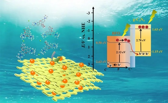
3.3. Reaction Mechanisms

As aforementioned, the photodegradation of RhB over 10BiFeO/hg-C3N4 was almost 100% within 60 min, far beyond the hg-C3N4, BiFeO3, and 10BiFeO/bg-C3N4 catalysts. The apparent discrepancies in photocatalytic performance will be discussed from micro-nano structure, adsorption properties, and the formation of the internal field.

3.3.1. Micro-Nano Structure

The large specific surface area of the prepared 10BiFeO/hg-C3N4 and well-dispersed BiFeO3 particles on the hg-C3N4 support (Figure 7) increased the contact probabilities between the reactants and the catalysts. In addition, the unique honeycomb morphology of 10BiFeO/hg-C3N4 formed multiple reflections of light through interlaminar pore structure. This multi-reflection can effectively improve the utilization of light energy and promote the diffusion of the reactants [36].

3.3.2. Adsorption of Reactant Molecules on the Catalysts

In the typical heterogeneous catalytic degradation of RhB, the key step is the adsorption of the reactant molecules onto the catalyst’s surface. The RhB solution with photocatalysts was stirred in a dark room for 30 min to achieve a stable absorption–desorption equilibrium before light irradiation. The UV/vis absorption of the dye solution was used to record the amount of the reactant molecules that were adsorbed on the surface of the catalysts in the dark. As shown in Figure 8a, the capacity of the adsorption molecules for hg-C3N4 and bg-C3N4 was similar. The adsorption ability was enhanced by a factor of approximately seven when BiFeO3 particles were deposited on graphite C3N4. The enhancement indicated that the strong Stern layers with the dye molecules formed on the surface of BiFeO3 ferroelectrics [44]. As shown in Figure 8b, the spontaneous polarization of ferroelectrics can induce surface macroscopic charges. The induced charges could be compensated by the charged species from the external environment [44]. It has been reported that the polarized ferroelectrics showed stronger adsorption of dye molecules on C+ and C surfaces than the non-ferroelectrics surface. This interaction between the dipole moment of a polar molecule (or induced dipole moment) and the polarization of ferroelectric domains can promote the bond-breaking of the adsorbed molecules and enhance photocatalytic activity [45,46,47].
The degradation of anionic dye methyl red (MR) was carried out following the same reaction conditions as the degradation of RhB to further identify the importance of the formation of stern layers with chemisorbed molecules. The adsorption of methyl red onto the catalysts was not evident (Figure S2). Figure S3 showed a 19% difference in MR degradation rate between the hg-C3N4 and 10BiFeO/hg-C3N4 within 60 min, and this difference is lower than that in RhB degradation. This indicated that the adsorption of RhB onto the catalysts played a vital role. Despite the lack of dye adsorption, 10BiFeO/hg-C3N4 still shows the highest activity in the photodegradation of methyl red among all tested catalysts.

3.3.3. The Internal Field

The separation efficiency of the photogenerated electron–hole pairs significantly impacted the photocatalytic activity. The fluorescence spectra and photoelectrochemical experiments were carried out to study the charge transfer resistance and the separation efficiency of the charge carriers. The room temperature PL spectra with an excitation wavelength of 325 nm for the hg-C3N4, 10BiFeO/hg-C3N4, and 10BiFeO/bg-C3N4 were displayed in Figure 9a. The composites have much lower PL intensity than the pure samples, meaning that the recombination of photogenerated electron–hole pairs can be effectively suppressed. The PL intensity of 10BiFeO/hg-C3N4 was weaker than that of 10BiFeO/bg-C3N4, which can be attributed to the special nanoarchitecture. The larger surface area and thinner thickness of honeycomb-like g-C3N4 promoted charge separation and migration [36]. Meanwhile, the efficient formation of heterojunction originated from the strong interaction between the well-dispersed BiFeO3 particles and honeycomb-like g-C3N4. The strong interaction contributes to the inhibition of photogenerated electron–hole recombination. The charge separation dynamics of the hg-C3N4, 10BiFeO/hg-C3N4, and 10BiFeO/bg-C3N4 were investigated by the time-resolved photoluminescence spectra (Figure 9b). In addition, the photocurrent–time response curves and EIS curves for the samples indicated that the 10BiFeO/hg-C3N4 had improved charger carrier mobility due to its highest current density (Figure 9c) and smallest charge transfer resistance according to the smallest hemicycle radius (Figure 9d) [39,48].
We employed UV-vis absorption spectra to examine the optical absorption properties of the synthesized pure graphite C3N4, BiFeO3, and the composite samples. As shown in Figure 10a,b, the absorption peaks of the pure graphite C3N4 and BiFeO3 were well consistent with the previous literature [22], and the absorption edge was located at around 450 nm and 600 nm, respectively. The absorption peak intensity for the hg-C3N4 was stronger than bg-C3N4 in the ultraviolet and visible light region due to the large surface area and the multiple reflections of incident light in the honeycomb structure [36]. The 10BiFeO/hg-C3N4 presented stronger peak intensity than 10BiFeO/bg-C3N4 attributed to the same reason, as shown in Figure 10b.
The band gap was calculated according to the following Equation (1) [46]:
(αhν)n = k(hν − Eg)
where h is Planck’s constant, ν is vibration frequency, α represents the absorption coefficient, Eg is the band gap, and k is the proportional constant. Figure 10c suggests that the band gap of the BiFeO3 and hg-C3N4 were 2.13 eV and 2.78 eV, respectively. Based on the band gap, we further accessed the conduction band (CB) and valence band (VB) levels of the BiFeO3 and hg-C3N4 by using the Mulliken electronegativity formula [49]. The CB and VB for the BiFeO3 were estimated to be +2.63 eV and +0.50 eV (vs. NHE), and the CB and VB of the hg-C3N4 were +1.63 eV and −1.15 eV, respectively (Figure 10d). The absorption edge of the composites was well aligned with the pure graphite C3N4 and BiFeO3. This indicated that the band gap of the graphite C3N4 and BiFeO3 in the composites was almost unchanged [28].
DMPO spin-trapping ESR was carried out to identify the possible reactive species involved in the degradation of RhB. The free radicals generated by BiFeO3, hg-C3N4, and 10BiFeO/hg-C3N4 were captured under visible light illumination. In Figure 11a, the ESR spectra of BiFeO3 indicated that only •OH radicals could be formed during the photoreaction. Considering the band structure of BiFeO3, the holes from BiFeO3 have the sufficient capacity to oxidize OH/H2O into •OH radicals due to its more positive VB potential (2.63 eV vs. NHE) than E (OH/•OH) (1.99 eV vs. NHE) and E(H2O/•OH) (2.38 eV vs. NHE) [50]. The signal of DMPO-•OH can also be observed in the hg-C3N4, and the •OH may come from the H2O2 produced by the reaction of O2 and H2O [51]. Meanwhile, the more negative CB in the hg-C3N4 can directly reduce O2 to form O2. Regarding the 10BiFeO/hg-C3N4, both the signals of DMPO-•OH and DMPO-O2 can be clearly observed. Furthermore, to rule out the possibility that DMPO-•OH originated from the degradation of the adduct DMPO-•OOH between DMPO and O2 [52,53], we investigate the degradation of coumarin to detect hydroxyl radicals preliminarily. As shown in Figure S5, for the 10BiFeO/hg-C3N4, the luminescence intensity of coumarin decreased rapidly with irradiation time due to the formation of •OH radicals, which can well support the results of ESR spin trapping experiments. Based on the possible reactive species, tertbutanol (t-BuOH), benzoquinone (BQ), and triethanolamine (TEOA) were chosen as quenchers of •OH radical, O2 radical, and h+, respectively, to investigate the role of active radicals on the degradation rate of RhB over 10BiFeO/hg-C3N4 [20]. From Figure 11b, the photodegradation rate has no noticeable change after introducing t-BuOH and TEOA into the reaction system. While after the addition of BQ, the photodegradation was significantly inhibited, suggesting that the O2 radical was the critical active species.
The proper band alignment between hg-C3N4 and BiFeO3 led to the formation of an efficient heterostructure and facilitated charge separation. Two mainstream charge-transfer mechanisms, type-II and Z-scheme, have been widely acknowledged [19]. For the type-Ⅱ mode, the two coupled semiconductors generate electron and hole pairs under irradiation. The photogenerated electrons were transferred from hg-C3N4 to BiFeO3, while the photogenerated holes were moved in the opposite direction. The trapping experiments suggested that O2 radicals are the main reactive species in the degradation reaction. The accumulated electrons in the BiFeO3 CB cannot generate O2 radicals because of its more positive CB edge potential than the reduction potential of O2. Therefore, it is hard to explain the mechanism of this reaction using the predicted type-II heterojunction. Regarding the Z-scheme mode, the photogenerated electrons in the BiFeO3 CB directly recombined with the photogenerated holes. The reserved photogenerated electrons in the CB of hg-C3N4 and holes in the VB of BiFeO3 still maintain the strong redox ability. The accumulated electrons in hg-C3N4 CB have enough energy to generate O2 radicals, effectively overcoming the limitation in type-Ⅱ mode.
The photo-deposition of PbO2 and Ag particles on the 10BiFeO/hg-C3N4 was carried out to gain further insight into the transfer route of the photogenerated carriers. In general, PbO2 nanoparticles characterize the site of the photogenerated hole flow, and Ag can reveal the photogenerated electron route [54,55]. As shown in Figure 12, according to the lattice fringe spacings from the plane of different species, the PbO2 nanoparticles were primarily photo-deposited onto the BiFeO3 particles, and the Ag nanoparticles are mainly distributed on hg-C3N4. The different distribution routes demonstrated that the photogenerated holes reserved in BiFeO3 and the electrons tend to remain in hg-C3N4. In situ irradiated XPS spectra were also performed to reveal the charge transfer between hg-C3N4 and BiFeO3. Upon exposure to photon illumination, C 1s and N 1s peaks move to lower binding energy, while the Bi 4f and Fe 2p peaks move in the opposite direction. The moving directions suggested a charge transfer from BiFeO3 to hg-C3N4 under light irradiation (Figure 13) [56]. These results provided convincing evidence that the Z-scheme mode is the proper reaction mechanism for RhB photocatalyzed degradation over 10BiFeO/hg-C3N4.
Overall, a possible mechanism for the photodegradation of RhB over 10BiFeO/hg-C3N4 can be deduced and shown in Figure 14. Under visible irradiation, the excited electrons on the BiFeO3 CB migrated to the hg-C3N4 VB on the interface between BiFeO3 and hg-C3N4. As a result, it left a trail of electrons and holes on the hg-C3N4 CB and BiFeO3 VB. The interfacial field was built within the interface of two regions, substantially increasing the separation efficiency of the charge carrier. This Z-scheme heterojunction exhibited both high charge separation and remarkable redox ability. The internal electric field, caused by the spontaneous polarization of BiFeO3 species, can increase the band bending and facilitate the migration of photogenerated carriers in the opposite direction. The superposition of these two fields can make the photogenerated carriers separate in space and effectively reduce the recombination opportunity. In addition, the ferroelectricity of BiFeO3 ensures a tightly bound layer of the dye molecule. The tightly bound layer can reduce the required energy to break bonds and enhance photocatalytic performance.

4. Conclusions

In this study, we synthesized a novel 10BiFeO/hg-C3N4 type-Z heterojunction photocatalyst via a simple, economical, non-toxic, and energy-saving one-step method. The characterization results showed that the BiFeO3 particles are well dispersed on the honeycomb-like g-C3N4, and the close contact led to the strong interaction between the two species. All of the composites showed a stronger photocatalytic degradation capacity to RhB than pure graphite C3N4 and BiFeO3 under the same conditions. The 10BiFeO/hg-C3N4 presented the best catalytic performance. The mechanism with respect to the photocatalytic performance improvement over the composites was discussed in detail. The unique honeycomb-like morphology of 10BiFeO/hg-C3N4 promoted the reactant diffusion and utilization of light energy. The spontaneous polarization of ferroelectric BiFeO3 induced a greater degree of binding between the polar RhB cation and the catalyst surface. Moreover, the synergistic effect between ferroelectric polarization and direct Z-scheme heterojunction leads to the efficient separation and the transfer of photogenerated carriers.

Supplementary Materials

The following supporting information can be downloaded at: https://www.mdpi.com/article/10.3390/nano12223970/s1, Figure S1: N2 physisorption isotherms of (a) bg-C3N4 and hg-C3N4; (b) 10BFO/bg-C3N4 and 10BFO/hg-C3N4; Figure S2: UV-Vis spectra of initial methyl red solution and after dark adsorption-desorption equilibrium for 30 min with 10BFO/hg-C3N4; Figure S3: Photodegradation profiles of methyl red with different catalysts under solar simulator within 60 min; Figure S4: The particle size distribution of the samples; Figure S5: Disappearance of coumarin under irradiation.

Author Contributions

H.C. conceived and designed the experiments; H.C., Z.W. and Y.L. carried out the synthetic experiment and photo-catalytic performance of the as-prepared samples; H.C., Z.W., G.C., Y.W., J.S. and L.Z. measured the as-prepared samples and analyzed the data; H.C. wrote the paper; Z.W. and G.C. revised the paper; J.M. and X.C. provided precise instruction. All authors have read and agreed to the published version of the manuscript.

Funding

This research was funded by the Young Academic Leaders Project of North University of China (No. 11045501), National Natural Science Foundation of China (No. 62001431, No. 22002143) and Shanxi Province Science Foundation for Youths (No. 201901D211223).

Institutional Review Board Statement

Not applicable.

Informed Consent Statement

Not applicable.

Data Availability Statement

The data presented in this study are available in this article.

Conflicts of Interest

The authors declare no conflict of interest.

References

  1. Lops, C.; Ancona, A.; Cesare, D.K.; Dumontel, B.; Garino, N.; Canavese, G.; Hérnandez, S.; Cauda, V. Sonophotocatalytic degradation mechanisms of rhodamine B dye via radicals generation by micro- and nano-particles of ZnO. Appl. Catal. B Environ. 2019, 243, 629–640. [Google Scholar] [CrossRef] [PubMed]
  2. Alakhras, F.; Alhajri, E.; Haounati, R.; Ouachtak, H.; Addi, A.A.; Saleh, T.A. A comparative study of photocatalytic degradation of rhodamine B using natural-based Zeolite composites. Surf. Interfaces 2020, 20, 100611. [Google Scholar] [CrossRef]
  3. Wang, T.; Liu, S.; Mao, W.; Bai, Y.; Chiang, K.; Shah, K.; Paz-Ferreiro, J. Novel Bi2WO6 loaded N-biochar composites with enhanced photocatalytic degradation of rhodamine B and Cr(VI). J. Hazard. Mater. 2020, 389, 121827. [Google Scholar] [CrossRef] [PubMed]
  4. Gao, Q.; Lei, Q.; Miao, R.; Gao, M.; Liu, H.; Yang, Q.; Liu, Y.; Song, F.; Yu, Y.; Yang, W. Bi-doped graphitic carbon nitride nnanotubes boost the photocatalytic degradation of rhodamine B. New J. Chem. 2022, 46, 3588–3594. [Google Scholar] [CrossRef]
  5. Al-Buriahi, A.K.; Al-Gheethi, A.A.; Senthil Kumar, P.; Radin Mohamed, R.M.S.; Yusof, H.; Alshalif, A.F.; Khalifa, N.A. Elimination of rhodamine B from textile wastewater using nanoparticle photocatalysts: A review for sustainable approaches. Chemosphere 2022, 287, 132162. [Google Scholar] [CrossRef] [PubMed]
  6. Yao, Y.; Sun, M.; Yuan, X.; Zhu, Y.; Lin, X.; Anandan, S. One-step hydrothermal synthesis of N/Ti3+ co-doping multiphasic TiO2/BiOBr heterojunctions towards enhanced sonocatalytic performance. Ultrason. Sonochem. 2018, 49, 69–78. [Google Scholar] [CrossRef]
  7. Xu, D.; Ma, H. Degradation of rhodamine B in water by ultrasound-assisted TiO2 photocatalysis. J. Clean. Prod. 2021, 313, 127758. [Google Scholar] [CrossRef]
  8. Wenderich, K.; Mul, G. Methods, mechanism, and applications of photodeposition in photocatalysis: A review. Chem. Rev. 2016, 116, 14587–14619. [Google Scholar] [CrossRef]
  9. Wen, J.; Xie, J.; Chen, X.; Li, X. A review on g-C3N4-based photocatalysts. Appl. Surf. Sci. 2017, 391, 72–123. [Google Scholar] [CrossRef]
  10. Fattahimoghaddam, H.; Mahvelati-Shamsabadi, T.; Lee, B.-K. Efficient photodegradation of rhodamine B and tetracycline over robust and green g-C3N4 nanostructures: Supramolecular design. J. Hazard. Mater. 2021, 403, 123703. [Google Scholar] [CrossRef]
  11. Zhang, X.; Xie, X.; Wang, H.; Zhang, J.; Pan, B.; Xie, Y. Enhanced photoresponsive ultrathin graphitic-phase C3N4 nanosheets for bioimaging. J. Am. Chem. Soc. 2013, 135, 18–21. [Google Scholar] [CrossRef]
  12. Cheng, F.; Wang, H.; Dong, X. The amphoteric properties of g-C3N4 nanosheets and fabrication of their relevant heterostructure photocatalysts by an electrostatic re-assembly route. Chem. Commun. 2015, 51, 7176–7179. [Google Scholar] [CrossRef]
  13. Zhang, M.; Yang, Y.; An, X.; Hou, L. A critical review of g-C3N4-based photocatalytic membrane for water purification. Chem. Eng. J. 2021, 412, 128663. [Google Scholar] [CrossRef]
  14. Lan, H.; Li, L.; An, X.; Liu, F.; Chen, C.; Liu, H.; Qu, J. Microstructure of carbon nitride affecting synergetic photocatalytic sctivity: Hydrogen bonds vs. structural defects. Appl. Catal. B Environ. 2017, 204, 49–57. [Google Scholar] [CrossRef]
  15. Zhao, T.; Xing, Z.; Xiu, Z.; Li, Z.; Yang, S.; Zhou, W. Oxygen-doped MoS2 nanospheres/CdS quantum dots/g-C3N4 nanosheets super-architectures for prolonged charge lifetime and enhanced visible-light-driven photocatalytic performance. ACS Appl. Mater. Interfaces 2019, 11, 7104–7111. [Google Scholar] [CrossRef]
  16. Wu, M.; He, X.; Jing, B.; Wang, T.; Wang, C.; Qin, Y.; Ao, Z.; Wang, S.; An, T. Novel carbon and defects co-modified g-C3N4 for highly efficient photocatalytic degradation of bisphenol A under visible light. J. Hazard. Mater. 2020, 384, 121323. [Google Scholar] [CrossRef]
  17. Katsumata, K.; Motoyoshi, R.; Matsushita, N.; Okada, K. Preparation of graphitic carbon nitride (g-C3N4)/WO3 composites and enhanced visible-light-driven photodegradation of acetaldehyde gas. J. Hazard. Mater. 2013, 260, 475–482. [Google Scholar] [CrossRef]
  18. Zhou, C.; Lai, C.; Huang, D.; Zeng, G.; Zhang, C.; Cheng, M.; Hu, L.; Wan, J.; Xiong, W.; Wen, M.; et al. Highly porous carbon nitride by supramolecular preassembly of monomers for photocatalytic removal of sulfamethazine under visible light driven. Appl. Catal. B Environ. 2018, 220, 202–210. [Google Scholar] [CrossRef]
  19. Low, J.; Yu, J.; Jaroniec, M.; Wageh, S.; Al-Ghamdi, A.A. Heterojunction photocatalysts. Adv. Mater. 2017, 29, 1601694. [Google Scholar] [CrossRef]
  20. Wei, H.; McMaster, W.A.; Tan, J.Z.Y.; Cao, L.; Chen, D.; Caruso, R.A. Mesoporous TiO2/g-C3N4 microspheres with enhanced visible-light photocatalytic activity. J. Phys. Chem. C. 2017, 121, 22114–22122. [Google Scholar] [CrossRef]
  21. Guan, R.; Li, J.; Zhang, J.; Zhao, Z.; Wang, D.; Zhai, H.; Sun, D. Photocatalytic performance and mechanistic research of ZnO/g-C3N4 on degradation of methyl orange. ACS Omega 2019, 4, 20742–20747. [Google Scholar] [CrossRef] [PubMed] [Green Version]
  22. Yang, Y.; Zhu, J.; He, Y.; Li, M.; Liu, Y.; Chen, M.; Cao, D. Charge transfer in photocatalysis of direct Z-scheme g-C3N4-based ferroelectric heterojunction. J. Alloys Compd. 2022, 893, 162270. [Google Scholar] [CrossRef]
  23. Li, Y.; Qin, T.; Chen, W.; Huang, M.; Xu, J.; Lv, J. Construction of a switchable g-C3N4/BiVO4 heterojunction from the Z-Scheme to the type II by incorporation of pyromellitic diimide. Cryst. Growth Des. 2022, 22, 1645–1653. [Google Scholar] [CrossRef]
  24. Subhiksha, V.; Kokilavani, S.; Sudheer Khan, S. Recent advances in degradation of organic pollutant in aqueous solutions using bismuth based photocatalysts: A review. Chemosphere 2022, 290, 133228. [Google Scholar] [CrossRef] [PubMed]
  25. Sun, H.; Ai, Y.; Li, D.; Tang, Z.; Shao, Z.; Liang, Q. Bismuth iron oxide nanocomposite supported on graphene oxides as the high efficient, stable and reusable catalysts for the reduction of nitroarenes under continuous flow conditions. Chem. Eng. J. 2017, 314, 328–335. [Google Scholar] [CrossRef]
  26. Irfan, S.; Zheng, Z.; Li, F.; Chen, Y.X.; Liang, G.X.; Luo, J.T.; Ping, F. Critical review: Bismuth ferrite as an emerging visible light active nanostructured photocatalyst. J. Mater. Res. Technol. 2019, 8, 6375–6389. [Google Scholar] [CrossRef]
  27. Zou, X.; You, L.; Chen, W.; Ding, H.; Wu, D.; Wu, T.; Chen, L.; Wang, J. Mechanism of polarization fatigue in BiFeO3. ACS Nano 2012, 6, 8997–9004. [Google Scholar] [CrossRef]
  28. Deng, X.-Z.; Song, C.; Tong, Y.-L.; Yuan, G.; Gao, F.; Liu, D.-Q.; Zhang, S.-T. Enhanced photocatalytic efficiency of C3N4/BiFeO3 heterojunctions: The synergistic effects of band alignment and ferroelectricity. Phys. Chem. Chem. Phys. 2018, 20, 3648–3657. [Google Scholar] [CrossRef]
  29. Lan, H.; Tang, Y.; Zhang, X.; You, S.; Tang, Q.; An, X.; Liu, H.; Qu, J. Decomplexation of Cu(II)-EDTA over oxygen-doped g-C3N4: An available resource towards environmental sustainability. Chem. Eng. J. 2018, 345, 138–146. [Google Scholar] [CrossRef]
  30. Zhang, M.; Yang, Y.; An, X.; Zhao, J.; Bao, Y.; Hou, L. Exfoliation method matters: The microstructure-dependent photoactivity of g-C3N4 nanosheets for water purification. J. Hazard. Mater. 2022, 424, 127424. [Google Scholar] [CrossRef]
  31. Li, Y.; Wang, M.-Q.; Bao, S.-J.; Lu, S.; Xu, M.; Long, D.; Pu, S. Tuning and thermal exfoliation graphene-like carbon nitride nanosheets for superior photocatalytic activity. Ceram. Int. 2016, 42, 18521–18528. [Google Scholar] [CrossRef]
  32. Zou, L.-R.; Huang, G.-F.; Li, D.-F.; Liu, J.-H.; Pan, A.-L.; Huang, W.-Q. A facile and rapid route for synthesis of g-C3N4 nanosheets with high adsorption capacity and photocatalytic activity. RSC Adv. 2016, 6, 86688–86694. [Google Scholar] [CrossRef]
  33. Xian, T.; Yang, H.; Shen, X.; Jiang, J.L.; Wei, Z.Q.; Feng, W.J. Preparation of high-quality BiFeO3 nanopowders via a polyacrylamide gel route. J. Alloys Compd. 2009, 480, 889–892. [Google Scholar] [CrossRef]
  34. Lan, F.; Aarons, J.; Shu, Y.; Zhou, X.; Jiao, H.; Wang, H.; Guan, Q.; Li, W. Anchoring strategy for highly active copper nanoclusters in hydrogenation of renewable biomass-derived compounds. Appl. Catal. B Environ. 2021, 299, 120651. [Google Scholar] [CrossRef]
  35. Wang, B.; Yue, Y.; Jin, C.; Lu, J.; Wang, S.; Yu, L.; Guo, L.; Li, R.; Hu, Z.-T.Z.; Pan, Z.; et al. Hydrochlorination of acetylene on single-atom Pd/N-doped carbon catalysts: Importance of pyridinic-N synergism. Appl. Catal. B Environ. 2020, 272, 118944. [Google Scholar] [CrossRef]
  36. Wang, Z.; Guan, W.; Sun, Y.; Dong, F.; Zhou, Y.; Ho, W.-K. Water-assisted production of honeycomb-like g-C3N4 with ultralong carrier: Lifetime and outstanding photocatalytic activity. Nanoscale 2015, 7, 2471–2479. [Google Scholar] [CrossRef]
  37. Wang, Z.; Ban, L.; Meng, P.; Li, H.; Zhao, Y. Ethynylation of formaldehyde over CuO/SiO2 catalysts modified by Mg species: Effects of the existential states of Mg species. Nanomaterials 2019, 9, 1137. [Google Scholar] [CrossRef] [Green Version]
  38. Wang, S.; Chen, D.; Niu, F.; Zhang, N.; Qin, L.; Huang, Y. Hydrogenation-induced surface oxygen vacancies in BiFeO3 nanoparticles for enhanced visible light photocatalytic performance. J. Alloys Compd. 2016, 688, 399–406. [Google Scholar] [CrossRef]
  39. Ge, L.; Xu, Y.; Ding, L.; You, F.; Liu, Q.; Wang, K. Perovskite-type BiFeO3/ultrathin graphite-like carbon nitride nanosheets p-n heterojunction: Boosted visible-lightdriven photoelectrochemical activity for fabricating ampicillin aptasensor. Biosens. Bioelectron. 2019, 124, 33–39. [Google Scholar] [CrossRef]
  40. Vignesh, S.; Muppudathi, A.L.; Sundar, J.K. Multifunctional performance of gC3N4-BiFeO3-Cu2O hybrid nanocomposites for magnetic separable photocatalytic and antibacterial activity. J. Mater. Sci. Mater. Electron. 2018, 29, 10784–10801. [Google Scholar] [CrossRef]
  41. Huo, H.; Hu, X.; Wang, H.; Li, J.; Xie, G.; Tan, X.; Jin, Q.; Zhou, D.; Li, C.; Qiu, G.; et al. Synergy of photocatalysis and adsorption for simultaneous removal of hexavalent chromium and methylene blue by g-C3N4/BiFeO3/carbon nanotubes ternary composites. Int. J. Environ. Res. Public Health 2019, 16, 3219. [Google Scholar] [CrossRef] [PubMed] [Green Version]
  42. Zhu, D.; Zhou, Q. Nitrogen doped g-C3N4 with the extremely narrow band gap for excellent photocatalytic sctivities under visible light. Appl. Catal. B Environ. 2021, 281, 119474. [Google Scholar] [CrossRef]
  43. Khan, M.A.; Nadeem, M.A.; Idriss, H. Ferroelectric polarization effect on surface chemistry and photo-catalytic activity: A review. Surf. Sci. Rep. 2016, 71, 1–31. [Google Scholar] [CrossRef]
  44. Cui, Y.; Briscoe, J.; Dunn, S. Effect of Ferroelectricity on Solar-Light-Driven Photocatalytic activity of BaTiO3—Influence on the carrier separation and stern layer formation. Chem. Mater. 2013, 25, 4215–4223. [Google Scholar] [CrossRef]
  45. Dunn, S.; Jones, P.M.; Gallardo, D.E. Photochemical growth of silver nanoparticles on c- and c+ domains on lead Zirconate Titanate thin films. J. Am. Chem. Soc. 2007, 129, 8724–8728. [Google Scholar] [CrossRef] [PubMed]
  46. Dunn, S.; Shaw, C.P.; Huang, Z.; Whatmore, R.W. Ultrahigh resolution of lead Zirconate Titanate 30/70 domains as imaged by piezoforce microscopy. Nanotechnology 2002, 13, 456–459. [Google Scholar] [CrossRef]
  47. Dunn, S.; Tiwari, D.; Jones, P.M.; Gallardo, D.E. Insights into the relationship between inherent materials properties of PZT and photochemistry for the development of nanostructured silver. J. Mater. Chem. 2007, 17, 4460–4463. [Google Scholar] [CrossRef]
  48. Cai, X.; Wang, F.; Wang, R.; Xi, Y.; Wang, A.; Wang, J.; Teng, B.; Bai, S. Synergism of surface strain and interfacial polarization on Pd@Au core–shell cocatalysts for highly efficient photocatalytic CO2 reduction over TiO2. J. Mater. Chem. A 2020, 8, 7350–7359. [Google Scholar] [CrossRef]
  49. He, Y.; Wang, P.; Zhu, J.; Yang, Y.; Liu, Y.; Chen, M.; Cao, D.; Yan, X. Synergistical dual strategies based on in situ-converted heterojunction and reduction-induced surface oxygen vacancy for enhanced photoelectrochemical performance of TiO2. ACS Appl. Mater. Interfaces 2019, 11, 37322–37329. [Google Scholar] [CrossRef]
  50. Sepahvand, H.; Sharifnia, S. Photocatalytic overall water splitting by Z-scheme g-C3N4/BiFeO3 heterojunction. Int. J. Hydrog. Energy 2019, 44, 23658–23668. [Google Scholar] [CrossRef]
  51. Zhu, B.; Xia, P.; Li, Y.; Ho, W.; Yu, J. Fabrication and photocatalytic activity enhanced mechanism of direct Z-scheme g-C3N4/Ag2WO4 photocatalyst. Appl. Surf. Sci. 2017, 391, 175–183. [Google Scholar] [CrossRef]
  52. Finkelstein, E.; Rosen, G.M.; Rauckman, E.J. Production of hydroxyl radical by decomposition of superoxide spin-trapped adducts. Mol. Pharmacol. 1982, 21, 262–265. [Google Scholar]
  53. Buettner, G.R. The spin trapping of superoxide and hydroxyl free radicals with DMPO (5,5-dimethylpyrroline-N-oxide): More about iron. Free Radic. Res. Commun. 1993, 19, S79–S87. [Google Scholar] [CrossRef] [Green Version]
  54. Ma, Y.; Lv, P.; Duan, F.; Sheng, J.; Lu, S.; Zhu, H.; Du, M.; Chen, M. Direct Z-scheme Bi2S3/BiFeO3 heterojunction nanofibers with enhanced photocatalytic sctivity. J. Alloys Compd. 2020, 834, 155158. [Google Scholar] [CrossRef]
  55. Jiang, W.; Zong, X.; An, L.; Hua, S.; Miao, X.; Luan, S.; Wen, Y.; Tao, F.F.; Sun, Z. Consciously constructing heterojunction or direct Z-Scheme photocatalysts by regulating electron flow direction. ACS Catal. 2018, 8, 2209–2217. [Google Scholar] [CrossRef]
  56. Zhang, C.; Huang, Y.; Zhao, B.; Yu, Y.; Yu, Y.; Zhang, B. CuOx clusters decorated TiO2 for photocatalytic oxidation of nitrogen in air into nitric oxide under ambient conditions. J. Catal. 2022, 409, 70–77. [Google Scholar] [CrossRef]
Figure 1. XRD patterns of following catalysts: (a) hg-C3N4, bg-C3N4, 10BiFeO/hg-C3N4, and 10BiFeO/bg-C3N4; (b) xBiFeO/hg-C3N4 (5%, 10%, and 20% BiFeO3).
Nanomaterials 12 03970 g001
Figure 2. FT-IR transmission spectra of (a) hg-C3N4 and bg-C3N4; (b) 10BiFeO/hg-C3N4, and 10BiFeO/bg-C3N4.
Nanomaterials 12 03970 g002
Figure 3. TEM images of synthesized samples: (a) bg-C3N4; (b) hg-C3N4; (c) 10BiFeO/bg-C3N4; and (d) 10BiFeO/hg-C3N4.
Nanomaterials 12 03970 g003
Figure 4. XPS spectra profiles (a) survey of hg-C3N4, 10BiFeO/hg-C3N4, 10BiFeO/bg-C3N4, and BiFeO; (b) Bi 4f of 10BiFeO/hg-C3N4, 10BiFeO/bg-C3N4, and BiFeO; (c) Fe 2p of 10BiFeO/hg-C3N4, 10BiFeO/bg-C3N4, and BiFeO; (d) O 1s of 10BiFeO/hg-C3N4, 10BiFeO/bg-C3N4, and BiFeO; (e) C 1s of hg-C3N4, 10BiFeO/hg-C3N4, and 10BiFeO/bg-C3N4; (f) N 1s of hg-C3N4, 10BiFeO/hg-C3N4, and 10BiFeO/bg-C3N4.
Nanomaterials 12 03970 g004
Figure 5. Photocatalytic degradation of RhB: (a) the synthesized g-C3N4, BiFeO and BiFeO/g-C3N4 samples; (b) the pseudo-first-order kinetics curves of BiFeO, hg-C3N4 and 10BiFeO/hg-C3N4; (c) xBiFeO/hg-C3N4 (5%, 10%, and 20% BiFeO3); (d) electrically poled 10BiFeO/hg-C3N4.
Nanomaterials 12 03970 g005
Figure 6. (a) Recycling runs for the photocatalytic RhB in the presence of 10BiFeO/hg-C3N4 under visible light irradiation; (b) M-H hysteresis loop of 10BiFeO/hg-C3N4 after the photocatalytic measurements.
Nanomaterials 12 03970 g006
Figure 7. Schematics of honeycomb-like morphology.
Nanomaterials 12 03970 g007
Figure 8. (a) Adsorption of RhB by g-C3N4 and BiFeO/g-C3N4 under dark conditions for 30 min; (b) internal polarization and adsorption mechanisms.
Nanomaterials 12 03970 g008
Figure 9. Steady-state PL spectra (a), time-resolved PL spectra (b), photocurrent response density (c) and EIS Nyquist plots (d) of hg-C3N4,10BiFeO/hg-C3N4 and 10BiFeO/bg-C3N4.
Nanomaterials 12 03970 g009
Figure 10. (a) UV-visible spectra of hg-C3N4 and bg-C3N4; (b) UV-visible spectra of BiFeO, 10BiFeO/hg-C3N4 and 10BiFeO/bg-C3N4; (c) estimated band gaps of BiFeO, hg-C3N4 and 10BiFeO/hg-C3N4; (d) band gap structure of 10BiFeO/hg-C3N4.
Nanomaterials 12 03970 g010
Figure 11. (a) ESR signals of BiFeO, hg-C3N4 and 10BiFeO/hg-C3N4; (b) Photocatalytic activity of 10BiFeO/hg-C3N4 for the degradation of RhB in the presence of different scavengers.
Nanomaterials 12 03970 g011
Figure 12. HRTEM images of (a) Ag and (b) PbO2 photo deposited in the existence of a BiFeO/hg-C3N4 sample, respectively.
Nanomaterials 12 03970 g012
Figure 13. XPS spectra of BiFeO/g-C3N4 in the dark and light irradiation: (a) Bi 4f, (b) Fe 2p, (c) C 1s, (d) N 1s.
Nanomaterials 12 03970 g013
Figure 14. Schematic illustration of the charge-carrier migration mechanism of Z-scheme heterojunction for 10BiFeO/g-C3N4.
Nanomaterials 12 03970 g014
Table 1. Structural and textural properties of the catalysts.
SampleBiFeO Content (wt. %) aDBiFeO (nm) bABET (m2/g) cVTotal (cm3/g) c
bg-C3N4--38.80.11
hg-C3N4--61.40.19
10BiFeO/bg-C3N410.729.318.20.07
10BiFeO/hg-C3N49.218.541.80.12
5BiFeO/hg-C3N44.312.449.60.14
20BiFeO/hg-C3N421.536.822.70.09
a The content of BiFeO3 in the catalysts were detected by ICP-AES; b The mean crystallite size of BiFeO3 calculated from XRD; c The BET surface area, pore size and volume were determined by N2 physical adsorption.
Publisher’s Note: MDPI stays neutral with regard to jurisdictional claims in published maps and institutional affiliations.

