Article

Identification of a Novel Osteogenetic Oligodeoxynucleotide (osteoDN) That Promotes Osteoblast Differentiation in a TLR9-Independent Manner

1
Department of Science and Technology, Graduate School of Medicine, Science and Technology, Shinshu University, 8304 Minami-minowa, Kami-ina, Nagano 399-4598, Japan
2
Department of Agriculture, Graduate School of Science and Technology, Shinshu University, 8304 Minami-minowa, Kami-ina, Nagano 399-4598, Japan
3
Department of Agricultural and Life Sciences, Faculty of Agriculture, Shinshu University, 8304 Minami-minowa, Kami-ina, Nagano 399-4598, Japan
4
Department of Biomolecular Innovation, Institute for Biomedical Sciences, Shinshu University, 8304 Minami-minowa, Kami-ina, Nagano 399-4598, Japan
*
Author to whom correspondence should be addressed.
Current address: Cellular and Molecular Biotechnology Research Institute, National Institute of Advanced Industrial Science and Technology, Central 5-41, 1-1-1 Higashi, Tsukuba 305-8565, Ibaraki, Japan.
Nanomaterials 2022, 12(10), 1680; https://doi.org/10.3390/nano12101680
Submission received: 1 April 2022 / Revised: 6 May 2022 / Accepted: 13 May 2022 / Published: 14 May 2022

Abstract

:
Dysfunction of bone-forming cells, osteoblasts, is one of the causes of osteoporosis. Accumulating evidence has indicated that oligodeoxynucleotides (ODNs) designed from genome sequences have the potential to regulate osteogenic cell fate. Such osteogenetic ODNs (osteoDNs) targeting and activating osteoblasts can be the candidates of nucleic acid drugs for osteoporosis. In this study, the ODN library derived from the Lacticaseibacillus rhamnosus GG genome was screened to determine its osteogenetic effect on murine osteoblast cell line MC3T3-E1. An 18-base ODN, iSN40, was identified to enhance alkaline phosphatase activity of osteoblasts within 48 h. iSN40 also induced the expression of osteogenic genes such as Msx2, osterix, collagen type 1α, osteopontin, and osteocalcin. Eventually, iSN40 facilitated calcium deposition on osteoblasts at the late stage of differentiation. Intriguingly, the CpG motif within iSN40 was not required for its osteogenetic activity, indicating that iSN40 functions in a TLR9-independent manner. These data demonstrate that iSN40 serves as a novel osteogenetic ODN (osteoDN) that promotes osteoblast differentiation. iSN40 provides a potential seed of the nucleic acid drug that activating osteoblasts for osteoporosis therapy.

1. Introduction

The bone is a metabolically active organ that remodels continuously throughout life. However, bone remodeling function is impaired with aging, resulting in reduced bone mass and micro-architectural deterioration [1]. It finally causes osteoporosis and increases fracture risk, which is related to around 200 million people and annually 8.9 million fractures in the world [2]. One of the pathological events expressing osteoporosis is the dysfunction of bone-forming cells, osteoblasts. Differentiation from osteoblasts into osteocytes is regulated by canonical Wnt/β-catenin signaling pathway [3]. It stimulates two bone-specific transcription factors, Runx2 and osterix [4], which coordinately regulate the expression of osteogenic genes such as collagen type 1α, osteopontin, osteocalcin, and receptor activator of NF-κB ligand (RANKL) [5]. However, osteoblasts of elderly people present dysregulated expression of these bone-related genes [6,7,8], indicating the decreased bone-forming capacity of aged osteoblasts. Therefore, re-activation of osteoblasts is a key therapeutic strategy for osteoporosis.
Several agents have been developed to regulate osteoblast function [9]. Teriparatide, a parathyroid hormone (PTH) fragment, directly targets PTH receptor on osteoblasts to induce osteoprotegerin and activate bone anabolism [10]. Denosumab, a decoy receptor for RANKL, increases bone mineralization by preventing bone resorption [11]. Currently, it is the most preferred treatment for post-menopausal osteoporosis [12]. Sclerostin is an inhibitor of Wnt signaling [13]. Thus, romosozumab, a sclerostin antibody, activates the Wnt pathway to stimulate bone formation [14]. Their beneficial effects in osteoporosis therapy have been robustly proven. However, with the current technology, these protein/peptide-based drugs are expensive and unstable to be produced in large quantities for the treatment of a large number of osteoporosis patients all over the world. To overcome this problem, other types of molecules targeting osteoblasts have been studied.
As nucleic acids offer several advantages, such as chemical synthesis, low-cost manufacturing, and stability during storage, they are potential nanomolecules for next-generation drugs. A wide variety of nucleotides have been clinically applied; antisense nucleotides that regulate gene expression [15], aptamers that target proteins [16], and ligands of Toll-like receptors (TLRs) that modulate the innate immune system [17] can specifically access their diverse targets. Furthermore, some oligodeoxynucleotides (ODNs) have been reported to regulate osteogenic cell fate. A 27-base cytosine (C)-rich ODN designed from the human mitochondrial genome, named MT01 ([ACC CCC TCT]3), was initially identified as an immunosuppressive ODN that inhibits the proliferation of human peripheral blood mononuclear cells [18]. Intriguingly, MT01 promoted the proliferation and differentiation of the human osteoblast-like cell line MG63 [19], rat bone marrow mesenchymal stem cells [20], and murine osteoblast cell line MC3T3-E1 [21] by upregulating Runx2 phosphorylation via activation of the ERK/MAPK pathway [22]. Thus, MT01 treatment reduced alveolar bone loss in rat periodontitis in vivo [20]. These studies suggest that genomic ODNs could be used as drug seeds for bone regeneration.
For muscle, an ODN library designed from the genome sequence of the lactic acid bacterium Lacticaseibacillus rhamnosus GG [23] has been screened to determine its effect on myogenic cell fate. The 18-base guanine (G)-rich ODN, iSN04, was found to promote differentiation and suppress inflammation of myogenic precursor cells, myoblasts [24,25,26,27,28]. iSN04, termed myogenetic ODN (myoDN), formed a globular structure of 1-nm radius and served as an anti-nucleolin aptamer in a TLR-independent manner [24]. The discovery of myoDN demonstrates that bacterial genome sequences are promising platforms for providing novel ODNs that can control cell differentiation. To authenticate this concept, this study explored the ODN library used for myoDN study to identify an osteogenetic ODN (osteoDN) that facilitates the differentiation and calcification of osteoblasts.

2. Materials and Methods

2.1. Chemicals

ODN sequences used in this study are listed in Table S1. Phosphorothioated (PS) ODNs and 6-carboxyfluorecein (6-FAM)-conjugated PS-ODNs were synthesized and purified using HPLC (GeneDesign, Osaka, Japan). PS-ODNs and ascorbic acid (AA) (Fujifilm Wako Chemicals, Osaka, Japan) were dissolved in endotoxin-free water. An equal volume of solvent was used as negative control.

2.2. Cell Culture

MC3T3-E1 cell line (RCB1126) was provided by the RIKEN BRC (Tsukuba, Japan) through the Project for Realization of Regenerative Medicine and the National Bio-Resource Project of the MEXT, Japan. The cells were cultured at 37 °C with 5% CO2 throughout the experiments. Undifferentiated MC3T3-E1 cells were maintained in a growth medium (GM) consisting of α-MEM (Nacalai, Osaka, Japan), 10% fetal bovine serum (FBS) (HyClone; GE Healthcare, Chicago, UT, USA), and a mixed solution of 100 units/mL penicillin and 100 μg/mL streptomycin (P/S) (Nacalai). To induce osteogenic differentiation, confluent MC3T3-E1 cells were cultured in a differentiation medium (DM) consisting of α-MEM, 10% FBS, 10 nM dexamethasone (Fujifilm Wako Chemicals), 10 mM β-glycerophosphate (Nacalai), and P/S. To facilitate differentiation, 15 or 50 μg/mL AA was added to GM or DM as required.

2.3. Alkaline Phosphatase (ALP) Staining

For screening, MC3T3-E1 cells were seeded on 96-well plates (1.0 × 10⁴ cells/100 μL GM/well) for screening or on 12-well plates (1.0 × 10⁴ cells/well) for high-resolution imaging. The next day, the medium was replaced with GM or DM containing 10 μM PS-ODNs. After 48 h, ALP enzymatic activity of the cells was visualized using ALP Stain Kit (Fujifilm Wako Chemicals) according to the manufacturer’s instruction. Phase-contrast images were obtained using EVOS FL Auto microscope (AMAFD1000; Thermo Fisher Scientific, Waltham, MA, USA). ALP-positive area was quantified using ImageJ software version 1.52a (Wayne Rasband; National Institute of Health, Bethesda, MD, USA).

2.4. Alizarin Staining

MC3T3-E1 cells were seeded on 24-well plates (3.0–5.0 × 10⁴ cells/well) and cultured in GM until they become confluent. The medium was then replaced with DM containing PS-ODNs every 3–4 days. The cells were fixed with 2% paraformaldehyde and stained with 1% w/v alizarin red S (Fujifilm Wako Chemicals). Bright-field images were captured using EVOS FL Auto microscope. Alizarin-positive area was quantified using ImageJ.

2.5. Quantitative Real-Time RT-PCR (qPCR)

MC3T3-E1 cells were seeded on 60-mm dishes (3.0 × 10⁵ cells/dish). The next day, the medium was replaced with GM or DM containing 10 μM iSN40. Total RNA was isolated using NucleoSpin RNA Plus (Macherey-Nagel, Düren, Germany) and reverse transcribed using ReverTra Ace qPCR RT Master Mix (TOYOBO, Osaka, Japan). qPCR was performed using GoTaq qPCR Master Mix (Promega, Madison, WI, USA) with the StepOne Real-Time PCR System (Thermo Fisher Scientific). The amount of each transcript was normalized to that of the 3-monooxygenase/tryptophan 5-monooxygenase activation protein zeta gene (Ywhaz) and presented as fold-changes. The primer sequences are listed in Table S2.

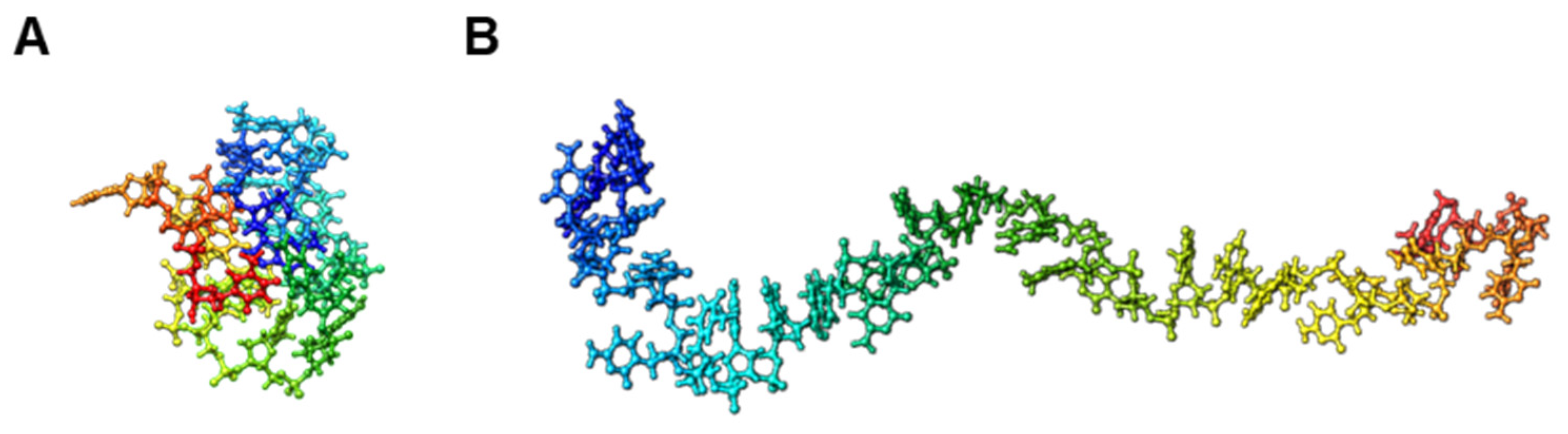
2.6. Trivial Trajectory Parallelization of Multicanonical Molecular Dynamics (TTP-McMD)

Starting with the simulation of iSN40 and MT01 structures built from their DNA sequences using NAB in AmberTools [29], an enhanced ensemble method, TTP-McMD [30], was used to sample the equilibrated conformations at 310 K. In the TTP-McMD, energy range of the multicanonical ensemble covered a temperature range from 280 K to 380 K. Sixty trajectories were used, and the production run was conducted for 40 ns in each trajectory (total 2.4 μs). Throughout the simulation, amber ff12SB force field [31] was used, whereas the solvation effect was represented by the generalized-born model [32].

2.7. Statistical Analysis

The results are presented as the mean ± standard error. Statistical comparisons between two groups were performed using unpaired two-tailed Student’s t-test and among multiple groups using one-way analysis of variance followed by Scheffe’s F-test. The statistical significance at p values are indicated in all the figures.

3. Results

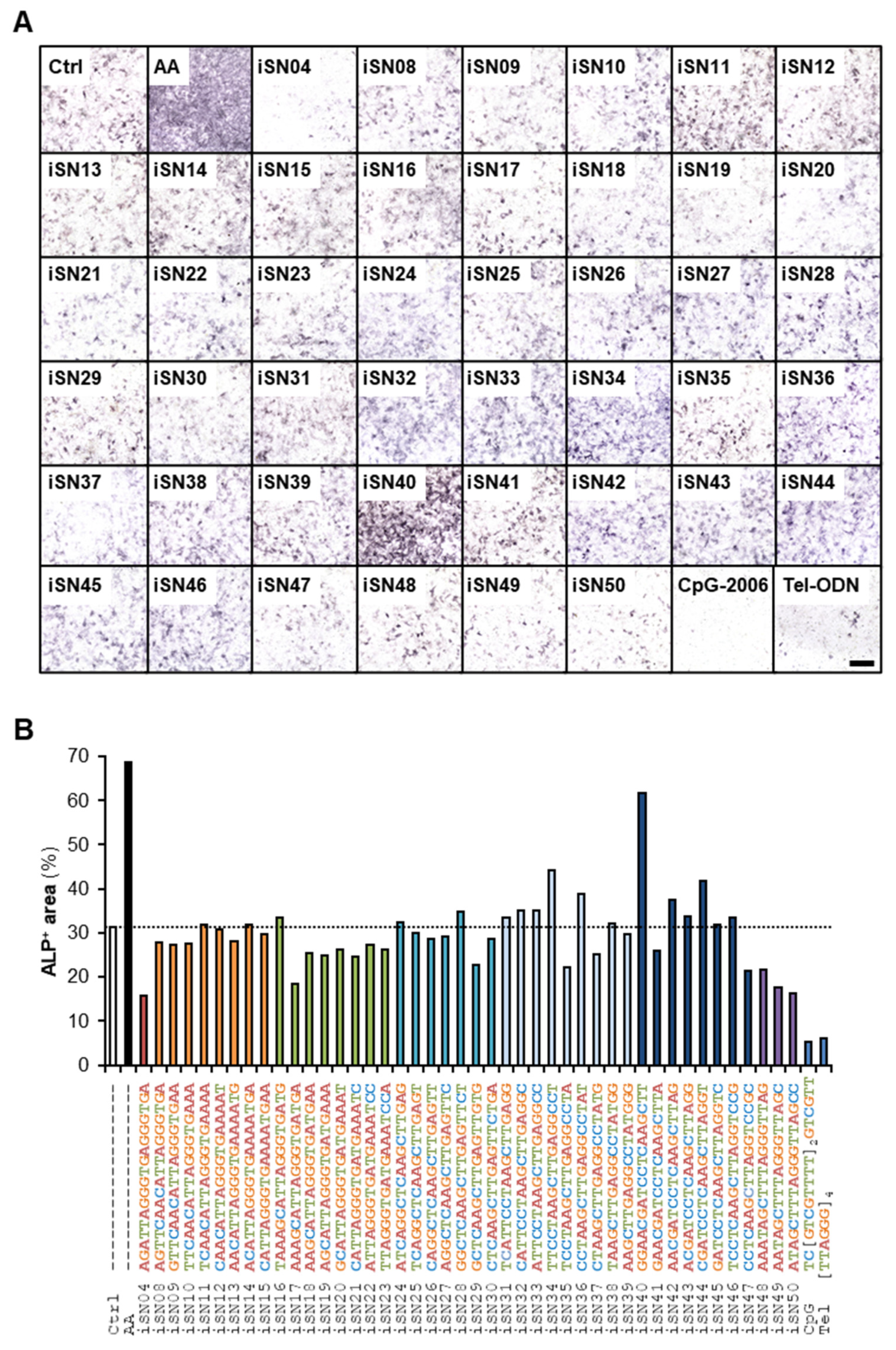
3.1. iSN40 Promotes Osteoblast Differentiation

Forty-four 18-base PS-ODNs designed from the Lacticaseibacillus rhamnosus GG genome (iSN04 and iSN08-iSN50) were administered to MC3T3-E1 cells. In addition, two immunomodulatory PS-ODNs were concomitantly tested: CpG-2006 is a TLR9 ligand that initiates inflammatory cascades [33], and Tel-ODN is a human telomeric ODN that suppresses immunological reactions depending on TLR3/7/9 [34]. The osteogenic effects of these PS-ODNs were investigated by measuring ALP enzymatic activity in the cells, a standard marker of osteoblast differentiation (Figure 1A). iSN40 (GGA ACG ATC CTC AAG CTT) markedly increased ALP-positive area to the same extent as AA, positive control for promoting osteogenic differentiation (Figure 1B). However, other PS-ODNs did not enhance ALP activity, indicating sequence-dependent osteogenetic activity of iSN40. iSN40-induced ALP activity was reproducibly confirmed by high-resolution images of the experiment performed independently of the screening (Figure S1). These results demonstrate that iSN40 serves as an osteoDN that promotes osteoblast differentiation.

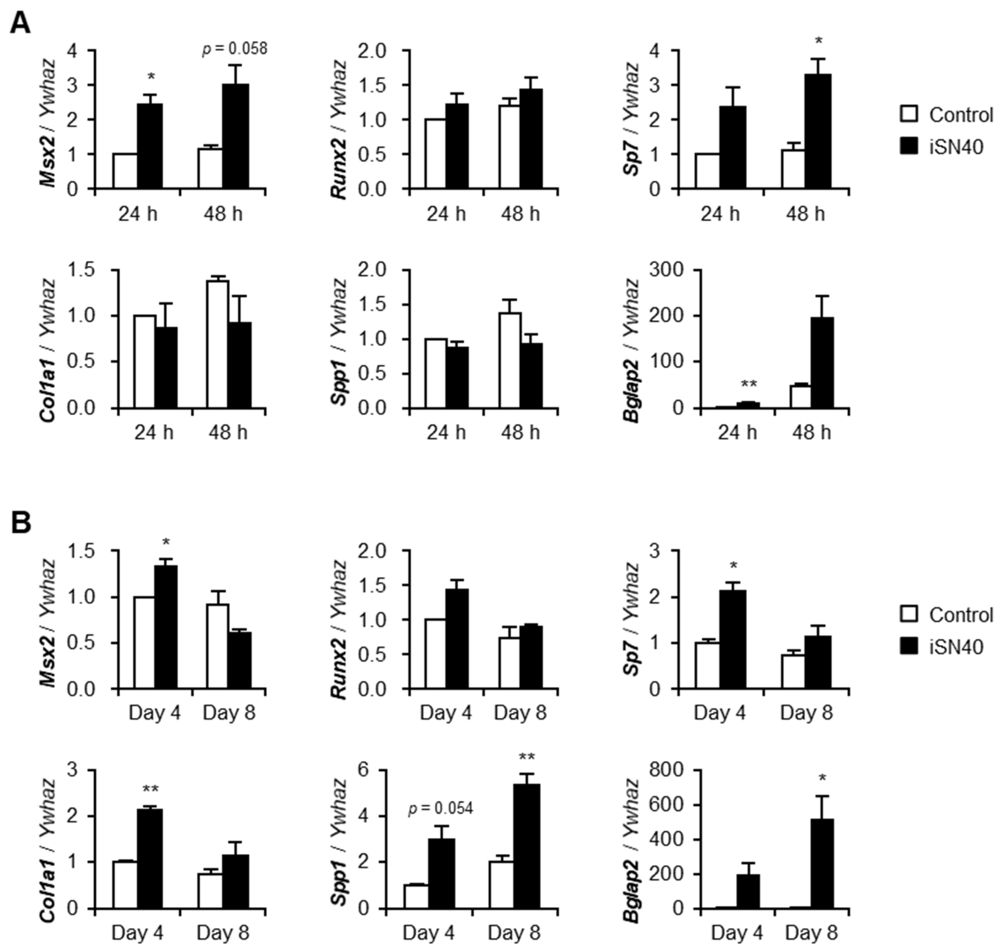
3.2. iSN40 Modulates Osteogenic Gene Expression

The effect of iSN40 on osteogenic gene expression in MC3T3-E1 cells was investigated by qPCR. To analyze the early stage of differentiation, MC3T3-E1 cells were treated with iSN40 in GM containing AA for 24–48 h (Figure 2A). iSN40 increased the mRNA levels of Msx2, a homeobox transcription factor that promotes osteogenesis through bone morphogenic protein 2-induced signaling. In contrast, iSN40 did not alter the levels of Runx2, a master regulator that determines osteoblast lineage and inhibits bone maturation [35]. Interestingly, iSN40 significantly upregulated the expression of osterix (Sp7), a downstream target of Runx2. iSN40 did not improve the mRNA levels of immature myoblast markers, collagen type 1α (Col1a1) and osteopontin (Spp1); however, iSN40 markedly induced osteocalcin (Bglap2), a hormone released from the bone [36]. To investigate the late stage of differentiation, MC3T3-E1 cells were treated with iSN40 in DM containing AA for 4–8 days (Figure 2B). iSN40 significantly elevated the levels of Msx2 on day 4 but not that of Runx2. Notably, by day 8, iSN40 upregulated the expression of osterix, collagen type 1α, osteopontin, and osteocalcin, which act downstream of Runx2 [37]. Compared with the results at the early stage, the effect of iSN40 on these gene expression was more significant at the late stage, suggesting a long-term activity of iSN40. These data indicate that iSN40 facilitates osteoblast differentiation by modulating osteogenic gene expression through the transcription factor Msx2 rather than Runx2.

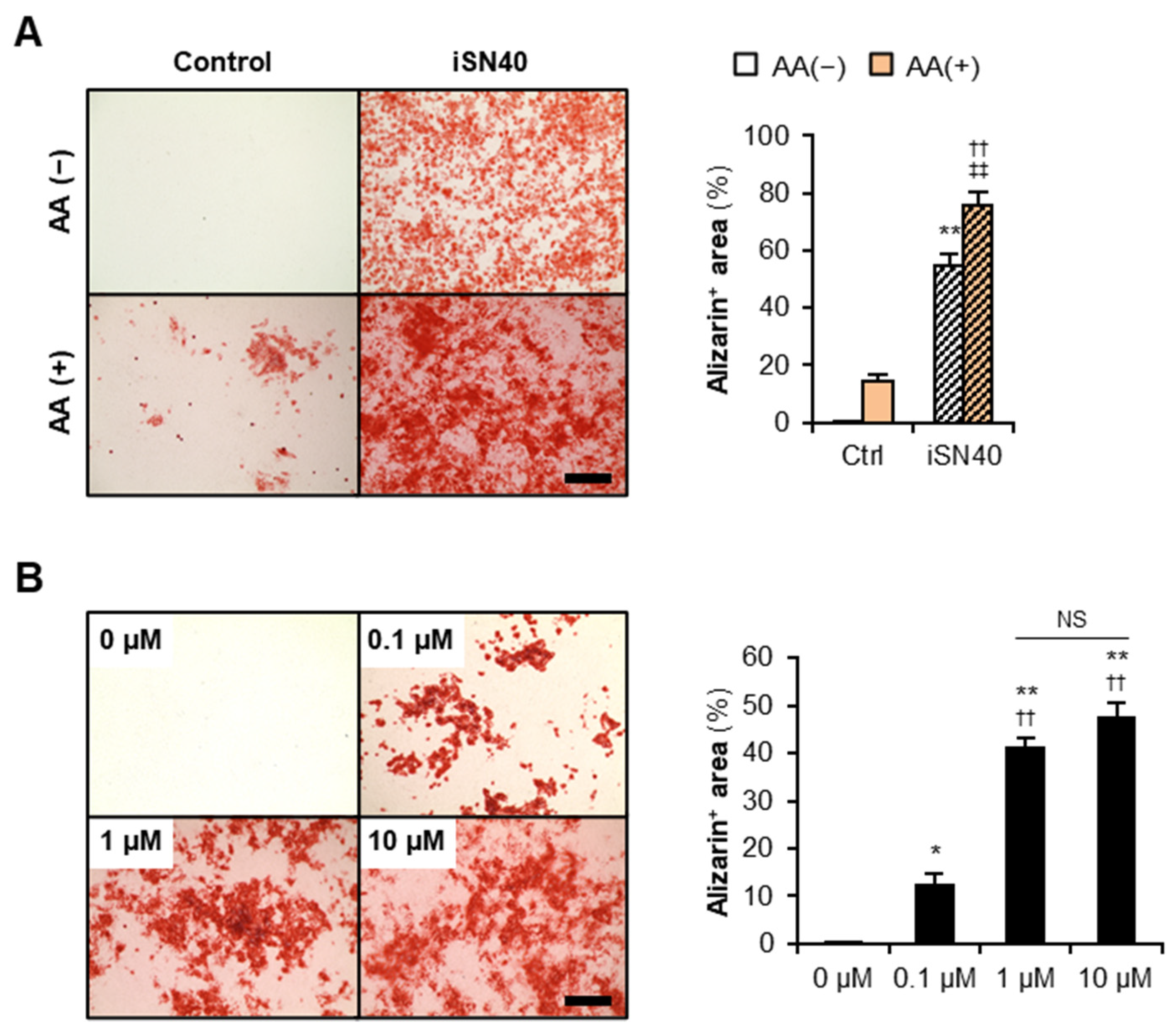
3.3. iSN40 Promotes Osteoblast Calcification

To investigate the effect of iSN40 on osteoblast calcification, MC3T3-E1 cells were treated with iSN40 in DM with or without AA for 12 days and then subjected to alizarin staining to visualize calcium deposition on the cells (Figure 3A). Alizarin-positive area was significantly increased by 10 μM iSN40 rather than by 15 μg/mL AA. Moreover, compared with individual treatments, co-treatment with iSN40 and AA further facilitated calcification, indicating that iSN40 and AA synergistically promote osteoblast mineralization. Although iSN40 was administered at a concentration of 10 μM in the above experiments, the dose response analysis revealed that 1 μM iSN40 was sufficient to induce osteoblast calcification in the presence of 15 μg/mL AA (Figure 3B). We used phosphorothioated iSN40 (PS-iSN40) in this study to avoid degradation by nucleases, whereas native iSN40 did not promote calcification even in the presence of 50 μg/mL AA (Figure S2). This demonstrates that phosphorothioation is necessary for iSN40 to function as an osteoDN.

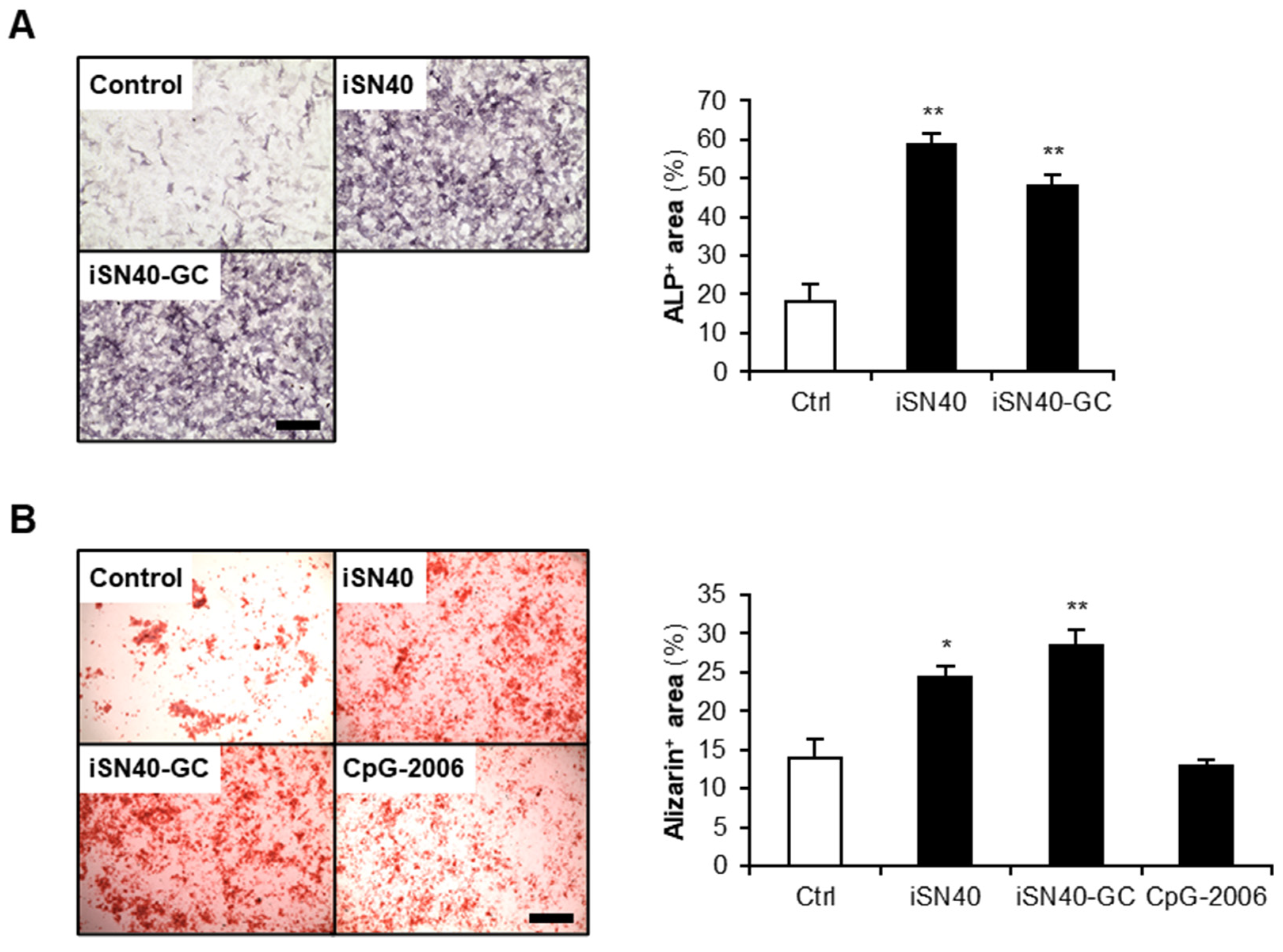
3.4. Osteogenetic Action of iSN40 Is TLR9-Independent

iSN40 (GGA ACG ATC CTC AAG CTT) contained a CpG motif. It has been studied that ODNs possessing unmethylated CpG motifs (CpG-ODNs) serve as TLR9 ligands and initiate innate immune system-induced inflammatory responses [38]. To examine the impact of the CpG motif within iSN40 on its osteogenetic activity, iSN40-GC (GGA AGC ATC CTC AAG CTT) was constructed, in which the CpG motif was substituted with GC. iSN40-GC enhanced ALP activity and calcification of MC3T3-E1 cells to the same extent as iSN40 (Figure 4). Conversely, a well-known TLR9 ligand, CpG-2006 [33], did not facilitate osteoblast mineralization (Figure 4B), which was further confirmed through ALP staining results (Figure 1 and Figure S1). In addition, RT-PCR revealed that MC3T3-E1 cells do not express TLR9 (Figure S3), corresponding to the previous study [39]. These results demonstrate that osteogenetic action of iSN40 is independent of its CpG motif and TLR9.

4. Discussion

The present study identified that iSN40, which is an 18-base ODN derived from the genome sequence of lactic acid bacteria, serves as an osteoDN that promotes differentiation and mineralization of osteoblasts. It could be the candidate of nucleic acid drug activating osteoblasts for osteoporosis. Although a direct target and action mechanism of iSN40 are unknown, the study results revealed that the osteogenetic function of iSN40 is TLR9-independent. The CpG motif within iSN40 was not required for its activity; A validated TLR9 ligand, CpG-2006 [33], enhanced neither ALP activity nor calcification of MC3T3-E1 cells; moreover, TLR9 was not expressed in MC3T3-E1 cells. TLR9-independent effect of iSN40 is crucial for its clinical application. The innate immune system, including IL-6 secretion, is closely related to bone homeostasis [40]. For example, CpG-ODNs generally upregulate interleukin (IL)-6 production via TLR9 [17]. IL-6 is a pro-osteoclastogenic cytokine [3] that is highly produced in aged osteoblasts [7]. Therefore, TLR9-independent osteogenetic activity of iSN40 is a favorable characteristic for osteoporosis therapy. However, it is still ambiguous whether iSN40 never serves as a CpG-ODN even in TLR9-expressing cells. Although human osteoblasts and bone-derived mesenchymal stem cells do not express TLR9 as MC3T3-E1 cells do not [41], other types of cells existing in bone tissue such as osteoclasts express TLR9 as a mediator of bone metabolism [42]. Thus, immunological and pro-inflammatory effects of iSN40 on TLR9-expressing cells in bone tissue need to be elucidated for future application in clinical settings.
iSN40 upregulated the expression of osteogenic genes such as osterix, collagen type Iα, osteopontin, and osteocalcin, which are induced by Runx2 [37]. However, iSN40 did not alter Runx2 expression. The transcriptional activity of Runx2 is regulated via post-transcriptional modifications including phosphorylation, acetylation, sumoylation, and ubiquitination, which are mediated by numerous factors [43]. iSN40 may target such modulators to enhance Runx2 activity. For instance, another osteoDN, MT01, has been reported to enhance Runx2 phosphorylation via activation of ERK and p38 MAPK [22], which potentiates the Runx2/osterix transcriptional machinery [44]. The effect of iSN40 on Runx2 protein needs to be examined in further studies.
iSN41-iSN47 are PS-ODNs analogous to iSN40; however, they did not enhance ALP activity of MC3T3-E1 cells. When iSN40 operates as an antisense nucleotide, iSN41-iSN47 would present partial osteogenetic activities. To discuss the mechanism of action of iSN40, a previous study reporting iSN04 is helpful. iSN04 is designed from the lactic acid bacteria, genome as same as iSN40, and serves as a myoDN that promotes myoblast differentiation in a TLR-independent manner [24]. iSN04 is taken up into cytoplasm without any carriers, forms a G-stacking structure, and physically interacts with nucleolin to interfere its function, indicating that iSN04 is an anti-nucleolin aptamer [24]. Thus, it is sufficiently possible that iSN40 works as an aptamer for target proteins. Administration of 6-FAM-conjugated iSN40 and iSN40-GC to MC3T3-E1 cells showed that they were autonomously incorporated into the cytoplasm within 30 min (Figure 5), suggesting that iSN40 acts intracellularly, not on the plasma membrane.
To further discuss the potential of iSN40 as an aptamer, conformation of iSN40 in water at 310 K was simulated using the TTP-McMD (Figure 6A). iSN40 exhibited a compact globular structure (average radius: 1.01 nm) similar to that of iSN04 [24]. Interestingly, the predicted structure of another osteoDN, MT01, was substantially different from that of iSN40 (Figure 6B), suggesting that the action mechanisms of iSN40 and MT01 are likely to be dissimilar. Indeed, iSN40 did not alter the mRNA levels of Runx2, but MT01 significantly induced Runx2 transcription [19,20]. To determine whether iSN40 is an aptamer, the iSN40-binding protein in osteoblasts needs to be identified. This would further promote the development and application of iSN40 as an osteoDN for bone regeneration.

5. Conclusions

The present study successfully identified a novel osteoDN, iSN40, which is an 18-base ODN designed from the lactic acid bacteria genome. iSN40 promoted the differentiation and calcification of osteoblasts by modulating osteogenic gene expression in a TLR9-independent manner. It demonstrated that the ODN library derived from bacteria genome can be a platform to discover the ODN targeting and regulating osteoblasts. Activation of osteoblasts by ODNs would provide an alternative strategy for osteoporosis therapy by promoting bone formation and mineralization.

6. Patents

Shinshu University has been assigned the invention of osteoDN by T.T., Y.N., K.U., and T.S., and Japan Patent Application 2021-122713 has been filed on 27 July 2021.

Supplementary Materials

The Supplementary Materials are available online at: https://www.mdpi.com/article/10.3390/nano12101680/s1. Figure S1: iSN40 enhances ALP activity in MC3T3-E1 cells, Figure S2: Phosphorothioation is required for iSN40 to exert osteogenetic activity, Figure S3: Expression of TLR genes in MC3T3-E1 cells, Table S1: ODN sequences, Table S2: Primer sequences for RT-PCR. References [45,46,47,48,49,50] are cited in the Supplementary Materials.

Author Contributions

Y.N. and T.T. designed the study. T.T. wrote the manuscript. Y.N. and M.M. performed experiments and data analyses. K.U. calculated the ODN structures. T.S. designed and provided the ODN library. All authors have read and agreed to the published version of the manuscript.

Funding

This research was funded by The Japan Society for the Promotion of Science, grant numbers 19K05948 and 22K05554.

Informed Consent Statement

Not applicable.

Data Availability Statement

The raw data supporting the conclusions of this article will be made available by the authors, without undue reservation.

Acknowledgments

The preprint has been posted on bioRxiv [51].

Conflicts of Interest

The authors declare no conflict of interest.

References

  1. Boros, K.; Freemont, T. Physiology of ageing of the musculoskeletal system. Best Pract. Res. Clin. Rheumatol. 2017, 31, 203–217. [Google Scholar] [CrossRef] [PubMed]
  2. Pisani, P.; Renna, M.D.; Conversano, F.; Casciaro, E.; Di Paola, M.; Quarta, E.; Muratore, M.; Casciaro, S. Major osteoporotic fragility fractures: Risk factor updates and societal impact. World J. Orthop. 2016, 7, 171–181. [Google Scholar] [CrossRef] [PubMed]
  3. Corrado, A.; Cici, D.; Rotondo, C.; Maruotti, N.; Cantatore, F.P. Molecular basis of bone aging. Int. J. Mol. Sci. 2020, 21, 3679. [Google Scholar] [CrossRef] [PubMed]
  4. Yang, X.; Wang, G.; Wang, Y.; Zhou, J.; Yuan, H.; Li, X.; Liu, Y.; Wang, B. Histone demethylase KDM7A reciprocally regulates adipogenic and osteogenic differentiation via regulation of C/EBPalpha and canonical Wnt signalling. J. Cell. Mol. Med. 2019, 23, 2149–2162. [Google Scholar] [CrossRef]
  5. Rutkovskiy, A.; Stenslokken, K.O.; Vaage, I.J. Osteoblast differentiation at a glance. Med. Sci. Monit. Basic Res. 2016, 22, 95–106. [Google Scholar] [CrossRef] [Green Version]
  6. Zhang, X.; Zhao, G.; Zhang, Y.; Wang, J.; Wang, Y.; Cheng, L.; Sun, M.; Rui, Y. Activation of JNK signaling in osteoblasts is inversely correlated with collagen synthesis in age-related osteoporosis. Biochem. Biophys. Res. Commun. 2018, 504, 771–776. [Google Scholar] [CrossRef]
  7. Eriksen, C.G.; Olsen, H.; Husted, L.B.; Sorensen, L.; Carstens, M.; Soballe, K.; Langdahl, B.L. The expression of IL-6 by osteoblasts is increased in healthy elderly individuals: Stimulated proliferation and differentiation are unaffected by age. Calcif. Tissue Int. 2010, 87, 414–423. [Google Scholar] [CrossRef]
  8. Becerikli, M.; Jaurich, H.; Schira, J.; Schulte, M.; Dobele, C.; Wallner, C.; Abraham, S.; Wagner, J.M.; Dadras, M.; Kneser, U.; et al. Age-dependent alterations in osteoblast and osteoclast activity in human cancellous bone. J. Cell. Mol. Med. 2017, 21, 2773–2781. [Google Scholar] [CrossRef] [Green Version]
  9. Awasthi, H.; Mani, D.; Singh, D.; Gupta, A. The underlying pathophysiology and therapeutic approaches for osteoporosis. Med. Res. Rev. 2018, 38, 2024–2057. [Google Scholar] [CrossRef]
  10. Corrado, A.; Sanpaolo, E.R.; Di Bello, S.; Cantatore, F.P. Osteoblast as a target of anti-osteoporotic treatment. Postgrad. Med. 2017, 129, 858–865. [Google Scholar] [CrossRef]
  11. McClung, M.R.; Lewiecki, E.M.; Cohen, S.B.; Bolognese, M.A.; Woodson, G.C.; Moffett, A.H.; Peacock, M.; Miller, P.D.; Lederman, S.N.; Chesnut, C.H.; et al. Bone Loss Study Group. N. Engl. J. Med. 2006, 354, 821–831. [Google Scholar] [CrossRef] [PubMed]
  12. Diedhiou, D.; Cuny, T.; Sarr, A.; Norou Diop, S.; Klein, M.; Weryha, G. Efficacy and safety of denosumab for the treatment of osteoporosis: A systematic review. Ann. Endocrinol. 2015, 76, 650–657. [Google Scholar] [CrossRef] [PubMed]
  13. Canalis, E. Wnt signalling in osteoporosis: Mechanisms and novel therapeutic approaches. Nat. Rev. Endocrinol. 2013, 10, 575–583. [Google Scholar] [CrossRef] [PubMed]
  14. Miller, S.A.; St Onge, E.L.; Whalen, K.L. Romosozumab: A novel agent in the treatment for postmenopausal osteoporosis. J. Pharm. Technol. 2021, 37, 45–52. [Google Scholar] [CrossRef]
  15. Quemener, A.M.; Bachelot, L.; Forestier, A.; Donnou-Fournet, E.; Gilot, D.; Galibert, M.D. The powerful world of antisense oligonucleotides: From bench to bedside. Wiley Interdiscip. Rev. RNA 2020, 11, e1594. [Google Scholar] [CrossRef]
  16. Wang, T.; Chen, C.; Larcher, L.M.; Barrero, R.A.; Veedu, R.N. Three decades of nucleic acid aptamer technologies: Lessons learned, progress and opportunities on aptamer development. Biotechnol. Adv. 2019, 37, 28–50. [Google Scholar] [CrossRef]
  17. Nigar, S.; Shimosato, T. Cooperation of oligodeoxynucleotides and synthetic molecules as enhanced immune modulators. Front. Nutr. 2019, 6, 140. [Google Scholar] [CrossRef] [Green Version]
  18. Yang, G.; Wan, M.; Zhang, Y.; Sun, L.; Sun, R.; Hu, D.; Zhou, X.; Wang, L.; Wu, X.; Wang, L.; et al. Inhibition of a C-rich oligodeoxynucleotide on activation of immune cells in vitro and enhancement of antibody response in mice. Immunology 2010, 131, 501–512. [Google Scholar] [CrossRef]
  19. Feng, Z.; Shen, Y.; Wang, L.; Cheng, L.; Wang, J.; Li, Q.; Shi, W.; Sun, X. An oligodeoxynucleotide with promising modulation activity for the proliferation and activation of osteoblast. Int. J. Mol. Sci. 2011, 12, 2543–2555. [Google Scholar] [CrossRef]
  20. Shen, Y.; Feng, Z.; Lin, C.; Hou, X.; Wang, X.; Wang, J.; Yu, Y.; Wang, L.; Sun, X. An oligodeoxynucleotide that induces differentiation of bone marrow mesenchymal stem cells to osteoblasts in vitro and reduces alveolar bone loss in rats with periodontitis. Int. J. Mol. Sci. 2012, 13, 2877–2892. [Google Scholar] [CrossRef] [Green Version]
  21. Yu, W.; Zheng, Y.; Li, H.; Lin, H.; Chen, Z.; Tian, Y.; Chen, H.; Zhang, P.; Xu, X.; Shen, Y. The Toll-like receptor ligand, CpG oligodeoxynucleotides, regulate proliferation and osteogenic differentiation of osteoblast. J. Orthop. Surg. Res. 2020, 15, 327. [Google Scholar] [CrossRef] [PubMed]
  22. Hou, X.; Shen, Y.; Zhang, C.; Zhang, L.; Qin, Y.; Yu, Y.; Wang, L.; Sun, X. A specific oligodeoxynucleotide promotes the differentiation of osteoblasts via ERK and p38 MAPK pathways. Int. J. Mol. Sci. 2012, 13, 7902–7914. [Google Scholar] [CrossRef] [PubMed]
  23. Nigar, S.; Yamamoto, Y.; Okajima, T.; Shigemori, S.; Sato, T.; Ogita, T.; Shimosato, T. Synergistic oligodeoxynucleotide strongly promotes CpG-induced interleukin-6 production. BMC Immunol. 2017, 18, 44. [Google Scholar] [CrossRef] [Green Version]
  24. Shinji, S.; Umezawa, K.; Nihashi, Y.; Nakamura, S.; Shimosato, T.; Takaya, T. Identification of the myogenetic oligodeoxynucleotides (myoDNs) that promote differentiation of skeletal muscle myoblasts by targeting nucleolin. Front. Cell Dev. Biol. 2021, 8, 616706. [Google Scholar] [CrossRef] [PubMed]
  25. Nakamura, S.; Yonekura, S.; Shimosato, T.; Takaya, T. Myogenetic oligodeoxynucleotide (myoDN) recovers the differentiation of skeletal muscle myoblasts deteriorated by diabetes mellitus. Front. Physiol. 2021, 12, 679152. [Google Scholar] [CrossRef] [PubMed]
  26. Nihashi, Y.; Shinji, S.; Umezawa, K.; Shimosato, T.; Ono, T.; Kagami, H.; Takaya, T. Myogenetic oligodeoxynucleotide complexed with berberine promotes differentiation of chicken myoblasts. Anim. Sci. J. 2021, 92, e13597. [Google Scholar] [CrossRef]
  27. Nihashi, Y.; Yamamoto, M.; Shimosato, T.; Takaya, T. Myogenetic oligodeoxynucleotide restores differentiation and reverses inflammation of myoblasts aggravated by cancer-conditioned medium. bioRxiv 2021, 469038. [Google Scholar] [CrossRef]
  28. Nohira, N.; Shinji, S.; Nakamura, S.; Nihashi, Y.; Shimosato, T.; Takaya, T. Myogenetic oligodeoxynucleotides as anti-nucleolin aptamers inhibit the growth of embryonal rhabdomyosarcoma cells. bioRxiv 2021, 464889. [Google Scholar] [CrossRef]
  29. Macke, T.J.; Case, D.A. Modeling unusual nucleic acid structures. In Molecular Modeling of Nucleic Acids; Leontis, N.B., SantaLucia, J., Eds.; American Chemical Society: Washington, DC, USA, 1998; pp. 379–393. [Google Scholar]
  30. Ikebe, J.; Umezawa, K.; Kamiya, N.; Sugihara, T.; Yonezawa, Y.; Takano, Y.; Nakamura, H.; Higo, J. Theory for trivial trajectory parallelization of multicanonical molecular dynamics and application to a polypeptide in water. J. Comput. Chem. 2011, 32, 1286–1297. [Google Scholar] [CrossRef]
  31. Maier, J.A.; Matinez, C.; Kasavajhala, K.; Wickstrom, L.; Hauser, K.E.; Simmerling, C. ff14SB: Improving the accuracy of protein side chain and backbone parameters from ff99SB. J. Chem. Theory Comput. 2015, 11, 3696–3713. [Google Scholar] [CrossRef] [Green Version]
  32. Tsui, V.; Case, D.A. Theory and applications of the generalized Born solvation model in macromolecular simulations. Biopolymers 2000, 56, 275–291. [Google Scholar] [CrossRef]
  33. Pohar, J.; Lainscek, D.; Fukui, R.; Yamamoto, C.; Miyake, K.; Jerala, R.; Bencina, M. Species-specific minimal sequence motif for oligodeoxyribonucleotides activating mouse TLR9. J. Immunol. 2015, 195, 4396–4405. [Google Scholar] [CrossRef] [PubMed] [Green Version]
  34. Sackesen, C.; van de Veen, W.; Akdis, M.; Soyer, O.; Zumkehr, J.; Ruckert, B.; Stanic, B.; Kalayci, O.; Alkan, S.S.; Gursel, I.; et al. Suppression of B-cell activation and IgE, IgA, IgG1 and IgG4 production by mammalian telomeric oligonucleotides. Allergy 2013, 68, 593–603. [Google Scholar] [CrossRef] [PubMed] [Green Version]
  35. Komori, T. Regulation of osteoblast differentiation by Runx2. Adv. Exp. Med. Biol. 2010, 658, 43–49. [Google Scholar]
  36. Moser, S.C.; van der Eerden, B.C.J. Osteocalcin: A versatile bone-derived hormone. Front. Endocrinol. 2018, 9, 794. [Google Scholar] [CrossRef] [PubMed] [Green Version]
  37. Liu, T.M.; Lee, E.H. Transcriptional regulatory cascades in Runx2-dependent bone development. Tissue Eng. Part B Rev. 2013, 19, 254–263. [Google Scholar] [CrossRef] [PubMed] [Green Version]
  38. Vollmer, J.; Krieg, A.M. Immunotherapeutic applications of CpG oligodeoxynucleotide TLR9 agonists. Adv. Drug Deliv. Rev. 2009, 61, 195–2004. [Google Scholar] [CrossRef]
  39. Nemoto, E.; Honda, T.; Kanaya, S.; Takada, H.; Shimauchi, H. Expression of functional Toll-like receptors and nucleotide-binding oligomerization domain proteins in murine cementoblasts and their upregulation during cell differentiation. J. Periodontal Res. 2008, 43, 585–593. [Google Scholar] [CrossRef]
  40. Charles, J.F.; Nakamura, M.C. Bone and the innate immune system. Curr. Osteoporos. Rep. 2014, 12, 1–8. [Google Scholar] [CrossRef] [Green Version]
  41. El-Sayed, K.M.F.; Boeckler, J.; Dorfer, C.E. TLR expression profile of human alveolar bone proper-derived stem/progenitor cells and osteoblasts. J. Craniomaxillofac. Surg. 2017, 45, 2054–2060. [Google Scholar] [CrossRef]
  42. Amcheslavsky, A.; Bar-Shavit, Z. Toll-like receptor 9 ligand blocks osteoclast differentiation through induction of phosphatase. J. Bone Miner. Res. 2007, 22, 1301–1310. [Google Scholar] [CrossRef] [PubMed]
  43. Vimalraj, S.; Arumugam, B.; Miranda, P.J.; Selvamurugan, N. Runx2: Structure, function, and phosphorylation in osteoblast differentiation. Int. J. Biol. Macromol. 2015, 78, 202–208. [Google Scholar] [CrossRef] [PubMed]
  44. Artigas, N.; Urena, C.; Rodriguez-Carballo, E.; Rosa, J.L.; Ventura, F. Mitogen-activated protein kinase (MAPK)-regulated interactions between Osterix and Runx2 are critical for the transcriptional osteogenic program. J. Biol. Chem. 2014, 289, 27105–27117. [Google Scholar] [CrossRef] [PubMed] [Green Version]
  45. Deng, H.; Kuang, P.; Cui, H.; Luo, Q.; Liu, H.; Lu, Y.; Fang, J.; Zuo, Z.; Deng, J.; Li, Y.; et al. Sodium fluoride induces apoptosis in mouse splenocytes by activating ROS-dependent NF-κB signaling. Oncotarget 2017, 8, 114428–114441. [Google Scholar] [CrossRef] [PubMed] [Green Version]
  46. Tian, Y.; Xu, Y.; Fu, Q.; Dong, Y. Osterix is required for Sonic hedgehog-induced osteoblastic MC3T3-E1 cell differentiation. Cell Biochem. Biophys. 2012, 64, 169–176. [Google Scholar] [CrossRef] [PubMed]
  47. Takayama, T.; Dai, J.; Tachi, K.; Shohara, R.; Kasai, H.; Imamura, K.; Yamano, S. The potential of stromal cell-derived factor-1 delivery using a collagen membrane for bone regeneration. J. Biomater. Appl. 2017, 31, 1049–1061. [Google Scholar] [CrossRef]
  48. Gao, J.; Feng, Z.; Wang, X.; Zeng, M.; Liu, J.; Han, S.; Xu, J.; Chen, L.; Cao, K.; Long, J.; et al. SIRT3/SOD2 maintains osteoblast differentiation and bone formation by regulating mitochondrial stress. Cell Death Differ. 2018, 25, 229–240. [Google Scholar] [CrossRef] [Green Version]
  49. Lee, D.J.; Tseng, H.C.; Wong, S.W.; Wang, Z.; Deng, M.; Ko, C.C. Dopaminergic effects on in vitro osteogenesis. Bone Res. 2015, 3, 15020. [Google Scholar] [CrossRef] [Green Version]
  50. Veazey, K.J.; Colding, M.C. Selection of stable reference genes for quantitative rt-PCR comparisons of mouse embryonic and extra-embryonic stem cells. PLoS ONE 2011, 6, e27592. [Google Scholar] [CrossRef] [Green Version]
  51. Nihashi, Y.; Miyoshi, M.; Umezawa, K.; Shimosato, T.; Takaya, T. Identification of a novel osteogenetic oligodeoxynucleotide (osteoDN) that promotes osteoblast differentiation in a TLR9-independent manner. bioRxiv 2022, 485101. [Google Scholar] [CrossRef]
Figure 1. Identification of iSN40 as an osteoDN. (A) ALP staining images of MC3T3-E1 cells treated with 10 μM PS-ODNs in GM for 48 h. Scale bar, 200 μm. (B) Quantification of ALP-positive area.
Nanomaterials 12 01680 g001
Figure 2. iSN40 induces osteogenic gene expression in MC3T3-E1 cells. (A) qPCR results of MC3T3-E1 cells treated with 10 μM iSN40 in GM with 15 μg/mL AA for 24 or 48 h. (B) qPCR results of MC3T3-E1 cells treated with 10 μM iSN40 in DM with 15 μg/mL AA for 4 or 8 days. * p < 0.05, ** p < 0.01 vs. control at each time point (Student’s t test). n = 3.
Nanomaterials 12 01680 g002
Figure 3. iSN40 induces calcification of MC3T3-E1 cells. (A) Representative images and quantification of alizarin staining of MC3T3-E1 cells treated with 10 μM iSN40 in DM with or without 15 μg/mL AA for 12 days. Scale bar, 200 μm. ** p < 0.01 vs. control-AA(−), †† p < 0.01 vs. control-AA(+), ‡‡ p < 0.01 vs. iSN40-AA(−) (Scheffe’s F-test). n = 4. (B) Representative images and quantification of alizarin staining of MC3T3-E1 cells treated with 0.1, 1, or 10 μM iSN40 in DM with 15 μg/mL AA for 9 days. Scale bar, 200 μm. * p < 0.05, ** p < 0.01 vs. 0 μM; †† p < 0.01 vs. 0.1 μM; NS, not significant (Scheffe’s F-test). n = 4.
Nanomaterials 12 01680 g003
Figure 4. Action of iSN40 is TLR9-independent. (A) Representative images and quantification of ALP staining of MC3T3-E1 cells treated with 10 μM of iSN40 or iSN40-GC in DM with 50 μg/mL AA for 48 h. Scale bar, 100 μm. ** p < 0.01 vs. control (Scheffe’s F-test). n = 4. (B) Representative images and quantification of alizarin staining of MC3T3-E1 cells treated with 1 μM PS-ODNs in DM with 50 μg/mL AA for 9 days. Scale bar, 200 μm. * p < 0.05, ** p < 0.01 vs. control (Scheffe’s F-test). n = 4.
Nanomaterials 12 01680 g004
Figure 5. Intracellular incorporation of iSN40. Representative fluorescent images of MC3T3-E1 cells treated with 5 μg/mL 6-FAM-iSN40 and 6-FAM-iSN40-GC in GM for 30- or 120-min. Scale bar, 25 μm.
Nanomaterials 12 01680 g005
Figure 6. The most thermodynamically stable conformations of the ODNs in water at 310K simulated by the TTP-McMD. (A) iSN40. (B) MT01.
Nanomaterials 12 01680 g006
Publisher’s Note: MDPI stays neutral with regard to jurisdictional claims in published maps and institutional affiliations.

