Article

Gas-Phase Fluorination of g-C3N4 for Enhanced Photocatalytic Hydrogen Evolution

1
School of Materials Science and Engineering, Tianjin University, Tianjin 300072, China
2
Key Laboratory of Advanced Ceramics and Machining Technology Ministry of Education, Tianjin 300072, China
*
Authors to whom correspondence should be addressed.
Nanomaterials 2022, 12(1), 37; https://doi.org/10.3390/nano12010037
Submission received: 30 November 2021 / Revised: 19 December 2021 / Accepted: 21 December 2021 / Published: 23 December 2021
(This article belongs to the Special Issue Fluorinated Nanocarbons and Their Applications)

Abstract

:
Graphitic carbon nitride (g-C3N4) has attracted much attention because of its potential for application in solar energy conservation. However, the photocatalytic activity of g-C3N4 is limited by the rapidly photogenerated carrier recombination and insufficient solar adsorption. Herein, fluorinated g-C3N4 (F-g-CN) nanosheets are synthesized through the reaction with F2/N2 mixed gas directly. The structural characterizations and theoretical calculations reveal that fluorination introduces N vacancy defects, structural distortion and covalent C-F bonds in the interstitial space simultaneously, which lead to mesopore formation, vacancy generation and electronic structure modification. Therefore, the photocatalytic activity of F-g-CN for H2 evolution under visible irradiation is 11.6 times higher than that of pristine g-C3N4 because of the enlarged specific area, enhanced light harvesting and accelerated photogenerated charge separation after fluorination. These results show that direct treatment with F2 gas is a feasible and promising strategy for modulating the texture and configuration of g-C3N4-based semiconductors to drastically enhance the photocatalytic H2 evolution process.

1. Introduction

The drastically increasing energy consumption globally requires clean and sustainable energy as alternatives to fossil fuels. Hydrogen energy is considered as the most promising candidate due to its potential application in fuel cells without carbon emissions. As such, the hydrogen evolution process has been widely studied, involving water splitting driven by light and with the help of a photocatalyst [1,2,3]. Graphitic carbon nitride (g-C3N4) has been demonstrated as a promising solar-to-fuel, metal-free, polymeric photocatalyst due to its suitable band structure, highly thermal and chemical stability, low cost and convenient preparation [4,5,6]. However, there are some intrinsic drawbacks to g-C3N4, such as its low electrical conductivity, insufficient light absorption, poor surface area and rapid recombination of photogenerated charge carriers [7,8]. Therefore, a great many strategies have been explored to improve its photocatalytic performance, including morphology design, elemental doping, heterojunction construction, nanocomposite hybridization and photosensitizer decoration [9,10,11]. Among these various strategies, it has been acknowledged that the introduction of non-metal heteroatoms is an effective method for enhancing the photocatalytic activity of semiconductors by expanding the absorption range of the solar spectrum, as highlighted in the pioneering study by Asahi et al. [12]. So far, various non-metal heteroatoms have been applied to synthesize doped g-C3N4 with significantly improved photocatalytic activity because of the narrowed bandgap, extended visible light absorption and promoted charge separation [13,14,15,16]. Fluorine (F) is the most electronegative element in the periodic table and the C-F bond is the strongest single covalent bond, meaning fluorination could provide maximum charge polarization to host materials, enhance the activity of energy-related reactions and improve the stability of compounds [17]. Nowadays, F-doped, nanostructured carbons have attracted tremendous interest and obtained great success, having controllable mechanical, electronic and magnetic properties [18,19,20]. F-doped g-C3N4 can be synthesized by either heating a mixture of dicyandiamide as the precursor with inorganic fluorides at high temperature [21,22,23] or via post-fluorination of g-C3N4 with the F-containing solution hydrothermally [24,25,26,27,28,29]. Both experimental and theoretical investigations have revealed that the structural distortion induced by F-doping enhances the photocatalytic activity of g-C3N4 because of the narrowed bandgap and accelerated separation of photogenerated electron–hole pairs. However, F-doped g-C3N4 is currently achieved through the nucleophilic substitution reaction of F with positively charged carbon atoms in a g-C3N4 framework [30], which not only limits the doping level but also restricts the degree of structural modification. Therefore, it is desirable to explore novel F-doping routes to increase the doping level and to modulate the structure to improve the photocatalytic properties of g-C3N4 further.
In addition to the hydrothermal method involving the use of F- as the elemental source, treatment with F-containing gas is another widely applied method to introduce F into the carbonaceous basal plane, with the molar ratio of F to C (F/C) in the range of 0–1.12 [31], which is called the gas-phase fluorination process [32,33]. Based on the consideration that bulk g-C3N4 has a multi-layer structure similar to graphite [34], as well as the successful fluorination of graphite through the direct gas fluorination method [35], it can be expected that the reaction with F2 is likely to introduce F atoms into the g-C3N4 framework at relatively high levels. However, the existence of defect-rich, N-bridged “poly(tri-s-triazine)/poly(triazine)” derived from the incomplete de-amination or polymerization of the nitrogen-containing precursor in g-C3N4 [36], rather than achieving a perfectly conjugated structure such as in graphite, should endow g-C3N4 a different fluorination mechanism [37]. Therefore, the morphological and electronic changes in g-C3N4 with F2 gas are worth investigating.
In this work, we prepared fluorinated g-C3N4 (F-g-CN) using F2 gas at high temperatures for the first time. Unlike conventional reactions with F-, the extremely strong oxidizability of F2 gas introduces F atoms into the g-C3N4 framework at a relatively high level through the addition reaction, leads to drastic structural distortion due to the transformed hybridization mode from sp2 to sp3 and creates abundant nitrogen defects by etching bridged N atoms simultaneously, thereby improving the charge separation because of the affected π band and long-pair electron states. As a result, F-g-CN shows 11.6 times greater photocatalytic activity for hydrogen production under visible light compared to pristine g-C3N4 thanks to the increased surface area of the porous structure, enhanced light absorption and efficient charge separation. This study reveals the effectiveness of F2 gas in improving the photocatalytic activity of g-C3N4 because of the synergetic effects of nitrogen defects, structural distortion and fluorine species.

2. Materials and Methods

2.1. Preparation of the Samples

The pristine g-C3N4 was prepared by calcining melamine (3 g) at 550 °C in argon for 4 h with a heating rate of 2.3 °C/min in a tube furnace. F-g-CN was synthesized under a static fluorination atmosphere in a nickel reactor. At first, 0.3 g of synthesized g-C3N4 power was placed on a nickel boat and then transferred into the nickel reactor. After sealing up the reactor carefully, the air and moisture were excluded from this reactor by alternately evacuating and filling the reactor with N2 gas three times. Subsequently, mixed gas of F2/N2 (20 vol.% of F2) was entered into the reactor slowly until 0.1 MPa at room temperature. Then, the reactor was heated at a rate of 5 °C min−1 until reaching the target temperature and kept for 1 h to ensure sufficient fluorination. The schematic synthesis process of F-g-CN is illustrated in Figure 1. After the reaction, the residual F2 and byproducts were ejected from the reactor by N2 gas and absorbed on an alkali absorbent. Finally, F-g-CN powder was obtained after naturally cooling to room temperature.

2.2. Characterization

The morphology of the samples was characterized by field emission scanning electron microscopy (SEM, Hitachi S4800, Tokyo, Japan) and transmission electron microscopy (TEM, JEM2100F, Joel, Tokyo, Japan). Moreover, the energy-dispersive spectroscopy (EDS) profiles were recorded. The crystal structures were analyzed by X-ray diffraction (XRD, Bruker D8 advanced with Cu Kα radiation). The specific surface area and pore size distribution were obtained using nitrogen adsorption–desorption at 77 K on an Autosorb-iQ2-MP (Quantachrome Inst., Boynton Beach, FL, USA). Fourier transform infrared spectra (FTIR) were recorded on a Bruker tensor 27 FTIR spectrometer ranging from 4000 to 400 cm−1. X-ray photoelectron spectroscopy (XPS) results were measured in Thermo ESCALAB 250 with Al Kα X-rays. UV–Vis diffuse reflectance spectra (DRS) were performed with a UV-Vis spectrophotometer (Perkin Elmer Lambda750) using BaSO4 as the reflectance standard reference. Steady-state photoluminescence (PL) spectra were recorded on a spectrophotometer (Hitachi F-4600, Hitachi, Japan). The time-resolved spectra were detected using a fluorescence spectrometer (Edinburgh FLS980, Instruments Ltd., UK) under an excitation wavelength of 360 nm at room temperature.

2.3. Photoelectrochemical Measurement

The photoelectrochemical measurements were conducted on an electrochemical station (CHI660D, Shanghai Chenhua Limited, Shanghai, China) with a standard three-electrode system in 0.5 M Na2SO4 solution. A Pt foil was used as the counter electrode and Ag/AgCl (saturated KCl) was used as the reference electrode. The working electrode was fabricated by coating the slurry of the 5 mg photocatalyst dispersed in 0.5 mL ethanol and 20 μL nafion on a clean fluoride-doped tin oxide (FTO) glass. After being naturally dried in air, the electrode was tested under the light source of a 300 W Xe lamp with a filter (λ > 420 nm). The photocurrent responses of the photocatalyst were recorded using a 20 s on–off light cycle at a voltage of 0.0 V. Electrochemical impedance spectroscopy (EIS) results were measured in the frequency range of 0.05 Hz to 100 kHz with a perturbation of 5 mV.

2.4. Photocatalytic Measurement

The photocatalytic hydrogen production experiments of samples were evaluated in an online photocatalytic hydrogen production system (CEL-SPH2N, Ceaulight, Beijing, China) with a 420 nm cutoff filter. Typically, 50 mg of photocatalytic powder was suspended in an aqueous solution (100 mL) containing triethanolamine (10 vol.%) as a sacrificial electron donor. In addition, 3 wt.% Pt was introduced as co-catalyst via in situ photodeposition of H2PtCl6. The reaction solution was evacuated several times to remove air completely prior to radiation under visible light. The temperature of the reaction was maintained at 25 °C during the reaction. The produced hydrogen was measured with an online gas chromatograph (GC-14C, Shimadzu, Kyoto, Japan) equipped with a thermal conductive detector (TCD) using nitrogen as a carrier gas. The apparent quantum efficiency (AQE) for H2 evolution was measured under identical experimental conditions, except that monochromatic light was used for irradiation with a 420 nm band pass filter. The AQE was calculated based on Equation (1):
AQE   =   2   ×   number   of   evolved   H 2 number   of   incident   photos     ×   100 %

2.5. Theoretical Calculations

First-principle calculations within the framework of density functional theory (DFT) were performed with the DMol3 software package. Perdew–Burke–Ernzerh (PBE) exchange correlation functionals with general gradient approximation (GGA) were utilized for geometric optimization and single-point energy calculations. Effective core potentials with a double numerical basis were employed for the description of core electrons. In the structural optimization calculations, the convergence tolerances for energy, maximum force and maximum displacement were set to 1.0 × 10⁻⁶ Ha per atom, 1.0 × 10⁻⁴ Ha/Å and 1.0 × 10⁻³ Å, respectively. The energy tolerance for self-consistent field iterations in the energy calculations was set to 1.0 × 10⁻⁶ Ha. The Brillouin zone was sampled at the 5 K × 5 K × 3 K point, because there was no significant change in the calculated energies when a larger number of K points were applied. DFT-D corrections were carried out using the Grimme method for all calculations.

3. Results and Discussion

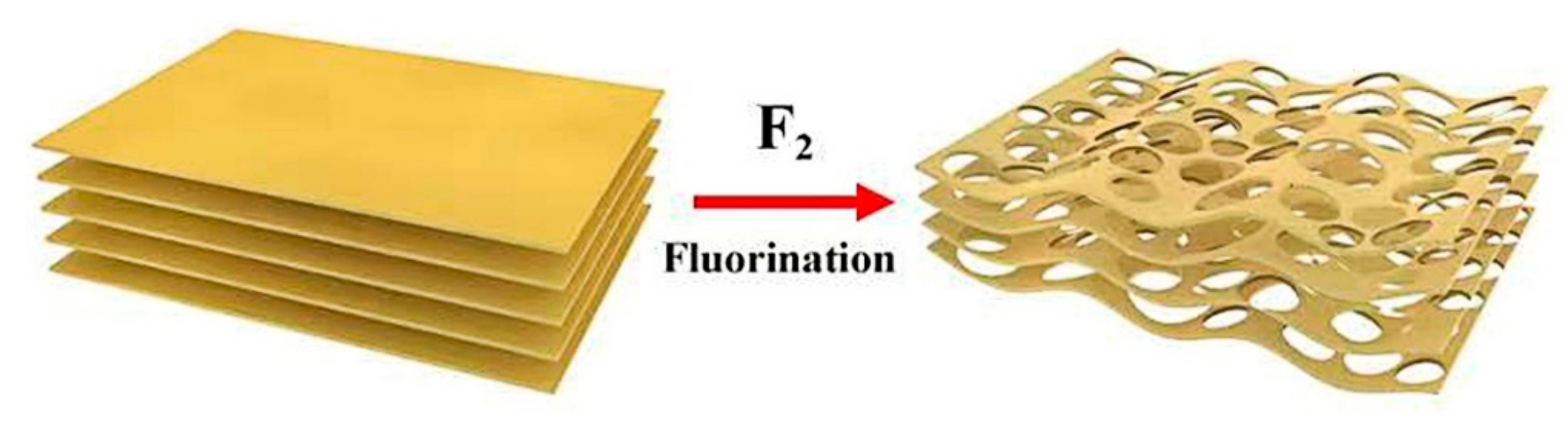
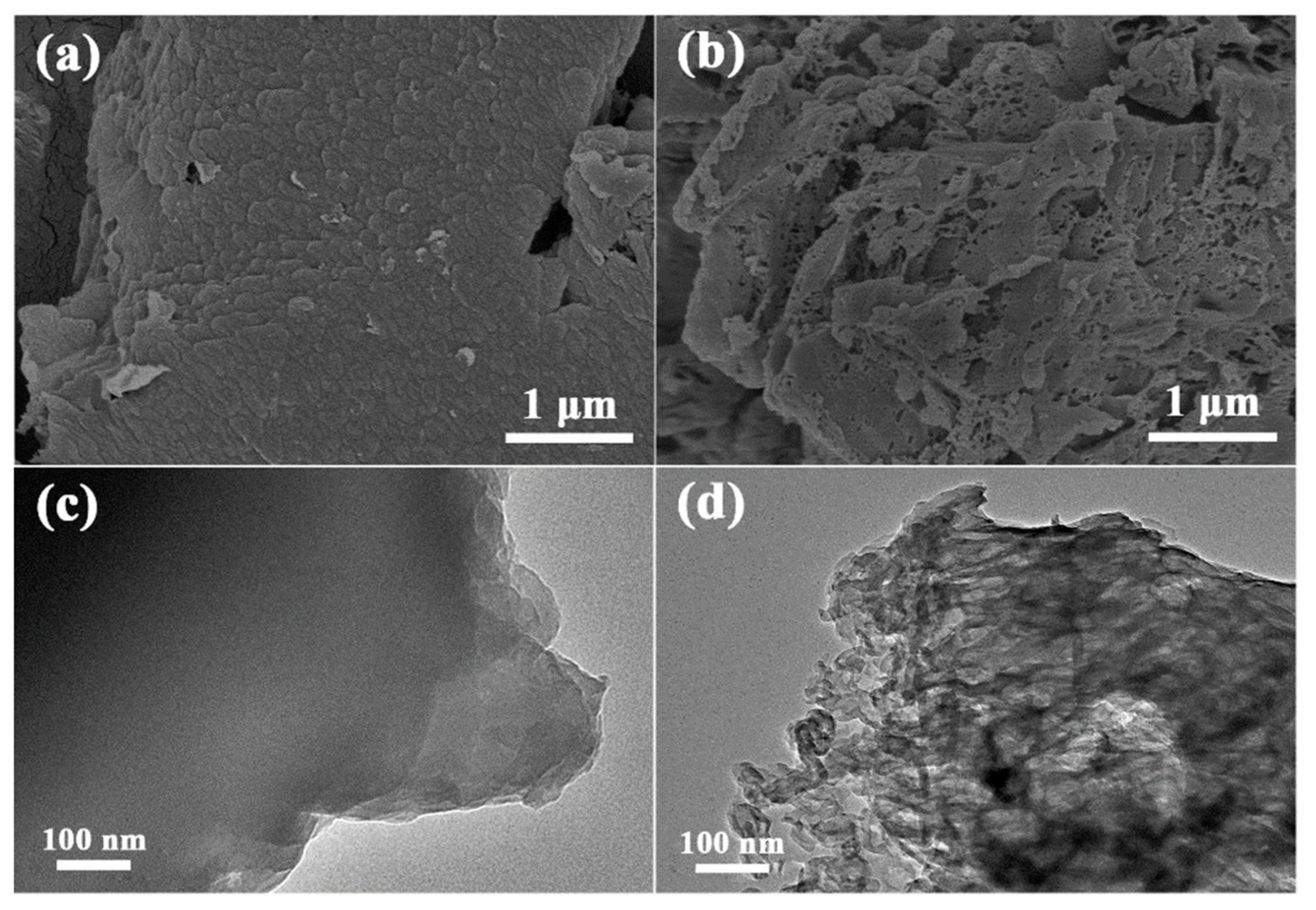
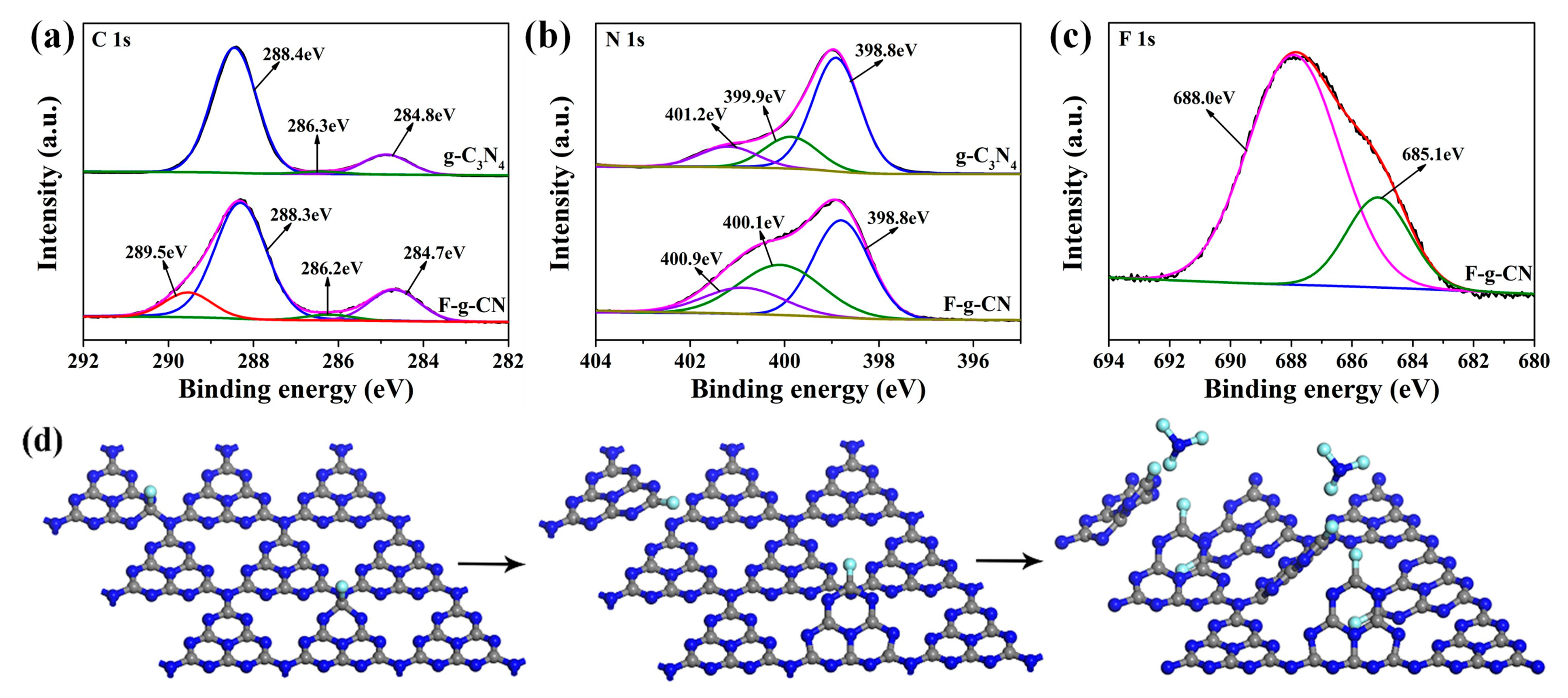
3.1. Morphology and Structural Analysis

The morphologies of g-C3N4 and prepared F-g-CN at different temperatures were examined by SEM at first. From the SEM image of g-C3N4 in Figure 2a, the dense stacked lamellar structure can be observed, which suppresses the capability of light absorption as well as the charge and mass transport [38]. The fluorination temperature is a crucial parameter in controlling the morphology and the fluorine content of g-C3N4; hence, g-C3N4 was fluorinated at 120, 150 and 180 °C, labeled as F-g-CN-120, F-g-CN-150 and F-g-CN-180, respectively, to investigate the photocatalytic activity. After the fluorination, all F-g-CN samples changed to porous and loose particles (Figure 2b and Figure S1a,b). The corresponding TEM image of g-C3N4 shown in Figure 2c further confirms that g-C3N4 is constructed of large and overlapped nanosheets. From the TEM images of F-g-CN in Figure 2d and Figure S1c,d, it can be noticed that the size of these layers is decreased remarkably and a large number of pores can be noticed clearly, consistent with its SEM image. In addition, these pores become larger with the increase in fluorination temperature, and the porous structure of F-g-CN obtained at 180 °C is seriously deteriorated due to the extensive fluorination at high temperatures. It should be noted that the much more transparent TEM image of F-g-CN than that of g-C3N4 indicates the reduced thickness by fluorination. The extremely reactivity of F2 gas destroys the structure of g-C3N4, leading to lots of pores in the g-C3N4 structure, similar to the porous g-C3N4 synthesized by strong oxidants [39]. Furthermore, the extremely low yield of F-g-CN fluorinated at 180 °C is insufficient for photocatalytic needs. Thus, F-g-CN fluorinated at 150 °C was selected for further investigation. The F-g-CN that appears in the following text refers to F-g-CN-150 if no special description is given.
Based on N2 adsorption–desorption measurements at 77 K for g-C3N4 and F-g-CN, both of them exhibit type IV isotherms with H3 hysteresis loops (Figure S2a), indicating the mesoporous structure of these samples [40]. Compared with g-C3N4, the shifted hysteresis loop of F-g-CN to the lower relative pressure region and the enlarged area of the hysteresis loop demonstrate the increased mesoporous structure after fluorination, in agreement with the morphological results. The specific surface areas calculated by the Brunauer–Emmett–Teller (BET) method for g-C3N4 and F-g-CN were 11.5 and 74.0 m2 g−1, respectively. The greatly increased surface area after fluorination can be ascribed to the generation of abundant pores. In addition, the pore size distributions of g-C3N4 and F-g-CN as analyzed using the Barrett–Joyner–Halenda (BJH) method further confirmed the plentiful mesopores in the range of 20–100 nm after the fluorination process (Figure S2b). Therefore, F2 gas has a great impact on the formation of the porous structure in g-C3N4.
The XRD patterns of g-C3N4 and F-g-CN in Figure 3a display a similar crystalline structure. The main diffraction peaks at 13.1° and 27.3° for all the samples correspond to the in-plane repetition of the tri-s-triazine motif and the periodic stacking of conjugated aromatic structures, respectively [37]. Note that the diffraction peak at 13.1° for F-g-CN almost disappears, which demonstrates the overwhelmingly destructed in-plane structure of g-C3N4 caused by the substantial mesopores, as revealed by the size distribution analysis [41]. In addition, the intensity of the diffraction peak at 27.3° for F-g-CN is weak in comparison with g-C3N4, suggesting the disturbed periodic structure of stacked g-C3N4 planes caused by the fluorination process [13]. The FTIR spectra of these samples (Figure 3b) confirm that the molecular skeletal vibration modes of F-g-CN are close to those of g-C3N4. The peak at 810 cm−1 is assigned to the characteristic breathing mode of triazine ring. The peaks between 900 and 1800 cm−1 are ascribed to typical stretching vibration modes of C=N and C-N heterocycles. The broad peak between 3000 and 3600 cm−1 corresponds to N-H and O-H stretching vibration modes [42]. However, the stretching vibration of fluorinated species occurs in the range of 1000 to 1400 cm−1, which is overlapped with the region associated with the skeletal stretching vibrations of aromatic C-N heterocycles from 900 to 1800 cm−1, so it is difficult to distinguish fluorinated species from the FTIR spectrum of F-g-CN. Moreover, the absence of the peak at 2180 cm−1 in F-g-CN demonstrates that nitrogen defects of cyano groups are not formed after the fluorination process [43].
The XPS measurement was carried out to investigate the chemical states and compositions of g-C3N4 and F-g-CN. The XPS survey spectrum of F-g-CN directly demonstrates the successful fluorination of g-C3N4 with the F concentration of 7.05 at. % (Figure S3). In addition, the elemental mapping images of F-g-CN (Figure S4) with homogeneous distribution of C, N and F elements further demonstrates the effective fluorination of g-C3N4. By comparing the high-resolution C1s spectra of g-C3N4 and F-g-CN (Figure 4a), it can be observed that besides the three peaks presented in g-C3N4, which are assigned to the adventitious carbon species (284.8 eV), C-NHX on the edges of heptazine units (286.3 eV) and N-C=N coordination in g-C3N4 framework (288.4 eV) [44,45], a new peak appears at 289.5 eV in the C1s of F-g-CN that is ascribed to C-F bond, because F atoms tend to bond with C atoms with lower electronegativity than nitrogen atoms [19]. Meanwhile, the high-resolution N1s spectra for these samples (Figure 4b) exhibit the similar features without any additional peaks, which further proves the formation of C-F bonds rather than N-F bonds in F-g-CN. The apparent three peaks at about 398.8, 399.9 and 401.2 eV correspond to sp2 hybridization nitrogen (C=N-C), sp3 hybridization nitrogen (N-C3) and amino functional groups (C-N-H), respectively [42,44]. The high-resolution F1s spectrum of F-g-CN (Figure 4c) can be resolved into two peaks. The peak at 685.1 eV is assigned to physically absorbed or entrapped F atoms because of the abundant formed mesopores and enlarged surface area [22], while the peak at 688.0 eV is identified as C-F bonds that have been revealed in its C1s spectrum [46]. The binding energies of C1s, N1s and F1s spectra of g-C3N4 and F-g-CN are listed in Table S1, which shows some variation after fluorination. However, the intensity ratio of N-sp2 to N-sp3 decreases from 3.34 to 1.32 after fluorination, which indicates the intensive structural distortion of the original conjugated g-C3N4 planes caused by formed C-F bonds [27].
To obtain more information about g-C3N4 structural deformation during fluorination, the first-principle DFT calculations were performed. The g-C3N4 crystal cell (7.12 Å × 7.12 Å × 9.30 Å) was modeled and optimized (Figure S5a). As mentioned in the discussion of the C1s spectrum of F-g-CN, F2 molecules tend to react with C atoms rather than N atoms. Taking this into consideration, the two nonequivalent carbon atoms in bulk g-C3N4 are designated C1 and C2 in Figure S5b. To determine the active site for fluorination, the Mulliken charge population analysis is applied to the bulk g-C3N4 model [47,48]. The C1 atom bound to the tri-coordinated bridged N atom has the higher Mulliken charge value, so these positively charged C atoms associate more readily with the F-bearing intermediate. Therefore, the F-g-CN model is built by introducing one F atom bonded with one C1 atom into each layer of g-C3N4. The F proportion obtained with this model is 6.67 at. %, which closely approximates the F proportion of 7.05 at. % (Table S2) determined from the XPS spectrum. In the optimized F-g-CN structure (Figure S6), fluorine atoms cause significant deformation of the C-N plane due to the disturbance of the conjugated heptazine unit. The C-F bond distance in F-g-CN is 1.33 Å, which is similar to the covalent C-F bond length in the CF4 crystal [49], indicating the strong bonding interactions between F and C atoms. However, the length of the bond between C1 and the tri-coordinated bridged N atom increases drastically from 1.47 Å to 2.90 Å. This is drastically longer than the covalent C-N bond, which shows there is no short range between these two atoms. Considering that the single bond of N-C3 is weaker than conjugated double bonds of C=N-C in the heptazine ring, the bridging nitrogen atoms are easily removed [50] after the presence of structural distortion induced by a high content of F under fluorine atmosphere. The unpaired electrons on the tri-coordinated bridged N atom resulting from the breakage of the C-N bond make it prone to reacting with F radicals. As a result, N atoms depart from the g-C3N4 framework in the form of NF3 gas during fluorination, leading N-vacancy defects in these positions, simultaneously associated with the formation of covalent C-F bond. The decrease in N responsible for these defects is revealed in the XPS survey spectrum of F-g-CN. Therefore, the reaction with F2 gas generates N-vacancy defects and causes structural distortion of g-C3N4, as illustrated in Figure 4d.

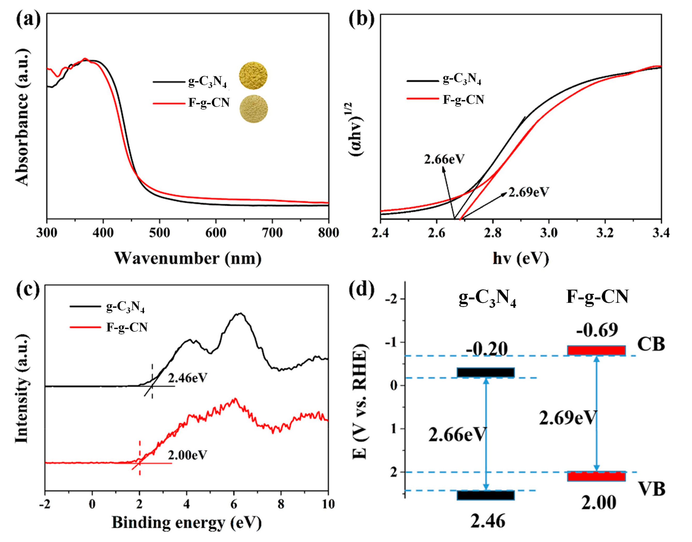
3.2. Optical and Electronic Properties

The optical absorption properties of the samples were studied by UV–Vis DRS spectra (Figure 5a). The absorption edge of F-g-CN shows an obvious blue shift compared with g-C3N4, which can be also represented by the color variation from yellow bulk to light yellow after the fluorination process (insets in Figure 5a). The corresponding bandgaps of the samples are determined by Tauc plots using the Kubelka–Munk function (Figure 5b), and the bandgap increases from 2.66 eV for g-C3N4 to 2.69 eV after fluorination, which is attributed to the structural distortion of F-g-CN [27]. Generally, the enlarged bandgap suffers from the shortcoming of light absorption, although an optical absorption tail in the longer wavelength observed in F-g-CN clearly implies the generation of a defect state or heteroatom level [51,52].
The influence of fluorination on the electronic structure of g-C3N4 is further investigated by valance band (VB) XPS spectra in Figure 5c. The positions of VB maximum of g-C3N4 and F-g-CN are validated as 2.46 and 2.00 eV, respectively. In combination with the bandgap derived from UV–Vis DRS, the conduction band (CB) edges of g-C3N4 and F-g-CN are estimated to be –0.20 and –0.69 eV, respectively. The CB minimum of F-g-CN is notably more negative than that of g-C3N4, which implies the more powerful reduction ability of the former [53]. The upshift of CB after fluorination is supposed to be caused by the vigorous structural distortion that is uncovered by the structural characterizations above. The schematic energy band structures of g-C3N4 and F-g-CN are illustrated in Figure 5d.
The steady PL spectra also depict a slight blue shift of the emission peak in F-g-CN (Figure 6a), consistent with their UV–Vis DRS spectra, which are proposed represent the induced structural distortion and formed C-F bonds. The time-resolved PL spectra were carried out at the wavelength of the maximum emission peak to measure the lifetime of the photogenerated charge carriers. The time-resolved PL spectra were carried out at the wavelength of the maximum emission peak to measure the lifetime of the photogenerated charge carriers. The fluorescence decay curves (Figure 6b) were fitted by using the following equation: I(t) = A1exp (−t/τ1) + A2exp (−t/τ2) + A3exp (−t/τ3). In order to relate the overall emission decay behaviors, the average lifetime (τave) was evaluated using the following equation: τave = (A1τ12 + A2τ22 + A3τ32)/(A1τ1 + A2τ2 + A3τ3). It was clearly deduced that after fluorination, the average emission lifetimes of g-C3N4 and F-g-CN were 6.39 and 8.62 ns, respectively. The fluorescence lifetime of F-g-CN was longer than that of g-C3N4, suggesting that more charge carriers took part in the photocatalytic reaction [54,55].
Transient photocurrents under visible light irradiation and EIS were implemented to analyze the photogenerated charge separation and transfer performances of the prepared samples. As displayed in Figure 6c, both g-C3N4 and F-g-CN electrodes shows positive transient photocurrents via alternate on–off cycles of visible light irradiation at a constant applied potential. Obviously, the photocurrent density values of g-C3N4, F-g-CN-120, F-g-CN-150 and F-g-CN-180 are 3.54, 4.58, 8.82 and 4.28 μAcm−2, respectively, indicating that F-g-CN-180 can provide more photogenerated charge carriers. The photocurrent density of the F-g-CN electrode is much higher than that of the g-C3N4 electrode, confirming the improved separation efficiency of photogenerated electron–hole pairs in F-g-CN, which is modulated by the distorted structure after the fluorination [27]. The density of state (DOS) of g-C3N4 (Figure 7a) shows that its VB is dominated by ring N atoms, while its CB is composed of C atoms and a small proportion of ring and inner N atoms. It can be noted that from the DOS of F-g-CN (Figure 7b), the CB of F-g-CN is dominated by the same atoms as g-C3N4, although its VB is comprised of ring N and bridged N atoms. The bridged N atoms in the VB of F-g-CN contribute to the mobility of photogenerated electron–hole pairs due to the presence of C–F bonds and the distorted structure [10]. Moreover, the arc radius of the EIS Nyquist plot for F-g-CN is markedly smaller than that of g-C3N4 (Figure 6d), which reflects the decreased charge transfer resistance at the interface and benefits the charge transfer after fluorination [56]. Therefore, the efficiency of the charge separation is much higher in F-g-CN than pristine g-C3N4.

3.3. Photocatalytic Performance

The photocatalytic performances of g-C3N4 and F-g-CN samples were evaluated with hydrogen evolution under visible light illumination (λ > 420 nm), where 3 wt.% Pt and TEOA were added in an aqueous solution as a co-catalyst and sacrificial agent, respectively. Both of the samples show the H2 evolution activity in Figure 8a and Figure S7b, while the average H2 evolution rates of g-C3N4, F-g-CN-120, F-g-CN-150 and F-g-CN-180 are 112, 675, 1298 and 370 μmol h−1 g−1, respectively. It can be seen that the H2 evolution rate of F-g-CN-150 is 11.6 times higher than that of g-C3N4 and the calculated AQE for F-g-CN-150 is as high as 4.53% at 420 nm, which highlights the outstanding superiority of F-g-CN-150 in photocatalytic H2 production (Table S3). Furthermore, there is no obvious decline in photocatalytic H2 evolution for five cycles under the same testing conditions (Figure 8b), indicating the sufficient stability and durability of F-g-CN as the photocatalyst. Based on the characterizations of F-g-CN, it can be claimed that the fluorination by F2 gas is an effective method to prominently promote the photocatalytic H2 production. Therefore, the synergetic effects of the highly porous structure, the considerable nitrogen vacancy defects and the relatively high degree of fluorination contribute to the excellent photocatalytic H2 evolution activity of F-g-CN.

4. Conclusions

In summary, a porous structure, nitrogen vacancy defects and fluorinated species were effectively introduced into the g-C3N4 framework via direct fluorination with F2 gas. Compared with the pristine g-C3N4, the photocatalytic H2 evolution activity of F-g-CN was improved significantly with a rate of 1298 μmol h−1 g1, which was 11.6 times higher than that of g-C3N4 under the same conditions. Based on the first-principle DFT calculations, detachment of bridged N atoms in tri-s-triazine from the g-C3N4 framework occurred during fluorination, resulting in N vacancy defects and covalent C-F bonds at the edge of the fragment. The specific surface area of F-g-CN was markedly enlarged due to the formation of mesopores in the g-C3N4 structure. Electron transition was also affected by the N vacancy defects, as well as by the presence of introduced F atoms and the structural distortion induced by fluorination. As a result of these synergetic effects, the separation of photogenerated charge carriers and utilization of solar energy were improved. Our study has highlighted a unique means of improving the performance of photocatalysts through fluorination. These results are instructive for understanding the complex effects of fluorination to g-C3N4, which inspired us to optimize the photocatalytic performance by engineering a photocatalytic texture and electronic structure.

Supplementary Materials

The following are available online at https://www.mdpi.com/article/10.3390/nano12010037/s1: Figure S1: SEM and TEM images of F-g-CN obtained by treating g-C3N4 with F2 gas at 120 °C (a,c) and 180 °C (b,d). Figure S2: (a) N2 adsorption–desorption isotherms and (b) the corresponding pore size distribution curves of g-C3N4 and F-g-CN. Figure S3: The XPS survey spectra of g-C3N4 and F-g-CN. Figure S4: (a) SEM image of F-g-CN and (b–d) its corresponding EDS elemental mapping images. Figure S5: (a) The optimized structure of the g-C3N4 model viewed from different directions: the blue and grey balls represent nitrogen and carbon atoms, respectively; (b) the corresponding deformation charge density of g-C3N4 and the increase and decrease in total electron density with the density of isolated atoms subtracted are denoted as blue and red, respectively. Figure S6: The optimized structure of F-g-CN model viewed from different directions. The blue, grey and cyan balls represent nitrogen, carbon and fluorine atoms, respectively. Figure S7: (a) Transient photocurrent responses of g-C3N4 and F-g-CN with different fluorinated temperatures. (b) Time-dependent photocatalytic H2 evolution over the pristine and fluorinated g-C3N4 samples. Table S1: The C 1s and N 1s binding energies, the intensity ratio of Nsp2/Nsp3 and the atomic ratio of C/N determined from XPS data in g-C3N4 and F-g-CN. Table S2: The surface atom contents of different materials. Table S3: Different g-C3N4-based materials used for photocatalytic H2 evolution.

Author Contributions

L.S. conducted the experiment, characterized the material properties and wrote the original draft. Y.L. evaluated the material properties, theoretical calculations and wrote the original draft. W.F. designed the experiment and revised the paper. All authors have read and agreed to the published version of the manuscript.

Funding

This work was financially supported by the National Key R&D Program of China (No. 2016YFA0202302), the State Key Program of National Natural Science Foundation of China (No. 51633007 and 52130303) and the National Natural Science Foundation of China (No. 51773147 and 51973151).

Conflicts of Interest

The authors declare no conflict of interest.

References

  1. Yan, H.J.; Yang, J.H.; Ma, G.J.; Wu, G.P.; Zong, X.; Lei, Z.B.; Shi, J.Y.; Li, C. Visible-light-driven hydrogen production with extremely high quantum efficiency on Pt-PdS/CdS photocatalyst. J. Catal. 2009, 266, 165–168. [Google Scholar] [CrossRef]
  2. Chen, X.B.; Liu, L.; Yu, P.Y.; Mao, S.S. Increasing solar absorption for photocatalysis with black hydrogenated titanium dioxide nanocrystals. Science 2011, 331, 746–750. [Google Scholar] [CrossRef] [PubMed]
  3. Qi, Y.; Zhao, Y.; Gao, Y.Y.; Li, D.; Li, Z.; Zhang, F.X.; Li, C. Redox-based visible-light-driven Z-scheme overall water splitting with apparent quantum efficiency exceeding 10%. Joule 2018, 2, 2393–2402. [Google Scholar] [CrossRef] [Green Version]
  4. Wang, X.C.; Maeda, K.; Thomas, A.; Takanabe, K.; Xin, G.; Carlsson, J.M.; Domen, K.; Antonietti, M. A metal-free polymeric photocatalyst for hydrogen production from water under visible light. Nat. Mater. 2009, 8, 76–80. [Google Scholar] [CrossRef]
  5. Ong, W.J.; Tan, L.L.; Ng, Y.H.; Yong, S.T.; Chai, S.P. Graphitic carbon nitride (g-C3N4)-based photocatalysts for artificial photosynthesis and environmental remediation: Are we a step closer to achieving sustainability? Chem. Rev. 2016, 116, 7159–7329. [Google Scholar] [CrossRef] [PubMed]
  6. Zhou, Z.X.; Zhang, Y.Y.; Shen, Y.F.; Liu, S.Q.; Zhang, Y.J. Molecular engineering of polymeric carbon nitride: Advancing applications from photocatalysis to biosensing and more. Chem. Soc. Rev. 2018, 47, 2298–2321. [Google Scholar] [CrossRef]
  7. Fu, J.W.; Zhu, B.C.; Jiang, C.J.; Cheng, B.; You, W.; Yu, J.G. Hierarchical porous O-doped g-C3N4 with enhanced photocatalytic CO2 deduction activity. Small 2017, 13, 1603938. [Google Scholar] [CrossRef] [PubMed]
  8. Jorge, A.B.; Dedigama, I.; Miller, T.S.; Shearing, P.; Brett, D.J.L.; McMilan, P.F. Carbon nitride materials as efficient catalyst supports for proton exchange membrane water electrolyzers. Nanomaterials 2018, 6, 432. [Google Scholar] [CrossRef] [PubMed] [Green Version]
  9. Cao, S.W.; Low, J.X.; Yu, J.G.; Jaroniec, M. Polymeric photocatalysts based on graphitic carbon nitride. Adv. Mater. 2015, 27, 2150–2176. [Google Scholar] [CrossRef]
  10. Liu, J.; Liu, Y.; Liu, N.Y.; Han, Y.Z.; Zhang, X.; Huang, H.; Lifshitz, Y.; Lee, S.T.; Zhong, J.; Kang, Z.H. Metal-free efficient photocatalyst for stable visible water splitting via a two-electron pathway. Science 2015, 347, 970–974. [Google Scholar] [CrossRef] [PubMed]
  11. Yu, F.T.; Wang, Z.Q.; Zhang, S.C.; Ye, H.N.; Kong, K.Y.; Gong, X.Q.; Hua, J.L.; Tian, H. Molecular engineering of donor-acceptor conjugated polymer/g-C3N4 heterostructures for significantly enhanced hydrogen evolution under visible-light irradiation. Adv. Funct. Mater. 2018, 28, 1804512. [Google Scholar] [CrossRef]
  12. Asahi, R.; Morikawa, T.; Ohwaki, T.; Aoki, K.; Taga, Y. Visible-light photocatalysis in nitrogen-doped titanium oxides. Science 2001, 293, 269–271. [Google Scholar] [CrossRef] [PubMed]
  13. Liu, G.; Niu, P.; Sun, C.H.; Smith, S.C.; Chen, Z.G.; Lu, G.Q.; Cheng, H.M. Unique electronic structure induced high photoreactivity of sulfur-doped graphitic C3N4. J. Am. Chem. Soc. 2010, 132, 11642–11648. [Google Scholar] [CrossRef] [PubMed]
  14. Ran, J.R.; Ma, T.Y.; Gao, G.P.; Du, X.W.; Qiao, S.Z. Porous P-doped graphitic carbon nitride nanosheets for synergistically enhanced visible-light photocatalytic H2 production. Energy Environ. Sci. 2015, 8, 3708–3717. [Google Scholar] [CrossRef]
  15. Zhang, G.G.; Zhang, M.W.; Ye, X.X.; Qiu, X.Q.; Sen, L.; Wang, X.C. Iodine modified carbon nitride semiconductors as visible light photocatalysts for hydrogen evolution. Adv. Mater. 2014, 26, 805–809. [Google Scholar] [CrossRef] [PubMed]
  16. Praus, P.; Smykalova, S.; Foniok, K.; Velisek, P.; Cvejn, D.; Zadny, J.; Storch, J. Post-synthetic derivatization of graphitic carbon nitride with methanesulfonyl chloride: Synthesis, characterization and photocatalysis. Nanomaterials 2020, 10, 193. [Google Scholar] [CrossRef] [Green Version]
  17. Jeon, I.Y.; Ju, M.J.; Xu, J.T.; Choi, H.J.; Seo, J.M.; Kim, M.J.; Choi, I.T.; Kim, H.M.; Kim, J.C.; Lee, J.J.; et al. Edge-fluorinated graphene nanoplatelets as high performance electrodes for dye-sensitized solar cells and lithium ion batteries. Adv. Funct. Mater. 2015, 25, 1170–1179. [Google Scholar] [CrossRef]
  18. Karlicky, F.; Datta, K.K.R.; Otyepka, M.; Zboril, R. Halogenated graphenes: Rapidly growing family of graphene derivatives. ACS Nano 2013, 7, 6434–6464. [Google Scholar] [CrossRef]
  19. Feng, W.; Long, P.; Feng, Y.Y.; Li, Y. Two-dimensional fluorinated graphene: Synthesis, structures, properties and applications. Adv. Sci. 2016, 3, 1500413. [Google Scholar] [CrossRef]
  20. Chen, X.Y.; Fan, K.; Liu, Y.; Li, Y.; Liu, X.Y.; Feng, W.; Wang, X. Recent advances in fluorinated graphene from synthesis to applications: Critical review on functional chemistry and structure engineering. Adv. Mater. 2021, 2101665. [Google Scholar] [CrossRef] [PubMed]
  21. Yong, W.; Yan, D.; Antonietti, M.; Lic, H.R.; Chen, X.F.; Wang, X.C. Excellent visible-light photocatalysis of fluorinated polymeric carbon nitride solids. Chem. Mater. 2010, 22, 5119–5121. [Google Scholar]
  22. Gao, D.; Liu, Y.; Song, M.; Shi, S.; Si, M.; Xue, D. Manifestation of high-temperature ferromagnetism in fluorinated graphitic carbon nitride nanosheets. J. Mater. Chem. C 2015, 3, 12230–12235. [Google Scholar] [CrossRef]
  23. Xu, M.Q.; Chai, B.; Yan, J.T.; Wang, H.B.; Ren, Z.D.; Paik, K.W. Facile synthesis of fluorine doped graphitic carbon nitride with enhanced visible light photocatalytic activity. Nano 2016, 11, 1650137. [Google Scholar] [CrossRef]
  24. Palanivel, B.; Hu, C.; Shkir, M.; AlFaify, S.; Ibrahim, F.A.; Hamdy, M.S.; Mani, A. Fluorine doped g-C3N4 coupled NiFe2O4 heterojunction: Consumption of H2O2 for production of hydroxyl radicals towards paracetamol degradation. Colloid Interface Sci. Commun. 2021, 42, 100410. [Google Scholar] [CrossRef]
  25. Zeng, L.; Ding, X.; Sun, Z.Z.; Hua, W.M.; Song, W.L.; Liu, S.Y.; Huang, L.M. Enhancement of photocatalytic hydrogen evolution activity of g-C3N4 induced by structural distortion via post-fluorination treatment. Appl. Catal. B Environ. 2018, 227, 276–284. [Google Scholar] [CrossRef]
  26. Lin, W.; Lu, K.C.; Zhou, S.J.; Wang, J.; Mu, F.H.; Wang, Y.; Wu, Y.; Kong, Y. Defects remodeling of g-C3N4 nanosheets by fluorine-containing solvothermal treatment to enhance their photocatalytic activities. Appl. Surf. Sci. 2019, 474, 194–202. [Google Scholar] [CrossRef]
  27. Ma, F.K.; Sun, C.L.; Shao, Y.L.; Wu, Y.Z.; Huang, B.B.; Hao, X.P. One-step exfoliation and fluorination of g-C3N4 nanosheets with enhanced photocatalytic activities. New. J. Chem. 2017, 41, 3061–3067. [Google Scholar] [CrossRef]
  28. Xue, M.T.; Tan, G.Q.; Liu, T.; Lv, L.; Li, B.; Zhang, D.; Dang, M.Y.; Ren, H.J.; Xia, A. Insights into the improved photocatalytic performance of fluorine surface modified mpg-C3N4 at room temperature under aqueous conditions. Appl. Catal. A Gen. 2019, 578, 89–97. [Google Scholar] [CrossRef]
  29. Mirzaei, A.; Chen, Z.; Haghighat, F.; Yerushalmi, L. Magnetic fluorinated mesoporous g-C3N4 for photocatalytic degradation of amoxicillin: Transformation mechanism and toxicity assessment. Appl. Catal. B Environ. 2019, 242, 337–348. [Google Scholar] [CrossRef]
  30. Wang, H.; Zhang, X.D.; Xie, J.F.; Zhang, J.J.; Ma, P.; Pan, B.C.; Xie, Y. Structural distortion in graphitic-C3N4 realizing an efficient photoreactivity. Nanoscale 2015, 7, 5152–5156. [Google Scholar] [CrossRef] [PubMed]
  31. Guerin, K.; Pinheiro, J.P.; Dubois, M.; Fawal, Z.; Masin, F.; Yazami, R.; Hamwi, A. Synthesis and characterization of highly fluorinated graphite containing sp2 and sp3 carbon. Chem. Mater. 2004, 16, 1786–1792. [Google Scholar] [CrossRef]
  32. Peng, C.; Li, Y.; Yao, F.N.; Fu, H.Y.; Zhou, R.X.; Feng, Y.Y.; Feng, W. Ultrahigh-energy-density fluorinated calcinated macadamia nut shell cathodes for lithium/fluorinated carbon batteries. Carbon 2019, 153, 783–791. [Google Scholar] [CrossRef]
  33. Fulvio, P.F.; Brown, S.S.; Adcock, J.; Mayes, R.T.; Guo, B.; Sun, X.G.; Dai, S. Low-temperature fluorination of soft-templated mesoporous carbons for a high-power lithium/carbon fluoride battery. Chem. Mater. 2011, 23, 4420–4427. [Google Scholar] [CrossRef]
  34. Niu, P.; Zhang, L.L.; Liu, G.; Cheng, H.M. Graphene-like carbon nitride nanosheets for improved photocatalytic activities. Adv. Funct. Mater. 2012, 22, 4763–4770. [Google Scholar] [CrossRef]
  35. Wang, L.; Li, Y.Y.; Wang, S.; Zhou, P.F.; Zhao, Z.D.; Li, X.W.; Zhuo, S.P. Fluorinated nanographite as a cathode material for lithium primary batteries. ChemElectroChem 2019, 6, 2201–2207. [Google Scholar] [CrossRef]
  36. Lotsch, B.V.; Doeblinger, M.; Sehnert, J.; Seyfarth, L.; Senker, J.; Oeckler, O.; Schnick, W. Unmasking melon by a complementary approach employing electron diffraction, solid-state NMR spectroscopy, and theoretical calculations-structural characterization of a carbon nitride polymer. Chem. Eur. J. 2007, 13, 4969–4980. [Google Scholar] [CrossRef]
  37. Liang, Q.H.; Li, Z.; Huang, Z.H.; Kang, F.Y.; Yang, Q.H. Holey graphitic carbon nitride nanosheets with carbon vacancies for highly improved photocatalytic hydrogen production. Adv. Funct. Mater. 2015, 25, 6885–6892. [Google Scholar] [CrossRef]
  38. Chetia, T.R.; Ansari, M.S.; Qureshi, M. Graphitic carbon nitride as a photovoltaic booster in quantum dot sensitized solar cells: A synergistic approach for enhanced charge separation and injection. J. Mater. Chem. A 2016, 4, 5528–5541. [Google Scholar] [CrossRef]
  39. Yang, L.Q.; Huang, J.F.; Li, S.; Cao, L.Y.; Yu, Q.; Jie, Y.N.; Jie, F.; Ouyang, H.B.; Ye, J.H. A surface modification resultant thermally oxidized porous g-C3N4 with enhanced photocatalytic hydrogen production. Appl. Catal. B Environ. 2017, 204, 335–345. [Google Scholar] [CrossRef]
  40. Wang, H.; Bian, Y.R.; Hu, J.T.; Dai, L.M. Highly crystalline sulfur-doped carbon nitride as photocatalyst for efficient visible-light hydrogen generation. Appl. Catal. B Environ. 2018, 238, 592–598. [Google Scholar] [CrossRef]
  41. Meng, N.N.; Jian, R.; Yang, L.; Yi, H.; Petit, T.; Zhang, B. Engineering oxygen-containing and amino groups into two-dimensional atomically-thin porous polymeric carbon nitrogen for enhanced photocatalytic hydrogen production. Energy Environ. Sci. 2018, 11, 566–571. [Google Scholar] [CrossRef] [Green Version]
  42. She, X.J.; Wu, J.J.; Zhong, J.; Xu, H.; Yang, Y.C.; Vajtai, R.; Lou, J.; Liu, Y.; Du, D.L.; Li, H.M.; et al. Oxygenated monolayer carbon nitride for excellent photocatalytic hydrogen evolution and external quantum efficiency. Nano Energy 2016, 27, 138–146. [Google Scholar] [CrossRef]
  43. Jiang, Y.B.; Sun, Z.Z.; Tang, C.; Zhou, Y.X.; Zeng, L.; Huang, L.M. Enhancement of photocatalytic hydrogen evolution activity of porous oxygen doped g-C3N4 with nitrogen defects induced by changing electron transition. Appl. Catal. B Environ. 2019, 240, 30–38. [Google Scholar] [CrossRef]
  44. Yu, H.J.; Shi, R.; Zhao, Y.X.; Bian, T.; Zhao, Y.F.; Zhou, C.; Waterhouse, G.I.N.; Wu, L.Z.; Tung, C.H.; Zhang, T.R. Alkali-assisted synthesis of nitrogen deficient graphitic carbon nitride with tunable band structures for efficient visible-light-driven hydrogen evolution. Adv. Mater. 2017, 29, 1605148. [Google Scholar] [CrossRef] [PubMed]
  45. Xing, W.N.; Chen, G.; Li, C.M.; Han, Z.H.; Hu, Y.D.; Meng, Q.Q. Doping effect of non-metal group in porous ultrathin g-C3N4 nanosheets towards synergistically improved photocatalytic hydrogen evolution. Nanoscale 2018, 10, 5239–5245. [Google Scholar] [CrossRef] [PubMed]
  46. Wang, N.; Fan, H.; Sun, J.C.; Han, Z.W.; Dong, J.; Ai, S.Y. Fluorine-doped carbon nitride quantum dots: Ethylene glycol-assisted synthesis, fluorescent properties, and their application for bacterial imaging. Carbon 2016, 109, 141–148. [Google Scholar] [CrossRef]
  47. Delley, B. An all-electron numerical method for solving the local density functional for polyatomic molecules. J. Chem. Phys. 1990, 92, 508–517. [Google Scholar] [CrossRef]
  48. Delley, B. From molecules to solids with the DMol3 approach. J. Chem. Phys. 2000, 113, 7756–7764. [Google Scholar] [CrossRef]
  49. Wolf, A.K.; Glinnemann, J.; Schmidt, M.U. Packing of tetrahedral EX4 molecules with E = C, Si, Ge, Sn, Pb and X = F, Cl, Br, I. CrystEngComm 2008, 10, 1364–1371. [Google Scholar] [CrossRef]
  50. Wang, Y.J.; Li, L.B.; Wei, Y.Y.; Xue, J.; Chen, H.; Ding, L.; Caro, J.; Wang, H.H. Water transport with ultralow friction through partially exfoliated g-C3N4 nanosheet membranes with self-supporting spacers. Angew. Chem. Int. Ed. 2017, 56, 8974–8980. [Google Scholar] [CrossRef]
  51. Li, Y.F.; Yang, M.; Xing, Y.; Liu, X.C.; Yang, Y.; Wang, X.; Song, S.Y. Preparation of carbon-rich g-C3N4 nanosheets with enhanced visible light utilization for efficient photocatalytic hydrogen production. Small 2017, 13, 1701552. [Google Scholar] [CrossRef] [PubMed]
  52. Niu, P.; Yin, L.C.; Yang, Y.Q.; Liu, G.; Cheng, H.M. Increasing the visible light absorption of graphitic carbon nitride (Melon) photocatalysts by homogeneous self-modification with nitrogen vacancies. Adv. Mater. 2014, 26, 8046–8052. [Google Scholar] [CrossRef]
  53. Han, Q.; Wang, B.; Gao, J.; Cheng, Z.H.; Zhao, Y.; Zhang, Z.P.; Qu, L.T. Atomically thin mesoporous nanomesh of graphitic C3N4 for high-efficiency photocatalytic hydrogen evolution. ACS Nano 2016, 10, 2745–2751. [Google Scholar] [CrossRef] [PubMed]
  54. Liu, W.; Cao, L.L.; Cheng, W.R.; Cao, Y.J.; Liu, X.K.; Zhang, W.; Mou, X.L.; Jin, L.L.; Zheng, X.S.; Che, W.; et al. Single-site active cobalt-based photocatalyst with a long carrier lifetime for spontaneous overall water splitting. Angew. Chem. Int. Ed. 2017, 129, 9440–9445. [Google Scholar] [CrossRef]
  55. Wen, W.; Wang, J.B.; Tong, R.F.; Liu, D.N.; Huang, H.; Yu, Y.; Zhou, Z.K.; Chu, P.K.; Yu, X.F. A low-cost metal-free photocatalyst based on black phosphorus. Adv. Sci. 2019, 6, 1801321. [Google Scholar] [CrossRef] [Green Version]
  56. Kim, H.; Gim, S.; Jeon, T.H.; Kim, H.; Choi, W. Distorted carbon nitride structure with substituted benzene moieties for enhanced visible light photocatalytic activities. ACS Appl. Mater. Interfaces 2017, 9, 40360–40368. [Google Scholar] [CrossRef]
Figure 1. The schematic synthesis process of F-g-CN.
Nanomaterials 12 00037 g001
Figure 2. SEM and TEM images of (a,c) g-C3N4 and (b,d) F-g-CN synthesized at 150 °C.
Nanomaterials 12 00037 g002
Figure 3. (a) XRD patterns and (b) FTIR spectra of g-C3N4 and F-g-CN.
Nanomaterials 12 00037 g003
Figure 4. High-resolution XPS spectra of g-C3N4 and F-g-CN: (a) C1s; (b) N1s; (c) F1s. (d) Schematic diagram of the g-C3N4 fluorination process. The blue, gray and cyan circles represent nitrogen, carbon and fluorine atoms, respectively.
Nanomaterials 12 00037 g004
Figure 5. (a) UV-Vis absorption spectra of g-C3N4 and F-g-CN and (b) their corresponding bandgaps. (c) VB XPS spectra of g-C3N4 and F-g-CN. (d) Schematic illustration of the g-C3N4 and F-g-CN band structures.
Nanomaterials 12 00037 g005
Figure 6. (a) PL spectra and (b) time-resolved PL spectra of g-C3N4 and F-g-CN. (c) Transient photocurrent responses and (d) EIS plots of g-C3N4 and F-g-CN.
Nanomaterials 12 00037 g006
Figure 7. Calculated density of state (DOS) values for (a) g-C3N4 and (b) F-g-CN from their optimized structures.
Nanomaterials 12 00037 g007
Figure 8. (a) Photocatalytic H2 evolution rates for g-C3N4 and F-g-CN. (b) Cycling tests of H2 evolution with F-g-CN-150.
Nanomaterials 12 00037 g008
Publisher’s Note: MDPI stays neutral with regard to jurisdictional claims in published maps and institutional affiliations.

