
Advances in Polyhydroxyalkanoate Nanocarriers for Effective Drug Delivery: An Overview and Challenges


 and
and
Abstract
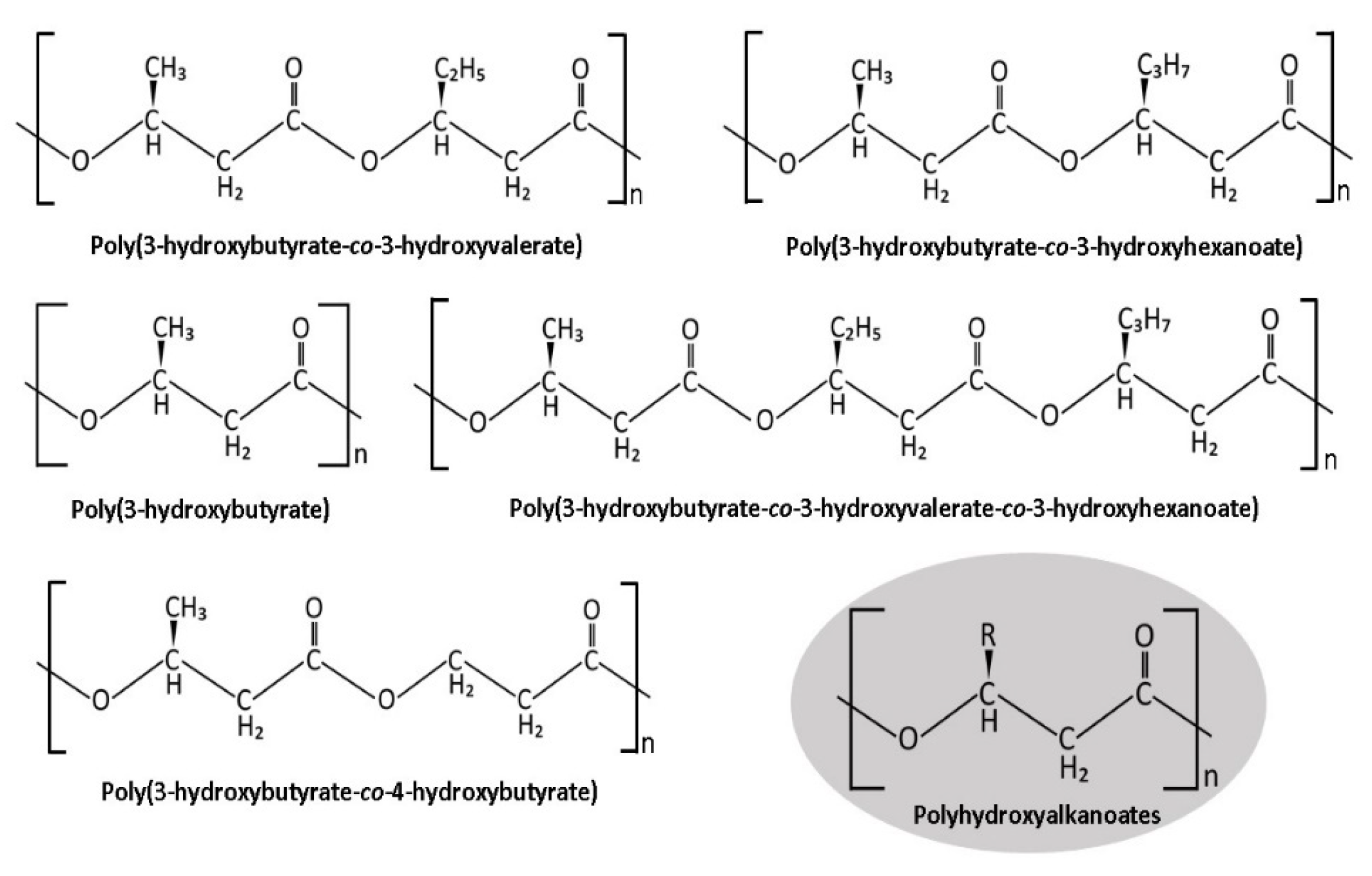
1. Introduction
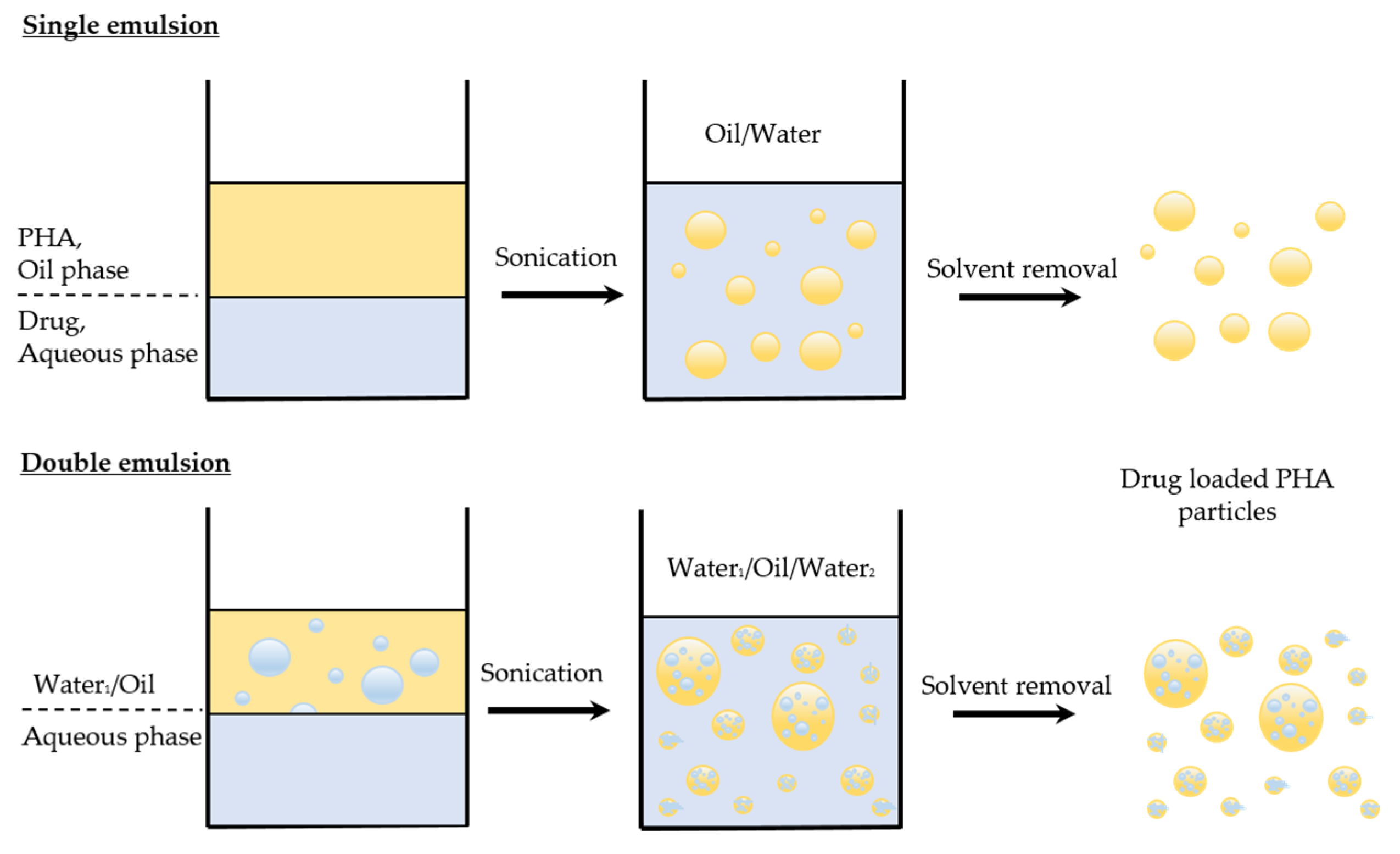
2. Formulation of PHA Nanocarriers

| Polymer | Drug | Size (nm) | Drug Loading (%) | Formulation Method | Key Findings | Ref. |
|---|---|---|---|---|---|---|
| Poly(3-R-hydroxyalkanoate) | Calcein and Nile red | 155 | - | Nanoprecipitation | Unsaturated PHA is suitable to make controlled release nanomedicine. | [45] |
| Poly(3-hydroxybutyrate-co-3-hydroxyvalerate-co-3-hydroxyhexanoate) (PHBVHHx) | Azathioprine | 95.7 | - | Modified emulsion | The particles have acceptable toxicity and slow clearance from kidneys, with a higher therapeutic effect than polylactic acid (PLA) nanoparticles when tested in a murine systemic lupus erythematosus model. | [32] |
| Poly(3-hydroxybutyrate-co-3-hydroxyhexanoate) (PHBHHx) | Curcumin | 273 ± 84 | 15–30 | Solvent evaporation | Lyophilization is suitable for preserving the nanoparticles at 4 °C. The particles had high apoptotic activity and localization into MDA-MB-231 cells. | [33] |
| Poly(3-hydroxybutyrate-co-3-hydroxyhexanoate) (PHBHHX) | Etoposide | 180–1500 | 2.92–8.77 | Modified solvent evaporation | Folic acid-conjugated nanoparticles have higher selectivity to cancer cells than fibroblast cells. | [34] |
| Poly(3-hydroxyvalerate-co-4-hydroxybutyrate) | Cisplatin | 155 ± 5 | 9.58 ± 1 | Emulsification–solvent evaporation | Cisplatin-loaded PHA nanoparticles accumulated in tumour cells and showed significant tumour deterioration compared to free drug treatment. | [35] |
| Poly(3-hydroxybutyrate-co-3-hydroxyvalerate) (PHBV) | Nile red | 166–426 | - | Oil-in-water emulsion | The nanoparticles penetrated the skin of the BALB/c mouse model without adverse effects. | [46] |
| Poly (3-hydroxybutyrate-co-12 mol% 3-hydroxyhexanoate) (PHBHHx) and Poly (3-hydroxybutyrate-co-5 mol% 3-hydroxyhexanoate) (PHBV) | TGX-221 | 195–220 | 8.5–8.8 | Modified emulsification/solvent diffusion | The encapsulation of TGX-221 in PHA nanoparticles could mitigate the poor bioavailability and limited in vivo half-life of the TGX-221. | [39] |
| Poly-3-hydroxybutyrate-co-5 mol% 3-hydroxyvalerate (PHBV-S), poly-3-hydroxybutyrate-co-11 mol% 3-hydroxyvalerate (PHBV-11) and poly-3-hydroxybutyrate-co-15 mol% 3-hydroxyvalerate (PHBV-15) | Ellipticine | 184–283 | - | Modified emulsification–solvent evaporation | The particles showed no inhibition of the A549 cancer cell line at various tested concentrations (i.e., 250.0, 62.5, and 15.6 μg/mL). | [47] |
| Poly(3-hydroxybutyrate-co-3-hydroxyhexanoate) (PHBHHx) | Rapamycin | 200 | 8.47–8.52 | Emulsification–solvent evaporation | The particles showed an efficient entrapment of 91.9% and a sustained release of rapamycin for almost 10 days. Cellular uptake of PEG200 end-capped nanoparticles was significantly higher than that of non-PEG nanoparticles in a human prostate cancer cell line and a murine macrophage cell line. | [40] |
| Polyhydroxybutyrate, poly(hydroxybutyrate-co-hydroxyvalerate) P(HB-HV) with 12 and 50% HV | 5,10,15,20-Tetrakis(4-hydroxy-phenyl)-21H, 23H-porphine | 169.0–211.2 | 0.91–46.64 | Emulsification-diffusion | The particles showed a concentration and time-dependent photocytotoxicity in a human colon adenocarcinoma cell line. | [48] |
| Poly(3-hydroxyoctanoate-co-3-hydroxyhexanoate) (PHOHHx) | - | 44–90 | - | Dialysis | A series of diblock copolymers of PHOHHx with poly(ethylene glycol) (PEG) were synthesized using “click” chemistry and assembled into micelles for drug delivery. | [29] |
| Poly(3-hydroxybutyrate-co-3-hydroxyvalerate) P(3HB-co-3HV) or poly(3-hydroxybutyrate-co-4-hydroxybutyrate) P(3HB-co-4HB) | Thymoquinone | 112–162 | - | Modified emulsification–solvent evaporation | The chemical combination of PHA copolymers and mPEG-based nanoparticles was nontoxic and biocompatible to prenatal rat neuronal hippocampal and NIH/3T3 fibroblast cells in vitro. | [49] |
| Polyhydroxybutyrate (PHB) | NuBCP-9 | 126 ± 8 | - | Double emulsion solvent evaporation | PEG-conjugated PHB nanoparticles showed a sustained release of NuBCP-9 for up to 26 days and efficient cellular uptake in a time-dependent manner in MCF-7 cells. A 90% tumour regression was seen when particles were administered intraperitoneally twice a week for three weeks in an Ehrlich syngeneic mouse model. | [50] |
| Polyhydroxybutyrate (PHB) | Nile red | - | - | Oil-in-water emulsion solvent evaporation | PHB functionalized with tumour-specific ligand nanoparticles showed a specific affinity to MDA-MB-231 breast cancer cells. | [51] |
| Poly(3-hydroxybutyrate-co-3-hydroxyhexanoate) (PHBHHx) | Rhodamine B isothiocyanate | 100–200 | - | Oil-in-water emulsion | The recombinant human a1-acid glycoprotein or recombinant human epidermal growth factor functionalized nanoparticles were taken up by macrophages and hepatocellular carcinoma cells. | [52] |
| Poly(3-hydroxybutyrate-co-3-hydroxyvalerate) (PHBV) | - | 133–300 | - | Miniemulsification and emulsion/solvent evaporation | An increase of the polymer concentration led to a larger particle size due to a change in viscosity. | [53] |
| Poly([R,S]-3-hydroxybutyrate) (PHB) | Doxorubicin and sorafenib | 199.3–250.5 | 2.6–8.4 | Nanoprecipitation | Co-encapsulation of dual anticancer drugs was achieved. A sustained and faster drug release was observed for doxorubicin and sorafenib, respectively. | [41] |
| Poly(hydroxioctanoate-co-hexanoate) | - | 63 ± 4 | - | Emulsion-solvent evaporation | The particles interacted with pulmonary surfactant proteins and lipids, which may limit the use of PHA for pulmonary drug delivery. | [54] |
| Polyhydroxyalkanoate (PHA) | - | 145–159 | - | Oil-in-water emulsion | The PHA nanoparticles showed antibacterial activity against S. aureus, S. pneumoniae, E. coli, K. pneumoniae, and P. aeruginosa. | [55] |
| Poly(3-hydroxybutyrate-co-3-hydroxyoctanoate) (P(HB-HO)) | Doxorubicin | 240 | 29.6 | Water-in-oil-in-water solvent extraction/evaporation | Doxorubicin-loaded folate-mediated nanoparticles were readily internalized by HeLa cells in vitro. | [42] |
| Polyhydroxybutyrates (PHB) | Concanavalin-A and etoposide | 239.43 ± 5.25 | - | Multi-emulsion | Iron oxide particles were successfully coated with PHB. The cytotoxicity of these magnetic PHB particles were reported against cancer and non-cancer cells. | [56] |
| Poly(hydroxybutyrate-co-hydroxyvalerate) (PHBV) | Fingolimod | 250 | 0–22.5 | Single and double evaporation | The optimal preparation of PHBV nanoparticles required a polymer concentration of 1.32%, a PVA concentration of 0.42%, and 5 mg of the drug. | [43] |
3. Applications of PHA Nanocarriers
3.1. Treatment of Cancer
3.2. Treatment of Infectious Diseases
3.3. Other Applications
4. Challenges and Author’s Perspective
5. Conclusions
Author Contributions
Funding
Data Availability Statement
Conflicts of Interest
References
- Jain, K.K. Drug Delivery Systems an Overview. Methods Mol. Biol. 2008, 437, 1–50. [Google Scholar] [PubMed]
- Maiti, S.; Sen, K.K. Introductory Chapter: Drug Delivery Concepts. In Advanced Technology for Delivering Therapeutics; Maiti, S., Sen, K.K., Eds.; InTech: London, UK, 2017. [Google Scholar]
- Neubert, R.H.H. Potentials of New Nanocarriers for Dermal and Transdermal Drug Delivery. Eur. J. Pharm. Biopharm. 2011, 77, 1–2. [Google Scholar] [CrossRef] [PubMed]
- Jeevanandam, J.; Barhoum, A.; Chan, Y.S.; Dufresne, A.; Danquah, M.K. Review on Nanoparticles and Nanostructured Materials: History, Sources, Toxicity and Regulations. Beilstein J. Nanotechnol. 2018, 9, 1050–1074. [Google Scholar] [CrossRef] [PubMed]
- Rizvi, S.A.A.; Saleh, A.M. Applications of Nanoparticle Systems in Drug Delivery Technology. Saudi Pharm. J. 2018, 26, 64–70. [Google Scholar]
- Mirza, A.Z.; Siddiqui, F.A. Nanomedicine and Drug Delivery: A Mini Review. Int. Nano Lett. 2014, 4, 94. [Google Scholar] [CrossRef]
- Ud Din, F.; Aman, W.; Ullah, I.; Qureshi, O.S.; Mustapha, O.; Shafique, S.; Zeb, A. Effective Use of Nanocarriers as Drug Delivery Systems for the Treatment of Selected Tumors. Int. J. Nanomed. 2017, 12, 7291–7309. [Google Scholar] [CrossRef]
- De Villiers, M.M.; Aramwit, P.; Kwon, G.S. Nanotechnology in Drug Delivery; de Villiers, M.M., Aramwit, P., Kwon, G.S., Eds.; Springer: New York, NY, USA, 2009. [Google Scholar] [CrossRef]
- Scicluna, M.C.; Vella-Zarb, L. Evolution of Nanocarrier Drug-Delivery Systems and Recent Advancements in Covalent Organic Framework–Drug Systems. ACS Appl. Nano Mater. 2020, 3, 3097–3115. [Google Scholar] [CrossRef]
- Tan, G.-Y.A.; Chen, C.-L.; Li, L.; Ge, L.; Wang, L.; Razaad, I.; Li, Y.; Zhao, L.; Mo, Y.; Wang, J.-Y. Start a Research on Biopolymer Polyhydroxyalkanoate (PHA): A Review. Polymers 2014, 6, 706–754. [Google Scholar] [CrossRef]
- Kathiraser, Y.; Aroua, M.K.; Ramachandran, K.B.; Tan, I.K.P. Chemical Characterization of Medium-Chain-Length Polyhydroxyalkanoates (PHAs) Recovered by Enzymatic Treatment and Ultrafiltration. J. Chem. Technol. Biotechnol. 2007, 82, 847–855. [Google Scholar] [CrossRef]
- Dhandayuthapani, B.; Yoshida, Y.; Maekawa, T.; Kumar, D.S. Polymeric Scaffolds in Tissue Engineering Application: A Review. Int. J. Polym. Sci. 2011, 2011, 290602. [Google Scholar] [CrossRef]
- Griffin, G. (Ed.) Chemistry and Technology of Biodegradable Polymers, 1994th ed.; Chapman and Hall: London, UK, 1993. [Google Scholar]
- Nigmatullin, R.; Thomas, P.; Lukasiewicz, B.; Puthussery, H.; Roy, I. Polyhydroxyalkanoates, a Family of Natural Polymers, and Their Applications in Drug Delivery. J. Chem. Technol. Biotechnol. 2015, 90, 1209–1221. [Google Scholar] [CrossRef]
- Shrivastav, A.; Kim, H.-Y.; Kim, Y.-R. Advances in the Applications of Polyhydroxyalkanoate Nanoparticles for Novel Drug Delivery System. Biomed Res. Int. 2013, 2013, 581684. [Google Scholar] [CrossRef]
- Li, Z.; Yang, J.; Loh, X.J. Polyhydroxyalkanoates: Opening Doors for a Sustainable Future. NPG Asia Mater. 2016, 8, e265. [Google Scholar] [CrossRef]
- Kourmentza, C.; Plácido, J.; Venetsaneas, N.; Burniol-Figols, A.; Varrone, C.; Gavala, H.N.; Reis, M.A.M. Recent Advances and Challenges towards Sustainable Polyhydroxyalkanoate (PHA) Production. Bioengineering 2017, 4, 55. [Google Scholar] [CrossRef]
- National Cancer Institute. Cancer and Nanotechnology; National Cancer Institute: Rockville, MD, USA, 2020. [Google Scholar]
- Awasthi, R.; Roseblade, A.; Hansbro, P.M.; Rathbone, M.J.; Dua, K.; Bebawy, M. Nanoparticles in Cancer Treatment: Opportunities and Obstacles. Curr. Drug Targets 2018, 19, 1696–1709. [Google Scholar] [CrossRef] [PubMed]
- Aghebati-Maleki, A.; Dolati, S.; Ahmadi, M.; Baghbanzhadeh, A.; Asadi, M.; Fotouhi, A.; Yousefi, M.; Aghebati-Maleki, L. Nanoparticles and Cancer Therapy: Perspectives for Application of Nanoparticles in the Treatment of Cancers. J. Cell. Physiol. 2020, 235, 1962–1972. [Google Scholar] [CrossRef] [PubMed]
- Babu, A.; Templeton, A.K.; Munshi, A.; Ramesh, R. Nanoparticle-Based Drug Delivery for Therapy of Lung Cancer: Progress and Challenges. J. Nanomater. 2013, 2013, 14. [Google Scholar] [CrossRef]
- Lu, X.-Y.; Wu, D.-C.; Li, Z.-J.; Chen, G.-Q. Polymer Nanoparticles. Prog. Mol. Biol. Transl. Sci. 2011, 104, 299–323. [Google Scholar] [PubMed]
- Blanco, F.G.; Hernández, N.; Rivero-Buceta, V.; Maestro, B.; Sanz, J.M.; Mato, A.; Hernández-Arriaga, A.M.; Prieto, M.A. From Residues to Added-Value Bacterial Biopolymers as Nanomaterials for Biomedical Applications. Nanomaterials 2021, 11, 1492. [Google Scholar] [CrossRef]
- Elmowafy, E.; Abdal-Hay, A.; Skouras, A.; Tiboni, M.; Casettari, L.; Guarino, V. Polyhydroxyalkanoate (PHA): Applications in Drug Delivery and Tissue Engineering. Expert Rev. Med. Devices 2019, 16, 467–482. [Google Scholar] [CrossRef]
- Nobes, G.A.; Marchessault, R.H.; Maysinger, D. Polyhydroxyalkanoates: Materials for Delivery Systems. Drug Deliv. 1998, 5, 167–177. [Google Scholar] [CrossRef] [PubMed]
- Michalak, M.; Kurcok, P.; Hakkarainen, M. Polyhydroxyalkanoate-Based Drug Delivery Systems: Polyhydroxyalkanoate-Based Drug Delivery Systems. Polym. Int. 2017, 66, 617–622. [Google Scholar] [CrossRef]
- Puppi, D.; Pecorini, G.; Chiellini, F. Biomedical Processing of Polyhydroxyalkanoates. Bioengineering 2019, 6, 108. [Google Scholar] [CrossRef]
- Rao, J.P.; Geckeler, K.E. Polymer Nanoparticles: Preparation Techniques and Size-Control Parameters. Prog. Polym. Sci. 2011, 36, 887–913. [Google Scholar] [CrossRef]
- Babinot, J.; Guigner, J.-M.; Renard, E.; Langlois, V. A Micellization Study of Medium Chain Length Poly(3-Hydroxyalkanoate)-Based Amphiphilic Diblock Copolymers. J. Colloid Interface Sci. 2012, 375, 88–93. [Google Scholar] [CrossRef] [PubMed]
- Iqbal, M.; Zafar, N.; Fessi, H.; Elaissari, A. Double Emulsion Solvent Evaporation Techniques Used for Drug Encapsulation. Int. J. Pharm. 2015, 496, 173–190. [Google Scholar] [CrossRef]
- Staff, R.H.; Landfester, K.; Crespy, D. Recent Advances in the Emulsion Solvent Evaporation Technique for the Preparation of Nanoparticles and Nanocapsules. In Hierarchical Macromolecular Structures: 60 Years after the Staudinger Nobel Prize II; Springer: Cham, Switzerland, 2013; pp. 329–344. [Google Scholar]
- Hu, J.; Wang, M.; Xiao, X.; Zhang, B.; Xie, Q.; Xu, X.; Li, S.; Zheng, Z.; Wei, D.; Zhang, X. A Novel Long-Acting Azathioprine Polyhydroxyalkanoate Nanoparticle Enhances Treatment Efficacy for Systemic Lupus Erythematosus with Reduced Side Effects. Nanoscale 2020, 12, 10799–10808. [Google Scholar] [CrossRef] [PubMed]
- Kilicay, E.; Karahaliloglu, Z.; Hazer, B.; Tekin, I.Ö.; Denkbas, E.B. Concanavaline A Conjugated Bacterial Polyester-Based PHBHHx Nanoparticles Loaded with Curcumin for Breast Cancer Therapy. J. Microencapsul. 2016, 33, 274–285. [Google Scholar] [CrossRef] [PubMed]
- Kılıçay, E.; Demirbilek, M.; Türk, M.; Güven, E.; Hazer, B.; Denkbas, E.B. Preparation and Characterization of Poly(3-Hydroxybutyrate-Co-3-Hydroxyhexanoate) (PHBHHX) Based Nanoparticles for Targeted Cancer Therapy. Eur. J. Pharm. Sci. 2011, 44, 310–320. [Google Scholar] [CrossRef] [PubMed]
- Shah, M.; Ullah, N.; Choi, M.H.; Kim, M.O.; Yoon, S.C. Amorphous Amphiphilic P(3HV-Co-4HB)-b-MPEG Block Copolymer Synthesized from Bacterial Copolyester via Melt Transesterification: Nanoparticle Preparation, Cisplatin-Loading for Cancer Therapy and in Vitro Evaluation. Eur. J. Pharm. Biopharm. 2012, 80, 518–527. [Google Scholar] [CrossRef]
- Piddubnyak, V.; Kurcok, P.; Matuszowicz, A.; Głowala, M.; Fiszer-Kierzkowska, A.; Jedliński, Z.; Juzwa, M.; Krawczyk, Z. Oligo-3-Hydroxybutyrates as Potential Carriers for Drug Delivery. Biomaterials 2004, 25, 5271–5279. [Google Scholar] [CrossRef] [PubMed]
- Michalak, M.; Marek, A.A.; Zawadiak, J.; Kawalec, M.; Kurcok, P. Synthesis of PHB-Based Carrier for Drug Delivery Systems with PH-Controlled Release. Eur. Polym. J. 2013, 49, 4149–4156. [Google Scholar] [CrossRef]
- Masood, F. Polymeric Nanoparticles for Targeted Drug Delivery System for Cancer Therapy. Mater. Sci. Eng. C Mater. Biol. Appl. 2016, 60, 569–578. [Google Scholar] [CrossRef] [PubMed]
- Lu, X.-Y.; Ciraolo, E.; Stefenia, R.; Chen, G.-Q.; Zhang, Y.; Hirsch, E. Sustained Release of PI3K Inhibitor from PHA Nanoparticles and in Vitro Growth Inhibition of Cancer Cell Lines. Appl. Microbiol. Biotechnol. 2011, 89, 1423–1433. [Google Scholar] [CrossRef] [PubMed]
- Lu, X.-Y.; Li, M.-C.; Zhu, X.-L.; Fan, F.; Wang, L.-L.; Ma, J.-G. Microbial Synthesized Biodegradable PHBHHxPEG Hybrid Copolymer as an Efficient Intracellular Delivery Nanocarrier for Kinase Inhibitor. BMC Biotechnol. 2014, 14, 4. [Google Scholar] [CrossRef] [PubMed]
- Babos, G.; Rydz, J.; Kawalec, M.; Klim, M.; Fodor-Kardos, A.; Trif, L.; Feczkó, T. Poly(3-Hydroxybutyrate)-Based Nanoparticles for Sorafenib and Doxorubicin Anticancer Drug Delivery. Int. J. Mol. Sci. 2020, 21, 7312. [Google Scholar] [CrossRef]
- Zhang, C.; Zhao, L.; Dong, Y.; Zhang, X.; Lin, J.; Chen, Z. Folate-Mediated Poly(3-Hydroxybutyrate-Co-3-Hydroxyoctanoate) Nanoparticles for Targeting Drug Delivery. Eur. J. Pharm. Biopharm. 2010, 76, 10–16. [Google Scholar] [CrossRef] [PubMed]
- Rezaie Shirmard, L.; Bahari Javan, N.; Khoshayand, M.R.; Kebriaee-Zadeh, A.; Dinarvand, R.; Dorkoosh, F.A. Nanoparticulate Fingolimod Delivery System Based on Biodegradable Poly (3-Hydroxybutyrate-Co-3-Hydroxyvalerate) (PHBV): Design, Optimization, Characterization and in-Vitro Evaluation. Pharm. Dev. Technol. 2017, 22, 860–870. [Google Scholar] [CrossRef] [PubMed]
- Han, F.Y.; Thurecht, K.J.; Whittaker, A.K.; Smith, M.T. Bioerodable PLGA-Based Microparticles for Producing Sustained-Release Drug Formulations and Strategies for Improving Drug Loading. Front. Pharmacol. 2016, 7, 185. [Google Scholar] [CrossRef]
- Pignatello, R.; Impallomeni, G.; Cupri, S.; Puzzo, G.; Curcio, C.; Rizzo, M.G.; Guglielmino, S.; Ballistreri, A. Unsaturated Poly(Hydroxyalkanoates) for the Production of Nanoparticles and the Effect of Cross-Linking on Nanoparticle Features. Materials 2019, 12, 868. [Google Scholar] [CrossRef]
- Eke, G.; Kuzmina, A.M.; Goreva, A.V.; Shishatskaya, E.I.; Hasirci, N.; Hasirci, V. In Vitro and Transdermal Penetration of PHBV Micro/Nanoparticles. J. Mater. Sci. Mater. Med. 2014, 25, 1471–1481. [Google Scholar] [CrossRef]
- Masood, F.; Chen, P.; Yasin, T.; Fatima, N.; Hasan, F.; Hameed, A. Encapsulation of Ellipticine in Poly-(3-Hydroxybutyrate-Co-3-Hydroxyvalerate) Based Nanoparticles and Its in Vitro Application. Mater. Sci. Eng. C Mater. Biol. Appl. 2013, 33, 1054–1060. [Google Scholar] [CrossRef] [PubMed]
- Pramual, S.; Assavanig, A.; Bergkvist, M.; Batt, C.A.; Sunintaboon, P.; Lirdprapamongkol, K.; Svasti, J.; Niamsiri, N. Development and Characterization of Bio-Derived Polyhydroxyalkanoate Nanoparticles as a Delivery System for Hydrophobic Photodynamic Therapy Agents. J. Mater. Sci. Mater. Med. 2016, 27, 40. [Google Scholar] [CrossRef] [PubMed]
- Shah, M.; Naseer, M.I.; Choi, M.H.; Kim, M.O.; Yoon, S.C. Amphiphilic PHA-MPEG Copolymeric Nanocontainers for Drug Delivery: Preparation, Characterization and in Vitro Evaluation. Int. J. Pharm. 2010, 400, 165–175. [Google Scholar] [CrossRef] [PubMed]
- Kapoor, S.; Gupta, D.; Kumar, M.; Sharma, S.; Gupta, A.K.; Misro, M.M.; Singh, H. Intracellular Delivery of Peptide Cargos Using Polyhydroxybutyrate Based Biodegradable Nanoparticles: Studies on Antitumor Efficacy of BCL-2 Converting Peptide, NuBCP-9. Int. J. Pharm. 2016, 511, 876–889. [Google Scholar] [CrossRef]
- Lee, J.; Jung, S.-G.; Park, C.-S.; Kim, H.-Y.; Batt, C.A.; Kim, Y.-R. Tumor-Specific Hybrid Polyhydroxybutyrate Nanoparticle: Surface Modification of Nanoparticle by Enzymatically Synthesized Functional Block Copolymer. Bioorg. Med. Chem. Lett. 2011, 21, 2941–2944. [Google Scholar] [CrossRef] [PubMed]
- Yao, Y.-C.; Zhan, X.-Y.; Zhang, J.; Zou, X.-H.; Wang, Z.-H.; Xiong, Y.-C.; Chen, J.; Chen, G.-Q. A Specific Drug Targeting System Based on Polyhydroxyalkanoate Granule Binding Protein PhaP Fused with Targeted Cell Ligands. Biomaterials 2008, 29, 4823–4830. [Google Scholar] [CrossRef]
- Leimann, F.V.; Cardozo Filho, L.; Sayer, C.; Araújo, P.H.H. Poly(3-Hydroxybutyrate-Co-3-Hydroxyvalerate) Nanoparticles Prepared by a Miniemulsion/Solvent Evaporation Technique: Effect of Phbv Molar Mass and Concentration. Braz. J. Chem. Eng. 2013, 30, 369–377. [Google Scholar] [CrossRef]
- Cañadas, O.; García-García, A.; Prieto, M.A.; Pérez-Gil, J. Polyhydroxyalkanoate Nanoparticles for Pulmonary Drug Delivery: Interaction with Lung Surfactant. Nanomaterials 2021, 11, 1482. [Google Scholar] [CrossRef]
- El-Malek, F.A.; Rofeal, M.; Farag, A.; Omar, S.; Khairy, H. Polyhydroxyalkanoate Nanoparticles Produced by Marine Bacteria Cultivated on Cost Effective Mediterranean Algal Hydrolysate Media. J. Biotechnol. 2021, 328, 95–105. [Google Scholar] [CrossRef] [PubMed]
- Erdal, E.; Kavaz, D.; Şam, M.; Demirbilek, M.; Demirbilek, M.E.; Sağlam, N.; Denkbaş, E.B. Preparation and Characterization of Magnetically Responsive Bacterial Polyester Based Nanospheres for Cancer Therapy. J. Biomed. Nanotechnol. 2012, 8, 800–808. [Google Scholar] [CrossRef] [PubMed]
- Di Mascolo, D.; Basnett, P.; Palange, A.L.; Francardi, M.; Roy, I.; Decuzzi, P. Tuning core hydrophobicity of spherical polymeric nanoconstructs for docetaxel delivery. Polym. Int. 2016, 65, 741–746. [Google Scholar] [CrossRef]
- Kilicay, E.; Erdal, E.; Hazer, B.; Türk, M.; Denkbas, E.B. Antisense oligonucleotide delivery to cancer cell lines for the treatment of different cancer types. Artif. Cells Nanomed. Biotechnol. 2016, 44, 1938–1948. [Google Scholar] [CrossRef] [PubMed]
- Chaturvedi, K.; Ganguly, K.; Kulkarni, A.R.; Nadagouda, M.N.; Stowbridge, J.; Rudzinski, W.E.; Aminabhavi, T.M. Ultra-small fluorescent bile acid conjugated PHB–PEG block copolymeric nanoparticles: Synthesis, characterization and cellular uptake. RSC Adv. 2013, 3, 7064–7070. [Google Scholar] [CrossRef]
- Kim, H.Y.; Ryu, J.H.; Chu, C.W.; Son, G.M.; Jeong, Y.I.; Kwak, T.W.; Kim, D.H.; Chung, C.W.; Rhee, Y.H.; Kang, D.H.; et al. Paclitaxel-incorporated nanoparticles using block copolymers composed of poly(ethylene glycol)/poly(3-hydroxyoctanoate). Nanoscale Res. Lett. 2014, 9, 525. [Google Scholar] [CrossRef]
- Pandian, S.R.K.; Deepak, V.; Nellaiah, H.; Sundar, K. PEG–PHB-glutaminase nanoparticle inhibits cancer cell proliferation in vitro through glutamine deprivation. In Vitro Cell. Dev. Biol.-Anim. 2015, 51, 372–380. [Google Scholar] [CrossRef]
- Akbal, Ö.; Erdal, E.; Vural, T.; Kavaz, D.; Denkbaş, E.B. Comparison of protein- and polysaccharide-based nanoparticles for cancer therapy: Synthesis, characterization, drug release, and interaction with a breast cancer cell line. Artif. Cells Nanomed. Biotechnol. 2017, 45, 193–203. [Google Scholar] [CrossRef]
- Vilos, C.; Morales, F.A.; Solar, P.; Herrera, N.S.; Nilo, F.D.G.; Aguayo, D.A.; Mendoza, H.L.; Comer, J.; Bravo, M.L.; Gonzalez, P.A.; et al. Paclitaxel-PHBV nanoparticles and their toxicity to endometrial and primary ovarian cancer cells. Biomaterials 2013, 34, 4098–4108. [Google Scholar] [CrossRef]
- Xiong, Y.-C.; Yao, Y.-C.; Zhan, X.-Y.; Chen, G.-Q. Application of Polyhydroxyalkanoates Nanoparticles as Intracellular Sustained Drug-Release Vectors. J. Biomater. Sci. Polym. Ed. 2010, 21, 127–140. [Google Scholar] [CrossRef]
- Sasikumar, P.; Ayyasamy, P.M. Design and Characterization of Poly-Hydroxy Butyric Acid (PHB) Based Polymeric Nanoparticles for Controlled Release of Doxorubicin for Cancer Treatment. Int. J. Curr. Microbiol. App. Sci. 2015, 4, 311–317. [Google Scholar]
- Kim, H.N.; Lee, J.; Kim, H.Y.; Kim, Y.R. Enzymatic synthesis of a drug delivery system based on polyhydroxyalkanoate-protein block copolymers. Chem. Commun. 2009, 46, 7104–7106. [Google Scholar] [CrossRef] [PubMed]
- Janku, F.; Yap, T.A.; Meric-Bernstam, F. Targeting the PI3K pathway in cancer: Are we making headway? Nat. Rev. Clin. Oncology 2018, 15, 273–291. [Google Scholar] [CrossRef] [PubMed]
- Saxton, R.A.; Sabatini, D.M. mTOR Signaling in Growth, Metabolism, and Disease. Cell 2017, 168, 960–976. [Google Scholar] [CrossRef] [PubMed]
- Hyman, D.M.; Smyth, L.M.; Donoghue, M.T.A.; Westin, S.N.; Bedard, P.L.; Dean, E.J.; Bando, H.; El-Khoueiry, A.B.; Pérez-Fidalgo, J.A.; Mita, A.; et al. AKT Inhibition in Solid Tumors With AKT1 Mutations. J. Clin. Oncol. 2017, 35, 2251–2259. [Google Scholar] [CrossRef]
- Kwiatkowski, D.J.; Choueiri, T.K.; Fay, A.P.; Rini, B.I.; Thorner, A.R.; de Velasco, G.; Tyburczy, M.E.; Hamieh, L.; Albiges, L.; Agarwal, N.; et al. Mutations in TSC1, TSC2, and MTOR Are Associated with Response to Rapalogs in Patients with Metastatic Renal Cell Carcinoma. Clin. Cancer Res. 2016, 22, 2445–2452. [Google Scholar] [CrossRef] [PubMed]
- Moulder, S.; Helgason, T.; Janku, F.; Wheler, J.; Moroney, J.; Booser, D.; Albarracin, C.; Morrow, P.K.; Atkins, J.; Koenig, K.; et al. Inhibition of the phosphoinositide 3-kinase pathway for the treatment of patients with metastatic metaplastic breast cancer. Ann. Oncol. 2015, 26, 1346–1352. [Google Scholar] [CrossRef]
- Pearson, H.B.; Li, J.; Meniel, V.S.; Fennell, C.M.; Waring, P.; Montgomery, K.G.; Rebello, R.J.; Macpherson, A.A.; Koushyar, S.; Furic, L.; et al. Identification of Pik3ca Mutation as a Genetic Driver of Prostate Cancer That Cooperates with Pten Loss to Accelerate Progression and Castration-Resistant Growth. Cancer Discov. 2018, 8, 764–779. [Google Scholar] [CrossRef]
- Yang, X.; Yang, J.-A.; Liu, B.-H.; Liao, J.-M.; Yuan, F.; Tan, Y.-Q.; Chen, Q.-X. TGX-221 inhibits proliferation and induces apoptosis in human glioblastoma cells. Oncol. Rep. 2017, 38, 2836–2842. [Google Scholar] [CrossRef][Green Version]
- Zhao, H.-F.; Wang, J.; Jiang, H.-R.; Chen, Z.-P.; To, S.-S.T. PI3K p110β isoform synergizes with JNK in the regulation of glioblastoma cell proliferation and migration through Akt and FAK inhibition. J. Exp. Clin. Cancer Res. 2016, 35, 78. [Google Scholar] [CrossRef]
- Chen, R.; Zhao, Y.; Huang, Y.; Yang, Q.; Zeng, X.; Jiang, W.; Liu, J.; Thrasher, J.B.; Forrest, M.L.; Li, B. Nanomicellar TGX221 blocks xenograft tumor growth of prostate cancer in nude mice. Prostate 2015, 75, 593–602. [Google Scholar] [CrossRef]
- Wee, S.; Wiederschain, D.; Maira, S.-M.; Loo, A.; Miller, C.; deBeaumont, R.; Stegmeier, F.; Yao, Y.-M.; Lengauer, C. PTEN-deficient cancers depend on PIK3CB. Proc. Natl. Acad. Sci. USA 2008, 105, 13057. [Google Scholar] [CrossRef]
- Baskaran, R.; Lee, J.; Yang, S.-G. Clinical development of photodynamic agents and therapeutic applications. Biomater. Res. 2018, 22, 25. [Google Scholar] [CrossRef]
- Yalcin, S.; Unsoy, G.; Mutlu, P.; Khodadust, R.; Gunduz, U. Polyhydroxybutyrate-coated magnetic nanoparticles for doxorubicin delivery: Cytotoxic effect against doxorubicin-resistant breast cancer cell line. Am. J. Ther. 2014, 21, 453–461. [Google Scholar] [CrossRef] [PubMed]
- Yalcin, S.; Gündüz, U. Synthesis and biological activity of siRNA and Etoposide with magnetic nanoparticles on drug resistance model MCF-7 Cells: Molecular docking study with MRP1 enzyme. Nanomed. J. 2021, 8, 98–105. [Google Scholar]
- Conte, R.; Valentino, A.; Di Cristo, F.; Peluso, G.; Cerruti, P.; Di Salle, A.; Calarco, A. Cationic Polymer Nanoparticles-Mediated Delivery of miR-124 Impairs Tumorigenicity of Prostate Cancer Cells. Int. J. Mol. Sci. 2020, 21, 869. [Google Scholar] [CrossRef]
- Memari, E.; Maghsoudi, A.; Yazdian, F.; Yousefi, M.; Mohammadi, M. Synthesis of PHB-co-PEI nanoparticles as gene carriers for miR-128-encoding plasmid delivery to U87 glioblastoma cells. Colloids Surfaces A: Physicochem. Eng. Asp. 2020, 599, 124898. [Google Scholar] [CrossRef]
- Shan, Z.-N.; Tian, R.; Zhang, M.; Gui, Z.-H.; Wu, J.; Ding, M.; Zhou, X.-F.; He, J. miR128-1 inhibits the growth of glioblastoma multiforme and glioma stem-like cells via targeting BMI1 and E2F3. Oncotarget 2016, 7, 78813–78826. [Google Scholar] [CrossRef][Green Version]
- She, X.; Yu, Z.; Cui, Y.; Lei, Q.; Wang, Z.; Xu, G.; Xiang, J.; Wu, M.; Li, G. miR-128 and miR-149 enhance the chemosensitivity of temozolomide by Rap1B-mediated cytoskeletal remodeling in glioblastoma. Oncol. Rep. 2014, 32, 957–964. [Google Scholar] [CrossRef]
- Amini, F.; Semnani, D.; Karbasi, S.; Banitaba, S.N. A novel bilayer drug-loaded wound dressing of PVDF and PHB/Chitosan nanofibers applicable for post-surgical ulcers. Int. J. Polym. Mater. 2018, 68, 772–777. [Google Scholar] [CrossRef]
- Kundrat, V.; Cernekova, N.; Kovalcik, A.; Enev, V.; Marova, I. Drug Release Kinetics of Electrospun PHB Meshes. Materials 2019, 12, 1924. [Google Scholar] [CrossRef]
- Naveen, N.; Kumar, R.; Balaji, S.; Uma, T.; Natrajan, T.; Sehgal, P. Synthesis of Nonwoven Nanofibers by Electrospinnin—A Promising Biomaterial for Tissue Engineering and Drug Delivery. Adv. Eng. Mater. 2010, 12, B380–B387. [Google Scholar] [CrossRef]
- Zhao, X.; Niu, Y.; Mi, C.; Gong, H.; Yang, X.; Cheng, J.; Zhou, Z.; Liu, J.; Peng, X.; Wei, D. Electrospinning nanofibers of microbial polyhydroxyalkanoates for applications in medical tissue engineering. J. Appl. Polym. Sci. 2021, 59, 1994–2013. [Google Scholar] [CrossRef]
- Neto, G.R.D.A.; Barcelos, M.V.; Ribeiro, M.E.A.; Folly, M.M.; Rodriguez, R.J.S. Formulation and characterization of a novel PHBV nanocomposite for bone defect filling and infection treatment. Mater. Sci. Eng. C 2019, 104, 110004. [Google Scholar] [CrossRef] [PubMed]
- Kandhasamy, S.; Perumal, S.; Madhan, B.; Umamaheswari, N.; Banday, J.A.; Perumal, P.T.; Santhanakrishnan, V.P. Synthesis and Fabrication of Collagen-Coated Ostholamide Electrospun Nanofiber Scaffold for Wound Healing. ACS Appl. Mater. Interfaces 2017, 9, 8556–8568. [Google Scholar] [CrossRef]
- Kuntzler, S.G.; de Almeida, A.C.A.; Costa, J.A.V.; de Morais, M.G. Polyhydroxybutyrate and phenolic compounds microalgae electrospun nanofibers: A novel nanomaterial with antibacterial activity. Int. J. Biol. Macromol. 2018, 113, 1008–1014. [Google Scholar] [CrossRef] [PubMed]
- Lightfoot Vidal, S.; Rojas, C.; Bouza Padín, R.; Pérez Rivera, M.; Haensgen, A.; González, M.; Rodríguez-Llamazares, S. Synthesis and characterization of polyhydroxybutyrate-co-hydroxyvalerate nanoparticles for encapsulation of quercetin. J. Bioact. Compat. Polym. 2016, 31, 439–452. [Google Scholar] [CrossRef]
- Mukheem, A.; Muthoosamy, K.; Manickam, S.; Sudesh, K.; Shahabuddin, S.; Saidur, R.; Akbar, N.; Sridewi, N. Fabrication and Characterization of an Electrospun PHA/Graphene Silver Nanocomposite Scaffold for Antibacterial Applications. Materials 2018, 11, 1673. [Google Scholar] [CrossRef]
- Mukheem, A.; Shahabuddin, S.; Akbar, N.; Anwar, A.; Sarih, N.M.; Sudesh, K.; Khan, N.; Sridewi, N. Fabrication of biopolymer polyhydroxyalkanoate/chitosan and 2D molybdenum disulfide–doped scaffolds for antibacterial and biomedical applications. Appl. Microbiol. Biotechnol. 2020, 104, 3121–3131. [Google Scholar] [CrossRef] [PubMed]
- Mukheem, A.; Shahabuddin, S.; Akbar, N.; Miskon, A.; Sarih, N.M.; Sudesh, K.; Khan, N.A.; Saidur, R.; Sridewi, N. Boron Nitride Doped Polyhydroxyalkanoate/Chitosan Nanocomposite for Antibacterial and Biological Applications. Nanomaterials 2019, 9, 645. [Google Scholar] [CrossRef]
- Phukon, P.; Radhapyari, K.; Konwar, B.K.; Khan, R. Natural polyhydroxyalkanoate–gold nanocomposite based biosensor for detection of antimalarial drug artemisinin. Mater. Sci. Eng. C 2014, 37, 314–320. [Google Scholar] [CrossRef]
- Wu, C.-S.; Wang, S.-S. Bio-Based Electrospun Nanofiber of Polyhydroxyalkanoate Modified with Black Soldier Fly’s Pupa Shell with Antibacterial and Cytocompatibility Properties. ACS Appl. Mater. Interfaces 2018, 10, 42127–42135. [Google Scholar] [CrossRef]
- Xu, P.; Yang, W.; Niu, D.; Yu, M.; Du, M.; Dong, W.; Chen, M.; Jan Lemstra, P.; Ma, P. Multifunctional and robust polyhydroxyalkanoate nanocomposites with superior gas barrier, heat resistant and inherent antibacterial performances. Chem. Eng. J. 2020, 382, 122864. [Google Scholar] [CrossRef]
- Xing, Z.-C.; Chae, W.-P.; Baek, J.-Y.; Choi, M.-J.; Jung, Y.; Kang, I.-K. In Vitro Assessment of Antibacterial Activity and Cytocompatibility of Silver-Containing PHBV Nanofibrous Scaffolds for Tissue Engineering. Biomacromolecules 2010, 11, 1248–1253. [Google Scholar] [CrossRef] [PubMed]
- Mayorga, J.L.C.; Randazzo, W.; Fabra, M.J.; Lagaron, J.; Aznar, R.; Sánchez, G. Antiviral properties of silver nanoparticles against norovirus surrogates and their efficacy in coated polyhydroxyalkanoates systems. LWT Food Sci. Technol. 2017, 79, 503–510. [Google Scholar] [CrossRef]
- Douglass, M.; Hopkins, S.; Pandey, R.; Singha, P.; Norman, M.; Handa, H. S-Nitrosoglutathione-Based Nitric Oxide-Releasing Nanofibers Exhibit Dual Antimicrobial and Antithrombotic Activity for Biomedical Applications. Macromol. Biosci. 2021, 21, 2000248. [Google Scholar] [CrossRef]
- Li, W.; Cicek, N.; Levin, D.B.; Logsetty, S.; Liu, S. Bacteria-triggered release of a potent biocide from core-shell polyhydroxyalkanoate (PHA)-based nanofibers for wound dressing applications. J. Biomater. Sci. Polym. Ed. 2020, 31, 394–406. [Google Scholar] [CrossRef]
- Peng, X.; Chen, Y.; Li, Y.; Wang, Y.; Zhang, X. A Long-Acting BMP-2 Release System Based on Poly(3-hydroxybutyrate) Nanoparticles Modified by Amphiphilic Phospholipid for Osteogenic Differentiation. BioMed Res. Int. 2016, 2016, 5878645. [Google Scholar] [CrossRef]
- Chaturvedi, K.; Ganguly, K.; Kulkarni, A.R.; Rudzinski, W.E.; Krauss, L.; Nadagouda, M.N.; Aminabhavi, T.M. Oral insulin delivery using deoxycholic acid conjugated PEGylated polyhydroxybutyrate co-polymeric nanoparticles. Nanomedicine 2015, 10, 1569–1583. [Google Scholar] [CrossRef]
- Volova, T.; Shishatskaya, E.; Sevastianov, V.; Efremov, S.; Mogilnaya, O. Results of biomedical investigations of PHB and PHB/PHV fibers. Biochem. Eng. J. 2003, 16, 125–133. [Google Scholar] [CrossRef]
- Miller, K.W.; Pang, K.-Y.Y. General anaesthetics can selectively perturb lipid bilayer membranes. Nature 1976, 263, 253–255. [Google Scholar] [CrossRef]
- Pang, K.-Y.Y.; Miller, K.W. Cholesterol modulates the effects of membrane perturbers in phospholipid vesicles and biomembranes. Biochim. Et Biophys. Acta BBA Biomembr. 1978, 511, 1–9. [Google Scholar] [CrossRef]
- Turkyilmaz, S.; Chen, W.-H.; Mitomo, H.; Regen, S.L. Loosening and Reorganization of Fluid Phospholipid Bilayers by Chloroform. J. Am. Chem. Soc. 2009, 131, 5068–5069. [Google Scholar] [CrossRef][Green Version]
- Nguyen, S.T.; Nguyen, H.T.-L.; Truong, K.D. Comparative cytotoxic effects of methanol, ethanol and DMSO on human cancer cell lines. Biomed. Res. Ther. 2020, 7, 3855–3859. [Google Scholar] [CrossRef]
- Murugan, P.; Han, L.; Gan, C.-Y.; Maurer, F.H.; Sudesh, K. A new biological recovery approach for PHA using mealworm, Tenebrio molitor. J. Biotechnol. 2016, 239, 98–105. [Google Scholar] [CrossRef] [PubMed]
- Brigham, C.J.; Sinskey, A.J. Applications of polyhydroxyalkanoates in the medical industry. Int. J. Biotechnol. Wellness Ind. 2012, 1, 52–60. [Google Scholar] [CrossRef]
- Koller, M.; Niebelschütz, H.; Braunegg, G. Strategies for recovery and purification of poly[(R)-3-hydroxyalkanoates] (PHA) biopolyesters from surrounding biomass. Eng. Life Sci. 2013, 13, 549–562. [Google Scholar] [CrossRef]
- Zinn, M.; Witholt, B.; Egli, T. Occurrence, synthesis and medical application of bacterial polyhydroxyalkanoate. Adv. Drug Deliv. Rev. 2001, 53, 5–21. [Google Scholar] [CrossRef]
- Cherkin, A. Destruction of bacterial endotoxin pyrogenicity by hydrogen peroxide. Immunochemistry 1975, 12, 625–627. [Google Scholar] [CrossRef]
- Lee, S.Y.; Choi, J.-I.; Han, K.; Song, J.Y. Removal of Endotoxin during Purification of Poly(3-Hydroxybutyrate) from Gram-Negative Bacteria. Appl. Environ. Microbiol. 1999, 65, 2762–2764. [Google Scholar] [CrossRef] [PubMed]
- Mohapatra, S.; Maity, S.; Dash, H.R.; Das, S.; Pattnaik, S.; Rath, C.C.; Samantaray, D. Bacillus and biopolymer: Prospects and challenges. Biochem. Biophys. Rep. 2017, 12, 206–213. [Google Scholar] [CrossRef]
- Valappil, S.P.; Boccaccini, A.R.; Bucke, C.; Roy, I. Polyhydroxyalkanoates in Gram-positive bacteria: Insights from the genera Bacillus and Streptomyces. Antonie Van Leeuwenhoek 2006, 91, 1–17. [Google Scholar] [CrossRef] [PubMed]
- Hazer, B.; Steinbüchel, A. Increased diversification of polyhydroxyalkanoates by modification reactions for industrial and medical applications. Appl. Microbiol. Biotechnol. 2007, 74, 1–12. [Google Scholar] [CrossRef]
- Lim, J.; You, M.; Li, J.; Li, Z. Emerging bone tissue engineering via Polyhydroxyalkanoate (PHA)-based scaffolds. Mater. Sci. Eng. C 2017, 79, 917–929. [Google Scholar] [CrossRef] [PubMed]
- Liu, Q.; Zhang, H.; Deng, B.; Zhao, X. Poly(3-hydroxybutyrate) and Poly(3-hydroxybutyrate-co-3-hydroxyvalerate): Structure, Property, and Fiber. Int. J. Polym. Sci. 2014, 2014, 374368. [Google Scholar] [CrossRef]
- Valappil, S.P.; Misra, S.K.; Boccaccini, A.R.; Roy, I. Biomedical applications of polyhydroxyalkanoates, an overview of animal testing and in vivo responses. Expert Rev. Med. Devices 2006, 3, 853–868. [Google Scholar] [CrossRef] [PubMed]
- Larsson, M.; Markbo, O.; Jannasch, P. Melt processability and thermomechanical properties of blends based on polyhydroxyalkanoates and poly(butylene adipate-co-terephthalate). RSC Adv. 2016, 6, 44354–44363. [Google Scholar] [CrossRef]
- Ahmed, T.; Marçal, H.; Lawless, M.; Wanandy, N.S.; Chiu, A.; Foster, L.J.R. Polyhydroxybutyrate and its Copolymer with Polyhydroxyvalerate as Biomaterials: Influence on Progression of Stem Cell Cycle. Biomacromolecules 2010, 11, 2707–2715. [Google Scholar] [CrossRef]
- Ferreira, B.M.P.; Duek, E.A.R. Pins composed of poly (L-lactic acid)/poly (3-hydroxybutyrate-co-hydroxyvalerate) PLLA/PHBV blends: Degradation in vitro. J. Appl. Biomater. Biomech. 2005, 3, 50–60. [Google Scholar] [CrossRef]
- Wang, Y.; Chen, R.; Cai, J.; Liu, Z.; Zheng, Y.; Wang, H.; Li, Q.; He, N. Biosynthesis and Thermal Properties of PHBV Produced from Levulinic Acid by Ralstonia eutropha. PLoS ONE 2013, 8, e60318. [Google Scholar] [CrossRef]
- Wu, C.A.; Pettit, A.R.; Toulson, S.; Grøndahl, L.; Mackie, E.J.; Cassady, A.I. Responsesin vivoto purified poly (3-hydroxybutyrate-co-3-hydroxyvalerate) implanted in a murine tibial defect model. J. Biomed. Mater. Res. Part A 2008, 91A, 845–854. [Google Scholar] [CrossRef]
- Yagmurlu, M.F.; Korkusuz, F.; Gürsel, I.; Korkusuz, P.; Örs, Ü.; Hasirci, V. Sulbactam-cefoperazone polyhydroxybutyrate-co-hydroxyvalerate (PHBV) local antibiotic delivery system: In vivo effectiveness and biocompatibility in the treatment of implant-related experimental osteomyelitis. J. Biomed. Mater. Res. 1999, 46, 494–503. [Google Scholar] [CrossRef]
- Ali, I.; Jamil, N. Polyhydroxyalkanoates: Current applications in the medical field. Front. Biol. 2016, 11, 19–27. [Google Scholar] [CrossRef]
- Pachekoski, W.M.; Agnelli, J.A.M.; Belem, L.P. Thermal, mechanical and morphological properties of poly (hydroxybutyrate) and polypropylene blends after processing. Mater. Res. 2009, 12, 159–164. [Google Scholar] [CrossRef]
- Wu, L.P.; Wang, D.; Parhamifar, L.; Hall, A.; Chen, G.Q.; Moghimi, S.M. Poly(3-hydroxybutyrate-co-R-3-hydroxyhexanoate) nanoparticles with polyethylenimine coat as simple, safe, and versatile vehicles for cell targeting: Population characteristics, cell uptake, and intracellular trafficking. Adv. Healthc. Mater. 2014, 3, 817–824. [Google Scholar] [CrossRef]
- Moore, J.E.; Soares, J.S.; Rajagopal, K.R. Biodegradable stents: Biomechanical modeling challenges and opportunities. Cardiovas. Eng. Technol. 2010, 1, 52–65. [Google Scholar] [CrossRef]
- Li, Z.; Loh, X.J. Water soluble polyhydroxyalkanoates: Future materials for therapeutic applications. Chemi Soc. Rev. 2015, 44, 2865–2879. [Google Scholar] [CrossRef] [PubMed]
- Chanprateep, S. Current trends in biodegradable polyhydroxyalkanoates. J. Biosci. Bioeng. 2010, 110, 621–632. [Google Scholar] [CrossRef] [PubMed]
- Chen, G.Q. New challenges and opportunities for industrial biotechnology. Microb. Cell Fact. 2012, 11, 111. [Google Scholar] [CrossRef] [PubMed]
- Wong, H.S.J.; Huong, K.H.; Shafie, N.A.H.; Amirul, A.A.A. Genetic incorporation of oil-utilizing ability in Cupriavidus malaysiensis USMAA2-4 for sustainable polyhydroxyalkanoates production from palm olein and 1-pentanol. J. Biotechnol. 2021, 337, 71–79. [Google Scholar] [CrossRef]
- Köseoglu, S.S.; Lawhon, J.T.; Lusas, E.W. Membrane processing of crude vegetable oils: Pilot plant scale remoyal of solvent from oil miscellas. J. Am. Oil Chem. Soc. 1990, 67, 315–322. [Google Scholar] [CrossRef]
- Li, X.; Du, Y.; Wu, G.; Li, Z.; Li, H.; Sui, H. Solvent extraction for heavy crude oil removal from contaminated soils. Chemosphere 2012, 88, 245–249. [Google Scholar] [CrossRef]
- Daly, S.R.; Fathi, A.; Bahramian, B.; Manavitehrani, I.; McClure, D.D.; Valtchev, P.; Schindeler, A.; Dehghani, F.; Kavanagh, J.M. A green process for the purification of biodegradable poly (β-hydroxybutyrate). J. Supercrit. Fluids 2018, 135, 84–90. [Google Scholar] [CrossRef]
- Policastro, G.; Panico, A.; Fabbricino, M. Improving biological production of poly (3-hydroxybutyrate-co-3-hydroxyvalerate) (PHBV) co-polymer: A critical review. Rev. Environ. Sci. Bio/Technol. 2021, 20, 1–35. [Google Scholar] [CrossRef]
- Akaraonye, E.; Keshavarz, T.; Roy, I. Production of polyhydroxyalkanoates: The future green materials of choice. J. Chem. Technol. Biotechnol. 2010, 85, 732–743. [Google Scholar] [CrossRef]
- Koller, M. Biodegradable and biocompatible polyhydroxy-alkanoates (PHA): Auspicious microbial macromolecules for pharmaceutical and therapeutic applications. Molecules 2018, 23, 362. [Google Scholar] [CrossRef]
- Możejko-Ciesielska, J.; Kiewisz, R. Bacterial polyhydroxyalkanoates: Still fabulous? Microbiol. Res. 2016, 192, 271–282. [Google Scholar] [CrossRef] [PubMed]


Publisher’s Note: MDPI stays neutral with regard to jurisdictional claims in published maps and institutional affiliations. |
© 2022 by the authors. Licensee MDPI, Basel, Switzerland. This article is an open access article distributed under the terms and conditions of the Creative Commons Attribution (CC BY) license (https://creativecommons.org/licenses/by/4.0/).
Share and Cite
Prakash, P.; Lee, W.-H.; Loo, C.-Y.; Wong, H.S.J.; Parumasivam, T. Advances in Polyhydroxyalkanoate Nanocarriers for Effective Drug Delivery: An Overview and Challenges. Nanomaterials 2022, 12, 175. https://doi.org/10.3390/nano12010175
Prakash P, Lee W-H, Loo C-Y, Wong HSJ, Parumasivam T. Advances in Polyhydroxyalkanoate Nanocarriers for Effective Drug Delivery: An Overview and Challenges. Nanomaterials. 2022; 12(1):175. https://doi.org/10.3390/nano12010175
Chicago/Turabian StylePrakash, Priyanka, Wing-Hin Lee, Ching-Yee Loo, Hau Seung Jeremy Wong, and Thaigarajan Parumasivam. 2022. "Advances in Polyhydroxyalkanoate Nanocarriers for Effective Drug Delivery: An Overview and Challenges" Nanomaterials 12, no. 1: 175. https://doi.org/10.3390/nano12010175
APA StylePrakash, P., Lee, W.-H., Loo, C.-Y., Wong, H. S. J., & Parumasivam, T. (2022). Advances in Polyhydroxyalkanoate Nanocarriers for Effective Drug Delivery: An Overview and Challenges. Nanomaterials, 12(1), 175. https://doi.org/10.3390/nano12010175