Article

Study on Performance Improvements in Perovskite-Based Ultraviolet Sensors Prepared Using Toluene Antisolvent and CH3NH3Cl

Department of Electrical Engineering, Gachon University, 1342 Seongnam Daero, Seongnam-Si 13120, Korea
*
Author to whom correspondence should be addressed.
Nanomaterials 2021, 11(4), 1000; https://doi.org/10.3390/nano11041000
Submission received: 20 March 2021 / Revised: 9 April 2021 / Accepted: 10 April 2021 / Published: 13 April 2021
(This article belongs to the Section Nanophotonics Materials and Devices)

Abstract

:
In this study, a simply structured perovskite-based ultraviolet C (UVC) sensor was prepared using a one-step, low-temperature solution-processing coating method. The UVC sensor utilized CH3NH3PbBr3 perovskite as the light-absorbing layer. To improve the characteristics of CH3NH3PbBr3, an antisolvent process using toluene and the addition of CH3NH3Cl were introduced. The device with these modifications exhibited a response rise/fall time of 15.8/16.2 ms, mobility of 158.7 cm2/V·s, responsivity of 4.57 mA/W, detectivity of 1.02 × 10¹³ Jones, and external quantum efficiency of 22.32% under the 254-nm UV illumination. Therefore, this methodology could be a good approach in facilitating UVC detection.

1. Introduction

Ultraviolet (UV) sensors have received significant research interest owing to their promising applications in communications, image sensing, environmental monitoring, astronomy, and medicine [1,2,3,4,5]. The UV range can be divided into three areas depending on the wavelength range, of UVA (320–400 nm), UVB (280–320 nm), and UVC (100–280 nm). Of the UV rays emitted by the sun, most UVA radiation reaches the Earth’s surface, most UVB is absorbed by the ozone layer with only a small amount reaching the surface, and UVC is completely absorbed by the ozone layer and atmosphere and does not reach the Earth surface.
While UVC is rare on the surface of the Earth, it can be emitted from lightning strikes or artificial sources such as arc welding, mercury lamps, and UV sterilization lamps. Even extremely low exposure to UVC can have a detrimental effect on the human body. The short-wavelength UV rays of less than 300 nm do not penetrate the epidermis when they touch the skin. However, long-wavelength UV rays have a strong penetrating power and can adversely affect the skin, eyes, and immune system, and are considered as harmful to the human body. Because UV rays are invisible to human vision, many safety accidents are caused by exposure to UV rays at industrial sites.
In addition, when the insulation levels of high-voltage transmission lines and transformers have deteriorated, power loss can occur via arc discharge. With continuous discharge, the insulating facility deteriorates further and the power system is destroyed; thus, large-scale power failure may occur.
Therefore, it is essential to develop semiconductor-based optical sensor systems that can convert incident radiation signals into electrical signals. In general, semiconductor-based sensors employ two detection methods. The first method uses a semiconductor with a wide bandgap, such as AlGaN, MgZnO, Ga2O3, ZnGa2O4, MoS2, or diamond. However, these materials require high-temperature and expensive processing methods, such as molecular beam epitaxy, chemical vapor deposition, pulsed laser deposition, atomic layer deposition, and magnetron sputtering [6,7,8,9,10]. The second detection method uses a narrow-bandgap Si diode sensor equipped with a UV filter. However, the high-performance UVC detection is difficult to achieve with Si diodes, because photogenerated carriers cannot easily reach the depletion layer of the semiconductor.
Recently, organic–inorganic perovskites have received much attention in radiation emission and detection, including solar cell applications. The high-quality perovskite crystals can be grown easily using a simple cold-solution method. The perovskite thin films coated by a simple low-temperature solution method can be uniform with good pinhole control by increasing the nucleus density of the perovskite through an antisolvent process. In addition, when Br ions are substituted with Cl, the bandgap increases. Perovskites have a large absorption coefficient of approximately 105 cm−1 in the UVC spectral range, as well as high mobility. Therefore, perovskite materials are expected to show high sensitivity and fast UVC response performance in UVC detection sensors when used with UV filters. However, the use of perovskites for UVC detection has rarely been reported. With the addition of an antisolvent process, a uniform and pinhole-controlled film can be manufactured, and by adding CH3NH3Cl, the bandgap is adjusted, resulting in improved UVC performance.
In this study, a CH3NH3PbBr3 thin film was fabricated on an etched indium-doped tin oxide (ITO) electrode using a one-step process to form a perovskite film-based UV sensor. To improve the surface of the prepared CH3NH3PbBr3 thin film and control the bandgap, a UVC sensor with a simple structure was fabricated through an antisolvent process with the addition of CH3NH3Cl. Consequently, the modified CH3NH3PbBr3 thin film exhibited improved surface properties, bandgap control, and mobility compared to those of the bare CH3NH3PbBr3. In addition, the perovskite-based UVC sensor exhibited a responsiveness of 4.57 mA/W, detectability of 1.02 × 10¹³ Jones, and an external quantum efficiency (EQE) of 22.32% under the 254-nm UV illumination. Besides, the simply structured UVC sensor fabricated using a low-temperature solution process showed high detectivity. The reported perovskite-based UVC sensors yield improved UVC sensing using materials with potential for further development.

2. Materials and Methods

2.1. Materials

Lead(II) bromide (PbBr2, 99.999% trace metals basis), N,N-dimethylformamide (DMF, 99.8%), toluene (99.8%), and dimethyl sulfoxide (DMSO, 99.7%) were purchased from Sigma Aldrich (Saint Louis, MO, USA). Methylammonium bromide (CH3NH3Br, MABr) and methylammonium chloride (CH3NH3Cl, MACl) were obtained from GreatCell Solar (Queanbeyan, Australia). ITO deposited on a quartz glass substrate with a thickness of 150 nm was obtained from RND Korea (Gwangmyeong, Korea). AZ GXR-601 photoresist (PR) and AZ 300 MIF were obtained from AZ Electronic Materials (Wiesbaden Germany). All materials were used without further purification.

2.2. Preparation of ITO

The quartz glass on which ITO was deposited was washed with distilled water, acetone, and 2-propanol for 20 min each with an ultrasonic cleaner. The washed ITO glass was then dried in nitrogen and 50 μL of AZ GXR-601 solution was spin-coated onto the dried substrate at 4000 rpm for 60 s. The coated PR solution was fired at 95 °C for 1 min and then cooled to room temperature. Subsequently, UV irradiation was performed for 100 s in an exposure machine using a patterned photomask. After the exposure process was completed, the ITO glass was immersed in AZ 300 MIF to remove the part exposed to UV, and then fired at 120 °C for 1 min and cooled to room temperature. The ITO was etched using an ITO etching solution and washed with acetone and distilled water to remove the AZ GXR-601 residue.

2.3. Synthesis of Perovskite

The MAPbBr3 solution was mixed using a 1:1 molar ratio of PbBr2 to MABr, and 100 μL of DMSO was added and mixed in 1 mL of DMF. The mixture was stirred at room temperature for 6 h. After stirring, the solution was filtered through a 0.45-μm syringe filter just before coating. In the solution to which MACl was added, PbBr2, MABr, and MACl were dissolved in 1 mL of DMF at molar ratios of 1:1:0.05–0.20 (in 0.05-M increments), respectively, and 100 μL of DMSO was added. The resulting solution was stirred at room temperature for 6 h. The stirred solution was filtered through a 0.45-μm syringe filter just before coating and use.

2.4. Preparation of a UV Sensor Based on Perovskite Films

To fabricate the perovskite sensor, an etched quartz ITO substrate was first washed in distilled water, acetone, and 2-propanol with ultrasonic waves for 20 min each. After removing the residue by blowing nitrogen on the washed substrate, it was dried for 30 min at 80 °C in a dryer. Polyimide tapes were attached to both ends of the dried ITO substrate to expose the electrodes. To prepare the MAPbBr3 thin film, 50 μL of the MAPbBr3 solution was dropped onto the etched ITO substrate and spin-coated at 3000 rpm for 45 s. In order to introduce an antisolvent process, MAPbBr3 was coated by dropping 60 μL of toluene for 25 s during coating. The coated MAPbBr3 thin film was fired at 140 °C for 15 min and then cooled to room temperature. For comparison, a bare sample without the antisolvent processing was also produced. A schematic of the fabricated device is shown in Figure 1a.

2.5. Characterization and Device Measurement

The shape and microstructure of the synthesized perovskite layer were investigated by field-emission scanning electron microscopy (FE-SEM, S-4700, Hitachi, Tokyo, Japan) and X-ray diffractometry (XRD, D/MAX-2200, Rigaku, Tokyo, Japan) at the Smart Materials Research Center for IoT of Gachon University. The electrical properties of the devices were measured using a UV–visible (UV-vis) spectrometer (UV-Vis 8453, Agilent, Santa Clara, CA, USA). The electrical properties of the devices were examined using a semiconducting characterization system (2400 Sourcemeter, Keithley, Cleveland, OH, USA) equipped with a probe station (M 150, Cascade, Beaverton, OR, USA). A 254-nm UV lamp (VL6.LC, Vilber, France) was used as the light source for UV irradiation.

3. Results and Discussion

Characteristics of the Prepared Perovskite Film

The XRD patterns of the bare MAPbBr3 and MAPbBr3 with the antisolvent process are shown in Figure 1b. The pattern of MAPbBr3 showed sharp diffraction peaks at 2θ = 14.94°, 26.02°, 30.16°, 33.92°, 45.90°, 53.56°, and 62.66°, corresponding to the (100), (111), (200), (210), (300), (222), and (400) planes, respectively [11,12,13]. The prepared MAPbBr3 thin films show no impurity diffraction peaks and the uniform pure tetragonal phase coatings are formed. The crystallite sizes, calculated using the full widths at half maximum and the Debye–Scherrer equation, are approximately 41.9 and 38.1 nm for the bare and antisolvent-processed samples.
The XRD patterns of the MAPbBr3 thin films with 0.05, 0.10, 0.15, and 0.20 M of added MACl are given in Figure 1c. The sharp peaks corresponding to the (100), (200), and (300) planes in each XRD pattern confirm that the MAPbBr3 thin films with 0.05–0.20 M added MACl were all in pure phase state. The peaks shifted to larger diffraction angles as the Cl content increased. The XRD peak of the (100) plane of the MAPbBr3 thin film to which 0.20 M of MACl was added was shifted from 14.94° to 14.96°. The measured d100 spacing of the MAPbBr3 thin film decreased from 2.995 Å (at 14.94°) to 2.983 Å (at 14.96°) with the addition of MACl, owing to the mixing of Cl with a smaller atomic radius [14,15,16].
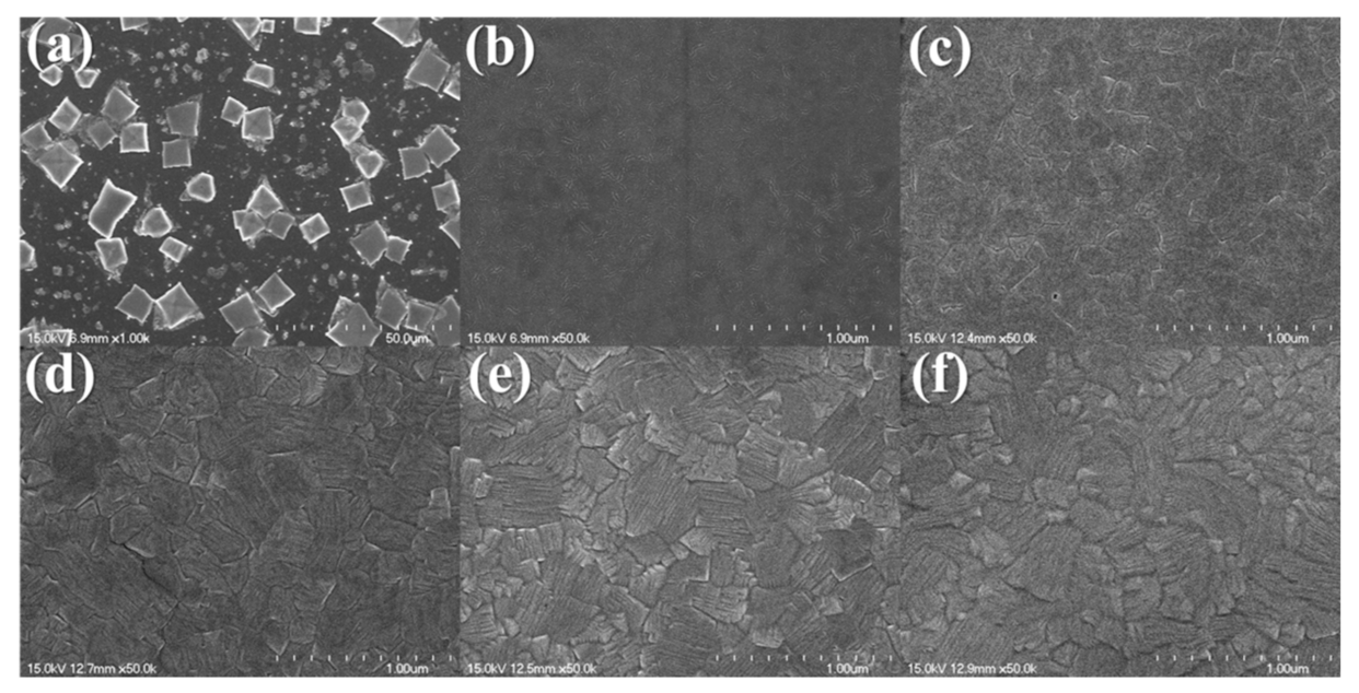
The SEM images of the MAPbBr3 thin film are presented in Figure 2. The cubic shape MAPbBr3 crystal are seen in Figure 2a. The size of the crystals ranged from 5 to 15 µm. The crystal grains were separated, and because of the one-step coating method, in most cases, the surface was not wholly covered, and the crystal distribution was not uniform [17,18]. In Figure 2b, an SEM image is shown for the MAPbBr3 thin film with the antisolvent process. Compared with Figure 2a, the antisolvent-processed MAPbBr3 thin film showed a smooth and pinhole-controlled perovskite surface. The toluene-added MAPbBr3 thin films exhibited superior crystallinity and uniformity compared to the films attained through the conventional spin-coating method. It is known that the uniformity of the perovskite thin film can significantly affect the UV sensor performance [19,20].
In Figure 2c–f, the surfaces of the MAPbBr3 thin film with 0.05–0.20 M MACl are shown. The surfaces of the MAPbBr3 thin films to which 0.05–0.20M of MACl are added are thin films with large grain sizes. In Figure 2a–c, only relatively small grains were observed. In Figure 2d,e, the larger grains are observed due to grain growth, while Figure 2f shows blurry grain boundaries due to degradation from the intense chemical treatment. As the amount of MACl was increased, the grain size generally increased, while, in the MAPbBr3 thin film containing 0.10 M MACl, the grains of approximately 100 nm remained. However, large grains of 500 nm or more were dominant.
The UV-vis absorbance spectrum of the MAPbBr3 thin film is shown in Figure 3a. MAPbBr3 has a strong absorption band in the range of 200–510 nm [21,22]. Therefore, a UV sensor based on MAPbBr3 has a strong advantage in detecting UV rays in the UVC region. The sharp band edge of MAPbBr3 was clearly observed, indicating the direct bandgap of MAPbBr3. The bandgap of the bare MAPbBr3 was estimated by extrapolating the linear range from (F(R∞)hν)2 to the photon energy (hν) intercept, depending on estimations by the Tauc and Davis–Mott models [23,24], is 2.27 eV [25,26]. The bandgap of the antisolvent-processed MAPbBr3 was 2.28 eV; the crystal size and the bandgap of the film decreased and increased, respectively, compared to those of the bare film. This is because the electron–hole pairs were much closer with non-negligible Coulomb interactions, resulting in a higher overall kinetic energy [27]. The bandgaps of MAPbBr3 with 0.05–0.20 M added MACl increased as the concentration of MACl increased.
The electrical characteristics of the MAPbBr3 thin films are shown in Figure 3c. The electrical characteristics, including the semiconductor type, resistivity, carrier concentration and mobility were determined using the Hall measurements. From the measurements, it was confirmed that all MAPbBr3 thin films had carrier concentrations of more than 1013 cm−3 and p-type characteristics. The resistivity values were 0.0941 and 0.1094 Ω·cm for the bare and antisolvent-processed MAPbBr3 thin films, and those for the MAPbBr3 films with 0.05, 0.10, 0.15, and 0.20 M MACl were 0.1051, 0.0924, 0.1306, and 0.1320 Ω·cm, respectively. The mobilities were 113.8, 134.4, 141.5, 158.7, 123.1 and 120.6 cm2/V·s for bare MAPbBr3, antisolvent-processed MAPbBr3, and MAPbBr3 with 0.05, 0.10, 0.15, and 0.20-M added MACl, respectively. The mobility increased as the amount of MACl was increased to 0.10 M, while it decreased for amounts exceeding 0.10 M. This was attributed to the increase in the diffusion distance and the decrease in the trap density with the addition of MACl. [28] The diffusion coefficient can be specified using the Einstein equation ( D = μ K B T q , where KB is the Boltzmann constant, T is the temperature of the sample, and q is the amount of charge). Because the diffusion coefficient is proportional to the diffusion distance, a higher mobility can lead to an increased diffusion distance.
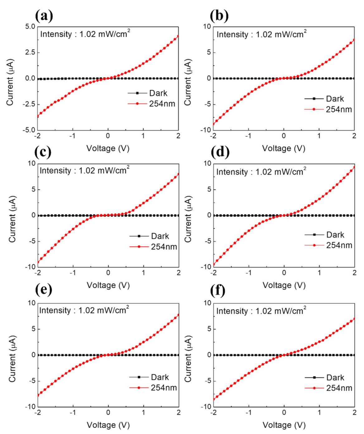
To analyze the UVC detection characteristics, a series of sensor structures were fabricated and compared using bare MAPbBr3, antisolvent-processed MAPbBr3, and MAPbBr3 with 0.05–0.20 M MACl thin films deposited on the etched ITO. The current–voltage (I–V) curves of the MAPbBr3-based sensors are displayed in Figure 4. The I–V characteristics were measured by switching the bias voltage from −2 V to 2 V at a scan rate of 0.1 V in a dark room where light was blocked and under a 254-nm light source with 1.02 mW/cm2 output.
The coated perovskite formed a Schottky barrier owing to contact with the ITO electrode. Under the applied voltage, ion movement and carrier trapping in the active region occurred at the ITO/perovskite interface. This indicated that an ohmic contact was formed between the perovskite and ITO electrodes. For all samples, an apparent increase in current was detected under the 254 nm light at 1.02 mW/cm2. The I–V curves obtained under the 254 nm irradiation maintained rectified shapes even with the addition of various concentrations of MACl. In this measurement, the MAPbBr3 films with the addition of MACl showed that a significant photocurrent could be photogenerated.
The reactivity (R) and specific detectivity (D*) are the main parameters used to evaluate the UV sensors. R, which reflects the sensor response to incident light, is determined as R = (IlightIdark)/APop, where Ilight is the photocurrent under 254-nm UV light, Idark is the dark current, A is the active area of the sensor and Pop is the incident light power [29,30]. The detectivity D* reflects the performance of the sensor, which can be checked by the performance of the signal generated by the main noise and light source in the dark [31,32]. D* is expressed as D* = R/(2qJdark), where q is the amount of charge and Jdark is the dark current density.
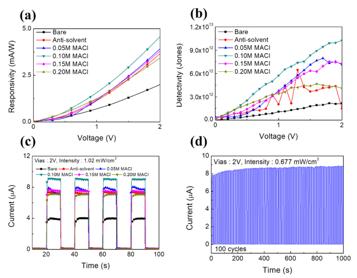
In Figure 5a, the responsivity curves of the MAPbBr3-based sensors are given. The responsivity curves showed that the value of R gradually increased as the bias voltage increased from 0 to 2 V. This was attributed to the increase in the conversion efficiency of the photodetector from photons to charge. At the bias voltage of 2 V under the 254-nm illumination at 1.02 mW/cm2, the R values were 2.00, 3.65, 3.91, 4.57, 3.79 and 3.43 mA/W for bare, antisolvent-processed, 0.05-, 0.10-, 0.15- and 0.20-M MACl-added MAPbBr3, respectively.
The detectivity curves of MAPbBr3 with added MACl are shown in Figure 5b. Under an applied 2V bias voltage, the D* values were 2.13 × 10¹² and 1.49 × 10¹² for bare and antisolvent-processed MAPbBr3. The D* values were 7.14 × 10¹², 1.02 × 10¹³, 7.28 × 10¹², and 4.11 × 10¹² for MAPbBr3 with 0.05, 0.10, 0.15, and 0.20 M of added MACl. Because D* depends on the dark current value, it is strongly influenced by the noise generated in measuring the dark current.
The EQE, another determinant of sensor performance, is defined as the number of electrons generated per incident photon [33,34,35], as follows: EQE = Rhc/eλ, where h is Planck’s constant, c is the speed of light, and λ is the wavelength of the incident light. At 2 V, EQEs were 9.77% and 17.84% for the bare and antisolvent-processed MAPbBr3, and 19.13%, 22.32%, 18.55% and 16.76% for MAPbBr3 with 0.05, 0.10, 0.15 and 0.20 M of added MACl, respectively.
In Figure 5c, the time-dependent photoresponses are shown for MAPbBr3-based sensors measured at a bias voltage of 2V and power intensity of 1.02 mW/cm2. The response time reflects the capacity of the photodetector to follow rapidly changing optical signals. The rise and fall times characterize the response speed of a light detector, and they are usually defined as the time intervals required for the photocurrent to rise, or fall, from 10 to 90% of its peak value, or vice versa. All fabricated samples showed excellent response characteristics, with rise/fall times of 32.6 ms/32.1 ms and 30 ms/28 ms for bare and antisolvent-processed MAPbBr3 and 29.8 ms/29 ms, 15.8 ms/16.2 ms, 25.8 ms/32.1 ms, and 35.1 ms/39 ms for MAPbBr3 with 0.05, 0.10, 0.15, and 0.20-M added MACl, respectively.
The ON/OFF repeatability of the operation of a sensor using an MAPbBr3 thin film with 0.10 M of added MACl under a 254-nm light source with 0.677 mW/cm2 output is given in Figure 5d. The sensor was irradiated for 5 s. The tested sensor showed reproducibility with consistent performance over 100 ON/OFF iterations. In the first iteration, the photocurrent was 7.06 μA; and after 100 repetitions was 8.78 μA. This shows that the reproducibility was maintained continuously relative to the initial photocurrent.
The parameters of the simply structured perovskite-based UVC detector developed in this study are similar to those of the photodetectors reported in other studies (Table 1). Compared to previously reported photodetectors, the proposed detector can be manufactured with a simple structure using a solution process at a low temperature, and it shows high-performance detection.

4. Conclusions

In summary, a simply structured perovskite-based optical sensor was fabricated through a low-temperature solution process. It showed a fast response speed (rise/fall time) of 15.8/16.2 ms, mobility of 158.7 cm2/V·s, responsivity of 4.57 mA/W, detectivity of 1.02 × 10¹³ Jones, and EQE of 22.32%. Therefore, the sensor reported in this study provides a promising solution for UVC detection.

Author Contributions

Investigation, S.G.S.; Supervision, H.W.C.; Validation, C.W.B. and H.W.C.; Writing—original draft, S.G.S.; Writing—review & editing, C.W.B. and H.W.C. All authors have read and agreed to the published version of the manuscript.

Funding

This research was supported by the Basic Science Research Capacity Enhancement Project through a Korea Basic Science Institute (National Research Facilities and Equipment Center) grant funded by the Ministry of Education (2019R1A6C1010016). This work was supported by the Korea Institute of Energy Technology Evaluation and Planning (KETEP) and the Ministry of Trade, Industry & Energy (MOTIE) of the Republic of Korea (No. 20194030202290).

Institutional Review Board Statement

Not applicable.

Informed Consent Statement

Not applicable.

Data Availability Statement

Not applicable.

Conflicts of Interest

The authors declare no conflict of interest.

References

  1. Boggess, A.; Carr, F.A.; Evans, D.C.; Fischel, D.; Freeman, H.R.; Fuechsel, C.F.; Klinglesmith, D.A.; Krueger, V.L.; Longanecker, G.W.; Moore, J.V.; et al. The IUE spacecraft and instrumentation. Nat. Cell Biol. 1978, 275, 372–377. [Google Scholar] [CrossRef]
  2. Xu, Z.; Ding, H.; Sadler, B.M.; Chen, G. Analytical performance study of solar blind non-line-of-sight ultraviolet short-range communication links. Opt. Lett. 2008, 33, 1860–1862. [Google Scholar] [CrossRef]
  3. Lin, R.; Zheng, W.; Zhang, D.; Zhang, Z.; Liao, Q.; Yang, L.; Huang, F. High-performance graphene/β-Ga2O3 heterojunction deep-ultraviolet photodetector with hot-electron excited carrier multiplication. ACS Appl. Mater. Interfaces 2018, 10, 22419–22426. [Google Scholar] [CrossRef] [PubMed]
  4. Zheng, W.; Lin, R.; Ran, J.; Zhang, Z.; Ji, X.; Huang, F. Vacuum-Ultraviolet Photovoltaic Detector. ACS Nano 2018, 12, 425–431. [Google Scholar] [CrossRef] [PubMed]
  5. Zheng, W.; Lin, R.; Zhang, Z.; Huang, F. Vacuum-Ultraviolet Photodetection in Few-Layered h-BN. ACS Appl. Mater. Interfaces 2018, 10, 27116–27123. [Google Scholar] [CrossRef] [PubMed]
  6. Gökkavas, M.; Butun, S.; Caban, P.; Strupinski, W.; Ozbay, E. Integrated AlGaN quadruple-band ultraviolet photodetectors. Semicond. Sci. Technol. 2012, 27, 065004. [Google Scholar] [CrossRef]
  7. Ju, Z.G.; Shan, C.X.; Jiang, D.Y.; Zhang, J.Y.; Yao, B.; Zhao, D.X.; Shen, D.Z.; Fan, X.W. MgxZn1−xO-based photodetectors covering the whole solar-blind spectrum range. Appl. Phys. Lett. 2008, 93, 173505. [Google Scholar] [CrossRef]
  8. Mendoza, F.; Makarov, V.; Weiner, B.R.; Morell, G. Solar-blind field-emission diamond ultraviolet detector. Appl. Phys. Lett. 2015, 107, 201605. [Google Scholar] [CrossRef] [Green Version]
  9. Chen, H.; Liu, H.; Zhang, Z.; Hui, L.; Fang, X. Nanostructured Photodetectors: From Ultraviolet to Terahertz. Adv. Mater. 2016, 28, 403–433. [Google Scholar] [CrossRef]
  10. Zhang, D.; Zheng, W.; Lin, R.; Li, T.; Zhang, Z.; Huang, F. High quality β-Ga2O3 film grown with N2O for high sensitivity solar-blind-ultraviolet photodetector with fast response speed. J. Alloys Compd. 2018, 735, 150–154. [Google Scholar] [CrossRef]
  11. Zhumekenov, A.A.; Burlakov, V.M.; Saidaminov, M.I.; Alofi, A.; Haque, A.; Turedi, B.; Davaasuren, B.; Dursun, I.; Cho, N.; El-Zohry, A.M.; et al. The Role of Surface Tension in the Crystallization of Metal Halide Perovskites. ACS Energy Lett. 2017, 2, 1782–1788. [Google Scholar] [CrossRef] [Green Version]
  12. Tisdale, J.T.; Smith, T.; Salasin, J.R.; Ahmadi, M.; Johnson, N.; Ievlev, A.V.; Koehler, M.; Rawn, C.J.; Lukosi, E.; Hu, B. Precursor purity effects on solution-based growth of MAPbBr3 single crystals towards efficient radiation sensing. CrystEngComm 2018, 20, 7818–7825. [Google Scholar] [CrossRef]
  13. Saidaminov, M.I.; Abdelhady, A.L.; Murali, B.; Alarousu, E.; Burlakov, V.M.; Peng, W.; Dursun, I.; Wang, L.; He, Y.; Maculan, G.; et al. High-quality bulk hybrid perovskite single crystals within minutes by inverse temperature crystallization. Nat. Commun. 2015, 6, 7586. [Google Scholar] [CrossRef] [Green Version]
  14. Wang, L.; Yuan, G.D.; Duan, R.F.; Huang, F.; Wei, T.B.; Liu, Z.Q.; Wang, J.X.; Li, J.M. Tunable bandgap in hybrid perovskite CH3NH3Pb(Br3−yXy) single crystals and photodetector applications. AIP Adv. 2016, 6, 045115. [Google Scholar] [CrossRef] [Green Version]
  15. Senocrate, A.; Kim, G.Y.; Grätzel, M.; Maier, J. Thermochemical stability of hybrid halide perovskites. ACS Energy Lett. 2019, 4, 2859–2870. [Google Scholar] [CrossRef] [Green Version]
  16. Lu, H.; Zhang, H.; Yuan, S.; Wang, J.; Zhan, Y.; Zheng, L. An optical dynamic study of MAPbBr3 single crystals passivated with MAPbCl3/I3-MAPbBr3 heterojunctions. Phys. Chem. Chem. Phys. 2017, 19, 4516–4521. [Google Scholar] [CrossRef]
  17. Mhamdi, A.; Mehdi, H.; Bouazizi, A.; Garcia-Belmonte, G. One-step methylammonium lead bromide films: Effect of annealing treatment. J. Mol. Struct. 2019, 1192, 1–6. [Google Scholar] [CrossRef]
  18. Tidhar, Y.; Edri, E.; Weissman, H.; Zohar, D.; Hodes, G.; Cahen, D.; Rybtchinski, B.; Kirmayer, S. Crystallization of methyl ammonium lead halide perovskites: Implications for photovoltaic applications. J. Am. Chem. Soc. 2014, 136, 13249–13256. [Google Scholar] [CrossRef] [PubMed]
  19. Dualeh, A.; Tétreault, N.; Moehl, T.; Gao, P.; Nazeeruddin, M.K.; Grätzel, M. Effect of Annealing Temperature on Film Morphology of Organic-Inorganic Hybrid Pervoskite Solid-State Solar Cells. Adv. Funct. Mater. 2014, 24, 3250–3258. [Google Scholar] [CrossRef]
  20. Eperon, G.E.; Burlakov, V.M.; Docampo, P.; Goriely, A.; Snaith, H.J. Morphological Control for High Performance, Solution-Processed Planar Heterojunction Perovskite Solar Cells. Adv. Funct. Mater. 2014, 24, 151–157. [Google Scholar] [CrossRef]
  21. Mehdi, H.; Mhamdi, A.; Hannachi, R.; Bouazizi, A. MAPbBr3 perovskite solar cells via a two-step deposition process. RSC Adv. 2019, 9, 12906–12912. [Google Scholar] [CrossRef] [Green Version]
  22. Chen, L.C.; Wu, J.R.; Tseng, Z.L.; Chen, C.C.; Chang, S.H.; Huang, J.K.; Lee, K.L.; Cheng, H.M. Annealing effect on (FAPbI3)1−x (MAPbBr3)x perovskite films in inverted-type perovskite solar cells. Materials 2016, 9, 747. [Google Scholar] [CrossRef] [Green Version]
  23. Tauc, J.; Grigorovici, R.; Vancu, A. Optical Properties and Electronic Structure of Amorphous Germanium. Phys. Status solidi (b) 1966, 15, 627–637. [Google Scholar] [CrossRef]
  24. Davis, E.A.; Mott, N. Conduction in non-crystalline systems V. Conductivity, optical absorption and photoconductivity in amorphous semiconductors. Philos. Mag. 1970, 22, 0903–0922. [Google Scholar] [CrossRef]
  25. Wang, K.-H.; Li, L.-C.; Shellaiah, M.; Sun, K.W. Structural and Photophysical Properties of Methylammonium Lead Tribromide (MAPbBr3) Single Crystals. Sci. Rep. 2017, 7, 1–14. [Google Scholar] [CrossRef] [Green Version]
  26. Kong, L.; Liu, G.; Gong, J.; Hu, Q.; Schaller, R.D.; Dera, P.; Tang, Y. Simultaneous band-gap narrowing and carrier-lifetime prolongation of organic–inorganic trihalide perovskites. Proc. Natl. Acad. Sci. USA 2016, 113, 8910–8915. [Google Scholar] [CrossRef] [Green Version]
  27. Singh, M.; Goyal, M.; Devlal, K. Size and shape effects on the band gap of semiconductor compound nanomaterials. J. Taibah Univ. Sci. 2018, 12, 470–475. [Google Scholar] [CrossRef] [Green Version]
  28. Xie, F.X.; Su, H.; Mao, J.; Wong, K.S.; Choy, W.C.H. Evolution of Diffusion Length and Trap State Induced by Chloride in Perovskite Solar Cell. J. Phys. Chem. C 2016, 120, 21248–21253. [Google Scholar] [CrossRef]
  29. Yu, J.; Javaid, K.; Liang, L.; Wu, W.; Liang, Y.; Song, A.; Cao, H. High-performance visible-blind ultraviolet photodetector based on IGZO TFT coupled with p–n heterojunction. ACS Appl. Mater. Interfaces 2018, 10, 8102–8109. [Google Scholar] [CrossRef] [PubMed]
  30. Cai, J.; Chen, X.; Hong, R.; Yang, W.; Wu, Z. High-performance 4H-SiC-based pin ultraviolet photodiode and investigation of its capacitance characteristics. Opt. Commun. 2014, 333, 182–186. [Google Scholar] [CrossRef]
  31. Inamdar, S.; Ganbavle, V.; Shaikh, S.; Rajpure, K. Effect of the buffer layer on the metal-semiconductor-metal UV photodetector based on Al-doped and undoped ZnO thin films with different device structures. Phys. Status Solidi (A) 2015, 212, 1704–1712. [Google Scholar] [CrossRef]
  32. Singh, S. Al doped ZnO based metal–semiconductor–metal and metal–insulator–semiconductor–insulator–metal UV sensors. Optik 2016, 127, 3523–3526. [Google Scholar] [CrossRef]
  33. Li, S.; Yan, Z.; Liu, Z.; Chen, J.; Zhi, Y.; Guo, D.; Li, P.; Wu, Z.; Tang, W. A self-powered solar-blind photodetector with large V oc enhancing performance based on the PEDOT:PSS/Ga2O3 organic–inorganic hybrid heterojunction. J. Mater. Chem. C 2020, 8, 1292–1300. [Google Scholar] [CrossRef]
  34. Kim, J.O.; Sengupta, S.; Barve, A.V.; Sharma, Y.D.; Adhikary, S.; Lee, S.J.; Noh, S.K.; Allen, M.S.; Allen, J.W.; Chakrabarti, S.; et al. Multi-stack InAs/InGaAs sub-monolayer quantum dots infrared photodetectors. Appl. Phys. Lett. 2013, 102, 011131. [Google Scholar] [CrossRef]
  35. Chen, J.-H.; Jing, Q.; Xu, F.; Lu, Z.-D.; Lu, Y.-Q. High-sensitivity optical-fiber-compatible photodetector with an integrated CsPbBr_3–graphene hybrid structure. Optica 2017, 4, 835–838. [Google Scholar] [CrossRef]
  36. Zhang, Z.; Zheng, W.; Lin, R.; Huang, F. High-sensitive and fast response to 255 nm deep-UV light of CH3NH3PbX3 (X = Cl, Br, I) bulk crystals. R. Soc. Open Sci. 2018, 5, 180905. [Google Scholar] [CrossRef] [Green Version]
  37. Maculan, G.; Sheikh, A.D.; Abdelhady, A.L.; Saidaminov, M.I.; Haque, A.; Murali, B.; Alarousu, E.; Mohammed, O.F.; Wu, T.; Bakr, O.M. CH3NH3PbCl3 Single Crystals: Inverse Temperature Crystallization and Visible-Blind UV-Photodetector. J. Phys. Chem. Lett. 2015, 6, 3781–3786. [Google Scholar] [CrossRef] [PubMed] [Green Version]
  38. Wei, T.C.; Tsai, D.S.; Ravadgar, P.; Ke, J.J.; Tsai, M.L.; Lien, D.H.; Huang, C.Y.; Horng, R.H.; He, J.H. See-Through Ga2O3 Solar-Blind Photodetectors for Use in Harsh Environments. IEEE J. Sel. Top. Quantum Electron. 2014, 20, 112–117. [Google Scholar]
  39. Tong, G.; Li, H.; Zhu, Z.; Zhang, Y.; Yu, L.; Xu, J.; Jiang, Y. Enhancing Hybrid Perovskite Detectability in the Deep Ultraviolet Region with Down-Conversion Dual-Phase (CsPbBr3–Cs4PbBr6) Films. J. Phys. Chem. Lett. 2018, 9, 1592–1599. [Google Scholar] [CrossRef] [PubMed]
  40. Zheng, E.; Yuh, B.; Tosado, G.A.; Yu, Q. Solution-processed visible-blind UV-A photodetectors based on CH3NH3PbCl3 perovskite thin films. J. Mater. Chem. C 2017, 5, 3796–3806. [Google Scholar] [CrossRef] [Green Version]
Figure 1. (a) Schematic of the fabricated sample and XRD patterns of MAPbBr3: (b) bare and antisolvent-processed, (c) with 0.05, 0.10, 0.15, and 0.20 M added MACl.
Nanomaterials 11 01000 g001
Figure 2. FE-SEM images of MAPbBr3: (a) bare, (b) antisolvent-processed, and with (c) 0.05 M, (d) 0.10 M, (e) 0.15 M, (f) 0.20 M added MACl.
Nanomaterials 11 01000 g002
Figure 3. (a) UV-vis absorbance of MAPbBr3, (b) dependence of absorption of the photon energy, (c) electrical characteristics of MAPbBr3.
Nanomaterials 11 01000 g003
Figure 4. Current-voltage (I–V) characteristics of perovskite-based UV sensors. (a) Bare, (b) antisolvent-processed, and containing (c) 0.05-M MACl, (d) 0.10-M MACl, (e) 0.15-M MACl, (f) 0.20-M MACl.
Nanomaterials 11 01000 g004
Figure 5. (a) Responsivity curves of MAPbBr3 sensors, (b) detectivity curves of MAPbBr3 sensors, (c) transient photoresponses, (d) stability of the prepared device for 100 ON/OFF switching cycles under the illumination of 254-nm light with an intensity of 0.677 mW/cm2.
Nanomaterials 11 01000 g005
Table 1. Comparison of important parameters of various UV detectors.
MaterialsLight
(nm)
MethodVoltage
(V)
Responsivity
(mA/W)
Detectivity
(Jones)
EQE
(%)
CH3NH3PbBr3
[this study]
254Solution24.571.02 × 101322.2
CH3NH3PbCl3
[36]
255Single crystals5450-219
CH3NH3PbBr3
[36]
255Single crystals5300-146
CH3NH3PbI3
[36]
255Single crystals5120-58
CH3NH3PbCl3
[37]
365Single crystals1546.901.2 × 1010-
Ga2O3
[38]
185MOCVD *100.32.8 × 10100.2
CsPbBr3-Cs4PbBr6
[39]
254Vapor049.401.2 × 101231
CH3NH3PbCl3
[40]
398Solution−1711.2 × 101023
* Metal–organic chemical vapor deposition.
Publisher’s Note: MDPI stays neutral with regard to jurisdictional claims in published maps and institutional affiliations.