
RETRACTED: Chitin, Chitosan, and Its Derivatives for Wound Healing: Old and New Materials


Abstract
:1. Introduction
2. Chitin for Wound Healing
2.1. In Vitro Studies

| Preparation | Cells | Major results | Ref. |
|---|---|---|---|
| Chitin, its derivatives | Fibroblast | Chitin and its derivatives showed almost no acceleratory effects on proliferation | [36] |
| Chitin/their oligomer/monomer | 3T6 | The migratory activity was reduced by chitin, chitosan and GlcN | [37] |
| Chitin/their oligomer/monomer | HUVECs | The migratory activity was enhanced by chitin, chitosan and GlcNAc | [37] |
| Chitin | Bovine PMNs | Chitin and chitosan activated bovine PMNs | [38] |
| Chitin | Canine PMNs | Chitin and chitosan activated canine PMNs | [39] |
| Chitin | Canine PMNs | Chitin and chitosan induced complement-mediated chemotactic activities | [40] |
| Chitin | Canine PMNs | Supernatants of canine PMNs incubated with chitin and chitosan contained high enough LTB4 and PGE2 concentrations | [41] |
| Chitin | Macrophage | Chitin is a size-dependent stimulator of macrophage IL-17A production and IL-17AR expression and demonstrated that these responses are TLR-2 and MyD88-dependent | [42] |
2.2. Animal Studies
| Preparation | Animal | Major results | Ref. |
|---|---|---|---|
| Chitin | Dog | Numbers of MN and PMN cells were larger in the chitin group than in the control group. Formation of granulating tissue around the implant was identified in the chitin group | [43] |
| Chitin-sponge | Dog, cow, cats, etc. | Chitin-sponges were applied in 30 cases as filling agents for surgical tissue defects, in 25 trauma cases, and in 31 cases of abscess as a wound dressing or tissue defect-filling agent. In 77 out of 86 cases (89.5%), good healing developed | [44] |
| Chitin-cotton | Dog, cow, cats, etc. | Chitin-cotton was applied in 8 cases of trauma and 12 cases of abscess as a wound dressing or tissue defect-filling agent. In 18 out of 20 cases (90.0%), good healing developed | [44] |
| Chitin-flake | Dog, cow, cats, etc. | Chitin-flake was applied in 9 cases of trauma as a wound dressing or tissue defect-filling agent. In 8 out of 9 cases (88.9%), good healing developed | [44] |
| Chitin/NWF | Dog | The amount of PGE2 in the exudate induced by chitin/NWF was about 5 times as high as that in the exudate induced by NWF | [45] |
| Chitin | Dog | Chitin activated the complement components C3 and C5, but not C4 | [46] |
| Chitin | Rat | Compared to chitosan, chitin at the higher concentration (10 mg/mL) induced stable collagen synthesis without scatter in the early wound-healing process | [47] |
3. Chitosan for Wound Healings
3.1. In Vitro Study
| Preparation | Cells | Major results | Ref. |
|---|---|---|---|
| Chitosan | HaCaT | Chitosan exhibited a molecular weight-dependent negative effects on cell viability and proliferation | [49] |
| Chitosan | Fibroblast | Chitosan with high DDA strongly stimulated proliferation | [50] |
| Chitosan, their oligomer/monomer | 3T6 | The migratory activity was reduced by chitosan and GlcN | [37] |
| Chitosan, their oligomer/monomer | HUVECs | The migratory activity was enhanced by chitosan | [37] |
| Chitosan-based membranes | Human PMNs | PMNs, in the presence of chitosan, secrete lysozyme. The materials do not stimulate the production of ROS | [51] |
| Chitosan | Human PMNs | PMNs, stimulated with G-CSF and chitosan, accumulated osteopontin mRNA and released osteopontin | [52] |
| Chitosan | Macrophage | Chitosan had a stimulatory effect on both macrophage nitric oxide (NO) production and chemotaxis | [53] |
3.2. In Vivo Study
| Preparation | Animal | Major results | Ref. |
|---|---|---|---|
| Cotton fiber-type chitosan | Dog | Cotton fiber-type chitosan showed the accelerated granulation in experimental open skin wounds on beagles during the early phase of wound healing | [62] |
| Chitosan membrane | Rat | Open skin wounds in rats covered with the asymmetric chitosan membrane were hemostatic and healed quickly | [64] |
| Chitosan acetate bandage (HemCom) | Mouse | Chitosan acetate bandage had an overall beneficial effect on wound healing, especially during the early period where its antimicrobial effect is most important | [65] |
| Chitosan powder | Rats | Chitosan greatly prevented the extension of burns in the early phase | [66] |
| Chitosan | Rats | The highest wound-healing rate was found in the group treated with high-molecular weight and high-DDA chitosan. Burns treated with high molecular weight chitosan had significantly more epithelial tissue, and the best re-epithelialization and fastest wound closure were obtained in the high-molecular-weight chitosan treatment group | [67] |
| Chitosan hydrogel | Rats | The wound beds of the animals treated with the chitosan hydrogel were considerably smaller than those of the untreated controls | [68] |
| Chitosan | Dogs | Chitosan was well-tolerated and promoted good tissue regeneration | [69] |
| Chitosan | Dogs | Chitosan was well-tolerated and promoted good tissue regeneration | [69] |
3.3. Clinical Study
4. Materials Based on Chitin and Chitosan for Wound Healing
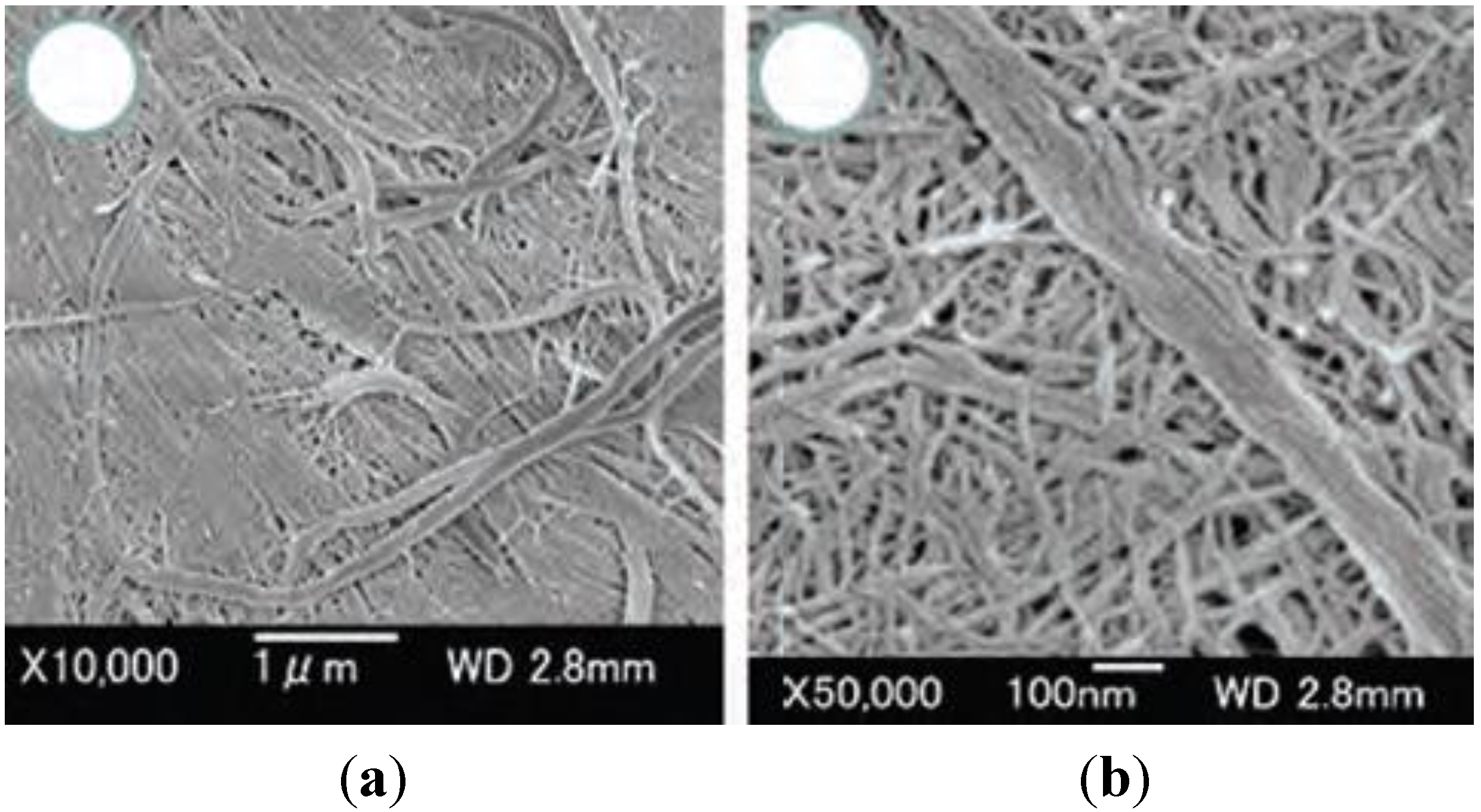
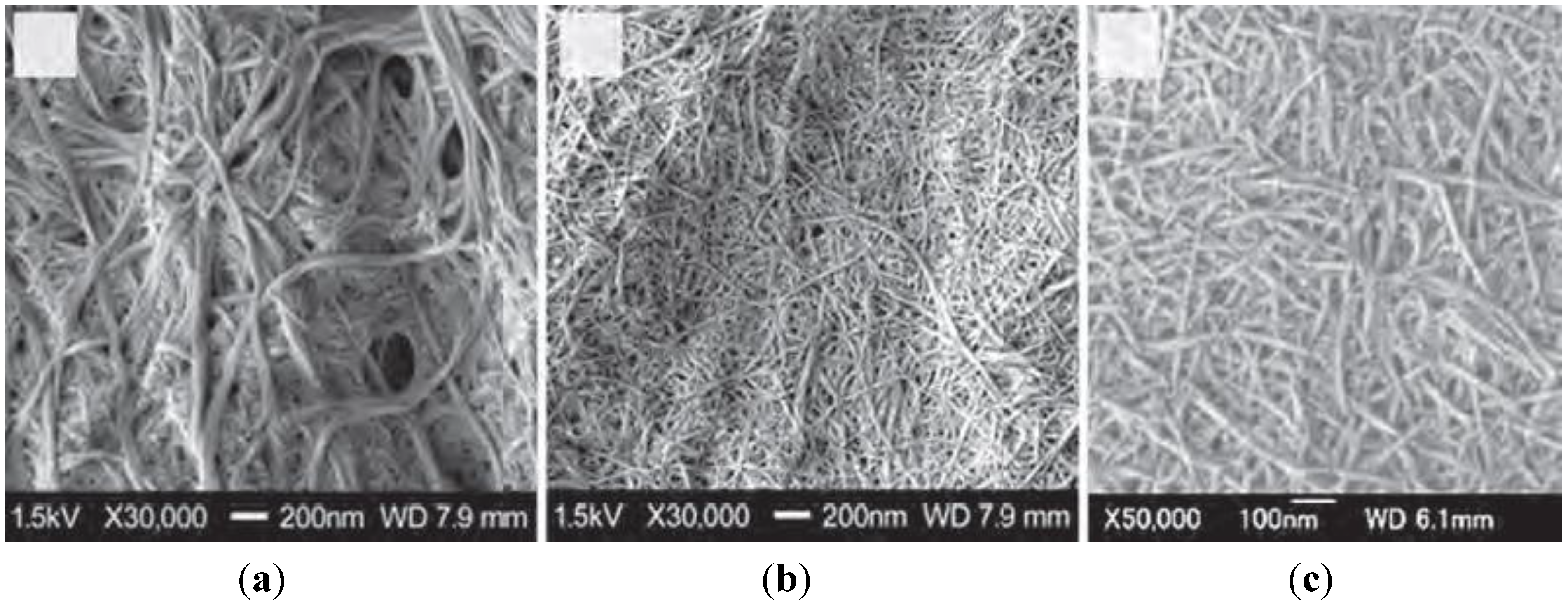
4.1. Chitin and Chitosan Nanofibers
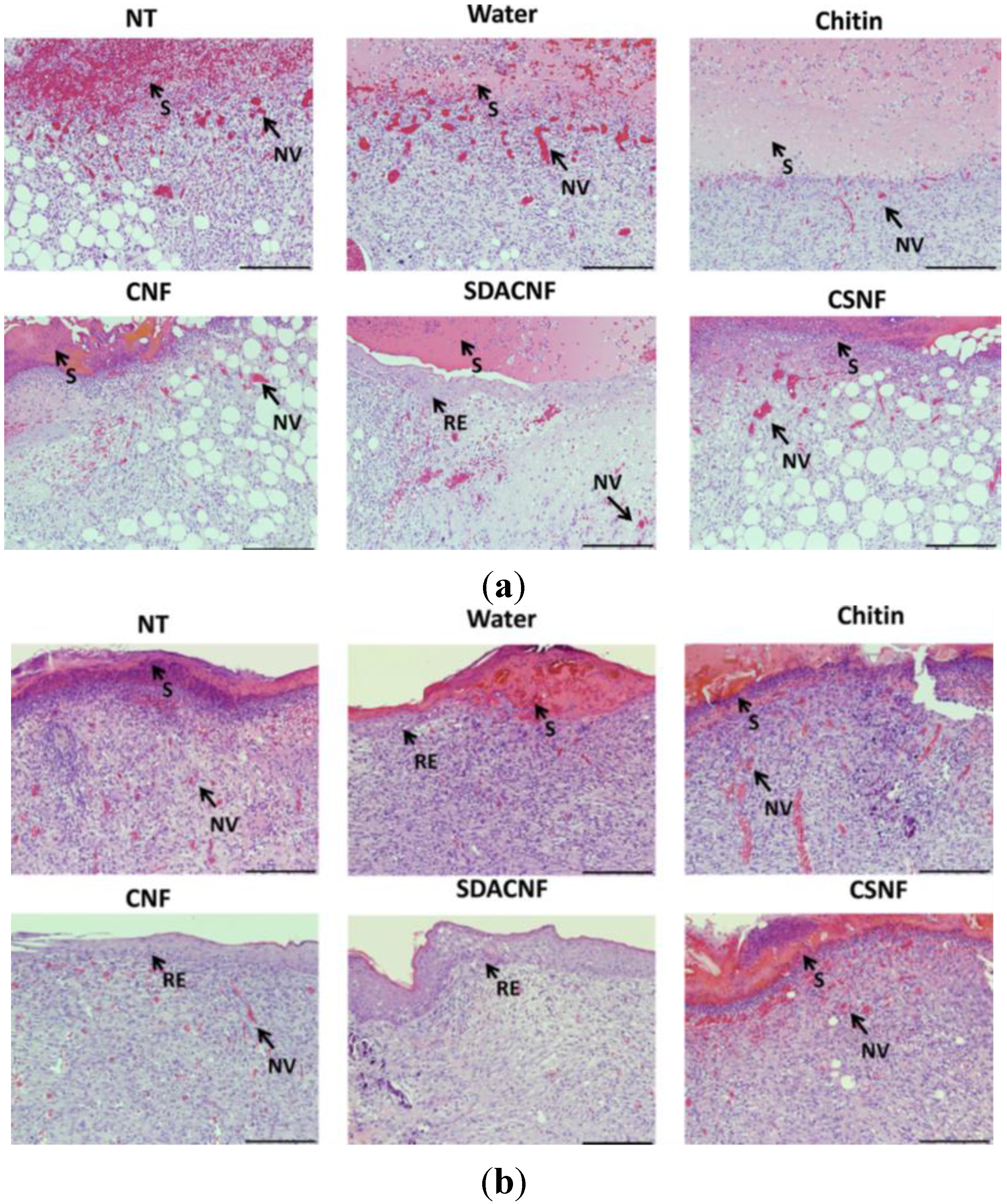
| I | P | R | |
|---|---|---|---|
| Day 4 | |||
| NT | +++ | + | − |
| Water | +++ | + | − |
| Chitin | ++/+++ | ++ | − |
| CNF | ++/+++ | ++ | − |
| SDACNF | ++ | ++ | + |
| CSNF | ++/+++ | ++ | − |
| Day 8 | |||
| NT | ++ | + | + |
| Water | ++ | ++ | + |
| Chitin | ++ | ++ | −/+ |
| CNF | + | ++ | ++ |
| SDACNF | + | +++ | +++ |
| CSNF | ++/+++ | ++/+++ | + |
4.2. Other Formulations Based on Chitin and Chitosan
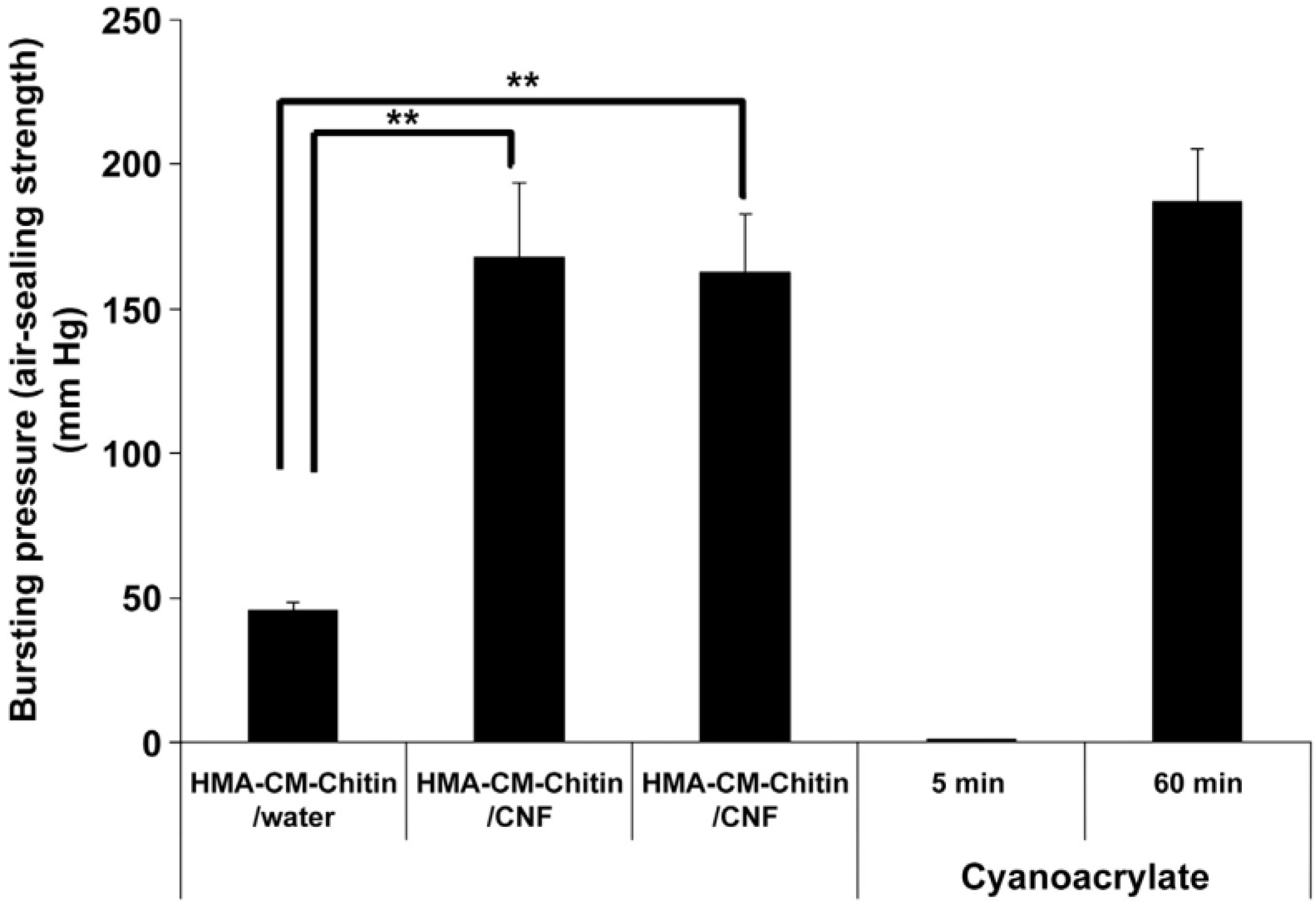
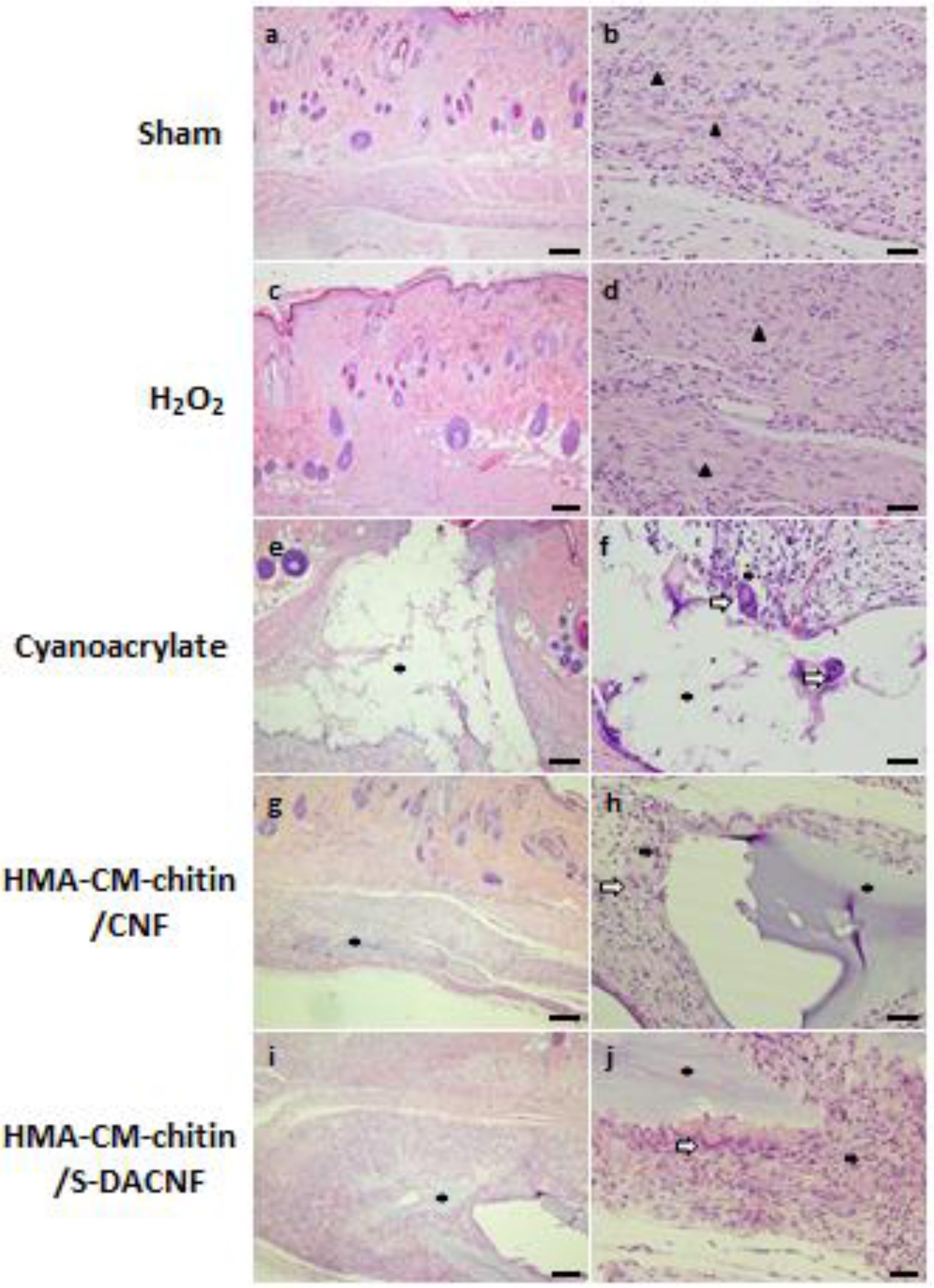
5. Tissue Adhesives Based on Chitin and Chitosan
6. Conclusions
Author Contributions
Conflicts of Interest
References
- Muzzarelli, R.A.A. Chitin nanostructures in living organisms. In Chitin: Formation and Diagenesis; Gupta, N., Ed.; Springer: Dordrecht, The Netherlands, 2011; Volume 34, pp. 1–34. [Google Scholar]
- Kurita, K. Controlled functionalization of the polysaccharide chitin. Prog. Polym. Sci. 2001, 269, 1921–1971. [Google Scholar] [CrossRef]
- Rinaudo, M. Chitin and chitosan: Properties and applications. Prog. Polym. Sci. 2006, 31, 603–632. [Google Scholar] [CrossRef]
- Pillai, K.S.; Paul, W.; Sharma, C.P. Chitin and chitosan polymers: Chemistry, solubility and fiber formation. Prog. Polym. Sci. 2009, 34, 641–678. [Google Scholar] [CrossRef]
- Jayakumar, R.; Prabaharan, M.; Nair, S.V.; Tamura, H. Novel chitin and chitosan nanofibers in biomedical applications. Biotechnol. Adv. 2010, 28, 142–150. [Google Scholar] [CrossRef] [PubMed]
- Jayakumar, R.; Prabaharan, M.; Sudheesh Kumar, P.T.; Nair, S.V.; Tamura, H. Biomaterials based on chitin and chitosan in wound dressing applications. Biotechnol. Adv. 2011, 29, 322–337. [Google Scholar] [CrossRef] [PubMed]
- Azuma, K.; Ifuku, S.; Osaki, T.; Okamoto, Y.; Minami, S. Preparation and Biomedical Applications of Chitin and Chitosan Nanofibers. J. Biomed. Nanotechnol. 2014, 10, 2891–2920. [Google Scholar] [CrossRef]
- Nagahama, H.; Kashiki, T.; Nwe, N.; Jayakumar, R.; Furuike, T.; Tamura, H. Preparation of biodegradable chitin/gelatin membranes with GlcNAc for tissue engineering applications. Carbohydr. Polym. 2008, 73, 456–463. [Google Scholar] [CrossRef]
- Nagahama, H.; Nwe, N.; Jayakumar, R.; Koiwa, S.; Furuike, T.; Tamura, H. Novel biodegradable chitin membranes for tissue engineering applications. Carbohydr. Polym. 2008, 73, 295–302. [Google Scholar] [CrossRef]
- Tamura, H.; Furuike, T.; Nair, S.V.; Jayakumar, R. Biomedical applications of chitin hydrogel membranes and scaffolds. Carbohydr. Polym. 2011, 84, 820–824. [Google Scholar] [CrossRef]
- Yusof, N.L.B.M.; Wee, A.; Lim, L.Y.; Khor, E. Flexible chitin films as potential wound-dressing materials: Wound model studies. J. Biomed. Mater. Res. A 2003, 66, 224–232. [Google Scholar] [CrossRef] [PubMed]
- Marreco, P.R.; Moreira, P.L.; Genari, S.C.; Moraes, A.M. Effects of different sterilization methods on the morphology, mechanical properties and cytotoxicity of chitosan membranes used as wound dressings. J. Biomed. Mater. Res. B Appl. Biomater. B 2004, 71, 268–277. [Google Scholar] [CrossRef]
- Jayakumar, R.; Nwe, N.; Tokura, S.; Tamura, H. Sulfated chitin and chitosan as novel biomaterials. Int. J. Biol. Macromol. 2007, 40, 175–181. [Google Scholar] [CrossRef] [PubMed]
- Jayakumar, R.; Nwe, N.; Nagahama, H.; Tamura, H. Synthesis, characterization and biospecific degradation behavior of sulfated chitin. Macromol. Symp. 2008, 264, 163–167. [Google Scholar] [CrossRef]
- Jayakumar, R.; Divya Rani, V.V.; Shalumon, K.T.; Sudheesh Kumar, P.T.; Nair, S.V.; Furuike, T.; Tamura, H. Bioactive and osteoblast cell attachment studies of novel α- and β-chitin membranes for tissue engineering applications. Int. J. Biol. Macromol. 2009, 45, 260–264. [Google Scholar] [CrossRef] [PubMed]
- Madhumathi, K.; Binulal, N.S.; Nagahama, H.; Tamura, H.; Shalumon, K.T.; Selvamurugan, N.; Nair, S.V.; Jayakumar, R. Preparation and characterization of novel α-chitin-hydroxyapatite composite membranes for tissue engineering applications. Int. J. Biol. Macromol. 2009, 44, 1–5. [Google Scholar] [CrossRef] [PubMed]
- Shalumon, K.T.; Binulal, N.S.; Selvamurugan, N.; Nair, S.V.; Menon, D.; Furuike, T.; Tamura, H.; Jayakumar, R. Electrospinning of carboxymethyl chitin/poly(vinyl alcohol) nanofibrous scaffolds for tissue engineering applications. Carbohydr. Polym. 2009, 77, 863–869. [Google Scholar] [CrossRef]
- Shalumon, K.T.; Anulekha, K.H.; Girish, C.M.; Nair, S.V.; Jayakumar, R. Single step electrospinning of chitosan/poly(caprolactone) nanofibers using formic acid/acetone solvent mixture. Carbohydr. polym. 2010, 80, 413–419. [Google Scholar] [CrossRef]
- Gopalan, N.K.; Dufresne, A. Crab shell chitin whisker reinforced natural rubber nanocomposites. 1. Processing and swelling behavior. Biomacromolecules 2003, 4, 657–665. [Google Scholar] [CrossRef] [PubMed]
- Fan, Y.; Saito, T.; Isogai, A. Preparation of chitin nanofibers from squid pen beta-chitin by simple mechanical treatment under acid conditions. Biomacromolecules 2008, 9, 1919–1923. [Google Scholar] [CrossRef] [PubMed]
- Ifuku, S.; Nogi, M.; Abe, K.; Yoshioka, M.; Morimoto, M.; Saimoto, H.; Yano, H. Preparation of chitin nanofibers with a uniform width as alpha-chitin from crab shells. Biomacromolecules 2009, 10, 1584–1588. [Google Scholar] [CrossRef] [PubMed]
- Yusof, N.L.B.M.; Lim, L.Y.; Khor, E. Preparation and characterization of chitin beads as a wound dressing precursor flexible chitin films as potential wound-dressing materials: Wound model studies. J. Biomed. Mater. Res. 2001, 54, 59–68. [Google Scholar] [CrossRef] [PubMed]
- Jayakumar, R.; Reis, R.L.; Mano, J.F. Phosphorous containing chitosan beads for controlled oral drug delivery. J. Bioact. Compat. Polym. 2006, 21, 327–340. [Google Scholar] [CrossRef]
- Prabaharan, M. Chitosan derivatives as promising materials for controlled drug delivery. J. Biomater. Appl. 2008, 23, 5–36. [Google Scholar] [CrossRef] [PubMed]
- Anitha, A.; Divya Rani, V.V.; Krishna, R.; Sreeja, V.; Selvamurugan, N.; SNair, S.V.; Tamura, H.; Jayakumar, H. Synthesis, characterization, cytotoxicity and antibacterial studies of chitosan, O-carboxymethyl and N,O-carboxymethyl chitosan nanoparticles. Carbohydr. Polym. 2009, 78, 672–677. [Google Scholar] [CrossRef]
- Anitha, A.; Deepa, N.; Chennazhi, K.P.; Nair, S.V.; Tamura, H.; Jayakumar, R. Development of mucoadhesive thiolated chitosan nanoparticles for biomedical applications. Carbohydr. Polym. 2010, 83, 66–73. [Google Scholar] [CrossRef]
- Peter, M.; Sudheesh Kumar, P.T.; Binulal, N.S.; Nair, S.V.; Tamura, H.; Jayakumar, R. Development of novel chitin/nano bioactive glass ceramic nanocomposite scaffolds for tissue engineering applications. Carbohydr. Polym. 2009, 78, 926–931. [Google Scholar] [CrossRef]
- Peter, M.; Binulol, N.S.; Soumya, S.; Nair, S.V.; Tamura, H.; Jayakumar, R. Nanocomposite scaffolds of bioactive glass ceramic nanoparticles disseminated chitosan matrix for tissue engineering applications. Carbohydr. Polym. 2010, 79, 284–289. [Google Scholar] [CrossRef]
- Prabaharan, M.; Jayakumar, R. Chitosan-graft-β-cyclodextrin scaffolds with controlled drug release capability for tissue engineering applications. Int. J. Biol. Macromol. 2009, 44, 320–325. [Google Scholar] [CrossRef] [PubMed]
- Maeda, Y.; Jayakumar, R.; Nagahama, H.; Furuike, T.; Tamura, H. Synthesis, characterization and bioactivity studies of novel β-chitin scaffolds for tissue-engineering applications. Int. J. Biol. Macromol. 2008, 42, 463–467. [Google Scholar] [CrossRef] [PubMed]
- Muramatsu, K.; Masuda, S.; Yoshihara, Y.; Fujisawa, A. In vitro degradation behavior of freeze-dried carboxymethyl-chitin sponges processed by vacuum-heating and gamma irradiation. Polym. Degradat. Stabil. 2003, 81, 327–332. [Google Scholar] [CrossRef]
- Portero, A.; Teijeiro-Osorio, D.; Alonso, M.J.; Remunan-Lopez, C. Development of chitosan sponges for buccal administration of insulin. Carbohydr. Polym. 2007, 68, 617–625. [Google Scholar] [CrossRef]
- Dai, T.; Tanaka, M.; Huang, Y.Y.; Hamblin, M.R. Chitosan preparations for wounds and burns: Antimicrobial and wound-healing effects. Exp. Rev. Anti Infect. Ther. 2011, 9, 857–879. [Google Scholar] [CrossRef]
- Boateng, J.S.; Matthews, K.H.; Stevens, H.N.; Eccleston, G.M. Wound healing dressings and drug delivery systems: A review. J. Pharm. Sci. 2008, 97, 2892–2923. [Google Scholar] [CrossRef] [PubMed]
- Beanes, S.R.; Dang, C.; Soo, C.; Ting, K. Skin repair and scar formation: The central role of TGF-β. Exp. Rev. Mol. Med. 2003, 5, 1–22. [Google Scholar] [CrossRef]
- Mori, T.; Okumura, M.; Matsuura, M.; Ueno, K.; Tokura, S.; Okamoto, Y.; Minami, S.; Fujinaga, T. Effects of chitin and its derivatives on the proliferation and cytokine production of fibroblasts in vitro. Biomaterials 1997, 18, 947–951. [Google Scholar] [CrossRef] [PubMed]
- Okamoto, Y.; Watanabe, M.; Miyatake, K.; Morimoto, M.; Shigemasa, Y.; Minami, S. Effects of chitin/chitosan and their oligomers/monomers on migrations of fibroblasts and vascular endothelium. Biomaterials 2002, 23, 1975–1979. [Google Scholar] [CrossRef] [PubMed]
- Usami, Y.; Okamoto, Y.; Minami, S.; Matsuhashi, A.; Kumazawa, N.H.; Tanioka, S.; Shigemasa, Y. Chitin and chitosan induce migration of bovine polymorphonuclear cells. J. Vet. Med. Sci. 1994, 56, 761–762. [Google Scholar] [CrossRef] [PubMed]
- Usami, Y.; Okamoto, Y.; Minami, S.; Matsuhashi, A.; Kumazawa, N.H.; Tanioka, S.; Shigemasa, Y. Migration of canine neutrophils to chitin and chitosan. J. Vet. Med. Sci. 1994, 56, 1215–1216. [Google Scholar] [CrossRef] [PubMed]
- Usami, Y.; Minami, S.; Okamoto, Y.; Matsuhashi, A.; Shigemasa, Y. Influence of chain length of N-acetyl-d-glucosamine and d-glucosamine residues on direct and complement-mediated chemotactic activities for canine polymorphonuclear cells. Carbohydar. Polym. 1997, 32, 115–122. [Google Scholar] [CrossRef]
- Usami, Y.; Okamoto, Y.; Takayama, T.; Shigemasa, Y.; Minami, S. Chitin and chitosan stimulate canine polymorphonuclear cells to release leukotrien B4 and prostaglandin E2. J. Biomed. Mater. Res. 1998, 42, 517–522. [Google Scholar] [CrossRef] [PubMed]
- Da Silva, C.A.; Hartl, D.; Liu, W.; Lee, C.G.; Elias, J.A. TLR-2 and IL-17A in chitin-induced macrophage activation and acute inflammation. J. Immunol. 2008, 181, 4279–4286. [Google Scholar] [CrossRef] [PubMed]
- Okamoto, Y.; Minami, S.; Matsuhashi, A.; Sashiwa, H.; Saimoto, H.; Shigemasa, Y.; Tanigawa, T.; Tanaka, Y.; Tokura, S. Polymeric N-acetyl-d-glucosamine (chitin) induces histionic activation in dogs. J. Vet. Med. Sci. 1993, 55, 739–742. [Google Scholar] [CrossRef] [PubMed]
- Okamoto, Y.; Minami, S.; Matsuhashi, A.; Sashiwa, H.; Saimoto, H.; Shigemasa, Y.; Tanigawa, T.; Tanaka, Y.; Tokura, S. Application of polymeric N-acetyl-d-glucosamine (chitin) to veterinary practice. J. Vet. Med. Sci. 1993, 55, 743–747. [Google Scholar] [CrossRef] [PubMed]
- Minami, S.; Okamoto, Y.; Matsuhashi, A.; Sashiwa, H.; Saimoto, H.; Shigemasa, Y.; Tanigawa, T.; Suzuki, T.; Tanioka, S.; Tanaka, Y. Polymeric N-acetyl-d-glucosamine (chitin) induces prostaglandin E2 in dogs. J. Vet. Med. Sci. 1995, 57, 377–378. [Google Scholar] [CrossRef] [PubMed]
- Minami, S.; Suzuki, H.; Okamoto, Y.; Fujinaga, T.; Shigemasa, Y. Chitin and chitosan activate complement via the alternative pathway. Carbohydr. Polym. 1998, 36, 151–155. [Google Scholar] [CrossRef]
- Suzuki, Y.; Okamoto, Y.; Morimoto, M.; Sashiwa, H.; Saimoto, H.; Tanioka, S.; Shigemasa, Y.; Minami, S. Influence of physicochemical properties of chitin and chitosan on complement activation. Carbohydr. Polym. 2000, 42, 307–310. [Google Scholar] [CrossRef]
- Kojima, K.; Okamoto, Y.; Kojima, K.; Miyatake, K.; Fujise, H.; Shigemasa, Y.; Minami, S. Effects of chitin and chitosan on collagen synthesis in wound healing. J. Vet. Med. Sci. 2004, 66, 1595–1598. [Google Scholar] [CrossRef]
- Wiegand, C.; Winter, D.; Hipler, U.C. Molecular-weight-dependent toxic effects of chitosans on the human keratinocyte cell line HaCaT. Skin Pharmacol. Physiol. 2010, 23, 164–170. [Google Scholar] [CrossRef] [PubMed]
- Howling, G.I.; Dettmar, P.W.; Goddard, P.A.; Hampson, F.; Dornish, M.; Wood, E.J. The effect of chitin and chitosan on the proliferation of .human skin fibroblasts and keratinocytes in vitro. Biomaterials 2001, 22, 2959–2966. [Google Scholar] [CrossRef] [PubMed]
- Santos, T.C.; Marques, A.P.; Silva, S.S.; Oliveira, J.M.; Mano, J.F.; Castro, A.G.; Reis, R.L. In vitro evaluation of the behaviour of human polymorphonuclear neutrophils in direct contact with chitosan-based membranes. J. Biotechnol. 2007, 132, 218–226. [Google Scholar] [CrossRef] [PubMed]
- Ueno, H.; Murakami, M.; Okumura, M.; Kadosawa, T.; Uede, T.; Fujinaga, T. Chitosan accelerates the production of osteopontin from polymorphonuclear leukocytes. Biomaterials 2001, 22, 1667–1673. [Google Scholar] [CrossRef] [PubMed]
- Rabea, E.I.; Badawy, M.E.; Stevens, C.V.; Smagghe, G.; Steurbaut, W. Chitosan as antimicrobial agent: Applications and mode of action. Biomacromolecules 2003, 4, 1457–1465. [Google Scholar] [CrossRef] [PubMed]
- Li, P.; Poon, Y.F.; Li, W.; Zhu, H.Y.; Yeap, S.H.; Cao, Y.; Qi, X.; Zhou, C.; Lamrani, M.; Beuerman, R.W.; et al. A polycationic antimicrobial and biocompatible hydrogel with microbe membrane suctioning ability. Nature Mater. 2011, 10, 149–156. [Google Scholar] [CrossRef]
- Kong, M.; Chen, X.G.; Xing, K.; Park, H. Antimicrobial properties of chitosan and mode of action: a state of the art review. Int. J. Food Microbiol. 2010, 144, 51–63. [Google Scholar] [CrossRef] [PubMed]
- Andres, Y.; Giraud, L.; Gerente, C.; Le Cloirec, P. Antibacterial effects of chitosan powder: Mechanisms of action. Environ. Technol. 2007, 28, 1357–1363. [Google Scholar] [CrossRef] [PubMed]
- Raafat, D.; von Bargen, K.; Haas, A.; Sahl, H.G. Insights into the mode of action of chitosan as an antibacterial compound. Appl. Environ. Microbiol. 2008, 74, 3764–3773. [Google Scholar] [CrossRef] [PubMed]
- Tang, H.; Zhang, P.; Kieft, T.L.; Ryan, S.J.; Baker, S.M.; Wiesmann, W.P.; Rogelj, S. Antibacterial action of a novel functionalized chitosan-arginine against Gram-negative bacteria. Acta Biomater. 2010, 6, 2562–2571. [Google Scholar] [CrossRef] [PubMed]
- No, H.K.; Park, N.Y.; Lee, S.H.; Meyers, S.P. Antibacterial activity of chitosans and chitosan oligomers with different molecular weights. Int. J. Food Microbiol. 2002, 74, 65–72. [Google Scholar] [CrossRef] [PubMed]
- Muzzarelli, R.; Tarsi, R.; Filippini, O.; Giovanetti, E.; Biagini, G.; Varaldo, P.E. Antimicrobial properties of N-carboxybutyl chitosan. Antimicrob. Agents Chemother. 1990, 10, 2019–2023. [Google Scholar] [CrossRef]
- Seyfarth, F.; Schliemann, S.; Elsner, P.; Hipler, U.C. Antifungal effect of high- and low-molecular-weight chitosan hydrochloride, carboxymethyl chitosan, chitosan oligosaccharide and N-acetyl-d-glucosamine against Candida albicans, Candida krusei and Candida glabrata. Int. J. Pharmaceut. 2008, 353, 139–148. [Google Scholar]
- Ueno, H.; Yamada, H.; Tanaka, I.; Kaba, N.; Matsuura, M.; Okumura, M.; Kadosawa, T.; Fujinaga, T. Accelerating effects of chitosan for healing at early phase of experimental open wound in dogs. Biomaterials 1999, 20, 1407–1414. [Google Scholar] [CrossRef] [PubMed]
- Okamoto, Y.; Shibazaki, K.; Minami, S.; Matsuhashi, A.; Tanioka, S.; Shigemasa, Y. Evaluation of chitin and chitosan on open would healing in dogs. J. Vet. Med. Sci. 1995, 57, 851–854. [Google Scholar] [CrossRef] [PubMed]
- Mi, F.L.; Shyu, S.S.; Wu, Y.B.; Lee, S.T.; Shyong, J.Y.; Huang, R.N. Fabrication and characterization of a sponge-like asymmetric chitosan membrane as a wound dressing. Biomaterials 2001, 22, 165–173. [Google Scholar] [CrossRef] [PubMed]
- Burkatovskaya, M.; Castano, A.P.; Demidova-Rice, T.N.; Tegos, G.P.; Hamblin, M.R. Effect of chitosan acetate bandage on wound healing in infected and noninfected wounds in mice. Wound Repair Regen. 2008, 16, 425–431. [Google Scholar] [CrossRef] [PubMed]
- Jin, Y.; Ling, P.X.; He, Y.L.; Zhang, T.M. Effects of chitosan and heparin on early extension of burns. Burns. 2007, 33, 1027–1031. [Google Scholar] [CrossRef] [PubMed]
- Alsarra, I.A. Chitosan topical gel formulation in the management of burn wounds. Int. J. Biol. Macromol. 2009, 45, 16–21. [Google Scholar] [CrossRef] [PubMed]
- Ribeiro, M.P.; Espiga, A.; Silva, D.; Baptista, P.; Henriques, J.; Ferreira, C.; Silva, J.C.; Borges, J.P.; Pires, E.; Chaves, P.; Correia, I.J. Development of a new chitosan hydrogel for wound dressing. Wound Repair Regen. 2009, 17, 817–824. [Google Scholar] [CrossRef] [PubMed]
- Boucard, N.; Viton, C.; Agay, D.; Mari, E.; Roger, T.; Chancerelle, Y.; Domard, A. The use of physical hydrogels of chitosan for skin regeneration following third-degree burns. Biomaterials 2007, 28, 3478–3488. [Google Scholar] [CrossRef] [PubMed]
- Diegelmann, R.F.; Dunn, J.D.; Lindblad, W.J.; Cohen, I.K. Analysis of the effects of chitosan on inflammation, angiogenesis, fibroplasia, and collagen deposition in polyvinyl alcohol sponge implants in rat wounds. Wound Repair Regen. 1996, 4, 48–52. [Google Scholar] [CrossRef] [PubMed]
- Okamoto, Y.; Tomita, T.; Minami, S.; Matsuhashi, A.; Kumazawa, N.H.; Tanioka, S.; Shigemasa, Y. Effects of chitosan on experimental abscess with Staphylococcus aureus in dogs. J. Vet. Med. Sci. 1995, 57, 765–767. [Google Scholar] [CrossRef] [PubMed]
- Burkatovskaya, M.; Tegos, G.P.; Swietlik, E.; Demidova, T.N.; Castano, A.P.; Hamblin, M.R. Use of chitosan bandage to prevent fatal infections developing from highly contaminated wounds in mice. Biomaterials 2006, 27, 4157–4164. [Google Scholar] [CrossRef] [PubMed]
- Ong, S.Y.; Wu, J.; Moochhala, S.M.; Tan, M.H.; Lu, J. Development of a chitosan-based wound dressing with improved hemostatic and antimicrobial properties. Biomaterials 2008, 29, 4323–4332. [Google Scholar] [CrossRef] [PubMed]
- França, R.; Mbeh, D.A.; Samani, T.D.; Le Tien, C.; Mateescu, M.A.; Yahia, L.; Sacher, E. The effect of ethylene oxide sterilization on the surface chemistry and in vitro cytotoxicity of several kinds of chitosan. J. Biomed. Mater. Res. B Appl. Biomater. 2013, 101, 1444–1455. [Google Scholar] [CrossRef] [PubMed]
- Mayol, L.; De Stefano, D.; Campani, V.; De Falco, F.; Ferrari, E.; Cencetti, C.; Matricardi, P.; Maiuri, L.; Carnuccio, R.; Gallo, A.; et al. Design and characterization of a chitosan physical gel promoting wound healing in mice. J. Mater. Sci. Mater. Med. 2014, 25, 1483–1493. [Google Scholar] [CrossRef] [PubMed]
- Biagini, G.; Bertani, A.; Muzzarelli, R.; Damadei, A.; Di Benedetto, G.; Belligolli, A.; Riccotti, G.; Zucchini, C.; Rizzoli, C. Wound management with N-carboxybutyl chitosan. Biomaterials 1991, 12, 281–286. [Google Scholar] [CrossRef] [PubMed]
- Stone, C.A.; Wright, H.; Clarke, T.; Powell, R.; Devaraj, V.S. Healing at skin graft donor sites dressed with chitosan. Br. J. Plast. Surg. 2000, 53, 601–606. [Google Scholar] [CrossRef] [PubMed]
- Valentine, R.; Athanasiadis, T.; Moratti, S.; Hanton, L.; Robinson, S.; Wormald, P.J. The efficacy of a novel chitosan gel on hemostasis and wound healing after endoscopic sinus surgery. Am. J. Rhinol. Allergy 2010, 24, 70–75. [Google Scholar] [CrossRef] [PubMed]
- Azad, A.K.; Sermsintham, N.; Chandrkrachang, S.; Stevens, W.F. Chitosan membrane as a wound-healing dressing: Characterization and clinical application. J. Biomed. Mater. Res. B Appl. Biomater. 2004, 69, 216–222. [Google Scholar] [CrossRef] [PubMed]
- Akncbay, H.; Senel, S.; Ay, Z.Y. Application of chitosan gel in the treatment of chronic periodontitis. J. Biomed. Mater. Res. B Appl. Biomater. 2007, 80, 290–296. [Google Scholar] [CrossRef] [PubMed]
- Boynueğri, D.; Ozcan, G.; Senel, S.; Uç, D.; Uraz, A.; Oğüş, E.; Cakilci, B.; Karaduman, B. Clinical and radiographic evaluations of chitosan gel in periodontal intraosseous defects: A pilot study. J. Biomed. Mater. Res. B Appl. Biomater. 2009, 90, 461–466. [Google Scholar] [CrossRef] [PubMed]
- Xia, Y.; Yang, P.; Sun, Y.; Wu, Y.; Mayers, B.; Gates, B.; Yin, Y.; Kim, F.; Yan, H. One-dimensional nanostructures: Synthesis, characterization, and applications. Adv. Mater. 2003, 15, 353–389. [Google Scholar] [CrossRef]
- Li, D.; Xia, Y. Electrospinning of nanofibers: Reinventing the wheel? Adv. Mater. 2004, 16, 1151–1170. [Google Scholar] [CrossRef]
- Huang, Z.M.; Zhang, Y.Z.; Kotaki, M.; Ramakrishna, S. A review on polymer nanofibers by electrospinning and their applications in nanocomposites. Compos. Sci. Technol. 2003, 63, 2223–2253. [Google Scholar] [CrossRef]
- Ramakrishna, S.; Fujihara, K.; Teo, W.E.; Yong, T.; Ma, Z.; Ramaseshan, R. Electrospun nanofibers: Solving global issues. Mater. Today 2006, 9, 40–50. [Google Scholar] [CrossRef]
- Muzzarelli, R.A.; Mehtedi, M.E.; Mattioli-Belmonte, M. Emerging biomedical applications of nano-chitins and nano-chitosans obtained via advanced eco-friendly technologies from marine resources. Mar. Drugs 2014, 12, 5468–5502. [Google Scholar] [CrossRef] [PubMed]
- Araki, J. Electrostatic or steric? Preparations and characterizations of well-dispersed systems containing rod-like nanowhiskers of crystalline polysaccharides. Soft Matter 2013, 9, 4125–4141. [Google Scholar] [CrossRef]
- Ifuku, S.; Nogi, M.; Abe, K.; Yoshioka, M.; Morimoto, M.; Saimoto, H.; Yano, H. Simple preparation of chitin nanofibers with a width of 10–20 nm from prawn shell under neutral conditions. Carbohydr. Polym. 2011, 84, 762–764. [Google Scholar] [CrossRef]
- Bruin, P.; Jonkman, M.F.; Meijer, H.J.; Pennings, A.J. A new porous polyetherurethane wound covering. J. Biomed. Mater. Res. 1990, 24, 217–226. [Google Scholar] [CrossRef] [PubMed]
- Matsuda, K.; Suzuki, S.; Isshiki, N.; Yoshioka, K.; Wada, R.; Okada, T.; Ikeda, Y. Influence of glycosaminoglycans on the collagen sponges component of a bilayer artificial skin. Biomaterials 1990, 11, 351–355. [Google Scholar] [CrossRef]
- Suzuki, S.; Matsuda, K.; Isshiki, N.; Tamada, Y.; Ikada, Y. Experimental study of newly developed bilayer artificial skin. Biomaterials 1990, 11, 356–360. [Google Scholar] [CrossRef] [PubMed]
- Naseri, N.; Algan, C.; Jacobs, V.; John, M.; Oksman, K.; Mathew, A.P. Electrospun chitosan-based nanocomposite mats reinforced with chitin nanocrystals for wound dressing. Carbohydr. Polym. 2014, 109, 7–15. [Google Scholar] [CrossRef] [PubMed]
- Naseri, N.; Mathew, A.P.; Girandon, L.; Fröhlich, M.; Oksman, K. Porous electrospun nanocomposite mats based on chitosan–cellulose nanocrystals for wound dressing: Effect of surface characteristics of nanocrystals. Cellulose 2015, 22, 521–534. [Google Scholar] [CrossRef]
- Muzzarelli, R.A.A. Biochemical significance of exogenous chitins and chitosans in animals and patients. Carbohydr. Polym. 1993, 20, 7–16. [Google Scholar] [CrossRef]
- Kelechi, T.J.; Mueller, M.; Hankin, C.S.; Bronstone, A.; Samies, J.; Bonham, P.A. A randomized, investigator-blinded, controlled pilot study to evaluate the safety and efficacy of a poly-N-acetyl glucosamine-derived membrane material in patients with venous leg ulcers. J. Am. Acad. Dermatol. 2012, 66, E209–E215. [Google Scholar] [CrossRef] [PubMed]
- Fischer, T.H.; Hays, W.E.; Valeri, C.R. Poly-N-acetyl glucosamine fibers accelerate hemostasis in patients treated with antiplatelet drugs. J. Trauma. 2011, 71, S176–S182. [Google Scholar] [CrossRef] [PubMed]
- Lindner, H.B.; Zhang, A.G.; Eldridge, J.; Demcheva, M.; Tsichilis, P.; Seth, A.; Vournakis, J.; Muise-Helmericks, R.C. Anti-bacterial effects of poly-N-acetyl-glucosamine nanofibers in cutaneous wound healing: Requirement for Akt1. PLoS One 2011, 6, e18996. [Google Scholar] [CrossRef] [PubMed]
- Mattioli-Belmonte, M.; Zizzi, A.; Lucarini, G.; Giantomassi, F.; Biagini, G.; Tucci, F.; Orlando, G.; Provinciali, M.; Carezzi, F.; Morganti, P. Chitin nanofibrils linked to chitosan glycolate as spray, gel and gauze preparations for wound repair. J. Bioact. Comp. Polym. 2007, 22, 525–553. [Google Scholar] [CrossRef]
- Chilarski, A.; Szosland, L.; Krucińska, I.; Kiekens, P.; Błasińska, A.; Schoukens, G.; Cisło, R.; Szumilewicz, J. Novel dressing materials accelerating wound healing made from dibutyrylchitin. Fibers Tex. East. Eur. 2007, 15, 77–81. [Google Scholar]
- Blasinska, A.; Drobnik, J. Effects of nonwoven mats of Di-O-butyrylchitin and related polymers on the process of wound healing. Biomacroloecules 2008, 9, 776–782. [Google Scholar] [CrossRef]
- Jang, S.I.; Mok, J.Y.; Jeon, I.H.; Park, K.H.; Nguyen, T.T.; Park, J.S.; Hwang, H.M.; Song, M.S.; Lee, D.; Chai, K.Y. Effect of electrospun non-woven mats of dibutyryl chitin/poly(lactic acid) blends on wound healing in hairless mice. Molecules 2012, 17, 2992–3007. [Google Scholar] [CrossRef] [PubMed]
- Ito, I.; Osaki, T.; Ifuku, S.; Saimoto, H.; Takamori, Y.; Kurozumi, S.; Imagawa, T.; Azuma, K.; Tsuka, T.; Okamoto, Y.; et al. Evaluation of the effects of chitin nanofibrils on skin function using skin models. Carbohydr Polym. 2014, 101, 464–470. [Google Scholar] [CrossRef] [PubMed]
- Tashiro, T. Antibacterial and bacterium adsorbing macromolecules. Macromol. Mater. Eng. 2001, 286, 63–87. [Google Scholar] [CrossRef]
- Ignatova, M.; Manolova, N.; Rashkov, I. Novel antibacterial fibers of quaternized chitosan and poly(vinyl pyrrolidine) prepared by electrospinning. Eur. Polym. J. 2007, 43, 1112–1122. [Google Scholar] [CrossRef]
- Ignatova, M.; Starbova, K.; Markova, N.; Manolova, N.; Rashkov, I. Electrospun nano-fibre mats with antibacterial properties from quaternized chitosan and poly(vinyl alcohol). Carbohydr. Res. 2006, 341, 2098–2107. [Google Scholar] [CrossRef] [PubMed]
- Zhou, Y.; Yang, D.; Chen, X.; Xu, Q.; Lu, F.; Nie, J. Electrospun water-soluble carboxyethyl chitosan/poly(vinyl alcohol) nanofibrous membrane as potential wound dressing for skin regeneration. Biomacromolecules 2008, 9, 349–354. [Google Scholar] [CrossRef] [PubMed]
- Charernsriwilaiwat, N.; Opanasopit, P.; Rojanarata, T.; Ngawhirunpat, T. Lysozyme-loaded, electrospun chitosan-based nanofiber mats for wound healing. Int. J. Pharm. 2012, 427, 379–384. [Google Scholar] [CrossRef] [PubMed]
- Zhou, Y.; Yang, H.; Liu, X.; Mao, J.; Gu, S.; Xu, W. Electrospinning of carboxyethyl chitosan/poly(vinyl alcohol)/silk fibroin nanoparticles for wound dressings. Int. J. Biol. Macromol. 2013, 53, 88–92. [Google Scholar] [CrossRef] [PubMed]
- Huang, L.Y.; Liu, T.Y.; Liu, K.H.; Liu, Y.Y.; Chao, C.S.; Tung, W.L.; Yang, M.C. Electrospinning of amphipathic chitosan nanofibers for surgical implants application. J. Nanosci. Nanotechnol. 2012, 12, 5066–5070. [Google Scholar] [CrossRef] [PubMed]
- Li, Y.; Chen, F.; Nie, J.; Yang, D. Electrospun poly(lactic acid)/chitosan core–shell structure nanofibers from homogeneous solution. Carbohydr. Polym. 2012, 90, 1445–1451. [Google Scholar] [CrossRef] [PubMed]
- Chen, Z.; Mo, X.; He, C.; Wang, H. Intermolecular interactions in electrospun collagen-chitosan complex nanofibers. Carbohydr. Polym. 2008, 72, 410–418. [Google Scholar] [CrossRef]
- Wang, X.; You, C.; Hu, X.; Zheng, Y.; Li, Q.; Feng, Z.; Sun, H.; Gao, C.; Han, C. The roles of knitted mesh-reinforced collagen-chitosan hybrid scaffold in the one-step repair of full-thickness skin defects in rats. Acta. Biomater. 2013, 9, 7822–7832. [Google Scholar] [CrossRef] [PubMed]
- Kossovich, L.Y.; Salkovskiy, Y.; Kirillova, I.V. Electrospun chitosan nanofiber materials as burn dressing. IFMBE Proc. 2010, 31, 1212–1214. [Google Scholar]
- Cai, Z.X.; Mo, X.M.; Zhang, K.H.; Fan, L.P.; Yin, A.L.; He, C.L.; Wang, H.S. Fabrication of chitosan/silk fibroin composite nanofibers for wound-dressing applications. Int. J. Mol. Sci. 2010, 11, 3529–3539. [Google Scholar] [CrossRef] [PubMed]
- Abdelgawad, A.M.; Hudson, S.M.; Rojas, O.J. Antimicrobial wound dressing nanofiber mats from multicomponent (chitosan/silver-NPs/polyvinyl alcohol) systems. Carbohydr Polym. 2014, 100, 166–178. [Google Scholar] [CrossRef] [PubMed]
- Gomes, S.R.; Rodrigues, G.; Martins, G.G.; Roberto, M.A.; Mafra, M.; Henriques, C.M.; Silva, J.C. In vitro and in vivo evaluation of electrospun nanofibers of PCL, chitosan and gelatin: A comparative study. Mater. Sci. Eng. C Mater. Biol. Appl. 2015, 46, 348–358. [Google Scholar] [CrossRef] [PubMed]
- Xu, F.; Weng, B.; Gilkerson, R.; Materon, L.A.; Lozano, K. Development of tannic acid/chitosan/pullulan composite nanofibers from aqueous solution for potential applications as wound dressing. Carbohydr Polym. 2015, 115, 16–24. [Google Scholar] [CrossRef] [PubMed]
- Izumi, R.; Komada, S.; Ochi, K.; Karasawa, L.; Osaki, T.; Murahata, Y.; Tsuka, T.; Imagawa, T.; Itoh, N.; Okamoto, Y.; et al. Favorable effects of superficially deacetylated chitin nanofibrils on the wound healing process. Carbohydr. Polym. 2015, 123, 461–467. [Google Scholar] [CrossRef]
- Kondo, T. Timing of skin wounds. Legal Med. 2007, 9, 109–114. [Google Scholar] [CrossRef] [PubMed]
- Erba, P.; Adini, A.; Demcheva, M.; Valeri, C.R.; Orgill, D.P. Poly-N-acetyl glucosamine fibers are synergistic with vacuum-assisted closure in augmenting the healing response of diabetic mice. J. Trauma. 2011, 71, S187–S193. [Google Scholar] [CrossRef] [PubMed]
- Pesin Suntar, I.; Kupeli Akkol, E.; Yılmazer, D.; Baykal, T.; Alper, M.; Kırmızıbekmez, H.; Yesilada, E. Investigations on the in vivo wound healing potential of Hypericum perforatum L. J. Ethnopharmacol. 2010, 127, 468–477. [Google Scholar] [CrossRef] [PubMed]
- Ifuku, S.; Ikuta, A.; Egusa, M.; Kaminaka, H.; Izawa, H.; Morimoto, M.; Saimoto, H. Preparation of high-strength transparent chitosan film reinforced with surface-deacetylated chitin nanofibers. Carbohydr. Polym. 2013, 98, 1198–1202. [Google Scholar] [CrossRef] [PubMed]
- Getie Gebre, M.; Mariam, T.; Reitz, R.; Neubert, R.H. Evaluation of the release profiles of flavonoids from topical formulations of the crude extract of the leaves of Dodonea viscosa (Sapindaceae). Pharmazie 2002, 57, 320–322. [Google Scholar] [PubMed]
- Shetty, S.; Udupa, S.; Udupa, L. Evaluation of antioxidant and wound healing effects of alcoholic and aqueous extract of Ocimum sanctum Linn in rats. Evid. Based Complement. Alternat. Med. 2008, 5, 95–101. [Google Scholar] [CrossRef] [PubMed]
- Azuma, K.; Osaki, T.; Eakuda, T.; Ifuku, S.; Saimoto, H.; Imagawa, T.; Okamoto, Y.; Minami, S. Beneficial and preventive effect of chitin nanofibrils in a dextran sulfate sodium-induced acute ulcerative colitis model. Carbohydr. Polym. 2012, 87, 1399–1403. [Google Scholar] [CrossRef]
- Nalwa, H.S. Handbook of Nanostructured Biomaterials and Their Applications in Nanobiotechnology; American Scientific Publishers: Los Angeles, CA, USA, 2005; Volumes 1–2. [Google Scholar]
- Nalwa, H.S. Encyclopedia of Nanoscience and Nanotechnology; American Scientific Publishers: Los Angeles, CA, USA, 2004; Volumes 1–10. [Google Scholar]
- Nalwa, H.S. Encyclopedia of Nanoscience and Nanotechnology; American Scientific Publishers: Los Angeles, CA, USA, 2011; Volumes 11–25. [Google Scholar]
- Cho, Y.W.; Cho, Y.N.; Chung, S.H.; Yoo, G.; Ko, S.W. Water-soluble chitin as a wound healing accelerator. Biomaterials 1999, 20, 2139–2145. [Google Scholar] [CrossRef] [PubMed]
- Loke, W.K.; Lau, S.K.; Yong, L.L.; Khor, E.; Sum, C.K. Wound dressing with sustained anti-microbial capability. J. Biomed. Mater. Res. Appl. Biomater. 2000, 53, 8–17. [Google Scholar] [CrossRef]
- Han, S.S. Topical formulations of water-soluble chitin as a wound healing assistant-evaluation on open wounds using a rabbit ear model. Fibers Polym. 2005, 6, 219–223. [Google Scholar] [CrossRef]
- Pietramaggiori, G.; Yang, H.J.; Scherer, S.S.; Kaipainen, A.; Chan, R.K.; Alperovich, M.; Newalder, J.; Demcheva, M.; Vournakis, J.N.; Valeri, C.R.; Hechtman, H.B.; Orgill, D.P. Effects of poly-N-acetyl glucosamine (pGlcNAc) patch on wound healing in db/db mouse. J. Trauma. 2008, 64, 803–808. [Google Scholar] [CrossRef] [PubMed]
- Nagahama, H.; Rani, V.V.; Shalumon, K.T.; Jayakumar, R.; Nair, S.V.; Koiwa, S.; Furuike, T.; Tamura, H. Preparation, characterization, bioactive and cell attachment studies of alpha-chitin/gelatin composite membranes. Int. J. Biol. Macromol. 2009, 44, 333–337. [Google Scholar] [CrossRef] [PubMed]
- Tamura, H.; Nagahama, H.; Tokura, S. Preparation of chitin hydrogel under mild conditions. Cellulose 2006, 13, 357–364. [Google Scholar] [CrossRef]
- Lee, D.W.; Lim, H.; Chong, H.N.; Shim, W.S. Advances in chitosan material and its hybrid derivatives: a review. Open Biomater. J. 2009, 1, 10–20. [Google Scholar] [CrossRef]
- Ishihara, M.; Ono, K.; Sato, M.; Nakanishi, K.; Saito, Y.; Yura, H.; Matsui, T.; Hattori, H.; Fujita, M.; Kikuchi, M.; Kurita, A. Acceleration of wound contraction and healing with a photocrosslinkable chitosan hydrogel. Wound Repair Regen. 2001, 9, 513–521. [Google Scholar] [CrossRef] [PubMed]
- Ishihara, M.; Nakanishi, K.; Ono, K.; Sato, M.; Kikuchi, M.; Saito, Y.; Yura, H.; Matsui, T.; Hattori, H.; Uenoyama, M.; Kurita, A. Photocrosslinkable chitosan as a dressing for wound occlusion and accelerator in healing process. Biomaterials 2002, 23, 833–840. [Google Scholar] [CrossRef] [PubMed]
- Kweon, D.K.; Song, S.B.; Park, Y.Y. Preparation of water-soluble chitosan/heparin complex and its application as wound healing accelerator. Biomaterials 2003, 24, 1595–1601. [Google Scholar] [CrossRef] [PubMed]
- Murakami, K.; Aoki, H.; Nakamura, S.; Nakamura, S.; Takikawa, M.; Hanzawa, M.; Kishimoto, S.; Hattori, H.; Tanaka, Y.; Kiyosawa, T.; Sato, Y.; Ishihara, M. Hydrogel blends of chitin/chitosan, fucoidan and alginate as healing-impaired wound dressings. Biomaterials 2010, 31, 83–90. [Google Scholar] [CrossRef] [PubMed]
- Yang, X.; Yang, K.; Wu, S.; Chen, X.; Yu, F.; Li, J.; Ma, M.; Zhu, Z. Cytotoxicity and wound healing properties of PVA/ws-chitosan/glycerol hydrogels made by irradiation followed by freeze-thawing. Radiat. Phys. Chem. 2010, 79, 606–611. [Google Scholar] [CrossRef]
- Sung, J.H.; Hwang, M.R.; Kim, J.O.; Lee, J.H.; Kim, Y.I.; Kim, J.H.; Chang, S.W.; Jin, S.G.; Kim, J.A.; Lyoo, W.S.; et al. Gel characterisation and in vivo evaluation of minocycline-loaded wound dressing with enhanced wound healing using polyvinyl alcohol and chitosan. Int. J. Pharm. 2010, 392, 232–240. [Google Scholar] [CrossRef] [PubMed]
- Liu, R.; Xu, X.; Zhuang, X.; Cheng, B. Solution blowing of chitosan/PVA hydrogel nanofiber mats. Carbohydr. Polym. 2014, 101, 1116–1121. [Google Scholar] [CrossRef] [PubMed]
- Aoyagi, S.; Onishi, H.; Machida, Y. Novel chitosan wound dressing loaded with minocycline for the treatment of severe burn wounds. Int. J. Pharm. 2007, 330, 138–145. [Google Scholar] [CrossRef] [PubMed]
- Xu, H.; Ma, L.; Shi, H.; Gao, G.; Han, C. Chitosan-hyaluronic acid hybrid film as a novel wound dressing: in vitro and in vivo studies. Polym. Adv. Technol. 2007, 18, 869–875. [Google Scholar] [CrossRef]
- Dong, Y.; Liu, H.Z.; Xu, L.; Li, G.; Ma, Z.N.; Han, F.; Yao, H.M.; Sun, Y.H.; Li, S.M. A novel CHS/ALG bi-layer composite membrane with sustained antimicrobial efficacy used as wound dressing. Chin. Chem. Lett. 2010, 21, 1011–1014. [Google Scholar] [CrossRef]
- Kofuji, K.; Huang, Y.; Tsubaki, K.; Kokido, F.; Nishikawa, K.; Isobe, T.; Murata, Y. Preparation and evaluation of a novel wound dressing sheet comprised of β-glucan-chitosan complex. React. Funct. Polym. 2010, 70, 784–789. [Google Scholar] [CrossRef]
- Rai, M.; Yadav, A.; Gade, A. Silver nanoparticles as a new generation of antimicrobials. Biotechnol. Adv. 2009, 27, 76–83. [Google Scholar] [CrossRef] [PubMed]
- Luo, C.; Zhang, Y.; Zeng, X.; Zeng, Y.; Wang, Y. The role of poly(ethylene glycol) in the formation of silver nanoparticles. J. Colloid Interface Sci. 2005, 288, 444–448. [Google Scholar] [CrossRef] [PubMed]
- Jing, A.; Xiaoyan, Y.; Qingzhi, L.; Desong, W. Preparation of chitosan-graft-(methyl methacrylate)/Ag nanocomposite with antimicrobial activity. Polym. Int. 2010, 59, 62–70. [Google Scholar] [CrossRef]
- Lu, S.Y.; Gao, W.J.; Gu, H.Y. Construction, application and biosafety of silver nanocrystalline chitosan wound dressing. Burns 2008, 34, 623–628. [Google Scholar] [CrossRef] [PubMed]
- Cohen, M.I. The theory of real materials. Ann. Rev. Mater. Sci. 2000, 30, 1–26. [Google Scholar] [CrossRef]
- Wang, Z.L. Zinc oxide nanostructures: growth, properties and applications. J. Phys. Condens. Matter. 2004, 16, R829–R858. [Google Scholar] [CrossRef]
- Sharma, A.; Rao, P.; Mathur, R.P.; Ameta, S.C. Photocatalytic reactions of xylidine ponceau on semiconducting zinc oxide powder. J. Photochem. Photobiol. A 1995, 86, 197–200. [Google Scholar] [CrossRef]
- Li, L.H.; Deng, J.C.; Deng, H.R.; Liu, Z.L.; Li, X.L. Preparation, characterization and antimicrobial activities of chitosan/Ag/ZnO blend films. Chem. Eng. J. 2010, 16, 378–382. [Google Scholar] [CrossRef]
- Vicentini, D.S.; Smania, A., Jr.; Laranjeira, M.C.M. Chitosan/poly (vinyl alcohol) films containing ZnO nanoparticles and plasticizers. Mater. Sci. Eng. 2010, C30, 503–508. [Google Scholar] [CrossRef]
- Anisha, B.S.; Biswas, R.; Chennazhi, K.P.; Jayakumar, R. Chitosan-hyaluronic acid/nano silver composite sponges for drug resistant bacteria infected diabetic wounds. Int. J. Biol. Macromol. 2013, 62, 310–320. [Google Scholar] [CrossRef] [PubMed]
- 1Kumar, P.T.; Lakshmanan, V.K.; Anilkumar, T.V.; Ramya, C.; Reshmi, P.; Unnikrishnan, A.G.; Nair, S.V.; Jayakumar, R. Flexible and microporous chitosan hydrogel/nano ZnO composite bandages for wound dressing: in vitro and in vivo evaluation. ACS Appl. Mater. Interfaces 2012, 4, 2618–2629. [Google Scholar] [CrossRef] [PubMed]
- Jaikumar, D.; Sajesh, K.M.; Soumya, S.; Nimal, T.R.; Chennazhi, K.P.; Nair, S.V.; Jayakumar, R. Injectable alginate-O-carboxymethyl chitosan/nano fibrin composite hydrogels for adipose tissue engineering. Int. J. Biol. Macromol. 2015, 74, 318–326. [Google Scholar] [CrossRef] [PubMed]
- Sudheesh Kumar, P.T.; Raj, M.; Govinth, P.; Nair, S.V.; Chennazhi, K.P.; Jayakumar, R. In vitro and In vivo Evaluation of Micro-Porous Chitosan Hydrogel/Nano Fibrin Composite Bandage for Skin Tissue Regeneration. Tissue Eng. Part A 2013, 19, 380–392. [Google Scholar] [CrossRef] [PubMed]
- Mehdizadeh, M.; Yang, J. Design strategies and applications of tissue bioadhesives. Macromol. Biosci. 2013, 13, 271–288. [Google Scholar] [CrossRef] [PubMed]
- Wheat, J.C.; Wolf, J.S. Advances in bioadhesives, tissue sealants, and hemostatic agents. Urol. Clin. North Am. 2009, 36, 265–275. [Google Scholar] [CrossRef] [PubMed]
- Spotnitz, W.D.; Burks, S. Hemostats, sealants, and adhesives: components of the surgical toolbox. Transfusion 2008, 48, 1502–1516. [Google Scholar] [CrossRef]
- Valbonesi, M. Fibrin glues of human origin. Best Pract. Res. Clin. Haematol. 2006, 19, 191–203. [Google Scholar] [CrossRef] [PubMed]
- Spotnitz, W.D.; Prabhu, R. Fibrin sealant tissue adhesive-review and update. J. Long Term Eff. Med. Implants 2005, 15, 245–270. [Google Scholar] [CrossRef] [PubMed]
- Braunwald, N.S.; Gay, W.; Tatooles, C.J. Evaluation of crosslinked gelatin as a tissue adhesive and hemostatic agent: An experimental study. Surgery 1966, 59, 1024–1030. [Google Scholar] [PubMed]
- Elvin, C.M.; Vuocolo, T.; Brownlee, A.G.; Sando, L.; Huson, M.G.; Liyou, N.E.; Stockwell, P.R.; Lyons, R.E.; Kim, M.; Edwards, G.A.; et al. A highly elastic tissue sealant based on photopolymerised gelatin. Biomaterials 2010, 31, 8323–8331. [Google Scholar] [CrossRef] [PubMed]
- Ryan, B.M.; Stockbrugger, R.W.; Ryan, J.M. A pathophysiologic, gastroenterologic, and radiologic approach to the management of gastric varices. Gastroenterology 2004, 126, 1175–1189. [Google Scholar] [CrossRef] [PubMed]
- Radosevich, M.; Goubran, H.I.; Burnouf, T. Fibrin sealant: Scientific rationale, production methods, properties, and current clinical use. Vox Sang. 1997, 72, 133–143. [Google Scholar] [CrossRef] [PubMed]
- Leggat, P.A.; Kedjarune, U.; Smith, D.R. Toxicity of cyanoacrylate adhesives and their occupational impacts for dental staff. Industrial Health 2004, 42, 207–211. [Google Scholar] [CrossRef] [PubMed]
- Smith, T.W.; DeGirolami, U.; Crowell, R.M. Neuropathological changes related to the transorbital application of ethyl 2-cyanoacrylate adhesive to the basal cerebral arteries of cats. J. Neurosurg. 1985, 62, 108–114. [Google Scholar] [CrossRef] [PubMed]
- Ono, K.; Saito, Y.; Yura, H.; Ishikawa, K.; Kurita, A.; Akaike, T.; Ishihara, M. Photocrosslinkable chitosan as a biological adhesive. J. Biomed. Mater. Res. 2000, 49, 289–295. [Google Scholar] [CrossRef] [PubMed]
- Renbutsu, E.; Hirose, M.; Omura, Y.; Nakatsubo, F.; Okamura, Y.; Okamoto, Y.; Saimoto, H.; Shigemasa, Y.; Minami, S. Preparation and biocompatibility of novel UV-curable chitosan derivatives. Biomacromolecules 2005, 6, 2385–2388. [Google Scholar] [CrossRef] [PubMed]
- Nie, W.; Yuan, X.; Zhao, J.; Zhou, Y.; Bao, H. Rapidly in situ forming chitosan/ε-polylysine hydrogels for adhesive sealants and hemostatic materials. Carbohydr. Polym. 2013, 96, 342–348. [Google Scholar] [CrossRef] [PubMed]
- Lih, E.; Lee, J.S.; Park, K.M.; Park, K.D. Rapidly curable chitosan-PEG hydrogels as tissue adhesives for hemostasis and wound healing. Acta Biomater. 2012, 8, 3261–3269. [Google Scholar] [CrossRef] [PubMed]
- Barton, M.J.; Morley, J.W.; Stoodley, M.A.; Shaikh, S.; Mahns, D.A.; Lauto, A. Long term recovery of median nerve repair using laser-activated chitosan adhesive films. J. Biophotonics. 2015, 8, 196–207. [Google Scholar] [CrossRef] [PubMed]
- Barton, M.J.; Morley, J.W.; Mahns, D.A.; Mawad, D.; Wuhrer, R.; Fania, D.; Frost, S.J.; Loebbe, C.; Lauto, A. Tissue repair strength using chitosan adhesives with different physical-chemical characteristics. J. Biophotonics. 2014, 7, 948–955. [Google Scholar] [CrossRef] [PubMed]
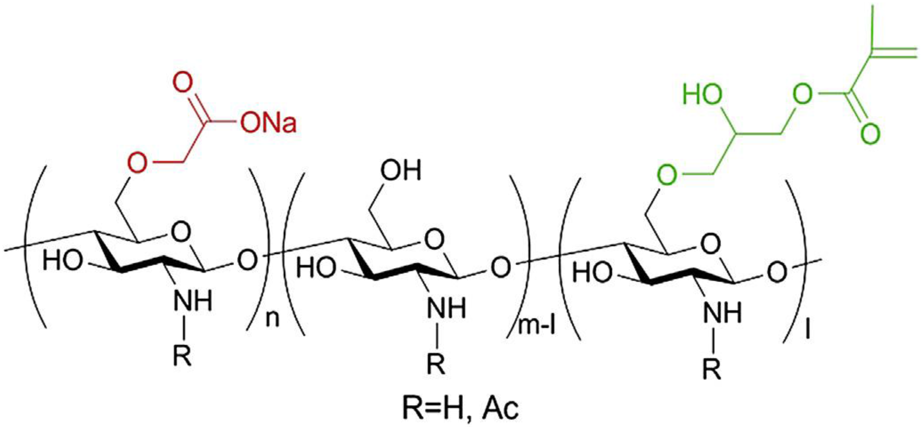
- Azuma, K.; Nishihara, M.; Shimizu, H.; Itou, Y.; Takashima, O.; Osaki, T.; Itoh, N.; Imagawa, T.; Murahata, Y.; Tsuka, T.; et al. Biological adhesive based on carboxymethyl chitin derivatives and chitin nanofibers. Biomaterials 2015, 42, 20–29. [Google Scholar] [CrossRef] [PubMed]
© 2015 by the authors. Licensee MDPI, Basel, Switzerland. This article is an open access article distributed under the terms and conditions of the Creative Commons Attribution license ( http://creativecommons.org/licenses/by/4.0/).
Share and Cite
Azuma, K.; Izumi, R.; Osaki, T.; Ifuku, S.; Morimoto, M.; Saimoto, H.; Minami, S.; Okamoto, Y. RETRACTED: Chitin, Chitosan, and Its Derivatives for Wound Healing: Old and New Materials. J. Funct. Biomater. 2015, 6, 104-142. https://doi.org/10.3390/jfb6010104
Azuma K, Izumi R, Osaki T, Ifuku S, Morimoto M, Saimoto H, Minami S, Okamoto Y. RETRACTED: Chitin, Chitosan, and Its Derivatives for Wound Healing: Old and New Materials. Journal of Functional Biomaterials. 2015; 6(1):104-142. https://doi.org/10.3390/jfb6010104
Chicago/Turabian StyleAzuma, Kazuo, Ryotaro Izumi, Tomohiro Osaki, Shinsuke Ifuku, Minoru Morimoto, Hiroyuki Saimoto, Saburo Minami, and Yoshiharu Okamoto. 2015. "RETRACTED: Chitin, Chitosan, and Its Derivatives for Wound Healing: Old and New Materials" Journal of Functional Biomaterials 6, no. 1: 104-142. https://doi.org/10.3390/jfb6010104
APA StyleAzuma, K., Izumi, R., Osaki, T., Ifuku, S., Morimoto, M., Saimoto, H., Minami, S., & Okamoto, Y. (2015). RETRACTED: Chitin, Chitosan, and Its Derivatives for Wound Healing: Old and New Materials. Journal of Functional Biomaterials, 6(1), 104-142. https://doi.org/10.3390/jfb6010104