


Abstract
Diabetes mellitus continues to impose a substantial global health burden, underscoring the need for therapeutic systems capable of achieving precise, adaptive, and patient-friendly glycemic control. Conventional diabetes treatments, including repeated insulin injections and oral hypoglycemic agents, are often constrained by non-physiological drug release, poor adherence, systemic side effects, and the persistent risk of hypoglycemia. In this context, glucose-responsive nanomedicine has emerged as a promising platform for next-generation diabetes therapy by enabling self-regulated and glucose-triggered delivery of insulin and other antidiabetic agents. This review highlights recent advances in glucose-responsive nanomedicine, focusing on the principal sensing mechanisms, including glucose oxidase-based, phenylboronic acid-based, and lectin-mediated systems, as well as the nanoscale carriers engineered to support them, such as polymeric nanoparticles, nanogels, micelles, liposomes, and hybrid nanostructures. These smart platforms offer significant potential to improve drug stability, enhance targeting efficiency, reduce dosing frequency, and more closely mimic endogenous insulin secretion. The review further examines their emerging role in precision diabetes care, particularly in combination with continuous glucose monitoring technologies, wearable devices, and closed-loop therapeutic systems. Despite notable progress at the preclinical level, important barriers to clinical translation remain, including challenges related to biocompatibility, long-term safety, reproducibility, scalable manufacturing, and regulatory approval. Collectively, glucose-responsive nanomedicine represents a rapidly advancing and clinically relevant field with the potential to redefine diabetes management through intelligent and personalized therapeutic strategies. This review provides a focused overview of current developments, key translational challenges, and future directions toward clinical implementation.
1. Introduction
Diabetes mellitus (DM) is a group of chronic metabolic disorders characterized by persistent hyperglycemia resulting from defects in insulin secretion, insulin action, or both [1]. Type 1 diabetes mellitus (T1DM) accounts for ~10% of the diabetic patient population; it is primarily caused by autoimmune damage to the insulin-producing β-cells of the pancreatic islets, leading to insulin deficiency [2,3]. In contrast, type 2 diabetes mellitus (T2DM), the most prevalent form of diabetes worldwide, is marked by insulin resistance (IR) and a progressive decline in β-cell function and insulin secretion [4]. In both forms, DM contributes to serious complications due to prolonged poor glycemic control, such as diabetic retinopathy, nephropathy, neuropathy, cardiovascular disease, and stroke [3,5].
Diabetes is a major global health challenge because of its rapidly increasing prevalence. According to the latest World Health Organization (WHO) report, the global number of adults living with diabetes increased significantly from 200 million in 1990 to 830 million in 2022, while the global prevalence doubled from 7% to 14% over the same period [6,7]. Empirical evidence has demonstrated that only 55.8% of people living with DM had been diagnosed in 2023, while only 21.1% were receiving the required diabetes management medication [8]. Moreover, the burden of diabetes and the coverage of diabetes medication are unequally distributed, with low- and middle-income countries (LMICs) being particularly affected [6]. Critically, the global challenges of DM are not limited to its rising prevalence; persistent gaps in early diagnosis, access to treatment, and continuity of long-term care influence population health outcomes, resulting in increased disability and premature mortality. For instance, the WHO reported that diabetes directly caused 1.6 million deaths in 2021, while disability-adjusted life years (DALYs) more than doubled between 2000 and 2021 [8].
Moreover, health-related quality of life (HRQoL) is profoundly affected by diabetes, with lifelong self-management, insulin use, frequent glucose monitoring, fear of hypoglycemia, and the risk of complications often reducing physical functioning, emotional well-being, and everyday participation [9]. Accordingly, HRQoL represents a significant outcome in diabetes care, complementing the clinical assessment of glycemic control [10]. Thus, enhancing diabetes therapy is crucial not only for achieving glycemic control but also for reducing disability, premature mortality, treatment burden, and the overall population health impact of the disease [11].
Conventional diabetes treatments include oral glucose-lowering agents and exogenous insulin therapy. These treatments remain fundamental for managing hyperglycemia and delaying disease complications. However, diabetes therapies are usually administered according to fixed schedules or patient-adjusted regimens. They do not intrinsically regulate drug release in response to real-time glucose changes [12]. This limitation is evident in subcutaneous insulin therapy, in which standard injections and pump-based systems cannot reproduce the rapid, glucose-dependent insulin secretion of healthy pancreatic β-cells [13].
A primary challenge in insulin therapy is maintaining stable glycemic control despite fluctuating physiological insulin requirements. Insulin needs may vary with nutritional status, physical activity, illness, stress, and individual sensitivity, necessitating ongoing monitoring and dose adjustments to minimize both hyperglycemia and treatment-related hypoglycemia. Moreover, the burden of multiple daily subcutaneous insulin injections may result in poor treatment adherence, compromising effective diabetes management [1,14]. This challenge persists despite the availability of newer microneedle systems, which are more adaptable, acceptable, and user-friendly than traditional methods [15]. Additionally, oral hypoglycemic agents have their own limitations, such as variability in patient response, drug-specific adverse effects, and continued adherence requirements [16].
Given the limitations of conventional diabetes therapies, a shift toward a smart, self-regulated therapeutic system that can automatically respond to real-time changes in blood glucose levels is desirable. Glucose-responsive drug delivery systems represent an innovative approach to diabetes management [12]. They are designed to detect fluctuations in glucose concentration and initiate therapeutic release, thereby increasing drug availability during hyperglycemia while minimizing unnecessary exposure when glucose levels decrease [12,13,17]. Therefore, smart delivery systems have the potential to improve both the precision and safety of diabetes treatment [18].
Within this field, nanomedicine has contributed to significant advancements in the treatment of various medical conditions [19] and is actively being studied in diabetes management [2,20,21]. Glucose-responsive nanomedicine refers to the medical application of engineered nanomaterials to enhance the stability, delivery, and controlled release of therapeutic agents through novel nanocarrier systems [20,22,23,24,25,26]. Glucose-responsive nanomedicine mainly relies on three sensing mechanisms: glucose oxidase, phenylboronic acid, and lectin-mediated mechanisms. Incorporating these mechanisms into nanoscale carriers, including polymeric nanoparticles, nanogels, micelles, liposomes, and hybrid nanostructures, allows these systems to be classified as glucose-responsive nanomedicine [2,12,27,28,29].
This review examines glucose-responsive nanomedicine as an advanced approach to improving diabetes therapy. It focuses on systems that improve glycemic control, reduce unnecessary insulin exposure, and prevent hypoglycemia. The review evaluates key glucose-sensing mechanisms and their use in nanocarriers. It also highlights the strengths and limitations of these platforms, focusing on responsiveness, safety, stability, and their potential for clinical translation.
Methodology and Literature Search Strategy
This narrative review was informed by a structured literature search conducted using PubMed/MEDLINE, Scopus, and Web of Science. Studies published between January 2000 and 15 July 2026 were considered. The search combined terms related to diabetes and insulin delivery with terms related to glucose-responsive nanomedicine, including “glucose-responsive,” “glucose-sensitive,” “self-regulated insulin delivery,” “nanoparticle,” “nanogel,” “liposome,” “micelle,” “hydrogel,” “microneedle,” “glucose oxidase,” “phenylboronic acid,” and “concanavalin A.” The reference lists and citations of relevant articles were also manually screened to identify additional studies. The final literature search was conducted on 15 July 2026.
English-language, peer-reviewed primary studies and relevant review articles were considered when they addressed glucose-sensing mechanisms, glucose-triggered drug release, nanocarrier design, preclinical evaluation, safety, manufacturing, or clinical translation. Conventional drug-delivery systems without a direct glucose-responsive mechanism, non-diabetes applications, conference abstracts, and non-peer-reviewed publications were excluded from the main analysis. Study selection was based on relevance to the review objectives and the scientific contribution of each article. As this was a narrative review, no formal risk-of-bias assessment or meta-analysis was performed.
2. Diabetes Therapy: Current Challenges and Unmet Needs
Despite advances in technology and medicine, maintaining optimal glycemic control remains a major clinical challenge [30]. The worldwide prevalence of diabetes is continually increasing and is associated with significant morbidity and mortality resulting from both microvascular and macrovascular complications [31,32]. Although current therapeutic approaches have improved patient outcomes, many individuals continue to experience inadequate glycemic control, treatment-related adverse effects, and poor patient adherence. Advances in diabetes management, such as improved glycemic control, cardiovascular risk reduction, and earlier detection through screening programs, have contributed to reduced rates of vascular complications. Consequently, life expectancy has increased, contributing to the emergence of additional complications such as cancer, dementia, postoperative infections, and respiratory infections [32].
2.1. Limitations of Insulin Injections and Oral Hypoglycemic Drugs
In type 1 diabetes and advanced cases of type 2 diabetes, where oral antidiabetic agents are inadequate for achieving optimal glycemic control, insulin remains the primary and most effective treatment [33]. Insulin therapy replaces deficient endogenous insulin in type 1 diabetes, thereby facilitating glucose uptake into tissues, maintaining serum glucose levels within the target range, and decreasing hepatic glucose production [34]. Although insulin therapy is an integral part of diabetes management, it is predominantly administered subcutaneously; this route of administration is associated with significant limitations, including needle-related anxiety, psychological insulin resistance, and local injection-site complications, all of which compromise treatment adherence and glycemic control, underscoring the need for alternative delivery routes [35]. Studies have shown that when patients with type 2 diabetes are prescribed subcutaneous insulin, they may perceive this as an indication that their condition has “worsened” or that alternative treatments have “failed,” consequently leading to delays in or refusal of treatment. Patient awareness of the potential complications of hypoglycemia, coupled with the requirement for precise insulin administration and comprehensive training, adds further complexity to therapy. This, combined with the demands of frequent glucose monitoring and continuous dose adjustments, contributes to heightened treatment-related anxiety and consequent reluctance to initiate insulin therapy [35].
Although subcutaneous insulin administration is traditionally regarded as the most reliable route, it demonstrates pharmacokinetic variability influenced by external factors. For instance, local blood flow at the injection site can significantly modulate insulin absorption, whereby increased perfusion during heat exposure or after exercise can accelerate systemic absorption, whereas reduced perfusion after exposure to cold temperatures can delay absorption. The injection site can also result in variable absorption rates, as abdominal administration accelerates absorption, whereas thigh and gluteal administration results in delayed absorption due to reduced blood flow in these areas [35].
A common complication of subcutaneous insulin administration is lipodystrophy, a disorder in which repeated injections at the same site result in localized adipose tissue changes [35]. Lipodystrophy falls into two categories: lipohypertrophy, characterized by thickening of the subcutaneous fat, and lipoatrophy, characterized by loss of subcutaneous fat at the injection site. In both cases, localized adipose tissue changes affect insulin pharmacokinetics, specifically influencing the rate of insulin absorption into the bloodstream. This intra-individual variability in insulin absorption causes inconsistent pharmacodynamic responses to the same insulin dose administered at different times, contributing to glycemic excursions and hypoglycemic episodes and resulting in reduced glycemic control [36].
Oral hypoglycemic agents are central to type 2 diabetes management and reduce serum glucose through a variety of mechanisms, including increasing insulin sensitivity in peripheral tissues, stimulating pancreatic β-cells to secrete insulin, increasing urinary glucose excretion, stimulating endogenous incretin release, and reducing glucose and carbohydrate absorption in the gastrointestinal tract [37]. However, the effectiveness of oral hypoglycemic agents is associated with β-cell function, particularly the ability of pancreatic β-cells to produce insulin. Clinical trials such as the UK Prospective Diabetes Study (UKPDS) and A Diabetes Outcome Progression Trial (ADOPT) have demonstrated that the progression of type 2 diabetes mellitus is closely associated with a progressive decline in pancreatic β-cell function. Since the efficacy of oral hypoglycemic agents is somewhat dependent on adequate β-cell activity, the progressive decline in β-cell function results in a subsequent reduction in therapeutic responsiveness, resulting in diminished glycemic control as the disease advances [38].
Oral hypoglycemic agents are associated with class-specific adverse effects that can negatively influence patient adherence and treatment success (Table 1). Biguanides are associated with gastrointestinal side effects and vitamin B12 deficiency [39]. Thiazolidinediones (TZDs) are associated with fluid retention, mediated through increased renal sodium absorption in the collecting ducts, causing peripheral edema and potentially precipitating congestive heart failure. In postmenopausal women, TZDs act through the activation of peroxisome proliferator-activated receptor-γ (PPAR-γ), promoting the differentiation of mesenchymal stem cells into adipocytes and inhibiting osteoblastogenesis, thereby significantly increasing the risk of bone fractures [40]. Sodium-glucose cotransporter 2 (SGLT2) inhibitors reduce renal glucose reabsorption, resulting in glucosuria, which may promote pathogen growth in the urinary tract and increase the risk of genitourinary tract infections. These agents are also associated with acute kidney injury and diabetic ketoacidosis [41].
Table 1.
Adverse effects of oral hypoglycemic agents.
2.2. Poor Glycemic Control and Risk of Hypoglycemia
Poor glycemic control and treatment-induced hypoglycemia arise from the interaction between treatment-related factors and individual patient variability. In T1DM, hypoglycemia arises when standard doses of exogenous insulin are administered despite reduced circulating glucose, such as when meals are delayed or missed, the patient has engaged in prolonged physical activity, or when alcohol is consumed without carbohydrate intake [42]. During exercise, peripheral skeletal muscle glucose uptake increases due to translocation of glucose transporter type 4 (GLUT4) to the cell surface [43]. In the presence of exogenous insulin, this effect is further amplified through insulin-activated GLUT4 activity, subsequently causing a rapid drop in serum glucose levels [44]. The absence of counterregulatory hormonal response, specifically glucagon and catecholamine, prevents the body’s normal physiological mechanism to restore blood glucose during hypoglycemia [45]. During alcohol intake, the liver stimulates ethanol metabolism, subsequently increasing the NADH/NAD+ ratio, thereby inhibiting gluconeogenesis, in turn exacerbating the current hypoglycemic state [46].
Oral hypoglycemic agents that are utilized in T2DM also pose the risk of hypoglycemia. Sulfonylureas exert their effects through a glucose-independent mechanism by directly stimulating the release of insulin from pancreatic β cells, irrespective of serum glucose levels. This unregulated insulin release can predispose patients to hypoglycemia [47]. Similarly, Meglitinides bind to sulfonylurea receptor 1 (SUR1) to close ATP-sensitive potassium (KATP) channels, resulting in the secretion of insulin regardless of serum glucose levels [48]. Evidently, conventional standardized therapeutics used in both T1DM and T2DM can cause hypoglycemia [49].
2.3. Adherence Issues and Non-Physiological Drug Delivery
Patient adherence issues and the limitations of current diabetic treatments in replicating physiological insulin secretion are major barriers to achieving stable glycemic control [50]. Under normal physiological conditions, insulin is delivered to the liver via the portal circulation, resulting in portal insulin concentrations that are two to fourfold higher than those in the peripheral circulation. Exogenous insulin administration bypasses the portal-hepatic route, resulting in glycemic variability characterized by recurrent episodes of hyperglycemia and hypoglycemia (Figure 1) [51,52].
Figure 1.
Portal–systemic blood insulin gradient and alternative routes of insulin administration. Red arrows indicate arterial blood flow, blue arrows indicate venous blood flow, and dashed black arrows indicate the inhaled, transdermal, and oral routes of insulin administration. Reprinted from Ref. [51].
Accordingly, patients are required to adhere to complex treatment regimens including close blood glucose monitoring, 14, counting, and recurrent dose adjustments (Table 2) [53]. Studies have demonstrated that higher medication regimen complexity was associated with poorer glycemic control and lower patient adherence rates [54,55,56]. Low adherence can manifest in a variety of ways including delayed doses, missed doses, or the intentional reduction in prescribed doses. A survey looking into patient adherence reported that over 50% of T1DM and T2DM intentionally omitted insulin doses, noting that adherence rates decreased as the number of times a day the medication should be taken increased [57]. Consequently, these studies have emphasized the importance of identifying alternative less complex insulin delivery pathways to improve patient adherence.
Table 2.
Factors contributing to poor adherence and limitations of conventional insulin therapy [53].
2.4. Need for More Precise and Adaptive Treatment Strategies
Conventional diabetes therapies are associated with several limitations that contribute to clinical challenges, including glycemic variability, treatment-induced hypoglycemia, and suboptimal treatment adherence. Consequently, there is a growing need for individualized and adaptive treatment approaches that can accommodate individual patient characteristics, treatment responses, and changing metabolic demands. Current approaches lack the precision and specificity to account for inter-individual variability in insulin sensitivity and counter-regulatory hormonal responses, resulting in variable glycemic control. Studies have shown the potential of developing a closed-loop artificial pancreas, allowing calculated and individualized delivery of basal insulin and demonstrating evidence of reducing glycemic variability incidences [58]. The closed-loop insulin system utilizes subcutaneous insulin administration; however, it does so according to a calculated algorithm in response to real-time changes in serum glucose levels rather than releasing a standardized amount of insulin with each administration (Figure 2). This system overcomes limitations of conventional therapies including the psychosocial fear of injections and the anxiety associated with traditional subcutaneous administration of insulin. Clinical trials have demonstrated promising outcomes, underscored the potential of personalized medicine, and reinforced the need for continued research and broader clinical implementation in diabetes management [59].
Figure 2.
Schematic of the configuration of closed-loop insulin delivery. The circular arrows indicate the direction of the closed-loop feedback process from glucose measurement and sensing to algorithmic modulation and insulin delivery. A continuous glucose monitor (CGM) transmits information about interstitial glucose concentrations to an algorithm hosted on a smartphone or insulin pump that translates information from the glucose sensor and computes the amount of insulin to deliver. An insulin pump delivers a rapid-acting insulin analogue subcutaneously. Insulin delivery is modulated in real time by the control algorithm. Communication between system components is wireless. CSII, continuous subcutaneous insulin infusion. Reprinted from Ref. [59].
3. Principles of Glucose-Responsive Nanomedicine
The development of glucose-responsive nanomedicine has been considered a promising strategy to improve insulin delivery through glucose-dependent drug release. By integrating the glucose sensing and nanocarrier systems, these platforms are designed to achieve more adaptive, efficient, and personalized diabetes management.
3.1. Concept and Functional Basis of Glucose-Responsive Nanomedicine
Glucose-responsive nanomedicine (GRN) can be defined as an advanced drug delivery approach designed to release insulin in response to variations in blood glucose concentrations. While conventional methods of insulin delivery rely on scheduled administration, GRN uses an on-demand, feedback-controlled drug delivery process that responds to changes in blood glucose concentrations [60]. The aim of developing GRN-based drug delivery systems is to mimic the function of pancreatic β-cells by releasing insulin according to blood glucose levels [61,62].
For the purposes of this review, a system is considered genuinely glucose-responsive only when glucose directly interacts with a glucose-recognition element and consequently regulates drug release. By contrast, systems that provide only sustained release or respond independently to pH, temperature, degradation, or other stimuli without directly sensing glucose are not classified as glucose-responsive systems [60,61,62,63,64].
Typically, β-cells sense blood glucose levels and release insulin when these levels are high. Likewise, glucose-responsive nanocarriers are designed to sense the surrounding glucose environment and release their drug cargo when the glucose level exceeds a certain threshold [63]. The glucose dependence of this process makes GRN-based systems suitable for closed-loop drug delivery, as shown in Figure 3, where drug release can be modulated according to fluctuations in physiological glucose levels without the need for constant external monitoring. In this way, insulin release closely mimics the natural regulatory mechanism of healthy pancreatic tissue [62,64].
Figure 3.
Schematic of a closed-loop insulin delivery system in the body. Blue arrows indicate the direction of the feedback loop: glucose sensing activates the insulin delivery system, which releases insulin, while effective insulin regulates glucose through its effects on the liver and peripheral tissues. Blue boxes represent the insulin delivery system and effective insulin, red boxes identify glucose and the physiological compartments involved in glucose regulation, and green labels identify the glucose-sensing and insulin-release processes. Reprinted from Ref [65].
3.2. How Glucose-Triggered Drug Release Occurs
Glucose-responsive nanomedicine contains three primary functional components: (1) a glucose sensing element, (2) a drug reservoir, and (3) a responsive carrier matrix. The glucose-sensing element detects changes in glucose concentration and translates them into a biochemical signal that induces a structural or physicochemical change in the carrier matrix, subsequently triggering drug release from the reservoir when a predetermined glucose concentration is reached [66,67]. These three components work together to deliver a self-regulating approach for the administration of the desired drug based on demand while minimizing unwanted therapeutic agent exposure [68].
The effectiveness of glucose-responsive nanomedicine relies on its ability to detect changes in glucose levels and produce an appropriate therapeutic response. All these systems share the same general goal: to release therapeutic agents when needed. However, each of those platforms uses a different molecular pathway that interacts with other molecules to be able to interpret changes in glucose concentrations and trigger the release of the appropriate drugs [69]. Several glucose-sensing strategies have been developed to enable this responsiveness, each utilizing distinct biochemical interactions to detect glucose and initiate drug release [70,71].
Currently available glucose-responsive system designs almost always rely on glucose-responsive elements such as phenylboronic acid (PBA), glucose oxidase (GOx), and concanavalin A (ConA). Continued advances in glucose-responsive nanotechnology have increased interest in these systems as potential platforms for future diabetes therapy [61,72].
3.3. Benefits over Conventional Delivery Systems
One major advantage of glucose-responsive nanomedicine is its ability to achieve tighter glycemic control as the therapeutic release is responsive to metabolic needs, where glucose concentrations can be maintained within a narrower physiological range [73]. Improved glycemic control is important in the management of diabetes as persistent fluctuations in glucose have been associated with the development of both microvascular and macrovascular complications [74].
Veiseh et al. have pointed out the significance of providing physiologically controlled insulin delivery. Physiological demands for insulin vary during the day depending on the intake of glucose. However, conventional insulin delivery methods have failed to provide accurate physiological delivery. This mismatch may contribute to periods of inadequate glycemic control and excessive insulin exposure. This is addressed by the design of glucose-responsive nanomedicine, which controls the drug release according to the actual glucose level, mimicking the physiological processes of insulin release and glucose control [75].
In addition to better glycemic regulation, glucose-responsive systems can decrease the likelihood of drug-induced hypoglycemia [67]. As glucose levels decrease toward the normal range, less medication is released, thereby reducing unnecessary drug exposure. This mechanism ensures greater safety and becomes one of the major advantages of glucose-responsive devices for delivering insulin [76].
Another important benefit of glucose-responsive nanomedicine is the possibility for better patient compliance and quality of life. Automated drug delivery may reduce the need for frequent therapeutic interventions and improve the management of chronic diseases. Glucose-responsive nanomedicine may also reduce the overall treatment burden and improve the overall quality of the patient experience [64].
3.4. Relevance to Precision Diabetes Therapy
Precision medicine focuses on optimizing clinical outcomes by adjusting treatment strategies to meet the physiological requirements of each patient [77]. Controlling blood glucose levels in patients with diabetes remains difficult because glucose levels fluctuate in response to diet, exercise, stress, and disease [78]. This has created a growing need for therapeutic strategies that adjust treatment according to the patient’s physiological state.
GRN is consistent with the goals of precision-based diabetes treatment because it enables dose control according to the patient’s actual blood glucose level rather than a predetermined dosing schedule [79]. This approach may enable personalized diabetes treatment by adapting to changes in metabolic requirements. According to Caturano et al., glucose-responsive nanomedicine represents a promising strategy for precision insulin delivery by enabling treatment to be tailored to real-time physiological conditions [64]. Similarly, Wang et al. stated that glucose-responsive delivery systems are designed to mimic endogenous insulin secretion and improve the accuracy of insulin administration in response to changing glucose levels [68].
The significance of glucose-responsive nanomedicine for precision medicine is further underscored by improvements in smart nanocarrier design. Current-generation nanocarriers can be designed with specific physicochemical properties to improve insulin-delivery efficiency, glucose responsiveness, biocompatibility, and controlled release [73]. Such adaptability enables the development of more personalized therapeutic platforms capable of addressing differences in patient physiology and disease progression, supporting the future transition toward individualized diabetes care [80,81].
4. Major Glucose-Sensing Mechanisms
The glucose-sensing mechanism is the core of any glucose-responsive nanocarrier, and it determines how the system detects rising glucose levels and translates that signal into active insulin release. There are three main mechanisms by which these systems operate: glucose oxidase (GOx)-based systems, phenylboronic acid (PBA)-based systems, and concanavalin A (ConA)-based systems. Each of these groups differs in terms of sensitivity, stability, physiological compatibility, and immunogenicity risk. The mechanism chosen determines the entire downstream system, from sensing to what kind of nanocarrier can be used, the path of administration (injected, oral, skin patch), and the rules related to drug storage. In addition to these subtopics, there are emerging systems and hybrid mechanisms that have been developed to address the limitations of individual mechanisms. Figure 4 illustrates the fundamental distinction between protein-based strategies, which use GOx or ConA, and protein-free strategies, which rely on PBA. As shown, protein-based strategies incorporate the sensing element as an active biochemical component within the carrier, whereas protein-free PBA-based strategies use small synthetic molecules, reducing the risk of immunogenicity but requiring precise chemical engineering [82,83,84,85].
Figure 4.
Schematic diagram of glucose-responsive material fabricated from (i) protein-based and (ii) protein-free strategies. The use of glucose-binding proteins ConA and GOx represents two major examples of protein-based strategies. The second design uses PBA-based molecular receptors to impart glucose-responsive properties. Reprinted from Ref. [82].
4.1. Glucose Oxidase (GOx)-Based Systems
GOx is a flavoprotein enzyme that is derived from the fungus Aspergillus niger, with the role of oxidizing glucose. The mechanism of this system is the conversion of glucose into glucono-δ-lactone which rapidly breaks down into gluconic acid. This product is responsible for a pH drop locally, from ~7.4 to ~6.5. H2O2 is also formed as a byproduct of this reaction. These two products then act as separate triggers: the pH drop causes pH-sensitive polymers in the carrier to swell and open, while the H2O2 cleaves oxidation-sensitive chemical bonds, also opening the carrier. Catalase is frequently co-encapsulated to decompose H2O2 into water and oxygen, which may reduce oxidative damage and partially replenish oxygen. However, catalase does not eliminate the risks associated with transient H2O2 accumulation, and regenerated oxygen may also weaken hypoxia-dependent release mechanisms. System performance therefore depends on oxygen availability, enzyme stability, diffusion, and the relative concentrations of GOx and catalase. To maintain enzymatic activity in vivo, GOx must be immobilized within the carrier architecture rather than freely loaded. To do this, strategies include covalent grafting to the polymer matrix and encapsulation within a protective polymeric shell [83,84,85,86,87]. One glucose molecule generates both an acid and an oxidant, which is considered a strong and amplified signal from a small input. This is why this system is commonly used in injectable nanoparticles and transdermal microneedle patches, where the local pH shift stays contained within the carrier [84,87,88]. Tai et al. loaded GOx, catalase, and insulin into pH-sensitive polymer nanovesicles, where GOx converts glucose to gluconic acid, dropping local pH and triggering insulin release through disassembly. Results showed that a single subcutaneous injection was able to maintain normal blood glucose levels in diabetic mice for up to 5 days with no inflammatory response, as shown in Figure 5 [89]. This study demonstrated that a purely enzymatic pH-driven mechanism is sufficient to achieve sustained glycemic control without needing synthetic chemical triggers. Table 3 summarizes the major GOx-based carrier platforms and their corresponding glucose-triggered release mechanisms [85,90].
Figure 5.
In vivo glucose regulation by nanovesicles in STZ-induced diabetic mice. (A) BG levels in STZ-induced diabetic mice after treatment with PBS solution, vesicles encapsulating both enzyme and insulin [VS(E+I)], or vesicles encapsulating insulin only [VS(I)]. p < 0.01 compared with the PBS group. (B) BG levels were continuously monitored during the first 12 h after administration of PBS solution, VS(E+I), or VS(I). Data represent the mean ± SD (n = 7). Abbreviations: BG, blood glucose; PBS, phosphate-buffered saline; SD, standard deviation; STZ, streptozotocin; VS(E+I), vesicles encapsulating enzyme and insulin; VS(I), vesicles encapsulating insulin only. Reprinted from Ref. [89]. An asterisk (*) indicates p < 0.01 compared with the PBS group.
Table 3.
GOx-mediated platforms [90].
Despite their signal-amplifying capacity, GOx-based systems face several physiological limitations. Restricted oxygen availability may reduce glucose oxidation, while excessive H2O2 can damage surrounding tissues and decrease enzyme activity. Enzyme denaturation, leakage and immune recognition may further reduce long-term stability. Moreover, diffusion of glucose and oxygen through the carrier and surrounding tissue can delay insulin release compared with responses measured under simplified in vitro conditions [86,87,90].
4.2. Phenylboronic Acid-Based Systems (PBA)
PBA is a small synthetic molecule containing a boronic acid group attached to a phenyl ring. This forms reversible covalent bonds when encountering glucose, specifically with the two adjacent hydroxyl groups on the glucose molecule. In contrast to GOx, PBA contains no protein component, which allows for more chemical stability and less immunological reaction risk. In terms of mechanism, PBA forms cross-links with the carrier’s polymer network, keeping it compact and sealed under normal glucose levels [91,92]. When glucose rises, free molecules compete for the PBA binding sites, breaking the cross-links, increasing the network’s hydrophilicity and causing osmotic swelling and carrier disassembly, releasing insulin. The main engineering limitation of this structure is that native PBA only reaches its glucose-binding tetrahedral form above its pKa (~8.8), which is too alkaline for the human body [85,93]. Strategies to improve glucose binding at physiological pH include introducing electron-withdrawing substituents that lower the pKa of the boronic acid group, incorporating neighboring amines that stabilize boronate formation, and using multivalent PBA architectures. However, these modifications do not necessarily guarantee sufficiently selective or rapid glucose responsiveness under physiological conditions [91,92,93,94]. This is preferred when a fully synthetic and stable system is required. It is incorporated across many carrier types like nanoparticles, micelles, nanogels, and transdermal hydrogel patches [83]. Table 4 summarizes PBA derivatives and their corresponding hydrogel platforms over the past ten years, highlighting combinations used to achieve glucose-responsive drug delivery over the past decade [95]. Kim et al. developed POSS-APBA nanoparticles that self-assemble into micelles at pH 7.4 (physiological pH), achieving 73.2% insulin entrapment efficiency. Insulin release was found to be directly proportional to glucose concentration across the clinically relevant range, confirming that substituent-driven pKa shifting of the boronic acid group alone is sufficient for precise glucose-responsive delivery without any enzymatic component (Figure 6) [85,94].
Table 4.
Summary of research on PBA in glucose-responsive hydrogels in the past ten years [95].
Figure 6.
In vitro glucose-responsive release of insulin from POSS-APBA@Insulin NPs. (a,c) Continual insulin release; (b) typical insulin-release curves at glucose concentrations of 0–3.75 mg/mL; and (d) insulin release at glucose concentrations of 1 and 2 mg/mL at pH 7.4 and 37 °C. Abbreviations: APBA, 3-aminophenylboronic acid; NPs, nanoparticles; POSS, polyhedral oligomeric silsesquioxane. Reprinted from Ref. [94].
PBA derivatives recognize cis-diol structures rather than glucose exclusively. Consequently, competing sugars and glycosylated biomolecules in biological fluids may occupy PBA-binding sites and reduce glucose selectivity. Binding and release are also affected by local pH, ionic strength, protein adsorption and the accessibility of PBA groups within the carrier. Therefore, glucose responsiveness demonstrated in simple buffer solutions may overestimate performance under physiological conditions, and selectivity, basal leakage and repeated reversibility should be evaluated in biologically relevant media [92,93,94].
4.3. Lectin/Concanavalin A-Based Systems (ConA)
Lectins are proteins that bind carbohydrates; Concanavalin A (ConA) is the most studied of the lectin family. ConA is extracted from jack beans, and exists in tetramer form, with each subunit containing a metal-ion-dependent glucose-binding site that works in coordination with examples of metal ions like Ca2+ and Mn2+ [96,97]. Although ConA can bind glucose at physiological pH, its affinity is not exclusive to glucose and depends on pH, ionic conditions and divalent metal ions [96,97,98]. ConA is cross-linked with glycosylated insulin or a glycopolymer to form a stable network that can hold insulin in place under normal glucose levels. When glucose levels rise, free glucose molecules competitively displace the glycopolymer from ConA, weakening the network in a manner that is concentration-dependent, as competitive binding equilibrium shifts with rising glucose, releasing insulin proportionally [84,99]. This system is primarily used within injectable hydrogel depots and implantable reservoir devices, where spatial confinement of the gel matrix ensures that competitive displacement produces a controlled release rather than passive diffusion [100]. Mansoor et al. conducted an experiment where ConA was loaded into a chitosan–Pluronic F-127 hydrogel, demonstrating glucose-proportional insulin release across simulated glycemic conditions over one week, reaching 97% cumulative release at hyperglycemic levels with >80% cell viability in pancreatic cell lines (Figure 7). However, ConA leakage, non-specific carbohydrate binding, structural instability, immunogenicity and mitogenic T-cell activation remain important barriers to clinical translation [100,101].
Figure 7.
In vitro insulin release profiles from the hydrogel in solutions of differing glucose release media concentrations at 37 °C. Reprinted from Ref. [101].
4.4. Hybrid and Emerging Glucose-Responsive Mechanisms
Each main mechanism mentioned has a specific limitation. For example, GOx uses oxygen, PBA has slower binding kinetics compared to enzymatic systems, and ConA can cause inflammation [101]. Hybrid systems are emerging as the new phase of glucose-responsive mechanisms, as they combine two mechanisms to mitigate their individual limitations. The goal of this field is faster, more precise release with fewer side effects [85]. Table 5 maps out representative dual-responsive injectable hydrogel systems that combine glucose-sensing with secondary stimuli, demonstrating expanded release duration and improved glycemic control relative to single-mechanism systems. More recently, research has moved toward nanoparticle-based hybrid designs, where the smaller scale and tighter structural control of these carriers allow for a faster and more tunable release response compared to hydrogel platforms [82,83].
Table 5.
Representative examples of dual-responsive injectable hydrogels for insulin delivery [83].
GOx and Hypoxia: These systems exploit the fact that GOx consumes oxygen, and this depletion acts as a second trigger, causing reductive cleavage of hypoxia-sensitive linkers in the polymer, accelerating the release of insulin. This mechanism is faster than GOx alone [84,102,103].
Dual pH and H2O2 Responsiveness: These systems are designed to respond simultaneously to both byproducts of the GOx reaction rather than relying on a single trigger. Each trigger acts on a distinct structural component, with pH affecting ionizable polymer groups and H2O2 acting on oxidation-sensitive linkages. Neither trigger alone fully dismantles the carrier, but together they promote near-complete carrier disassembly. This combined response may reduce unwanted drug leakage at normal glucose levels [71,84,88,104,105].
PBA and Supramolecular Assembly: These systems combine the glucose-binding ability of PBA with host–guest chemistry, in which molecules self-assemble into a stable structure that dissociates when glucose levels rise. A cyclodextrin ring accommodates a PBA-tagged component through host–guest interactions, forming a stable nanoparticle at normal glucose levels. When glucose concentrations increase, glucose binding disrupts these interactions, destabilizing the assembly and triggering insulin release [105,106,107,108,109].
PBA-Insulin Conjugates: Rather than encapsulating insulin within a carrier, this approach modifies insulin by attaching PBA directly to the insulin molecule. At normal glucose levels, PBA promotes the binding of insulin to albumin in the blood, limiting its availability. Rising glucose concentrations alter this interaction and promote the release of insulin into circulation [110,111]. For broader clinical context, MK-2640 (Merck), which is an insulin-saccharide conjugate rather than a PBA-insulin conjugate, reached Phase 1 trials but did not demonstrate sufficient glucose-responsive performance for clinical use. This outcome illustrates the broader challenge of tuning glucose-binding affinity to the euglycemic range (~4–10 mmol/L) while maintaining reversible binding [84,112].
The three glucose-sensing mechanisms examined in this section are GOx, PBA, and ConA. Each has a distinct trade-off profile in terms of response speed, physiological compatibility, and immunogenic risk. None of these individual mechanisms alone meets all the requirements of an ideal clinical system. GOx produces a strong biochemical signal but raises concerns regarding enzyme stability and immune responses in vivo. PBA avoids these enzyme-related limitations but often requires structural modification to function effectively at physiological pH. ConA provides natural glucose-binding capability but has not advanced clinically because of its mitogenicity and associated safety concerns. Hybrid and emerging systems have therefore been developed to address these limitations by combining multiple sensing elements, aiming to achieve broader operating ranges, more appropriate response thresholds, and improved safety. The nanocarrier cannot be designed independently of the sensing mechanism because its composition, assembly, and drug-release behavior are constrained by the mechanism selected from the outset [82,83,84,85].
Although GOx-, PBA-, and ConA-based systems all enable glucose-responsive drug delivery, they differ considerably in their glucose-sensing mechanisms, physicochemical characteristics, and translational potential. To facilitate direct comparison, Table 6 summarizes the principal strengths, translational challenges, and clinical implications of the major glucose-sensing mechanisms discussed in this review.
Table 6.
Comparative analysis of the major glucose-sensing mechanisms used in glucose-responsive nanomedicine.
Each method for glucose detection has its advantages and disadvantages that define its utility for clinical use. The GOx-based systems show excellent glucose sensitivity but are limited by enzyme-related issues such as oxygen dependence and hydrogen peroxide production. PBA-based systems are a promising and protein-free alternative with improved design flexibility but need further optimization to enable effective glucose sensing in physiological conditions. ConA-based systems have good glucose-binding properties but are mainly limited by immunogenicity and biocompatibility problems. Thus, the current research is increasingly orientated towards hybrid platforms that integrate complementary sensing mechanisms to achieve better responsiveness, alleviating the constraints of individual systems. Because the sensing mechanism determines the material requirements and release behavior of the delivery system, the following section examines the nanocarrier architectures used to implement these glucose-responsive mechanisms.
5. Nanoparticles and Platform Design
Various nanoparticle platforms have been studied for drug delivery. Each formulation of the platform provides a unique characteristic in terms of drug encapsulation and loading, stimuli-responsiveness and release performance. Examples of nanocarrier platforms include: (i) polymeric nanoparticles, (ii) nanogels, (iii) liposomes, (iv) micelles, and finally (v) multifunctional platforms.
5.1. Polymeric Nanoparticles
Polymeric nanoparticles are made from biodegradable polymers, including chitosan, alginate, poly(lactic-co-glycolic acid) (PLGA), dextran, and hyaluronic acid. These particles generally range from 1 to 100 nm in size, although particles up to approximately 200 nm are commonly used in biomedical applications. They are bioengineered to improve drug protection and enhance targeted bioavailability depending on varying body conditions. In terms of drug loading, the therapeutic agent can either be entrapped in the polymeric matrix (the wall of the nanoparticle) or embedded in the core by surface adsorption and chemical attachment to the polymer surface. The drug is then released in specific ways, including the diffusion of the drug out of the encapsulation, the degradation of the polymer, and the swelling of the polymer [113].
Polymeric nanoparticles are generally classified into nanocapsules and nanospheres. Nanocapsules are core–shell structures consisting of a polymeric membrane surrounding a core that contains the therapeutic agent. In contrast, nanospheres consist of a polymeric matrix in which the drug is uniformly dispersed throughout the polymeric network rather than concentrated within a central core (Figure 8). These structural differences influence the drug-loading mechanism and release profile [114].
Figure 8.
Schematic representation of the structure of nano-capsules and nanospheres. Reprinted from Ref. [114].
Polymeric nanoparticles have been used to deliver various drugs including anti-cancer and antibiotics. In diabetes research [29]. Heidarisasan et al. performed an in vivo experiment on diabetic rats, evaluating insulin-loaded trimethyl chitosan nanoparticles (polymeric nanoparticles), and found that they improved both renal and oxidative stress markers when compared to conventional injectable insulin. Additionally, results show that there has been greater control over hyperglycemia in nanoparticle formulations, even though the difference was not statistically significant (Figure 9) [115].
Figure 9.
(A) shows glucose levels and (B) shows weight changes during 10 weeks in all groups. Abbreviations: C = Normal control, DM = Diabetic control, DM+NP = Diabetic group treated with nanoparticle, DM+N.in = Diabetic group treated with oral nanoinsulin, DM+SC.in = Diabetic group treated with subcutaneous injected insulin. Reprinted from [115].
5.2. Nanogels
Nanogels are defined as soft, nanosized hydrogel particles that consist of crosslinked polymer networks holding a high-water content. This unique composition provides excellent biocompatibility, making them highly effective for encapsulating hydrophilic therapeutics like insulin and various biomacromolecules. Their three-dimensional hydrated structure is capable of holding medications while maintaining their biological activity, and through specific structural adjustments, they can even be adapted to carry hydrophobic agents. A primary benefit of nanogels is their stimulus-responsive behavior. Drug release from these particles can be activated via multiple pathways including diffusion, the breakdown or degradation of the polymer network, pH-induced swelling or shrinking, ion displacement, or the application of external energy input. Each of these triggers modifies the gel structure to facilitate the release of the loaded drug [116,117,118].
Within the field of diabetes therapy, this responsive nature is highly beneficial. Nanogels can be specifically designed to initiate insulin release when exposed to glucose-related triggers, such as fluctuations in glucose concentration, shifts in local pH, or specific enzyme-mediated reactions. Consequently, nanogels have emerged as promising self-regulated platforms designed for controlled antidiabetic drug delivery [119].
Li et al. developed glucose and hydrogen peroxide-responsive nanogels for insulin delivery. In this system, glucose oxidase converts glucose into gluconic acid and hydrogen peroxide, and the generated hydrogen peroxide destabilizes the oxidation-sensitive nanogel, thereby accelerating insulin release. The nanogels demonstrated glucose-dependent insulin release, favorable biocompatibility, and glucose-lowering activity following subcutaneous administration in diabetic mice [120]. This system therefore meets the definition of genuine glucose responsiveness because insulin release is regulated by glucose recognition and its downstream biochemical products.
5.3. Liposomes
Liposomes are nanosized vesicular carriers made up of one or more phospholipid bilayers that surround an aqueous core. Their structure mimics biological systems, they demonstrate excellent biocompatibility, and they possess the capacity to hold both hydrophilic and hydrophobic drugs, making them attractive options for drug-delivery systems. Hydrophilic drugs are typically inserted into the aqueous core, whereas hydrophobic drugs are embedded within the lipid bilayer. Figure 10 demonstrates the structure of a liposome made from a lipid bilayer [121]. Following administration, drug release can occur through several pathways including diffusion, destabilization of the membrane, fusion with biological membranes, or breakdown of the liposomal structure [122].
Figure 10.
Schematic structure of a liposome. The hydrophilic core is enclosed by a hydrophobic bilayer made of phospholipids and cholesterol, enabling the delivery of both hydrophilic agents within the core and hydrophobic agents within the bilayer. Reprinted from Ref. [121].
In the context of diabetes therapy, liposomes have garnered attention as carriers for insulin and other antidiabetic medications. Their structure can be adjusted to produce stimulus-responsive systems, which enable drug release when exposed to glucose-related triggers such as variations in glucose concentration, pH changes, or enzymatic reactions. This characteristic establishes liposomes as valuable platforms for controlled and potentially self-regulated antidiabetic drug delivery [123].
Liu et al. created glucose-responsive multivesicular liposomes intended for insulin delivery and demonstrated encouraging results in both laboratory and animal studies. In laboratory experiments, the system released increased amounts of insulin at elevated glucose concentrations, exhibited pulsatile release when glucose levels changed, and remained fairly stable at physiological pH. In animal experiments, using rats with chemically induced type 1 diabetes, the insulin-loaded liposomes swiftly decreased blood glucose, sustained it within the normal range, and showed superior performance compared to free insulin in glucose tolerance tests, with glucose returning to normal in roughly 2 h following the challenge. These observations demonstrate that glucose-responsive liposomes serve as a valuable intelligent platform for insulin delivery in diabetes [124].
5.4. Micelles
Micelles are self-assembled nanosized carriers formed from amphiphilic molecules that possess a hydrophobic interior and a hydrophilic exterior. They are particularly well-suited for drugs with limited water solubility because the hydrophobic interior can hold lipophilic compounds, while the outer layer enhances stability and biocompatibility. Beyond improving solubility and bioavailability, micelles can shield the enclosed drug and facilitate controlled release. Drug release typically occurs through diffusion, breakdown of the micelle structure, degradation of the polymer, or changes triggered by external stimuli [125].
Gaballa and Theato developed glucose-responsive polymeric micelles assembled through reversible boronic acid–diol interactions between a phenylboronic acid-containing block copolymer and a diol-functionalized polymer. At neutral pH, increasing glucose concentrations competitively disrupted these interactions, promoting micelle dissociation and insulin release. The complex micelles exhibited improved glucose responsiveness under physiologically relevant conditions compared with conventional phenylboronic acid-based micelles [126].
5.5. Hybrid and Multifunctional Nanoplatforms
Hybrid and multifunctional nanoplatforms integrate two or more material systems into a single carrier to combine the benefits of each constituent. In diabetes therapy, these platforms are valuable because they can enhance drug loading, stability, glucose responsiveness, and release control within one unified system. By incorporating polymers, lipids, inorganic materials, or bioactive molecules, hybrid systems can achieve more targeted and flexible antidiabetic drug delivery compared to single-component nanocarriers [127].
Various hybrid combinations have been investigated to optimize glucose-responsive insulin delivery, including (i) polymer–lipid hybrids, which strengthen insulin encapsulation and stability for oral delivery [128]; (ii) polymer–enzyme systems containing glucose oxidase, which enhance glucose sensitivity and promote accelerated insulin release upon triggering [129]; (iii) polymer–inorganic hybrids, such as mesoporous silica-based systems, which offer substantial loading capacity along with self-regulated release behavior [130]; and (iv) microgel–nanoparticle hybrid platforms, which have shown sustained glycemic control in diabetic models [131].
5.6. Comparative Performance of Glucose-Responsive Delivery Platforms
To facilitate comparison across different carrier architectures and sensing mechanisms, Table 7 summarizes the reported performance, preclinical evidence and principal translational limitations of representative glucose-responsive insulin-delivery platforms.
Table 7.
Comparative performance of representative glucose-responsive insulin-delivery platforms.
Overall, no platform currently combines rapid responsiveness, minimal basal leakage, complete reversibility, high insulin loading and prolonged glycemic control. GOx-based systems remain limited by oxygen dependence, enzyme instability and H2O2 generation, while PBA-based systems face concerns regarding physiological selectivity and competing biological diols; ConA-based systems additionally raise immunogenicity and safety concerns [67,94,101,103,124,131]. Inconsistent reporting also prevents direct comparison and highlights the need for standardized evaluation under physiologically relevant glucose conditions.
Having considered the principal sensing mechanisms and carrier architectures, the following section evaluates their therapeutic applications, preclinical performance, and routes of administration.
6. Therapeutic Applications and Recent Advances
6.1. Therapeutic Applications
Glucose-responsive nanomedicine has emerged as a highly promising strategy to improve the management of diabetes through the development of self-regulated drug delivery systems. Smart nanoplatforms such as these are capable of sensing fluctuations in blood glucose concentrations and subsequently releasing insulin as well as other therapeutic agents in a controlled manner.
Unlike conventional insulin therapy that often results in fluctuating glucose levels and poor adherence from patients, glucose-responsive systems aim to mimic physiological insulin secretion closely, thereby reducing the risk of hyperglycemia and hypoglycemia [132,133]. Recent advances in the field of nanotechnology have significantly improved the sensitivity, therapeutic efficiency, and design of such systems, making them increasingly relevant in diabetes therapy (Figure 11).
Figure 11.
Overview of glucose-responsive insulin delivery systems, integrating glucose-responsive units, carrier materials, and principal dosage forms. Reprinted from Ref. [132].
Insulin delivery remains the principal therapeutic application of glucose-responsive nanomedicine. Representative systems include GOx-containing hypoxia-sensitive vesicles, PBA-functionalized carriers, and hybrid GOx/PBA platforms, which have demonstrated glucose-triggered insulin release and glycemic regulation in preclinical models [132,134,135,136,137,138,139]. Their sensing mechanisms and carrier architectures are discussed in detail in Section 4 and Section 5. Beyond insulin delivery, glucose-responsive nanomedicine is being explored for the delivery of other antidiabetic agents, including glucagon-like peptide-1 (GLP-1) receptor agonists, gene-based therapeutics, and metformin [140]. The encapsulation of the agents mentioned within nanoscale carriers enhances bioavailability, improves drug stability, and protects against premature degradation. Nanocarriers can also facilitate targeted drug delivery to the pancreas or insulin-sensitive organs, therefore improving therapeutic outcomes, while simultaneously minimizing systemic side effects. Recent studies have further explored the use of nanotechnology for co-delivery strategies, in which insulin and other antidiabetic compounds are incorporated into a single glucose-responsive platform to achieve synergistic therapeutic effects [141].
6.2. Preclinical Performance Evaluation
In vitro and in vivo studies have demonstrated the significant therapeutic potential of glucose-responsive nanomedicine. In vitro investigations evaluated glucose-triggered release kinetics, biocompatibility, and nanoparticle stability as well as responsiveness under simulated physiological conditions [142]. Meanwhile, in vivo studies in diabetic animal models have shown improved glycemic control, reduced fluctuations of glucose, and prolonged therapeutic activity as well as decreased frequency of insulin administration [143]. Some nanoplatforms have demonstrated their ability to reduce inflammatory responses and oxidative stress associated with the progression of diabetes [144]. These findings highlight the potential of these systems to improve long-term diabetes outcomes and patient quality of life.
6.3. Administration Routes and Emerging Technologies
Recent innovations in the field of smart nanomedicine design have focused on enhancing safety, responsiveness, and integration with advanced technology in healthcare. Wearable glucose monitoring devices and closed-loop insulin delivery systems are increasingly being combined with glucose-responsive nanocarriers to create automated platforms for diabetes management [136]. Microneedle-based systems that incorporate glucose-sensitive nanoparticles have recently emerged as minimally invasive approaches that allow painless and continuous insulin administration [137,145]. Researchers are also exploring biomimetic nanomaterials and multifunctional hybrid nanoparticles that are capable of simultaneous glucose sensing, drug delivery, and real-time monitoring of blood glucose levels [141]. These developments all support the growing shift towards personalized medicine, especially in diabetes care.
Several challenges continue to arise, despite these advances, which limit the clinical translation of glucose-responsive nanomedicine. Long-term biocompatibility, reproducibility, and regulatory approvals remain significant barriers [135]. Furthermore, variations in delayed responsiveness and potential immunogenicity should be addressed before clinical implementation becomes feasible. Nevertheless, ongoing progress in materials, nanotechnology, and biomedical engineering continues to accelerate development towards safer and more effective glucose-responsive systems, which are expected to play a transformative role in the future of diabetes therapy by enabling adaptive, patient-centered treatment strategies [145]. These unresolved barriers and their implications for clinical translation are examined in detail in the following section.
7. Clinical Translation and Future Challenges
Nanomedicine research has rapidly expanded, spanning a broad spectrum of disease applications, including chronic conditions such as diabetes [146]. However, significant physiological and biological barriers continue to impede clinical translation, resulting in only a limited proportion of nanoformulations progressing to clinical application [146]. The multifactorial pathophysiology of diabetes mellitus coupled with the inherently complex physicochemical nature of nanomedicine systems results in various translational barriers that limit clinical implementation [147,148]. These limitations include biocompatibility, toxicity concerns, long-term safety, reproducibility, scalability, manufacturing limitations, regulatory barriers, and clinical translation.
7.1. Biocompatibility and Toxicity Concerns
For chronic conditions, such as diabetes mellitus, biocompatibility issues are even more pronounced due to prolonged and repeated exposure to nanomedicine therapies [149]. After entering biological fluids, nanoparticles rapidly adsorb proteins and other biomolecules, forming a dynamic protein corona that influences aggregation, cellular uptake, biodistribution, immune recognition and clearance. Opsonin-rich coronas may promote uptake by hepatic and splenic macrophages of the mononuclear phagocyte system, whereas dysopsonins may prolong circulation. Surface engineering can influence, but cannot completely control, corona composition because it varies with particle properties and the surrounding biological environment, as shown in Figure 12 [150,151,152]. Nanoparticles composed of silver can undergo surface-driven electron-transfer reactions and metal ion release, resulting in the generation of reactive oxygen species that decrease glutathione levels and increase reactive oxygen species (ROS) levels, eventually causing mitochondrial dysfunction and inflammation [153,154]. More recent studies have reported the protective effects of silver nanoparticles on pancreatic β cells, and accordingly, increasing attention has been given to utilizing their anti-inflammatory and antioxidant properties to improve insulin sensitivity and glycemic control, while mitigating the cytotoxic effects associated with systemic nanoparticle accumulation [154]. A subset of nanoparticles is engineered to target the endothelium for therapeutic delivery; however, their subsequent activation and increase in ROS result in nitric oxide depletion, exacerbating vascular inflammation and raising concerns about vascular toxicity [155].
Figure 12.
The formation of protein corona and exchange of adsorbed proteins over time in biological conditions. Notes: (A) Immediately upon exposure. (B) After a longer exposure time, with displacement of proteins among the hard corona, soft corona, and cellular environment. (C) Major factors affecting protein corona pattern divided into two categories: nanoparticle and environment. Abbreviation: NP, nanoparticle. Reprinted from Ref. [151].
7.2. Long-Term Safety and Reproducibility
Nanoparticle disposition depends on size, shape, surface charge, composition, protein-corona formation and biodegradability. As summarized in Figure 13, clearance may involve material degradation, renal elimination, uptake by the mononuclear phagocyte system and hepatobiliary excretion. Small particles or degradation products may undergo renal elimination, whereas larger or opsonized particles are more commonly taken up by hepatic and splenic macrophages. These pathways do not guarantee complete clearance; slow degradation and repeated administration may lead to accumulation in the liver, spleen, kidneys or lungs [156,157]. Consequences of bioaccumulation include progressive organ burden, reduced detoxification, disturbance of normal physiological clearance systems, oxidative stress, and chronic inflammation [157,158]. At the molecular level, protein corona formation determines the biological identity and fate of the nanoparticle; however, even for biodegradable nanoparticle systems, degradation kinetics must be evaluated as incomplete breakdown can stimulate accumulation of residual material in organs [157]. For example, polymers such as PLGA degrade into lactic acid and glycolic acid; accumulation of these metabolites creates an acidic microenvironment that can trigger inflammatory signaling pathways and autocatalysis of PLGA, resulting in faster polymer degradation [159].
Figure 13.
Molecular clearance: The liver contains diverse enzymes and an aqueous environment to metabolize nearly any nanoparticle at the molecular level, breaking down particles over time to incorporate them into bile for excretion. Black arrows indicate movement toward hepatic processing, whereas the green arrow indicates biliary excretion into the intestine. The colored bands associate the nanoparticle classes with their representative degradation or clearance mechanisms. PGAs include polyglutamic and polyglycolic acids; biopolymers include chitosan, dextran, and hyaluronic acids; inorganics include silica, gold, and carbon; PLAs include all polylactic acids; and hygroscopes include water-attracting compounds such as bile salts and wetting agents such as Tween. Reprinted from Ref. [157].
Reproducibility and batch-to-batch variability represent significant limitations in the production of nanoparticles, as minor deviations in synthesis conditions can result in significant nanoparticle property changes [158,159]. The lack of standardized physicochemical characterization in nanomedicine and the variability among experimental designs across nanomedicine studies highlight the reproducibility issues in nanomedicine. Minute changes in the preparation methodology or formulation conditions of nanoparticles can result in significant changes in zeta potential, morphology, drug loading, release kinetics, and polydispersity, consequently leading to batch-to-batch variability, hindering clinical translation [160].
Experimental studies of polydopamine nanoparticles further demonstrate that controlled oxidation conditions and polymerization time can modify critical quality attributes, including particle size, zeta potential, morphology, surface roughness, and functional surface accessibility [161,162].
7.3. Scale-Up and Manufacturing Limitations
As noted above, limited reproducibility and pronounced batch-to-batch variability remain critical challenges in nanomedicine implementation. Consequently, scalability is one of the most significant barriers limiting the clinical translation of glucose-responsive nanomedicine, as physicochemical conditions can be difficult to reproduce consistently within industrial manufacturing settings [163,164]. Glucose-responsive nanomedicine relies on multiple sequential production steps, including nanoprecipitation, self-assembly, surface functionalization, and ligand conjugation. The transition from laboratory-scale to industrial-scale nanomedicine production poses scalability challenges, as each unit operation or production step can independently influence critical quality attributes (CQAs), such as particle size, encapsulation efficiency, and stability [165]. Consequently, insufficient control at any step can introduce deviations that propagate through subsequent steps. Therefore, multistep manufacturing processes may allow deviations to accumulate across different stages, resulting in significant alterations in the final nanoparticle product. Transitioning from small-scale production to industrial manufacturing also presents significant cost burdens [166].
In addition to technical complexity, industrial manufacturing must comply with Good Manufacturing Practice (GMP) requirements while maintaining standardized nanoparticle physicochemical properties. Manufacturing standards and GMP quality guidelines are still evolving in the field of nanomedicine, and a universally standardized nanomedicine-specific regulatory framework has not yet been established [167]. Before translating laboratory-scale nanomedicine synthesis to large-scale industrial production, GMP-compliant pilot manufacturing processes should be established to support reproducible scale-up and the production of materials for clinical evaluation [168]. A concise summary of the barriers hindering the clinical translation of glucose-responsive nanomedicine is presented in Table 8.
Table 8.
Summary of major barriers limiting clinical translation of glucose-responsive nanomedicine.
7.4. Regulatory Barriers
Nanomedicines are regulated within existing medicinal-product frameworks, supplemented by nanomaterial- and product-specific guidance. The U.S. Food and Drug Administration (FDA) and the European Medicines Agency (EMA) apply established requirements for quality, safety and efficacy while requesting additional characterization and manufacturing data when nanoscale attributes affect product performance. However, no single harmonized international pathway exists, and data requirements may remain product- and jurisdiction-specific [168,169,170,171].
For example, Lipodox®, a liposomal doxorubicin product, was approved by the FDA as generic nanomedicine, whereas the EMA did not approve the product, claiming it did not demonstrate bioequivalence of free doxorubicin [170]. This is an example of how the same product can be interpreted and regulated differently across jurisdictions due to the lack of defined criteria, highlighting the need for harmonized global standards in nanomedicine implementation.
Moreover, to facilitate industrial manufacture, glucose-responsive nanomedicine systems must be stable during global transport and long-term storage [172]. Studies report that vesicular nanocarriers specifically are prone to aggregation secondary to Ostwald ripening, resulting in loss of uniformity and reduced stability [173]. Moreover, liposomes are composed of phospholipid bilayers, susceptible to oxidation and consequent liposomal breakdown, causing the encapsulated drug to leak out. Nanoparticles with low zeta potential demonstrate reduced repulsion and therefore are more likely to aggregate [174]. This nanoparticle aggregation can increase particle size, alter drug release kinetics, and reduce the stability of the nanoparticle, resulting in unpredictable drug delivery [175]. Stability limitations during storage, encompassing nanoparticle aggregation, unintended drug leakage, and alterations in drug release profiles, underscore the need for rigorous regulatory oversight to ensure product consistency and therapeutic efficacy [172,173,174,175,176,177].
7.5. Clinical Development Status and Translational Potential
Most glucose-responsive nanomedicine platforms remain at the in vitro or animal-study stage. Although GOx-, PBA-, and ConA-based nanocarriers have demonstrated glucose-triggered drug release and glycemic control in experimental models, their clinical safety and efficacy have not been established. MK-2640 is the most advanced glucose-responsive insulin to have undergone human testing; however, it is a soluble insulin-saccharide conjugate rather than a nanocarrier. In Phase 1 testing, MK-2640 was generally well tolerated but showed approximately 25-fold lower potency than regular human insulin and insufficient glucose-responsive performance [112]. Table 9 highlights the substantial gap between promising preclinical evidence and clinical development. Therefore, the clinical prospects of nanocarrier-based systems should be interpreted as future potential rather than evidence of clinical readiness.
Table 9.
Clinical development status of glucose-responsive therapeutic systems.
Table 10 shows that several strategies have been proposed to overcome challenges related to protein corona formation, immune recognition, bioaccumulation, manufacturing reproducibility, scalability, regulatory uncertainty, and patient acceptance [178,179]. For example, studies have demonstrated the potential of PEGylation to reduce protein adsorption and suppress protein corona formation, subsequently improving nanoparticle stability in physiological fluids. Certain manufacturing protocols have been developed in accordance with GMP requirements, including microfluidic manufacturing protocols for mRNA vaccine lipid nanoparticles. These protocols specify controlled production steps, including defined microfluidic mixing ratios and laminar-flow mixing, followed by tangential-flow filtration [179]. Experimental studies have also evaluated autonomous chemical platforms that integrate artificial intelligence (AI) modules, particularly closed-loop optimization processes, into nanoparticle production to continuously adjust reaction conditions in real time. These studies demonstrated a highly reproducible nanoparticle production workflow that may improve scalability and consistency [180].
Table 10.
Future strategies to facilitate clinical translation of glucose-responsive nanomedicine.
8. Conclusions and Future Perspectives
Glucose-responsive nanomedicine represents a significant advancement in diabetes therapy by enabling self-regulated, glucose-triggered delivery of insulin and antidiabetic agents. This review has highlighted the major glucose-sensing mechanisms, including glucose oxidase-based, phenylboronic acid-based, and lectin-mediated systems, along with diverse nanocarrier platforms such as polymeric nanoparticles, nanogels, liposomes, micelles, and hybrid nanostructures. These smart systems demonstrate superior glycemic control, enhanced drug stability, and improved patient adherence compared to conventional therapies in preclinical models.
However, significant barriers to clinical translation remain, including biocompatibility concerns, long-term safety validation, manufacturing scalability, and regulatory approval challenges. Future development should focus on rational design strategies, biocompatible biodegradable carriers, comprehensive clinical trials, and collaborative efforts between academia and industry to establish standardized manufacturing protocols. With continued advancement and systematic resolution of translational barriers, glucose-responsive nanomedicine has substantial potential to redefine diabetes management through intelligent, personalized therapeutic strategies that more closely mimic physiological insulin secretion and improve patient outcomes.
Author Contributions
Conceptualization, A.A.; methodology, A.A. and A.B.; investigation, A.A., A.B., S.A., F.R., A.Z., H.N.Z. and R.A.; data curation, A.A., A.B., S.A., F.R., A.Z., H.N.Z. and R.A.; formal analysis, A.A., A.B., S.A., F.R., A.Z., H.N.Z. and R.A.; writing—original draft preparation, A.A., A.B., S.A., F.R., A.Z., H.N.Z. and R.A.; writing—review and editing, A.A., A.B., S.A., F.R., A.Z., H.N.Z., R.A. and G.R.D.; visualization, A.A.; supervision, G.R.D.; project administration, A.A. and G.R.D. All authors have read and agreed to the published version of the manuscript.
Funding
This research received no external funding.
Institutional Review Board Statement
Not applicable.
Informed Consent Statement
Not applicable.
Data Availability Statement
No new data were created or analyzed in this study. Data sharing is not applicable to this article.
Acknowledgments
The authors thank the School of Postgraduate Studies and Research, RCSI-Bahrain, for the Article Processing Fee support.
Conflicts of Interest
The authors declare no conflicts of interest.
References
- Tegegne, B.A.; Adugna, A.; Yenet, A.; Belay, W.Y.; Yibeltal, Y.; Dagne, A.; Teffera, Z.H.; Amare, G.A.; Abebaw, D.; Tewabe, H.; et al. A critical review on diabetes mellitus type 1 and type 2 management approaches: From lifestyle modification to current and novel targets and therapeutic agents. Front. Endocrinol. 2024, 15, 1440456. [Google Scholar] [CrossRef] [Scilit] [PubMed]
- Andreadi, A.; Lodeserto, P.; Todaro, F.; Meloni, M.; Romano, M.; Minasi, A.; Bellia, A.; Lauro, D. Nanomedicine in the treatment of diabetes. Int. J. Mol. Sci. 2024, 25, 7028. [Google Scholar] [CrossRef] [Scilit] [PubMed]
- Holt, R.I.G.; DeVries, J.H.; Hess-Fischl, A.; Hirsch, I.B.; Kirkman, M.S.; Klupa, T.; Ludwig, B.; Nørgaard, K.; Pettus, J.; Renard, É.; et al. The management of type 1 diabetes in adults. A consensus report by the American Diabetes Association (ADA) and the European Association for the Study of Diabetes (EASD). Diabetologia 2021, 64, 2609–2652, Erratum in Diabetologia 2022, 65, 255. https://doi.org/10.1007/s00125-021-05600-6. [Google Scholar] [CrossRef] [Scilit] [PubMed]
- American Diabetes Association Professional Practice Committee. 2. Diagnosis and classification of diabetes: Standards of care in diabetes-2024. Diabetes Care 2024, 47, S20–S42. [Google Scholar] [CrossRef] [Scilit] [PubMed]
- Islam, K.; Islam, R.; Nguyen, I.; Malik, H.; Pirzadah, H.; Shrestha, B.; Lentz, I.B.; Shekoohi, S.; Kaye, A.D. Diabetes Mellitus and Associated Vascular Disease: Pathogenesis, Complications, and Evolving Treatments. Adv. Ther. 2025, 42, 2659–2678. [Google Scholar] [CrossRef] [Scilit] [PubMed]
- World Health Organization. Diabetes [Internet]. 2024. Available online: https://www.who.int/news-room/fact-sheets/detail/diabetes (accessed on 15 May 2026).
- NCD Risk Factor Collaboration (NCD-RisC). Worldwide trends in diabetes prevalence and treatment from 1990 to 2022: A pooled analysis of 1108 population-representative studies with 141 million participants. Lancet 2024, 404, 2077–2093. [Google Scholar] [CrossRef] [Scilit] [PubMed]
- Stafford, L.K.; Gage, A.; Xu, Y.Y.; Conrad, M.; Beltran, I.B.; Boyko, E.J.; Duncan, B.B.; Hay, S.I.; Lenox, H.; Lozano, R.; et al. Global, regional, and national cascades of diabetes care, 2000–2023: A systematic review and modelling analysis using findings from the Global Burden of Disease Study. Lancet Diabetes Endocrinol. 2025, 13, 924–934. [Google Scholar] [CrossRef] [Scilit] [PubMed]
- Teli, M.; Thato, R.; Rias, Y.A. Predicting factors of health-related quality of life among adults with type 2 diabetes: A systematic review. SAGE Open Nurs. 2023, 9, 23779608231185921, Erratum in SAGE Open Nurs. 2023, 9, 23779608231202935. https://doi.org/10.1177/23779608231202935. [Google Scholar] [CrossRef] [Scilit] [PubMed]
- Oluchi, S.E.; Manaf, R.A.; Ismail, S.; Shahar, H.K.; Mahmud, A.; Udeani, T.K. Health related quality of life measurements for di-abetes: A systematic review. Int. J. Environ. Res. Public Health 2021, 18, 9245. [Google Scholar] [CrossRef] [Scilit] [PubMed]
- Liu, G.; Li, T.; Chen, C.; Xiao, N. Health-related quality of life and psychosocial outcomes in patients with type 2 diabetes mellitus: A bibliometric analysis. J. Diabetes Res. 2025, 2025, 5164503. [Google Scholar] [CrossRef] [Scilit] [PubMed]
- Kaushal, A.; Musafir, A.; Sharma, G.; Rani, S.; Singh, R.K.; Kumar, A.; Bhadada, S.K.; Barnwal, R.P.; Singh, G. Revolutionizing Diabetes Management Through Nanotechnology-Driven Smart Systems. Pharmaceutics 2025, 17, 777. [Google Scholar] [CrossRef] [Scilit] [PubMed]
- Brouillard, J.E. Update on the Status of Glucose-Responsive Insulins. Clin. Diabetes 2025, 43, 322–323. [Google Scholar] [CrossRef] [Scilit] [PubMed]
- Gong, Y.; Wei, T.; Liu, Y.; Wang, J.; Yan, J.; Yang, D.; Luo, S.; Weng, J.; Zheng, X. Continuous subcutaneous insulin infusion versus multiple daily injection therapy in pregnant women with type 1 diabetes. J. Diabetes 2024, 16, e13558. [Google Scholar] [CrossRef] [Scilit] [PubMed]
- Chellathurai, M.S.; Mahmood, S.; Sofian, Z.M.; Hee, C.W.; Sundarapandian, R.; Ahamed, H.N.; Kandasamy, C.S.; Hilles, A.R.; Hashim, N.M.; Janakiraman, A.K. Biodegradable polymeric insulin microneedles—A design and materials perspective review. Drug Deliv. 2024, 31, 2296350. [Google Scholar] [CrossRef] [Scilit] [PubMed]
- Tan, S.Y.; Wong, J.M.; Sim, Y.J.; Wong, S.S.; Elhassan, S.M.; Tan, S.H.; Lim, G.L.; Tay, N.R.; Annan, N.C.; Bhattamisra, S.K.; et al. Type 1 and 2 diabetes mellitus: A review on current treatment approach and gene therapy as potential intervention. Diabetes Metab. Syndr. 2019, 13, 364–372. [Google Scholar] [CrossRef] [Scilit] [PubMed]
- Kitaoka, S.; Kojima, M.; Koita, M.; Koyama, H.; Mori, C.; Okabe, M.; Ando, R.; Kobayashi, K.; Watanabe, R.; Takano, Y.; et al. Fully dissolved glucose-responsive insulin delivery system based on a self-immolative insulin prodrug and glucose oxidase. Chem. Sci. 2025, 16, 16645–16658. [Google Scholar] [CrossRef] [Scilit] [PubMed]
- Fuchs, S.; Caserto, J.S.; Liu, Q.; Wang, K.; Shariati, K.; Hartquist, C.M.; Zhao, X.; Ma, M. A Glucose-Responsive Cannula for Automated and Electronics-Free Insulin Delivery. Adv. Mater. 2024, 36, e2403594. [Google Scholar] [CrossRef] [Scilit] [PubMed]
- Thatte, A.S.; Billingsley, M.M.; Weissman, D.; Melamed, J.R.; Mitchell, M.J. Emerging strategies for nanomedicine in autoimmunity. Adv. Drug Deliv. Rev. 2024, 207, 115194. [Google Scholar] [CrossRef] [Scilit] [PubMed]
- Alsaei, A.; Zarwi, A.; Binrajab, A.; Rahimi, F.; AlAnsari, R.; Kumar, M.P.; Butler, A.E.; Atkin, S.L.; Deen, G.R. Alginate-based biomaterials: From fundamental “Egg-Box” chemistry to diverse biomedical and metabolic management of obesity and diabetes. Gels 2026, 12, 250. [Google Scholar] [CrossRef] [Scilit] [PubMed]
- Lemmerman, L.R.; Das, D.; Higuita-Castro, N.; Mirmira, R.G.; Gallego-Perez, D. Nanomedicine-based strategies for diabetes: Diagnostics, monitoring, and treatment. Trends Endocrinol. Metab. 2020, 31, 448–458. [Google Scholar] [CrossRef] [Scilit] [PubMed]
- Simos, Y.V.; Spyrou, K.; Patila, M.; Karouta, N.; Stamatis, H.; Gournis, D.; Dounousi, E.; Peschos, D. Trends of nanotechnology in type 2 diabetes mellitus treatment. Asian J. Pharm. Sci. 2021, 16, 62–76. [Google Scholar] [CrossRef] [Scilit] [PubMed]
- Nelson, K.M.; Irvin-Choy, N.; Hoffman, M.K.; Gleghorn, J.P.; Day, E.S. Diseases and conditions that impact maternal and fetal health and the potential for nanomedicine therapies. Adv. Drug Deliv. Rev. 2021, 170, 425–438. [Google Scholar] [CrossRef] [Scilit] [PubMed]
- Zhang, Y.; Yu, J.; Qiang, L.; Gu, Z. Nanomedicine for obesity treatment. Sci. China Life Sci. 2018, 61, 373–379. [Google Scholar] [CrossRef] [Scilit] [PubMed]
- Li, D.; Liu, Y.; Wu, N. Application progress of nanotechnology in regenerative medicine of diabetes mellitus. Diabetes Res. Clin. Pract. 2022, 190, 109966. [Google Scholar] [CrossRef] [Scilit] [PubMed]
- Li, Y.; Zhang, W.; Zhao, R.; Zhang, X. Advances in oral peptide drug nanoparticles for diabetes mellitus treatment. Bioact. Mater. 2022, 15, 392–408. [Google Scholar] [CrossRef] [Scilit] [PubMed]
- Guan, W.; Zhang, L. Applications and prospects of biomaterials in diabetes management. Front Bioeng. Biotechnol. 2025, 13, 1547343. [Google Scholar] [CrossRef] [Scilit] [PubMed]
- Mitchell, M.J.; Billingsley, M.M.; Haley, R.M.; Wechsler, M.E.; Peppas, N.A.; Langer, R. Engineering precision nanoparticles for drug delivery. Nat. Rev. Drug Discov. 2021, 20, 101–124. [Google Scholar] [CrossRef] [Scilit] [PubMed]
- Abd-Alhussain, G.K.; Alatrakji, M.Q.Y.M.; Ahmed, S.J.; Fawzi, H.A. Efficacy of oral insulin nanoparticles for the management of hyperglycemia in a rat model of diabetes induced with streptozotocin. J. Med. Life. 2024, 17, 217–225. [Google Scholar] [CrossRef] [Scilit] [PubMed]
- Liu, J.; Ren, Z.H.; Qiang, H.; Wu, J.; Shen, M.; Zhang, L.; Lyu, J. Trends in the incidence of diabetes mellitus: Results from the Global Burden of Disease Study 2017 and implications for diabetes mellitus prevention. BMC Public Health 2020, 20, 1415. [Google Scholar] [CrossRef] [Scilit] [PubMed]
- GBD 2021 Diabetes Collaborators. Global, regional, and national burden of diabetes from 1990 to 2021, with projections of prevalence to 2050: A systematic analysis for the Global Burden of Disease Study 2021. Lancet 2023, 402, 203–234. [Google Scholar] [CrossRef] [Scilit] [PubMed]
- Tomic, D.; Shaw, J.E.; Magliano, D.J. The burden and risks of emerging complications of diabetes mellitus. Nat. Rev. Endocrinol. 2022, 18, 525–539. [Google Scholar] [CrossRef] [Scilit] [PubMed]
- von Scholten, B.J.; Kreiner, F.F.; Gough, S.C.L.; von Herrath, M. Current and future therapies for type 1 diabetes. Diabetologia 2021, 64, 1037–1048. [Google Scholar] [CrossRef] [Scilit] [PubMed]
- DiMeglio, L.A.; Evans-Molina, C.; Oram, R.A. Type 1 diabetes. Lancet 2018, 391, 2449–2462. [Google Scholar] [CrossRef] [Scilit] [PubMed]
- Garcia-Saenz, M.; Gomez-Romero, O.J.; Espinosa-Cardenas, E.; Ramirez-Renteria, C.; Doval-Caballero, J.L.E.; Uribe-Cortes, D.; Ferreira-Hermosillo, A. Advances in insulin delivery: Transdermal and needle-free technologies as emerging strategies to improve metabolic control and treatment adherence. Life 2026, 16, 377. [Google Scholar] [CrossRef] [Scilit] [PubMed]
- Gentile, S.; Strollo, F.; Ceriello, A.; on behalf of the AMD-OSDI Injection Technique Study Group. Lipodystrophy in insulin-treated subjects and other injection-site skin reactions: Are we sure everything is clear? Diabetes Ther. 2016, 7, 401–409. [Google Scholar] [CrossRef] [Scilit] [PubMed]
- Lorenzati, B.; Zucco, C.; Miglietta, S.; Lamberti, F.; Bruno, G. Oral hypoglycemic drugs: Pathophysiological basis of their mechanism of action. Pharmaceuticals 2010, 3, 3005–3020. [Google Scholar] [CrossRef] [Scilit] [PubMed]
- Saisho, Y. Importance of beta cell function for the treatment of type 2 diabetes. J. Clin. Med. 2014, 3, 923–943. [Google Scholar] [CrossRef] [Scilit] [PubMed]
- Froldi, G. View on metformin: Antidiabetic and pleiotropic effects, pharmacokinetics, side effects, and sex-related differences. Pharmaceuticals 2024, 17, 478. [Google Scholar] [CrossRef] [Scilit] [PubMed]
- Lebovitz, H.E. Thiazolidinediones: The forgotten diabetes medications. Curr. Diab Rep. 2019, 19, 151. [Google Scholar] [CrossRef] [Scilit] [PubMed]
- Pelletier, R.; Ng, K.; Alkabbani, W.; Labib, Y.; Mourad, N.; Gamble, J.M. Adverse events associated with sodium-glucose co-transporter 2 inhibitors: An overview of quantitative systematic reviews. Ther. Adv. Drug Saf. 2021, 12, 2042098621989134. [Google Scholar] [CrossRef] [Scilit] [PubMed]
- McNeilly, A.D.; McCrimmon, R.J. Impaired hypoglycaemia awareness in type 1 diabetes: Lessons from the lab. Diabetologia 2018, 61, 743–750. [Google Scholar] [CrossRef] [Scilit] [PubMed]
- Cryer, P.E. Hypoglycemia in type 1 diabetes mellitus. Endocrinol. Metab. Clin. N. Am. 2010, 39, 641–654. [Google Scholar] [CrossRef] [Scilit] [PubMed]
- Yardley, J.E.; Sigal, R.J. Exercise strategies for hypoglycemia prevention in individuals with type 1 diabetes. Diabetes Spectr. 2015, 28, 32–38. [Google Scholar] [CrossRef] [Scilit] [PubMed]
- Rickels, M.R. Hypoglycemia-associated autonomic failure, counterregulatory responses, and therapeutic options in type 1 dia-betes. Ann. N. Y. Acad. Sci. 2019, 1454, 68–79. [Google Scholar] [CrossRef] [Scilit] [PubMed]
- White, N.D. Alcohol use in young adults with type 1 diabetes mellitus. Am. J. Lifestyle Med. 2017, 11, 433–435. [Google Scholar] [CrossRef] [Scilit] [PubMed]
- Sola, D.; Rossi, L.; Schianca, G.C.; Maffioli, P.; Bigliocca, M.; Mella, R.; Corlianò, F.; Fra, G.P.; Bartoli, E.; Derosa, G. Sulfonylureas and their use in clinical practice. Arch. Med. Sci. 2015, 11, 840–848. [Google Scholar] [CrossRef] [Scilit] [PubMed]
- Leonard, C.E.; Han, X.; Brensinger, C.M.; Bilker, W.B.; Cardillo, S.; Flory, J.H.; Hennessy, S. Comparative risk of serious hypoglycemia with oral antidiabetic monotherapy: A retrospective cohort study. Pharmacoepidemiol Drug Saf. 2017, 27, 9–18. [Google Scholar] [CrossRef] [Scilit] [PubMed]
- Nakhleh, A.; Shehadeh, N. Hypoglycemia in diabetes: An update on pathophysiology, treatment, and prevention. World J. Diabetes 2021, 12, 2036–2049. [Google Scholar] [CrossRef] [Scilit] [PubMed]
- Lee, D.S.U.; Lee, H. Adherence and persistence rates of major antidiabetic medications: A review. Diabetol. Metab. Syndr. 2022, 14, 12. [Google Scholar] [CrossRef] [Scilit] [PubMed]
- Matteucci, E.; Giampietro, O.; Covolan, V.; Guistarini, D.; Fanti, P.; Rossi, R. Insulin administration: Present strategies and future directions for a noninvasive (possibly more physiological) delivery. Drug Des. Devel Ther. 2015, 9, 3109–3118. [Google Scholar] [CrossRef] [Scilit] [PubMed]
- Shah, R.B.; Patel, M.; Maahs, D.M.; Shah, V.N. Insulin delivery methods: Past, present and future. Int. J. Pharm. Investig. 2016, 6, 1–9. [Google Scholar] [CrossRef] [PubMed]
- Kerr, D.; Messing, R.; Resch, A. Actionable self-monitoring of blood glucose: Redefining the role for patients using multiple daily injection therapy. J. Diabetes Sci. Technol. 2011, 5, 1584–1590. [Google Scholar] [CrossRef] [Scilit] [PubMed]
- Rahman, N.A.; Lim, M.T.; Thevendran, S.; Hamdi, N.A.; Sivasampu, S. Medication regimen complexity and medication burden among patients with type 2 diabetes mellitus: A retrospective analysis. Front Pharmacol. 2022, 13, 808190. [Google Scholar] [CrossRef] [Scilit] [PubMed]
- Gimeno, E.J.; Bogelund, M.; Larsen, S.; Okkels, A.; Reitzel, S.B.; Ren, H.; Orozco-Beltran, D. Adherence and persistence to basal insulin among people with type 2 diabetes in Europe: A systematic literature review and meta-analysis. Diabetes Ther. 2024, 15, 1047–1067. [Google Scholar] [CrossRef] [Scilit] [PubMed]
- Evans, M.; Engberg, S.; Faurby, M.; Fernandes, J.D.R.; Hudson, P.; Polonsky, W. Adherence to and persistence with antidiabetic medications and associations with clinical and economic outcomes in people with type 2 diabetes mellitus: A systematic liter-ature review. Diabetes Obes. Metab. 2022, 24, 377–390. [Google Scholar] [CrossRef] [Scilit] [PubMed]
- Peyrot, M.; Rubin, R.R.; Kruger, D.F.; Travis, L.B. Correlates of insulin injection omission. Diabetes Care 2010, 33, 240–245. [Google Scholar] [CrossRef] [Scilit] [PubMed]
- Ho, D.; Quake, S.R.; McCabe, E.R.B.; Chng, W.J.; Chow, E.K.; Ding, X.; Gelb, B.D.; Ginsburg, G.S.; Hassenstab, J.; Ho, C.-M.; et al. Enabling technologies for personalized and precision medicine. Trends Biotechnol. 2020, 38, 497–518. [Google Scholar] [CrossRef] [Scilit] [PubMed]
- Boughton, C.K.; Hovorka, R. New closed-loop insulin systems. Diabetologia 2021, 64, 1007–1015. [Google Scholar] [CrossRef] [Scilit] [PubMed]
- Wang, Z.; Wang, J.; Kahkoska, A.R.; Buse, J.B.; Gu, Z. Developing insulin delivery devices with glucose responsiveness. Trends Pharmacol. Sci. 2021, 42, 31–44. [Google Scholar] [CrossRef] [Scilit] [PubMed]
- Ishihara, K. Advances in bioinspired polymer hydrogel systems with biomedical functionalities. Sci. Technol. Adv. Mater. 2025, 26, 2469490. [Google Scholar] [CrossRef] [Scilit] [PubMed]
- Ravaine, V.; Ancla, C.; Catargi, B. Chemically controlled closed-loop insulin delivery. J. Control Release 2008, 132, 2–11. [Google Scholar] [CrossRef] [Scilit] [PubMed]
- Shen, D.; Yu, H.; Wang, L.; Khan, A.; Haq, F.; Chen, X.; Huang, Q.; Teng, L. Recent progress in design and preparation of glu-cose-responsive insulin delivery systems. J. Control Release 2020, 321, 236–258. [Google Scholar] [CrossRef] [Scilit] [PubMed]
- Caturano, A.; Nilo, R.; Nilo, D.; Russo, V.; Santonastaso, E.; Galiero, R.; Rinaldi, L.; Monda, M.; Sardu, C.; Marfella, R.; et al. Advances in nanomedicine for precision insulin delivery. Pharmaceuticals 2024, 17, 945. [Google Scholar] [CrossRef] [Scilit] [PubMed]
- Mohanty, A.R.; Ravikumar, A.; Peppas, N.A. Recent advances in glucose-responsive insulin delivery systems: Novel hydrogels and future applications. Regen. Biomater. 2022, 9, rbac056. [Google Scholar] [CrossRef] [Scilit] [PubMed]
- Webber, M.J.; Anderson, D.G. Smart approaches to glucose-responsive drug delivery. J. Drug Target. 2015, 23, 651–655. [Google Scholar] [CrossRef] [Scilit] [PubMed]
- Ji, K.; Wei, X.; Kahkoska, A.R.; Zhang, J.; Zhang, Y.; Xu, J.; Wei, X.; Liu, W.; Wang, Y.; Yao, Y.; et al. An orally administered glucose-responsive polymeric complex for high-efficiency and safe delivery of insulin in mice and pigs. Nat. Nanotechnol. 2024, 19, 1880–1891. [Google Scholar] [CrossRef] [Scilit] [PubMed]
- Kim, K.O. PH and glucose dual-responsive hybrid polymeric smart insulin carrier for diabetes treatment. Polymers 2026, 18, 1209. [Google Scholar] [CrossRef] [Scilit] [PubMed]
- Rege, N.K.; Phillips, N.F.B.; Weiss, M.A. Development of glucose-responsive ‘smart’ insulin systems. Curr. Opin. Endocrinol. Diabetes Obes. 2017, 24, 267–278. [Google Scholar] [CrossRef] [Scilit] [PubMed]
- Liu, M.; Wang, R.; Hoi, M.P.M.; Wang, Y.; Wang, S.; Li, G.; Vong, C.T.; Chong, C.M. Nano-based drug delivery systems for managing diabetes: Recent advances and future prospects. Int. J. Nanomed. 2025, 20, 6221–6252. [Google Scholar] [CrossRef] [Scilit] [PubMed]
- Zhang, C.; Hong, S.; Liu, M.D.; Yu, W.Y.; Zhang, M.K.; Zhang, L.; Zeng, X.; Zhang, X.Z. PH-sensitive MOF integrated with glucose oxidase for glucose-responsive insulin delivery. J. Control Release 2020, 320, 159–167. [Google Scholar] [CrossRef] [Scilit] [PubMed]
- Boakye-Yiadom, K.O.; Roy, D.; Zafar, H.; Raza, F. Phenylboronic acid derivatives: Advancing glucose-responsive insulin delivery and multifunctional biomedical applications. RSC Pharm. 2025, 2, 962–981. [Google Scholar] [CrossRef] [Scilit]
- Lakharwal, P.; Bose, R.; Thakur, S.; Byun, H.S. Smart nanomaterial-based therapeutics for the management of diabetes and its complications. Mater. Des. 2026, 264, 115805. [Google Scholar] [CrossRef] [Scilit]
- Offler, L.J.; Wells, L.K.; Palmer, T.M. The impact of glycaemic variability on vascular dysfunction in diabetes. Biomolecules 2025, 15, 1544. [Google Scholar] [CrossRef] [Scilit] [PubMed]
- Veiseh, O.; Tang, B.C.; Whitehead, K.A.; Anderson, D.G.; Langer, R. Managing diabetes with nanomedicine: Challenges and oppor-tunities. Nat. Rev. Drug Discov. 2015, 14, 45–57. [Google Scholar] [CrossRef] [Scilit] [PubMed]
- Wong, C.Y.; Al-Salami, H.; Dass, C.R. Potential of insulin nanoparticle formulations for oral delivery and diabetes treatment. J. Control Release 2017, 264, 247–275. [Google Scholar] [CrossRef] [Scilit] [PubMed]
- Marques, L.; Costa, B.; Pereira, M.; Silva, A.; Santos, J.; Saldanha, L.; Silva, I.; Magalhães, P.; Schmidt, S.; Vale, N. Advancing precision medicine: A review of innovative in silico approaches for drug development, clinical pharmacology and personalized healthcare. Pharmaceutics 2024, 16, 332. [Google Scholar] [CrossRef] [Scilit] [PubMed]
- Riddell, M.; Perkins, B.A. Exercise and glucose metabolism in persons with diabetes mellitus: Perspectives on the role for con-tinuous glucose monitoring. J. Diabetes Sci. Technol. 2009, 3, 914–923. [Google Scholar] [CrossRef] [Scilit] [PubMed]
- Gu, Z.; Aimetti, A.A.; Wang, Q.; Dang, T.T.; Zhang, Y.; Veiseh, O.; Cheng, H.; Langer, R.S.; Anderson, D.G. Injectable nano-network for glucose-mediated insulin delivery. ACS Nano 2013, 7, 4194–4201. [Google Scholar] [CrossRef] [Scilit] [PubMed]
- Dangana, R.S.; Ben Okon, M.; Orire, I.E.; Sanusi, I.O.; Terkimbi, S.D.; Aja, P.M.; Abubakar, I.B.; Anyim, G. Systematic review of peptide nanoparticles for improved diabetes outcomes: Insights and opportunities. Discov. Nano. 2025, 20, 41. [Google Scholar] [CrossRef] [Scilit] [PubMed]
- Wei, Y.; Chen, W.; Ma, Y.; Zhao, Y.; Dai, Z.; Liu, H.; Zhang, S.; Zhou, Y.; Ye, X.; Liang, B. Integrated individually addressable microneedle arrays for robust glucose monitoring and on-demand insulin releasing. Microsyst. Nanoeng. 2025, 11, 211. [Google Scholar] [CrossRef] [Scilit] [PubMed]
- Pal, S.; Rakshit, T.; Saha, S.; Jinagal, D. Glucose-Responsive Materials for Smart Insulin Delivery: From Protein-Based to Protein-Free Design. ACS Mater. Au. 2025, 5, 239–252. [Google Scholar] [CrossRef] [Scilit] [PubMed]
- Li, L.; Wang, Y. Advancements in Injectable Hydrogels for Controlled Insulin Delivery: A Comprehensive Review of the Design, Properties and Therapeutic Applications for Diabetes and Its Complications. Polymers 2025, 17, 780. [Google Scholar] [CrossRef] [Scilit] [PubMed]
- Li, K.; Zhang, X.; Gu, Z.; Wang, J. Advances in injectable and transdermal glucose-responsive insulin delivery systems. Zhejiang Da Xue Xue Bao Yi Xue Ban 2026, 55, 16–26. [Google Scholar] [CrossRef] [Scilit] [PubMed]
- Yao, Y.; Ji, K.; Wang, Y.; Gu, Z.; Wang, J. Materials and carriers development for glucose-responsive insulin. Acc. Mater. Res. 2022, 3, 960–970. [Google Scholar] [CrossRef] [Scilit]
- Volpatti, L.R.; Matranga, M.A.; Cortinas, A.B.; Delcassian, D.; Daniel, K.B.; Langer, R.; Anderson, D.G. Glucose-responsive nanoparticles for rapid and extended self-regulated insulin delivery. ACS Nano 2020, 14, 488–497. [Google Scholar] [CrossRef] [Scilit] [PubMed]
- Hu, X.; Yu, J.; Qian, C.; Lu, Y.; Kahkoska, A.R.; Xie, Z.; Jing, X.; Buse, J.B.; Gu, Z. H2O2-responsive vesicles integrated with transcutaneous patches for glucose-mediated insulin delivery. ACS Nano 2017, 11, 613–620. [Google Scholar] [CrossRef] [Scilit] [PubMed]
- Li, M.; Wang, N.; Liu, R.; Zhang, X.; He, W.; Zhang, W.; Li, J.; Peng, C.; Li, Y. PH and H2O2 dual-sensitive nanoparticles enable enhanced and safe glucose-responsive oral insulin delivery for diabetes mellitus treatment. Theranostics 2024, 14, 5596–5607. [Google Scholar] [CrossRef] [Scilit] [PubMed]
- Tai, W.; Mo, R.; Di, J.; Subramanian, V.; Gu, X.; Buse, J.B.; Gu, Z. Bio-inspired synthetic nanovesicles for glucose-responsive release of insulin. Biomacromolecules 2014, 15, 3495–3502. [Google Scholar] [CrossRef] [Scilit] [PubMed]
- Zhao, L.; Wang, L.; Zhang, Y.; Xiao, S.; Bi, F.; Zhao, J.; Gai, G.; Ding, J. Glucose oxidase-based glucose-sensitive drug delivery for diabetes treatment. Polymers 2017, 9, 255. [Google Scholar] [CrossRef] [Scilit] [PubMed]
- Morariu, S. Advances in the Design of Phenylboronic acid-based glucose-sensitive hydrogels. Polymers 2023, 15, 582. [Google Scholar] [CrossRef] [Scilit] [PubMed]
- Ma, Q.; Zhao, X.; Shi, A.; Wu, J. Bioresponsive functional phenylboronic acid-based delivery system as an emerging platform for diabetic therapy. Int. J. Nanomed. 2021, 16, 297–314. [Google Scholar] [CrossRef] [Scilit] [PubMed]
- Wang, C.; Lin, B.; Zhu, H.; Bi, F.; Xiao, S.; Wang, L.; Gai, G.; Zhao, L. Recent Advances in Phenylboronic Acid-Based Gels with Potential for Self-Regulated Drug Delivery. Molecules 2019, 24, 1089. [Google Scholar] [CrossRef] [Scilit] [PubMed]
- Kim, W.J.; Kwon, Y.J.; Cho, C.H.; Ye, S.K.; Kim, K.O. Insulin smart drug delivery nanoparticles of aminophenylboronic acid-POSS molecule at neutral pH. Sci. Rep. 2021, 11, 21894. [Google Scholar] [CrossRef] [Scilit] [PubMed]
- Sarkhel, S.; Shuvo, S.M.; Ansari, M.A.; Mondal, S.; Kapat, P.; Ghosh, A.; Sarkar, T.; Biswas, R.; Atanase, L.I.; Carauleanu, A. Nanotechnology-Based Approaches for the Management of Diabetes Mellitus: An Innovative Solution to Long-Lasting Challenges in Antidiabetic Drug Delivery. Pharmaceutics 2024, 16, 1572. [Google Scholar] [CrossRef] [Scilit] [PubMed]
- Coulibaly, F.S.; Youan, B.B. Concanavalin A-polysaccharides binding affinity analysis using a quartz crystal microbalance. Biosens. Bioelectron. 2014, 59, 404–411. [Google Scholar] [CrossRef] [Scilit] [PubMed]
- Kaushik, S.; Mohanty, D.; Surolia, A. The role of metal ions in substrate recognition and stability of concanavalin A: A molecular dynamics study. Biophys. J. 2009, 96, 21–34. [Google Scholar] [CrossRef] [Scilit] [PubMed]
- Locke, A.K.; Cummins, B.M.; Abraham, A.A.; Coté, G.L. PEGylation of concanavalin A to improve its stability for an in vivo glucose sensing assay. Anal. Chem. 2014, 86, 9091–9097. [Google Scholar] [CrossRef] [Scilit] [PubMed]
- Disotuar, M.M.; Chen, D.; Lin, N.P.; Chou, D.H. Glucose-Responsive Insulin Through Bioconjugation Approaches. J. Diabetes Sci. Technol. 2020, 14, 198–203. [Google Scholar] [CrossRef] [Scilit] [PubMed]
- Bercea, M.; Lupu, A. Recent Insights into Glucose-Responsive Concanavalin A-Based Smart Hydrogels for Controlled Insulin Delivery. Gels 2024, 10, 260. [Google Scholar] [CrossRef] [Scilit] [PubMed]
- Mansoor, S.; Adeyemi, S.A.; Kondiah, P.P.D.; Choonara, Y.E. A closed loop stimuli-responsive concanavalin A-loaded chitosan–pluronic hydrogel for glucose-responsive delivery of short-acting insulin prototyped in RIN-5F pancreatic cells. Biomedicines 2023, 11, 2545. [Google Scholar] [CrossRef] [Scilit] [PubMed]
- Yu, J.; Zhang, Y.; Ye, Y.; DiSanto, R.; Sun, W.; Ranson, D.; Ligler, F.S.; Buse, J.B.; Gu, Z. Microneedle-array patches loaded with hypoxia-sensitive vesicles provide fast glucose-responsive insulin delivery. Proc. Natl. Acad. Sci. USA 2015, 112, 8260–8265. [Google Scholar] [CrossRef] [Scilit] [PubMed]
- Yu, J.; Qian, C.; Zhang, Y.; Cui, Z.; Zhu, Y.; Shen, Q.; Ligler, F.S.; Buse, J.B.; Gu, Z. Hypoxia and H2O2 Dual-Sensitive Vesicles for Enhanced Glu-cose-Responsive Insulin Delivery. Nano Lett. 2017, 17, 733–739. [Google Scholar] [CrossRef] [Scilit] [PubMed]
- Xu, X.; Ran, Y.; Huang, C.; Yin, Z. Glucose and H2O2 Dual-responsive nanocomplex grafted with insulin prodrug for blood glucose regulation. Biomacromolecules 2022, 23, 1765–1776. [Google Scholar] [CrossRef] [Scilit] [PubMed]
- Zhou, D.; Li, S.; Fei, Z.; Zhou, P.; Zhao, Y.; Zhi, L.; Li, C.; Peng, X.; Liu, X.; Zhao, C. Glucose and pH Dual-Responsive Polymersomes with Multilevel Self-Regulation of Blood Glucose for Insulin Delivery. Biomacromolecules 2021, 22, 3971–3979. [Google Scholar] [CrossRef] [Scilit] [PubMed]
- Xu, X.; Shang, H.; Zhang, T.; Shu, P.; Liu, Y.; Xie, J.; Zhang, D.; Tan, H.; Li, J. A stimuli-responsive insulin delivery system based on reversible phenyl-boronate modified cyclodextrin with glucose triggered host-guest interaction. Int. J. Pharm. 2018, 548, 649–658. [Google Scholar] [CrossRef] [Scilit] [PubMed]
- Zhang, Y.H.; Zhang, Y.M.; Zhao, Q.H.; Liu, Y. Simultaneous expression and transportation of insulin by supramolecular polysac-charide nanocluster. Sci. Rep. 2016, 6, 22654. [Google Scholar] [CrossRef] [Scilit] [PubMed]
- Dong, Z.; Kang, Y.; Yuan, Q.; Luo, M.; Gu, Z. H2O2-responsive nanoparticle based on the supramolecular self-assemble of cy-clodextrin. Front Pharmacol. 2018, 9, 552. [Google Scholar] [CrossRef] [Scilit] [PubMed]
- Javanbakht, S.; Darvishi, S.; Dorchei, F.; Hosseini-Ghalehno, M.; Dehghani, M.; Pooresmaeil, M.; Suzuki, Y.; Ain, Q.U.; Rubio, L.R.; Shaabani, A.; et al. Cyclodextrin Host-Guest Recognition in Glucose-Monitoring Sensors. ACS Omega 2023, 8, 33202–33228. [Google Scholar] [CrossRef] [Scilit] [PubMed]
- Chou, D.H.-C.; Webber, M.J.; Tang, B.C.; Lin, A.B.; Thapa, L.S.; Deng, D.; Truong, J.V.; Cortinas, A.B.; Langer, R.; Anderson, D.G. Glucose-responsive insulin activity by covalent modifi-cation with aliphatic phenylboronic acid conjugates. Proc. Natl. Acad. Sci. USA 2015, 112, 2401–2406. [Google Scholar] [CrossRef] [Scilit] [PubMed]
- Hoeg-Jensen, T. Review: Glucose-sensitive insulin. Mol. Metab. 2021, 46, 101107. [Google Scholar] [CrossRef] [Scilit] [PubMed]
- Krug, A.W.; Visser, S.A.G.; Tsai, K.; Kandala, B.; Fancourt, C.; Thornton, B.; Morrow, L.; Kaarsholm, N.C.; Bernstein, H.S.; Stoch, S.A.; et al. Clinical Evaluation of MK-2640: An Insulin Analog With Glucose-Responsive Properties. Clin. Pharmacol. Ther. 2019, 105, 417–425. [Google Scholar] [CrossRef] [Scilit] [PubMed]
- Zielińska, A.; Carreiró, F.; Oliveira, A.M.; Neves, A.; Pires, B.; Venkatesh, D.N.; Durazzo, A.; Lucarini, M.; Eder, P.; Silva, A.M.; et al. Polymeric Nanoparticles: Production, Charac-terization, Toxicology and Ecotoxicology. Molecules 2020, 25, 3731. [Google Scholar] [CrossRef] [Scilit] [PubMed]
- Shakya, A.K.; Al-Sulaibi, M.; Naik, R.R.; Nsairat, H.; Suboh, S.; Abulaila, A. Review on PLGA Polymer Based Nanoparticles with Antimicrobial Properties and Their Application in Various Medical Conditions or Infections. Polymers 2023, 15, 3597. [Google Scholar] [CrossRef] [Scilit] [PubMed]
- Heidarisasan, S.; Ziamajidi, N.; Karimi, J.; Abbasalipourkabir, R. Effects of insulin-loaded chitosan-alginate nanoparticles on RAGE expression and oxidative stress status in the kidney tissue of rats with type 1 diabetes. Iran. J. Basic Med. Sci. 2018, 21, 1035–1042. [Google Scholar] [CrossRef] [Scilit] [PubMed]
- Kabanov, A.V.; Vinogradov, S.V. Nanogels as pharmaceutical carriers: Finite networks of infinite capabilities. Angew. Chem. Int. Ed. Engl. 2009, 48, 5418–5429. [Google Scholar] [CrossRef] [Scilit] [PubMed]
- Yin, Y.; Hu, B.; Yuan, X.; Cai, L.; Gao, H.; Yang, Q. Nanogel: A Versatile Nano-Delivery System for Biomedical Applications. Pharmaceutics 2020, 12, 290. [Google Scholar] [CrossRef] [Scilit] [PubMed]
- Ferozekhan, S.; Umashankar, M.S.; Damodharan, N. A Comprehensive Review of Nanogel-Based Drug Delivery Systems. Cureus 2024, 16, e68633. [Google Scholar] [CrossRef] [Scilit] [PubMed]
- Delgado-Pujol, E.J.; Martínez, G.; Casado-Jurado, D.; Vázquez, J.; León-Barberena, J.; Rodríguez-Lucena, D.; Torres, Y.; Alcudia, A.; Begines, B. Hydrogels and Nanogels: Pioneering the Future of Advanced Drug Delivery Systems. Pharmaceutics 2025, 17, 215. [Google Scholar] [CrossRef] [Scilit] [PubMed]
- Li, C.; Liu, X.; Liu, Y.; Huang, F.; Wu, G.; Liu, Y.; Zhang, Z.; Ding, Y.; Lv, J.; Ma, R.; et al. Glucose and H2O2 dual-sensitive nanogels for enhanced glucose-responsive insulin delivery. Nanoscale 2019, 11, 9163–9175. [Google Scholar] [CrossRef] [Scilit] [PubMed]
- Torres, J.; Oses, J.V.; Rabasco-Álvarez, A.M.; González-Rodríguez, M.L.; García, M.C. Innovations in Cancer Therapy: Endogenous Stimuli-Responsive Liposomes as Advanced Nanocarriers. Pharmaceutics 2025, 17, 245. [Google Scholar] [CrossRef] [Scilit] [PubMed]
- Liu, P.; Chen, G.; Zhang, J. A Review of Liposomes as a Drug Delivery System: Current Status of Approved Products, Regulatory Environments, and Future Perspectives. Molecules 2022, 27, 1372. [Google Scholar] [CrossRef] [Scilit] [PubMed]
- Umar, M.; Sherazi, T.A.; Talha, M.; Ramadan, M.F.; Ahmed, A.E.; Taha, R.; Naqvi, S.A.R. Nanoliposome-based drug delivery systems for the treatment of diabetes mellitus: A review. RSC Adv. 2025, 15, 49119–49143. [Google Scholar] [CrossRef] [Scilit] [PubMed]
- Liu, G.; He, S.; Ding, Y.U.; Chen, C.; Cai, Q.; Zhou, W. Multivesicular Liposomes for Glucose-Responsive Insulin Delivery. Pharmaceutics 2021, 14, 21. [Google Scholar] [CrossRef] [Scilit] [PubMed]
- Hwang, D.; Ramsey, J.D.; Kabanov, A.V. Polymeric micelles for the delivery of poorly soluble drugs: From nanoformulation to clinical approval. Adv. Drug Deliv. Rev. 2020, 156, 80–118. [Google Scholar] [CrossRef] [Scilit] [PubMed]
- Gaballa, H.; Theato, P. Glucose-Responsive Polymeric Micelles via Boronic Acid-Diol Complexation for Insulin Delivery at Neutral pH. Biomacromolecules 2019, 20, 871–881. [Google Scholar] [CrossRef] [Scilit] [PubMed]
- Mansoor, S.; Kondiah, P.P.D.; Choonara, Y.E.; Pillay, V. Polymer-Based Nanoparticle Strategies for Insulin Delivery. Polymers 2019, 11, 1380. [Google Scholar] [CrossRef] [Scilit] [PubMed]
- Boushra, M.; Tous, S.S.; Fetih, G.N.; Xue, H.Y.; Wong, H.L. Development of bi-polymer lipid hybrid nanocarrier (BLN) to improve the entrapment and stability of insulin for efficient oral delivery. J. Drug Deliv. Sci. Technol. 2019, 49, 632–641. [Google Scholar] [CrossRef] [Scilit]
- Chai, Z.; Dong, H.; Sun, X.; Fan, Y.; Wang, Y.; Huang, F. Development of glucose oxidase-immobilized alginate nanoparticles for enhanced glucose-triggered insulin delivery in diabetic mice. Int. J. Biol. Macromol. 2020, 159, 640–647. [Google Scholar] [CrossRef] [Scilit] [PubMed]
- Hou, L.; Zheng, Y.; Wang, Y.; Hu, Y.; Shi, J.; Liu, Q.; Zhang, H.; Zhang, Z. Self-Regulated Carboxyphenylboronic Acid-Modified Mesoporous Silica Nanoparticles with “Touch Switch” Releasing Property for Insulin Delivery. ACS Appl. Mater. Interfaces 2018, 10, 21927–21938. [Google Scholar] [CrossRef] [Scilit] [PubMed]
- Volpatti, L.R.; Facklam, A.L.; Cortinas, A.B.; Lu, Y.C.; Matranga, M.A.; MacIsaac, C.; Hill, M.C.; Langer, R.; Anderson, D.G. Microgel en-capsulated nanoparticles for glucose-responsive insulin delivery. Biomaterials 2021, 267, 120458. [Google Scholar] [CrossRef] [Scilit] [PubMed]
- Li, Z.; Jiang, J.; Zhu, Z.; Luo, C.; Xu, C.; Chen, C.; Ren, G.; Huang, R.; He, D. Material-driven glucose responsiveness in insulin delivery systems: Carrier selection and performance. Front Endocrinol. 2026, 17, 1825247. [Google Scholar] [CrossRef] [Scilit] [PubMed]
- Zhang, J.; Wei, X.; Liu, W.; Wang, Y.; Kahkoska, A.R.; Zhou, X.; Zheng, H.; Zhang, W.; Sheng, T.; Zhang, Y.; et al. Week-long normoglycaemia in diabetic mice and minipigs via a subcutaneous dose of a glu-cose-responsive insulin complex. Nat. BioMed Eng. 2024, 8, 1214–1225. [Google Scholar] [CrossRef] [Scilit] [PubMed]
- Ghazwani, M.; Hani, U.; Kyada, A.; Ballal, S.; Issa, B.S.; Abosaoda, M.K.; Singh, A.; Sabarivani, A.; Ray, S. Advancements in insulin delivery: The potential of natural polymers for improved diabetes management. Front Bioeng. Biotechnol. 2025, 13, 1566743. [Google Scholar] [CrossRef] [Scilit] [PubMed]
- Sarkhel, S.; Shuvo, S.M.; Sarkar, T.; Ghosh, A.; Kapat, P.; Biswas, R. Cutting-edge nanotechnology in diabetes care: Metamorphosing diagnostics and treatment strategies. Future J. Pharm. Sci. 2026, 12, 41. [Google Scholar] [CrossRef] [Scilit]
- Leung, H.M.C.; Forlenza, G.P.; Prioleau, T.O.; Zhou, X. Noninvasive glucose sensing in vivo. Sensors 2023, 23, 7057. [Google Scholar] [CrossRef] [Scilit] [PubMed]
- Patel, R.; Sharma, N.; Kaur, G. Nanomedicine in the treatment of diabetes: Emerging nanotherapeutic strategies of novel drug delivery systems. Eur. Med. J. 2025, 10, 45–56. [Google Scholar] [CrossRef] [Scilit]
- Roy, P.G.; Zhang, J.; Bhattacharya, K.; Banerjee, P.; Shen, J.; Zhou, S. One-pot synthesis of phenylboronic acid-based microgels for tunable gate of glucose-responsive insulin release at physiological pH. Molecules 2025, 30, 3059. [Google Scholar] [CrossRef] [Scilit] [PubMed]
- Ramadan, H.; Moustafa, N.; Ahmed, R.R.; El-Shahawy, A.A.G.; Eldin, Z.E.; Al-Jameel, S.S.; Amin, K.A.; Ahmed, O.M.; Abdul-Hamid, M. Therapeutic effect of oral insu-lin-chitosan nanobeads pectin-dextrin shell on streptozotocin-diabetic male albino rats. Heliyon 2024, 10, e35636. [Google Scholar] [CrossRef] [Scilit] [PubMed]
- Zeng, J.; Tang, X.; Qin, D.; Yu, L.; Zhou, X.; Feng, C.; Mi, J.; Pan, H.; Wu, J.; Huang, B.; et al. Engineered GLP-1R-targeting nanoplatforms: Multimodal therapeutics in human diseases. J. Nanobiotechnology 2025, 23, 682. [Google Scholar] [CrossRef] [Scilit] [PubMed]
- Mageta, M.S.; Maigari, Y.N.; Tang, Y.; Qian, C. Innovations and challenges in bioresponsive materials for effective drug delivery in diabetes mellitus. Acta Mater. Med. 2025, 4, 549–569. [Google Scholar] [CrossRef] [Scilit]
- Dinh, C.T.; Nguyen, L.P.; Pham, U.T.; Nguyen, A.M.; Do, H.T.M.; Tran, T.Q.; Luu, P.D.; Doan, T.D.; Bui, M.T.H.; Nguyen, D.T. Systematic development and characterization of en-zyme-free, borax-crosslinked microneedles for glucose-responsive insulin delivery and in vivo glycemic mitigation. Pharmaceutics 2025, 17, 1578. [Google Scholar] [CrossRef] [Scilit] [PubMed]
- Karim, A.M. Smart antidiabetic nanomedicine: A revolutionized therapeutic approach for treatment of diabetes mellitus. Bioengineering 2025, 12, 1309. [Google Scholar] [CrossRef] [Scilit] [PubMed]
- Tai, Q.D.; Tang, Y.; Xie, S.T.; Ye, Y.Y.; Tang, X.; Lyu, Q.; Fan, Z.-J.; Liao, Y.-H. Glucose-responsive nanozyme hydrogel for glycemic control and catalytic anti-infective therapy in diabetic wound healing. Mater. Today Bio. 2025, 35, 102405. [Google Scholar] [CrossRef] [Scilit] [PubMed]
- Moonla, C.; Reynoso, M.; Chang, A.Y.; Saha, T.; Surace, S.; Wang, J. Microneedle-Based multiplexed monitoring of diabetes bi-omarkers: Capabilities beyond glucose toward closed-loop theranostic systems. ACS Sens. 2025, 10, 5363–5379. [Google Scholar] [CrossRef] [Scilit] [PubMed]
- Tuguntaev, R.G.; Hussain, A.; Fu, C.; Chen, H.; Tao, Y.; Huang, Y.; Liu, L.; Liang, X.-J.; Guo, W. Bioimaging guided pharmaceutical evaluations of nano-medicines for clinical translations. J. Nanobiotechnol 2022, 20, 236. [Google Scholar] [CrossRef] [Scilit] [PubMed]
- Mlynarska, E.; Czarnik, W.; Dzieza, N.; Jedraszak, W.; Majchrowicz, G.; Prusinowski, F.; Stabrawa, M.; Rysz, J.; Franczyk, B. Type 2 diabetes mellitus: New patho-genetic mechanisms, treatment and the most important complications. Int. J. Mol. Sci. 2025, 26, 1094. [Google Scholar] [CrossRef] [Scilit] [PubMed]
- Zhang, X.Q.; Xu, X.; Bertrand, N.; Pridgen, E.; Swami, A.; Farokhzad, O.C. Interactions of nanomaterials and biological systems: Im-plications to personalized nanomedicine. Adv. Drug Deliv. Rev. 2012, 64, 1363–1384. [Google Scholar] [CrossRef] [Scilit] [PubMed]
- Mayordomo, N.M.; Zatarain-Beraza, A.; Valerio, F.; Álvarez-Méndez, V.; Turegano, P.; Herranz-García, L.; de Aguileta, A.L.; Cattani, N.; Álvarez-Alonso, A.; Fanarraga, M.L. The protein corona paradox: Challenges in achieving true biomimetics in nanomedicines. Biomimetics 2025, 10, 276. [Google Scholar] [CrossRef] [Scilit] [PubMed]
- Liu, J.; Liu, Z.; Pang, Y.; Zhou, H. The interaction between nanoparticles and the immune system: Application in the treatment of inflammatory diseases. J. Nanobiotechnol 2022, 20, 127. [Google Scholar] [CrossRef] [Scilit] [PubMed]
- Nguyen, V.H.; Lee, B.J. Protein corona: A new approach for nanomedicine design. Int. J. Nanomed. 2017, 12, 3137–3151. [Google Scholar] [CrossRef] [Scilit] [PubMed]
- Zhang, T.; Tang, J.Z.; Fei, X.; Li, Y.; Song, Y.; Qian, Z.; Peng, Q. Can nanoparticles and nano-protein interactions bring a bright future for insulin delivery? Acta Pharm. Sin. B 2021, 11, 651–667. [Google Scholar] [CrossRef] [Scilit] [PubMed]
- Murali, S.; Thomas, J.; Abraham, J.; Kanna, V.; Ramachandiran, S.; Suruthi, S.S. Nanodiagnostics and nanotoxicology: A systematic review and meta-analysis on nanoparticle applications and safety in metabolic organs. OpenNano 2026, 27, 100266. [Google Scholar] [CrossRef] [Scilit]
- Paul, S.; Sarkar, I.; Sarkar, N.; Bose, A.; Chakraborty, M.; Chakraborty, A.; Mukherjee, S. Silver nanoparticles in diabetes mellitus: Therapeutic potential and mechanistic insights. Bull. Natl. Res. Cent. 2024, 48, 33. [Google Scholar] [CrossRef] [Scilit]
- Mauricio, M.D.; Guerra-Ojeda, S.; Marchio, P.; Valles, S.L.; Aldasoro, M.; Escribano-Lopez, I.; Herance, J.R.; Rocha, M.; Vila, J.M.; Victor, V.M. Nanoparticles in medicine: A focus on vascular oxidative stress. Oxid. Med. Cell Longev. 2018, 2018, 6231482. [Google Scholar] [CrossRef] [Scilit] [PubMed]
- Edo, G.I.; Ali, A.B.M.; Okolie, M.C.; Orogu, J.O.; Emumejaye, K.; Oghroro, E.E.A.; Jikah, A.N.; Yousif, E.; Igbuku, U.A.; Owheruo, J.O.; et al. Toxicity of nanostructures in drug delivery ap-plications. Pharm. Sci. Adv. 2026, 4, 100117. [Google Scholar] [CrossRef] [Scilit] [PubMed]
- Mathis, B.J.; Zaboronok, A.; Shi, Y.; Nagumo, Y.; Nishiyama, H.; Hiramatsu, Y. Nanoparticle clearance and new horizons in engineered drug delivery. Pharmaceutics 2026, 18, 471. [Google Scholar] [CrossRef] [Scilit] [PubMed]
- Gopalakrishnan, M.; Gopalakrishnan, J.; Jayaprakash, N. Smart nanomaterials enabling drug delivery and glucose monitoring for diabetes management. Discov. Nano 2026, 21, 144. [Google Scholar] [CrossRef] [Scilit] [PubMed]
- Makadia, H.K.; Siegel, S.J. Poly lactic-co-glycolic acid (PLGA) as biodegradable controlled drug delivery carrier. Polymers 2011, 3, 1377–1397. [Google Scholar] [CrossRef] [Scilit] [PubMed]
- Costa, C.; Padrela, L. Progress on drug nanoparticle manufacturing: Exploring the adaptability of batch bottom-up approaches to continuous manufacturing. J. Drug Deliv. Sci. Technol. 2025, 111, 107120. [Google Scholar] [CrossRef] [Scilit]
- Nucera, A.; Guzzi, R.; Desiderio, G.; Ferraro, A.; Dal Poggetto, G.; Castriota, M.; Gennari, O. Size and surface properties of polydo-pamine nanoparticles tunable via controlled oxidation conditions. Mater. Adv. 2025, 6, 6956–6969. [Google Scholar] [CrossRef] [Scilit]
- Benvenuto, M.; Castagnaro, I.B.; Bonaventura, C.P.; Nucera, A.; Guzzi, R.; Desiderio, G.; Policicchio, A.; Castriota, M.; La Deda, M.; Gennari, O. Mapping polydopamine nanoparticle maturation over 1-240 hours: Linking morphology, surface accessibility, and redox response. Surf. Interfaces 2026, 88, 109127. [Google Scholar] [CrossRef] [Scilit]
- Soares, S.; Sousa, J.; Pais, A.; Vitorino, C. Nanomedicine: Principles, properties, and regulatory issues. Front Chem. 2018, 6, 360. [Google Scholar] [CrossRef] [Scilit] [PubMed]
- Wang, J.; Wang, Z.; Yu, J.; Kahkoska, A.R.; Buse, J.B.; Gu, Z. Glucose-responsive insulin and delivery systems: Innovation and trans-lation. Adv. Mater. 2020, 32, e1902004. [Google Scholar] [CrossRef] [Scilit] [PubMed]
- Herdiana, Y.; Wathoni, N.; Shamsuddin, S.; Muchtaridi, M. Scale-up polymeric-based nanoparticles drug delivery systems: De-velopment and challenges. OpenNano 2022, 7, 100048. [Google Scholar] [CrossRef] [Scilit]
- Fortune, A.; Aime, A.; Raymond, D.; Kumar, S. Nanotechnology in medicine: A double-edged sword for health outcomes. Health Nanotechnol. 2025, 1, 9. [Google Scholar] [CrossRef] [Scilit]
- Dordevic, S.; Gonzalez, M.M.; Conejos-Sanchez, I.; Carreira, B.; Pozzi, S.; Acurcio, R.C.; Satchi-Fainaro, R.; Florindo, H.F.; Vicent, M.J. Current hurdles to the translation of nanomedicines from bench to the clinic. Drug Deliv. Transl. Res. 2022, 12, 500–525. [Google Scholar] [CrossRef] [Scilit] [PubMed]
- Souto, E.B.; Silva, G.F.; Dias-Ferreira, J.; Zielinska, A.; Ventura, F.; Durazzo, A.; Lucarini, M.; Novellino, E.; Santini, A. Nanopharmaceutics: Part I-clinical trials legislation and good manufacturing practices (GMP) of nanotherapeutics in the EU. Pharmaceutics 2020, 12, 146. [Google Scholar] [CrossRef] [Scilit] [PubMed]
- Muhlebach, S. Regulatory challenges of nanomedicines and their follow-on versions: A generic or similar approach? Adv. Drug Deliv. Rev. 2018, 131, 122–131. [Google Scholar] [CrossRef] [Scilit] [PubMed]
- Wang, Y.; Grainger, D.W. Regulatory considerations specific to liposome drug development as complex drug products. Front Drug Deliv. 2022, 2, 901281. [Google Scholar] [CrossRef] [Scilit]
- Desai, N.; Rana, D.; Patel, M.; Bajwa, N.; Prasad, R.; Vora, L.K. Nanoparticle therapeutics in clinical perspective: Classification, mar-keted products, and regulatory landscape. Small 2025, 21, 2502315. [Google Scholar] [CrossRef] [Scilit] [PubMed]
- Alshawwa, S.Z.; Kassem, A.A.; Farid, R.M.; Mostafa, S.K.; Labib, G.S. Nanocarrier drug delivery systems: Characterization, limitations, future perspectives and implementation of artificial intelligence. Pharmaceutics 2022, 14, 883. [Google Scholar] [CrossRef] [Scilit] [PubMed]
- Alarcon, R.; Walter, M.; Paez, M.; Azocar, M.I. Ostwald ripening and antibacterial activity of silver nanoparticles capped by an-ti-inflammatory ligands. Nanomaterials 2023, 13, 428. [Google Scholar] [CrossRef] [Scilit] [PubMed]
- Gonzalez-Garibay, A.S.; Sanchez-Hernandez, I.M.; Torres-Gonzalez, O.R.; Hernandez-Avina, A.S.; Villarreal-Amezquita, A.A.; Pa-dilla-Camberos, E. Antidiabetic activity of silver nanoparticles biosynthesized with Stenocereus queretaroensis flower extract. Pharmaceuticals 2025, 18, 1310. [Google Scholar] [CrossRef] [Scilit] [PubMed]
- Souto, E.B.; Souto, S.B.; Campos, J.R.; Severino, P.; Pashirova, T.N.; Zakharova, L.Y.; Silva, A.M.; Durazzo, A.; Lucarini, M.; Izzo, A.A.; et al. Nanoparticle delivery systems in the treatment of diabetes complications. Molecules 2019, 24, 4209. [Google Scholar] [CrossRef] [Scilit] [PubMed]
- Yang, J.; Cao, Z. Glucose-responsive insulin release: Analysis of mechanisms, formulations, and evaluation criteria. J. Control Release 2017, 263, 231–239. [Google Scholar] [CrossRef] [Scilit] [PubMed]
- Sharma, A.; Faber, H.; Okimi, N.; Anthopoulos, T.D.; Yano, T. Advances in saliva-based glucose sensors. Biosens. Bioelectron. X 2026, 28, 100730. [Google Scholar] [CrossRef] [Scilit]
- Xu, Y.; De Keersmaecker, H.; Braeckmans, K.; De Smedt, S.; Cani, P.D.; Preat, V.; Beloqui, A. Targeted nanoparticles towards increased L cell stimulation as a strategy to improve oral peptide delivery in incretin-based diabetes treatment. Biomaterials 2020, 255, 120209. [Google Scholar] [CrossRef] [Scilit] [PubMed]
- Maeki, M.; Uno, S.; Niwa, A.; Okada, Y.; Tokeshi, M. Microfluidic technologies and devices for lipid nanoparticle-based RNA de-livery. J. Control Release 2022, 344, 80–96. [Google Scholar] [CrossRef] [Scilit] [PubMed]
- Gao, F.; Li, H.; Chen, Z.; Yi, Y.; Nie, S.; Cheng, Z.; Liu, Z.; Guo, Y.; Liu, S.; Qin, Q.; et al. A chemical autonomous robotic platform for end-to-end synthesis of nano-particles. Nat. Commun. 2025, 16, 7558. [Google Scholar] [CrossRef] [Scilit] [PubMed]
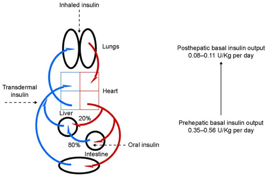
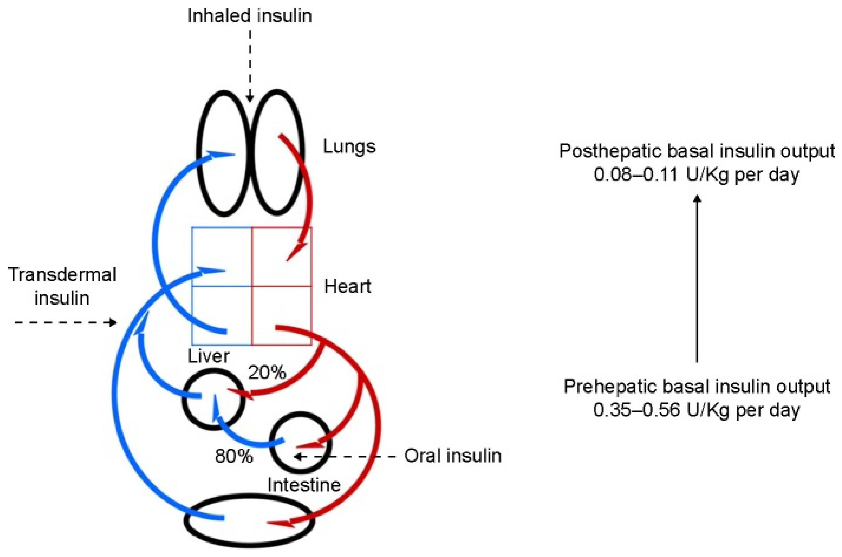
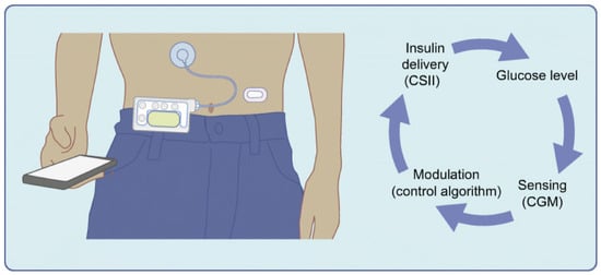
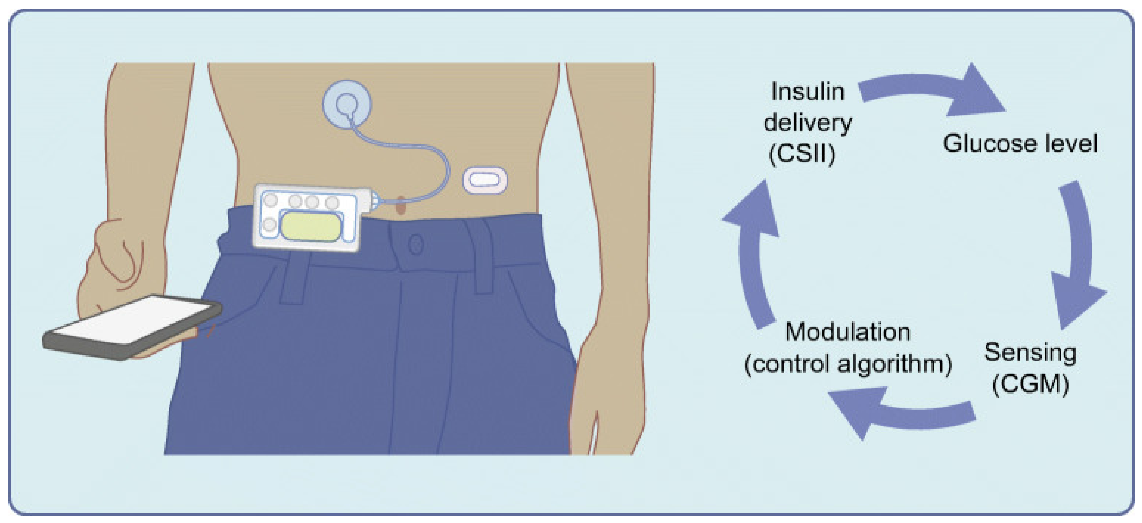
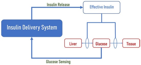
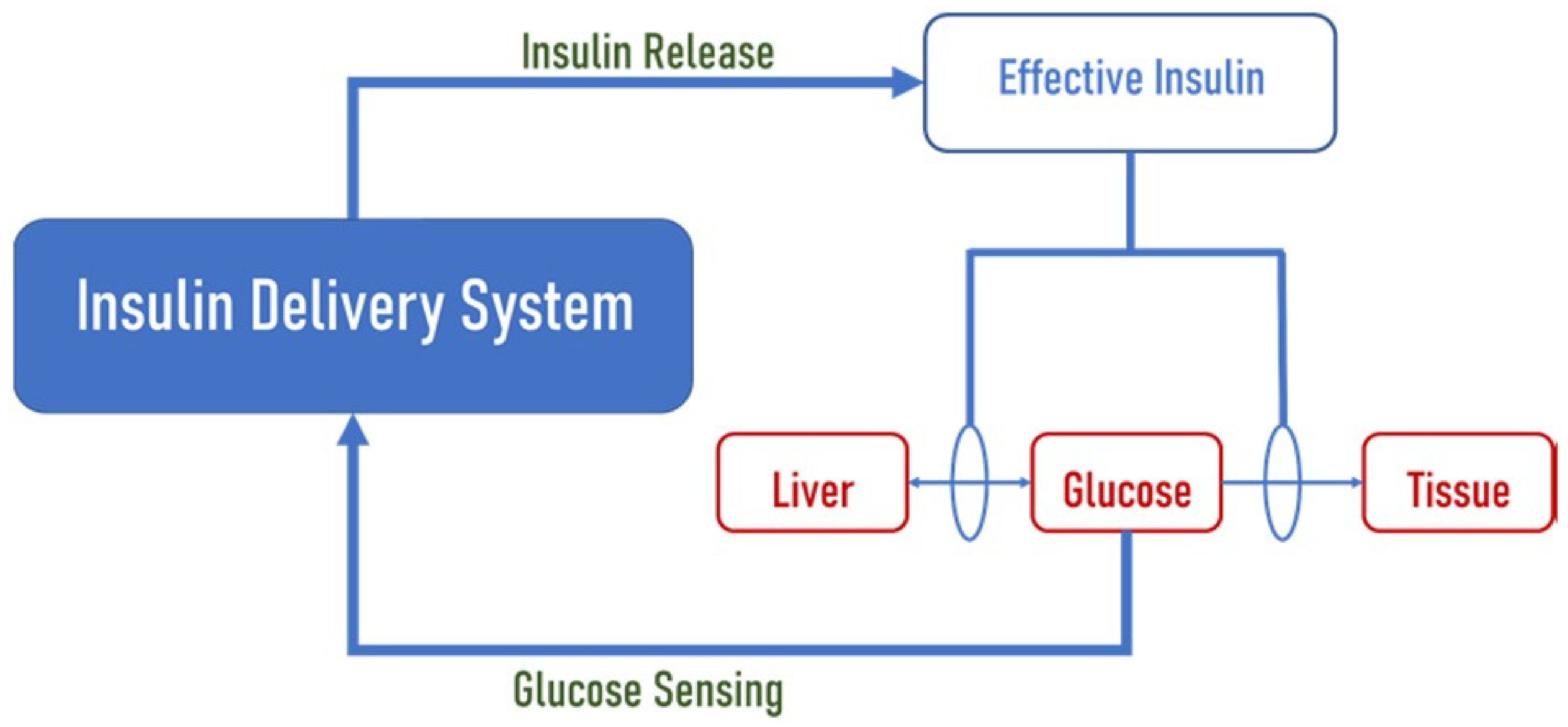
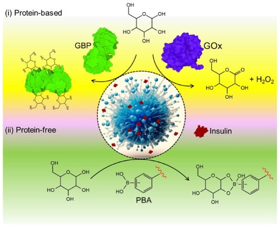
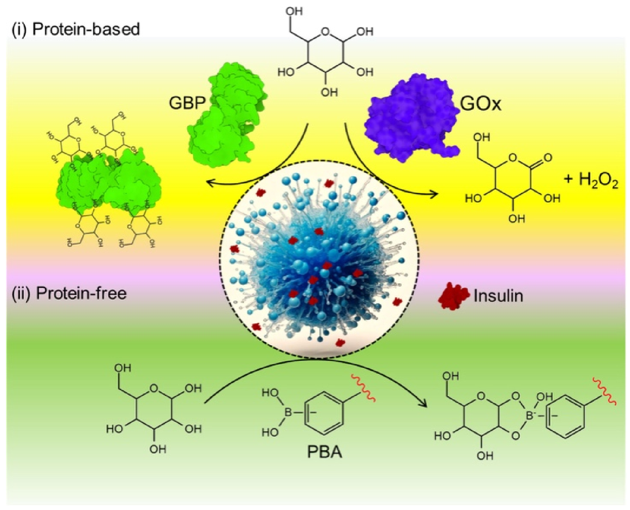
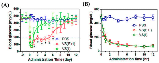
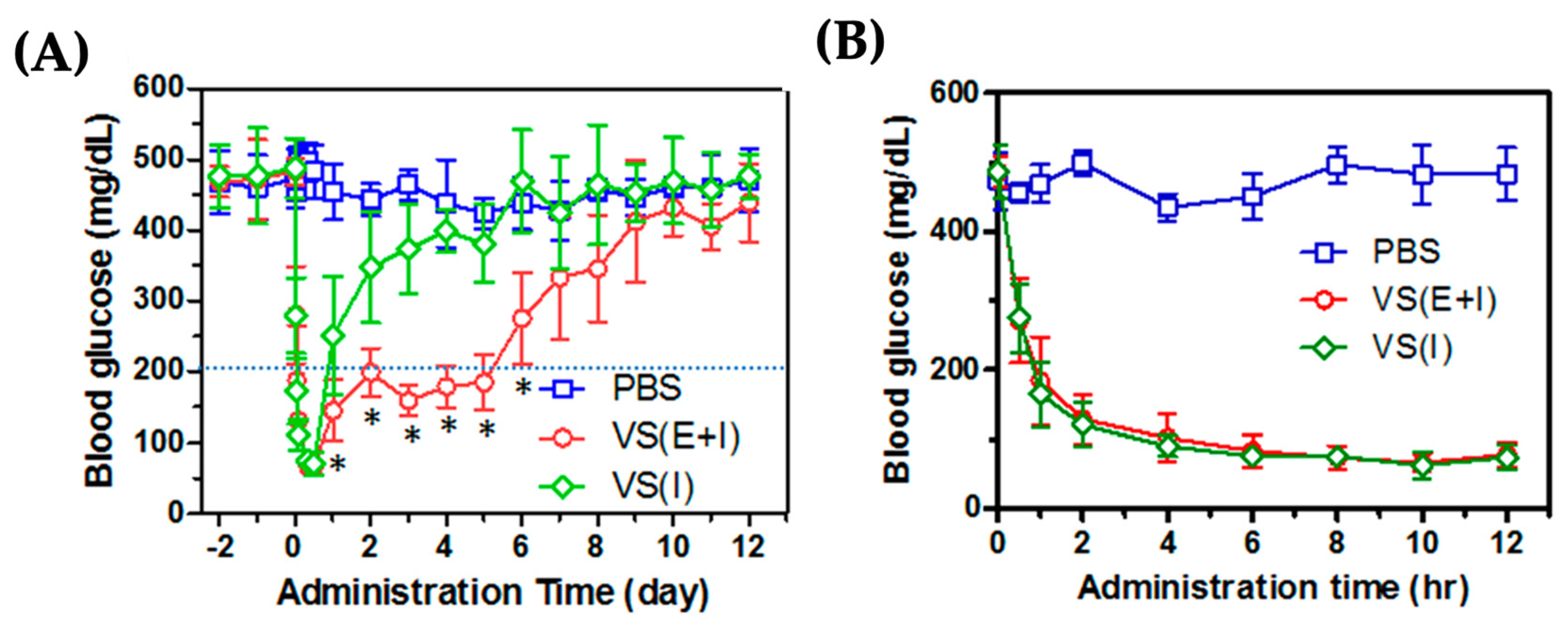
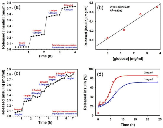
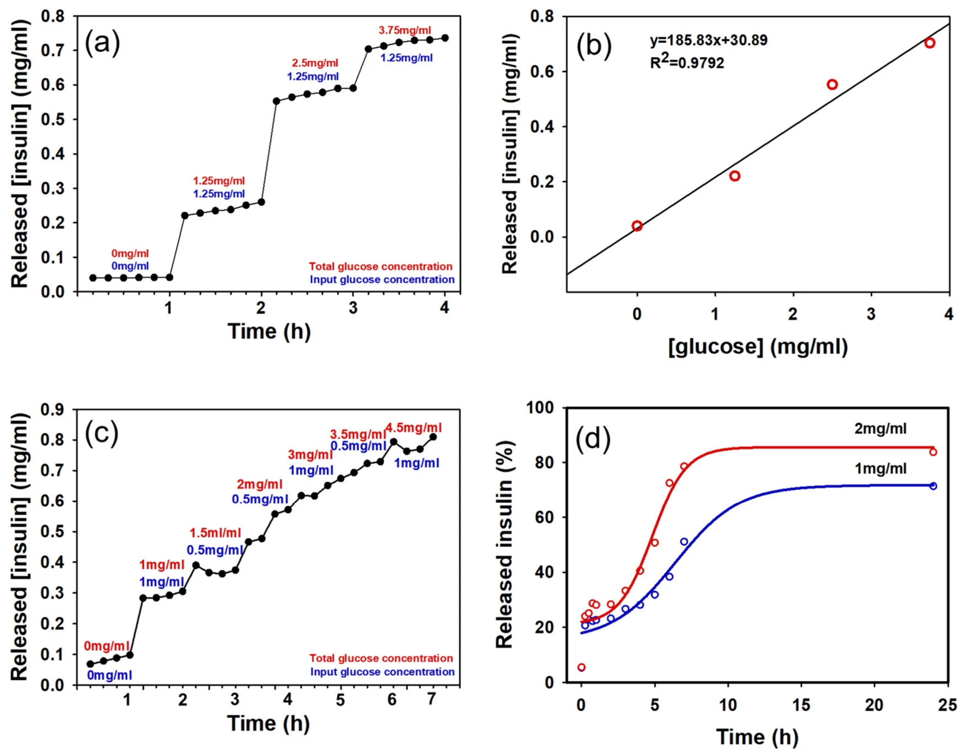
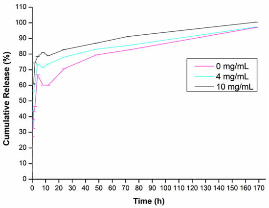
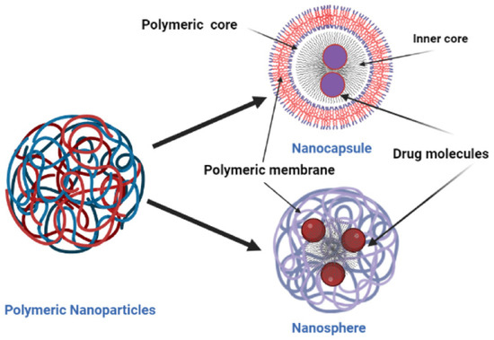
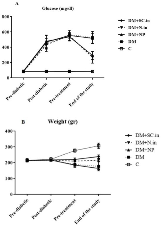
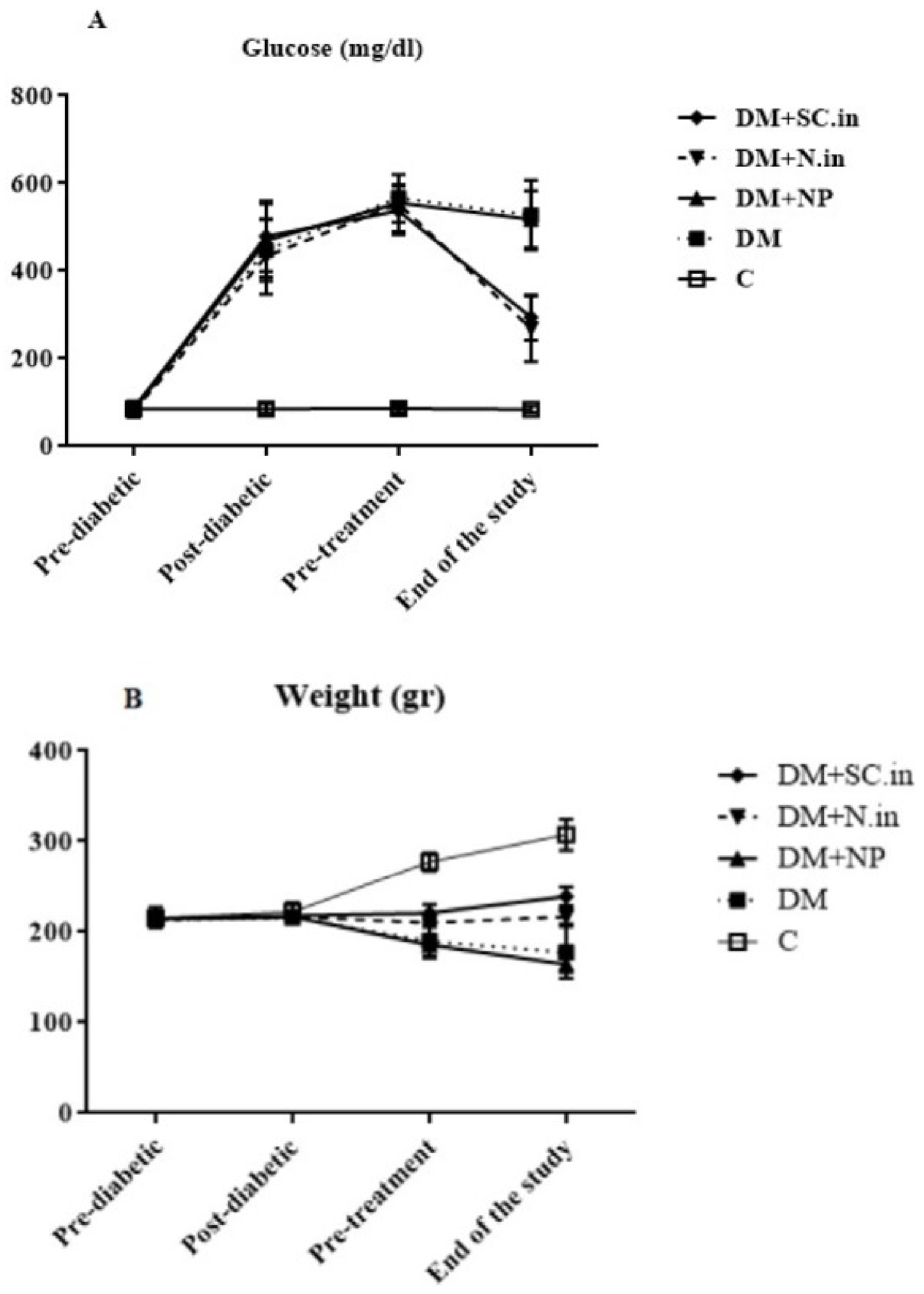
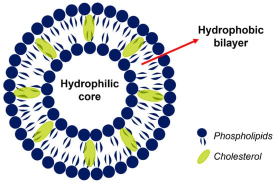
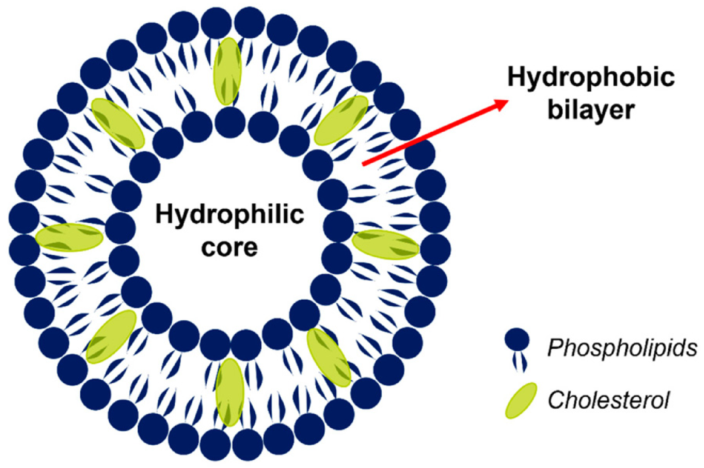
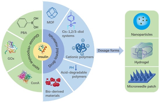
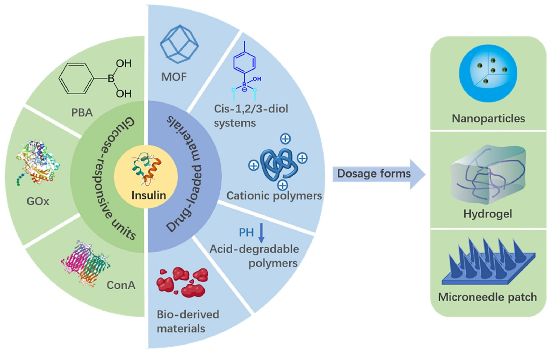
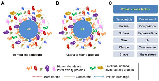
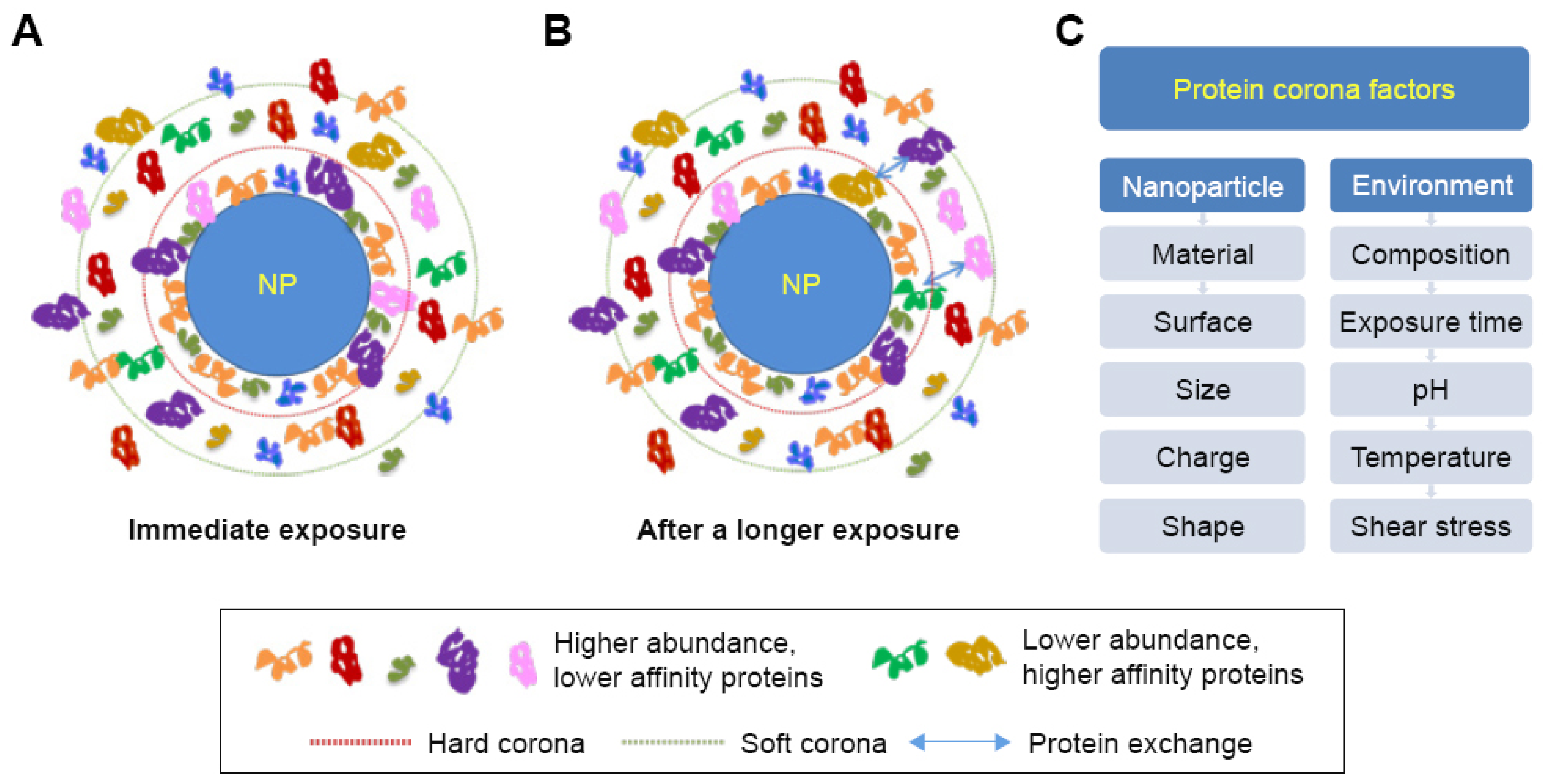
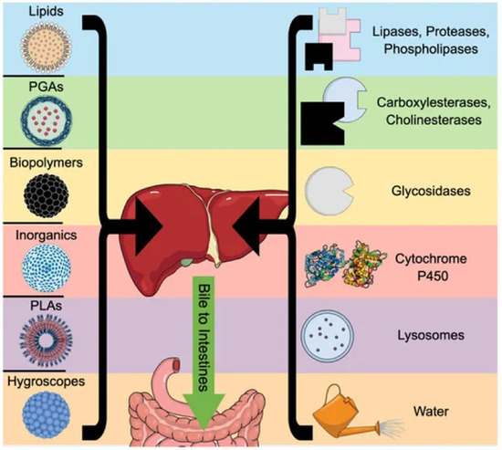
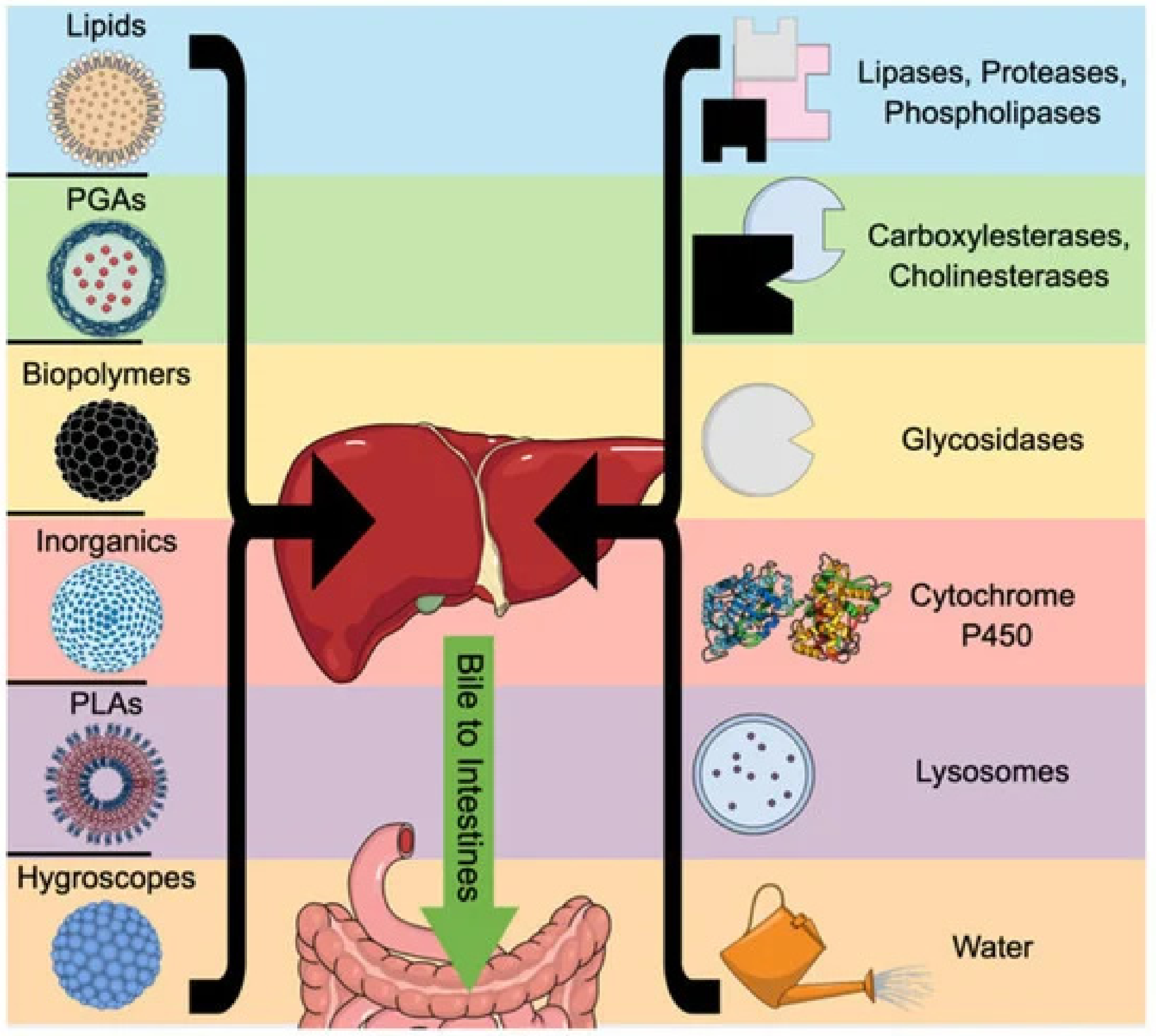

Disclaimer/Publisher’s Note: The statements, opinions and data contained in all publications are solely those of the individual author(s) and contributor(s) and not of MDPI and/or the editor(s). MDPI and/or the editor(s) disclaim responsibility for any injury to people or property resulting from any ideas, methods, instructions or products referred to in the content. |
© 2026 by the authors. Licensee MDPI, Basel, Switzerland. This article is an open access article distributed under the terms and conditions of the Creative Commons Attribution (CC BY) license.