Functional Adhesives for a Restorative Future
 , , , , and
Abstract
1. Introduction
2. Methodology
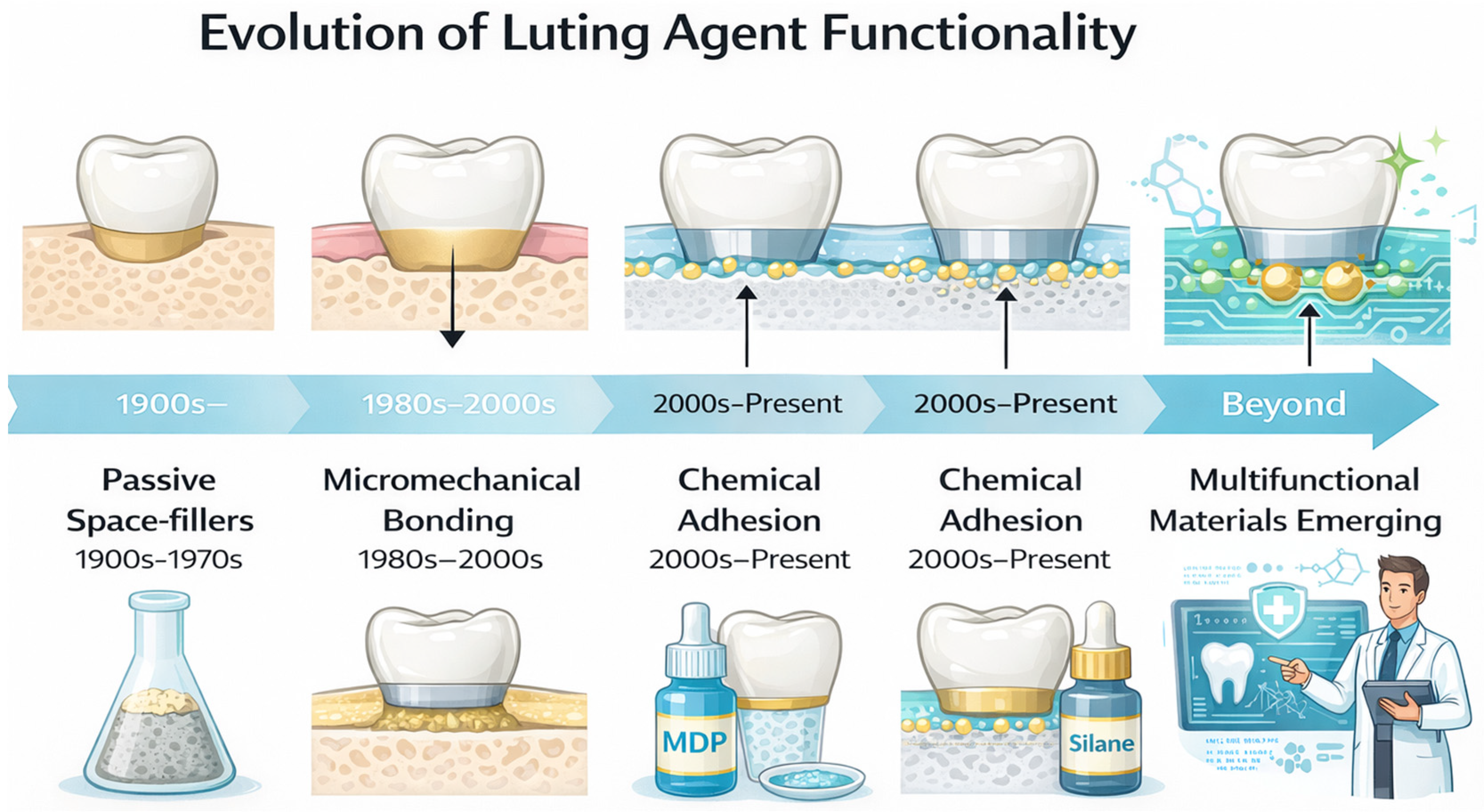
3. The Historical Evolution of Cementation: From Mechanical Luting to Adhesive Dentistry
3.1. The Old Luting Era: A Mechanical Philosophy
3.2. The Transitory Era: The Rise of Resin Cements
3.3. The Modern Adhesive Era: The Engineering of an Interface
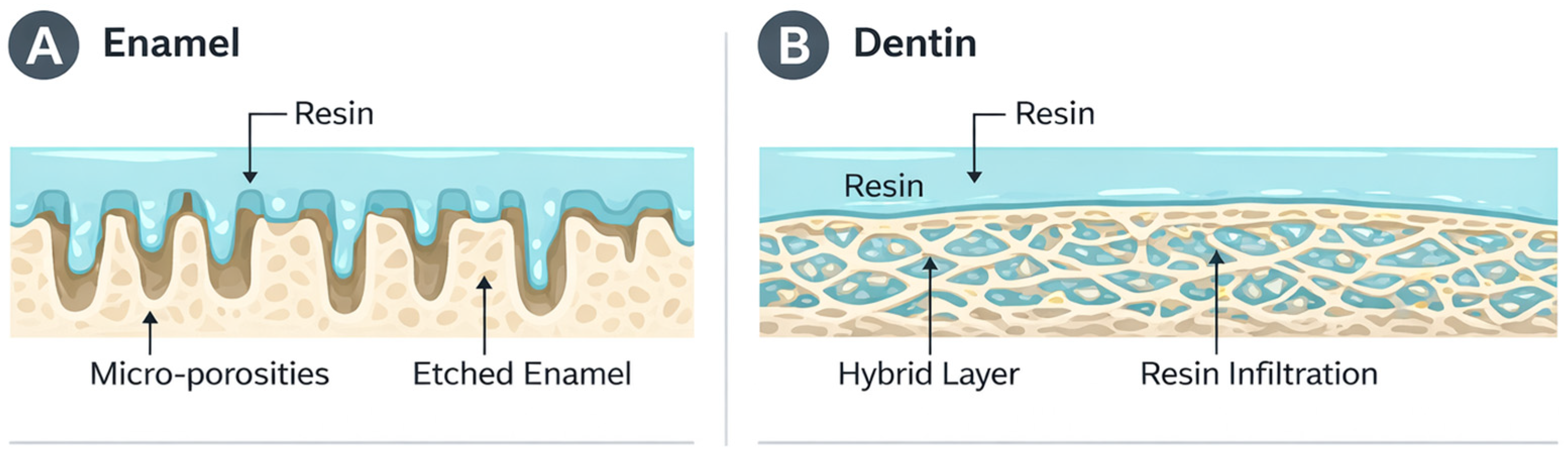
4. The Biological Substrates
4.1. Enamel as a Substrate
4.2. Dentin as a Substrate
5. Physicochemical Properties of Functional Luting Agents
5.1. Polymerization Kinetics and Degree of Conversion
5.2. Physical Properties
5.3. Absorption and Water Solubility
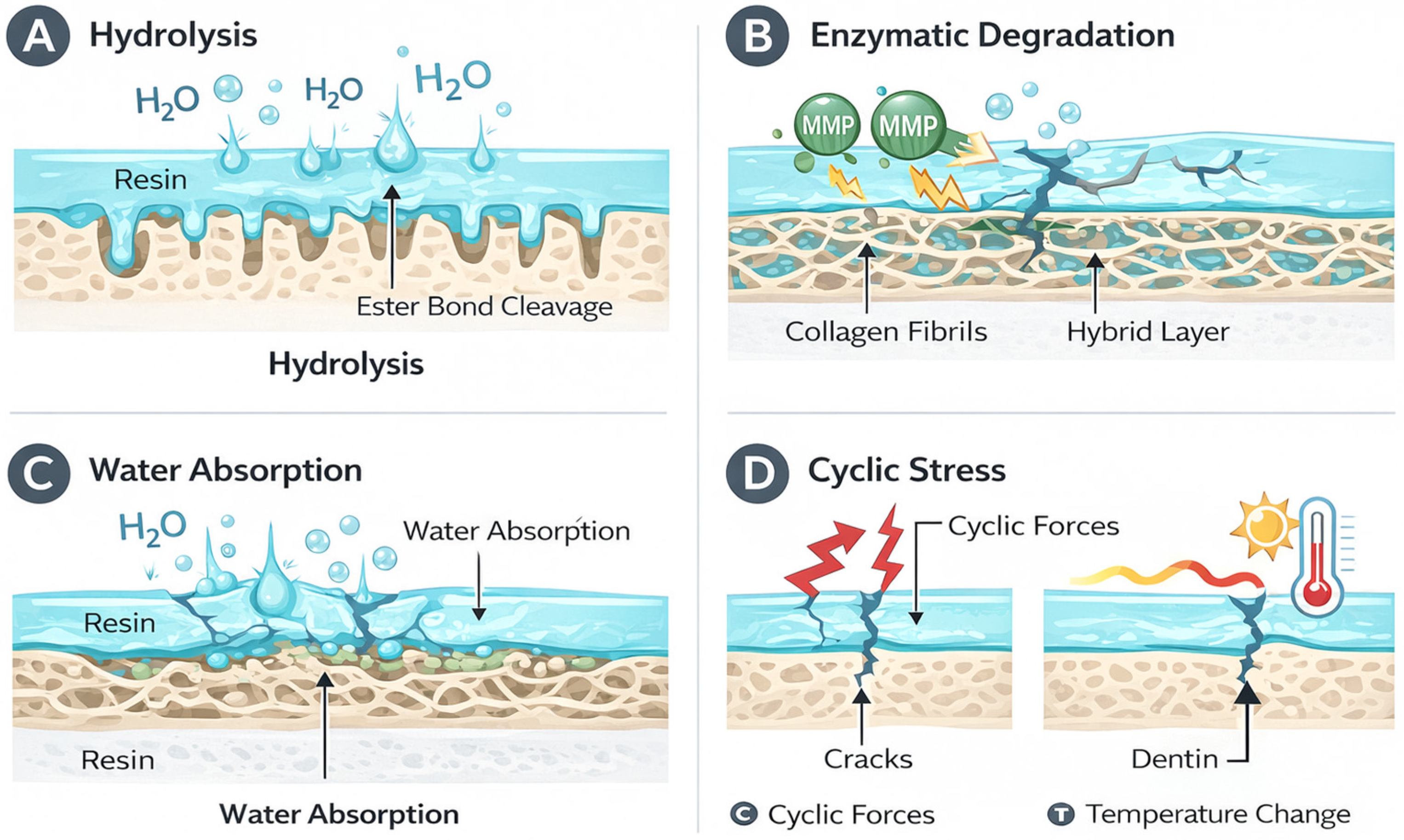
6. Mechanisms of Bond Degradation and Interface Durability
7. Material-Specific Adhesive Strategies
7.1. Glass-Ceramics (Lithium Disilicate & Leucite)
7.2. Zirconia (Polycrystalline)
7.3. Resin-Matrix Ceramics/Hybrid Ceramics (e.g., PICN)
7.4. Indirect Composite Restorations
8. Discussion
9. Limitations of This Review
10. Future Directions
11. Conclusions
Supplementary Materials
Author Contributions
Funding
Institutional Review Board Statement
Informed Consent Statement
Data Availability Statement
Conflicts of Interest
Abbreviations
| IDS | Immediate dentin sealing |
| DC | Degree of conversion |
| HEMA | Hydroxyethyl methacrylate |
| GICs | Glass ionomer cements |
| RMGICs | Resin-modified glass ionomer cements |
| MMPs | Matrix metalloproteinases |
| HF | Hydrofluoric acid |
| Al2O3 | Aluminum oxide |
| PICN | Polymer-infiltrated ceramic network |
| TEGDMA | Triethylene glycol dimethacrylate |
References
- Özcan, M.; Garcia, L.D.F.R.; Volpato, C.A.M. Bioactive Materials for Direct and Indirect Restorations: Concepts and Applications. Front. Dent. Med. 2021, 2, 647267. [Google Scholar] [CrossRef]
- Heboyan, A.; Vardanyan, A.; Karobari, M.I.; Marya, A.; Avagyan, T.; Tebyaniyan, H.; Mustafa, M.; Rokaya, D.; Avetisyan, A. Dental Luting Cements: An Updated Comprehensive Review. Molecules 2023, 28, 1619. [Google Scholar] [CrossRef] [PubMed]
- Manso, A.P.; Carvalho, R.M. Dental Cements for Luting and Bonding Restorations. Dent. Clin. N. Am. 2017, 61, 821–834. [Google Scholar] [CrossRef] [PubMed]
- Perdigão, J. Current Perspectives on Dental Adhesion: (1) Dentin Adhesion–Not There Yet. Jpn. Dent. Sci. Rev. 2020, 56, 190–207. [Google Scholar] [CrossRef] [PubMed]
- Nagaoka, N.; Yoshihara, K.; Feitosa, V.P.; Tamada, Y.; Irie, M.; Yoshida, Y.; Van Meerbeek, B.; Hayakawa, S. Chemical Interaction Mechanism of 10-MDP with Zirconia. Sci. Rep. 2017, 7, 45563. [Google Scholar] [CrossRef] [PubMed]
- Gunwal, M.; Gohil, C.; Bambawale, A.; Parakh, S.; Parakh, A.B.; Madanala, N. Clinical Response Evaluation of Two Different Luting Cements Utilized for Cementing Metal Class II Inlays: An In Vivo Study. World J. Dent. 2023, 14, 510–514. [Google Scholar] [CrossRef]
- Abad-Coronel, C.; Naranjo, B.; Valdiviezo, P. Adhesive Systems Used in Indirect Restorations Cementation: Review of the Literature. Dent. J. 2019, 7, 71. [Google Scholar] [CrossRef] [PubMed]
- Hardy, C.M.F.; Bebelman, S.; Leloup, G.; Hadis, M.A.; Palin, W.M.; Leprince, J.G. Investigating the Limits of Resin-Based Luting Composite Photopolymerization through Various Thicknesses of Indirect Restorative Materials. Dent. Mater. 2018, 34, 1278–1288. [Google Scholar] [CrossRef] [PubMed]
- Sofan, E.; Sofan, A.; Palaia, G.; Tenore, G.; Romeo, U.; Migliau, G. Sofan Classification Review of Dental Adhesive Systems: From the IV Generation to the Universal Type. Ann. Stomatol. 2017, 8, 1–17. [Google Scholar] [PubMed]
- Van Meerbeek, B. From Buonocore’s Pioneering Acid-Etch Technique to Self-Adhering Restoratives. A Status Perspective of Rapidly Advancing Dental Adhesive Technology. J. Adhes. Dent. 2020, 22, 7–34. [Google Scholar] [CrossRef] [PubMed]
- Carrilho, E.; Cardoso, M.; Marques Ferreira, M.; Marto, C.M.; Paula, A.; Coelho, A.S. 10-MDP Based Dental Adhesives: Adhesive Interface Characterization and Adhesive Stability—A Systematic Review. Materials 2019, 12, 790. [Google Scholar] [CrossRef] [PubMed]
- Tavangar, M.S.; Jafarpur, D.; Bagheri, R. Evaluation of Compressive Strength and Sorption/Solubility of Four Luting Cements. J. Dent. Biomater. 2017, 4, 387–393. [Google Scholar] [PubMed]
- Saleem, S.S. Comparative Evaluation of Antimicrobial Activities of Different Types of Luting Cements. Erbil Dent. J. 2024, 7, 208–216. [Google Scholar] [CrossRef]
- Turkistani, A.; Islam, S.; Shimada, Y.; Tagami, J.; Sadr, A. Dental Cements: Bioactivity, Bond Strength and Demineralization Progression around Restorations. Am. J. Dent. 2018, 31, 24B–31B. [Google Scholar] [PubMed]
- Amina; Rajput, G.; Ahmed, S.; Chaturvedi, S.; Addas, M.K.; Bhagat, T.V.; Gurumurthy, V.; Alqahtani, S.M.; Alobaid, M.A.; Alsubaiy, E.F.; et al. Comparison of Microleakage in Nanocomposite and Amalgam as a Crown Foundation Material Luted with Different Luting Cements under CAD-CAM Milled Metal Crowns: An In Vitro Microscopic Study. Polymers 2022, 14, 2609. [Google Scholar] [CrossRef] [PubMed]
- Saran, R.; Upadhya, N.P.; Ginjupalli, K.; Amalan, A.; Rao, B.; Kumar, S. Effect on Physical and Mechanical Properties of Conventional Glass Ionomer Luting Cements by Incorporation of All-Ceramic Additives: An In Vitro Study. Int. J. Dent. 2020, 2020, 8896225. [Google Scholar] [CrossRef] [PubMed]
- Kheur, M.; Kantharia, N.; Iakha, T.; Kheur, S.; Husain, N.A.-H.; Özcan, M. Evaluation of Mechanical and Adhesion Properties of Glass Ionomer Cement Incorporating Nano-Sized Hydroxyapatite Particles. Odontology 2020, 108, 66–73. [Google Scholar] [CrossRef] [PubMed]
- Srinivasan, S.R.; Mathew, M.G.; Jayaraman, J. Comparison of Three Luting Cements for Prefabricated Zirconia Crowns in Primary Molar Teeth: A 36-Month Randomized Clinical Trial. Pediatr. Dent. 2023, 45, 117–124. [Google Scholar] [PubMed]
- Varghese, E.; Samson, R.S.; Albaker, S.A.; Thomas, A.A.; Alqarni, A.S.; Dhanya, K.B. Evaluation of Microleakage of Stainless Steel Crowns and Pedo Jacket Crowns after Cementation with Different Luting Cements. J. Pharm. Bioallied Sci. 2023, 15, S451–S454. [Google Scholar] [CrossRef] [PubMed]
- Maletin, A.; Knežević, M.J.; Koprivica, D.Đ.; Veljović, T.; Puškar, T.; Milekić, B.; Ristić, I. Dental Resin-Based Luting Materials—Review. Polymers 2023, 15, 4156. [Google Scholar] [CrossRef] [PubMed]
- Schneider, L.F.J.; Ribeiro, R.B.; Liberato, W.F.; Salgado, V.E.; Moraes, R.R.; Cavalcante, L.M. Curing Potential and Color Stability of Different Resin-Based Luting Materials. Dent. Mater. 2020, 36, e309–e315. [Google Scholar] [CrossRef] [PubMed]
- Dimitriadi, M.; Petropoulou, A.; Zinelis, S.; Eliades, G. Degree of Conversion of Dual-Cured Composite Luting Agents: The Effect of Transition Metal-Based Touch-Cure Activators. J. Dent. 2024, 147, 105147. [Google Scholar] [CrossRef] [PubMed]
- Yurdagüven, G.Y.; Çiftçioğlu, E.; Kazokoğlu, F.Ş.; Kayahan, M.B. 5-Year Clinical Performance of Ceramic Onlay and Overlay Restorations Luted with Light-Cured Composite Resin. J. Dent. 2024, 149, 105258. [Google Scholar] [CrossRef] [PubMed]
- Cuevas-Suárez, C.E.; De Oliveira Da Rosa, W.L.; Vitti, R.P.; Da Silva, A.F.; Piva, E. Bonding Strength of Universal Adhesives to Indirect Substrates: A Meta-Analysis of in Vitro Studies. J. Prosthodont. 2020, 29, 298–308. [Google Scholar] [CrossRef] [PubMed]
- Watanabe, S.; Takamizawa, T.; Hayashi, K.; Aoki, R.; Barkmeier, W.; Latta, M.; Watanabe, H.; Miyazaki, M. Comparing Various Resin Luting Cement Systems in Different Etching Modes Through Bond Durability and Morphological Features. Oper. Dent. 2024, 49, 231–244. [Google Scholar] [CrossRef] [PubMed]
- Maravic, T.; Mazzitelli, C.; Mayer-Santos, E.; Mancuso, E.; Gracis, S.; Breschi, L.; Fuzzi, M. Current Trends for Cementation in Prosthodontics: Part 1—The Substrate. Polymers 2025, 17, 566. [Google Scholar] [CrossRef] [PubMed]
- Krummel, A.; Garling, A.; Sasse, M.; Kern, M. Influence of Bonding Surface and Bonding Methods on the Fracture Resistance and Survival Rate of Full-Coverage Occlusal Veneers Made from Lithium Disilicate Ceramic after Cyclic Loading. Dent. Mater. 2019, 35, 1351–1359. [Google Scholar] [CrossRef] [PubMed]
- Wiegand, A.; Lechte, C.; Kanzow, P. Adhesion to Eroded Enamel and Dentin: Systematic Review and Meta-Analysis. Dent. Mater. 2021, 37, 1845–1853. [Google Scholar] [CrossRef] [PubMed]
- Alghazzawi, T.F. Clinical Survival Rate and Laboratory Failure of Dental Veneers: A Narrative Literature Review. J. Funct. Biomater. 2024, 15, 131. [Google Scholar] [CrossRef] [PubMed]
- Popescu, M.; Malița, M.; Vorovenci, A.; Ștețiu, A.A.; Perieanu, V.Ș.; Costea, R.C.; David, M.; Costea, R.M.; Ștețiu, M.A.; Drăguș, A.C.; et al. Wet vs. Dry Dentin Bonding: A Systematic Review and Meta-Analysis of Adhesive Performance and Hybrid Layer Integrity. Oral 2025, 5, 63. [Google Scholar] [CrossRef]
- Costa, M.J.F.; Souza, B.B.T.L.C.D.; Sales-Junior, R.A.D.; Pomacóndor-Hernández, C.; Borges, B.C.D. Does the Application of Anti-Erosive Substances on Eroded Dentin Affect Bond Strength? A Systematic Review. Odovtos-Int. J. Dent. Sci. 2024, 27, 62–74. [Google Scholar] [CrossRef]
- Mokeem, L.S.; Garcia, I.M.; Melo, M.A. Degradation and Failure Phenomena at the Dentin Bonding Interface. Biomedicines 2023, 11, 1256. [Google Scholar] [CrossRef] [PubMed]
- Nikaido, T.; Tagami, J.; Yatani, H.; Ohkubo, C.; Nihei, T.; Koizumi, H.; Maseki, T.; Nishiyama, Y.; Takigawa, T.; Tsubota, Y. Concept and Clinical Application of the Resin-Coating Technique for Indirect Restorations. Dent. Mater. J. 2018, 37, 192–196. [Google Scholar] [CrossRef] [PubMed]
- Agrawal, A.; Nehal, R.; Gala, K.; Sachdev, S.S. Immediate Dentin Sealing: Advancing Bonding Efficacy and Clinical Success. Cureus 2025, 17, e78102. [Google Scholar] [CrossRef] [PubMed]
- Samartzi, T.-K.; Papalexopoulos, D.; Sarafianou, A.; Kourtis, S. Immediate Dentin Sealing: A Literature Review. Clin. Cosmet. Investig. Dent. 2021, 13, 233–256. [Google Scholar] [CrossRef] [PubMed]
- Varadan, P.; Balaji, L.; Manaswini, D.Y.; Rajan, R.M. Reinforced Immediate Dentin Sealing vs. Conventional Immediate Dentin Sealing on Adhesive Behavior of Indirect Restorations: A Systematic Review. J. Contemp. Dent. Pract. 2023, 23, 1066–1075. [Google Scholar] [CrossRef] [PubMed]
- Wang, J.; Song, W.; Zhu, L.; Wei, X. A Comparative Study of the Microtensile Bond Strength and Microstructural Differences between Sclerotic and Normal Dentine after Surface Pretreatment. BMC Oral Health 2019, 19, 216. [Google Scholar] [CrossRef] [PubMed]
- Mazzitelli, C.; Maravic, T.; Josic, U.; Mancuso, E.; Generali, L.; Checchi, V.; Breschi, L.; Mazzoni, A. Effect of Adhesive Strategy on Resin Cement Bonding to Dentin. J. Esthet. Restor. Dent. 2023, 35, 501–507. [Google Scholar] [CrossRef] [PubMed]
- Fallahzadeh, F.; Safarzadeh-Khosroshahi, S.; Atai, M. Dentin Bonding Agent with Improved Bond Strength to Dentin through Incorporation of Sepiolite Nanoparticles. J. Clin. Exp. Dent. 2017, 9, e738–e742. [Google Scholar] [CrossRef] [PubMed][Green Version]
- Coelho, A.; Vilhena, L.; Antunes, M.; Amaro, I.; Paula, A.; Marto, C.M.; Saraiva, J.; Ferreira, M.M.; Carrilho, E.; Ramalho, A. Effect of Different Cavity Disinfectants on Adhesion to Dentin of Permanent Teeth. J. Funct. Biomater. 2022, 13, 209. [Google Scholar] [CrossRef] [PubMed]
- Abdullah Alsadon, O. Adhesion Concepts and Techniques for Laboratory-Processed Indirect Dental Restorations. Saudi Dent. J. 2022, 34, 661–668. [Google Scholar] [CrossRef] [PubMed]
- Barbon, F.J.; Isolan, C.P.; Soares, L.D.; Bona, A.D.; De Oliveira Da Rosa, W.L.; Boscato, N. A Systematic Review and Meta-Analysis on Using Preheated Resin Composites as Luting Agents for Indirect Restorations. Clin. Oral Investig. 2022, 26, 3383–3393. [Google Scholar] [CrossRef] [PubMed]
- Ilie, N. Microstructural Dependence of Mechanical Properties and Their Relationship in Modern Resin-Based Composite Materials. J. Dent. 2021, 114, 103829. [Google Scholar] [CrossRef] [PubMed]
- Skapska, A.; Komorek, Z.; Cierech, M.; Mierzwinska-Nastalska, E. Comparison of Mechanical Properties of a Self-Adhesive Composite Cement and a Heated Composite Material. Polymers 2022, 14, 2686. [Google Scholar] [CrossRef] [PubMed]
- Assaf, J.; Hardan, L.; Kassis, C.; Bourgi, R.; Devoto, W.; Amm, E.; Moussa, C.; Sawicki, J.; Lukomska-Szymanska, M. Influence of Resin Cement Thickness and Elastic Modulus on the Stress Distribution of Zirconium Dioxide Inlay-Bridge: 3D Finite Element Analysis. Polymers 2021, 13, 3863. [Google Scholar] [CrossRef] [PubMed]
- Hajjaj, M.S.; Alhowirini, L.F.; Alghamdi, R.S.; Merdad, Y.M.; Filemban, H.K.; Bawazir, M.; Alothman, K.A.; Turkestani, N.A.; Alzahrani, S.J. Effects of Preheating on the Mechanical Properties of Dental Composites. Crystals 2025, 15, 632. [Google Scholar] [CrossRef]
- Kelch, M.; Stawarczyk, B.; Mayinger, F. Time-Dependent Degree of Conversion, Martens Parameters, and Flexural Strength of Different Dual-Polymerizing Resin Composite Luting Materials. Clin. Oral Investig. 2022, 26, 1067–1076. [Google Scholar] [CrossRef] [PubMed]
- Michailidou, S.; Dionysopoulos, D.; Papadopoulos, C.; Naka, O.; Andriotis, E.; Fatouros, D.; Tolidis, K. Effect of a Diode Laser (445 nm) on Polymerization Efficiency of a Preheated Resin Composite Used for Luting of Indirect Composite Restorations. Oper. Dent. 2023, 48, 513–523. [Google Scholar] [CrossRef] [PubMed]
- Jalalian, B.; Golkar, P.; Paktinat, A.; Ahmadi, E.; Panahande, S.A.; Ranjbar Omrani, L. Degree of Conversion of Resin-Modified Glass Ionomer Cement Containing Hydroxyapatite Nanoparticles. Front. Dent. 2020, 16, 415–420. [Google Scholar] [CrossRef] [PubMed]
- Ilie, N. Comparative Analysis of Static and Viscoelastic Mechanical Behavior of Different Luting Material Categories after Aging. Materials 2021, 14, 1452. [Google Scholar] [CrossRef] [PubMed]
- Kim, D.Y.; Aryan, N.; Lawson, N.C.; Cheon, K. Comparison of Luting Cement Solubility: A Narrative Review. Dent. J. 2024, 12, 365. [Google Scholar] [CrossRef] [PubMed]
- Thomas, M.; Mustafa, M.; Karkera, R.; Raj, A.N.; Isaac, L.; Reddy, R.N. Comparison of the Solubility of Conventional Luting Cements with That of the Polyacid Modified Composite Luting Cement and Resin-Modified Glass Ionomer Cement. J. Contemp. Dent. Pract. 2016, 17, 1016–1021. [Google Scholar] [CrossRef]
- Singh, H.; Rashmi, S.; Pai, S.; Kini, S. Comparative Evaluation of Fluoride Release from Two Different Glass Ionomer Cement and a Novel Alkasite Restorative Material-An in Vitro Study. Pesqui. Bras. Odontopediatria Clín. Integr. 2020, 20, e5209. [Google Scholar] [CrossRef]
- Basso, G.R.; Della Bona, Á.; Gobbi, D.L.; Cecchetti, D. Fluoride Release from Restorative Materials. Braz. Dent. J. 2011, 22, 355–358. [Google Scholar] [CrossRef] [PubMed]
- Pellizzari, V.; Michels, A.; Luiz, S.; De Souza, E.; Tabchoury, C.; Rached, R. Fluoride Ion Release of Self-Adhesive Resin Cements and Their Potential to Inhibit In Situ Enamel and Dentin Demineralization. Oper. Dent. 2017, 42, 548–558. [Google Scholar] [CrossRef] [PubMed]
- Muto, R.; Takamizawa, T.; Shiratsuchi, K.; Kasahara, Y.; Suda, S.; Watanabe, H.; Latta, M.A.; Miyazaki, M. Influence of Luting Strategies on Dentin Bond Performance of Self-Adhesive Resin Luting Cement in Combination with a Universal Adhesive. Clin. Oral Investig. 2024, 28, 478. [Google Scholar] [CrossRef] [PubMed]
- Goulart, M.; Borges Veleda, B.; Damin, D.; Bovi Ambrosano, G.M.; Coelho de Souza, F.H.; Erhardt, M.C.G. Preheated Composite Resin Used as a Luting Agent for Indirect Restorations: Effects on Bond Strength and Resin-Dentin Interfaces. Int. J. Esthet. Dent. 2018, 13, 86–97. [Google Scholar] [PubMed]
- Topdağı, B. Optimization of Bond Strength Between Heat-Polymerized PMMA and Contemporary CAD/CAM Framework Materials: A Comparative In Vitro Study. Polymers 2025, 17, 1488. [Google Scholar] [CrossRef] [PubMed]
- Ashhar, M.; Bansal, R.; Bansal, M.; Singh, D.; Soni, K.; Samra, R.K.; Gupta, S. Shear Bond Strength of Different Fifth-Generation Dentin Bonding Agents: An In Vitro Comparative Analysis. Cureus 2025, 17, e94490. [Google Scholar] [CrossRef] [PubMed]
- Abedi, F.; Siahvoshi, S.; Babaei, M.; Nourmohammadi, S. Comparative Analysis of Shear Bond Strength of Four Adhesive Systems on Primary Dentin. Clin. Exp. Dent. Res. 2025, 11, e70205. [Google Scholar] [CrossRef] [PubMed]
- Comino-Garayoa, R.; Peláez, J.; Tobar, C.; Rodríguez, V.; Suárez, M.J. Adhesion to Zirconia: A Systematic Review of Surface Pretreatments and Resin Cements. Materials 2021, 14, 2751. [Google Scholar] [CrossRef] [PubMed]
- Şenol, M.; Gürbüz, A.; Oyar, P. Bond Strength of Conventional Resin-Based Adhesive Cement and Self-Adhesive Resin Cement to CAD-CAM Restorative Materials. BMC Oral Health 2025, 25, 296. [Google Scholar] [CrossRef] [PubMed]
- David-Pérez, M.; Ramírez-Suárez, J.P.; Latorre-Correa, F.; Agudelo-Suárez, A.A. Degree of Conversion of Resin-Cements (Light-Cured/Dual-Cured) under Different Thicknesses of Vitreous Ceramics: Systematic Review. J. Prosthodont. Res. 2022, 66, 385–394. [Google Scholar] [CrossRef] [PubMed]
- Da Silva, F.A.S.; Paschoini, V.L.; Cortez, T.V.; Corona, S.A.M.; Souza-Gabriel, A.E. Physicochemical and Mechanical Properties of Preheated Composite Resins for Luting Ceramic Laminates. Odontology 2024, 112, 773–781. [Google Scholar] [CrossRef] [PubMed]
- Petropoulou, A.; Dimitriadi, M.; Zinelis, S.; Papathanasiou, I.; Eliades, G. Conversion and Tack-Curing of Light-Cured Veneer Luting Agents. J. Funct. Biomater. 2025, 16, 307. [Google Scholar] [CrossRef] [PubMed]
- Roesner, A.J.; Schmohl, L.; Hahnel, S.; Fuchs, F.; König, A.; Rauch, A. Acid Resistance of Self-Adhesive Resin Luting Cements–Changes in Surface Texture Parameters and Microhardness. Dent. Mater. 2022, 38, 1376–1384. [Google Scholar] [CrossRef] [PubMed]
- Desai, K.; Karthickraj, S.M. Comparative Evaluation of Bond Strengths Between Dual Cure Resin Cement and Light Cure Resin Cement in Root Surface Indirect Restorations: An In Vitro Analysis Study. Cureus 2024, 16, e55009. [Google Scholar] [CrossRef] [PubMed]
- Meharry, M.R.; Schwartz, J.; Montalvo, A.; Mueller, D.; Mitchell, J.C. Comparison of 2 Self-Adhesive Resin Cements with or without a Self-Etching Primer. Gen. Dent. 2020, 68, 22–28. [Google Scholar] [PubMed]
- Barbon, F.J.; Moraes, R.R.; Isolan, C.P.; Spazzin, A.O.; Boscato, N. Influence of Inorganic Filler Content of Resin Luting Agents and Use of Adhesive on the Performance of Bonded Ceramic. J. Prosthet. Dent. 2019, 122, 566.e1–566.e11. [Google Scholar] [CrossRef] [PubMed]
- Garner, J.R.; Wajdowicz, M.N.; DuVall, N.B.; Roberts, H.W. Selected Physical Properties of New Resin-Modified Glass Ionomer Luting Cements. J. Prosthet. Dent. 2017, 117, 277–282. [Google Scholar] [CrossRef] [PubMed]
- Sriamporn, T.; Thamrongananskul, N.; Klaisiri, A. The Effectiveness of Various Functional Monomers in Self-Adhesive Resin Cements on Prosthetic Materials. J. Int. Soc. Prev. Community Dent. 2022, 12, 332–335. [Google Scholar] [CrossRef] [PubMed]
- Bharali, K.; Das, M.; Jalan, S.; Paul, R.; Deka, A. To Compare and Evaluate the Sorption and Solubility of Four Luting Cements after Immersion in Artificial Saliva of Different pH Values. J. Pharm. Bioallied Sci. 2017, 9, 103. [Google Scholar] [CrossRef] [PubMed]
- da Silva, J.C.; Rogério Vieira, R.; Rege, I.C.C.; Cruz, C.A.d.S.; Vaz, L.G.; Estrela, C.; Castro, F.L.A. de Pre-Heating Mitigates Composite Degradation. J. Appl. Oral Sci. 2015, 23, 571–579. [Google Scholar] [CrossRef] [PubMed]
- Penteado, M.M.; Tribst, J.P.M.; Jurema, A.L.B.; Saavedra, G.S.F.A.; Borges, A.L.S. Influence of Resin Cement Rigidity on the Stress Distribution of Resin-Bonded Fixed Partial Dentures. Comput. Methods Biomech. Biomed. Eng. 2019, 22, 953–960. [Google Scholar] [CrossRef] [PubMed]
- Hajaj, T.; Lile, I.E.; Negru, R.M.; Niculescu, S.T.; Stuparu, S.; Rominu, M.; Sinescu, C.; Albu, P.; Titihazan, F.; Veja, I. Adhesive Performance of Zirconia and Lithium Disilicate Maryland Cantilever Restorations on Prepared and Non-Prepared Abutment Teeth: An In Vitro Comparative Study. Biomimetics 2025, 10, 413. [Google Scholar] [CrossRef] [PubMed]
- Chiapinotto, G.F.; Da Rosa, L.S.; Scotti, N.; Kleverlaan, C.J.; Valandro, L.F.; Pereira, G.K.R. Does Adhesive Luting Promote Improved Fatigue Performance of Lithium Disilicate Simplified Crowns? J. Mech. Behav. Biomed. Mater. 2022, 134, 105373. [Google Scholar] [CrossRef] [PubMed]
- Fraga, S.; Jager, N.; Campos, F.; Valandro, L.F.; Kleverlaan, C.J. Does Luting Strategy Affect the Fatigue Behavior of Bonded Y-TZP Ceramic? J. Adhes. Dent. 2018, 20, 307–315. [Google Scholar] [CrossRef] [PubMed]
- Sundfeld, D.; Palialol, A.R.M.; Fugolin, A.P.P.; Ambrosano, G.M.B.; Correr-Sobrinho, L.; Martins, L.R.M.; Pfeifer, C.S. The Effect of Hydrofluoric Acid and Resin Cement Formulation on the Bond Strength to Lithium Disilicate Ceramic. Braz. Oral Res. 2018, 32, e43. [Google Scholar] [CrossRef] [PubMed]
- Alkhurays, M. Influence of Different Luting Cements on the Shear Bond Strength of Pretreated Lithium Disilicate Materials. J. Contemp. Dent. Pract. 2019, 20, 1056–1060. [Google Scholar] [CrossRef]
- Mourouzis, P.; Koulaouzidou, E.; Palaghias, G.; Helvatjoglu-Antoniades, M. Color Match of Luting Composites and Try-in Pastes: The Impact on the Final Color of CAD/CAM Lithium Disilicate Restorations. Int. J. Esthet. Dent. 2018, 13, 98–109. [Google Scholar] [PubMed]
- Meister, J.; Kaschuba, N.; Romer, M.; Bourauel, C. Influence of Cementation on the Aesthetical Appearance of Full-Ceramic Restorations. Materials 2023, 16, 1236. [Google Scholar] [CrossRef] [PubMed]
- Caminha, L.I.; Dors, M.; Isolan, C.P.; Barbon, F.J.; Boscato, N. Effect of Preheated Composite Resin Luting Agents on the Color of Ceramic Laminates. Gen. Dent. 2022, 70, 67–73. [Google Scholar] [PubMed]
- Lima, L.C.; Aldarvis, J.; Amaral, F.L.; Vieira-Junior, W.F.; Turssi, C.P.; Basting, R.T.; Lima, A.; Barros, L.S.; França, F.M. Microhardness, Diametral Tensile Strength and Color Stability of Heated Resin Composites Used for Luting Ceramic Veneers. Am. J. Dent. 2024, 37, 191–196. [Google Scholar] [PubMed]
- Da Rosa, L.S.; Dapieve, K.S.; Dalla-Nora, F.; Rippe, M.P.; Valandro, L.F.; Sarkis-Onofre, R.; Pereira, G.K.R. Does Adhesive Luting Reinforce the Mechanical Properties of Dental Ceramics Used as Restorative Materials? A Systematic Review and Meta-Analysis. J. Adhes. Dent. 2022, 24, 209–222. [Google Scholar] [CrossRef] [PubMed]
- Spazzin, A.; Guarda, G.; Oliveira-Ogliari, A.; Leal, F.; Correr-Sobrinho, L.; Moraes, R. Strengthening of Porcelain Provided by Resin Cements and Flowable Composites. Oper. Dent. 2016, 41, 179–188. [Google Scholar] [CrossRef] [PubMed]
- Karaokutan, I.; Aykent, F.; Özdoğan, M. Comparison of the Color Change of Porcelain Laminate Veneers Produced by Different Materials After Luting with Three Resin Cements. Oper. Dent. 2023, 48, 166–175. [Google Scholar] [CrossRef] [PubMed]
- Awad, M.M.; Alhalabi, F.; Alotaibi, N.; Alzamil, F.; Binalrimal, S.; Alrahlah, A.; Ahmed, M.H. A Systematic Review and Meta-Analysis of Bond Strength Studies Associated with Self-Etching Primer and HF Acid Etching of Dental Glass-Ceramics. Int. J. Adhes. Adhes. 2022, 118, 103216. [Google Scholar] [CrossRef]
- Prochnow, C.; Castillo, D.C.; Pilecco, R.O.; Dal Piva, A.M.D.O.; Tribst, J.P.M.; Valandro, L.F.; Moraes, R.R.D.; Pereira, G.K.R. Influence of Different Evaluation Pastes on the Bond of Resin Cement to Lithium Disilicate Ceramic: An in Vitro Study. J. Prosthet. Dent. 2026, 135, 580.e1–580.e7. [Google Scholar] [CrossRef] [PubMed]
- Mazzitelli, C.; Paolone, G.; Sabbagh, J.; Scotti, N.; Vichi, A. Color Stability of Resin Cements after Water Aging. Polymers 2023, 15, 655. [Google Scholar] [CrossRef] [PubMed]
- Hawthan, M.; Larsson, C.; Chrcanovic, B.R. Survival of Fixed Prosthetic Restorations on Vital and Nonvital Teeth: A Systematic Review. J. Prosthodont. 2024, 33, 110–122. [Google Scholar] [CrossRef] [PubMed]
- Mihali, S.G.; Hiller, A. State-of-the-Art Zirconia and Glass–Ceramic Materials in Restorative Dentistry: Properties, Clinical Applications, Challenges, and Future Perspectives. Appl. Sci. 2025, 15, 12841. [Google Scholar] [CrossRef]
- D’Alessandro, C.; Josic, U.; Mazzitelli, C.; Maravic, T.; Graham, L.; Barausse, C.; Mazzoni, A.; Breschi, L.; Blatz, M.B. Is Zirconia Surface Etching a Viable Alternative to Airborne Particle Abrasion? A Systematic Review and Meta-Analysis of in Vitro Studies. J. Dent. 2024, 151, 105394. [Google Scholar] [CrossRef] [PubMed]
- Vivek, V.J.; Venugopal, P.; Divakar, N.; Bharath, S.; Sarin, K.; Mohammed, N. Comparison of Zirconia to Dentin Bonding Using Resin-Based Luting Cements and Resin-Modified Glass-Ionomer Cement: In Vitro. J. Pharm. Bioallied Sci. 2022, 14, S460–S463. [Google Scholar] [CrossRef] [PubMed]
- Sulaiman, T.A.; Suliman, A.A.; Abdulmajeed, A.A.; Zhang, Y. Zirconia Restoration Types, Properties, Tooth Preparation Design, and Bonding. A Narrative Review. J. Esthet. Restor. Dent. 2024, 36, 78–84. [Google Scholar] [CrossRef] [PubMed]
- Tayal, A.; Niyogi, A.; Adhikari, H.; Adhya, P.; Ghosh, A. Comparative Evaluation of Effect of One Coat 7 Universal and Tetric N-Bond Universal Adhesives on Shear Bond Strength at Resin–Zirconia Interface: An in Vitro Study. J. Conserv. Dent. 2021, 24, 336. [Google Scholar] [CrossRef] [PubMed]
- Liu, X.; Jiang, X.; Xu, T.; Zhao, Q.; Zhu, S. Investigating the Shear Bond Strength of Five Resin-Based Luting Agents to Zirconia Ceramics. J. Oral Sci. 2020, 62, 84–88. [Google Scholar] [CrossRef] [PubMed]
- Ansari, S.; Jahedmanesh, N.; Cascione, D.; Zafarnia, P.; Shah, K.C.; Wu, B.M.; Moshaverinia, A. Effects of an Etching Solution on the Adhesive Properties and Surface Microhardness of Zirconia Dental Ceramics. J. Prosthet. Dent. 2018, 120, 447–453. [Google Scholar] [CrossRef] [PubMed]
- Conner, C.; Andretti, F.; Hernandez, A.I.; Rojas-Rueda, S.; Azpiazu-Flores, F.X.; Morrow, B.R.; Garcia-Godoy, F.; Jurado, C.A.; Alshabib, A. Surface Evaluation of a Novel Acid-Etching Solution for Zirconia and Lithium Disilicate. Materials 2025, 18, 2912. [Google Scholar] [CrossRef] [PubMed]
- Vishnu, G.; Jeevanandan, G. Evaluation of Microleakage Using Different Luting Cements in Kedo Zirconia Crowns: An In Vitro Assessment. Cureus 2024, 16, e66237. [Google Scholar] [CrossRef] [PubMed]
- Sahin, I.; Karayilmaz, H.; Çiftçi, Z.Z.; Kirzioglu, Z. Fracture Resistance of Prefabricated Primary Zirconium Crowns Cemented with Different Luting Cements. Pediatr. Dent. 2018, 40, 443–448. [Google Scholar] [PubMed]
- Kitayama, S.; Nikaido, T.; Ikeda, M.; Alireza, S.; Miura, H.; Tagami, J. Internal Coating of Zirconia Restoration with Silica-Based Ceramic Improves Bonding of Resin Cement to Dental Zirconia Ceramic. Bio-Med. Mater. Eng. 2010, 20, 77–87. [Google Scholar] [CrossRef] [PubMed]
- Abhishek, G.; Vishwanath, S.K.; Nair, A.; Prakash, N.; Chakrabarty, A.; Malalur, A.K. Comparative Evaluation of Bond Strength of Resin Cements with and without 10-Methacryloyloxydecyl Dihydrogen Phosphate (Mdp) to Zirconia and Effect of Thermocycling on Bond Strength-An in Vitro Study. J. Clin. Exp. Dent. 2022, 14, e316–e320. [Google Scholar] [CrossRef] [PubMed]
- Yang, B.; Barloi, A.; Kern, M. Influence of Air-Abrasion on Zirconia Ceramic Bonding Using an Adhesive Composite Resin. Dent. Mater. 2010, 26, 44–50. [Google Scholar] [CrossRef] [PubMed]
- Hajjaj, M.S.; Barboud, H.M.; Almashabi, H.K.; Alzahrani, S.J.; Haimed, T.S.A.; Alnoury, A.S.; Sulaiman, T.A. Evaluation of Different Priming Agents with Conventional and Bioactive Self-Adhesive Resin Cements on Shear Bond Strength to Zirconia. Appl. Sci. 2023, 13, 8369. [Google Scholar] [CrossRef]
- Tyor, S.; Al-Zordk, W.; Sakrana, A.A. Fracture Resistance of Monolithic Translucent Zirconia Crown Bonded with Different Self-Adhesive Resin Cement: Influence of MDP-Containing Zirconia Primer after Aging. BMC Oral Health 2023, 23, 636. [Google Scholar] [CrossRef] [PubMed]
- Krifka, S.; Preis, V.; Rosentritt, M. Effect of Decontamination and Cleaning on the Shear Bond Strength of High Translucency Zirconia. Dent. J. 2017, 5, 32. [Google Scholar] [CrossRef] [PubMed]
- Feitosa, S.; Patel, D.; Borges, A.; Alshehri, E.; Bottino, M.; Özcan, M.; Valandro, L.; Bottino, M. Effect of Cleansing Methods on Saliva-Contaminated Zirconia—An Evaluation of Resin Bond Durability. Oper. Dent. 2015, 40, 163–171. [Google Scholar] [CrossRef] [PubMed]
- Alsultani, R.Z.; Gholam, M.K. Effectiveness of Various Cleaning Protocols in Enhancing Resin–Zirconia Bond Strength After Saliva Contamination. Prosthesis 2025, 7, 158. [Google Scholar] [CrossRef]
- Mine, A.; Kabetani, T.; Kawaguchi-Uemura, A.; Higashi, M.; Tajiri, Y.; Hagino, R.; Imai, D.; Yumitate, M.; Ban, S.; Matsumoto, M.; et al. Effectiveness of Current Adhesive Systems When Bonding to CAD/CAM Indirect Resin Materials: A Review of 32 Publications. Jpn. Dent. Sci. Rev. 2019, 55, 41–50. [Google Scholar] [CrossRef] [PubMed]
- Dikici, B.; Türkeş Başaran, E.; Can, E. Does the Type of Resin Luting Material Affect the Bonding of CAD/CAM Materials to Dentin? Dent. J. 2025, 13, 41. [Google Scholar] [CrossRef] [PubMed]
- Lamberti Miotti, L.; Cargnelutti Follak, A.; De Souza Gonçalves, L.; Aldrighi Münchow, E.; Henrique Susin, A. Bond Strength to Dentin of a Polymer-Infiltrate Ceramic-Network Material Cemented with Dual Resin Cements Submitted to Different Adhesive Strategies. Int. J. Adhes. Adhes. 2024, 128, 103551. [Google Scholar] [CrossRef]
- Awad, M.M.; Alhalabi, F.; Alshehri, A.; Salem, M.A.; Robaian, A.; Alghannam, S.; Alayad, A.S.; Almutairi, B.; Alrahlah, A. Silane-Containing Universal Adhesives Influence Resin-Ceramic Microtensile Bond Strength. Coatings 2023, 13, 477. [Google Scholar] [CrossRef]
- Rohr, N.; Flury, A.; Fischer, J. Efficacy of a Universal Adhesive in the Bond Strength of Composite Cements to Polymer-Infiltrated Ceramic. J. Adhes. Dent. 2017, 19, 417–424. [Google Scholar] [CrossRef] [PubMed]
- Calheiros-Lobo, M.J.; Vieira, T.; Carbas, R.; Da Silva, L.F.M.; Pinho, T. Effectiveness of Self-Adhesive Resin Luting Cement in CAD-CAM Blocks—A Systematic Review and Meta-Analysis. Materials 2023, 16, 2996. [Google Scholar] [CrossRef] [PubMed]
- Berkman, M.; Tuncer, S.; Tekçe, N.; Karabay, F.; Demirci, M. Microtensile Bond Strength Between Self-Adhesive Resin Cements and Resin Based Ceramic CAD/CAM Block. Odovtos-Int. J. Dent. Sci. 2020, 23, 215–224. [Google Scholar] [CrossRef]
- Bayas Salinas, A.; Reascos Flores, A.; Cascante-Calderón, M.G.; Villacís Altamirano, I.M. Effect of Thermocycling and Surface Treatments on the Bond Strength of a Hybrid PICN Ceramic. Acta Odontol. Latinoam. 2025, 38, 69–75. [Google Scholar] [CrossRef] [PubMed]
- Matinlinna, J.P.; Lung, C.Y.K.; Tsoi, J.K.H. Silane Adhesion Mechanism in Dental Applications and Surface Treatments: A Review. Dent. Mater. 2018, 34, 13–28. [Google Scholar] [CrossRef] [PubMed]
- Bezerra, A.; Gonçalves, G.; Alves, L.; Stamfor, T.; De Brito, O.; Monteiro, G. Bacterial Adhesion and In Situ Biodegradation of Preheated Resin Composite Used as a Luting Agent for Indirect Restorations. Oper. Dent. 2024, 49, 725–737. [Google Scholar] [CrossRef] [PubMed]
- Alsunbul, H.; Khan, A.A.; Alqahtani, Y.M.; Hassan, S.A.B.; Asiri, W.; Saadaldin, S.; Alharthi, R.; Aldegheishem, A. Using Functionalized Micron-Sized Glass Fibres for the Synergistic Effect of Glass Ionomer on Luting Material. J. Funct. Biomater. 2023, 14, 550. [Google Scholar] [CrossRef] [PubMed]
- Pandey, A. Recent Advances in Bioactive Dental Materials: A Paradigm Shift in Restorative Dentistry. Dental 2024, 6, 15. [Google Scholar] [CrossRef]
- Zailai, A.; Mubarki, O.; Alobaidan, A.N.; Alenazi, S.A.; Humedi, A.A.; Alshahrani, K.M.; Alfattah, K.M.; Baobied, M.S.; Alenizi, T.; Eishan, A.A.; et al. Clinical Efficacy of Bioactive and Smart Restorative Materials in Preventing Secondary Caries: A Systematic Review and Meta-Analysis. Cureus 2026, 18, e102221. [Google Scholar] [CrossRef] [PubMed]
- Zhang, K.; Zhang, N.; Weir, M.D.; Reynolds, M.A.; Bai, Y.; Xu, H.H.K. Bioactive Dental Composites and Bonding Agents Having Remineralizing and Antibacterial Characteristics. Dent. Clin. N. Am. 2017, 61, 669–687. [Google Scholar] [CrossRef] [PubMed]
- Wu, J.; Zhang, Q.; Weir, M.D.; Oates, T.W.; Zhou, C.; Chang, X.; Xu, H.H.K. Novel Self-Healing Dental Luting Cements with Microcapsules for Indirect Restorations. J. Dent. 2017, 66, 76–82. [Google Scholar] [CrossRef] [PubMed]
- Wang, X.; Ding, T. A Review on the Current State of Microcapsule-Based Self-Healing Dental Composites. J. Funct. Biomater. 2024, 15, 165. [Google Scholar] [CrossRef] [PubMed]
- Bacchi, A.; Spazzin, A.O.; De Oliveira, G.R.; Pfeifer, C.; Cesar, P.F. Resin Cements Formulated with Thio-Urethanes Can Strengthen Porcelain and Increase Bond Strength to Ceramics. J. Dent. 2018, 73, 50–56. [Google Scholar] [CrossRef] [PubMed]
- Ishikawa, K.; Yamauti, M.; Tichy, A.; Ikeda, M.; Ueno, T.; Wakabayashi, N.; Thanatvarakorn, O.; Prasansuttiporn, T.; Klein-Junior, C.A.; Takahashi, A.; et al. UV-Mediated Photofunctionalization of Indirect Restorative Materials Enhances Bonding to a Resin-Based Luting Agent. BioMed Res. Int. 2021, 2021, 9987860. [Google Scholar] [CrossRef] [PubMed]
- Vohra, F.; Altwaim, M.; Alshuwaier, A.; Al Deeb, M.; Alfawaz, Y.; Alrabiah, M.; Abduljabbar, T. Influence of Bioactive, Resin and Glass Ionomer Luting Cements on the Fracture Loads of Dentin Bonded Ceramic Crowns: Failure Loads of Ceramic Crowns Bonded with Bioactive Cement. Pak. J. Med. Sci. 2020, 36, 416–421. [Google Scholar] [CrossRef] [PubMed]
- Komatsu, K.; Suzumura, T.; Komatsu, E.; Matsuura, T.; Shibata, R.; Kusunoki, Y.; Choi, J.; Ogawa, T. Reimagining Bonding Interfaces: UV Photofunctionalization, a Novel Physicochemical Approach, Unlocks Titanium and Cement Adhesive Potential. Dent. Mater. 2025, 41, 1478–1488. [Google Scholar] [CrossRef]
- Tribst, J.P.M.; Etoeharnowo, L.; Tadros, M.; Feilzer, A.J.; Werner, A.; Kleverlaan, C.J.; Dal Piva, A.M.D.O. The Influence of Pre-Heating the Restoration and Luting Agent on the Flexural Strength of Indirect Ceramic and Composite Restorations. Biomater. Investig. Dent. 2023, 10, 2279066. [Google Scholar] [CrossRef] [PubMed]
- Falacho, R.I.; Marques, J.A.; Palma, P.J.; Roseiro, L.; Caramelo, F.; Ramos, J.C.; Guerra, F.; Blatz, M.B. Luting Indirect Restorations with Resin Cements versus Composite Resins: Effects of Preheating and Ultrasound Energy on Film Thickness. J. Esthet. Restor. Dent. 2022, 34, 641–649. [Google Scholar] [CrossRef] [PubMed]
- Gerdolle, D.; Browet, S.; Gresnigt, M. The Multi-Luting Concept: A New Approach to Facilitate the Adhesive Luting of Multiple Indirect Restorations. Swiss Dent. J. SSO 2024, 134, 72–83. [Google Scholar] [CrossRef] [PubMed]
- Elkaffas, A.A.; Alshehri, A.; Alqahtani, A.A.; Almudahi, A.F.; Alanazi, K.K.; Alhalabi, F.A.; Abuelqomsan, M.A.; Alqahtani, A.R. Fracture Resistance of Milled and 3D Printed Ultra-Thin Occlusal Veneers Made of CAD/CAM Resin-Based Ceramics Cemented by Variable Luting Approaches. BMC Oral Health 2025, 25, 1204. [Google Scholar] [CrossRef] [PubMed]
- Alshabib, A.; Berger, J.; Garcia, E.; Jurado, C.A.; Cabral, G.; Baldotto, A.; Riquieri, H.; Alrabiah, M.; Floriani, F. A Comprehensive Digital Workflow for Enhancing Dental Restorations in Severe Structural Wear. Bioengineering 2026, 13, 77. [Google Scholar] [CrossRef] [PubMed]
- Alghauli, M.A.; Alqutaibi, A.Y.; Wille, S.; Kern, M. Clinical Reliability of Self-Adhesive Luting Resins Compared to Other Adhesive Procedures: A Systematic Review and Meta-Analysis. J. Dent. 2023, 129, 104394. [Google Scholar] [CrossRef] [PubMed]
- Maroulakos, G.; Thompson, G.A.; Kontogiorgos, E.D. Effect of Cement Type on the Clinical Performance and Complications of Zirconia and Lithium Disilicate Tooth-Supported Crowns: A Systematic Review. Report of the Committee on Research in Fixed Prosthodontics of the American Academy of Fixed Prosthodontics. J. Prosthet. Dent. 2019, 121, 754–765. [Google Scholar] [CrossRef] [PubMed]
- Ghodsi, S.; Arzani, S.; Shekarian, M.; Aghamohseni, M. Cement Selection Criteria for Full Coverage Restorations: A Comprehensive Review of Literature. J. Clin. Exp. Dent. 2021, 13, e1154–e1161. [Google Scholar] [CrossRef] [PubMed]
- Taschner, M.; Stirnweiss, A.; Frankenberger, R.; Kramer, N.; Galler, K.M.; Maier, E. Fourteen Years Clinical Evaluation of Leucite-Reinforced Ceramic Inlays Luted Using Two Different Adhesion Strategies. J. Dent. 2022, 123, 104210. [Google Scholar] [CrossRef] [PubMed]
- Ashraf, H.; El Tannir, A.; El Zohairy, A.; Kamal, D. Clinical Performance of Indirect Hybrid Ceramic Onlay Restorations Cemented with Injectable Resin Composite versus Dual-Cure Resin Cement: An 18-Month Randomized Clinical Trial. BMC Oral Health 2025, 25, 1419. [Google Scholar] [CrossRef] [PubMed]
- Ding, J.; Jin, Y.; Feng, S.; Chen, H.; Hou, Y.; Zhu, S. Effect of Temporary Cements and Their Removal Methods on the Bond Strength of Indirect Restoration: A Systematic Review and Meta-Analysis. Clin. Oral Investig. 2022, 27, 15–30. [Google Scholar] [CrossRef] [PubMed]
- Eltoukhy, R.I.; Elkaffas, A.A.; Ali, A.I.; Mahmoud, S.H. Indirect Resin Composite Inlays Cemented with a Self-Adhesive, Self-Etch or a Conventional Resin Cement Luting Agent: A 5 Years Prospective Clinical Evaluation. J. Dent. 2021, 112, 103740. [Google Scholar] [CrossRef] [PubMed]
- Hardan, L.; Devoto, W.; Bourgi, R.; Cuevas-Suárez, C.E.; Lukomska-Szymanska, M.; Fernández-Barrera, M.Á.; Cornejo-Ríos, E.; Monteiro, P.; Zarow, M.; Jakubowicz, N.; et al. Immediate Dentin Sealing for Adhesive Cementation of Indirect Restorations: A Systematic Review and Meta-Analysis. Gels 2022, 8, 175. [Google Scholar] [CrossRef] [PubMed]
- Staněk, J.; Riad, A.; Le, A.; Bernát, M.; Hammal, M.; Azar, B. Survival of Prosthodontic Restorations Luted with Resin-Based versus Composite-Based Cements: Retrospective Cohort Study. Materials 2022, 15, 312. [Google Scholar] [CrossRef] [PubMed]
- Brunton, P.A.; Ratnayake, J.; Loch, C.; Veerasamy, A.; Cathro, P.; Lee, R. Indirect Restorations and Fixed Prosthodontics: Materials and Techniques Used by General Dentists of New Zealand. Int. J. Dent. 2019, 2019, 5210162. [Google Scholar] [CrossRef] [PubMed]
- Mohamed, S.G.A.; Hussein, H.G.A.; Mohamed, G.A.; Ibrahim, S.R.M. Different Zirconia Surface Treatments: Strategies to Enhance Adhesion in Zirconia-Based Dental Restorations. J. Pharm. Bioallied Sci. 2025, 17, 45–49. [Google Scholar] [CrossRef] [PubMed]
- Alsaeed, A.Y. Bonding CAD/CAM Materials with Current Adhesive Systems: An Overview. Saudi Dent. J. 2022, 34, 259–269. [Google Scholar] [CrossRef] [PubMed]
- Şahan, M.H.; Peşkersoy, C.; Kümbüloğlu, O.; Türkün, M. Effect of Different Adhesive Systems and Silane Application on Shear Bond Strength of Resin Cement to Indirect Restorations. J. Dent. Mater. Tech. 2023, 12, 104–110. [Google Scholar] [CrossRef]
- Siqueira, F.; Cardenas, A.M.; Gutierrez, M.F.; Malaquias, P.; Hass, V.; Reis, A.; Loguercio, A.D.; Perdigao, J. Laboratory Performance of Universal Adhesive Systems for Luting CAD/CAM Restorative Materials. J. Adhes. Dent. 2016, 18, 331–340. [Google Scholar] [CrossRef] [PubMed]
- Conejo, J. Current Adhesive Protocols for Indirect Ceramic Restorations. Compend. Contin. Educ. Dent. 2022, 43, 592–594. [Google Scholar] [PubMed]
- Habibzadeh, S.; Khamisi, F.; Mosaddad, S.A.; Fernandes, G.V.D.O.; Heboyan, A. Full-Ceramic Resin-Bonded Fixed Dental Prostheses: A Systematic Review. J. Appl. Biomater. Funct. Mater. 2024, 22, 22808000241250118. [Google Scholar] [CrossRef] [PubMed]
- Kolanko, J.; Bonsor, S. Does Immediate Dentine Sealing Improve Bonding Effectiveness of Glass Ceramic Restorations Compared to Delayed Dentine Sealing? Eur. J. Prosthodont. Restor. Dent. 2022, 30, 65–75. [Google Scholar] [CrossRef] [PubMed]
- Bragança, G.F.D.; Mazão, J.D.; Versluis, A.; Soares, C.J. Effect of Luting Materials, Presence of Tooth Preparation, and Functional Loading on Stress Distribution on Ceramic Laminate Veneers: A Finite Element Analysis. J. Prosthet. Dent. 2021, 125, 778–787. [Google Scholar] [CrossRef] [PubMed]
- Politano, G.; Van Meerbeek, B.; Peumans, M. Nonretentive Bonded Ceramic Partial Crowns: Concept and Simplified Protocol for Long-Lasting Dental Restorations. J. Adhes. Dent. 2018, 20, 495–510. [Google Scholar] [CrossRef] [PubMed]
- Solon-de-Mello, M.; Da Silva Fidalgo, T.K.; Dos Santos Letieri, A.; Masterson, D.; Granjeiro, J.M.; Monte Alto, R.V.; Maia, L.C. Longevity of Indirect Restorations Cemented with Self-adhesive Resin Luting with and without Selective Enamel Etching. A Systematic Review and Meta-analysis. J. Esthet. Restor. Dent. 2019, 31, 327–337. [Google Scholar] [CrossRef] [PubMed]
- Alvarenga, M.; Machado, L.; Prado, A.; Veloso, S.; Monteiro, G. Self-Adhesive Resin Cement versus Conventional Cements on the Failure Rate of Indirect Single-Tooth Restorations: A Systematic Review and Meta-Analysis of Randomized Clinical Trials. J. Prosthet. Dent. 2024, 132, 880.e1–880.e8. [Google Scholar] [CrossRef] [PubMed]
- Magne, P.; Razaghy, M.; Carvalho, M.A.; Soares, L.M. Luting of Inlays, Onlays, and Overlays with Preheated Restorative Composite Resin Does Not Prevent Seating Accuracy. Int. J. Esthet. Dent. 2018, 13, 318–332. [Google Scholar] [PubMed]
- Hardy, C.M.F.; Landreau, V.; Valassis, M.; Mercelis, B.; De Munck, J.; Van Meerbeek, B.; Leprince, J. Mini-iFT Confirms Superior Adhesive Luting Performance Using Light-Curing Restorative Composites. J. Adhes. Dent. 2021, 23, 539–548. [Google Scholar] [CrossRef] [PubMed]
- Labunet, A.; Kui, A.; Vigu, A.; Voina-Tonea, A.; Burde, A.; Sava, S. Preheated Composite for Prosthetic Cementation to Enamel and Dentin: A Scoping Review. Dent. J. 2026, 14, 69. [Google Scholar] [CrossRef] [PubMed]
- Chiodera, G.; Monterubbianesi, R.; Tosco, V.; Papini, O.; Orsini, G.; Putignano, A. Application of Bulk-Fill Composite to Simplify the Cementation of Indirect Restorations: The COMBO Technique. Dent. J. 2024, 12, 239. [Google Scholar] [CrossRef] [PubMed]
- Tsujimoto, A.; Barkmeier, W.W.; Takamizawa, T.; Watanabe, H.; Johnson, W.W.; Latta, M.A.; Miyazaki, M. Simulated Localized Wear of Resin Luting Cements for Universal Adhesive Systems with Different Curing Mode. J. Oral Sci. 2018, 60, 29–36. [Google Scholar] [CrossRef] [PubMed]
- Hoffmann, L.; Kessler, A.; Kunzelmann, K.-H. Three-Body Wear of Luting Composites and Influence of the ACTA Wheel Material. Dent. Mater. J. 2021, 40, 1226–1234. [Google Scholar] [CrossRef] [PubMed]
- Oshika, M.; Kishimoto, T.; Horie, T.; Alhotan, A.; Irie, M.; Sule, V.C.; Barkmeier, W.W.; Tsujimoto, A. Wear Resistance of Light-Cure Resin Luting Cements for Ceramic Veneers. J. Funct. Biomater. 2024, 16, 5. [Google Scholar] [CrossRef] [PubMed]
- Tosco, V.; Monterubbianesi, R.; Orilisi, G.; Sabbatini, S.; Conti, C.; Özcan, M.; Putignano, A.; Orsini, G. Comparison of Two Curing Protocols during Adhesive Cementation: Can the Step Luting Technique Supersede the Traditional One? Odontology 2021, 109, 433–439. [Google Scholar] [CrossRef] [PubMed]
- Stegall, D.; Tantbirojn, D.; Perdigao, J.; Versluis, A. Does Tack Curing Luting Cements Affect the Final Cure? J. Adhes. Dent. 2017, 19, 239–243. [Google Scholar] [CrossRef] [PubMed]
- Breschi, L.; Josic, U.; Maravic, T.; Mancuso, E.; Del Bianco, F.; Baldissara, P.; Mazzoni, A.; Mazzitelli, C. Selective Adhesive Luting: A Novel Technique for Improving Adhesion Achieved by Universal Resin Cements. J. Esthet. Restor. Dent. 2023, 35, 1030–1038. [Google Scholar] [CrossRef] [PubMed]
- Ishii, R.; Takamizawa, T.; Katsuki, S.; Iwase, K.; Shoji, M.; Sai, K.; Tsujimoto, A.; Miyazaki, M. Immediate Bond Performance of Resin Composite Luting Systems to Saliva-contaminated Enamel and Dentin in Different Curing Modes. Eur. J. Oral Sci. 2022, 130, e12854. [Google Scholar] [CrossRef] [PubMed]
- Buyukates, I.; Garoushi, S.; Vallittu, P.K.; Uctasli, S.; Lassila, L. Effect of Different Luting Protocols on the Bond Strength of Fiber-Reinforced CAD/CAM Blocks. Polymers 2026, 18, 160. [Google Scholar] [CrossRef] [PubMed]
- Merlo, E.G.; Della Bona, A.; Griggs, J.A.; Jodha, K.S.; Corazza, P.H. Mechanical Behavior and Adhesive Potential of Glass Fiber-Reinforced Resin-Based Composites for Use as Dentin Analogues. Am. J. Dent. 2020, 33, 310–314. [Google Scholar] [PubMed]
- Alshabib, A.; AlDosary, K.; Algamaiah, H. A Comprehensive Review of Resin Luting Agents: Bonding Mechanisms and Polymerisation Reactions. Saudi Dent. J. 2024, 36, 234–239. [Google Scholar] [CrossRef] [PubMed]
- Leung, G.K.-H.; Wong, A.W.-Y.; Chu, C.-H.; Yu, O.Y. Update on Dental Luting Materials. Dent. J. 2022, 10, 208. [Google Scholar] [CrossRef] [PubMed]



| Era | Philosophy | Materials | Primary Retention Mechanism | Limitations |
|---|---|---|---|---|
| Old Luting Era (1900s–1970s) [2,3,6] | Mechanical— Cement as a passive space-filler | Zinc phosphate, Zinc Polycarboxylate, Glass ionomer cements | Macro-mechanical (preparation geometry: taper, height, surface area). | No adhesion, high solubility, low strength, marginal degradation, required aggressive tooth preparation |
| Transitory Resin Cement Era (1980s–2000s) [7,8,9] | Micromechanical—First true bonding | Early Bis-GMA resin cements, Dual-cure cements, Early etch-and-rinse adhesives. | Micromechanical via resin tag formation in etched enamel; primitive hybrid layer in dentin. | Technique sensitivity, polymerization challenges under thick ceramics, hydrolytic degradation of hydrophilic adhesives |
| Modern Adhesive Era (2000s–Present) [4,5,10,11] | Biomimetic & Chemical— Durable, integrated interface | Universal adhesives (10-MDP), Self-etch systems, Bioactive cements | Combined micromechanical + stable chemical bonding (e.g., MDP-Ca/MDP-Zr salts) | Complexity of dentin bonding, zirconia adhesion still technique-sensitive, long-term degradation of some interfaces |
| Cement Type | Year Introduced | Clinical Significance | Advantages | Key Limitations |
|---|---|---|---|---|
| Zinc Phosphate | Late 1800s | Mainstay for decades; longest clinical history | Acceptable clinical performance; low cost | No adhesion to tooth structure; high solubility; low tensile strength; requires aggressive preparation designs |
| Polycarboxylate | 1960s | First chemically adhesive cement | Chemical bonding via calcium ion chelation | High viscosity; difficult handling; limited clinical adoption |
| Glass Ionomer Cement (GIC) | 1970s | Milestone in adhesive dentistry | Chemical bonding to tooth; cumulative release (23.67 ppm); favorable CTE (10–14 ppm/°C) close to tooth structure | Moisture sensitivity; low fracture toughness (0.5–1.5 MPa·m1/2); solubility; limited to low-stress areas |
| Resin-Modified GIC (RMGIC) | 1990s | Addressed GIC moisture sensitivity and initial strength | Improved mechanical properties; reduced moisture sensitivity; fluoride release | Inferior adhesive performance vs. resin cements; higher failure rates |
| Property | Conventional GIC | RMGIC | Self-Adhesive Resin Cement | Adhesive Resin Cement |
|---|---|---|---|---|
| Flexural Strength (MPa) [16,20,43,44] | 20–50 | 50–80 | 80–120 | 100–150 |
| Elastic Modulus (GPa) [43,44,45] | 2–6 | 4–8 | 5–10 | 8–15 |
| Degree of Conversion (%) [21,22,46,47,48,49] | N/A (acid–base reaction only) | 40–60 | 55–75 | 65–85 |
| Water Sorption (µg/mm3) [50,51] | 50–150 | 40–170 | 20–40 | 15–35 |
| Solubility (µg/mm3) [51,52] | 5–20 | 2–8 | 0.5–2.0 | 0.1–1.0 |
| Cumulative Release (ppm) [53,54,55] | 23.67 | 20.37 | Not specified | Not specified |
| Bond Strength to Dentin (MPa) [24,25,38,56,57,58,59] | 2–5 | 5–10 | 10–20 | 20–40 |
| Bond Strength to Zirconia (MPa) [27,60,61] | 0–2 | 1–3 | 5–15 | 15–30 |
| Step | Procedure | Principal Support |
|---|---|---|
| Surface Pretreatment [60,86,87] | 1. Etch with 4–9% hydrofluoric acid (HF) for 20–60 s. 2. Rinse thoroughly. 3. Apply silane coupling agent. | HF creates micro-retentive surface with etch depth of 10–50 µm and increases surface area by 10–20. Silane forms siloxane bonds (Si–O–Si) with ceramic and copolymerizes with resin matrix, yielding bond strengths of 25–40 MPa. |
| Tooth Management [26] | Selective enamel etching + immediate dentin sealing (IDS) as needed data | Maximizes enamel bond and reduces post-operative sensitivity. |
| Luting Agent [8,88] | Light-cure cement for thin veneers (<1.5 mm). Dual-cure cement for thicker crowns/onlays. | Light-cure offers better color stability (ΔE < 1.5 after aging). Dual-cure ensures polymerization under thick ceramics, with DC of 60–75%. |
| Clinical Outcome [23,75,89] | Excellent survival rates (95–98% at 5–10 years), reinforced structure. | - |
| Step | Procedure | Principal Support |
|---|---|---|
| Surface Pretreatment [5,102] | 1. Airborne-particle abrasion with 50 µm Al2O3 at 0.2–0.3 MPa. 2. Clean ultrasonically. 3. Apply MDP-containing primer or universal adhesive. | Air abrasion creates micromechanical retention with surface roughness (Ra) of 0.5–1.5 µm; 10-MDP forms stable MDP-ZrO2 salts with bond strengths of 15–30 MPa. |
| Luting Agent [61,103,104] | Adhesive resin cement containing MDP. Avoid self-adhesive cements alone. | MDP-containing cements outperform non-MDP systems by 30–50% in bond strength. |
| Critical Note [54,105,106,107] | Saliva contamination severely inhibits bonding (50–80% reduction). Reabrade or use alkaline cleaners if contaminated. | Contamination compromises the chemical interaction between MDP and zirconia, necessitating immediate re-treatment. |
| Step | Procedure | Principal Support |
|---|---|---|
| Surface Pretreatment [5,102] | 1. Airborne-particle abrasion (sandblasting) with 50 µm Al2O3. 2. Avoid HF etching (ineffective). | Sandblasting improves micromechanical retention, increasing bond strength by 40–60% compared to untreated surfaces. |
| Chemical Coupling [110,111,112] | Adhesive resin cement containing MDP. Avoid self-adhesive cements alone. | Bond strengths of 20–35 MPa are achievable with universal adhesive. |
| Luting Agent [113,114,115,116] | Saliva contamination severely inhibits bonding (50–80% reduction). Alkaline cleaners if contaminated. | Self-adhesive cements show lower bond strengths (10–20 MPa) without pretreatment. |
| Step | Procedure | Principal Support |
|---|---|---|
| Surface Pretreatment [109] | Airborne-particle abrasion with 50 µm Al2O3. | Increases bond strength by 50–100% compared to untreated surfaces. |
| Chemical Coupling [117] | Silane or universal adhesive. | Improves compatibility with resin cement; silane provides chemical coupling to glass filler particles. |
| Luting Agent [35,118] | Preheated composite (superior mechanical properties) or conventional resin cement. | Preheated composites offer high filler content (70–85 wt%) and strength (120–180 MPa). However, monitor for increased biodegradation (bacterial adhesion 2–3 or higher). |
Disclaimer/Publisher’s Note: The statements, opinions and data contained in all publications are solely those of the individual author(s) and contributor(s) and not of MDPI and/or the editor(s). MDPI and/or the editor(s) disclaim responsibility for any injury to people or property resulting from any ideas, methods, instructions or products referred to in the content. |
© 2026 by the authors. Licensee MDPI, Basel, Switzerland. This article is an open access article distributed under the terms and conditions of the Creative Commons Attribution (CC BY) license.
Share and Cite
Katsonis, A.; Tatarciuc, M.S.; Vitalariu, A.M.; Vasluianu, R.I.; Al-Ashkar, J.; Cioloca, C.H.; Katsoni, A.-S.; Perperidis, P.; Gradinaru, I.; Armencia, A.O.; et al. Functional Adhesives for a Restorative Future. J. Funct. Biomater. 2026, 17, 329. https://doi.org/10.3390/jfb17070329
Katsonis A, Tatarciuc MS, Vitalariu AM, Vasluianu RI, Al-Ashkar J, Cioloca CH, Katsoni A-S, Perperidis P, Gradinaru I, Armencia AO, et al. Functional Adhesives for a Restorative Future. Journal of Functional Biomaterials. 2026; 17(7):329. https://doi.org/10.3390/jfb17070329
Chicago/Turabian StyleKatsonis, Andreas, Monica Silvia Tatarciuc, Anca Mihaela Vitalariu, Roxana Ionela Vasluianu, Jamal Al-Ashkar, Catalina Holban Cioloca, Andrea-Simoni Katsoni, Panagiotis Perperidis, Irina Gradinaru, Adina Oana Armencia, and et al. 2026. "Functional Adhesives for a Restorative Future" Journal of Functional Biomaterials 17, no. 7: 329. https://doi.org/10.3390/jfb17070329
APA StyleKatsonis, A., Tatarciuc, M. S., Vitalariu, A. M., Vasluianu, R. I., Al-Ashkar, J., Cioloca, C. H., Katsoni, A.-S., Perperidis, P., Gradinaru, I., Armencia, A. O., & Stamatin, O. (2026). Functional Adhesives for a Restorative Future. Journal of Functional Biomaterials, 17(7), 329. https://doi.org/10.3390/jfb17070329