Review

Modified Polysaccharides: Potential Biomaterials for Bioprinting

1
College of Intelligence Science and Technology, National University of Defense Technology, Changsha 410073, China
2
National Key Laboratory of Equipment State Sensing and Smart Support, National University of Defense Technology, Changsha 410073, China
*
Authors to whom correspondence should be addressed.
These authors contributed equally to this work.
J. Funct. Biomater. 2025, 16(9), 338; https://doi.org/10.3390/jfb16090338
Submission received: 24 June 2025 / Revised: 8 August 2025 / Accepted: 25 August 2025 / Published: 9 September 2025

Abstract

Polysaccharides have emerged as promising biomaterials for 3D bioprinting due to their inherent biocompatibility, biodegradability, and structural diversity. However, their limited mechanical strength, insufficient bioactivity, and suboptimal printability hinder their direct application in fabricating complex tissue constructs. This review systematically summarizes universal modification strategies to address these challenges by tailoring polysaccharides’ physicochemical and biological properties. We first analyse the fundamental requirements of bioprinting materials, emphasising on the critical role of shear-thinning behaviours, post-printing structural fidelity, and cell-instructive functions. Subsequently, we highlight the advantages and limitations of representative polysaccharides, including chitosan, alginate, and hyaluronic acid. Chemical functionalisation, physical reinforcement, and biological hybridisation are proposed as versatile approaches to synergistically enhance printability, mechanical robustness, and bioactivity to tackle the limitations. Furthermore, dynamic crosslinking mechanisms enabling self-healing and stimuli-responsive behaviours are discussed as emerging solutions for constructing biomimetic architectures. Finally, we outline future directions in balancing material processability with cellular viability and scaling up modified polysaccharides for clinical translation. This review aims to provide a design blueprint for engineering polysaccharide-based bioinks toward next-generation regenerative medicine.

1. Introduction

In recent years, three-dimensional (3D) bioprinting [1,2,3,4] has attracted wide research interest in tissue engineering and clinical applications. This technology allows for unmatched architecture control, adaptability, and repeatability that can overcome the limits of conventional biofabrication techniques. We take extrusion-based 3D bioprinting as an example, the basic principle of which is to stack bioinks layer by layer following programmed paths to form 3D bioactive tissue with designed geometries [5], where the bioinks are biomaterials encapsulated with living cells. Bioprinting can quickly fabricate large-scale structures with high resolution, giving it great potential for tissue engineering [6,7,8,9,10]. The mechanical and biological properties of bioinks are key elements in bioprinting that determine structural stability and cell survivability and have triggered great interest in the field to seek the so-called “ideal” bioinks. To ensure the survival of cells, bioinks need to be biocompatible. To this end, natural polymer materials (polysaccharide [11,12,13], protein [14,15]) and synthetic polymer materials (polyethylene glycol [16]) are developed. Natural polymer materials that recapitulate the extracellular matrix (ECM) in structure and function are the easiest to recognise and accept by in vivo organisms [17]. The natural polymer materials consist predominantly of polysaccharide compounds (e.g., glycosaminoglycans, proteoglycans, and glycoproteins) [18], which support cellular growth and promote tissue regeneration. The polysaccharide-based functional materials thus have great application potential in tissue engineering and have been widely studied in the past decades.
Polysaccharides are one of the most common substances in nature, and play an extremely key role in organisms [19], including energy storage and structural support [20]. Benefiting from their availability and handleability, polysaccharides are considered a high-value renewable material [21]. The basic chemical structure of polysaccharides is relatively simple: monosaccharide molecules are sequentially linked by O-glycosidic bonds formed by dehydration and condensation. The source of materials and the degree of polymerisation influence the chemical structure, functional groups, molecular weights, and electrical charges of the resultant polysaccharides, leading to a great diversity of polysaccharides with completely different mechanical and biological properties. Despite the many outstanding properties of natural polysaccharides, such as biocompatibility, biodegradability, and hydrophilicity, most natural polysaccharides inevitably have drawbacks in particular aspects. For example, chitosan has poor solubility [22], alginate shows weak mechanical stability [23], and agarose exhibits an excessive gelling temperature for cells [24], which all hinder them from being directly used in bioprinting. On the other hand, the large numbers of chemically active functional groups [25] such as -COOH, -NH2, and-OH found in natural polysaccharides have enabled them to be physically or chemically modified on demand to achieve significantly improved mechanical and biological properties [26], making them candidate bioinks for bioprinting.
Herein, we systematically review conventional methodologies employed for the modification of diverse polysaccharides to enhance their suitability for bioprinting applications, while concurrently establishing comprehensive evaluation frameworks for assessing their bioprintability (Figure 1). Polysaccharides are categorized according to their biological origins into three primary classes: plant-derived, animal-derived, and microbial-derived polysaccharides. Initially, we elucidate the fundamental structural characteristics and physicochemical properties of these polysaccharides, followed by an in-depth analysis of potential modification strategies to optimise their performance in bioprinting and tissue engineering applications. Subsequently, we delineate the fundamental principles and critical parameters governing 3D bioprinting processes specifically pertaining to polysaccharide-based bioinks, with particular emphasis on quantitative printability assessment methodologies. Furthermore, we critically examine representative applications of modified polysaccharides in tissue engineering through bioprinting technologies. We anticipate that this comprehensive analysis will not only provide valuable insights for future advancements in polysaccharide research and derivative development, but also facilitate the expansion of these versatile biomaterials into applications extending beyond the realm of tissue engineering.

2. Classification and Modification of Polysaccharides

Polysaccharides have emerged as versatile biomaterials with broad applications across multiple disciplines, owing to their structural diversity, functional versatility, and abundant natural sources. Their inherent biomimicry of extracellular matrix (ECM) components renders them particularly attractive for tissue engineering applications. While native polysaccharides exhibit several advantageous characteristics—including excellent biocompatibility, pronounced hydrophilicity, and negligible toxicity—they are frequently limited by intrinsic drawbacks such as inadequate mechanical stability, suboptimal printability, and absence of bioactive functional groups.
To address these limitations, the diverse and tunable structures of polysaccharides enable strategic modifications to tailor their properties for specific applications. In this section, polysaccharides are systematically classified into three categories based on their biological origin: plant-derived, animal-derived, and microbial-derived polysaccharides. For each category, we analyse their fundamental physicochemical properties and inherent limitations, while proposing potential modification strategies (Table 1) to enhance their performance in bioprinting and tissue engineering applications.

2.1. Plant Polysaccharides

2.1.1. Cellulose

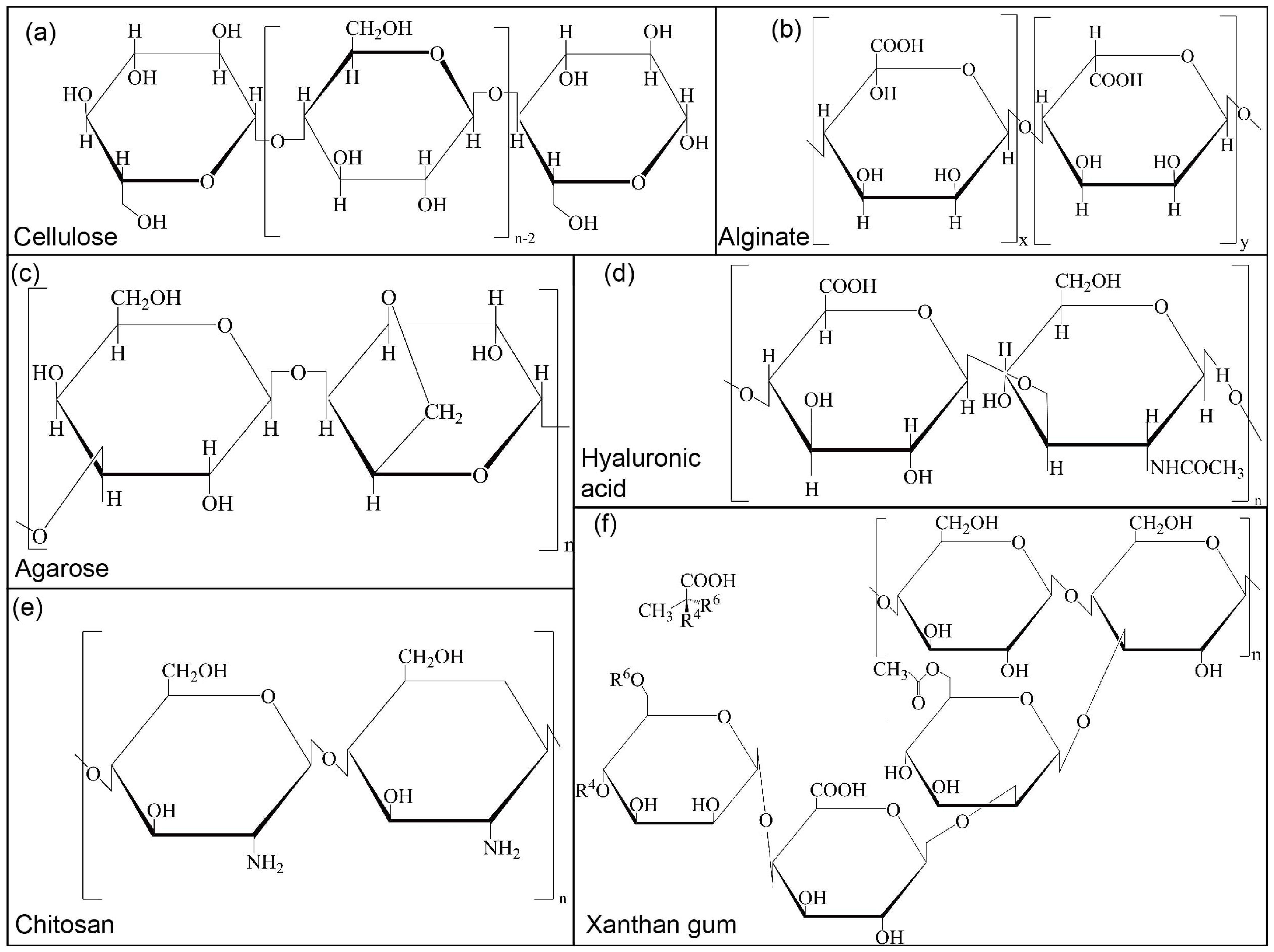
Cellulose [52,53] (Figure 2a) is the world’s most abundant renewable natural polymer and exists in many plant species. Many studies have indicated excellent advantages in terms of low cytotoxicity, low density, high hardness, and biocompatibility [54]. Since its discovery, it has been considered a polysaccharide with broad prospects in biomedicine, particularly as a bioink. It is a linear semicrystalline polysaccharide composed of D-glucose units connected by β (1–4). Hydrogen bonds and van der Waals forces are formed between cellulose chains when polymers are formed. Therefore, they exhibit desirable features such as high strength, rigidity, thermal stability, and low expansion coefficients [55]. However, the inherent limitations of cellulose as a bioink significantly constrain its biomedical applications, including poor aqueous solubility, incompatibility with hydrophobic polymers, inadequate dimensional stability under physiological conditions, and excessive hygroscopicity. Additionally, native cellulose exhibits restricted biodegradability in mammalian systems, being primarily degradable only by specific symbiotic bacteria and microbial cellulases. To overcome these challenges, strategic modifications are essential to (1) enhance its mechanical robustness through physical crosslinking or composite formation, (2) introduce bioactive functional groups to improve cellular interactions, and (3) optimise its physicochemical properties to ensure optimal biocompatibility and support cell viability. These modifications are crucial for transforming cellulose into a clinically relevant biomaterial for bioprinting applications.
Nanocellulose particles are produced through acid hydrolysis, which breaks hydrogen bonds and amorphous fibrous regions, resulting in nanoparticles (NPs) with well-defined shapes that exhibit high viscosity and shear dilution during printing. Most importantly, the mechanical properties of bioinks can be adjusted [56]. Torres-Rendon [57] printed rod-like structures using cellulose nanocrystals through bioprinting. Highly efficient and compatible cellulase degrades rod-like structures into hollow tubular structures, where mouse fibroblasts can grow and form a fused cell layer.
Cellulose nanofiber (CNF) is another cellulose derivative featuring enhanced mechanical properties, which attracts significant interest as a multifunctional biomaterial. CNF, as a biocompatible additive, is mixed with gelatin–alginate bioinks to improve the printability [58]. Results show that the rheological properties and printability of the bioink are greatly enhanced with the addition of CNF. CNF can also prepare bioinks compatible with pneumatic-based direct ink writing (DIW) [59].
Cellulose possesses abundant modifiable functional groups—particularly hydroxyl and carboxyl moieties [60]—that enable diverse chemical derivatisation strategies, including esterification, oxidation, polymer grafting, and etherification. Among these modifications, carboxymethyl cellulose (CMC) has emerged as a particularly valuable derivative, serving as both a viscosity modulator for bioinks [28] and a versatile platform for composite development.
The incorporation of CMC with complementary biopolymers (e.g., alginate, gelatin, chitosan) yields hybrid bioinks with optimised rheological properties, significantly enhancing printability through synergistic interactions. Furthermore, covalent crosslinking of CMC networks can substantially improve the structural integrity of bioprinted constructs. Notably, CMC-collagen peptide composites demonstrate dual functionality: (1) enhanced cellular adhesion through integrin-binding motifs and (2) improved oxidative stability [60]. Three-dimensional scaffolds fabricated from these composites effectively promote cell adsorption and spheroid aggregation [61,62], while exhibiting exceptional biocompatibility and tunable mechanical properties—critical attributes for advanced tissue engineering applications.

2.1.2. Alginate

Alginate [63,64] (Figure 2b) is a natural anionic biopolymer found mainly in brown algae’s cell walls and intercellular mucilage. It is an unbranched polysaccharide linked by a 1–4 glycosidic bond consisting of β-D-mannuronic acid (M) and α-L-gulonic acid (G) [65]. The G and M sequences of alginate from different sources differ greatly [66], which is why alginate’s physical and mechanical properties can vary significantly. The molecular structure of alginate is similar to that of its source. For example, alginate derived from algae has a wide range of relative molecular masses, whereas alginate derived from bacteria exhibits a high degree of polymerisation. Alginate has high viscosity in the presence of divalent cations [67]. Alginate is an ideal biomaterial because of its low toxicity, biodegradability, and biocompatibility. There are many reasons for these properties: (1) monovalent cations can solubilize sodium alginate gels crosslinked using cations; (2) such gels lack bioactive moieties and thus have poor cell-adhesion properties [68]; (3) they have weak mechanical properties when hydrated in water [69]; and (4) they have low electrical conductivity, thermal conductivity, and non-antibacterial properties.
As mentioned above, based on the number and length variation in G and M [70], the mechanical properties and gelation capabilities of alginates can be modified significantly to meet the requirements of bioprinting. When G-blocks crosslink with Ca2+ via the “egg-box” model, a high G/M ratio (>1.5) forms a rigid gel network, enhancing compressive strength. Conversely, M/G > 1.5 creates a looser network, increasing elongation (>50%) but reducing stiffness [71,72].
Alginate bioink made with high and low molecular weights in specific proportions has faultless processing performance [73]. Concentration is another factor, and most studies indicate that the 2–4% range is the optimal window in terms of printability and shape fidelity.
Mixing with other polymers is a vital alginate modification method [74,75]. Seunghyun prepared a composite bioink with alginate, tempo-oxidised cellulose nanofibrils (TOCNFs), and polydopamine NPs (PDANPs), which combined the advantages of each component to achieve superior printability, mechanical properties, and osteogenic properties [76]. The most suitable formulations for 3D cell printing and osteogenic differentiation were 1.5% alginate, 1.5% TOCNFs, and 0.5% PDANP. Additionally, printability was significantly improved through the hybridisation of alginate with carboxymethyl cellulose and montmorillonite clay [77]. To ensure printability and shape fidelity, rheological and swelling tests, filament breakage tests, and melting tests were performed to optimise the material composition. As the most common modifier for alginate, gelatin has been extensively used in cell-laden extrusion-based bioprinters with the advantage of biocompatibility, bioactivity, and its reversible gelation kinetics, supported by both in vivo and in vitro experiments [61,62]. Bin Yao [78] incorporated alginate lyase into alginate–gelatin bioinks to improve their degradation properties and acquire lower stiffness and higher porosity.
The RGD (arginine–glycine–aspartate) tripeptide sequence, naturally present in extracellular matrix proteins like fibronectin and laminin, serves as an effective modifier to enhance the bioactivity of alginate biomaterials. Research by Yuanjia Zhu et al. [79] demonstrated that RGD-modified sodium alginate, particularly at higher concentrations, significantly improves cellular responses through three key mechanisms: enhanced cell viability via strengthened integrin-mediated cell–matrix interactions, promoted integrin clustering that facilitates focal adhesion formation, and accelerated proliferation rates through activation of downstream signalling pathways.

2.1.3. Agarose

Agarose [80,81] (Figure 2c) is a natural linear polysaccharide composed of repetitive units of D-β-galactose (D-Gal) and 3,6-anhydro-α-L-galactose (L-AHG) [82]. Natural agarose (NA) hydrogel demonstrates excellent biocompatibility and unique physicochemical properties, including an intrinsic porous network structure, remarkable plasticity, and thermosensitive gelation behaviour. These characteristics render it particularly suitable for various biomedical applications, such as injectable hydrogels, self-healing biomaterials, and 3D-printed tissue scaffolds. The gelation process exhibits distinct thermal hysteresis, with the sol–gel transition occurring at a lower temperature than the gel–sol transition. This endows agarose with significant advantages as a physical hydrogel that maintains long-term stability at physiological temperatures without requiring chemical crosslinking agents. However, the relatively high melting temperature (>70 °C) necessary for dissolution poses challenges for cell-laden applications, as it may compromise cellular viability. Furthermore, while agarose demonstrates a high water absorption capacity, its inherent biological inertness and mechanical stiffness significantly limit cellular adhesion, proliferation, and differentiation. These limitations underscore the need for strategic modification of agarose to enhance its bioactivity while preserving its favourable mass transport properties for oxygen and nutrient diffusion.
Modification for agarose mainly includes physical and chemical methods. Physical methods modify physical properties such as concentration, and chemical methods alter agarose’s structure and composition (e.g., affixing functional groups). The combination of collagen, chitosan, bacterial cellulose, and agarose can effectively mimic an extracellular matrix. Qiang Zou [83] used three polysaccharide hydrogel composites of nanocellulose, agarose, and sodium alginate with seeded cells as bioinks and polyvinyl alcohol (PVA) as sacrificial material to construct complex structures. Analogously, Eman Mirdamadi [84] prepared agarose–alginate bioinks to print a high shape fidelity structure comparable to Pluronic without additional crosslinkers or sacrificial materials. Interestingly, this bioink could be used for extrusion-based 3D bioprinting, which may provide researchers with an easy-to-manufacture technique for developing complex engineered cartilaginous tissues.
Using native agarose, TEMPO, NaBr, and NaOCl as raw materials, carboxymethylated agarose (CA) was synthesised by chemical methods in Aurelien Forget’s laboratory [85]. CA hydrogels exhibited viscoelasticity and shear-thinning properties over a range of shear rates from 0.1 to 100 rad/s, presumably owing to the organization of CA into a β-sheet-like structure and low intermolecular H-bond formation. This rheological property allows CA to be extruded at room temperature. Yixue Su’s work shows that Pluronic F-127 was used as the fugitive bioinks, and CA as the support bioinks were used to print vessel channels [86].

2.2. Animal Polysaccharides

2.2.1. Hyaluronic Acid

Hyaluronic acid [87,88] (Figure 2d) is a negatively charged multifunctional polysaccharide composed of alternating disaccharide units of D-glucuronic acid and N-acetyl-D-glucosamine. It is an unbranched and unmodified polymer with a high molecular weight, determining its biological properties [89]. Natural hyaluronic acid exhibits excellent biocompatibility and bio-adhesion. However, the high degradation rate in living tissues and poor mechanical stability are indisputable. As a biomaterial, it has two important characteristics: (1) a passive molecular structure [90] and (2) functional groups with chemical modifications. It is unlikely that unmodified hyaluronic acid is suitable for bioink due to the lack of shape retention capability. There are many chemical modification methods available, including crosslinking and grafting. However, more researchers have directed their attention toward physical blending. For example, it is common to mix polysaccharides with positively charged proteins or surfactants to enhance their integrity [91].
The performance of hyaluronic acid (HA)-based bioinks in 3D bioprinting is critically dependent on three key parameters: adhesion properties, mechanical integrity, and controlled degradability, all of which significantly influence cellular viability and tissue development. The coordinated regulation of cell adhesion, mechanical integrity, and degradability constitutes a fundamental paradigm in tissue engineering design, collectively governing cellular survival, proliferation, and tissue regeneration. Mechanistically, (1) cell-adhesion-mediated integrin signalling suppresses apoptosis while activating PI3K-Akt pathways to promote cell cycle progression, with RGD peptides and other ligands directing stem cell differentiation; (2) mechanical integrity transduces biophysical cues via mechanotransduction pathways, where substrate stiffness dictates cellular responses, and structural collapse-induced stress shielding triggers apoptosis; (3) precise spatiotemporal control of degradability is critical—enzymatically cleavable matrices enable responsive growth factor release for vascularization, whereas hydrolytic degradation byproducts may elicit inflammatory responses, with excessive degradation compromising structural integrity and insufficient degradation impeding cellular infiltration while promoting fibrotic encapsulation. The strategic integration of these tripartite factors enables dynamic orchestration of pro-survival signalling, lineage specification cues, and tissue remodelling processes, thereby engineering an optimised regenerative microenvironment. Achieving optimal printability requires precise optimisation [92] of hyaluronic acid (HA) concentration (typically 2–10% w/v) and molecular weight (76–1550 kDa), as these parameters directly govern hydrogel formation kinetics and network density [93]. These optimal ranges may vary significantly, depending on specific bioprinting applications and desired scaffold properties.
To further enhance bioink performance, various modification strategies have been developed, including polymer blending with compounds such as methoxy polyethylene glycol (mPEG) and nanoparticles for precise tuning of rheological behaviour and mechanical properties, as well as gamma irradiation treatment, which provides a sterile and efficient crosslinking method for property modulation. These approaches collectively enable the development of tailored HA-based bioinks that meet the stringent requirements of specific tissue engineering applications while maintaining favourable biological responses.
Chemical modification is also popular in addition to physical modification. From the view of the molecular structure of hyaluronic acid, each disaccharide unit contains four hydroxyl groups, one amide, and one carboxyl group. In principle, two different modification methods can be used: crosslinking [94] and coupling [95]. For the former, hyaluronic acid chains are connected by two or more bonds, so mechanical, swelling, and rheological properties can be altered. Only one bond and special functional units are incorporated to implement labelling for the latter. Both methods can exist simultaneously [96]. Functional group modification is one of the most effective methods. For example, hyaluronic acid modified by mono-alkyl reagents and bis-alkylating agents can be used to construct scaffolds, and esterified hyaluronic acid can be used as a substrate material for the construction of novel supramolecular hydrogel networks [97].

2.2.2. Chitosan

Chitosan [98,99] (Figure 2e) is a natural alkaline polysaccharide produced by the deacetylation of chitin, which contains glucosamine and N-acetylglucosamine in a repetitive unit structure [100]. There are six hydroxyl groups and one amino group in this structure. Chitosan is widely distributed in the cell walls of crustaceans and certain plants. The glucosamine structure and the degree of deacetylation are both important features [101]. The solubility of chitosan increases with the degree of deacetylation. Based on its biocompatibility, biodegradability, and significant antibacterial activity [102], chitosan has numerous applications in the biomedical field, particularly for wound dressing [103]. Chitosan has osteoconductive properties that support the attachments of osteoblasts and the formation of mineralized bones [104]. However, the single-chemical structure of chitosan-based hydrogels [105], weak mechanical integrity, and low structural stability significantly limit their application in 3D bioprinting. Controlling the degree of deacetylation and molecular weight can significantly impact physicochemical properties such as crystallinity, solubility, and degradability. Additionally, there are many reactive functional groups in the molecular chains of chitosan. As a result, physical or chemical modifications have been implemented to obtain chitosan derivatives that meet tissue engineering needs [106].
Various products such as sponges, NPs, and gel particles can be obtained through physical modification such as ultrasonic treatment, ionizing radiation, and mechanical grinding [107]. The cavity effect of ultrasound disrupts the intermolecular or intramolecular hydrogen bonds of chitosan polymers. Based on this principle, Klaypradit [108] used an ultrasonic atomiser to obtain chitosan microencapsulated fish oil, and Choo [109] used an ultrasound-assisted drip irrigation system to prepare chitosan/hemispherical microbeads. Liu [110] combined high-pressure homogenization with wet grinding to break the bond connections in the crystals of chitosan and crushed chitosan particles into nanofibers. To produce mechanically controllable composites, Brysch [111] employed thermomechanical processing to enhance chitosan’s mechanical integrity, demonstrating that while room-temperature pressing fails to achieve consolidation, sintering at 180 °C yields substantial hardening (15 ± 0.7 μHV). This effect is further amplified to 26.1 ± 0.1 μHV at 220 °C with carbon nanostructure (CNS) incorporation, attributable to grain boundary refinement and improved chitosan–CNS interfacial cohesion. Yue [112] combined ultraviolet radiation with ozone treatment to tailor the degradation of chitosan. Ozone/UV irradiation accelerates chitosan degradation through hydroxyl radical generation via ozone photolysis, establishing a temperature-dependent duality in chitosan’s structural stability.
Chitosan also contains amine, acetylamino, and hydroxyl active sites suitable for chemical modification, which significantly improves rheological properties, antibacterial properties, and thermal stability [107]. The most common chemical modification is acylation with organic acid compounds, which breaks the hydrogen bonds in chitosan, changes its initial crystalline form, and increases its solubility [113]. Additionally, carboxymethylation, thioylation, graft copolymerisation, and quaternary ammonium salt modification [114] can improve cell biocompatibility and adhesion. Yang [115] reported a method to combine sulfonic acid groups with the amino groups of chitosan to form aminosulfonate compounds. Additionally, chitosan can be modified by phosphorylation, mercaptan, and alkylation to improve the antibacterial properties [116].
Nevertheless, conventional physical and chemical modification approaches often involve complex procedures, stringent reaction conditions, and potential environmental hazards [117]. In contrast, enzymatic modification has emerged as an eco-friendly and biocompatible alternative for polysaccharide functionalisation. This strategy was exemplified by Aberg et al. [118], who employed tyrosinase to successfully incorporate bioactive peptides into chitosan matrices, leading to substantially enhanced viscoelastic properties. Similarly, Wang et al. [119] demonstrated the efficacy of laccase-mediated crosslinking in creating innovative ternary conjugates of β-lactoglobulin, chitosan, and ferulic acid, which exhibited remarkable antioxidant capacity. These enzymatic approaches not only circumvent the limitations of traditional modification methods but also preserve the intrinsic biocompatibility of polysaccharides while imparting desired functional properties.

2.3. Microbial Polysaccharide (Xanthan Gum)

Xanthan gum [120] (Figure 2f) is a high-molecular-weight microbial polysaccharide that is synthesised through the polymerisation of D-glucose, D-mannose, and D-glucuronic acid residues at a ratio of 2:2:1 [121]. The material exhibits a high degree of pseudoplasticity and shear rate-/time-dependent viscosity due to the tunability of hydrophilic groups in its molecular structure [122]. Based on its two-stage quintuple right helix structure, it can stably exist over a wide temperature and pH range. Its O-acetyl and acetone residues can crosslink with Na+, Ca2+, Fe3+, and other cations to form high-performance hydrogels.
Rheological properties, including viscosity and shear thinning, can significantly affect the material’s bioprintability. Experimental results showed that the static viscosity and shear thinning of a xanthan gum solution are related to its concentration and molecular weight [123]. When the concentration exceeds 2000 mg/L, the viscosity exhibits an exponential relationship with the concentration [124]. Furthermore, with the addition of complementary modifiers such as phosphate, sodium lactate, and ethyl lactate, the dynamic viscosity decreases, but the shear-thinning property remains. The charge of xanthan gum can be neutralized in a salt solution, which changes the rheological properties of the gel. Jackson [125] used β-lactoglobulin (β-lg) to induce electrostatic attraction during network formation to modify xanthan gum. It was observed that the β-lg aggregates along xanthan chains in the form of a crosslinking agent, and the gelation process and gel network can be controlled by the ratio of β-lactoglobulin and xanthan chains, leading to tunable kinetic properties of the hydrogel. It has also been found that xanthan gum can be mixed with carbon nanotubes, graphene, and metal oxides [126]. In a magnetic field, NPs have been added to promote the self-assembly of xanthan gum and chitosan [127], and magnetic response polyelectrolyte composite hydrogels were fabricated through in situ ion complexation. These hydrogels benefit the adhesion and proliferation of NIH3T3 fibroblasts in a magnetic field, showing great potential in skin, cartilage, muscle, and connective tissue engineering applications.
Chemical crosslinking offers superior control over material properties compared to physical methods, particularly in tuning elasticity and mechanical strength [125]. This advantage is exemplified in the work of Soumya et al. [128], who developed alginate dialdehyde-xanthan gum composite hydrogels using ethyl orthosilicate as a chemical crosslinker. The resulting scaffolds demonstrated both enhanced mechanical integrity and improved 3T3 fibroblast adhesion and proliferation. Chemical modification strategies such as esterification and etherification have proven particularly effective for optimising xanthan gum properties, including its adhesive characteristics and swelling behaviour. Quan et al. [121] demonstrated this approach through the successful synthesis of hydrophobic xanthan gum via hexadecyl grafting onto the polysaccharide’s hydroxyl groups. Conversely, selective removal of functional groups can also serve as an effective modification strategy, as shown by Pinto et al. [7], who observed that alkaline deacetylation progressively increased viscosity with rising pH. These chemical modification techniques provide versatile tools for precisely engineering polysaccharide properties to meet specific biomedical requirements.
Ionizing radiation has emerged as an effective tool for polymer modification, offering precise control over network structure formation at remarkably low energy inputs. Hayrabolulu et al. [129] demonstrated this capability through the synthesis of xanthan gum hydrogels with tunable network architectures using γ-radiation doses as low as 1–2 kGy in controlled acetylene (C2H2) and carbon tetrachloride (CCl4) atmospheres. This radiation-induced crosslinking approach enables the fabrication of polysaccharide networks with tailored properties while maintaining the biocompatibility essential for biomedical applications. The method’s efficiency at such low radiation doses highlights its potential as an energy-efficient alternative to conventional chemical crosslinking techniques.

3. 3D Bioprinting Using Modified Polysaccharides

Additive manufacturing (AM) has been considered to be a promising technology for fabricating artificial tissue and organs for transplantation [130]. AM utilises layered deposition of materials to form complex 3D geometries that are difficult to manufacture via conventional subtractive methods [131]. Bioprinting, a branch of it, is dedicated to biomedicine and bioengineering applications, and has boosted the development of biofabrication in the past decade. A variety of 3D bioprinting methods using modified polysaccharides have been developed, including stereolithography [132], inkjet [133], laser-assisted [134], and extrusion-based bioprinting [135]. The inherent rheological and photosensitivity limitations of polysaccharides significantly increase their adaptation challenges for non-extrusion bioprinting technologies. Consequently, current modification strategies predominantly focus on extrusion-based bioprinting, which remains the most prevalent method due to its exceptional bioink compatibility (across a wide spectrum of formulations) and multi-material printing capability. This review specifically concentrates on quality assessment metrics for extrusion-based systems, as other bioprinting mechanisms have been extensively covered in the existing literature [136,137,138,139].

3.1. Factors Affecting the Printing Performance of Modified Polysaccharides

The factors affecting the printing performance of biological ink stem from two aspects, namely (1) the characteristics of bioink itself [140], particularly rheological properties, and (2) the parameters of the printing processes.
Rheological properties such as shear thinning, viscoelasticity [141], and yield stress [142] in combination depict the deformation and flow behaviours of the material under external stress, which significantly impact the printability of a bioink. In an extrusion printing system, a bioink changes from an amorphous-dominant state before extrusion to a liquid-dominant state during extrusion due to the lowered viscosity, owing to the shear-thinning property, before returning to an amorphous-dominant state after extrusion [143,144].
On the other hand, printing parameters including nozzle diameter [145], extrusion pressure [146,147,148,149], and translational speed [140,141] can also greatly affect the quality of printing. The influences of these parameters are always coupled. For example, a higher extrusion pressure typically requires a higher translational speed to achieve a preferable printing quality [150], while over-speeding can cause filament discontinuity and lead to failed printing. Similarly, a lowered nozzle diameter would require a higher pressure to initiate the extrusion and a lowered speed to compensate for the decreased flux in the nozzle.

3.2. Printability Assessment of Modified Polysaccharides
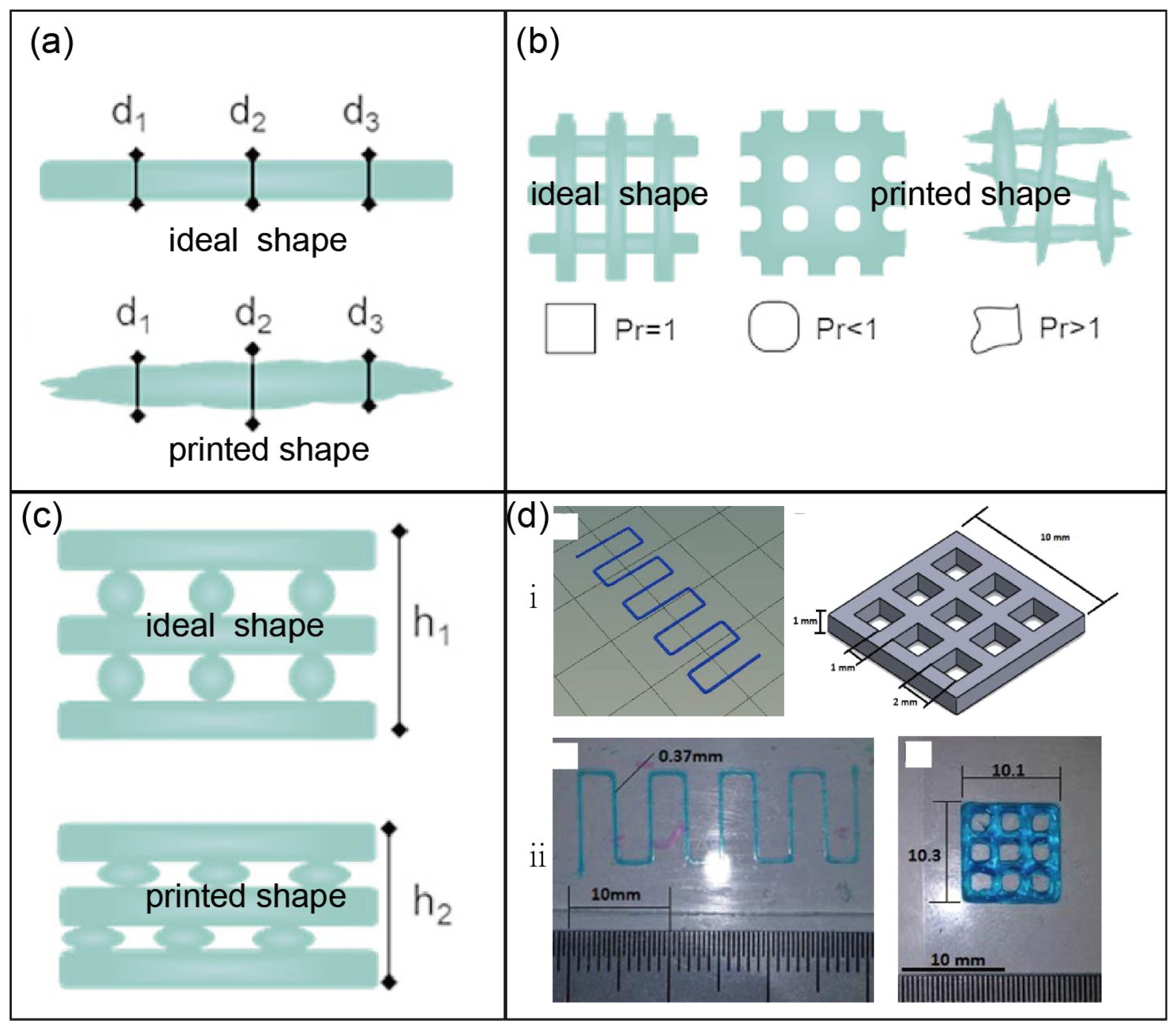
In extrusion-based bioprinting, deposited bioink filaments can undergo plastic deformation and fusion between adjacent filaments, resulting in decreased geometric resolution and unpredictable deviation from designed structures [151]. Hence, several methods have been proposed to assess the printing fidelity, attempting to identify the factors governing the printing process.
Sotorrí [152] defined printability as transporting a bioink to printer nozzles for layer-by-layer extrusion and deposition at a predictable deformation. Gillispie [153] provided a more intuitive definition: the ability of a material to be printed in a manner that results in desirable outcomes for a given application when subjected to a certain set of printing conditions. Additionally, Kang [154] stated that printability refers to the standardisation of printing accuracy and printing processes, including the selection of materials and the configuration of parameters.
Once a bioink is extruded, the diameter D of the cylindrical extrudate is dependent on the outlet flow rate Q and the translational speed of the nozzle V [155] Equation (1). A simple method to evaluate the printing quality is to measure the diameter of a filament at specific positions and compare them to the design sizes at the same locations (Figure 3a) [156,157]. Lin et al. [158] adopted a normalised evaluation approach, where the widths at 10 points along a filament were measured before calculating their mean W m and standard deviation W S D . A dimensionless number R N was then defined according to Equation (2), where a smaller R N value represents better printing quality.
D = 4 Q π V
R N = W S D W m .
Naghieh [158] proposed a method to define the “strand printability ( s P r )”, as shown in Equation (3), where D s is the designed diameter of a strand and D exp   is the measured diameter. The “pore printability ( p P r )” is described by Equation (4) [159], where L is the perimeter and A is the area of a printed lattice structure. An ideal material and printing setup would result in both s P r and p P r close to one.
s P r = 1 D s D exp .   D s
p P r = L 2 16 A .
Hazur [160] and colleagues focused on the goodness of printed squared structures and used the following equation to quantitatively describe the printability P:
P = 1 1 2 × ( L ¯ b x L o x + L ¯ b y L o y ) 1 + 1 × 1 + 1 2 × ( S D b x L o x + S D b y L o y ) ,
where L o x and L o y are the designed side lengths in x and y directions, L ̄ b x and L ̄ b y are the measured means of lengths, and S D b x and S D b y are the standard deviations of the lengths in corresponding directions. P can take values between zero and one, while a preferable printability feature is a P close to one.
However, current evaluation methods primarily focus on geometric fidelity—the congruence between printed and designed structures—while neglecting critical biological parameters including cell-adhesion efficiency and viability within the bioprinted constructs. We propose that a comprehensive printability assessment framework should incorporate two fundamental dimensions: (1) structural fidelity, encompassing shape accuracy and mechanical stability of the printed architecture; and (2) biological performance, evaluating cellular responses such as adhesion kinetics, proliferation rates, and long-term viability. This dual-aspect evaluation paradigm enables more rigorous and clinically relevant characterisation of bioink printability, bridging the gap between engineering precision and biological functionality essential for successful tissue engineering applications.
In recent years, semi-quantitative evaluations based on pore shape fidelity have gradually emerged, and the dimensionless parameter P r (0 < P r ≤ 1) defined in Equation (6) has become the most common evaluation index (Figure 3b), where L and A a are the perimeter and area of the printed structure, respectively. It is evident that as it approaches 1, the shape fidelity improves significantly [161]. In addition to evaluating the shape fidelity in the x-y plane, it is also necessary to assess the degree of collapse along the Z-axis direction. There is an obvious error between h 1 (designed) and h 2 (printed) in Figure 3c. For example, in Michael’s [162] and Ribeiro’s [151] studies, the differences between the designed and printed constructs were clarified (Figure 3d).
P r = L 2 16 A a
The degree of damage or survival rate of cells after printing is an important index for evaluating the biocompatibility of bioinks. Han [129] conducted numerical and experimental studies on cell damage for pneumatic extrusion-based 3D bioprinting and proposed a predictive function for the damaged cell ratio. The validity of the function was verified via living/dead cell staining, proving its capacity to provide appropriate guidance for 3D bioprinting by selecting proper nozzle geometries and operating pressure.
Figure 3. The assessment of shape fidelity: the assessment of cylindrical filament (a), pore (b), and height along the z-axis direction (c) (reprinted from Ref. [143]); (d) the differences between the design (i) and the printed constructs (ii) (reprinted with permission from Ref. [162], copyright 2018, Elsevier).
Jfb 16 00338 g003

4. Application of Modified Polysaccharides in Tissue Engineering

Polysaccharide-based biomaterials have unique biocompatibility and are highly similar in structure to natural extracellular matrices. The unique structures of polysaccharides make it easy to improve their properties through physical and chemical modification for tissue engineering applications.

4.1. Cartilage Tissue Engineering

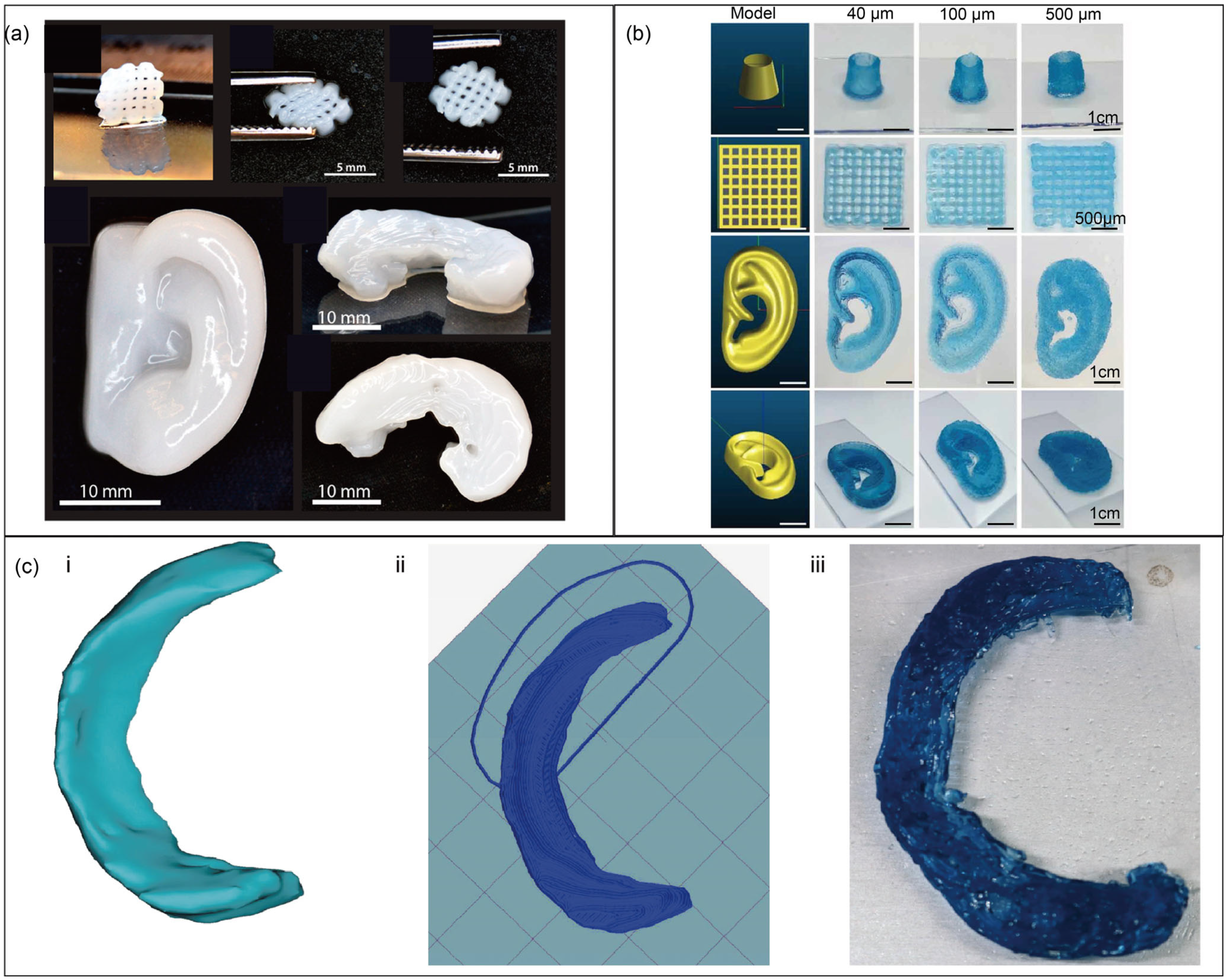
Chitosan has a significant effect on bone regeneration, yet its insufficient immunogenicity and biodegradability necessitate a modification with growth factors or scaffold materials to render it an effective alternative for autogenous bone transplantation [163]. He et al. [106] prepared a novel chitosan-based hydrogel with ethylenediamine tetraacetic acid and used calcium solution for physical crosslinking. Modified chitosan and pure chitosan were compounded to obtain a bioink with the advantages of rapid gelation and high printing accuracy that can be used to construct 3D chondrocyte scaffolds. Combining nanofibrillated cellulose (NFC) and alginate is suitable for chondrocyte growth. Markstedt et al. [164] successfully fabricated cell-laden human ears and menisci using bioprinting. The viability of human nasoseptal chondrocyte cells significantly dropped from 95.3 ± 0.1% to 69.9 ± 13.3% (p < 0.05) after bioink encapsulation but recovered to 72.8 ± 6.0% at day 1 and further increased to 85.7 ± 1.9% by day 7 post-printing. These results demonstrate that their bioink exhibits excellent printability and promotes cartilage regeneration (Figure 4a).
Ear deformities can cause significant psychological issues in patients, and various side effects accompany surgical treatment. Fortunately, biological 3D printing technology can be used to manufacture an auricle. Flégeau et al. [165] first prepared a tyramine-modified hyaluronic acid hydrogel (HA-TYR) and enzymatically crosslinked it by adding horseradish peroxidase and H2O2. This hydrogel exhibits tunable porosity, shear-thinning, and excellent thixotropic properties, with which an in vitro model of a human ear was bioprinted (Figure 4b).
Meniscal injury is another common disease. Three-dimensional bioprinting minimises the mismatch between artificial meniscus implants and natural knee tissue. Wenbin Luo [58] developed an improved bioink in which cellulose nanofiber (CNF) was mixed with gelatin–alginate bioinks to ensure high-precision bioprinting. They collected MRI information to reconstruct personalised meniscus models, which were then manufactured with CNF-modified gelatin–alginate bioinks (Figure 4c).
Figure 4. Biofabrication structures: the structures of grid, human ear, meniscus (a) (reprinted with permission from Ref. [164], copyright 2015, American Chemical Society), representative objects of 3D-printed objects using HA-TYR bioinks (b) (reprinted from Ref. [165]), 3D Meniscus model (c) (i,ii), and bioink-based, cell-free meniscal prototype (c) (iii) (reprinted from Ref. [58]).
Jfb 16 00338 g004

4.2. Vascular Tissue Engineering

The vascular system serves as the critical circulatory network responsible for nutrient transport and metabolic homeostasis in living organisms, making its structural and functional integrity essential for survival. Given the substantial global health burden imposed by vascular pathologies—including atherosclerosis, aneurysms, and peripheral artery disease—the development of effective vascular repair strategies represents a pressing clinical imperative.
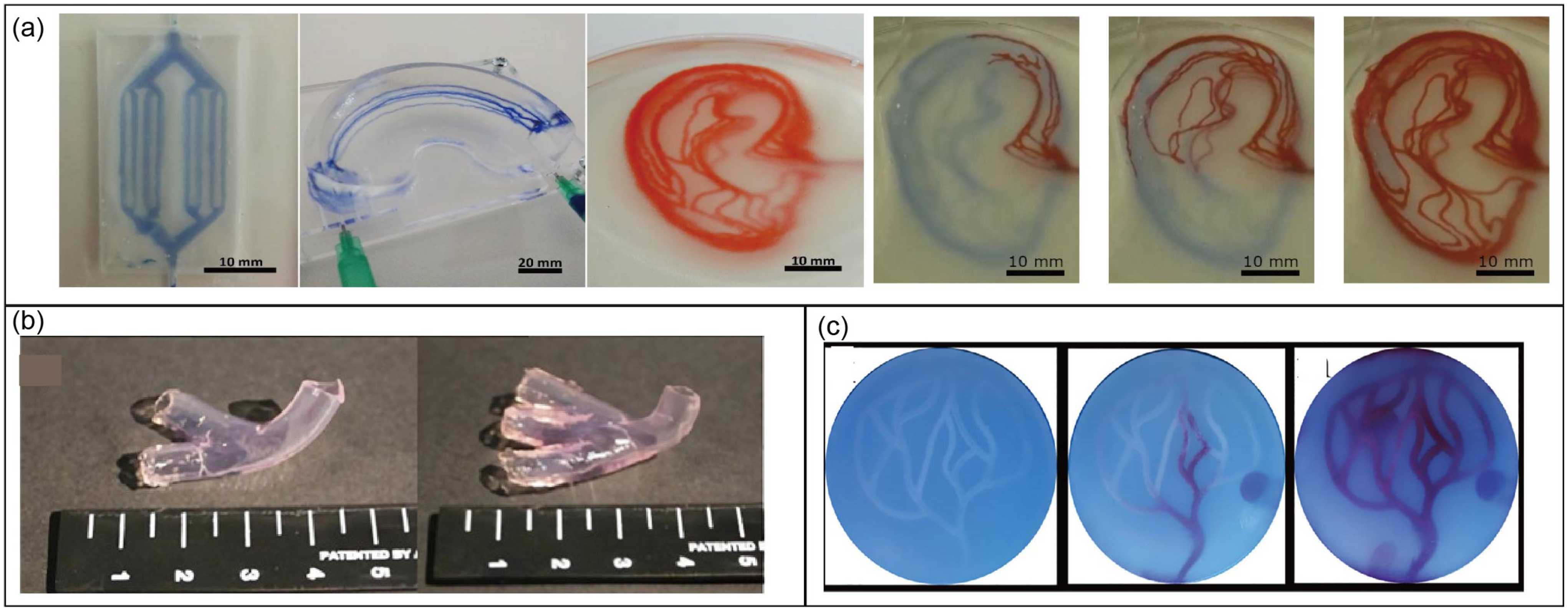
As early as 2017, Kreimendahl et al. [166] cultured endothelial cells [167] and fibroblasts [168] in a mixture of agarose, type-I collagen, and fibrinogen to form a capillary network. Their work proved that printability and angiogenesis are not mutually exclusive. A year later, Štumberger and Vihar [169] used gelatin and xanthan gum to create complex 3D structures, where xanthan gum was used as a sacrificial material to construct complex channels simulating the vascular structure of tissue (Figure 5a). In 2020, by using Pluronic F127 as a sacrificial component, Li et al. [120] proposed a hybrid biological ink composed of alginate (Alg) and silk fibroin (SF). Through rheological tests, Fourier transform infrared spectroscopy (FTIR) analysis, and cytocompatibility evaluation, this bioink was proven to be a promising coaxially extruded bioink. Based on the sacrifice of Pluronic F127, a hierarchical microchannel network was formed in the structure, laying the foundation for manufacturing large-scale blood vessels.
Antunes et al. [170] prepared an alginate tubular structure (Figure 5b) where the sacrificial component was gelatin. Because there is no natural cell-binding motif in alginate, type-I collagen or silk fibroin was added to improve the bioactivity of alginate-based hydrogels. Additionally, Ba2+ was used for secondary crosslinking to ensure stability in an aqueous environment. The mechanical properties were enhanced sufficiently after crosslinking to produce adequate vascular substitutes. Zou [83] used three polysaccharide hydrogel composites of nanocellulose, agarose, and sodium alginate with seeded cells as bioinks and polyvinyl alcohol as a sacrificial material to fabricate structures. The resulting nutrient network gradually formed a blood-vessel-like structure (Figure 5c).
In 2021, Dogan et al. [171] formulated an alginate/collagen type-I bioink supporting extrusion-based bioprinting, where human-induced pluripotent stem-cell-derived mesodermal progenitor cells (hiMPCs) retained their capabilities, giving rise to the formation of complex vessels. Vascular endothelial growth factor was added to encourage the production of blood vessels, and the formation of various vessels within bioprinted scaffolds was observed after 21 days of culture.

4.3. Skin Tissue Engineering

Skin, the largest organ of the human body, is the first line of defence against external threats. There are numerous examples of skin trauma that cause pathological changes in the body. Therefore, skin transplantation is an important treatment option. However, when skin is burned in a large area, few transplantable skin sources can meet the resulting demand. Therefore, the development of active skin substitutes with physiological functions has attracted the attention of many researchers. Sandri [172] used chitosan and glucosamine as raw materials and citric acid as a crosslinking agent to create scaffolds for repairing damaged skin. After 18 days of culture, epidermal skin was almost completely rebuilt, exhibiting considerable keratinisation. Su [38] made an agarose–polydopamine (APG) hydrogel with good biocompatibility. Based on the introduction of PDA, the cell migration rate on the surface of the APG was high. Notably, using APG promotes collagen deposition and angiogenesis, which can accelerate the healing of damaged skin.
Alginate has received increasing attention as a raw material for skin production. For example, Somasekharan et al. [173] created a bioink based on alginate blended with gelatin and diethylaminoethyl cellulose (DCEL). The optimal formulation for this bioink is composed of 2% alginate (w/v), 3.3% gelatin (w/v), and 0.93% DCEL (w/v). Fibroblasts and keratinocytes were co-cultured for 21 days, and histological analysis revealed the formation of dermal and epidermal equivalent structures.

4.4. Complex Biological Structures

While the complete realization of directly bioprinted functional tissues and organs remains an ambitious objective requiring significant further research and technological advancement, current progress is highly promising. Both commercial entities and academic research institutions are actively engaged in developing increasingly sophisticated in vitro biological constructs, systematically advancing the field toward this transformative medical capability.
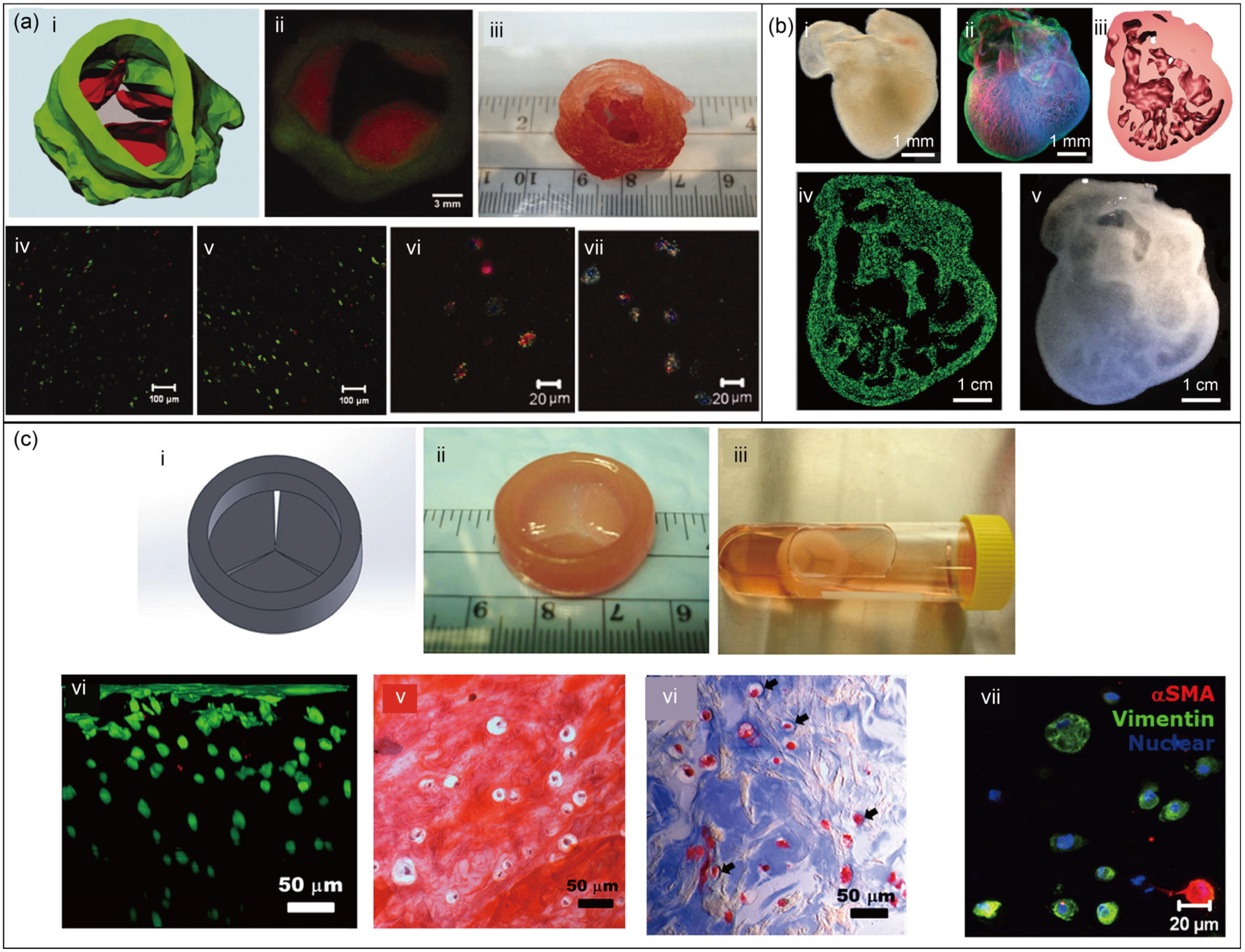
As shown in Figure 6a, encapsulated aortic root sinus smooth muscle cells (SMCs) and aortic valve leaflet interstitial cells (VICs) were encapsulated within separate alginate–gelatin hydrogel preparations. These preparations were then used to construct porcine aortic valves using the Fab@Home™ system for bioprinting. After 7 days of culture, SMCs and VICs exhibited over 80% viability [174]. A few years ago, the FRESH technique was developed to create 3D heart structures with complex internal and external architectures (Figure 6b), whose constituent materials included alginate, collagen, and fibrin. Although this construct did not contain cells, it demonstrated the potential of 3D bioprinting and modified polysaccharides for building complex organs [175]. In addition, 3D-printable formulations of hybrid hydrogels, based on methacrylated hyaluronic acid (Me-HA) and methacrylated gelatin (Me-Gel), were developed and used to bioprint heart valve conduits containing encapsulated human aortic valvular interstitial cells (HAVICs) (Figure 6c) [176]. Xuanyi Ma illustrates the first in vitro hepatic model that combinatorially mimics several in vivo features of the liver by providing a 3D culture environment for human-induced pluripotent stem cell (hiPSC)-derived hepatic cells in triculture with supporting cells, arranged in a biomimetic liver lobule pattern [177].

5. Summary and Outlook

The development of the ideal bioink formulation is an everlasting challenge. As a common and essential material to make up a creature, polysaccharides have shown remarkable potential in biomedicine, drug delivery, wound dressing, and cancer treatment. However, due to insufficient purity and strength, natural polysaccharides cannot be directly used as biomaterials for bioprinting. To provide a reference for further researchers concerning polysaccharides for bioprinting, this paper reviews the performance characteristics and modification methods for bioprinting, and tissue engineering applications. Chemical modification can modify molecular structures to obtain specific properties, but the operation often requires harsh experimental conditions and produces harmful by-products. Simultaneously, the employment of crosslinking agents and ultraviolet light exposure can harm cellular structures [178]. The operation of physical modification is relatively trivial, with little or no damage to the inherent structure, so it is difficult to change the bioactivity of polysaccharides. Enzymatic modification [179] has peculiarities including a clear objective, consummate manoeuvrability, and outstanding efficiency, but there is a mere handful of available enzymes that limit the application spectrum. In addition, the properties of polysaccharides are highly correlated with their sources and production batches [180]. Although the research on modified polysaccharides for bioprinting has made remarkable achievements, there is undoubtedly still significant room for improvement. Considering these issues, the following perspectives and research directions are provided.
Integrating artificial intelligence (AI) with large models to create an intelligent decision support system for polysaccharide modification is a valuable exploration, with the potential to enhance the efficiency and precision of the modification process. This system first relies on the vast amounts of experimental data already collected, utilising big data techniques for deep integration and cleaning to build a high-precision large model tailored for polysaccharide modification. Subsequently, through comprehensive model training, the system extracts the core features and underlying patterns within the polysaccharide modification process. In practical applications, based on the modification objectives and experimental conditions, the system recommends the optimal modification scheme, including methods and reaction conditions, and accurately predicts the modification outcomes. Furthermore, the system provides real-time monitoring and prediction of critical parameters during the experimental process, ensuring the stability and reproducibility of the entire modification process, thereby significantly enhancing the efficiency and precision of polysaccharide modification. This approach has been proven successful for applications in 3D bioprinting [181,182], biomedical engineering [183], and programmable metamaterials [184].
A critical gap persists in the field, as current methodologies lack robust, quantitative frameworks for objectively evaluating polysaccharide modifications. This challenge demands the establishment of a comprehensive assessment system developed through collaborative efforts among interdisciplinary experts spanning bioprinting, biomaterials science, bioengineering, and clinical medicine. Such a framework should incorporate multidimensional evaluation criteria, including (1) physicochemical characterisation of modified materials, (2) biological performance metrics, and (3) practical processing parameters. Specifically, this system must assess the scientific rationale of modification strategies, technical feasibility of modification processes, and functional printability of resulting materials. Most importantly, rigorous biological validation remains paramount—only through systematic in vitro and in vivo testing can researchers definitively determine whether modifications successfully achieve their intended biomimetic and functional objectives while maintaining essential biocompatibility.
The native physicochemical characteristics of polysaccharides fundamentally govern their modification potential and ultimate performance in 3D bioprinting applications. Critical consideration must be given to three key factors: (1) biological source characteristics (extraction origin and methods), (2) scalability of production processes, and (3) purification efficiency—all of which significantly influence material consistency and functionality. The three primary polysaccharide classifications exhibit distinct structure–property relationships: animal-derived polysaccharides typically offer superior bioactivity but present greater batch variability, plant-based variants provide robust mechanical properties yet require extensive processing, while microbial polysaccharides enable precise molecular control but may necessitate genetic engineering for optimal performance. These inherent advantages and limitations dictate their respective suitability for specific biofabrication applications, ranging from soft tissue mimics to load-bearing constructs.
Significant advancements have been made in engineering polysaccharides for bioprinting applications, yet substantial opportunities remain for further optimisation. The integration of artificial intelligence with experimental biochemistry represents a transformative approach to accelerate the development of polysaccharide-based bioinks. Machine learning algorithms can predict structure–function relationships and optimise modification parameters, while high-throughput experimental validation ensures biological relevance. This synergistic combination promises to enhance the clinical translation potential of polysaccharide biomaterials, facilitating the fabrication of complex, functional tissue constructs with improved physiological accuracy and therapeutic efficacy. Such advancements will be critical for generating viable tissue models and implantable grafts that meet the stringent requirements of regenerative medicine applications.

Author Contributions

Writing—original draft preparation, T.J., Y.Y. and Z.L. (Zening Lin); writing—review and editing, T.J., Y.H. and Z.L. (Zirong Luo); funding acquisition, T.J. and Z.L. (Zirong Luo). All authors have read and agreed to the published version of the manuscript.

Funding

This work is supported by the National Natural Science Foundation of China (52105039) and the Hunan Provincial Natural Science Foundation of China (2024JJ4043).

Institutional Review Board Statement

Not applicable.

Informed Consent Statement

Not applicable.

Data Availability Statement

No new data were created or analysed in this study. Data sharing is not applicable to this article.

Conflicts of Interest

The authors declare no conflicts of interest.

References

  1. Riccardo, L.; Oksana, D.; Carlos, E.G.-M.; Bruce, E.K.; Riccardo, R.; Jacob, S.; Kristi, S.A.; Shaochen, C.; Marcy, Z.-W.; Yu, S.Z. Light-based vat-polymerization bioprinting. Nat. Rev. Methods Primers 2023, 3, 46. [Google Scholar] [CrossRef]
  2. Daly, A.C.; Prendergast, M.E.; Hughes, A.J.; Burdick, J.A. Bioprinting for the Biologist. Cell 2021, 184, 18–32. [Google Scholar] [CrossRef]
  3. Zhu, J.; He, Y.; Wang, Y.; Cai, L.-H. Voxelated bioprinting of modular double-network bio-ink droplets. Nat. Commun. 2024, 15, 5902. [Google Scholar] [CrossRef] [PubMed]
  4. Urciuolo, A.; Giobbe, G.G.; Dong, Y.; Michielin, F.; Brandolino, L.; Magnussen, M.; Gagliano, O.; Selmin, G.; Scattolini, V.; Raffa, P.; et al. Hydrogel-in-hydrogel live bioprinting for guidance and control of organoids and organotypic cultures. Nat. Commun. 2023, 14, 3128. [Google Scholar] [CrossRef] [PubMed]
  5. Mahendiran, B.; Muthusamy, S.; Sampath, S.; Jaisankar, S.N.; Popat, K.C.; Selvakumar, R.; Krishnakumar, G.S. Recent trends in natural polysaccharide based bioinks for multiscale 3D printing in tissue regeneration: A review. Int. J. Biol. Macromol. 2021, 183, 564–588. [Google Scholar] [CrossRef] [PubMed]
  6. Li, N.; Song, J.; Zhu, G.; Li, X.; Liu, L.; Shi, X.; Wang, Y. Periosteum tissue engineering—A review. Biomater. Sci. 2016, 4, 1554–1561. [Google Scholar] [CrossRef]
  7. Bhamare, N.; Tardalkar, K.; Khadilkar, A.; Parulekar, P.; Joshi, M.G. Tissue engineering of human ear pinna. Cell Tissue Bank. 2022, 23, 441–457. [Google Scholar] [CrossRef]
  8. Moon, K.H.; Ko, I.K.; Yoo, J.J.; Atala, A. Kidney diseases and tissue engineering. Methods 2016, 99, 112–119. [Google Scholar] [CrossRef]
  9. Brody, H. Regenerative medicine. Nature 2016, 540, S49. [Google Scholar] [CrossRef]
  10. Nii, T.; Katayama, Y. Biomaterial-Assisted Regenerative Medicine. Int. J. Mol. Sci. 2021, 22, 8657. [Google Scholar] [CrossRef]
  11. Radeke, C.; Pons, R.; Mihajlovic, M.; Knudsen, J.R.; Butdayev, S.; Kempen, P.J.; Segeritz, C.P.; Andresen, T.L.; Pehmoller, C.K.; Jensen, T.E.; et al. Transparent and Cell-Guiding Cellulose Nanofiber 3D Printing Bioinks. ACS Appl. Mater. Interfaces 2023, 15, 2564–2577. [Google Scholar] [CrossRef]
  12. Petta, D.; D’Amora, U.; Ambrosio, L.; Grijpma, D.W.; Eglin, D.; D’Este, M. Hyaluronic acid as a bioink for extrusion-based 3D printing. Biofabrication 2020, 12, 032001. [Google Scholar] [CrossRef]
  13. Li, C.; Zheng, Z.; Jia, J.; Zhang, W.; Qin, L.; Zhang, W.; Lai, Y. Preparation and characterization of photocurable composite extracellular matrix-methacrylated hyaluronic acid bioink. J. Mater. Chem. B 2022, 10, 4242–4253. [Google Scholar] [CrossRef]
  14. Chakraborty, J.; Mu, X.; Pramanick, A.; Kaplan, D.L.; Ghosh, S. Recent advances in bioprinting using silk protein-based bioinks. Biomaterials 2022, 287, 121672. [Google Scholar] [CrossRef]
  15. Zheng, Z.; Wu, J.; Liu, M.; Wang, H.; Li, C.; Rodriguez, M.J.; Li, G.; Wang, X.; Kaplan, D.L. 3D Bioprinting of Self-Standing Silk-Based Bioink. Adv. Healthc. Mater. 2018, 7, 1701026. [Google Scholar] [CrossRef]
  16. Chester, D.; Theetharappan, P.; Ngobili, T.; Daniele, M.; Brown, A.C. Ultrasonic Microplotting of Microgel Bioinks. ACS Appl. Mater. Interfaces 2020, 12, 47309–47319. [Google Scholar] [CrossRef] [PubMed]
  17. Tchobanian, A.; Van Oosterwyck, H.; Fardim, P. Polysaccharides for tissue engineering: Current landscape and future prospects. Carbohydr. Polym. 2019, 205, 601–625. [Google Scholar] [CrossRef] [PubMed]
  18. Theocharis, A.D.; Skandalis, S.S.; Gialeli, C.; Karamanos, N.K. Extracellular matrix structure. Adv. Drug Deliv. Rev. 2016, 97, 4–27. [Google Scholar] [CrossRef] [PubMed]
  19. Xiao, R.; Grinstaff, M.W. Chemical synthesis of polysaccharides and polysaccharide mimetics. Prog. Polym. Sci. 2017, 74, 78–116. [Google Scholar] [CrossRef]
  20. DeVree, B.T.; Steiner, L.M.; Głazowska, S.; Ruhnow, F.; Herburger, K.; Persson, S.; Mravec, J. Current and future advances in fluorescence-based visualization of plant cell wall components and cell wall biosynthetic machineries. Biotechnol. Biofuels 2021, 14, 78. [Google Scholar] [CrossRef]
  21. Hassan, B.; Chatha, S.A.S.; Hussain, A.I.; Zia, K.M.; Akhtar, N. Recent advances on polysaccharides, lipids and protein based edible films and coatings: A review. Int. J. Biol. Macromol. 2018, 109, 1095–1107. [Google Scholar] [CrossRef]
  22. Sigroha, S.; Khatkar, A. Chitosan—A Naturally Derived Antioxidant Polymer with Diverse Applications. Curr. Org. Chem. 2017, 21, 333–341. [Google Scholar] [CrossRef]
  23. Simó, G.; Fernández-Fernández, E.; Vila-Crespo, J.; Ruipérez, V.; Rodríguez-Nogales, J.M. Research progress in coating techniques of alginate gel polymer for cell encapsulation. Carbohydr. Polym. 2017, 170, 1–14. [Google Scholar] [CrossRef]
  24. Gu, Y.; Cheong, K.L.; Du, H. Modification and comparison of three Gracilaria spp. agarose with methylation for promotion of its gelling properties. Chem. Cent. J. 2017, 11, 104. [Google Scholar] [CrossRef]
  25. Prasher, P.; Sharma, M.; Mehta, M.; Satija, S.; Aljabali, A.A.; Tambuwala, M.M.; Anand, K.; Sharma, N.; Dureja, H.; Jha, N.K.; et al. Current-status and applications of polysaccharides in drug delivery systems. Colloid Interface Sci. Commun. 2021, 42, 100418. [Google Scholar] [CrossRef]
  26. Jin, M.; Shi, J.; Zhu, W.; Yao, H.; Wang, D.A. Polysaccharide-Based Biomaterials in Tissue Engineering: A Review. Tissue Eng. Part B Rev. 2021, 27, 604–626. [Google Scholar] [CrossRef] [PubMed]
  27. Liu, X.; Wu, M.; Wang, M.; Hu, Q.; Liu, J.; Duan, Y.; Liu, B. Direct Synthesis of Photosensitizable Bacterial Cellulose as Engineered Living Material for Skin Wound Repair. Adv. Mater. 2022, 34, 2109010. [Google Scholar] [CrossRef] [PubMed]
  28. Kuzmenko, V.; Karabulut, E.; Pernevik, E.; Enoksson, P.; Gatenholm, P. Tailor-made conductive inks from cellulose nanofibrils for 3D printing of neural guidelines. Carbohydr. Polym. 2018, 189, 22–30. [Google Scholar] [CrossRef]
  29. Chinga-Carrasco, G. Potential and Limitations of Nanocelluloses as Components in Biocomposite Inks for Three-Dimensional Bioprinting and for Biomedical Devices. Biomacromolecules 2018, 19, 701–711. [Google Scholar] [CrossRef]
  30. Thakur, V.K.; Thakur, M.K. Processing and characterization of natural cellulose fibers/thermoset polymer composites. Carbohydr. Polym. 2014, 109, 102–117. [Google Scholar] [CrossRef]
  31. Piras, C.C.; Fernandez-Prieto, S.; De Borggraeve, W.M. Nanocellulosic materials as bioinks for 3D bioprinting. Biomater. Sci. 2017, 5, 1988–1992. [Google Scholar] [CrossRef]
  32. Torres-Rendon, J.G.; Kopf, M.; Gehlen, D.; Blaeser, A.; Fischer, H.; De Laporte, L.; Walther, A. Cellulose Nanofibril Hydrogel Tubes as Sacrificial Templates for Freestanding Tubular Cell Constructs. Biomacromolecules 2016, 17, 905–913. [Google Scholar] [CrossRef]
  33. Abraham, E.; Weber, D.E.; Sharon, S.; Lapidot, S.; Shoseyov, O. Multifunctional Cellulosic Scaffolds from Modified Cellulose Nanocrystals. ACS Appl. Mater. Interfaces 2017, 9, 2010–2015. [Google Scholar] [CrossRef] [PubMed]
  34. Zennifer, A.; Senthilvelan, P.; Sethuraman, S.; Sundaramurthi, D. Key advances of carboxymethyl cellulose in tissue engineering & 3D bioprinting applications. Carbohydr. Polym. 2021, 256, 117561. [Google Scholar] [CrossRef] [PubMed]
  35. Mohamed, A.L.; Soliman, A.A.F.; Ali, E.A.; Abou-Zeid, N.Y.; Nada, A.A. Hydrogel bioink based on clickable cellulose derivatives: Synthesis, characterization and In Vitro assessment. Int. J. Biol. Macromol. 2020, 163, 888–897. [Google Scholar] [CrossRef] [PubMed]
  36. Li, Q.; Jiang, Z.; Ma, L.; Yin, J.; Gao, Z.; Shen, L.; Yang, H.; Cui, Z.; Ye, H.; Zhou, H. A versatile embedding medium for freeform bioprinting with multi-crosslinking methods. Biofabrication 2022, 14, 035022. [Google Scholar] [CrossRef]
  37. Khoshnood, N.; Shahrezaee, M.H.; Shahrezaee, M.; Zamanian, A. Three-dimensional bioprinting of tragacanth/hydroxyapaptite modified alginate bioinks for bone tissue engineering with tunable printability and bioactivity. J. Appl. Polym. Sci. 2022, 139, e52833. [Google Scholar] [CrossRef]
  38. Ooi, H.W.; Mota, C.; Ten Cate, A.T.; Calore, A.; Moroni, L.; Baker, M.B. Thiol-Ene Alginate Hydrogels as Versatile Bioinks for Bioprinting. Biomacromolecules 2018, 19, 3390–3400. [Google Scholar] [CrossRef]
  39. Barcelo, X.; Eichholz, K.F.; Garcia, O.; Kelly, D.J. Tuning the Degradation Rate of Alginate-Based Bioinks for Bioprinting Functional Cartilage Tissue. Biomedicines 2022, 10, 1621. [Google Scholar] [CrossRef]
  40. Piras, C.C.; Smith, D.K. Multicomponent polysaccharide alginate-based bioinks. J. Mater. Chem. B 2020, 8, 8171–8188. [Google Scholar] [CrossRef]
  41. Lin, Z.; Wu, M.; He, H.; Liang, Q.; Hu, C.; Zeng, Z.; Cheng, D.; Wang, G.; Chen, D.; Pan, H.; et al. 3D Printing of Mechanically Stable Calcium-Free Alginate-Based Scaffolds with Tunable Surface Charge to Enable Cell Adhesion and Facile Biofunctionalization. Adv. Funct. Mater. 2019, 29, 1808439. [Google Scholar] [CrossRef]
  42. Gu, Y.; Schwarz, B.; Forget, A.; Barbero, A.; Martin, I.; Shastri, V.P. Advanced Bioink for 3D Bioprinting of Complex Free-Standing Structures with High Stiffness. Bioengineering 2020, 7, 141. [Google Scholar] [CrossRef] [PubMed]
  43. Nadernezhad, A.; Caliskan, O.S.; Topuz, F.; Afghah, F.; Erman, B.; Koc, B. Nanocomposite Bioinks Based on Agarose and 2D Nanosilicates with Tunable Flow Properties and Bioactivity for 3D Bioprinting. ACS Appl. Bio Mater. J. 2019, 2, 796–806. [Google Scholar] [CrossRef] [PubMed]
  44. Su, T.; Zhang, M.; Zeng, Q.; Pan, W.; Huang, Y.; Qian, Y.; Dong, W.; Qi, X.; Shen, J. Mussel-inspired agarose hydrogel scaffolds for skin tissue engineering. Bioact. Mater. 2021, 6, 579–588. [Google Scholar] [CrossRef]
  45. Poldervaart, M.T.; Goversen, B.; de Ruijter, M.; Abbadessa, A.; Melchels, F.P.W.; Oner, F.C.; Dhert, W.J.A.; Vermonden, T.; Alblas, J. 3D bioprinting of methacrylated hyaluronic acid (MeHA) hydrogel with intrinsic osteogenicity. PLoS ONE 2017, 12, e0177628. [Google Scholar] [CrossRef]
  46. Antich, C.; de Vicente, J.; Jimenez, G.; Chocarro, C.; Carrillo, E.; Montanez, E.; Galvez-Martin, P.; Marchal, J.A. Bio-inspired hydrogel composed of hyaluronic acid and alginate as a potential bioink for 3D bioprinting of articular cartilage engineering constructs. Acta Biomater. 2020, 106, 114–123. [Google Scholar] [CrossRef]
  47. Hossain Rakin, R.; Kumar, H.; Rajeev, A.; Natale, G.; Menard, F.; Li, I.T.S.; Kim, K. Tunable metacrylated hyaluronic acid-based hybrid bioinks for stereolithography 3D bioprinting. Biofabrication 2021, 13, 044109. [Google Scholar] [CrossRef]
  48. Zhou, K.; Feng, M.; Mao, H.; Gu, Z. Photoclick polysaccharide-based bioinks with an extended biofabrication window for 3D embedded bioprinting. Biomater. Sci. 2022, 10, 4479–4491. [Google Scholar] [CrossRef]
  49. Coskun, S.; Akbulut, S.O.; Sarikaya, B.; Cakmak, S.; Gumusderelioglu, M. Formulation of chitosan and chitosan-nanoHAp bioinks and investigation of printability with optimized bioprinting parameters. Int. J. Biol. Macromol. 2022, 222, 1453–1464. [Google Scholar] [CrossRef]
  50. He, Y.; Wang, F.; Wang, X.; Zhang, J.; Wang, D.; Huang, X. A photocurable hybrid chitosan/acrylamide bioink for DLP based 3D bioprinting. Mater. Des. 2021, 202, 109588. [Google Scholar] [CrossRef]
  51. Shen, Y.; Tang, H.; Huang, X.; Hang, R.; Zhang, X.; Wang, Y.; Yao, X. DLP printing photocurable chitosan to build bio-constructs for tissue engineering. Carbohydr. Polym. 2020, 235, 115970. [Google Scholar] [CrossRef]
  52. Demirtas, T.T.; Irmak, G.; Gumusderelioglu, M. A bioprintable form of chitosan hydrogel for bone tissue engineering. Biofabrication 2017, 9, 035003. [Google Scholar] [CrossRef] [PubMed]
  53. Liu, X.; Hao, M.; Chen, Z.; Zhang, T.; Huang, J.; Dai, J.; Zhang, Z. 3D bioprinted neural tissue constructs for spinal cord injury repair. Biomaterials 2021, 272, 120771. [Google Scholar] [CrossRef] [PubMed]
  54. Taniguchi Nagahara, M.H.; Caiado Decarli, M.; Inforçatti Neto, P.; da Silva, J.V.L.; Moraes, Â.M. Crosslinked alginate-xanthan gum blends as effective hydrogels for 3D bioprinting of biological tissues. J. Appl. Polym. Sci. 2022, 139, e52612. [Google Scholar] [CrossRef]
  55. Shapira, A.; Noor, N.; Oved, H.; Dvir, T. Transparent support media for high resolution 3D printing of volumetric cell-containing ECM structures. Biomed. Mater. 2020, 15, 045018. [Google Scholar] [CrossRef]
  56. Lim, W.; Shin, S.Y.; Cha, J.M.; Bae, H. Optimization of Polysaccharide Hydrocolloid for the Development of Bioink with High Printability/Biocompatibility for Coextrusion 3D Bioprinting. Polymers 2021, 13, 1773. [Google Scholar] [CrossRef]
  57. Wang, B.; Han, Y.; Lin, Q.; Liu, H.; Shen, C.; Nan, K.; Chen, H. In Vitro and In Vivo evaluation of xanthan gum-succinic anhydride hydrogels for the ionic strength-sensitive release of antibacterial agents. J. Mater. Chem. B 2016, 4, 1853–1861. [Google Scholar] [CrossRef]
  58. Luo, W.; Song, Z.; Wang, Z.; Wang, Z.; Li, Z.; Wang, C.; Liu, H.; Liu, Q.; Wang, J. Printability Optimization of Gelatin-Alginate Bioinks by Cellulose Nanofiber Modification for Potential Meniscus Bioprinting. J. Nanomater. 2020, 2020, 863428. [Google Scholar] [CrossRef]
  59. Das, R.; Lee, C.P.; Prakash, A.; Hashimoto, M.; Fernandez, J.G. Geometrical control of degradation and cell delivery in 3D printed nanocellulose hydrogels. Mater. Today Commun. 2022, 30, 103023. [Google Scholar] [CrossRef]
  60. Mu, H.; Wang, Y.; Wei, H.; Lu, H.; Feng, Z.; Yu, H.; Xing, Y.; Wang, H. Collagen peptide modified carboxymethyl cellulose as both antioxidant drug and carrier for drug delivery against retinal ischaemia/reperfusion injury. J. Cell. Mol. Med. 2018, 22, 5008–5019. [Google Scholar] [CrossRef]
  61. Yang, J.; He, H.; Li, D.; Zhang, Q.; Xu, L.; Ruan, C. Advanced strategies in the application of gelatin-based bioink for extrusion bioprinting. Bio-Des. Manuf. 2023, 6, 586–608. [Google Scholar] [CrossRef]
  62. Lin, Y.; Chen, S.; Liu, Y.; Guo, F.; Miao, Q.; Huang, H. A composite hydrogel scaffold based on collagen and carboxymethyl chitosan for cartilage regeneration through one-step chemical crosslinking. Int. J. Biol. Macromol. 2023, 226, 706–715. [Google Scholar] [CrossRef] [PubMed]
  63. Yalman, V.; Laçin, N.T. Development of humic acid and alginate-based wound dressing and evaluation on inflammation. Mater. Technol. 2019, 34, 705–717. [Google Scholar] [CrossRef]
  64. Wong, C.-C.; Lu, C.-X.; Cho, E.-C.; Lee, P.-W.; Chi, N.-W.; Lin, P.-Y.; Jheng, P.-R.; Chen, H.-L.; Mansel, B.W.; Chen, Y.-M.; et al. Calcium peroxide aids tyramine-alginate gel to crosslink with tyrosinase for efficient cartilage repair. Int. J. Biol. Macromol. 2022, 208, 299–313. [Google Scholar] [CrossRef]
  65. Rosiak, P.; Latanska, I.; Paul, P.; Sujka, W.; Kolesinska, B. Modification of Alginates to Modulate Their Physic-Chemical Properties and Obtain Biomaterials with Different Functional Properties. Molecules 2021, 26, 7264. [Google Scholar] [CrossRef]
  66. Hecht, H.; Srebnik, S. Structural Characterization of Sodium Alginate and Calcium Alginate. Biomacromolecules 2016, 17, 2160–2167. [Google Scholar] [CrossRef]
  67. Sahoo, D.R.; Biswal, T. Alginate and its application to tissue engineering. SN Appl. Sci. 2021, 3, 30. [Google Scholar] [CrossRef]
  68. Bouhadir, K.H.; Lee, K.Y.; Alsberg, E.; Damm, K.L.; Anderson, K.W.; Mooney, D.J. Degradation of Partially Oxidized Alginate and Its Potential Application for Tissue Engineering. Biotechnol. Prog. 2001, 17, 945–950. [Google Scholar] [CrossRef]
  69. Hurtado, A.; Aljabali, A.A.A.; Mishra, V.; Tambuwala, M.M.; Serrano-Aroca, Á. Alginate: Enhancement Strategies for Advanced Applications. Int. J. Mol. Sci. 2022, 23, 4486. [Google Scholar] [CrossRef]
  70. Dalheim, M.O.; Vanacker, J.; Najmi, M.A.; Aachmann, F.L.; Strand, B.L.; Christensen, B.E. Efficient functionalization of alginate biomaterials. Biomaterials 2016, 80, 146–156. [Google Scholar] [CrossRef]
  71. Ramos, P.E.; Silva, P.; Alario, M.M.; Pastrana, L.M.; Teixeira, J.A.; Cerqueira, M.A.; Vicente, A.A. Effect of alginate molecular weight and M/G ratio in beads properties foreseeing the protection of probiotics. Food Hydrocoll. 2018, 77, 8–16. [Google Scholar] [CrossRef]
  72. GhavamiNejad, A.; Ashammakhi, N.; Wu, X.Y.; Khademhosseini, A. Crosslinking Strategies for 3D Bioprinting of Polymeric Hydrogels. Small 2020, 16, 2002931. [Google Scholar] [CrossRef] [PubMed]
  73. Park, J.; Lee, S.J.; Chung, S.; Lee, J.H.; Kim, W.D.; Lee, J.Y.; Park, S.A. Cell-laden 3D bioprinting hydrogel matrix depending on different compositions for soft tissue engineering: Characterization and evaluation. Mater. Sci. Eng. C 2017, 71, 678–684. [Google Scholar] [CrossRef] [PubMed]
  74. Stanisci, A.; Aarstad, O.A.; Tondervik, A.; Sletta, H.; Dypas, L.B.; Skjak-Braek, G.; Aachmann, F.L. Overall size of mannuronan C5-Epimerases influences their ability to epimerize modified alginates and alginate gels. Carbohydr. Polym. 2018, 180, 256–263. [Google Scholar] [CrossRef] [PubMed]
  75. Bukhari, A.A.H.; Elsayed, N.H.; Monier, M. Development and characterization of photo-responsive cinnamoly modified alginate. Carbohydr. Polym. 2021, 260, 117771. [Google Scholar] [CrossRef]
  76. Im, S.; Choe, G.; Seok, J.M.; Yeo, S.J.; Lee, J.H.; Kim, W.D.; Lee, J.Y.; Park, S.A. An osteogenic bioink composed of alginate, cellulose nanofibrils, and polydopamine nanoparticles for 3D bioprinting and bone tissue engineering. Int. J. Biol. Macromol. 2022, 205, 520–529. [Google Scholar] [CrossRef]
  77. Habib, A.; Khoda, B. Development of clay based novel hybrid bio-ink for 3D bio-printing process. J. Manuf. Process. 2019, 38, 76–87. [Google Scholar] [CrossRef]
  78. Yao, B.; Hu, T.; Cui, X.; Song, W.; Fu, X.; Huang, S. Enzymatically degradable alginate/gelatin bioink promotes cellular behavior and degradation In Vitro and In Vivo. Biofabrication 2019, 11, 045020. [Google Scholar] [CrossRef]
  79. Zhu, Y.; Stark, C.J.; Madira, S.; Ethiraj, S.; Venkatesh, A.; Anilkumar, S.; Jung, J.; Lee, S.; Wu, C.A.; Walsh, S.K.; et al. Three-Dimensional Bioprinting with Alginate by Freeform Reversible Embedding of Suspended Hydrogels with Tunable Physical Properties and Cell Proliferation. Bioengineering 2022, 9, 807. [Google Scholar] [CrossRef]
  80. Singh, Y.P.; Bhardwaj, N.; Mandal, B.B. Potential of Agarose/Silk Fibroin Blended Hydrogel for In Vitro Cartilage Tissue Engineering. ACS Appl. Mater. Interfaces 2016, 8, 21236–21249. [Google Scholar] [CrossRef]
  81. Ionescu, A.-M.; Alaminos, M.; Cardona, J.d.l.C.; García-López Durán, J.d.D.; González-Andrades, M.; Ghinea, R.; Campos, A.; Hita, E.; Pérez, M.d.M. Investigating a novel nanostructured fibrin–agarose biomaterial for human cornea tissue engineering: Rheological properties. J. Mech. Behav. Biomed. Mater. 2011, 4, 1963–1973. [Google Scholar] [CrossRef]
  82. Jiang, C.; Liu, Z.; Cheng, D.; Mao, X. Agarose degradation for utilization: Enzymes, pathways, metabolic engineering methods and products. Biotechnol. Adv. 2020, 45, 107641. [Google Scholar] [CrossRef] [PubMed]
  83. Zou, Q.; Tian, X.; Luo, S.; Yuan, D.; Xu, S.; Yang, L.; Ma, M.; Ye, C. Agarose composite hydrogel and PVA sacrificial materials for bioprinting large-scale, personalized face-like with nutrient networks. Carbohydr. Polym. 2021, 269, 118222. [Google Scholar] [CrossRef] [PubMed]
  84. Lopez-Marcial, G.R.; Zeng, A.Y.; Osuna, C.; Dennis, J.; Garcia, J.M.; O’Connell, G.D. Agarose-Based Hydrogels as Suitable Bioprinting Materials for Tissue Engineering. ACS Biomater. Sci. Eng. 2018, 4, 3610–3616. [Google Scholar] [CrossRef] [PubMed]
  85. Forget, A.; Derme, T.; Mitterberger, D.; Heiny, M.; Sweeney, C.; Mudili, L.; Dargaville, T.R.; Shastri, V.P. Architecture-inspired paradigm for 3D bioprinting of vessel-like structures using extrudable carboxylated agarose hydrogels. Emergent Mater. 2019, 2, 233–243. [Google Scholar] [CrossRef]
  86. Su, Y.; Chu, B.; Gao, Y.; Wu, C.; Zhang, L.; Chen, P.; Wang, X.; Tang, S. Modification of agarose with carboxylation and grafting dopamine for promotion of its cell-adhesiveness. Carbohydr. Polym. 2013, 92, 2245–2251. [Google Scholar] [CrossRef]
  87. Lee, S.-w.; Kim, J.; Do, M.; Namkoong, E.; Lee, H.; Ryu, J.H.; Park, K. Developmental role of hyaluronic acid and its application in salivary gland tissue engineering. Acta Biomater. 2020, 115, 275–287. [Google Scholar] [CrossRef]
  88. Singh, D.; Wang, S.-B.; Xia, T.; Tainsh, L.; Ghiassi-Nejad, M.; Xu, T.; Peng, S.; Adelman, R.A.; Rizzolo, L.J. A biodegradable scaffold enhances differentiation of embryonic stem cells into a thick sheet of retinal cells. Biomaterials 2018, 154, 158–168. [Google Scholar] [CrossRef]
  89. Grandoch, M.; Bollyky, P.L.; Fischer, J.W. Hyaluronan: A Master Switch Between Vascular Homeostasis and Inflammation. Circ. Res. 2018, 122, 1341–1343. [Google Scholar] [CrossRef]
  90. Fallacara, A.; Baldini, E.; Manfredini, S.; Vertuani, S. Hyaluronic Acid in the Third Millennium. Polymers 2018, 10, 701. [Google Scholar] [CrossRef]
  91. Tiwari, S.; Bahadur, P. Modified hyaluronic acid based materials for biomedical applications. Int. J. Biol. Macromol. 2019, 121, 556–571. [Google Scholar] [CrossRef] [PubMed]
  92. Wolf, K.J.; Kumar, S. Hyaluronic Acid: Incorporating the Bio into the Material. ACS Biomater. Sci. Eng. 2019, 5, 3753–3765. [Google Scholar] [CrossRef] [PubMed]
  93. Townsend, J.M.; Sanders, M.E.; Kiyotake, E.A.; Detamore, M.S. Independent control of molecular weight, concentration, and stiffness of hyaluronic acid hydrogels. Biomed. Mater. 2022, 17, 6. [Google Scholar] [CrossRef] [PubMed]
  94. Tavakoli, S.; Krishnan, N.; Mokhtari, H.; Oommen, O.P.; Varghese, O.P. Fine-tuning Dynamic Cross–linking for Enhanced 3D Bioprinting of Hyaluronic Acid Hydrogels. Adv. Funct. Mater. 2024, 34, 2307040. [Google Scholar] [CrossRef]
  95. Gamarra, A.; Missagia, B.; Urpí, L.; Morató, J.; Muñoz-Guerra, S. Ionic coupling of hyaluronic acid with ethyl N-lauroyl l-arginate (LAE): Structure, properties and biocide activity of complexes. Carbohydr. Polym. 2018, 197, 109–116. [Google Scholar] [CrossRef]
  96. Fan, F.; Su, B.; Kolodychak, A.; Ekwueme, E.; Alderfer, L.; Saha, S.; Webber, M.J.; Hanjaya-Putra, D. Hyaluronic Acid Hydrogels with Phototunable Supramolecular Cross-Linking for Spatially Controlled Lymphatic Tube Formation. ACS Appl. Mater. Interfaces 2023, 15, 58181–58195. [Google Scholar] [CrossRef]
  97. Kotla, N.G.; Bonam, S.R.; Rasala, S.; Wankar, J.; Bohara, R.A.; Bayry, J.; Rochev, Y.; Pandit, A. Recent advances and prospects of hyaluronan as a multifunctional therapeutic system. J. Control. Release 2021, 336, 598–620. [Google Scholar] [CrossRef]
  98. Tao, F.; Cheng, Y.; Shi, X.; Zheng, H.; Du, Y.; Xiang, W.; Deng, H. Applications of chitin and chitosan nanofibers in bone regenerative engineering. Carbohydr. Polym. 2020, 230, 115658. [Google Scholar] [CrossRef]
  99. Li, J.; Cai, C.; Li, J.; Li, J.; Li, J.; Sun, T.; Wang, L.; Wu, H.; Yu, G. Chitosan-Based Nanomaterials for Drug Delivery. Molecules 2018, 23, 2661. [Google Scholar] [CrossRef]
  100. Balagangadharan, K.; Dhivya, S.; Selvamurugan, N. Chitosan based nanofibers in bone tissue engineering. Int. J. Biol. Macromol. 2017, 104, 1372–1382. [Google Scholar] [CrossRef]
  101. Chesnutt, B.M.; Yuan, Y.; Buddington, K.; Haggard, W.O.; Bumgardner, J.D. Composite Chitosan/Nano-Hydroxyapatite Scaffolds Induce Osteocalcin Production by Osteoblasts In Vitro and Support Bone Formation In Vivo. Tissue Eng. Part A 2009, 15, 2571–2579. [Google Scholar] [CrossRef]
  102. Ardean, C.; Davidescu, C.M.; Nemes, N.S.; Negrea, A.; Ciopec, M.; Duteanu, N.; Negrea, P.; Duda-Seiman, D.; Musta, V. Factors Influencing the Antibacterial Activity of Chitosan and Chitosan Modified by Functionalization. Int. J. Mol. Sci. 2021, 22, 7449. [Google Scholar] [CrossRef]
  103. Wu, P.; Xi, X.; Li, R.; Sun, G. Engineering Polysaccharides for Tissue Repair and Regeneration. Macromol. Biosci. 2021, 21, e2100141. [Google Scholar] [CrossRef]
  104. Levengood, S.L.; Zhang, M. Chitosan-based scaffolds for bone tissue engineering. J. Mater. Chem. B 2014, 2, 3161–3184. [Google Scholar] [CrossRef]
  105. Ji, J.; Wang, L.; Yu, H.; Chen, Y.; Zhao, Y.; Zhang, H.; Amer, W.A.; Sun, Y.; Huang, L.; Saleem, M. Chemical Modifications of Chitosan and Its Applications. Polym.-Plast. Technol. Eng. 2014, 53, 1494–1505. [Google Scholar] [CrossRef]
  106. Ahmed, S.; Annu; Ali, A.; Sheikh, J. A review on chitosan centred scaffolds and their applications in tissue engineering. Int. J. Biol. Macromol. 2018, 116, 849–862. [Google Scholar] [CrossRef]
  107. Rezaei, F.S.; Sharifianjazi, F.; Esmaeilkhanian, A.; Salehi, E. Chitosan films and scaffolds for regenerative medicine applications: A review. Carbohydr. Polym. 2021, 273, 118631. [Google Scholar] [CrossRef] [PubMed]
  108. Klaypradit, W.; Huang, Y.-W. Fish oil encapsulation with chitosan using ultrasonic atomizer. LWT-Food Sci. Technol. 2008, 41, 1133–1139. [Google Scholar] [CrossRef]
  109. Choo, C.K.; Kong, X.Y.; Goh, T.L.; Ngoh, G.C.; Horri, B.A.; Salamatinia, B. Chitosan/halloysite beads fabricated by ultrasonic-assisted extrusion-dripping and a case study application for copper ion removal. Carbohydr. Polym. 2016, 138, 16–26. [Google Scholar] [CrossRef] [PubMed]
  110. Liu, D.; Chang, P.R.; Chen, M.; Wu, Q. Chitosan colloidal suspension composed of mechanically disassembled nanofibers. J. Colloid Interface Sci. 2011, 354, 637–643. [Google Scholar] [CrossRef] [PubMed]
  111. Brysch, C.N.; Wold, E.; Patterson, M.; Ordoñez Olivares, R.; Eberth, J.F.; Robles Hernandez, F.C. Chitosan and chitosan composites reinforced with carbon nanostructures. J. Alloys Compd. 2014, 615, S515–S521. [Google Scholar] [CrossRef]
  112. Yue, W.; He, R.; Yao, P.; Wei, Y. Ultraviolet radiation-induced accelerated degradation of chitosan by ozone treatment. Carbohydr. Polym. 2009, 77, 639–642. [Google Scholar] [CrossRef]
  113. Kurita, K.; Ikeda, H.; Yoshida, Y.; Shimojoh, M.; Harata, M. Chemoselective Protection of the Amino Groups of Chitosan by Controlled Phthaloylation: Facile Preparation of a Precursor Useful for Chemical Modifications. Biomacromolecules 2002, 3, 1–4. [Google Scholar] [CrossRef] [PubMed]
  114. Kolodziejska, M.; Jankowska, K.; Klak, M.; Wszola, M. Chitosan as an Underrated Polymer in Modern Tissue Engineering. Nanomaterials 2021, 11, 3019. [Google Scholar] [CrossRef]
  115. Yang, J.; Xie, Q.; Zhu, J.; Zou, C.; Chen, L.; Du, Y.; Li, D. Preparation and In Vitro antioxidant activities of 6-amino-6-deoxychitosan and its sulfonated derivatives. Biopolymers 2015, 103, 539–549. [Google Scholar] [CrossRef] [PubMed]
  116. Shanmugam, A.; Kathiresan, K.; Nayak, L. Preparation, characterization and antibacterial activity of chitosan and phosphorylated chitosan from cuttlebone of Sepia kobiensis (Hoyle, 1885). Biotechnol. Rep. 2016, 9, 25–30. [Google Scholar] [CrossRef]
  117. Ahmed, S.; Ikram, S. Chitosan Based Scaffolds and Their Applications in Wound Healing. Achiev. Life Sci. 2016, 10, 27–37. [Google Scholar] [CrossRef]
  118. Aberg, C.M.; Chen, T.; Olumide, A.; Raghavan, S.R.; Payne, G.F. Enzymatic Grafting of Peptides from Casein Hydrolysate to Chitosan. Potential for Value-Added Byproducts from Food-Processing Wastes. J. Agric. Food Chem. 2004, 52, 788–793. [Google Scholar] [CrossRef]
  119. Wang, D.; Lv, P.; Zhang, L.; Yang, S.; Gao, Y. Structural and Functional Characterization of Laccase-Induced β-Lactoglobulin-Ferulic Acid-Chitosan Ternary Conjugates. J. Agric. Food Chem. 2019, 67, 12054–12060. [Google Scholar] [CrossRef] [PubMed]
  120. Hajikhani, M.; Khanghahi, M.M.; Shahrousvand, M.; Mohammadi-Rovshandeh, J.; Babaei, A.; Khademi, S.M.H. Intelligent superabsorbents based on a xanthan gum/poly (acrylic acid) semi-interpenetrating polymer network for application in drug delivery systems. Int. J. Biol. Macromol. 2019, 139, 509–520. [Google Scholar] [CrossRef]
  121. Piola, B.; Sabbatini, M.; Gino, S.; Invernizzi, M.; Reno, F. 3D Bioprinting of Gelatin-Xanthan Gum Composite Hydrogels for Growth of Human Skin Cells. Int. J. Mol. Sci. 2022, 23, 539. [Google Scholar] [CrossRef]
  122. Kumar, A.; Rao, K.M.; Han, S.S. Application of xanthan gum as polysaccharide in tissue engineering: A review. Carbohydr. Polym. 2018, 180, 128–144. [Google Scholar] [CrossRef]
  123. Zhong, L.; Oostrom, M.; Truex, M.J.; Vermeul, V.R.; Szecsody, J.E. Rheological behavior of xanthan gum solution related to shear thinning fluid delivery for subsurface remediation. J. Hazard. Mater. 2013, 244–245, 160–170. [Google Scholar] [CrossRef]
  124. Choppe, E.; Puaud, F.; Nicolai, T.; Benyahia, L. Rheology of xanthan solutions as a function of temperature, concentration and ionic strength. Carbohydr. Polym. 2010, 82, 1228–1235. [Google Scholar] [CrossRef]
  125. Arimura, T.; Omagari, Y.; Yamamoto, K.; Kadokawa, J.-i. Chemoenzymatic synthesis and hydrogelation of amylose-grafted xanthan gums. Int. J. Biol. Macromol. 2011, 49, 498–503. [Google Scholar] [CrossRef]
  126. Bhat, I.M.; Wani, S.M.; Mir, S.A.; Masoodi, F.A. Advances in xanthan gum production, modifications and its applications. Biocatal. Agric. Biotechnol. 2022, 42, 102328. [Google Scholar] [CrossRef]
  127. Pinto, E.P.; Furlan, L.; Vendruscolo, C.T. Chemical deacetylation natural xanthan (Jungbunzlauer®). Polímeros 2011, 21, 47–52. [Google Scholar] [CrossRef]
  128. Jena, S.R.; Dalei, G.; Das, S.; Nayak, J.; Pradhan, M.; Samanta, L. Harnessing the potential of dialdehyde alginate-xanthan gum hydrogels as niche bioscaffolds for tissue engineering. Int. J. Biol. Macromol. 2022, 207, 493–506. [Google Scholar] [CrossRef] [PubMed]
  129. Hayrabolulu, H.; Demeter, M.; Cutrubinis, M.; Şen, M. Radiation synthesis and characterization of xanthan gum hydrogels. Radiat. Phys. Chem. 2021, 188, 109613. [Google Scholar] [CrossRef]
  130. Bose, S.; Ke, D.; Sahasrabudhe, H.; Bandyopadhyay, A. Additive manufacturing of biomaterials. Prog. Mater. Sci. 2018, 93, 45–111. [Google Scholar] [CrossRef]
  131. Jiang, T.; Munguia-Lopez, J.G.; Flores-Torres, S.; Kort-Mascort, J.; Kinsella, J.M. Extrusion bioprinting of soft materials: An emerging technique for biological model fabrication. Appl. Phys. Rev. 2019, 6, 011310. [Google Scholar] [CrossRef]
  132. Luo, Y.; Pan, H.; Jiang, J.; Zhao, C.; Zhang, J.; Chen, P.; Lin, X.; Fan, S. Desktop-Stereolithography 3D Printing of a Polyporous Extracellular Matrix Bioink for Bone Defect Regeneration. Front. Bioeng. Biotechnol. 2020, 8, 589094. [Google Scholar] [CrossRef] [PubMed]
  133. Compaan, A.M.; Christensen, K.; Huang, Y. Inkjet Bioprinting of 3D Silk Fibroin Cellular Constructs Using Sacrificial Alginate. ACS Biomater. Sci. Eng. 2017, 3, 1519–1526. [Google Scholar] [CrossRef] [PubMed]
  134. Sorkio, A.; Koch, L.; Koivusalo, L.; Deiwick, A.; Miettinen, S.; Chichkov, B.; Skottman, H. Human stem cell based corneal tissue mimicking structures using laser-assisted 3D bioprinting and functional bioinks. Biomaterials 2018, 171, 57–71. [Google Scholar] [CrossRef]
  135. Lian, L.; Zhou, C.; Tang, G.; Xie, M.; Wang, Z.; Luo, Z.; Japo, J.; Wang, D.; Zhou, J.; Wang, M.; et al. Uniaxial and Coaxial Vertical Embedded Extrusion Bioprinting. Adv. Healthc. Mater. 2022, 11, 2102411. [Google Scholar] [CrossRef]
  136. Tembadamani, S.; Mohan, T.S.; Thrivikraman, G.; Muthuvijayan, V.; Barman, S.R. Engineering Polysaccharide Biomaterials: Modifications and Crosslinking Strategies for Soft Tissue Bioprinting. Macromol. Rapid Commun. 2025, in press. e00236. [Google Scholar] [CrossRef]
  137. Vajda, J.; Vihar, B.; Ćurić, L.Č.; Maver, U.; Vesenjak, M.; Dubrovski, P.D.; Milojević, M. Sr2+ vs. Ca2+ as post-processing ionic crosslinkers: Implications for 3D bioprinting of polysaccharide hydrogels in tissue engineering. J. Mater. Res. Technol. 2023, 23, 1805–1820. [Google Scholar] [CrossRef]
  138. Nagaraja, K.; Dhokare, P.; Bhattacharyya, A.; Noh, I. Recent advances in 3D bioprinting of polysaccharide-based bioinks for fabrication of bioengineered tissues. Mol. Syst. Des. Eng. 2024, 9, 977–999. [Google Scholar] [CrossRef]
  139. Kaith, A.; Jain, N.; Kaul, S.; Nagaich, U. Polysaccharide-infused bio-fabrication: Advancements in 3D bioprinting for tissue engineering and bone regeneration. Mater. Today Commun. 2024, 40, 109429. [Google Scholar] [CrossRef]
  140. Lin, S.; Li, B.; Yang, L.; Zhai, Y.; Wang, X.; Wang, C. New method for reducing viscosity and shear stress in hydrogel 3D printing via multidimension vibration. Comput. Methods Biomech. Biomed. Engin 2022, 25, 1796–1811. [Google Scholar] [CrossRef]
  141. Zhao, Y.; Li, Y.; Mao, S.; Sun, W.; Yao, R. The influence of printing parameters on cell survival rate and printability in microextrusion-based 3D cell printing technology. Biofabrication 2015, 7, 045002. [Google Scholar] [CrossRef] [PubMed]
  142. Kiyotake, E.A.; Douglas, A.W.; Thomas, E.E.; Nimmo, S.L.; Detamore, M.S. Development and quantitative characterization of the precursor rheology of hyaluronic acid hydrogels for bioprinting. Acta Biomater. 2019, 95, 176–187. [Google Scholar] [CrossRef] [PubMed]
  143. Schwab, A.; Levato, R.; D’Este, M.; Piluso, S.; Eglin, D.; Malda, J. Printability and Shape Fidelity of Bioinks in 3D Bioprinting. Chem. Rev. 2020, 120, 11028–11055. [Google Scholar] [CrossRef] [PubMed]
  144. Smith, P.T.; Basu, A.; Saha, A.; Nelson, A. Chemical modification and printability of shear-thinning hydrogel inks for direct-write 3D printing. Polymer 2018, 152, 42–50. [Google Scholar] [CrossRef]
  145. Rawal, P.; Tripathi, D.M.; Ramakrishna, S.; Kaur, S. Prospects for 3D bioprinting of organoids. Bio-Des. Manuf. 2021, 4, 627–640. [Google Scholar] [CrossRef]
  146. Distler, T.; Solisito, A.A.; Schneidereit, D.; Friedrich, O.; Detsch, R.; Boccaccini, A.R. 3D printed oxidized alginate-gelatin bioink provides guidance for C2C12 muscle precursor cell orientation and differentiation via shear stress during bioprinting. Biofabrication 2020, 12, 045005. [Google Scholar] [CrossRef]
  147. Nair, K.; Gandhi, M.; Khalil, S.; Yan, K.C.; Marcolongo, M.; Barbee, K.; Sun, W. Characterization of cell viability during bioprinting processes. Biotechnol. J. 2009, 4, 1168–1177. [Google Scholar] [CrossRef]
  148. Blaeser, A.; Duarte Campos, D.F.; Puster, U.; Richtering, W.; Stevens, M.M.; Fischer, H. Controlling Shear Stress in 3D Bioprinting is a Key Factor to Balance Printing Resolution and Stem Cell Integrity. Adv. Healthc. Mater. 2016, 5, 326–333. [Google Scholar] [CrossRef]
  149. Emmermacher, J.; Spura, D.; Cziommer, J.; Kilian, D.; Wollborn, T.; Fritsching, U.; Steingroewer, J.; Walther, T.; Gelinsky, M.; Lode, A. Engineering considerations on extrusion-based bioprinting: Interactions of material behavior, mechanical forces and cells in the printing needle. Biofabrication 2020, 12, 025022. [Google Scholar] [CrossRef]
  150. Adhikari, J.; Roy, A.; Das, A.; Ghosh, M.; Thomas, S.; Sinha, A.; Kim, J.; Saha, P. Effects of Processing Parameters of 3D Bioprinting on the Cellular Activity of Bioinks. Macromol. Biosci. 2021, 21, e2000179. [Google Scholar] [CrossRef]
  151. Ribeiro, A.; Blokzijl, M.M.; Levato, R.; Visser, C.W.; Castilho, M.; Hennink, W.E.; Vermonden, T.; Malda, J. Assessing bioink shape fidelity to aid material development in 3D bioprinting. Biofabrication 2017, 10, 014102. [Google Scholar] [CrossRef]
  152. Sotorrío, G.; Alonso, J.; Olsson, N.O.E.; Tenorio, J.A. Printability of materials for extrusion 3D printing technologies: A review of material requirements and testin. Mater. De Construcción 2021, 71, 344. [Google Scholar] [CrossRef]
  153. Gillispie, G.; Prim, P.; Copus, J.; Fisher, J.; Mikos, A.G.; Yoo, J.J.; Atala, A.; Lee, S.J. Assessment methodologies for extrusion-based bioink printability. Biofabrication 2020, 12, 022003. [Google Scholar] [CrossRef]
  154. Yu, K.; Zhang, X.; Sun, Y.; Gao, Q.; Fu, J.; Cai, X.; He, Y. Printability during projection-based 3D bioprinting. Bioact. Mater. 2022, 11, 254–267. [Google Scholar] [CrossRef]
  155. Fu, Z.; Naghieh, S.; Xu, C.; Wang, C.; Sun, W.; Chen, X. Printability in extrusion bioprinting. Biofabrication 2021, 13. [Google Scholar] [CrossRef]
  156. Wang, L.; Xu, M.E.; Luo, L.; Zhou, Y.; Si, P. Iterative feedback bio-printing-derived cell-laden hydrogel scaffolds with optimal geometrical fidelity and cellular controllability. Sci. Rep. 2018, 8, 2802. [Google Scholar] [CrossRef] [PubMed]
  157. Gao, T.; Gillispie, G.J.; Copus, J.S.; Pr, A.K.; Seol, Y.J.; Atala, A.; Yoo, J.J.; Lee, S.J. Optimization of gelatin-alginate composite bioink printability using rheological parameters: A systematic approach. Biofabrication 2018, 10, 034106. [Google Scholar] [CrossRef] [PubMed]
  158. Lin, Z.; Jiang, T.; Kinsella, J.M.; Shang, J.; Luo, Z. Assessing roughness of extrusion printed soft materials using a semi-quantitative method. Mater. Lett. 2021, 303, 130480. [Google Scholar] [CrossRef]
  159. Ouyang, L.; Yao, R.; Zhao, Y.; Sun, W. Effect of bioink properties on printability and cell viability for 3D bioplotting of embryonic stem cells. Biofabrication 2016, 8, 035020. [Google Scholar] [CrossRef]
  160. Hazur, J.; Detsch, R.; Karakaya, E.; Kaschta, J.; Tessmar, J.; Schneidereit, D.; Friedrich, O.; Schubert, D.W.; Boccaccini, A.R. Improving alginate printability for biofabrication: Establishment of a universal and homogeneous pre-crosslinking technique. Biofabrication 2020, 12, 045004. [Google Scholar] [CrossRef]
  161. Nelson, C.; Tuladhar, S.; Launen, L.; Habib, A. 3D Bio-Printability of Hybrid Pre-Crosslinked Hydrogels. Int. J. Mol. Sci. 2021, 22, 13481. [Google Scholar] [CrossRef]
  162. Giuseppe, M.D.; Law, N.; Webb, B.; Macrae, R.A.; Liew, L.J.; Sercombe, T.B.; Dilley, R.J.; Doyle, B.J. Mechanical behaviour of alginate-gelatin hydrogels for 3D bioprinting. J. Mech. Behav. Biomed. Mater. 2018, 79, 150–157. [Google Scholar] [CrossRef] [PubMed]
  163. Kozusko, S.D.; Riccio, C.; Goulart, M.; Bumgardner, J.; Jing, X.L.; Konofaos, P. Chitosan as a Bone Scaffold Biomaterial. J. Craniofacial Surg. 2018, 29, 1788–1793. [Google Scholar] [CrossRef] [PubMed]
  164. Markstedt, K.; Mantas, A.; Tournier, I.; Martinez Avila, H.; Hagg, D.; Gatenholm, P. 3D Bioprinting Human Chondrocytes with Nanocellulose-Alginate Bioink for Cartilage Tissue Engineering Applications. Biomacromolecules 2015, 16, 1489–1496. [Google Scholar] [CrossRef] [PubMed]
  165. Flegeau, K.; Puiggali-Jou, A.; Zenobi-Wong, M. Cartilage tissue engineering by extrusion bioprinting utilizing porous hyaluronic acid microgel bioinks. Biofabrication 2022, 14, 3. [Google Scholar] [CrossRef]
  166. Kreimendahl, F.; Kopf, M.; Thiebes, A.L.; Duarte Campos, D.F.; Blaeser, A.; Schmitz-Rode, T.; Apel, C.; Jockenhoevel, S.; Fischer, H. Three-Dimensional Printing and Angiogenesis: Tailored Agarose-Type I Collagen Blends Comprise Three-Dimensional Printability and Angiogenesis Potential for Tissue-Engineered Substitutes. Tissue Eng. Part C Methods 2017, 23, 604–615. [Google Scholar] [CrossRef]
  167. Mendes, A.C.; Baran, E.T.; Pereira, R.C.; Azevedo, H.S.; Reis, R.L. Encapsulation and survival of a chondrocyte cell line within xanthan gum derivative. Macromol. Biosci. 2012, 12, 350–359. [Google Scholar] [CrossRef]
  168. Schwarz, S.; Kuth, S.; Distler, T.; Gogele, C.; Stolzel, K.; Detsch, R.; Boccaccini, A.R.; Schulze-Tanzil, G. 3D printing and characterization of human nasoseptal chondrocytes laden dual crosslinked oxidized alginate-gelatin hydrogels for cartilage repair approaches. Mater. Sci. Eng. C 2020, 116, 111189. [Google Scholar] [CrossRef]
  169. Stumberger, G.; Vihar, B. Freeform Perfusable Microfluidics Embedded in Hydrogel Matrices. Materials 2018, 11, 2529. [Google Scholar] [CrossRef]
  170. Antunes, M.; Bonani, W.; Reis, R.L.; Migliaresi, C.; Ferreira, H.; Motta, A.; Neves, N.M. Development of alginate-based hydrogels for blood vessel engineering. Mater. Sci. Eng. C 2021, 134, 112588. [Google Scholar] [CrossRef]
  171. Dogan, L.; Scheuring, R.; Wagner, N.; Ueda, Y.; Schmidt, S.; Wörsdörfer, P.; Groll, J.; Ergün, S. Human iPSC-derived mesodermal progenitor cells preserve their vasculogenesis potential after extrusion and form hierarchically organized blood vessels. Biofabrication 2021, 13, 045028. [Google Scholar] [CrossRef] [PubMed]
  172. Sandri, G.; Rossi, S.; Bonferoni, M.C.; Miele, D.; Faccendini, A.; Del Favero, E.; Di Cola, E.; Icaro Cornaglia, A.; Boselli, C.; Luxbacher, T.; et al. Chitosan/glycosaminoglycan scaffolds for skin reparation. Carbohydr. Polym. 2019, 220, 219–227. [Google Scholar] [CrossRef] [PubMed]
  173. Somasekharan, L.T.; Raju, R.; Kumar, S.; Geevarghese, R.; Nair, R.P.; Kasoju, N.; Bhatt, A. Biofabrication of skin tissue constructs using alginate, gelatin and diethylaminoethyl cellulose bioink. Int. J. Biol. Macromol. 2021, 189, 398–409. [Google Scholar] [CrossRef] [PubMed]
  174. Duan, B.; Hockaday, L.A.; Kang, K.H.; Butcher, J.T. 3D Bioprinting of heterogeneous aortic valve conduits with alginate_gelatin hydrogels. J. Biomed. Mater. Res. Part A 2012, 101, 1255–1264. [Google Scholar] [CrossRef]
  175. Hinton, T.J.; Jallerat, Q.; Palchesko, R.N.; Park, J.H.; Grodzicki, M.S.; Shue, H.-J.; Ramadan, M.H.; Hudson, A.R.; Feinberg, A.W. Three-dimensional printing of complex biological structures by freeform reversible embedding of suspended hydrogels. Sci. Adv. 2015, 1, e1500758. [Google Scholar] [CrossRef]
  176. Duan, B.; Kapetanovic, E.; Hockaday, L.A.; Butcher, J.T. Three-dimensional printed trileaflet valve conduits using biological hydrogels and human valve interstitial cells. Acta Biomater. 2014, 10, 1836–1846. [Google Scholar] [CrossRef]
  177. Ma, X.; Qu, X.; Zhu, W.; Li, Y.S.; Yuan, S.; Zhang, H.; Liu, J.; Wang, P.; Lai, C.S.; Zanella, F.; et al. Deterministically patterned biomimetic human iPSC-derived hepatic model via rapid 3D bioprinting. Proc. Natl. Acad. Sci. USA 2016, 113, 2206–2211. [Google Scholar] [CrossRef]
  178. Teixeira, M.C.; Lameirinhas, N.S.; Carvalho, J.P.F.; Silvestre, A.J.D.; Vilela, C.; Freire, C.S.R. A Guide to Polysaccharide-Based Hydrogel Bioinks for 3D Bioprinting Applications. Int. J. Mol. Sci. 2022, 23, 6564. [Google Scholar] [CrossRef]
  179. Kaczmarek, M.B.; Struszczyk-Swita, K.; Li, X.; Szczesna-Antczak, M.; Daroch, M. Enzymatic Modifications of Chitin, Chitosan, and Chitooligosaccharides. Front. Bioeng. Biotechnol. 2019, 7, 243. [Google Scholar] [CrossRef]
  180. Zhou, Y.; Chen, X.; Chen, T.; Chen, X. A review of the antibacterial activity and mechanisms of plant polysaccharides. Trends Food Sci. Technol. 2022, 123, 264–280. [Google Scholar] [CrossRef]
  181. Jo, Y.; Hwang, D.G.; Kim, M.; Yong, U.; Jang, J. Bioprinting-assisted tissue assembly to generate organ substitutes at scale. Trends Biotechnol. 2023, 41, 93–105. [Google Scholar] [CrossRef]
  182. An, J.; Chua, C.K.; Mironov, V. Application of Machine Learning in 3D Bioprinting: Focus on Development of Big Data and Digital Twin. Int. J. Bioprinting 2024, 7, 342. [Google Scholar] [CrossRef]
  183. Shokrani, H.; Shokrani, A.; Seidi, F.; Kucińska-Lipka, J.; Makurat-Kasprolewicz, B.; Saeb, M.R.; Ramakrishna, S. Artificial intelligence for biomedical engineering of polysaccharides: A short overview. Curr. Opin. Biomed. Eng. 2023, 27, 100463. [Google Scholar] [CrossRef]
  184. Lee, S.; Kwon, J.; Kim, H.; Ritchie, R.O.; Gu, G.X. Advancing programmable metamaterials through machine learning-driven buckling strength optimization. Curr. Opin. Solid State Mater. Sci. 2024, 31, 101161. [Google Scholar] [CrossRef]
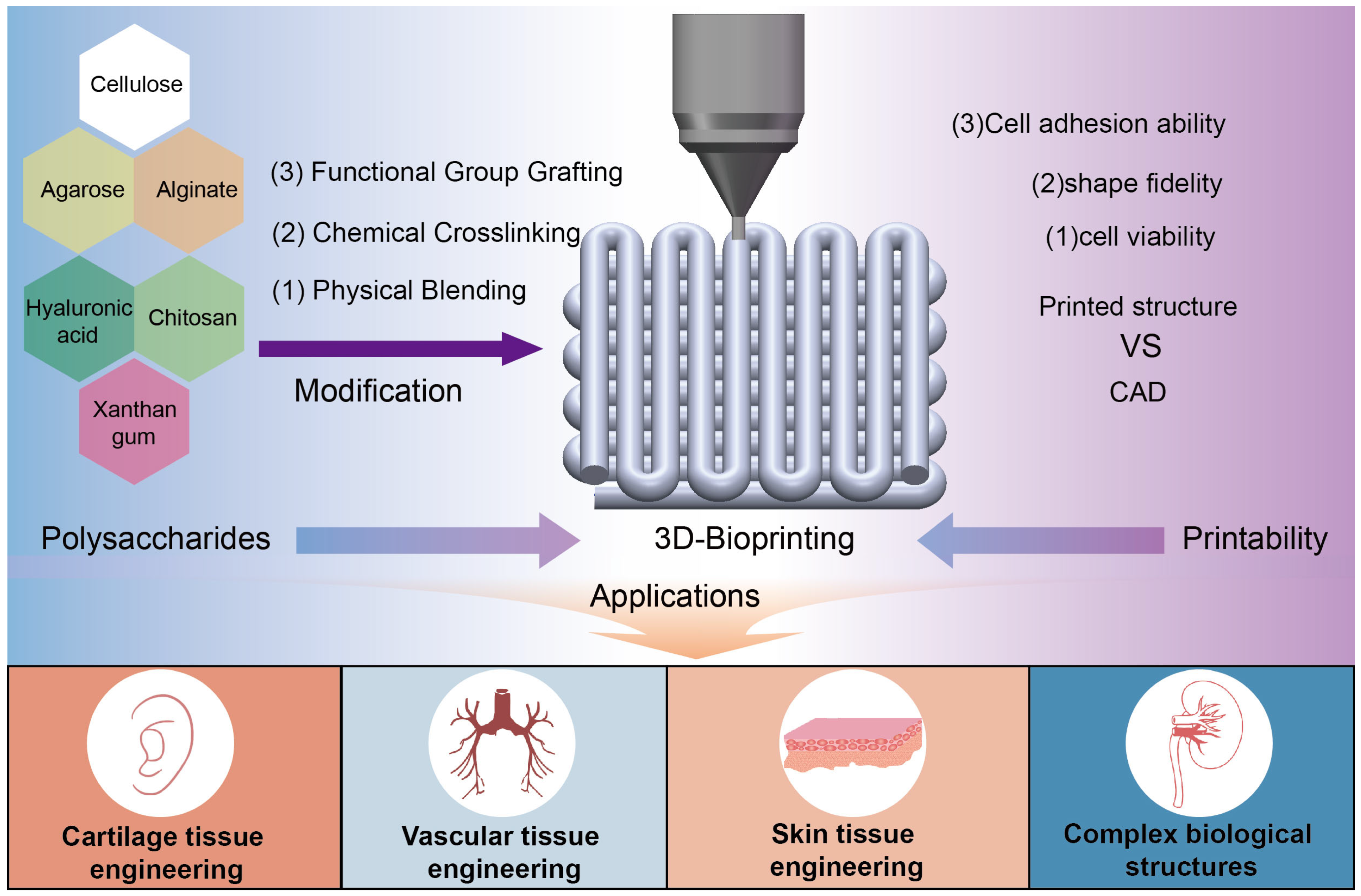
Figure 1. Schematic diagram of polysaccharides modification for 3D bioprinting and tissue engineering applications.
Jfb 16 00338 g001
Figure 2. The chemical construction of cellulose (a), alginate (b), agarose (c), hyaluronic acid (d), chitosan (e), and xanthan gum (f).
Jfb 16 00338 g002
Figure 5. Artificial blood vessel: the fabricated hydrogel microfluidic devices (a) (reprinted from Ref. [169]), alginate tubular structure (b) (reprinted with permission from Ref. [170], copyright 2022, Elsevier), bioprinted vessel-like networks with nutrient networks (c) (reprinted with permission from Ref. [83], copyright 2021, Elsevier).
Jfb 16 00338 g005
Figure 6. Complex biological structures: (a) an aortic valve conduit: (i) a micro-CT image, (ii) fluorescent image of first printed two layers, (iii) a printed aortic valve conduit in colour, (ivvii) images of fluorescent staining after 7-day culture (reprinted from Ref. [174]); (b) a 3D heart construct of chick embryo: (i) a dark-field image of an embryonic chick heart. (ii) A 3D image of the chick heart, (iii) the 3D CAD model of the embryonic heart, (iv) A cross section of the 3D-printed heart in fluorescent alginate (green), (v) A dark-field image of the 3D-printed heart (reprinted from Ref. [175]); (c) bioprinting of heart valve conduit: (i) heart valve model, (ii) the printed valve conduit, (iii) intact conduit after 7-day static culture, (ivvii) staining images (reprinted with permission from Ref. [176], copyright 2014, Elsevier).
Jfb 16 00338 g006
Table 1. Possible modification methods and 3D bioprinting applications of polysaccharides.
PolysaccharidesSubstrate MaterialMethod or Substance of ModificationTarget SubstanceApplicationsCharacteristicsRefs.
Plant polysaccharidesCelluloseMechanical shearingNanofibrillated cellulose3D bioprinting and the automated fabrication of complex tissue-mimicking constructsAccessibility, biocompatibility, and shear-thinning properties[11]
Nanometer granulationCellulose nanocrystalsBiobased cellulosic scaffold materialHydrophobicity, oleophilicity, and lipophilicity[27]
Alkalization/mercerisation and etherification reactionsCarboxymethyl cellulose Bioinks for printing bioconstructsGood viscosity modifier, shear alignment, and shape memory property[28]
The open-ring reaction of 1-azido-2,3-epoxypropane (AEP) with hydroxyethyl celluloseAzido-hydroxy-ethyl celluloseA novel bioink for bone tissue engineeringBiocompatibility, biodegradability, and printability[29]
Hydrophobic modification and hydroxypropyl methylationHydroxypropyl methyl celluloseBioinks for freeform writing of the millimetric complex tubular structuresTunable rheological properties, good stability, and compatibility with additives without strong hydrophilic groups[30]
AlginateTragacanth/hydroxyapatiteTragacanth/hydroxyapatite modified alginate bioinks Repair of significant bone tissue defectsImproving compressive strength, viscosity, printability properties, resolution, and shape fidelity[31]
Norbornene functionalisingModular alginate-based bioinksConstruction of complex multi-ink geometriesHigh cell survivability, stable 3D constructs[32]
Oxidised alginateDegradable alginate-based bioinksBioprinting functional cartilage tissueRapidly degrade, excellent shape fidelity[33]
Molecular weights, concentration, and viscosities ——Porous bioprinted constructs for bone tissue engineeringGood biocompatibility and tailorable performance [34]
ε-polylysine (ε-PL)ε-polylysine (ε-PL)-modified Alginate-based bioinks (Alg/ε-PL)Alginate-based scaffolds
For the precise and individualised therapy of tissue defects
Excellent self-supporting stability, mechanical stability[35]
AgaroseCarboxylated agarose——Bioink for Bioprinting of free-standing structures with high StiffnessPrinting high-aspect ratio objects possessing anatomically relevant curvature and architecture[36]
2D nanosilicate additivesNanocomposite agaroseStrong shear-thinning bioinks for extrusion 3D bioprinting applications Tunable flow properties and bioactivity[37]
PolydopamineAgarose-polydopamineHydrogel scaffolds for skin wound healingGood cell adhesion, biodegradability, and biocompatibility[38]
Animal polysaccharidesHyaluronic AcidMethacrylation of high-molecular-weight hyaluronic acidMethacrylated hyaluronic acid Scaffold materials for application in 3D-printed, tissue-engineered bone substitutesGood primary cell survival and excellent spontaneous osteogenic differentiation in vitro[39]
AlginateA new bioink for cartilage tissue 3D bioprintingHighly viable and functional bioprinted 3D hybrid structures for Articular cartilage regenerationPrintability, gelling abilities, stiffness, and good degradability[40]
Gelatin methacryloyl (GelMA), methacrylated hyaluronic acid (MAHA)Tunable MAHA-GelMA (metacrylated hyaluronic acid-based hybrid bioinks)Stereolithographic (SLA) 3D bioprintingExcellent mechanical strength, printability, and cell-adhesive nature[41]
Norbornene functional groups (Nor) and cysteamine hydrochloride (Cys)Hiol-norbornene photoclick polysaccharide-based bioink Bioprinting a liver model in vitroIncreased viscoelastic properties, reduced ROS (reactive oxygen species) accumulation, and superior shape fidelity [42]
ChitosanNanohydroxyapatite (nhap)Chitosan-nanohap bioinks3D cellular structures and bone tissue engineering applicationsHigh resolution, shape fidelity, and high printability index[43]
Acrylamide (AM), chitosan modified with methacryloyl groups (CHIMA)CHIMA/AMA favourable bioink for the DLP-based 3D printing in the field of tissue engineering and regenerative medicineEnhanced compression strength, improved elasticity, and favourable biocompatibility[44]
Grafting chitosan molecular chains with methacryloyl groupsA photocurable chitosan bioink (CHI-MA)A potential bioink for the DLP and other photocuring-based 3D printing technologiesHigh resolution, high fidelity, and good biocompatibility[45]
Nanostructured bone-like hydroxyapatite(HA)Chitosan-HA hydrogels3D bioprinting of tissue constructsEnabling good mechanical support after printing, providing highly active cell platforms[46]
Hyaluronic acid derivatives and Matrigel.NSC(neural stem cell)-laden scaffoldA neural tissue scaffoldFast gelation and spontaneous covalent crosslinking capability[47]
Microbial polysaccharideXanthan gum(XG)Alginate, strontium ionsCrosslinked alginate-xanthan gum blendSimple cellularized structures and microtissue models to complex organ bioprintingNoncytotoxic, shear-thinning, and easily sterilizable [48]
Calcium-alginate nanoparticlesAlginate-XG hybrid medium A promising support medium for 3D printing of tissues and organsAllowing long-term, high resolution, and accurate printing of bio-structures with a high degree of anatomical complexity[49]
Carboxymethyl cellulose Utilising extrusion-based 3D bioprintingTunability regarding pore size and mechanical strength optimisation[50]
Succinic anhydrideSuccinic anhydride (SA)-modified xanthan (XG–SA) derivativesPromising drug delivery materials for antibacterial applicationsHigher storage (G’) and loss (G’) modulus[51]
Disclaimer/Publisher’s Note: The statements, opinions and data contained in all publications are solely those of the individual author(s) and contributor(s) and not of MDPI and/or the editor(s). MDPI and/or the editor(s) disclaim responsibility for any injury to people or property resulting from any ideas, methods, instructions or products referred to in the content.