1. Introduction
The Yellow Sea, lying along the western edge of the Pacific Ocean and adjacent to China and the Korean Peninsula, is a shallow semi-enclosed marine basin that has recently been the focus of biodiversity surveys and taxonomic studies on nematodes [
1,
2,
3,
4]. A total of over 400 nematode species have been identified, with 119 recognized as new to science [
5].
Being the biggest marginal sea along the western Pacific and ranking third in size worldwide, the South China Sea lies adjacent to the margin of the Coral Triangle, a major center of global biodiversity. Gagarin and Tu [
6] carried out detailed taxonomic studies on South China Sea nematodes, documenting 62 species from the Vietnam coast, of which 22 were new to science. Chinese researchers have documented 366 species along the southern Chinese coastline of the South China Sea, including 20 newly described nematode species, of which six were from offshore sediments in the Beibu Gulf [
7,
8,
9], five from offshore regions of the Qiongzhou Strait [
10,
11,
12], and nine from Guangdong’s intertidal areas [
13,
14,
15,
16]. However, the overall diversity of nematodes in Chinese sea areas remains unclear, with new species being continually discovered; therefore, further taxonomic investigations in these regions are essential.
Halichoanolaimus de Man, 1886 is a widespread and species-rich genus of predatory nematodes of Selachinematidae, occurring broadly from shallow marine environments to the deep ocean floor [
17,
18]. At present, the genus comprises 30 valid species recorded worldwide [
19,
20]. Zograf et al. [
21] provided the key in 2015. The latest species descriptions were presented by Huang and Zhai [
19].
Cobbionema Filipjev, 1922 was proposed by Filipjev in 1922, and its type species is
Cobbionema acrocerca Filipjev, 1922 [
22]. At present, the genus comprises only six species documented globally [
20,
23]. The most recent species descriptions were provided by Leduc [
18] and Ahmed et al. [
23].
Linhystera Juario, 1974 [
24] is based on the type species
Linhystera problematica Juario, 1974 and currently contains five valid species around the world [
25]. The diagnosis of
Linhystera was provided by Fonseca and Bezerra [
26]. In the same year, Yu et al. emended the diagnosis of
Linhystera and published descriptions of two new species from depths of 29.5 m and 50 m, respectively—
Linhystera longiapophysis and
Linhystera breviapophysis—which are mainly characterized by possessing dorsocaudal gubernacular apophyses. They further proposed that the presence or absence of gubernacular apophyses is variable within the genus and accordingly divided all congeneric species into two groups based on this morphological trait [
27]. The latest species description, of
Linhystera filiformis, was published in 2024 by Lv et al., found in intertidal sandy sediment from Chinese sea areas [
25].
2. Materials and Methods
For the investigation of biodiversity in free-living marine nematodes, undisturbed sediment samples were collected using a 0.1 m2 modified Gray–O’Hara box corer during the 2018 Open Research Cruise in the Yellow Sea aboard the R/V Kexue Sanhao (sampling area: 32° N–38°50′ N, 120°20′ E–124°10′ E). Additional sediment samples were obtained from multiple deep-sea sites in the South China Sea in 2020 during cruises of the R/Vs Jiageng and Kexue (sampling area: 8° N–24° N, 108° E–120° E).
The surface sediment layer (0–8 cm depth) was sampled for meiofauna using a shortened syringe with a 2.9 cm diameter. Samples were preserved using the addition of 10% formalin solution at a 1:1 (
v/
v) ratio and subsequently preserved. Under laboratory conditions, the samples were carried out using a 0.1% Rose Bengal solution for a period of 24 h. The stained material was processed by sequential filtration through 500 and 31 µm mesh sieves, followed by rinsing with tap water to remove fine sediments and facilitate the separation of macrofauna from meiofauna. The fraction retained in the 31 µm sieve was subjected to density separation by centrifugation in Ludox-TM (50% colloidal silica suspension; Sigma-Aldrich, St. Louis, MO, USA) at a specific gravity of 1.15 g/mL [
28], enabling the isolation of meiofauna from heavier sediment particles. After three successive centrifugation steps, the supernatant was transferred into a 31 µm sieve to eliminate remaining Ludox-TM. The material retained in the sieve was then gently rinsed with distilled water and transferred into a Petri dish, and meiofaunal organisms were subsequently isolated under a stereomicroscope.
Nematodes collected were transferred into a cavity block that contained a mixture of 5% glycerol, 5% ethanol, and 90% distilled water (
v/
v) [
29]. Following gradual evaporation of ethanol, the specimens were placed in glycerin to produce permanent slides [
30]. Morphological observations and descriptions of the specimens were performed by means of a differential interference contrast microscope (Leica DM 2500) (CMS GmbH, Wetzlar, Germany, assembled in Shanghai, China), and micrographs were acquired with a Leica DMC 5400 digital camera (Leica Microsystems (Switzerland) Ltd., Heerbrugg, made in Germany). Figures were drawn as line illustrations by means of a camera lucida. All measurements were recorded using Leica LAS X software (version 3.3.3, Leica Microsystems, CMS GmbH, Wetzlar, Germany), and curved structures were measured following the midline of their curvature. All measurements are given in micrometers (µm). Type specimens are housed in the Marine Biological Museum, Chinese Academy of Sciences (Qingdao).
This manuscript adopts the following abbreviations: a = body length/maximum body diameter; abd = diameter of the body at the cloaca or anus; b = body length/pharynx length; c = body length/tail length; cbd = respective body diameter; c′ = tail length/cloacal or anus body diameter; V% = vulva distance from anterior end/total body length (%).
3. Results and Discussion
3.1. Halichoanolaimus parvulus sp. nov.
ZooBank registration: urn:lsid:zoobank.org:act:460477C9-5A4F-4762-A4E4-6B95DF054B5E
Class Chromadorea Inglis, 1983
Order: Chromadorida Chitwood, 1933.
Family: Selachinematidae Cobb, 1915.
Genus: Halichoanolaimus de Man, 1886.
Diagnosis (modified from Leduc [
18]): Lateral differentiation of the cuticle is evident, marked by punctations that are larger and spaced farther apart. The anterior sensilla are typically papilliform, or outer labial and cephalic sensilla short setae (
Halichoanolaimus funestus,
Halichoanolaimus pumilus). Buccal cavity is composed of two distinct regions: an anterior section bearing cuticularized rhabdions terminating in pointed denticles, and a posterior chamber encircled by three pairs of Y-shaped cuticularized rhabdions. Amphideal fovea multispiral, positioned at the midpoint of the buccal cavity. Pharynx lacks both anterior and posterior bulbs. The intestine is blind in adult stages. Precloacal supplements are typically papilliform or setiform. The tail comprises a conical proximal region and frequently an elongated cylindrical distal portion.
Diagnosis: Body 935–1095 µm long; amphideal fovea exhibiting 4.5–5 turns; spicules arched, middle portion broad, tapering distally; gubernaculum slender, composed of two separate lateral elements that taper distally; a short precloacal seta; in males, the tail is conico-cylindrical, the posterior region comprising about three-fifths of the tail being cylindrical.
Holotype and paratype material: Specimens included two males and three females: Holotype (m1) on slide 3875-1-0-3, paratype 1 (m2) on slide 3600-3-0-4, paratype 2 (f1) on slide 3875-1-0-3, and both paratype 3 (f2) and paratype 4 (f3) on slide 3875-1-0-2.
Type locality and habitat. The holotype (m1), along with all paratype females, was obtained from the muddy sediments of the seafloor at Station 3875-1 (38°45′ N, 121°10′ E, water depth 45 m) in the Yellow Sea, whereas paratype male 2 was obtained from muddy sediments of the seafloor at Station 3600-3 (36°0′ N, 122°50′ E, water depth 30 m).
Etymology: This species’ name “parvulus” denotes its comparatively smaller body size compared with other members of the genus.
Measurements: All morphometric data are presented in
Table 1.
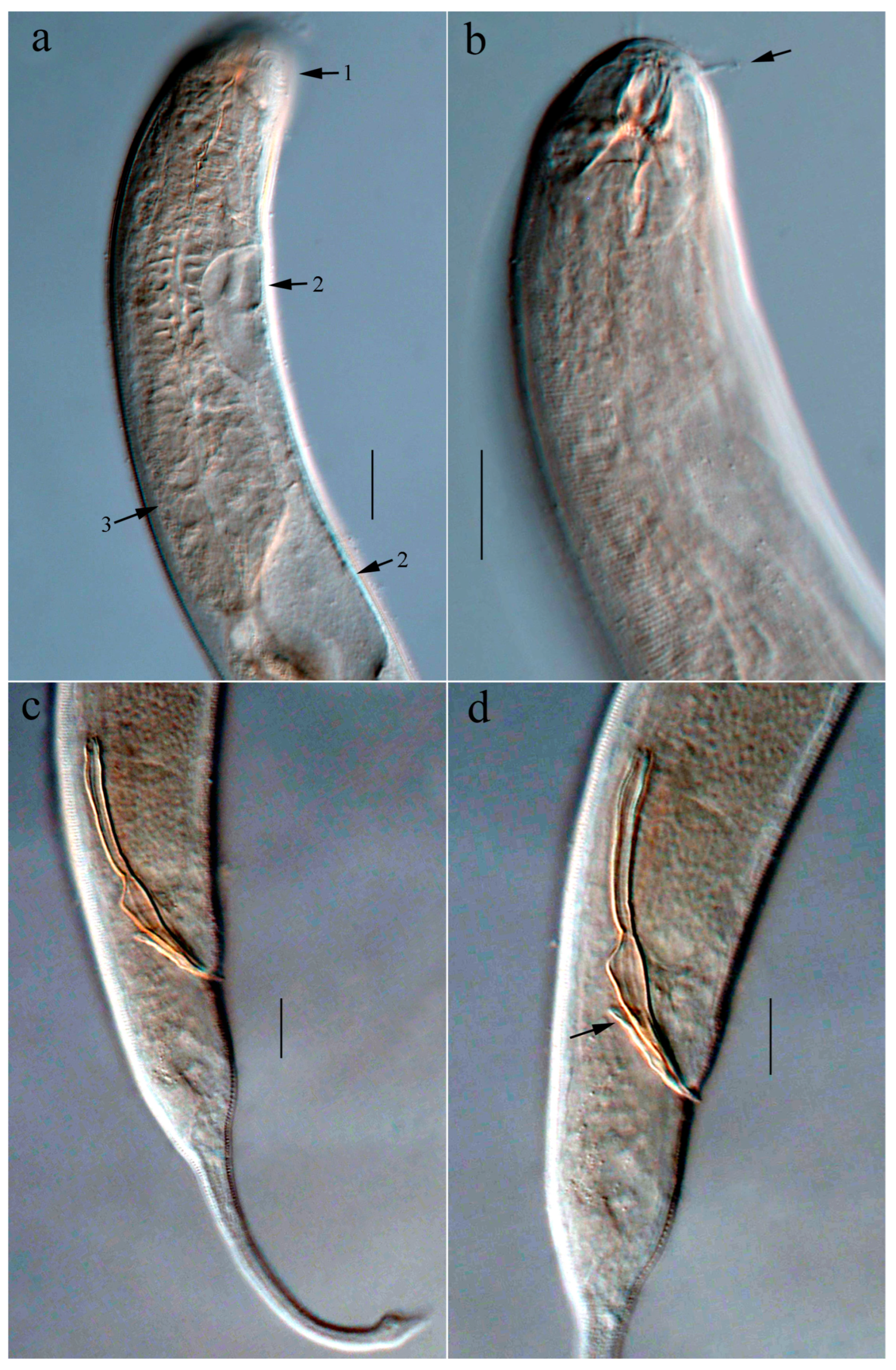
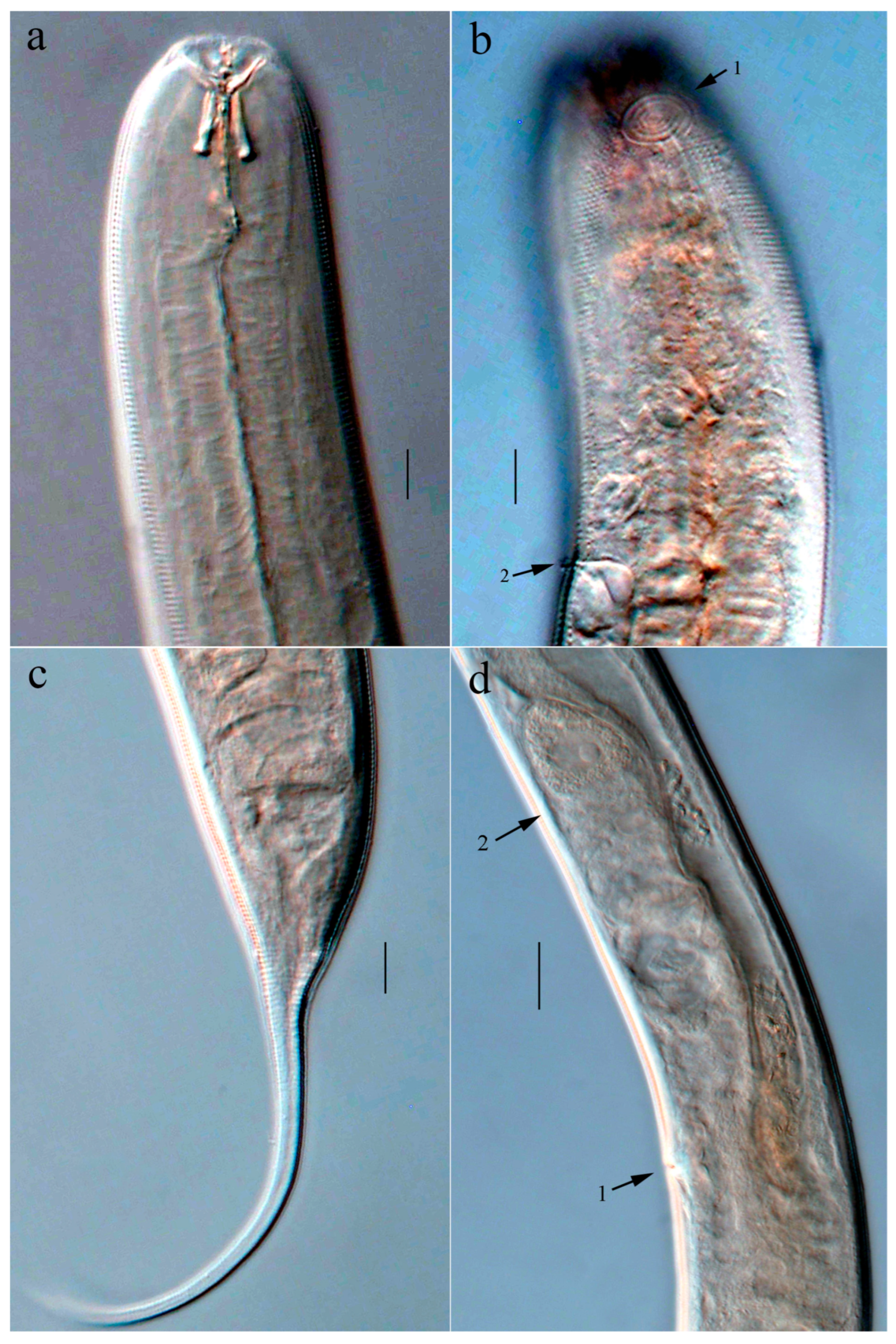
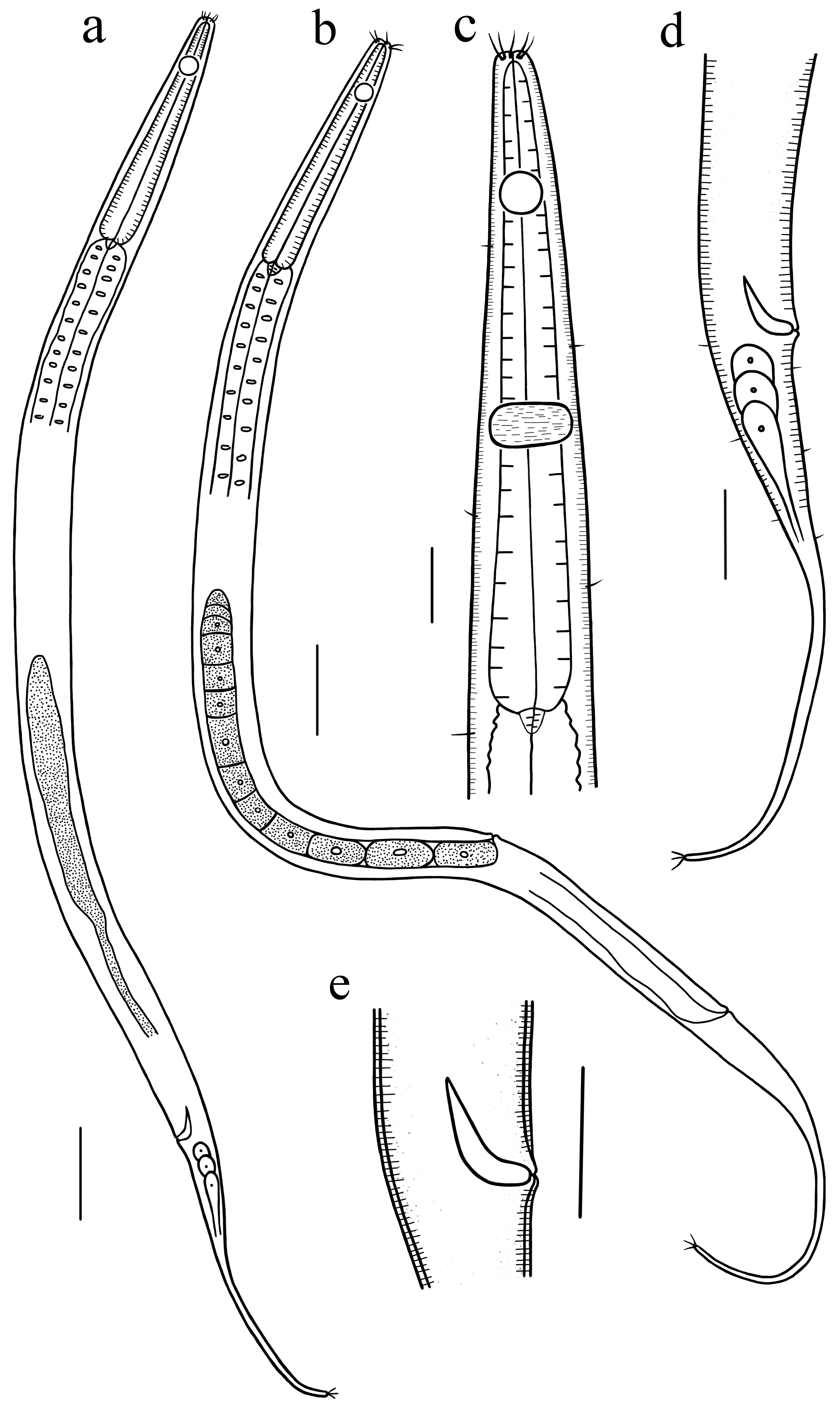
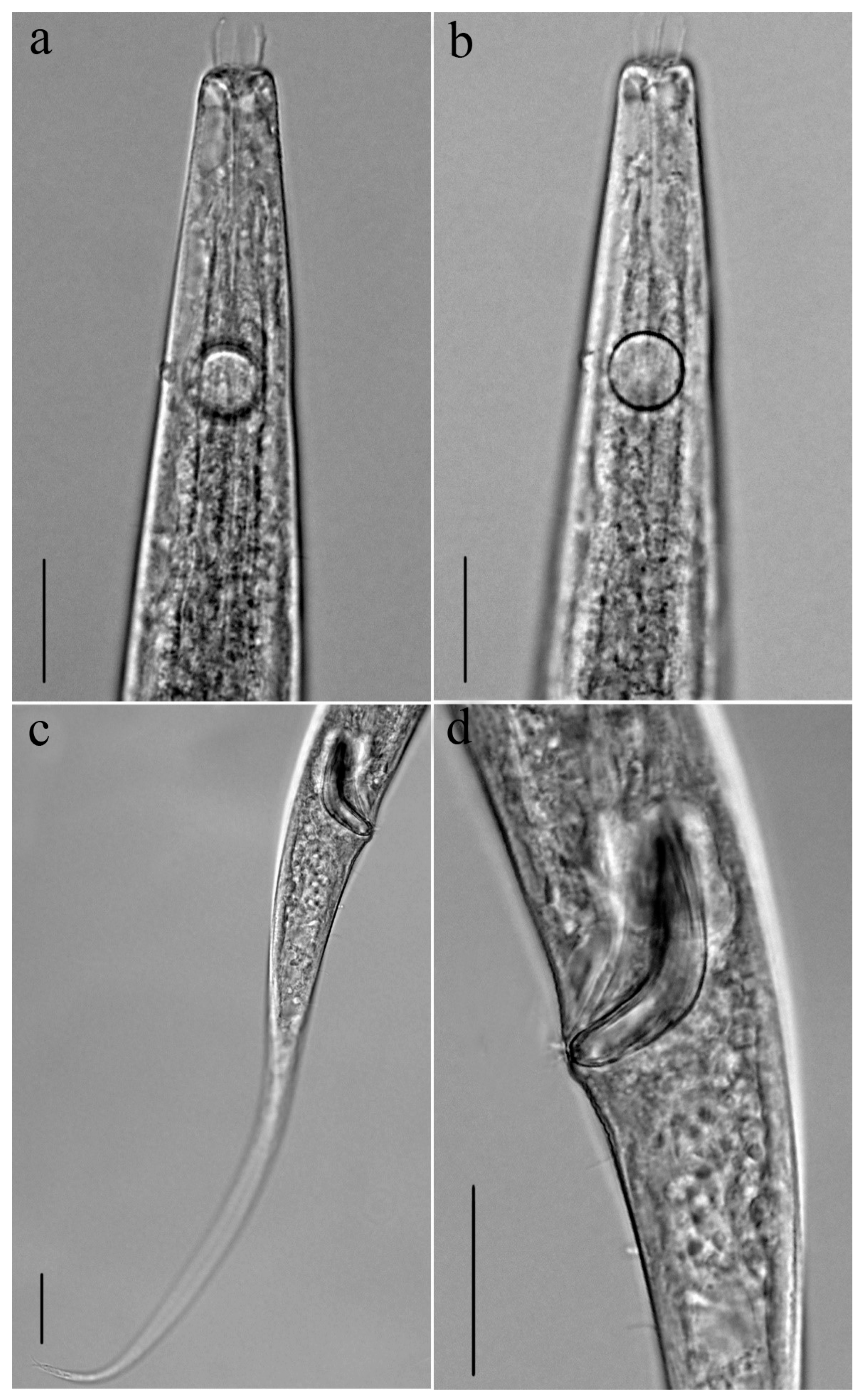
Description: Males: Body cylindrical, with a slight narrowing toward the posterior extremity. Transverse rows of punctations adorn the cuticle, while the lateral field bears punctations that are larger and less densely arranged. No cuticular pores were detected. Somatic setae short and robust, 2–3 µm long, arranged in longitudinal lines at cervical region. Lip region moderately rounded. All head sensilla papilliform. Six outer labial papillae, level with the four cephalic papillae. The amphideal fovea, which exhibits a multispiral structure of 4.5–5 turns, has a width that is approximately 48% of the body diameter and is located at the center of the buccal cavity. Buccal cavity large, with its anterior and posterior sections separated by a pair of denticle rows, and measures around 20 µm in depth. Within the cup-shaped anterior region of the buccal cavity, there are three groups of rhabdions, each group consisting of three sclerotized elements. In the narrower posterior region of the buccal cavity, three Y-shaped sclerotized rhabdions with swollen bases are present, each ranging from 8 to 10 µm in length. Exhibiting a cylindrical form, the pharynx has its anterior region surrounding the buccal cavity and lacks a posterior bulb. The lumen of the esophagus is cuticularized. The esophagus houses the nerve ring, which is positioned at its mid-length. Secretory–excretory system is observed. A small renette cell occurs at the cardia region. The ampulla is expanded. Positioned immediately behind the nerve ring, the excretory pore is in close proximity to it, 87–105 µm from the body’s anterior end. Surrounded by the intestine, the cardia is relatively small.
The reproductive system is diorchic and consists of two testes that are opposed and extended. The anterior testis lies on the right side of the intestine, while the posterior testis, which is smaller, is situated on the left. Spicules are paired structures, arched, proximal end not enlarged, middle portion slightly broad, tapering distally, about 1.4 cloacal body diameters in length. Gubernaculum exhibits a slender form, composed of two separate lateral pieces that taper distally and adjoin the dorsal side of the spicules. A short ventral precloacal seta 3 µm long, just anterior to the cloaca. Tail conico-cylindrical with three-fifths posterior cylindrical portion. Tail tip swollen with a spinneret, 3–4 µm long. Caudal setae short and distributed ventral-side. Three caudal glands are situated posterior to the intestinal blind end.
Females: Resembling the males, but amphideal fovea slightly smaller (32–39% of corresponding body diameter), intestine blind, without anus. Tail conical with a long filiform posterior part (taking the intestinal blind end as the starting point of the tail region), with swollen tip. Didelphic reproductive system contains two mutually opposed, reflexed ovaries. Positioned on the left of the intestine, the anterior ovary is opposed by the posterior ovary on the right side. Vulva is located slightly anterior to mid-body. Vagina sclerotized, straight and short.
Remarks:
Halichoanolaimus parvulus sp. nov. is distinguished by relatively smaller body, amphideal fovea consisting of 4.5–5 turns; in males, it measures 39–48% of the corresponding body diameter with four turns, and in females, 32–39% of the corresponding body diameter; spicules arched, have a broad central portion that tapers distally; gubernaculum slender comprising two separate lateral pieces tapering distally; only a single short precloacal seta observed; tail conico-cylindrical with three-fifths posterior cylindrical portion in males. The present species most resembles
Halichoanolaimus minutissimus Timm, 1961 in having small inconspicuous precloacal supplements [
31]. The new species also resembles
H. minutissimus with its larger amphid (5 turns versus 3.5 turns), longer spicules (46–49 µm versus 20 µm), longer gubernaculum (20–22 µm versus 10 µm), and shorter tail (c′ = 3.5–4.8 versus 11 in males). The new species additionally resembles
Halichoanolaimus consimilis Allgén, 1933 [
32],
Halichoanolaimus ossilagulus Leduc, 2020 [
18],
Halichoanolaimus ovalis Ditleversusen, 1921 [
33] and
Halichoanolaimus pumilus Leduc, 2020 [
18] in terms of body dimensions and tail morphology (conico-cylindrical). It can be distinguished from
H. consimilis by its relatively shorter spicules (46–49 versus 72 µm in
H. consimilis), only having a single precloacal seta (versus four precloacal supplements), and its arched spicules versus the inverted S-shape in
H. consimilis. The new species can be differentiated from
H. ossilagulus by its shorter spicules (46–49 versus 56–59 µm), one short precloacal seta versus 2–3 precloacal supplements, and longer tail with a swollen tip (c′ = 3.5–4.8 versus 2.4–3.2, tail tip tapered in
H. ossilagulus). The new species is readily differentiated from
H. ovalis by its number of precloacal supplements (a precloacal seta versus 7–9 papilliform supplements), shorter spicules (46–59 µm versus 68–73 µm), and shorter gubernaculum (20–22 µm versus 37–43 µm). The new species is separated from
H. pumilus by a slightly longer body length (935–1095 µm versus 756 µm), fewer turns of the amphideal fovea (4.5–5 versus 6.5), having a single precloacal seta versus three precloacal cylindrical cuticularized structures, and its tail tip being swollen (versus tapered).
3.2. Cobbionema sinica sp. nov.
ZooBank registration: urn:lsid:zoobank.org:act:BF7C3B39-335C-49B2-B9CC-FC83AC35C210
Class Chromadorea Inglis, 1983
Order: Chromadorida Chitwood, 1933.
Family: Selachinematidae Cobb, 1915.
Genus: Cobbionema Filipjev, 1922.
Diagnosis (modified from Leduc [
34], Tchesunov [
35], and Ahmed et al. [
23]): Cuticle bears fine transverse rows of dots and shows no lateral differentiation. Sensilla at the anterior end arranged in three circles: one circle of six inner labial sensilla, and six outer labial setae positioned slightly anterior to four cephalic setae. Four cephalic setae markedly exceeding the outer labial sensillae in length. Amphideal fovea multispiral with circular outline. Buccal cavity surrounded by anterior pharyngeal bulb, and forming anterior and posterior chambers through two transverse denticle bands. Anterior chamber barrel-shaped or cylindrical, with 12 or six cuticularized rhabdions; posterior buccal chamber cone-shaped, with three strongly cuticularized Y-shaped mandibles. Three well-developed, acute tines (one middorsal, two ventrosublateral), located slightly anterior to the anterior circle of denticles. Male possesses two opposed testes, one extended and the posterior one reflexed. Tail conical or conico-cylindrical.
Diagnosis: Length of cephalic setae is 6 µm; amphideal fovea shows 4–4.5 coils; the pharynx bears a prominent anterior bulb and enlarged posterior region; arrow-shaped spicules measure 2.1–2.5 cloacal body diameters; gubernaculum exhibits a rod-like shape, and is aligned with the distal portion of the spicules, 25–27 µm long; tail exhibits a conico-cylindrical shape, with the terminal end broadened and a distinct spinneret; precloacal supplements absent.
Holotype and paratype material: Two males and one female were obtained and examined: Holotype (m1) on slide 3600-4-2-2, paratype male 2 (m2) and female (f1) on slide 3400-1-0-1.
Type locality and habitat: Holotype (m1) was recovered from the muddy seabed sediment at Station 3600-4 (36°0′ N, 120°50′ E, water depth 30 m) in the Yellow Sea. Paratypes were obtained from the silty seabed sediment at Station 3400-1 (34°0′ N, 120°48′ E, water depth 48 m) in the Yellow Sea.
Etymology: The species’ name is derived from China, where it was first identified.
Measurements: All morphometric data are presented in
Table 1.
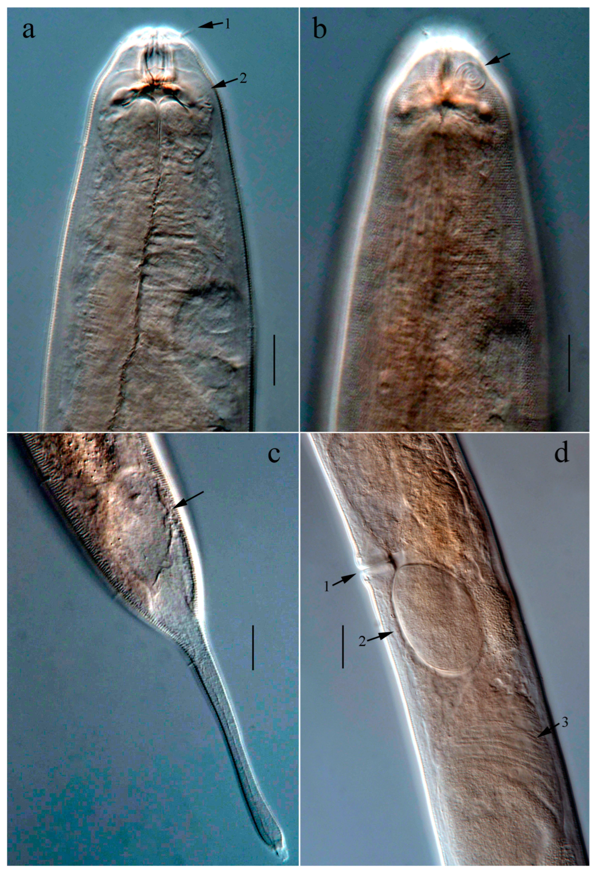
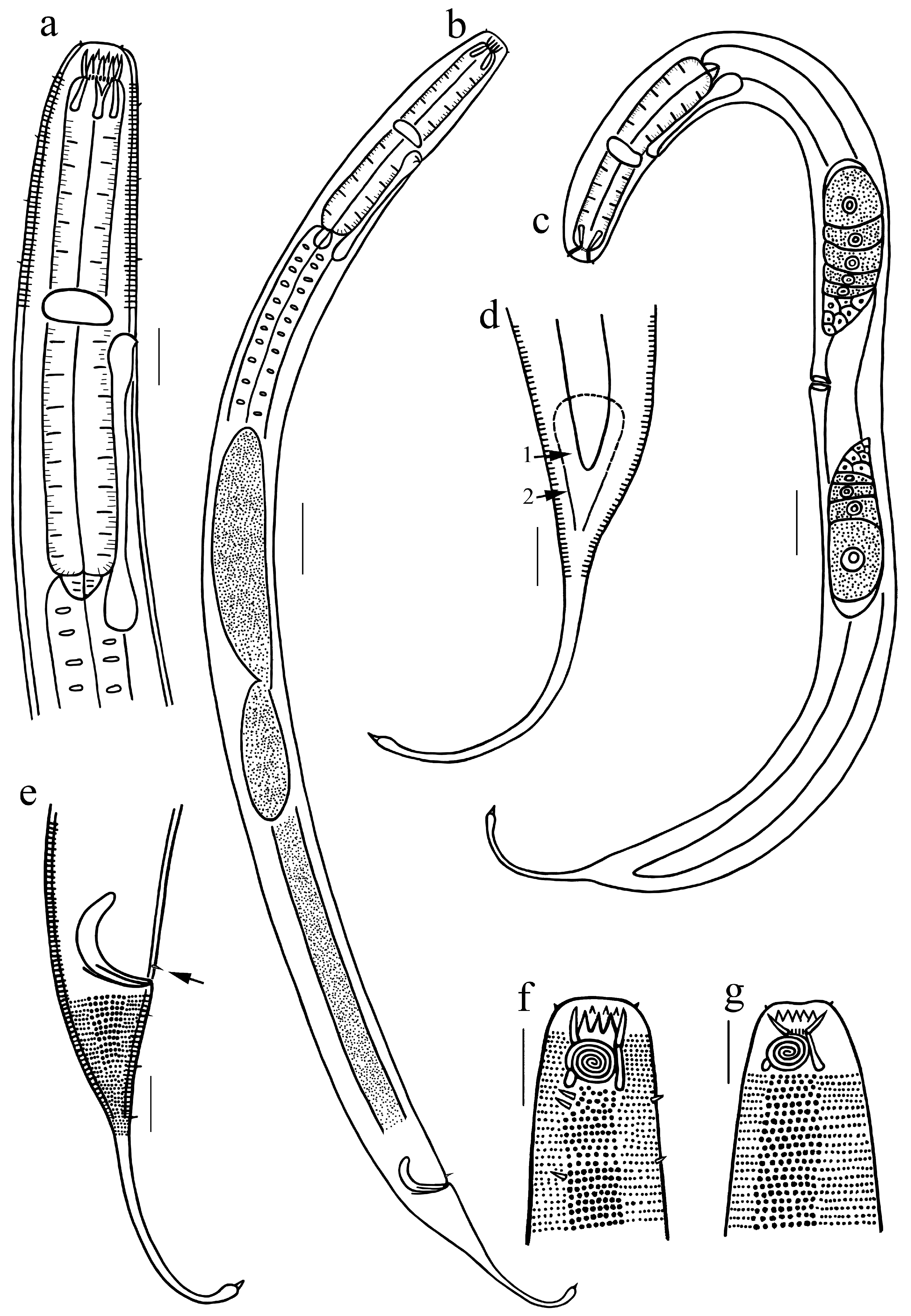
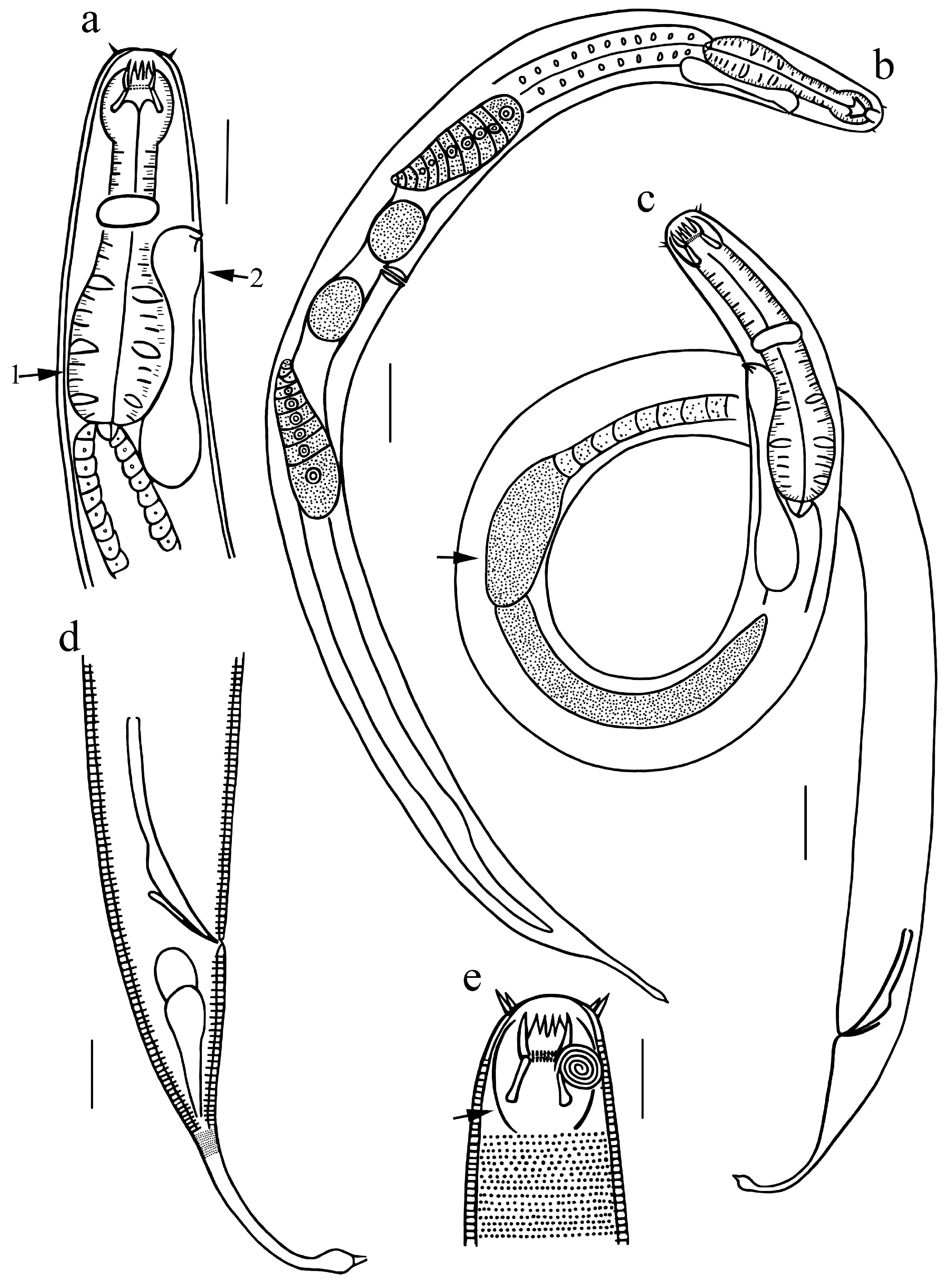
Description: Males: Body form cylindrical, narrowing towards both ends; cuticle ornamented with transverse punctation rows, lacking lateral differentiation. Cuticle pores not detected. Inner labial sensilla are papilliform, whereas the six outer labial setae, measuring approximately 4 µm in length, are positioned slightly anterior to the four cephalic setae, which are about 6 µm long. Somatic setae, which are short and sparsely present, exhibit an irregular distribution pattern across the body. Head blunt and rounded, not set off. Buccal cavity large, 30–34 µm deep and 10–11 µm wide at the middle, partitioned into two parts by two rows of denticles. Anterior region of the buccal cavity bears six cuticularized denticles, each of which is provided with two pairs of sharp projections at its posterior tip. The posterior portion of the buccal cavity contains three Y-shaped rhabdions that taper toward the posterior end. Amphideal fovea multispiral with 4.5 turns, approximately 15 µm in diameter (i.e., 47–48% of the body diameter at that position), and is positioned at the mid-length of the buccal cavity. Secretory–excretory system with pore opening at 65 µm from the anterior end. Large ventral gland just below the pharyngeal base. Pharynx relatively short, with a distinct anterior bulb and an enlarged posterior portion, cardia conical, surrounded by the intestine. Nerve ring indistinct, just anterior to excretory pore. Tail is conico-cylindrical, with a length equivalent to 3–4.7 cloacal body diameters, and is characterized by a distinctly expanded terminal region bearing a conical spinneret (8 µm long). Three caudal glands present, situated posterior to the cloacal.
Reproductive system features a pair of testes oriented in opposite directions. Anterior testis is outstretched and positioned to the left of the intestine, whereas the posterior testis is reflexed and located on the right side of the intestine. Spicules paired, 80–85 µm long, i.e., 2.1–2.5 cloacal body diameters, slightly curved and arrow-shaped, swelling dorsally near the distal end, then tapered gradually to the pointed tip. Gubernaculum rod-like, bent distal portion, 25–27 µm long, parallel to the distal ends of spicules, without apophyses. Pre-cloacal supplements not present.
Females: Resembling males, but body larger and amphideal fovea slightly smaller (4 turns and 37% of corresponding body diameter), intestine blind, without rectum and anus. Tail conico-cylindrical, 3.4 corresponding body diameters (the intestinal blind end defined as the starting point of the tail), with a slightly swollen terminal end and a 5 µm long spinneret. Reproductive system didelphic, comprising two opposed ovaries that are reflexed. Anterior ovary on the left of intestine; posterior ovary on the right of intestine. Vulva positioned before the mid-point of body, i.e., 40% of the total body length measured from the anterior extremity. Vagina sclerotized, straight and short, 23 µm long. Mature egg oval, approximately 86 µm long and 56 µm wide.
Remarks:
Cobbionema sinica sp. nov. is distinguished by having anterior esophageal bulb and an enlarged posterior esophageal portion, cephalic setae 6 µm long, amphideal fovea with 4–4.5 turns, spicules 2.1–2.5 cloacal body diameters long, arrow-shaped, gubernaculum rod-like, parallel to the distal part of spicules, 25–27 µm long, precloacal papilla absent; the tail is conico-cylindrical, featuring a swollen distal end and a distinct spinneret.
C. sinica resembles
Cobbionema trigamma Leduc, 2013 [
34] and
Cobbionema cylindrolaimoides Schuurmans Stekhoven, 1950 [
36] with respect to body dimensions and tail morphology. However, it can be distinguished from
C. trigamma by its longer tail, longer posterior cylindrical section and relatively long spinneret (c′ = 3.0–4.7 versus c′ = 2.2–2.8, cylindrical section shorter and digitate distal section). The new species can be distinguished from
C. cylindrolaimoides in that the latter possesses lateral alae and body pores, cephalic sensilla 8–10 µm long, a sexually dimorphic amphideal fovea with 3.5–4.25 turns in males and 3.25–3.5 turns in females, 89–98 µm long spicules, a single midventral pre-cloacal papilliform sensillum, and a shorter tail (c′ = 1.8–2). In body size, the new species is also comparable to
Cobbionema acuminata Ahmed, Bostrom & Holovachov, 2020 and
Cobbionema brevispicula Ahmed, Bostrom & Holovachov, 2020 [
23], but can be distinguished from the latter two species by its conico-cylindrical tail with enlarged extremity (versus conical tail with tapered extremity), longer spicules (80–85 µm versus 55–65 µm in
C. acuminata and 29–32 µm in
C. brevispicula), and absence of precloacal papilla (versus precloacal papilla present in the latter two species).
3.3. Linhystera longispicula sp. nov.
ZooBank registration: LSIDurn:lsid:zoobank.org:act:3457B40A-479E-48E9-AC16-DA1CFBF40A5F
Class Chromadorea Inglis, 1983
Order: Monhysterida Filipjev, 1929.
Family: Xyalidae Chitwood, 1951.
Genus: Linhystera Juario, 1974.
Diagnosis (modified based on Yu et al. [
27] and Lv et al. [
25]): Cuticle exhibiting fine striation. Anterior sensilla are organized in two circles (6 + 10): inner labial papilla in one circle and difficult to recognize, outer labial setae and cephalic setae of the second circle with approximately equal in length. Four subcephalic setae might be present. Buccal cavity minute and slit-like. Amphideal fovea circular, far from the anterior end. Females with a monorchic reproductive system, ovary outstretched, positioned to the left of the intestine. Males with a monorchic reproductive system; anterior testis located to the left of the intestine. Posterior testis reduced or absent. Spicules slender and simple. Gubernaculum present or absent. Gubernaculum apophysis present or absent. Tail conical-cylindrical or filiform, terminal setae present or absent.
Holotype and paratype material: Specimens included three males, one female, and one juvenile, all of which were examined and measured: holotype male (m1) on slide NH8-3, paratype male (m2, m3) on slide S7-1, paratype female (f1) and juvenile (juv) on slide S7-2.
Type locality and habitat: The holotype specimen was collected from subtidal muddy sediment at Station NH8 in the South China Sea (19°53′48″ N, 115°42′26″ E, water depth 1553 m). All paratype specimens were found in subtidal muddy sediment at Station S7 in the South China Sea (20°9′0″ N, 117°19′20″ E, water depth 1687 m).
Etymology: This species’ name refers to its elongate spicules.
Measurement: All morphometric data are presented in
Table 2.
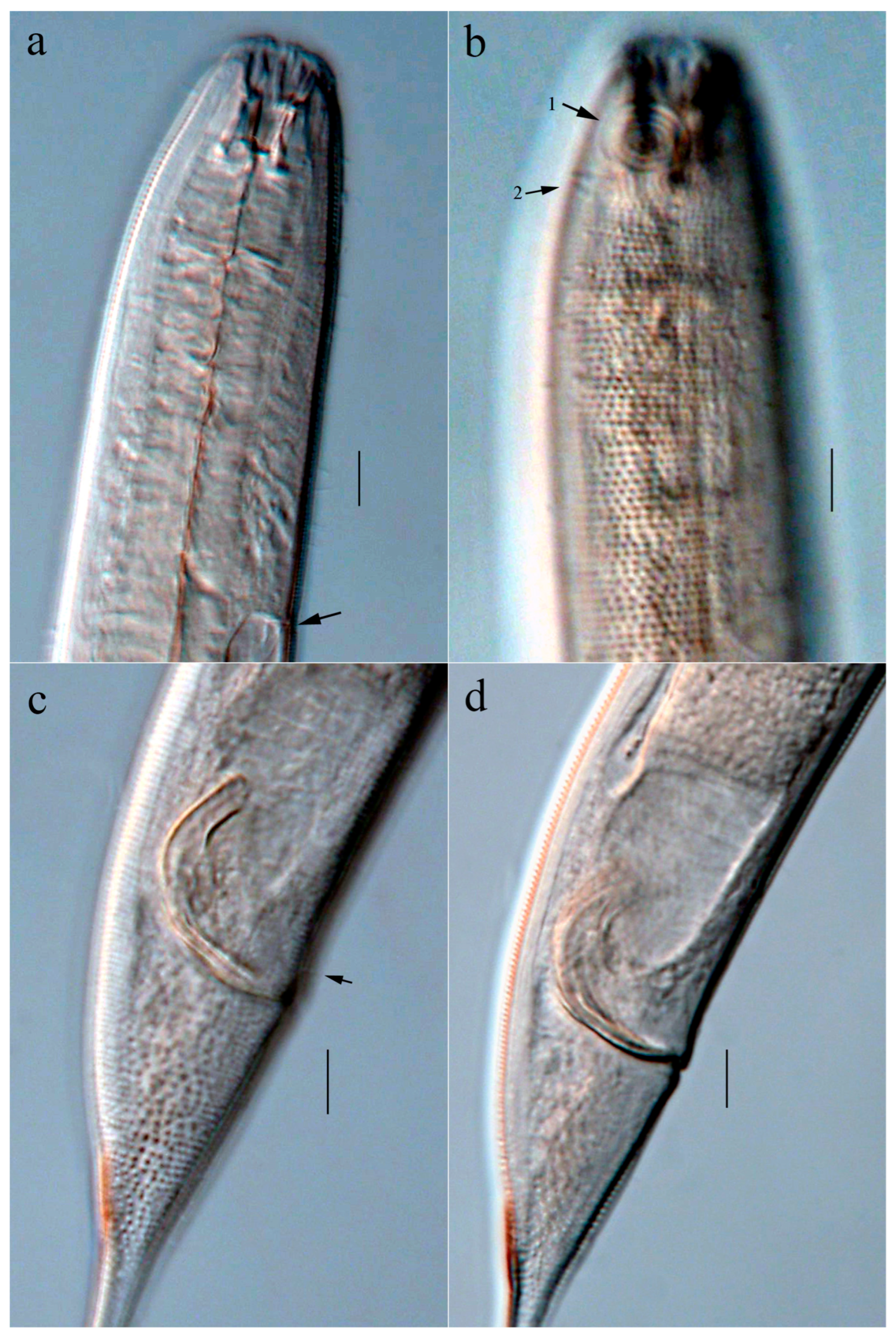
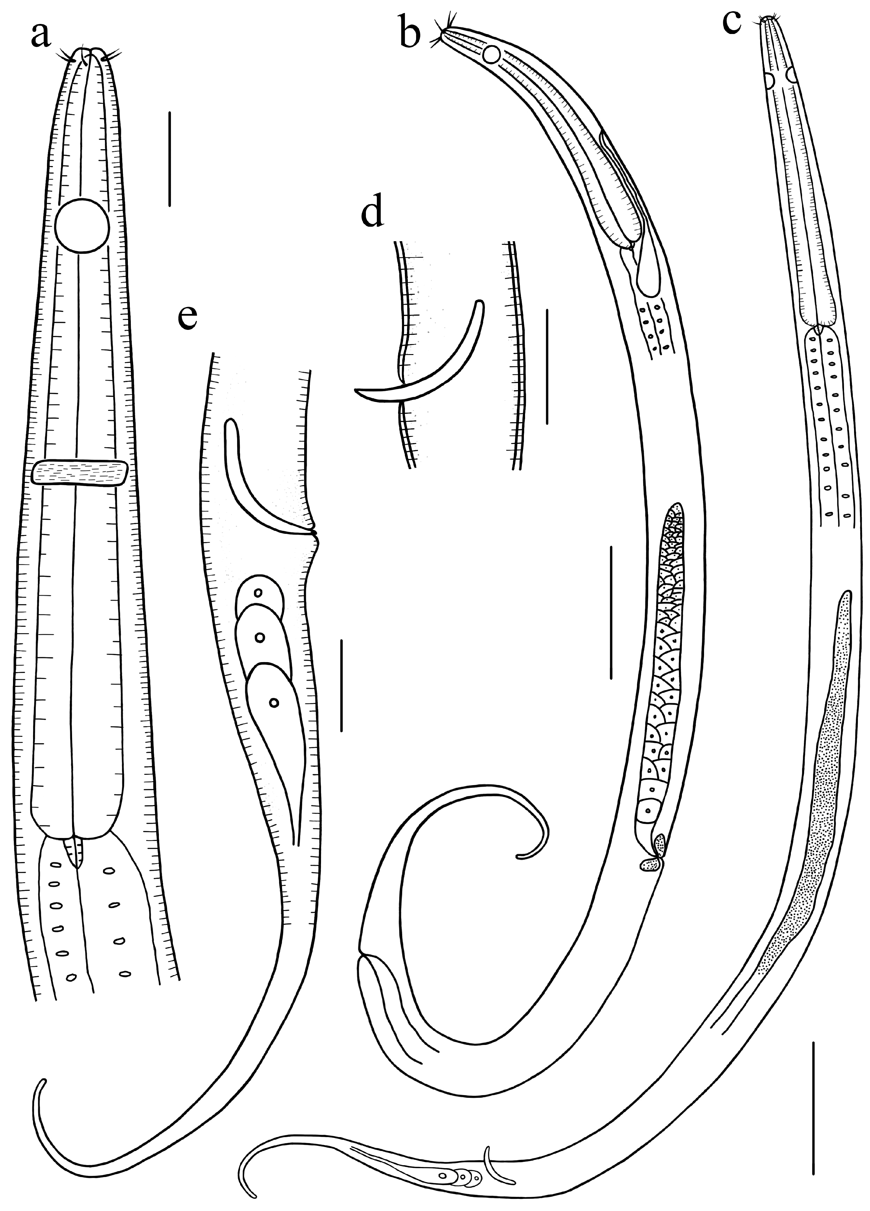
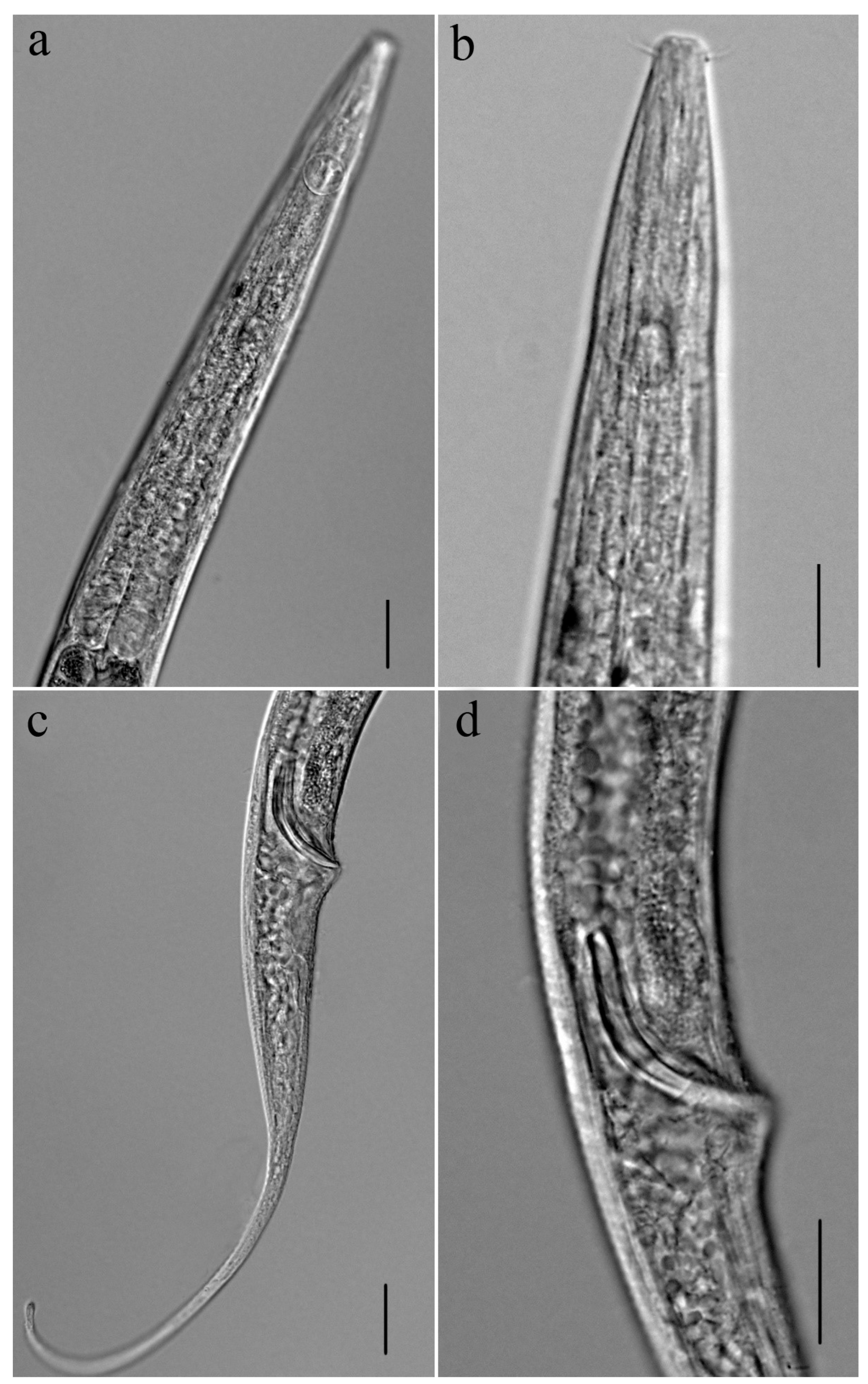
Description: Males: Body cylindrical, slender, narrowing toward both ends, length 1285–1399 µm. Cuticle with faint transverse striations, Cervical setae distributed irregularly at the amphideal region. Head narrow, 8–9 µm in diameter. Buccal cavity minute, slit-like. Inner labial sensilla not observed, four cephalic setae together with six outer labial setae lie almost in one ring and share the same length. Amphid circular, measuring 13–14 µm in diameter, corresponding to approximately 71% of the body diameter at that position, and located at 4.5 head diameter to the anterior end. Cylindrical esophagus with slight basal broadening. Well-developed conical cardia encircled by intestinal tissue. No ventral gland or excretory pore detected. Nerve ring located at the midpoint of the esophageal length. Tail conico-cylindrical, 199–240 µm long, cylindrical portion occupying about 50% of tail length; caudal setae present, distributed irregularly at the tail region, terminal setae absent. Three caudal glands present, situated posterior to the cloacal.
An anteriorly extended testis is positioned at the left of the intestine in the reproductive system. Spicules elongate, slender and arcuate, 45–47 µm long, approximately 2.2 times the body diameter at cloaca. Gubernaculum and precloacal supplements absent.
Females: Resembling the males in most features. A single ovary, extended toward the anterior, is positioned to the left of the intestine. Vulva is positioned at mid-body, at approximately 57% body length posterior to the anterior end. Vagina short and sclerotized. Spermatheca not observed.
3.4. Linhystera nanhaiensis sp. nov.
ZooBank registration: urn:lsid:zoobank.org:act:658FD2D4-2E80-43D9-B8BB-CFF478FD0E25
Class Chromadorea Inglis, 1983
Order: Monhysterida Filipjev, 1929.
Family: Xyalidae Chitwood, 1951.
Genus: Linhystera Juario, 1974.
Holotype and paratype material: Specimens included three males and one female, all of which were measured: holotype (m1) on slide 138-5-1, paratype male 2 (m2) and male 3 (m3) on slide S57, and female (f1) on slide on S55.
Type locality and habitat: The holotype was obtained from the muddy sediment on the sea floor at Station DIVE138 in the South China Sea (19°0′53″ N, 115°43′55″ E, water depth 2500 m). Paratypes were sampled from the silt sediment on the sea floor at Station S55 (18°0′9″ N, 114°0′12″ E, water depth 3257 m) and Station S57 (17°59′51″ N, 112°0′26″ E, water depth 2448 m) in the South China Sea.
Etymology: This species derives its name from the South China Sea, the collection site of the holotype.
Measurement: All morphometric data are presented in
Table 2.
Description: Males: Body form cylindrical, slender, tapering at both extremities, length 1478–1670 µm. Cuticle with faint transverse striations. Somatic setae present, short and distributed irregularly. Head narrow, 10.8–11.4 µm in diameter, buccal cavity minute and slit-like. Four cephalic setae, along with the six outer labial setae, are organized in a nearly continuous ring and have a similar length of approximately 10 µm. Circular amphideal fovea ranges from 10 to 11 µm in diameter, covering approximately 62% of the local body width, and is positioned 4.4 head diameters posterior to the anterior end. Esophagus cylindrical, slightly broadening towards the base, not expanded into a bulb. Well-developed and conical, the cardia is enclosed by surrounding intestinal tissue. The ventral gland and excretory pore were not detected. Nerve ring at the midpoint of esophageal length. Tail conico-cylindrical, 237–277 µm long, about 7.7 times of body diameter at cloaca, cylindrical part constituting roughly 60% of the tail’s length, caudal setae are distributed randomly on the ventral side, three terminal setae present, 4–5 µm for each. Three caudal glands present, situated posterior to the cloacal.
An anterior outstretched testis is present in the reproductive system and positioned left of the intestine. Spicules ox-horn-shaped, 46–50 µm in length, approximately 1.2 times the cloacal body diameter, anterior half tapering with pointed end, and posterior half inflating with blunt end. Gubernaculum and precloacal supplements absent.
Females: Most characteristics are similar to those of the male. Reproductive system includes a single anterior ovary, located to the left of the intestine. Vulva located at post-mid of body, 56.9% body length from the anterior body end. Vagina short and sclerotized. Spermatheca not observed.
Remarks:
Linhystera longispicula sp. nov. is distinguished by a cuticle with faint transverse striations; a minute, slit-like buccal cavity; four cephalic setae together with six outer labial setae lie in one ring, 3.3–4 µm long and equal in length; a circular amphideal fovea located far from the anterior end; slender, symmetrical spicules measuring 2.2 cloacal body diameters; absence of a gubernaculum; and a conico-cylindrical tail without terminal setae.
Linhystera nanhaiensis sp. nov. is characterized by a transversely striated cuticle; a minute, slit-like buccal cavity; four cephalic setae together with six outer labial setae lie in one ring, 9.5–11 µm long; a circular amphidial fovea situated distantly from the anterior end; a long conico-cylindrical tail with three terminal setae; and ox-horn-shaped spicules measuring 1.2 cloacal body diameters. It represents one of the largest species in the genus (the ratio of body length to maximum body diameter = 26–29). Both species conform closely to the diagnosis of the genus
Linhystera. Following the diagnostic criteria of Lv et al. [
25], the absence of a gubernaculum distinguishes these two new species from previously described congeners in which this structure is present or not reported.
Linhystera longispicula sp. nov. resembles
Linhystera problematica Juario, 1974 in having a conico-cylindrical tail with the cylindrical portion occupying about half of the tail length, but differs in its longer body (1285–1399 µm vs. 720–833 µm), larger amphidial fovea (13–14 µm vs. 5–8 µm), more posterior amphidial position (38–40 µm vs. 8–11 µm from the anterior end), and longer tail (199–240 µm without terminal setae vs. 95–106 µm with three terminal setae) [
22]. It is also similar to
Linhystera longa in body length (approximately 1300 µm), but differs in its lower a value (33 vs. 50–72), absence of cervical setal crown (vs. present), shorter outer labial setae (3.3–4 µm vs. 6 µm), and tail without terminal setae (vs. with three terminal setae) [
37]. Compared to
Linhystera filiformis, the new species can be further distinguished by its more posterior amphideal fovea (38–40 µm vs. 6–8 µm from the anterior end), larger maximum body diameter (35–39 µm vs. 19–22 µm), and conico-cylindrical tail without terminal setae (vs. filiform tail with three terminal setae) [
25].
Linhystera nanhaiensis sp. nov. resembles
Linhystera longa Pastor de Ward, 1985 in its relatively large body (>1200 µm) and irregularly distributed somatic setae, but differs by the absence of a crown of cervical setae (vs. present) and its more slender body (the ratio of body length to maximum body diameter = 27–29 vs. 50–72) [
37]. It also resembles
Linhystera problematica in its tail morphology, but differs in its longer body (1478–1670 µm vs. 720–833 µm), more posterior amphideal position (47–50 µm vs. 8–11 µm from the anterior end), and longer tail (237–277 µm without terminal setae vs. 95–106 µm with three terminal setae) [
22]. The two species newly identified from the South China Sea,
Linhystera longispicula and
L. nanhaiensis, are similar in body size and in lacking a gubernaculum, but can be clearly distinguished by spicule morphology: they are slender and arcuate in
L. longispicula versus ox-horn-shaped in
L. nanhaiensis. At present, the genus
Linhystera comprises seven recognized species worldwide, including the two newly described species presented herein. Of these, one species has been recorded from the Yellow Sea, two from the East China Sea, and four from the South China Sea.