Article

The Discovery and Delimitation of a New Cryptic Species of Spirinia (Nematoda: Desmodoridae) Using SSU and LSU rDNA Divergence †

1
Department of Environmental Science, Hanyang University, Seoul 04763, Republic of Korea
2
Center for Creative Convergence Education, Hanyang University, Seoul 04763, Republic of Korea
3
Research Institute of Natural Science, Hanyang University, Seoul 04763, Republic of Korea
*
Author to whom correspondence should be addressed.
urn:lsid:zoobank.org:pub:172B5CE6-E87B-4F4B-AB32-FC7183935F26.
J. Mar. Sci. Eng. 2025, 13(7), 1251; https://doi.org/10.3390/jmse13071251 (registering DOI)
Submission received: 2 June 2025 / Revised: 23 June 2025 / Accepted: 26 June 2025 / Published: 28 June 2025
(This article belongs to the Special Issue Biodiversity and Population Ecology of Marine Invertebrates)

Abstract

The cosmopolitan nematode Spirinia parasitifera has long been considered a single, morphologically variable species; however, mounting molecular evidence suggests that it represents a complex of cryptic taxa. In this study, we describe Spirinia koreana sp. nov., a new species collected from intertidal sediments of the Republic of Korea. The new species exhibits a high degree of morphological resemblance to both S. antipodea and S. parasitifera, with overlapping ranges in most morphological traits. While certain measurements, such as relatively shorter body length, more slender form (higher a ratio), moderately long tail length, and shorter spicule length differ from those in some described populations, no single morphological character alone reliably separates S. koreana from all previously reported specimens of S. parasitifera or S. antipodea. Nevertheless, molecular evidence from multiple genetic markers clearly supports its distinction as a separate species. Molecular data from mitochondrial COI, 18S rRNA, and 28S rRNA genes confirm the genetic distinctness of the Korean specimens from S. parasitifera and S. antipodea. Notably, S. koreana sp. nov. differs from other Spirinia species by 2.1–3.4% in 18S and up to 34.4% in 28S sequences, surpassing thresholds previously used to delimit marine nematode species. Our results emphasize the value of integrative taxonomy combining fine-scale morphology and multi-marker molecular data to uncover hidden diversity in meiofaunal nematodes.

1. Introduction

The genus Spirinia Gerlach, 1963, belongs to the family Desmodoridae and comprises a diverse group of free-living marine nematodes commonly found in coastal regions all over the world. Species belonging to the genus are typically distinguished from other desmodorids by a finely striated cuticle, a reduced buccal cavity (very small, with minute dorsal tooth or no teeth), a non-spiral amphid with a cuticle that entirely surrounds or partially (posteriorly) surrounds the amphid at the cephalic setae level, indistinct supplementary organs, and a conical tail. Among the 14 currently valid Spirinia species, S. parasitifera (Bastian, 1865) is particularly noteworthy for its cosmopolitan distribution, with records spanning Europe, the Mediterranean [1], the Americas [2,3,4], Asia [5], and Australia [6]. This wide geographic range is accompanied by substantial morphometrical intraspecific variation, especially in body and tail length, spicule size, and de Man indices (morphometric ratios), leading to the long-standing hypothesis that S. parasitifera may not be a single species but instead a cryptic species complex.
Leduc and Zhao (2019) were among the first to formally recognize this issue, proposing that at least three distinct species may exist within what has historically been identified as S. parasitifera. They also described S. antipodea as a new species, based on divergence in SSU and LSU rRNA gene sequences [7].
This study builds on previous work by further investigating the cryptic nature of S. parasitifera. For the first time in East Asia—aside from an earlier record from the Maldives [5]—we collected over 20 specimens (11 males, 12 females, and 4 additional specimens for molecular analysis) morphologically similar to S. parasitifera from Sunnyeobawi Beach, Incheon, a subtidal zone on the east coast of Republic of Korea. From these Korean specimens, we successfully obtained mitochondrial COI, 18S rRNA, and 28S rRNA sequences, and our results demonstrate that these individuals are genetically distinct from both S. parasitifera and S. antipodea.
Our findings support the growing consensus that S. parasitifera comprises multiple cryptic species. Furthermore, they highlight the critical role of molecular markers—particularly SSU and LSU rRNA—in delineating species boundaries and elucidating relationships among closely related genera such as Chromaspirina and Perspiria. In this study, we describe a new species, Spirinia koreana sp. nov., collected from intertidal sediments along the coast of Republic of Korea. We provide a comprehensive description including diagnostic morphological features, morphometric measurements, line drawings, and a molecular phylogenetic analysis based on three genetic markers (K2P distances and Bayesian Inference trees).

2. Materials and Methods

2.1. Sample Collection and Morphological Analysis

Nematode samples were qualitatively collected in March 2025 from Seonnyeobawi Beach, located southwest of Incheon on Yeongjongdo Island. Two replicate sediment samples were taken using a plastic acrylic corer: one immediately fixed in 5% neutralized formalin for morphological analysis and the other in 99% ethanol for molecular analysis. Upon return to the laboratory, the ethanol-fixed samples were stored in a freezer at −24 °C, while the formalin-fixed samples were kept at room temperature.
Formalin samples were transferred to a Petri dish, where nematodes were examined under a dissecting microscope (Olympus SZX7, Tokyo, Japan) and handpicked into a dish containing a solution of 10% glycerol and distilled water. This dish was placed in a dry oven at 40 °C for two to three days to allow gradual evaporation, enabling the nematodes to be transferred to pure glycerin without shrinkage.
Specimens were mounted on glass slides using the standard wax-ring method [8]. Nematodes of interest were observed and photographed using a compound microscope (Olympus BX51, Tokyo, Japan) equipped with a Canon EOS 90D camera (Tokyo, Japan). Morphometric values were measured and recorded using IC Measure v.2.0.0.286 and Helicon Focus v.8.3.3 software. All pencil drawings were produced using the same optical microscope equipped with a drawing tube, and final line illustrations were digitally rendered.

2.2. DNA Extraction and Amplification

Nematode specimens were handpicked from ethanol-fixed samples and transferred to temporary glass slides for quick observation under a compound microscope to ensure accurate molecular data linkage with species identification. Each specimen was photographed to create digital vouchers and provide photographic evidence for future reference. Individual specimens were then transferred into separate wells of a 12-well dish containing ultrapure water and rinsed for 20 min. DNA extraction was performed using the HotSHOT protocol described by Meeker et al. (2007) [9], yielding a total of 110 µL of DNA template. Each worm was placed in a microcentrifuge tube containing 95 µL of ultrapure water and 5 µL of 1 M NaOH. Tubes were incubated in a thermocycler at 95 °C for 20 min, followed by cooling at 4 °C for 5 min. After centrifugation, 10× g µL of 1 M Tris-HCl was added to neutralize the solution, producing DNA templates ready for downstream amplification. The extracted DNA templates were either used immediately for PCR or stored at −24 °C. DNA amplification was carried out using the IP-Taq PCR premix (COSMOgenetech, Seoul, Republic of Korea), with each 20 µL reaction mixture consisting of 6 µL of DNA template, 2 µL of ultrapure water, and 1 µL of each primer. Detailed information regarding primers, amplification conditions, and references is provided below (Table 1). Amplification success was assessed by visualizing PCR products on a 1% agarose gel. Products confirmed to be amplified were sent to Bioneer (Daejeon, Republic of Korea) for purification and sequencing.

2.3. Phylogenetic Analysis

All obtained sequences were visualized using FinchTV v.1.4.0 (Geospiza, Inc., Seattle, WA, USA; https://digitalworldbiology.com/FinchTV, accessed on 1 May 2025), and the quality of each peak was assessed by comparing the forward and reverse strands. The forward and reverse sequences were aligned using ClustalW implemented in MEGA v.11.0.13. [13,14]. For SSU rDNA sequences, which consisted of two fragments, the sequences were merged using the online tool EMBOSS Merger (https://www.bioinformatics.nl/cgi-bin/emboss/merger, accessed on 1 May 2025), to obtain a nearly full-length 18S rRNA sequence [15]. The assembled sequences were used in BLAST searches were conducted using the NCBI online BLAST interface (BLAST+ v2.15.0; https://blast.ncbi.nlm.nih.gov, accessed on 1 May 2025) to identify closely related species. Pairwise genetic distances (divergence) for mtCOI, SSU, and LSU rDNA sequences were calculated using MEGA 11 under the Kimura 2-parameter (K2P) model [16]. Due to the lack of available conspecific and congeneric sequences, mtCOI phylogenetic trees were not constructed. Instead, sequences retrieved from GenBank were aligned using MEGA11 for subsequent phylogenetic analysis. For the Maximum Likelihood (ML) approach, IQ-TREE (multicore v.2.0.3) [17] was used with the ModelFinder tool [18] to determine the best-fit model using the Akaike information criterion. The GTR + F + I + G4 model was determined to be the best-fitting substitution model for both the 18S and 28S rRNA datasets and was subsequently applied in IQ-TREE (web server) using the ultrafast bootstrap method with 1000 replicates [19]. For Bayesian Inference (BI) approach, the best-fit model was determined using jModelTest software v.2.1.7 [20]. The tree was generated using the MrBayes v.3.2.6 software [21]. The model prameters were as follows: Lset base = (0.2659 0.2002 0.2534 0.2805) nst = 6; rmat = (1.0000 2.9760 1.0000 1.0000 5.6381) rates = gamma shape = 0.5080; ncat = 4; and pinvar = 0.4580 with Microlaimus sp. set as the outgroup. Markov Chain Monte Carlo (MCMC) was run with ngen = 1,000,000; nchains = 4; sam-plefreq = 100; savebrlens = yes; printfreq = 1000; sump burnin = 250; and sumt burnin = 250. All trees were exported to FigTree v.1.4.4 for visualization modifications [22].

3. Results

3.1. Morphological Analysis

  • Systematics
Class Chromadorea Inglis, 1983
Order Desmodorida De Coninck, 1965
Family Desmodoridae Filipjev, 1922
Subfamily Spiriniinae Chitwood, 1936
Genus Spirinia Gerlach, 1963
  • Diagnosis (updated from Leduc & Zhao 2019)
Body with fine or distinct annulation and rounded or conical cephalic region. Annulated cephalic region; body annulations either completely or partially (posterior) surrounding amphidial fovea, but with few exceptions. Spiral amphidial fovea. Buccal cavity narrow, lightly cuticularized, no or small/inconspicuous dorsal tooth, no or minute ventrosublateral teeth. Pharynx with small pyriform, oval, or rounded posterior bulb without cuticularized lumen. Precloacal supplements usually absent. Mostly conical with a few exceptional conico-attenuated and usually short tail.
Type species: Sprinia parasitifera (Bastian, 1865) Gerlach, 1963.
  • List of valid species
  • Spirinia antipodea Leduc, 2019 (Leduc & Zhao 2019: 91–105, Figures 1–3; New Zealand, Pauatahanui Inlet, North Island gravel sand, intertidal zone) [7].
  • Spirinia gerlachi (Luc & De Coninck, 1959) Gerlach, 1963 (Luc & De Coninck 1959: 123–125, Figures 26–28; France, Roscoff Region, Coarse sand in a tidal pool behind Large gravel with Lithothamnium and Enteromorpha) [23].
  • Spirinia gnaigeri Ott, 1977 (Ott 1977: 134–137, Figures 40–45; Bermuda, intertidal sand flat in Tuckers Town Cove) [24].
  • Spirinia guanabarensis (Maria, Esteves, Smol, Vanreusel & Decraemer, 2009) Leduc & Verschelde, 2015 (Maria et al., 2009: 21–36, Figures 1 and 2; Brazil, Guanabara Bay, Bica Beach) [25].
  • Spirinia hopperi Coles, 1987 (Coles 1987: 79–101, Figures 1c, 3c, 7b and 9b; England, Wembury Bay, South Devon) [2].
  • Spirinia inaurita (Wieser & Hopper, 1967) Leduc & Verschelde, 2015 (Wieser & Hopper 1967: 273–274, Figure 37a–f; United States, Florida, Key Biscayne, bear cut area, flat around high-waterlevel, fine to medium sand and debris) [26].
  • Spirinia laevioides Gerlach, 1963 (Gerlach 1963: 69–70, Tafel 2d–g; Maldives, Fadiffolu Atoll, Island shore of Dirudi) [5].
  • Spirinia laevis (Bastian, 1865) Gerlach, 1963 (Bastian 1865: 160, Plate XIII Figures 204–206; United Kingdom, tidepools of Falmouth, Cornwall) [27].
  • Spirinia okemwai (Muthumbi, Verschelde & Vincx, 1995) Leduc & Verschelde, 2015 (Muthumbi, Verschelde & Vincx 1995: 182–186, Figures 1A–H and 2A–G; Kenya, intertidal sediments of the Ceriops mangrove in Gazi Bay) [28].
  • Spirinia parasitifera (Bastian, 1865) Gerlach, 1963 (Bastian 1865: 159, Plate XIII. Figures 201–203; England, Falmouth, amongst sand and small stones from tidepools) [27]
  • Spirinia parma (Ott, 1972) Leduc & Verschelde, 2015 (Ott 1972: 481–483, Figures 85–96; United States, Wilmington, North Carolina, Wrightsville Beach) [29].
  • Spirinia schneideri (Villot, 1875) Gerlach, 1963 (Villot 1875: 463–4644, Pl. XI, Figure 11a–c; France, Roscoff, Brittany) [30].
  • Spirinia sophia Da Silva, De Castro, Da Fonseca Cavalcanti & Da Fonseca-Genevois, 2009 (Da Silva, De Castro, Da Fonseca Cavalcanti & Da Fonseca-Genevois 2009: 39–44, Figures 5–7; Brazil, Campos Basin, off the coast of Rio de Janeiro) [31].
  • Spirinia verecunda Leduc & Verschelde, 2015 (Leduc & Verschelde 2015: 10–15, Figures 1–3; New Zealand, Chatham Rise) [32].
  • Spirinia koreana Son & Jeong, 2025 sp. nov. (Son & Jeong, 2025: Republic of Korea, Yeongjongdo Island, sandy intertidal zone) (This study).
  • Description
Spirinia koreana sp. nov. (Figure 1, Figure 2, Figure 3 and Figure 4, Table 2).
Zoobank registration: urn:lsid:zoobank.org:act:E71C5E4D-EFC8-4780-90B8-2140964F6CA1.
  • Locality: collected from a sandy subtidal zone of Sunnyeobawi Beach (37.439457, 126.378285), Jung-gu, Incheon, Republic of Korea.
  • Materials examined: Holotype (NIBRIV0000927233), allotype (NIBRIV0000927234), ten paratype males and eleven paratype females (NIBRIV0000927235–NIBRIV0000927240; multiple specimens on one slide, voucher number respective to slide) deposited to National Institute of Biological Resources (Republic of Korea). All specimens were collected on 28 March 2025.
  • Description:
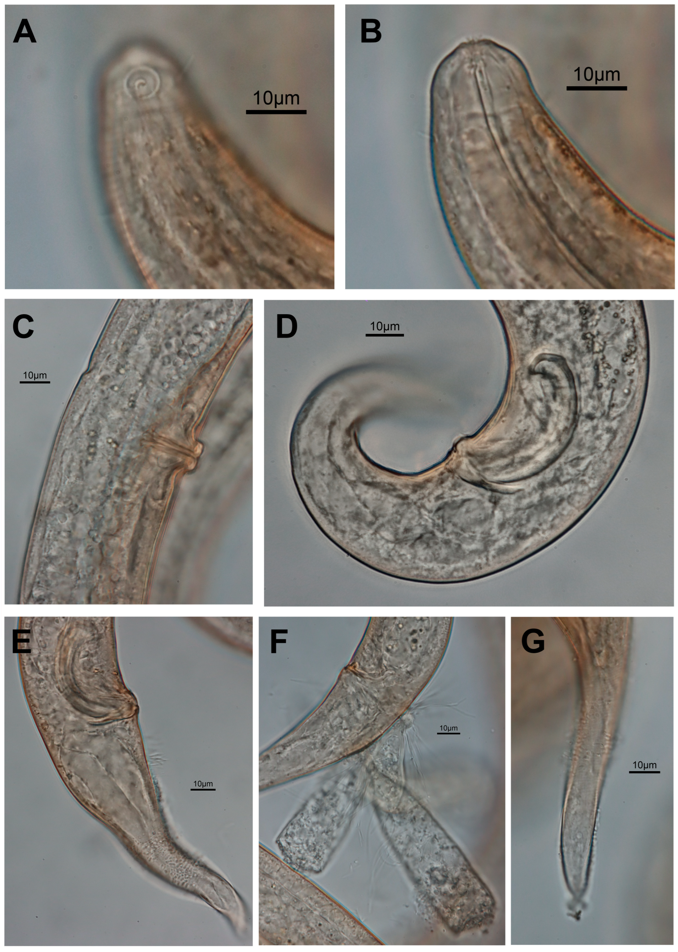
Male: Body cylindrical, long with brown/orange hue (Figure 3), usually with little or extreme coiling of the body (Figure 1C). Cuticle with fine striation with no lateral differentiation. Cheilostoma observed in the anterior extremity. Head region rounded, inner labial papillae and outer labial papillae not visible. Amphid unispiral and round, only posterior half partially surrounded by cuticle striation (Figure 3A). Cuticle striation near head tip/anterior to amphid not observed. Ratio of amphid width to body width at amphid always less than half (Table 3). Four cephalic setae slightly below the amphid, approximately 5–6 μm long. Buccal cavity small, seen with minute dorsal tooth, subventral teeth unclear. Pharynx muscular with cuticularized lumen, running narrow and straight until posterior pharyngeal bulb circular and vase-like. Nerve ring faintly present posterior to mid-level of pharynx. Cardia roughly half the size of pharyngeal bulb, present just below the pharyngeal bulb. Reproductive organ, testis, single outstretched located left to the intestine. Spicule curved with knob proximally. Slightly curved gubernaculum at the base of the spicule with slight apophyses. Precloacal supplement and setae absent. Tail ranging from conical to conico-attenuated (Figure 4) with some subventral setae. Three caudal glands of different sizes observed overlapping one another.
Female: Similar to male but slightly larger in size. Body ratio a (48), b (15), c (17), and c’ (3) similar to male without noticeable differences (Table 2). Reproductive system with two opposed and reflexed ovaries to the right of intestine. Vulva located near mid-body. Vulva protruding with constrictor muscle. Subventral setae present in females.
  • Diagnosis and relationships:
Spirinia koreana sp. nov. is morphologically similar to both S. parasitifera and S. antipodea, sharing several diagnostic features such as a unispiral amphid partially surrounded posteriorly by cuticular striations, a pharyngeal structure, and similar shapes in the spicules, gubernaculum, and conical tail. As with the relationship between S. antipodea and S. parasitifera, S. koreana can be distinguished from both species by at least one morphometric characteristic; however, these differences are not consistent across all comparisons. When morphometric data from geographically diverse populations of S. parasitifera are examined collectively, the measurements of both S. antipodea and S. koreana fall within the overall global range of variation reported for S. parasitifera (Table 3).
In terms of morphometric values, Spirinia koreana exhibits a shorter body length (1833–2282 µm) compared to most populations of S. parasitifera (1600–3796 µm) and S. antipodea (2182–2564 µm) (Table 3). The a ratio for S. koreana (44–54) falls within the higher range of S. parasitifera (28–59) and overlaps entirely with S. antipodea (44–59), indicating a relatively slender body form (Table 3), especially when compared to S. parasitifera populations from Florida, Cornwall, or Nova Scotia (Table S4). The pharynx length ratio (b) in S. koreana (13–17) is not a useful distinguishing character, as it overlaps with most S. parasitifera populations (13–21) and matches S. antipodea (17–19). The tail ratio (c) indicates that S. koreana possesses a moderately long tail, spanning the full range observed in S. parasitifera (14–29) and resembling S. antipodea (15–19), though shorter than the Cuban S. parasitifera population, which exhibits unusually high values (21–29) (Table S4). The c′ ratio (tail length to anal body diameter) of S. koreana (2.7–4.2) is slightly lower than that of S. antipodea (3.7–4.7) but falls within the range for S. parasitifera (2.1–4.7) (Table 3). The spicule length of S. koreana (49–64 µm) lies on the shorter end of the spectrum compared to S. antipodea (52–59 µm) and S. parasitifera populations from Scotland, the North Sea, and Cornwall (up to 100 µm) but is comparable to S. parasitifera from the Maldives and Florida. In summary, S. koreana can be distinguished by its relatively short body, more slender form (higher a ratio), moderately long tail, and shorter spicule length in males.
Although S. koreana falls within the overall morphometric range reported globally for S. parasitifera, a region-specific comparison, based on data from Leduc & Zhao (2019) (Table S4), reveals that S. koreana most closely resembles specimens from Nova Scotia and Woods Hole, USA. A closer examination of the morphometric variation among S. parasitifera populations suggests they can be grouped into three distinct clusters: Cluster 1. Shorter body and pharynx, relatively short tails with moderate c′ values, and shorter spicules (e.g., Maldives, Florida, USA); Cluster 2. Long, slender body with larger spicules (e.g., Scotland, Cornwall, North Sea); Cluster 3. Intermediate form with moderate body size (longer than Cluster 1, but less slender than Cluster 2) and spicule length (e.g., Nova Scotia, Woods Hole). Specimens from Cuba appear to be outliers, characterized by unusually long tails and pharynx lengths (high b values), deviating from all other groups (Table S4).
Taken together, the morphometric profile of S. koreana aligns most closely with Cluster 3, showing near-identical values across key measurements including body length (L), ratios (a, b, c, c′), spicule length, and V.

3.2. Molecular Analysis

A total of four specimens of Spirinia koreana sp. nov. were sequenced. We were able to obtain partial sequences of all three markers (mitochondrial COI, 18S rRNA, and 28S rRNA) of the new species for our investigation (Table 4). To assess genetic divergence among congeners, pairwise Kimura 2-parameter (K2P) distances were calculated using available Spirinia mtCOI sequences from GenBank. Due to the limited number of comparable sequences, only two additional Spirinia mtCOI sequences were included in the analysis: KX951912 (from Portugal) and MG659561 (from the Netherlands). While additional Spirinia mtCOI sequences are archived in GenBank, most did not align reliably with the Korean, Portuguese, and Dutch sequences and were therefore excluded from the analysis. Furthermore, the two sequences retrieved from GenBank were only identified to the genus level, limiting the taxonomic resolution of the comparison. Nevertheless, the Korean Spirinia specimens differed substantially from the two other sequences, with K2P distances of 25.9% and 23.6%, respectively (Table S1), indicating a high level of mitochondrial divergence consistent with species-level differentiation.
The 18S and 28S rRNA sequences of Spirinia koreana sp. nov. clearly indicate that it is genetically distinct from both S. parasitifera and S. antipodea. Three specimens of S. koreana (PV628730, PV628731, PV628732) are genetically identical (K2P distance = 0.0000), while the fourth (PV628733) shows only minimal divergence (0.42%), indicating strong intraspecific coherence. The new species differs from multiple S. parasitifera populations, including DQ394726, JN968281, JN968216, AY854217, and AM236044 (all from the UK) and MW078517 (Sweden), by 3.4–3.8%. It also shows a divergence of 2.1–2.5% from S. antipodea (MH472584, New Zealand), suggesting this is its closest known relative, though still a distinct taxon. The genetic distance to Spirinia sp. from Portugal (KX944162) is 2.6–2.9%, and the divergence from Perspiria elongata (EF527426, Brazil) is highest at 3.9–4.3%. These results, in combination with consistent morphological differences, strongly support the recognition of S. koreana as a separate species within the genus Spirinia.
The divergence is even more pronounced in the 28S rRNA gene. The Korean specimens differ from Swedish and German S. parasitifera sequences (OR589684–OR589686, KC755216) by 24.5%. Greater genetic distances are observed with the U.S. S. parasitifera isolate (GU003895) and S. antipodea from New Zealand (MH472585), which show 34.5% and 30.9% divergence, respectively. These levels of divergence strongly support the recognition of S. koreana as a distinct species within Spirinia.
Bayesian Inference (BI) analyses based on 18S and 28S rRNA datasets consistently recovered Spirinia koreana as a distinct and strongly supported clade within the subfamily Spiriniinae (Figure 4 and Figure 5). All four sequences of S. koreana from Republic of Korea clustered together with posterior probability (PP) = 1.00, confirming their conspecificity. In both trees, this clade was positioned either basally or as sister to other S. parasitifera lineages, supporting its status as a separate species.
In the 18S BI tree, S. koreana was recovered as sister to a moderately supported group (PP = 0.90) that included S. parasitifera sequences from Sweden (MW078517) and the UK (DQ394726, AY854217, AM236044). This larger assemblage also contained S. antipodea (MH472584) and Spirinia sp. from Portugal (KX944162), though this grouping was only weakly supported (PP = 0.77) (Figure 5). Notably, two additional UK sequences of S. parasitifera (JN968278, JN968216) formed a separate clade with strong support (PP = 1.00), providing further evidence for potential cryptic divergence within the S. parasitifera complex.
The 28S Bayesian Inference (BI) tree yielded a broadly similar topology to that of the 18S analysis (Figure 6), with Spirinia koreana forming a distinct and strongly supported clade (PP = 1.00). This clade was recovered as sister to a group comprising S. parasitifera sequences from Sweden (OR589684–OR589686) and a German isolate (KC755216). S. antipodea (MH472585) again branched separately, this time in association with the U.S. S. parasitifera sequence (GU003895), with moderate support (PP = 0.78).
Notably, only the 28S dataset recovered Spirinia as a monophyletic group, with high support (PP = 1.00). However, this result should be interpreted cautiously, as the number of available 28S rRNA sequences is limited, potentially influencing the observed topology. Importantly, neither Spiriniinae nor Desmodorinae were recovered as monophyletic in the 28S tree—an outcome consistent with previous molecular studies—and underscoring the need for a comprehensive taxonomic re-evaluation of these subfamilies.

4. Discussion

4.1. Morphological and Molecular Differentiation of Spirinia Koreana

The morphological traits of Spirinia koreana sp. nov. are definitely similar to those of S. parasitifera and S. antipodea. Particularly, S. koreana also has protist and bacteria-like structures in the tail region. The triangular suctorian protist observed is morphologically similar to those described by Bastian (1865) [27] and subsequently seen on Spirinia verecunda (see figs. 1J & 3E in [32]) and S. antipodea (see fig. 2B in [7]). Based on the morphology and host association, the epibiont in question is most likely Loricophrya susannae or a closely related suctorian ciliate. According to Baldrighi et al. (2020) [35], this species has been recorded on nematodes of the related genera Chromaspirinia and Perspiria [31], suggesting a preference for hosts with developed cuticular ornamentation, a trait common in Desmodoridae. Although the presence of suctorian ciliates is not genus-specific, their selective attachment based on host surface structures remains noteworthy. This supports the idea that morphological traits of basibionts, such as heavily ornamented cuticles, may influence epibiont colonization [35].
Phylogenetic analysis, granted with great lack of available sequences for Chromaspirinia and Perspiria, suggests a shared ancestry with Spirinia but with substantial genetic divergence. In the 18S BI tree, Perspiria elongata (originally described as Spirinia elongata) forms a sister clade to S. koreana, yet its long branch length indicates high molecular divergence (Figure 5). It is also molecularly most divergent from the new species with 3.9–4.3% based on 18S rRNA sequences. Similarly, Chromaspirinia pelitta does not group within the Spirinia clade, further supporting its genetic distinctness. These findings align with Leduc & Verschelde (2015), who argued that despite morphological similarities among Spirinia, Chromaspirinia, and Perspiria, they are genetically distinct lineages [32]. These results reinforce the necessity of using molecular data in tandem with morphology to clarify relationship within Desmodoridae.
In terms of molecular divergence, S. koreana differed from its congeners by 2.1–3.4% in SSU and 24.5–34.4% in LSU. These values are comparable to those reported for S. antipodea, which showed 2.1–4.6% divergence in 18S and 12.5–18.9% in LSU. Notably, S. koreana exhibited a greater level of divergence from other Spirinia species in LSU compared to S. antipodea (24.5–34.4% vs. 12.5–18.9%). Although no universal threshold exists for delineating interspecific boundaries, previous studies have observed SSU divergences of 2.4–7.5% between congeneric nematodes [36]. However, Bhadury et al. (2006) noted that while SSU rDNA can serve as a useful barcode marker, it often lacks sufficient resolution to distinguish closely related marine nematode taxa [37]. In contrast, Pereira et al. (2010) proposed that 28S rRNA is more effective for detecting cryptic diversity due to its greater resolving power [38]. Their study demonstrated that Mesacanthion sp. 1 and sp. 2 differed by only 1.06% in SSU but 12.09% in LSU, while Trileptium sp. 1 and sp. 2 showed 3.87% SSU divergence and 12.97% in LSU, suggesting cryptic speciation and supporting the superiority of LSU in distinguishing between closely related species. Our findings align with those of Pereira et al. (2010), indicating that while SSU can reveal interspecific differences to some degree, LSU provides a clearer divergence. Therefore, an integrative approach, combining detailed morphological examination with both SSU and LSU molecular data, will be essential for uncovering the true diversity and potential cryptic nature of species within Spirinia.

4.2. Ambiguous Distinction Between Spirinia and Perspiria

Perspiria was originally described as a subgenus of Spirinia by Wieser & Hopper (1967) [26] and was later elevated to generic status by Vincx & Gourbault (1989) [34]. Subsequently, Leduc & Verschelde (2015) [32] revised both genera (along with Chromaspirina) due to substantial morphological overlap and proposed several nomenclatural changes. Spirinia and Perspiria share numerous morphological features, including a spiral amphideal fovea, finely annulated cuticle, similar head structures and male copulatory organs, a narrow buccal cavity, minute or small teeth, and comparable sperm morphology. These shared traits have contributed to taxonomic ambiguity and confusion between the two genera.
Leduc & Verschelde (2015) [32] proposed two primary diagnostic characters to distinguish the genera: (1) tail shape (Spirinia: conical; Perspiria: conico-cylindrical to filiform) and (2) the position of the amphid relative to cuticle annulations (Spirinia: amphid entirely surrounded by annulations; Perspiria: amphid partially or not fully surrounded). However, there are exceptions to the traits. Several Spirinia species (e.g., S. gerlachi, S. gnaigeri, S. guanabarensis, some S. parasitifera, S. antipodea, and S. koreana sp. nov.) exhibit amphids that are only partially surrounded by cuticular striations. This issue is further complicated by the difficulty of observing amphid–annulation relationships. Many older species descriptions do not mention amphid annulation (e.g., S. gerlachi, S. gnaigeri, S. hopperi, S. inaurita, S. laevis, S. okemwai, S. parma, and S. schneideri), and in several cases, the illustra-tions do not depict annulation around the amphid at all (e.g., S. inaurita, some S. laevis, S. hopperi). Notably, S. hopperi was described as lacking cuticle annulation entirely, yet a fine striation is visible anterior to the amphid in a published SEM micrograph. While a partially surrounded amphid is an exception to the diagnosis of the genus Spirinia, more than five species (S. gerlachi, S. gnaigeri, some S. parasitifera, S. antipodea, and S. koreana sp. nov.) exhibit this trait (Table 5) and are still considered valid members of the genus based on the second diagnostic character.
The second diagnostic character, tail shape, also appears more ambiguous than initially presumed. The examination of over 20 S. koreana specimens revealed considerable intraspecific variation in tail morphology, ranging from conical to conico-attenuated, with some individuals exhibiting forms bordering on conico-cylindrical (Figure 4). This variation initially prompted consideration of assigning S. koreana to Perspiria, but comparison with congeners revealed many Spirinia species whose tails, though described as conical, are illustrated as conico-attenuated or gradually tapering, resembling the range of tail shapes observed in the new species (Table 5). For example, S. okemwai is described as having a conical tail, yet its illustrations and micrographs suggest a form closer to conico-cylindrical [28]. S. laevis was originally described and depicted as conical (Bastian, 1865) [27], but Gerlach (1950) later described and illustrated it with a short, cylindrical terminal portion [39]. A similar tail is seen in S. parma (Ott, 1977) [24].
Some Spirinia species (S. antipodea, certain populations of S. parasitifera, and S. koreana sp. nov.) possess gradually tapering, conico-attenuated tails that contrast with clearly conical-tailed species like S. hopperi, S. laeviodes, S. sophia, and S. verecunda (Table 5). To address this morphological gradient, we propose the use of the term conico-attenuated to describe tails that fall between conical and conico-cylindrical in shape. Accordingly, we recommend updating the diagnosis of Spirinia to include both conical and conico-attenuated tail forms.
Despite these complications, the two diagnostic traits proposed by Leduc & Verschelde (2015) [32] remain useful for distinguishing Spirinia from Perspiria. However, the observed exceptions and morphological variability—some of which blur the boundaries between the two genera—demonstrate that these traits are not universally consistent. Our 18S Bayesian tree supports the clustering of Spirinia, including S. koreana sp. nov., and places it near P. elongata (Figure 5). The long branch separating P. elongata suggests both phylogenetic proximity and distinctiveness. It also supports the continued use of the proposed diagnostic characters, even if exceptions exist.
Unfortunately, the lack of molecular data for other Perspiria species limits a more definitive assessment. While we accept the current diagnoses and revisions of both genera, we emphasize the importance of supplementing morphological identifications with molecular data. We urge nematologists to generate sequence data alongside morphological observations, which will be essential for future taxonomic revision. Until such data become available, both morphological and molecular evidence suggest that S. koreana sp. nov. is best placed within Spirinia, rather than Perspiria. Over time, as molecular data accumulate for both Perspiria and additional Spirinia species, it may become evident that conico-attenuated-tailed species are more closely related to Perspiria, potentially leading to the transfer of several species, including S. koreana. However, such taxonomic changes should be based on integrative evidence that confirms whether the proposed diagnostic traits align with phylogenetic relationships.

Supplementary Materials

The following supporting information can be downloaded at https://www.mdpi.com/article/10.3390/jmse13071251/s1. Table S1: Kimura 2-parameter distance between other species belonging to the genus Spirinia based on mtCOI alignment with 1000 bootstraps. Standard deviation is marked in blue. Table S2: Kimura 2-parameter distance between other species belonging to the genus Spirinia based on 18S rRNA alignment with 1000 bootstraps. Standard deviation is marked in blue. Table S3: Kimura 2-parameter distance between other species belonging to the genus Spirinia based on 28S rRNA alignment with 1000 bootstraps. Standard deviation is marked in blue. Table S4: Dimensions of key traits of Spirinia parasitifera and related species (updated from Leduc & Zhao 2019).

Author Contributions

Conceptualization, K.S. and R.J.; methodology, K.S. and R.J.; software, R.J.; validation, K.S. and R.J.; formal analysis, K.S. and R.J.; investigation, K.S. and R.J.; resources, R.J.; data curation, K.S. and R.J.; writing—original draft preparation, K.S. and R.J.; writing—review and editing, K.S. and R.J.; visualization, K.S. and R.J.; supervision, R.J.; project administration, R.J.; funding acquisition, R.J. All authors have read and agreed to the published version of the manuscript.

Funding

This research was funded by the National Institute of Biological Resources (NIBR), Ministry of Environment (MOE) of the Republic of Korea (NIBR202402107), and Basic Science Research Program through the National Research Foundation of Korea (NRF), funded by the Ministry of Education (2021R1I1A2043807).

Data Availability Statement

All specimens examined within this study were deposited in the National Institute of Biological Resources (NIBR), Republic of Korea. Molecular sequences obtained were submitted to GenBank, and their respective accession numbers are listed in Table 4.

Acknowledgments

During the preparation of this manuscript/study, the author(s) used ChatGPT GPT-4o for the purpose of correcting and improving grammar of written text. The authors have reviewed and edited the output and take full responsibility for the content of this publication.

Conflicts of Interest

The authors declare no conflicts of interest. The funders had no role in the design of this study; in the collection, analyses, or interpretation of data; in the writing of the manuscript; or in the decision to publish the results.

Abbreviations

The following abbreviations are used in this manuscript:
LTotal body length (µm)
aBody length divided by maximum body diameter
bBody length divided by pharynx length
cBody length divided by tail length
c’Tail length divided by anal body diameter
cbdCorresponding body diameter
spicSpicule length as an arc

References

  1. Louati, H.; Said, O.B.; Soltani, A.; Cravo-Laureau, C.; Duran, R.; Aissa, P.; Mahmoudi, E.; Pringault, O. Responses of a Free-Living Benthic Marine Nematode Community to Bioremediation of a Pah Mixture. Environ. Sci. Pollut. Res. Int. 2015, 22, 15307–15318. [Google Scholar] [CrossRef] [PubMed]
  2. Coles, J.W. Observations on the Marine Nematode Genus Spirinia Gerlach, 1963 (Desmodoridae: Spiriniinae) with Descriptions of Two New Species. Bull. Br. Mus. (Nat. Hist.) Zool. 1987, 53, 79–101. [Google Scholar]
  3. Venekey, V.; Fonseca-Genevois, V.G.; Santos, P.J.P. Biodiversity of Free-Living Marine Nematodes on the Coast of Brazil: A Review. Zootaxa 2010, 2568, 39–66. [Google Scholar] [CrossRef]
  4. Armenteros, M.; Ruiz-Abierno, A.; Decraemer, W. Revision of Desmodorinae and Spiriniinae (Nematoda: Desmodoridae) with Redescription of Eight Known Species. Eur. J. Taxon. 2014, 96, 1–32. [Google Scholar] [CrossRef]
  5. Gerlach, S.A. Freilebende Meeresnematoden Von Den Malediven Ii. Kiel. Meeresforsch 1963, 19, 67–103. [Google Scholar]
  6. Hourston, M.; Potter, I.C.; Warwick, R.M.; Valesini, F.J.; Clarke, K.R. Spatial and Seasonal Variations in the Ecological Characteristics of the Free-Living Nematode Assemblages in a Large Microtidal Estuary. Estuar. Coast. Shelf Sci. 2009, 82, 309–322. [Google Scholar] [CrossRef]
  7. Leduc, D.; Zhao, Z.Q. Morphological and Molecular Characterisation of Spirinia antipodea Leduc N. Sp.(Nematoda: Desmodoridae), a Cryptic Species Related to S. parasitifera, from the Coast of New Zealand. Nematology 2019, 21, 91–105. [Google Scholar] [CrossRef]
  8. Hopper, D. Drawing and Measuring Nematodes. In Laboratory Methods for Work with Plant and Soil Nematodes; Ministry of Agriculture, Fisheries and Food, Her Majesty’s Stationery Office: London, UK, 1970. [Google Scholar]
  9. Meeker, N.D.; Hutchinson, S.A.; Ho, L.; Trede, N.S. Method for Isolation of Pcr-Ready Genomic DNA from Zebrafish Tissues. Biotechniques 2007, 43, 610–614. [Google Scholar] [CrossRef] [PubMed]
  10. Derycke, S.; Vanaverbeke, J.; Rigaux, A.; Backeljau, T.; Moens, T. Exploring the Use of Cytochrome Oxidase C Subunit 1 (Coi) for DNA Barcoding of Free-Living Marine Nematodes. PLoS ONE 2010, 5, e13716. [Google Scholar] [CrossRef]
  11. Holterman, M.; van der Wurff, A.; van den Elsen, S.; van Megen, H.; Bongers, T.; Holovachov, O.; Bakker, J.; Helder, J. Phylum-Wide Analysis of Ssu Rdna Reveals Deep Phylogenetic Relationships among Nematodes and Accelerated Evolution toward Crown Clades. Mol. Biol. Evol. 2006, 23, 1792–1800. [Google Scholar] [CrossRef]
  12. De Ley, P.; De Ley, I.T.; Morris, K.; Abebe, E.; Mundo-Ocampo, M.; Yoder, M.; Heras, J.; Waumann, D.; Rocha-Olivares, A.; Burr, A.H.J.; et al. An Integrated Approach to Fast and Informative Morphological Vouchering of Nematodes for Applications in Molecular Barcoding. Philos. Trans. R. Soc. London. Ser. B Biol. Sci. 2005, 360, 1945–1958. [Google Scholar] [CrossRef]
  13. Tamura, K.; Stecher, G.; Kumar, S. Mega11: Molecular Evolutionary Genetics Analysis Version 11. Mol. Biol. Evol. 2021, 38, 3022–3027. [Google Scholar] [CrossRef] [PubMed]
  14. Thompson, J.D.; Higgins, D.G.; Gibson, T.J. Clustal-W—Improving the Sensitivity of Progressive Multiple Sequence Alignment through Sequence Weighting, Position-Specific Gap Penalties and Weight Matrix Choice. Nucleic Acids Res. 1994, 22, 4673–4680. [Google Scholar] [CrossRef]
  15. Bell, T.G.; Kramvis, A. Fragment Merger: An Online Tool to Merge Overlapping Long Sequence Fragments. Viruses 2013, 5, 824–833. [Google Scholar] [CrossRef] [PubMed]
  16. Kimura, M. A Simple Method for Estimating Evolutionary Rates of Base Substitutions through Comparative Studies of Nucleotide-Sequences. J. Mol. Evol. 1980, 16, 111–120. [Google Scholar] [CrossRef] [PubMed]
  17. Minh, B.Q.; Schmidt, H.A.; Chernomor, O.; Schrempf, D.; Woodhams, M.D.; Von Haeseler, A.; Lanfear, R. Iq-Tree 2: New Models and Efficient Methods for Phylogenetic Inference in the Genomic Era. Mol. Biol. Evol. 2020, 37, 1530–1534. [Google Scholar] [CrossRef]
  18. Kalyaanamoorthy, S.; Minh, B.Q.; Wong, T.K.F.; von Haeseler, A.; Jermiin, L.S. Modelfinder: Fast Model Selection for Accurate Phylogenetic Estimates. Nat. Methods 2017, 14, 587–589. [Google Scholar] [CrossRef]
  19. Minh, B.Q.; Nguyen, M.A.; von Haeseler, A. Ultrafast Approximation for Phylogenetic Bootstrap. Mol. Biol. Evol. 2013, 30, 1188–1195. [Google Scholar] [CrossRef]
  20. Darriba, D.; Taboada, G.L.; Doallo, R.; Posada, D. Jmodeltest 2: More Models, New Heuristics and High-Performance Computing. Nat. Methods 2012, 9, 772. [Google Scholar] [CrossRef]
  21. Huelsenbeck, J.P.; Ronquist, F. Mrbayes: Bayesian Inference of Phylogenetic Trees. Bioinformatics 2001, 17, 754–755. [Google Scholar] [CrossRef]
  22. Rambaut, A. Figtree, Version 1.4.4; Institute of Evolutionary Biology, University of Edinburgh: Edinburgh, UK, 2018. [Google Scholar]
  23. Luc, M.; De Coninck, L.A. Nématodes Libres Marins De La Région De Roscoff. Arch. De Zool. Expérimentale Et Générale 1959, 98, 103–165. [Google Scholar]
  24. Ott, J.A. New Freeliving Marine Nematodes from the West Atlantic I. Four New Species from Bermuda with a Discussion of the Genera Cytolaimium and Rhabdocoma Cobb, 1920. Zool. Anz. 1977, 198, 120–138. [Google Scholar]
  25. Maria, T.F.; Esteves, A.M.; Smol, N.; Vanreusel, A.; Decraemer, W. Chromaspirina guanabarensis Sp. N. (Nematoda: Desmodoridae) and a New Illustrated Dichotomous Key to Chromaspirina Species. Zootaxa 2009, 2092, 21–36. [Google Scholar] [CrossRef]
  26. Wieser, W.; Hopper, B. Marine Nematodes of the East Coast of North America. I. Florida. Bull. Mus. Comp. Zool. Harv. Coll. 1967, 135, 239–344. [Google Scholar]
  27. Bastian, H.C. Monograph of the Anguillulidae, or Free Nematoids, Marine, Land, and Freshwater; with Descriptions of 100 New Species. Trans. Linn. Soc. Lond. 1865, XXV, 73–184. [Google Scholar] [CrossRef]
  28. Muthumbi, A.; Verschelde, D.; Vincx, M. New Desmodoridae (Nematoda: Desmodoroidea): Three New Species from Ceriops Mangrove Sediments (Kenya) and One Related New Species from the North Sea. Cah. De Biol. Mar. 1995, 36, 181–195. [Google Scholar]
  29. Ott, J.A. Twelve New Species of Nematodes from an Intertidal Sandflat in North Carolina. Int. Rev. Der Gesamten Hydrobiol. Und Hydrogr. 1972, 57, 463–496. [Google Scholar] [CrossRef]
  30. Villot, A. Recherches Sur Les Helminthes Libres Ou Parasites Des Cotes De La Bretagne. Arch. De Zool. Expérimentale Et Générale 1875, 4, 451–482. [Google Scholar]
  31. Da Silva, M.C.; De Castro, F.J.V.; Cavalcanti, M.D.; Da Fonsêca-Genevois, V. Spirinia lara Sp. N. and Spirinia Sophia Sp. N. (Nematoda, Desmodoridae) from the Brazilian Continental Margin (Campos Basin, Rio De Janeiro). Zootaxa 2009, 2081, 31–45. [Google Scholar] [CrossRef]
  32. Leduc, D.; Verschelde, D. New Spirinia and Stygodesmodora Species (Nematoda, Spiriniinae) from the Southwest Pacific, and Revision of the Related Genera Spirinia, Chromaspirina and Perspiria. Eur. J. Taxon. 2015, 118, 1–25. [Google Scholar] [CrossRef]
  33. Platt, H.; Warwick, R. Free-Living Marine Nematodes; Part 2: British Chromadorids; Linnean Society of London: London, UK, 1988. [Google Scholar]
  34. Vincx, M.; Gourbault, N. Desmodoridae from the Bay of Morlay (Brittany) and the Southern Bight of the North Sea. Cah. De Biol. Mar. 1989, 30, 103–114. [Google Scholar]
  35. Baldrighi, E.; Dovgal, I.; Zeppilli, D.; Abibulaeva, A.; Michelet, C.; Michaud, E.; Franzo, A.; Grassi, E.; Cesaroni, L.; Guidi, L.; et al. The Cost for Biodiversity: Records of Ciliate-Nematode Epibiosis with the Description of Three New Suctorian Species. Diversity 2020, 12, 224. [Google Scholar] [CrossRef]
  36. Armenteros, M.; Rojas-Corzo, A.; Ruiz-Abierno, A.; Derycke, S.; Backeljau, T.; Decraemer, W. Systematics and DNA Barcoding of Free-Living Marine Nematodes with Emphasis on Tropical Desmodorids Using Nuclear Ssu Rdna and Mitochondrial Coi Sequences. Nematology 2014, 16, 979–989. [Google Scholar] [CrossRef]
  37. Bhadury, P.; Austen, M.C.; Bilton, D.T.; Lambshead, P.J.D.; Rogers, A.D.; Smerdon, G.R. Development and Evaluation of a DNA-Barcoding Approach for the Rapid Identification of Nematodes. Mar. Ecol. Prog. Ser. 2006, 320, 1–9. [Google Scholar] [CrossRef]
  38. Pereira, T.J.; Fonseca, G.; Mundo-Ocampo, M.; Guilherme, B.C.; Rocha-Olivares, A. Diversity of Free-Living Marine Nematodes (Enoplida) from Baja California Assessed by Integrative Taxonomy. Mar. Biol. 2010, 157, 1665–1678. [Google Scholar] [CrossRef]
  39. Gerlach, S.A. Über Einige Nematoden Aus Der Familie Der Desmodoriden. Zool. Anz. 1950, 145, 178–198. [Google Scholar]
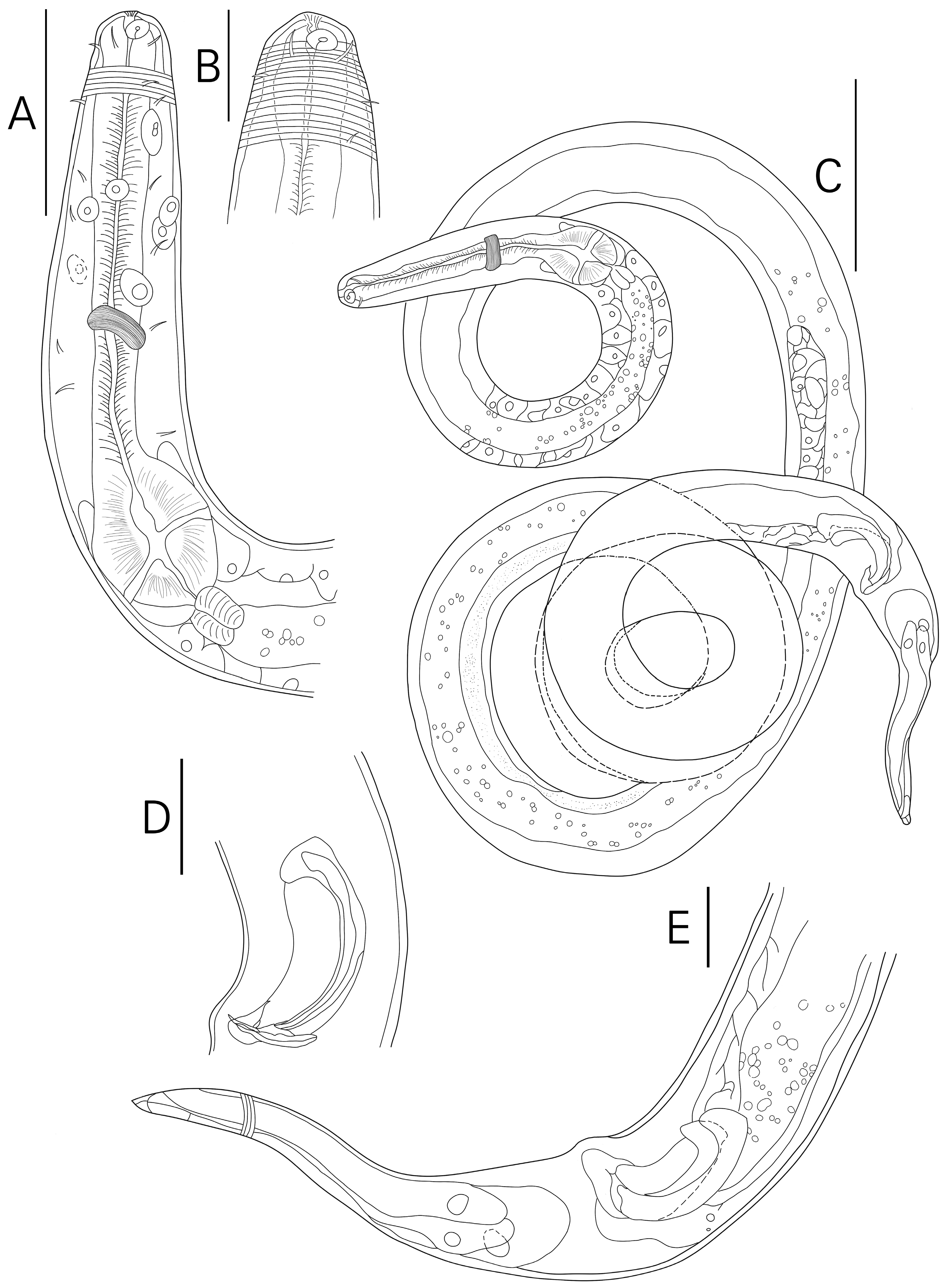
Figure 1. Spirinia koreana sp. nov. Male, holotype: (A) pharyngeal region; (B) anterior region; (C) habitus; (D) cloacal region; (E) tail region (scale bar: (A) = 50 μm; (B,E) = 20 μm; (C) = 100 μm; (D) = 25 μm).
Jmse 13 01251 g001
Figure 2. Sprinia koreana sp. nov. Female, allotype: (A) anterior region; (B) habitus; (C) tail region; (D) female reproductive system (scale bar: (A) = 20 μm; (B) = 100 μm; (C,D) = 50 μm).
Jmse 13 01251 g002
Figure 3. Spirinia koreana sp. nov. light micrographs―(A) ♀ cephalic region with amphid; (B) ♀ cephalic region with focus on buccal; (C) ♀ vulva protruding with constrictor muscle; (D) ♂ cloacal region showing spicule and gubernaculum with no bacteria on surface; (E) ♂ tail region covered with bacteria; (F) ♀ tail region with multiple protists attached.; (G) ♀ tail region covered with bacteria (scale bar: (AG) = 10 μm).
Jmse 13 01251 g003
Figure 4. Varying tail morphology of Spirinia koreana sp. nov. light micrographs―(A) ♂ paratype 11, conical tail; (B) ♂ paratype 14, conical tail; (C) ♀ paratype 13, conico-attenuated tail (D) ♀ paratype 15, conico-attenuated tail (scale bar: (AD = 50) μm) (all images were focus-stacked using Helicon Focus 6).
Jmse 13 01251 g004
Figure 5. Rooted Bayesian Inference phylogenetic tree of 18S rRNA region with Microlaimus robustidens. set as the outgroup. Sequences from this study are bolded; species belonging to the genus Spirinia are marked with blue (posterior probability support shown at each node).
Jmse 13 01251 g005
Figure 6. Rooted Bayesian Inference phylogenetic tree of 28S rRNA region with Microlaimus robustidens. set as the outgroup. Sequences from this study are bolded; species belonging to the genus Spirinia are marked with blue (posterior probability support shown at each node).
Jmse 13 01251 g006
Table 1. Information regarding amplification including primers, amplification conditions, and respective references.
Target GenePrimer (Direction)Sequence 5′-3′Amplification ConditionReference
mtCOIJB3 (f)TTTTTTGGGCATCCTGAGGTTTAT94 °C for 5 min, 5 cycles of 94 °C for 30 s, 54 °C for 30 s, and 72 °C for 30 s, followed by 35 cycles of denaturation at 94 °C for 30 s, annealing at 50 °C for 30 s, and extension at 72 °C for 30 s, followed by final step of 72 °C for 10 minDerycke et al., 2010 [10]
JB5 (r)AGCACCTAAACTTAAAACATAATGAAAATG
18S988F (f)CTCAAAGATTAAGCCATGC94 °C for 5 min, 5 cycles of 94 °C for 30 s, 45 °C for 30 s, and 72 °C for 70 s, followed by 35 cycles of denaturation at 94 °C for 30 s, annealing at 54 °C for 30 s, and extension at 72 °C for 70 s, followed by final step of 72 °C for 5 minHolterman et al., 2006 [11]
1912R (r)TTTACGGTCAGAACTAGGG
1813F (f)CTGCGTGAGAGGTGAAAT
2646R (r)GCTACCTTGTTACGACTTTT
28SD2A (f)ACAAGTACCGTGAGGGAAAGTTG95 °C for 5 min followed by 37 cycles of denaturation at 95 °C for 30 s, annealing at 56 °C for 1 min, and extension at 72 °C for 1 min 30 s min, followed by final step at 72 °C for 5 minDe Ley et al., 2005 [12]
D3B (r)TCGGAAGGAACCAGCTACTA
Table 2. Measurement of Spirinia koreana sp. nov. (All measurements are in µm; “n/a” indicates not applicable).
CharacterMaleFemale
HolotypeMale (n = 11) Mean ± Sd (Range)AllotypeFemale (n = 12) Mean ± Sd (Range)
L21002052 ± 98.4 (1833–2180)19902105 ± 113.7 (1858–2282)
a (L/gbd)48.848 ± 3.2 (43.6–54.5)44.248.9 ± 3.1 (45.3–53.4)
b (L/pharynx length)13.915 ± 1.2 (12.7–17)15.315.5 ± 0.8 (13.9–17.1)
c (L/tail length)15.817 ± 2.2 (12.5–22)1716.9 ± 1.4 (15.1–19.8)
c’ (tail length/anal body diameter)3.43 ± 0.3 (2.7–4)3.73.7 ± 0.2 (3.4–4.2)
Cephalic setae length5.66 ± 0.5 (5–6.8)5.65.5 ± 0.5 (4.4–6.4)
Cephalic setae cbd1716 ± 1.3 (13–18)1516.1 ± 0.9 (14.6–17.5)
Amphid (at center) cbd1615 ± 1.7 (12–18)1415.8 ± 0.8 (14–17.4)
Amphid height4.75 ± 0.5 (3.9–5.9)4.15 ± 0.5 (4.1–6)
Amphid diameter6.15 ± 0.5 (4.3–6.1)5.45.5 ± 0.6 (4.8–7)
Amphid diameter divided by cbd (%)38.135 ± 4.8 (28.3–42.3)38.634.9 ± 4.3 (29.8–46.7)
Amphid (at center) distance to anterior4.96 ± 1.5 (4–9.7)56.5 ± 1.7 (4.2–9.7)
Nerve ring distance to anterior7576 ± 7.8 (65–93)7780.2 ± 9.7 (64–94)
Nerve ring cbd3231 ± 0.7 (30–32)3131.9 ± 2.3 (27–34)
Pharynx length151134 ± 8.4 (122–151)130135.5 ± 7.2 (120–149)
Pharyngeal bulb width3029 ± 0.9 (28–30)2729.8 ± 2.2 (27–36)
Pharynx cbd3736 ± 0.9 (34–37)3535.9 ± 3.7 (30–46)
Max. body diameter4343 ± 2.2 (40–47)4543.2 ± 1.2 (41–45)
Spicule length (as arc)6455 ± 4.1 (49–64)n/an/a
Gubernaculum length (as arc)2222 ± 1.8 (18–24)n/an/a
Anal body diameter3936 ± 2.8 (30–41)3233.6 ± 2.1 (30–37)
Tail length (µm)133120 ± 12.8 (95–147)117124.7 ± 10.2 (103–140)
Vulva distance to anteriorn/an/a9491026.5 ± 52.1 (911–1119)
Vulva body diametern/an/a4745 ± 1.8 (41–48)
Vulva distance to anterior as percentage of total body lengthn/an/a48%0.5 ± 0 (0.5–0.5)
Table 3. Comparison of morphometric measurements of valid Spirinia species (updated from Leduc & Zhao 2019).
SpeciesSourceRegionLabcc’Spic (µm)V
S. gerlachi[23]France149437.51110.4~437
S. gnaigeri[24]Bermuda860–91320–239–109–112.8–3.550–5547–50
S. guanabarensis[25]Brazil2110–257937–4811–1417–222.6–3.358–7055–59
S. hopperi[2]England4100–510048–6720–2222–302.2–3110–13039–44
S. inaurita[26]USA1180–135038–40 (calc)11–1213–143.927–2849
S. laevioides[5]Maldives1348–136727–3213.5–1411.8–13.93–3.342–5243
S. laevis[27]UK~3600~40~10~25~7040
S. okemwai[28]Kenya966–123123–276.6–8.514.5–17.41.8–2.441–5255
S. parma[29]USA1276–160841.1–56.111.1–12.914.7–19.23.3–3.9263158
S. schneideri[23]France3424– 391926–32.517.6–18.627.6–48.41119–14040.2–48
S. sophia[31]Brazil1945–2180108–11217–2123–244–526–2755–57
S. verecunda[32]New Zealand757228.001523851–54
S. parasitifera[2,4,5,27,33,34]Global1600–379628–5912–2614–292–445–10046–53
S. antipodea[7]New Zealand2182–256444–5917–1915–193.7–4.752–5949–51
S. koreana sp. nov.Present studyRepublic of Korea1833–228244–5413–1712–222.7–4.249–6447–52
Table 4. GenBank accession number of sequences obtained in this study.
#Species NameIsolate NumberGenBank Accession Number
mtCOI18S28S
JB3988F, 1813FD2A
/JB5/1912R, 2646R/D3B
(~360 bp)(~1600 bp)(~750 bp)
1Spirinia koreana sp. nov.1APV637276PV628730PV630260
2Spirinia koreana sp. nov.1NPV637277PV628731PV630261
3Spirinia koreana sp. nov.1OPV637278PV628732PV630262
4Spirinia koreana sp. nov.1PPV637279 PV628733PV630263
Table 5. Summary of amphid and tail morphology across valid Spirinia species based on both textual descriptions and accompanying illustrations. Figure references correspond to those cited in the “Sources” column unless otherwise noted.
SpeciesSource/
Reference
Amphid Coverage (of Annulation)Tail Morphology
Textual DescriptionFigure DepictionTextual DescriptionFigure Depiction
S. gerlachiLuc & De Coninck 1959 [23]not mentionedposterior only (Figure 27)long conico-attenuatedconical/long conico-attenuated (Figure 28)
S. gnaigeriOtt 1977 [24]not mentionedposterior only (Figure 43)conicalconical/long conico-attenuated (Figure 44)
S. guanabarensisMaria et al., 2009 [25]largely surroundedmostly, but not entirely (Figures 1C and 2A,B)conicalconical/conico-attenuated (Figures 1D,E and 2F,G)
S. hopperiColes 1987 [2]not mentioned cuticle described as without striations entirelynot depicted (Figure 1c), but with SEM showing fine striation at anterior end of amphid (Figure 9B)tail long; fairly long in both sexesconical (Figure 3C)
S. inauritaWieser & Hopper 1967 [26]not mentionednot depicted (Figure 37A)not describedconical/long conico-attenuated (Figure 37E)
S. laevioidesGerlach 1963 [5]cuticle shows annulation that reaches up to the front edge of the amphidsentire (Figure 2e)conicalconical (Figure 2f)
S. laevisBastian 1865 [27]not mentioned–not depicted (Figure 204)
–entire (Gerlach 1950 [39]: Figure 6A,B)
–narrowing to a point
–cylindrical–conical with a rounded terminal cone (Gerlach 1950 [39])
–conical (Figures 205 and 206)
–cylindrical portion depicted (Gerlach 1950 [39]: Figure 6C)
S. okemwaiMuthumbi, Verschelde & Vincx 1995 [28]not mentionedentire (Figures 1B,G and 2A,B,D)conicalconico-cylindrical (Figures 1E,F and 2E,G)
S. parmaOtt 1972 [29]not mentionedpartially, beginning at front edge (Figures 87, 90 and 92)conicalmostly conical but with terminal cylindrical flagellate portion depicted (Figures 88, 89 and 91)
S. schneideriLuc & De Coninck 1959 [23]not mentioned–not depicted (Figure 31)
–not depicted (Coles 1987 [2]: Figure 1b), but with SEM showing fine striation well above anterior end of amphid (Coles 1987 [2]: Figure 9A)
Tail is different in males and females. In females, tail is conical. In males, tail is subhemispherical with terminal point.subhemispherical with terminal cylindrical point (Figures 32 and 33)
conical (Coles 1987 [2]: Figure 3B)
S. sophiaSilva et al., 2009 [31]amphid surrounded by transverse cuticular striationentire (Figures 5B,D and 6B,C)conicalconical (Figures 5E,F and 7G)
S. verecundaLeduc & Verschelde 2015 [32]annulations completely surrounding amphidentire (Figure 1A,B)conicalconical (Figure 1G,I,J)
S. parasitiferaBastian 1865 [27]: Gerlach 1963 [5]: Coles 1987 (UK, Canada, USA) [2]: Platt & Warwick 1988 [33]: Vincx & Gourbault 1989 [34]: Armenteros et al., 2014 [4]not mentioned (Bastian 1865 [27]; Gerlach 1963 [5]; Coles 1987 [2]; Platt & Warwick 1988 [33]; Vincx & Gourbault 1989 [34]; Armenteros et al., 2014 [4]) –not depicted (Bastian 1865 [27]: Figure 201)
–posterior only (Gerlach 1963 [5]: Tafel 1e, 1h; Armenteros et al., 2014 [4]: Figure 11A)
–entire (Gerlach 1963 [5]: Tafel 1f, 1g, 1c; Coles 1987 [2]: Figure 1a; Platt & Warwick 1988 [33]: Figure 148A; Vincx & Gourbault 1989 [34]: Figure 3c; Armenteros et al., 2014 [4]: Figure 12A,B)
–tapering gradually to a point posteriorly (Bastian 1865 [27])
–conical (Platt & Warwick 1988 [33])
–conical (Bastian 1865 [27]: Figures 202 and 203; Coles 1987 [2]: Figure 10A,C; Armenteros et al., 2014 [4]: Figure 11B)
–long conico-attenuated (Coles 1987 [2]: Figures 2A and 3A; Platt & Warwick 1988 [33]: Figure 148B; Vincx & Gourbault 1989 [34]: Figure 3M,N)
S. antipodeaLeduc & Zhao 2019 [7]amphid partially surrounded by cuticle annulationposterior only (Figure 1C,D)conical–conical (Figure 2B,D)
–conico-attenuated (Figures 2A and 3F)
S. koreana sp. nov.Present studyonly posterior half of amphid partially surrounded by cuticle striation posterior only (Figures 1B and 2A)conical/conico-attenuated–conical (Figures 2C, 3E and 4A,B)
–conico-attenuated (Figures 1E and 4C,D)
Disclaimer/Publisher’s Note: The statements, opinions and data contained in all publications are solely those of the individual author(s) and contributor(s) and not of MDPI and/or the editor(s). MDPI and/or the editor(s) disclaim responsibility for any injury to people or property resulting from any ideas, methods, instructions or products referred to in the content.