Article

Possible Evolutionary Precursors of Mast Cells: The ‘Granular Cell’ Immunocyte of Botrylloides leachii (Tunicata; Ascidiacea)

by
Nicolò Brunelli
1,
Stefano Dalle Palle
2 and
Francesca Cima
1,*
1
Laboratory of Biology of Ascidians, Department of Biology, University of Padova, 35131 Padova, PD, Italy
2
Laboratory of Microbial Ecology and Genomics, Istituto Zooprofilattico Sperimentale delle Venezie, 35020 Legnaro, PD, Italy
*
Author to whom correspondence should be addressed.
J. Mar. Sci. Eng. 2025, 13(4), 811; https://doi.org/10.3390/jmse13040811
Submission received: 28 February 2025 / Revised: 3 April 2025 / Accepted: 14 April 2025 / Published: 18 April 2025
(This article belongs to the Section Marine Biology)

Abstract

:
Vertebrate mast cells are the first cells to initiate the inflammatory response. The origin of these highly specialised innate immunity cells in chordates is an intriguing unanswered question, and tunicates represent the best candidates to address this question for their close relationship with vertebrates. In the colonial ascidian Botrylloides leachii, a particular cell type circulates in the haemolymph, namely, ‘granular cell’, which is a distinct immunocyte from both phagocytic and cytotoxic lines. Like mast cells and unlike basophils, granular cells were labelled with anti-c-kit antibody on their plasmalemma and exhibited a high content of heparin in their granules, as revealed by various histochemical techniques. Immunohistochemistry revealed the presence of heparin and histamine inside the same granules resembling the granules of mast cells. Histoenzymatic assays revealed the presence of mast cell enzymes such as β-glucuronidase, arylsulphatase, chloroacetyl esterase, and proteases. These cells degranulated after exposure to bacteria, compound 48/80, or heterologous plasma. During exposure to bacteria, they crowd into the perivisceral sinus and then infiltrate the epithelium of the postbranchial gut, where they release the content of their granules, a behaviour remarkably similar to that of the gastric leukopedesis of mast cells.

1. Introduction

Mast cells are considered sentinel cells of the vertebrate immune system and are located at strategic points in organisms such as connective tissues. In response to a proinflammatory stimulus, they degranulate, initiate, and modulate immune inflammatory activities in concert with other immunocytes [1,2]. In 1878, Paul Erhlich named these cells Mastzellen (meaning ‘fattening cells’) because he mistakenly believed that the granules were the result of the absorption of extracellular materials [3,4,5]. Although they are conventionally divided into subpopulations with various classifications [6,7], their morphology is easily recognisable. They are round cells 20–30 µm in diameter with amoeboid activity. Their cytoplasm contains basophilic granules 0.3–0.8 µm in diameter surrounded by a membrane. The nucleus, which is rather small and spherical, is usually centrally located, but is sometimes completely hidden by the large number of basophilic granules, which fill the cytoplasm and appear highly electron-dense under an electron microscope.
In mammals and other vertebrates, mast cells are widely distributed throughout vascularised tissues, particularly under and, in some cases, within epithelia, and in the vicinity of blood vessels, nerves, smooth muscle cells, mammary glands, and hair follicles [8]. They are present in large numbers near surfaces exposed to the external environment, where the potential for contact with pathogens and allergens increases [9]. They play an important role in the immune system because they are usually the first cells to come into contact with foreign bodies and initiate the inflammatory response by degranulating their content, in which the heparin matrix acts as an ion-exchange resin to which positively charged molecules are reversibly bound [10,11].
Once activated, mast cells release a number of preformed substances, such as histamine, heparin, and proteases, and secondarily release a number of de novo synthesised mediators, such as platelet-activating factor, leukotrienes, cytokines, and chemokines [12,13,14,15], which are useful in recruiting other immune cells to participate in the body’s defence. The mediator load of mast cells is the highest of any cell type [16]. Mast cells are key cells in the adaptive immune response associated with IgEs, immunoglobulins produced by plasma cells in allergic and immune responses to certain pathogens, for which they possess the specific membrane receptor FcεRI [17], and are also involved in interactions with other cells of the adaptive immunity such as T and B lymphocytes [18,19,20].
Mast cells are probably derived from an ancestral cell with defensive functions already present in nonvertebrate chordates prior to the evolution of adaptive immunity. Indeed, they are involved in innate immune defence by isolating pathogens and directly eliminating the invader [21]. In mammals, particularly humans and mice, mast cells appear to be able to neutralise bacteria by an intracellular elimination process similar to that of phagocytes [22]. They can sometimes exert bactericidal activity through DNA extracellular traps (ETosis), similar to those produced by neutrophils at the end of their degranulation activity [23].
Although mast cells primarily trigger the immediate innate response, which is acute inflammation to protect against infection, their evolutionary origin remains poorly understood, as little information is available on their activity in other vertebrates outside the mammalian class. It is likely that the phylogenetic ancestors of mast cells were transferred from their nonvertebrate common ancestor to vertebrates and subsequently incorporated into the network of adaptive immune processes. From this point of view, tunicates, which show characteristics of basal chordates and have only an innate immunity, are of particular interest for the study of the evolutionary origin of mast cells in vertebrates. Molecular studies have revealed the phylogenetic proximity of these two groups, which share a common ancestor [24,25,26]. Indeed, potential mast cell-like precursors of these cells have been found in tunicates [8]. The subphylum Tunicata appeared in the Cambrian about 518–500 million years ago [27,28] and the classes that have haemocytes are Ascidiacea, with benthic and sessile species, and Thaliacea, with pelagic species. Indeed, their haemolymph contains several types of circulating cells, some of which are also able to migrate from the bloodstream into tissues, where they can perform various immune functions, such as secreting cytotoxic substances, phagocytosing of cells and debris, encapsulating foreign materials, and even repairing damaged tissues [29].
The first discoveries of circulating ‘basophilic granulocytes’ in ascidians date back to the 1940s and 1960s with Undritz [30], Steinemann-Hussein [31], and Undritz and Steinemann [32] in phlebobranch solitary ascidians such as Ascidia mentula (Müller, 1776), Ciona intestinalis (Linnaeus, 1767) and Phallusia mammillata (Cuvier, 1815). These authors revealed the presence of circulating basophilic haemocytes by means of histological methods based on dye metachromasia, a characteristic change in the colour of staining exhibited by certain dyes when they bind to particular substances [33]. These studies were later abandoned, and it was not until thirty years later that basophilic granulocytes were reported in the haemolymph of the stolidobranch solitary ascidians Halocynthia roretzi (Drasche, 1884) [34,35] and Styela plicata (Lesueur, 1823) [36], the latter sharing with vertebrate basophils the presence of heparin and histamine within the same granule [37]. A ‘mast cell-like cell’ with colocalisation of heparin and histamine and the presence of proteases inside the granules has been reported in the solitary ascidian C. intestinalis [38], the aplousobranch colonial ascidian Diplosoma listerianum (Milne Edwards, 1841) [39], and the salp Thalia democratica (Forskål, 1775) [40].
The circulating haemocytes of Botrylloides leachii (Savigny, 1816), a stolidobranch colonial ascidian with a wide geographical distribution [41], have been classified on the basis of morpho-functional characteristics [42,43,44,45]. In actively filter-feeding colonies, they include (i) undifferentiated pluripotent cells (‘haemoblasts’, approximately 15% of all haemocytes [44]); (ii) vacuolated/storage cells (approximately 3% of all haemocytes [44]) containing pigment granules (‘pigmented cells’) or nitrogenous catabolites in the form of uric acid crystals (‘nephrocytes’); (iii) multinucleated cells (approximately 0.5% of all haemocytes [45]), which are formed from cell fusions and are the most common type in hibernating (torpor) colonies involved in colonial regeneration [46]; and (iv) immunocytes, i.e., cells involved in immune responses (approximately 82% of all haemocytes [44]). The immunocytes in turn belong to distinct differentiation pathways [42]: (i) a phagocytic line (‘hyaline amoebocyte’ and ‘macrophage-like cell’—3% and 19%, respectively [44]) that can actively move towards foreign cells or particles and ingest them; (ii) a cytotoxic line (‘granular amoebocyte’ and ‘morular cell’—1% and 49%, respectively [44]) that is responsible for cytotoxicity related to the induction of severe oxidative stress until cell death; (iii) a compartmentalised cell line involved in foreign cell/particle agglutination (‘compartmentalised amoebocyte’ and ‘compartmentalised cell’—5% and 4%, respectively [44]); and (iv) a granular cell line (1% [44]) that contains acidic polysaccharides in the basophilic granules, but the function of which remains unknown. Under transmission electron microscopy (TEM), some granular cells have been observed to cross the basal lamina and infiltrate the intestinal epithelium. This ability led to some hypotheses about their function. They could be haemocytes with nutritive functions (‘trophocytes’, i.e., circulating cells that store nutrients and are involved in bud morphogenesis in colonial ascidians [47]) or be involved in immune surveillance of the digestive tract (‘immunocytes’).
The aim of this study was therefore to discover the similarity of content of granular cells to that of vertebrate mast cells. The functions of granular cells in the immune system of B. leachii were analysed by means of histochemical, immunohistochemical, enzymatic, ultrastructural, and functional investigations of both cultured cells and sections of colonies to provide new insights into the evolutionary origins and ancestral role of mast cell-like cells in chordates.

2. Results

The granular cells of B. leachii are easily recognisable in haemocyte monolayers. They possess a variable shape. Most are spherical, but also appear amoeboid. Even their size is variable, with the approximate length of the maximum axis ranging between 7 and 12 µm. They are specifically characterised by the presence of packed refractive granules that completely fill the cytoplasm and are clearly visible in living unstained cells (Figure 1A,B). Under TEM, the granules (0.2 to 1 µm in size) appeared as roundish vacuoles containing homogeneous, finely granular, electron-dense material (Figure 1D,E). Moreover, in an immunohistochemical assay in which an anti-c-kit antibody, i.e., a specific marker of a tyrosine kinase receptor of vertebrate mast cells [48], was used, only the granular cells were labelled (Figure 1C).

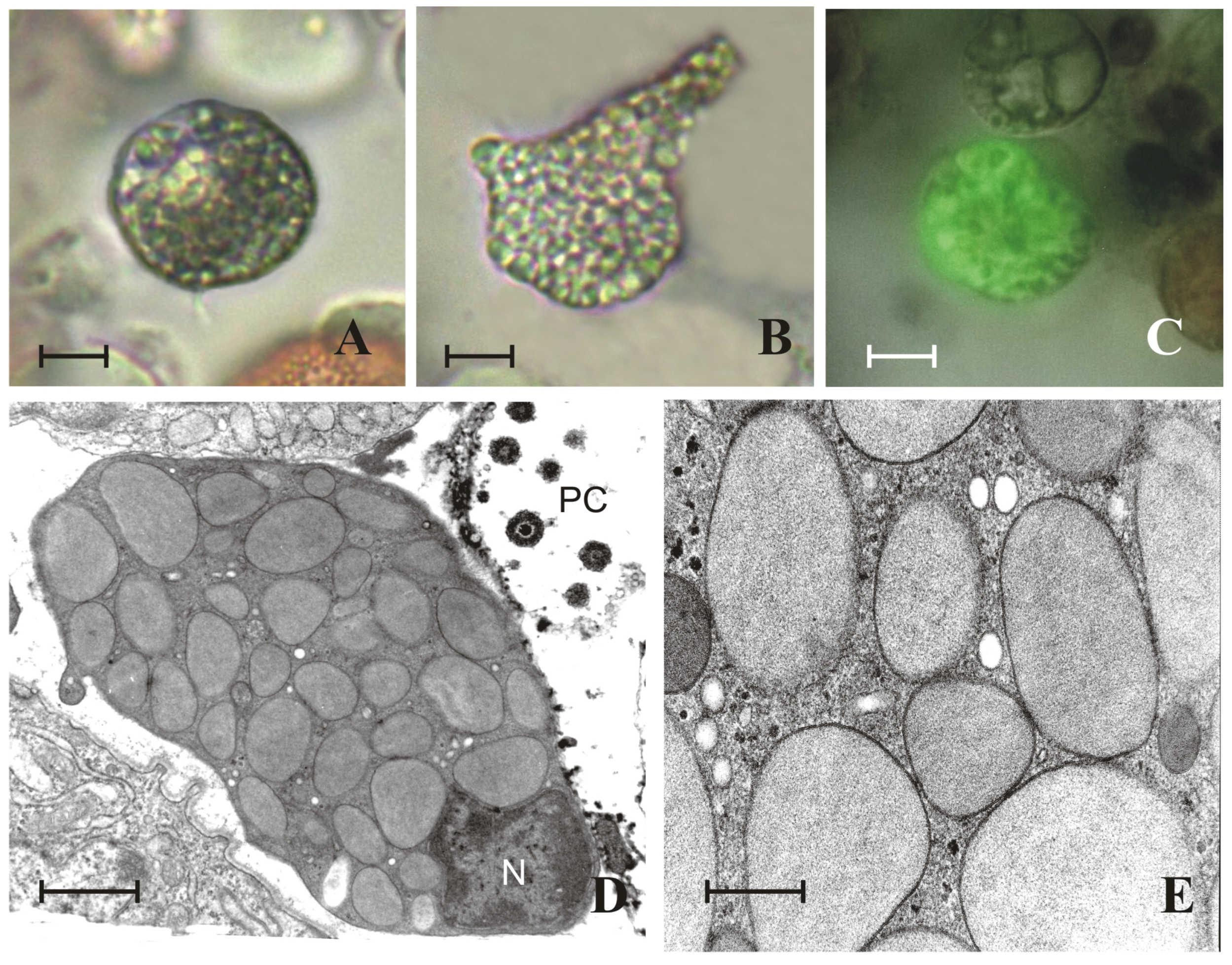
2.1. Granules of Granular Cells Contain a Heparin–Histamine System

Histochemical assays revealed that the cytoplasmic granules showed metachromatic pink–violet staining with toluidine blue (Figure 2A) and 1,9-dimethyl-methylene blue (DMB) (Figure 2B), highlighting the presence of polyanionic molecules such as glycosaminoglycans (GAGs), which are rich in sulphate groups, as supported by the brown colour after Geyer’s reaction (Figure 2C). Granules appeared green after treatment with Ehrlich’s trichrome mixture, revealing their basophilic nature on the basis of their specific affinity for methyl green (Figure 2D). Evidence of the presence of heparinoid molecules rich in sulphate groups is the red staining after Csaba’s reaction (Figure 2E) and the affinity to berberine sulphate (Figure 2F).
The presence of both heparin and histamine inside the granules was confirmed by immunohistochemical treatment: labelling of anti-heparin antibody was detected by red fluorescence of Cy3 fluorochrome (Figure 3A), and that of anti-histamine was detected by green fluorescence of the FITC fluorochrome (Figure 3B). Immunohistochemistry under TEM showed that heparin and histamine were present in the same granule (Figure 3C).
Heparinase I and heparinase III have been employed to discriminate the presence of heparin from other heparinoids, such as heparan sulphate inside the granules of granular cells. Heparinase I is a heparin hydrolase that specifically hydrolyses bonds between hexosamine and the O-sulphate of hyaluronic acid in heparin, and heparinase III is a heparan sulphate hydrolase that specifically hydrolyses the 1→4 bonds between hexosamine and glucuronic acid in heparan sulphate. Compared with both the untreated (control) and heparinase III-treated monolayers, the fluorescence of granular cells via immunohistochemistry with an anti-heparin antibody was significantly lower in the heparinase I-treated monolayers, suggesting that heparinase I digested heparin contained in the granules. The presence of heparin instead of heparan sulphate is also supported because compared with the control, the heparinase III-treated monolayers did not significantly differ (Figure 4).

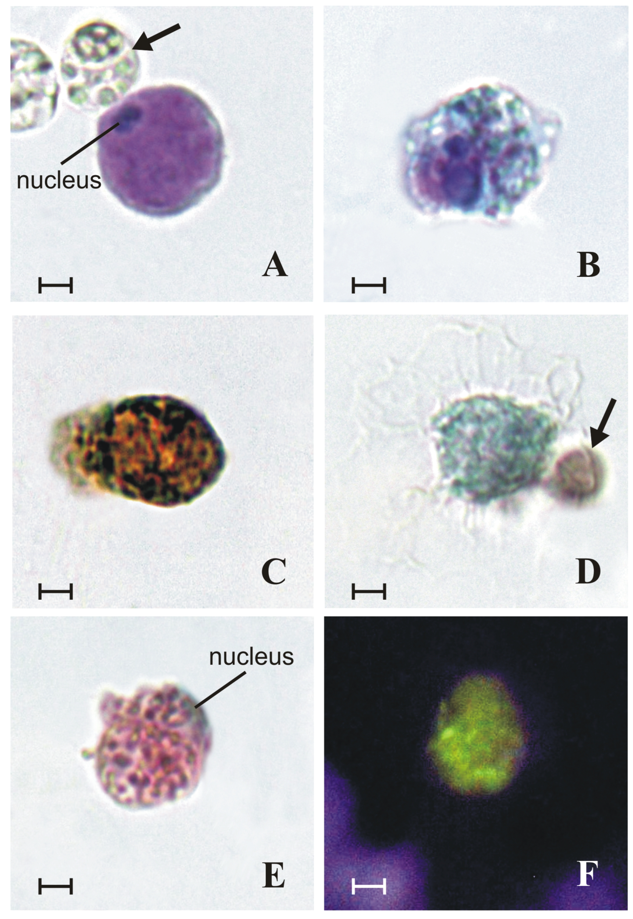
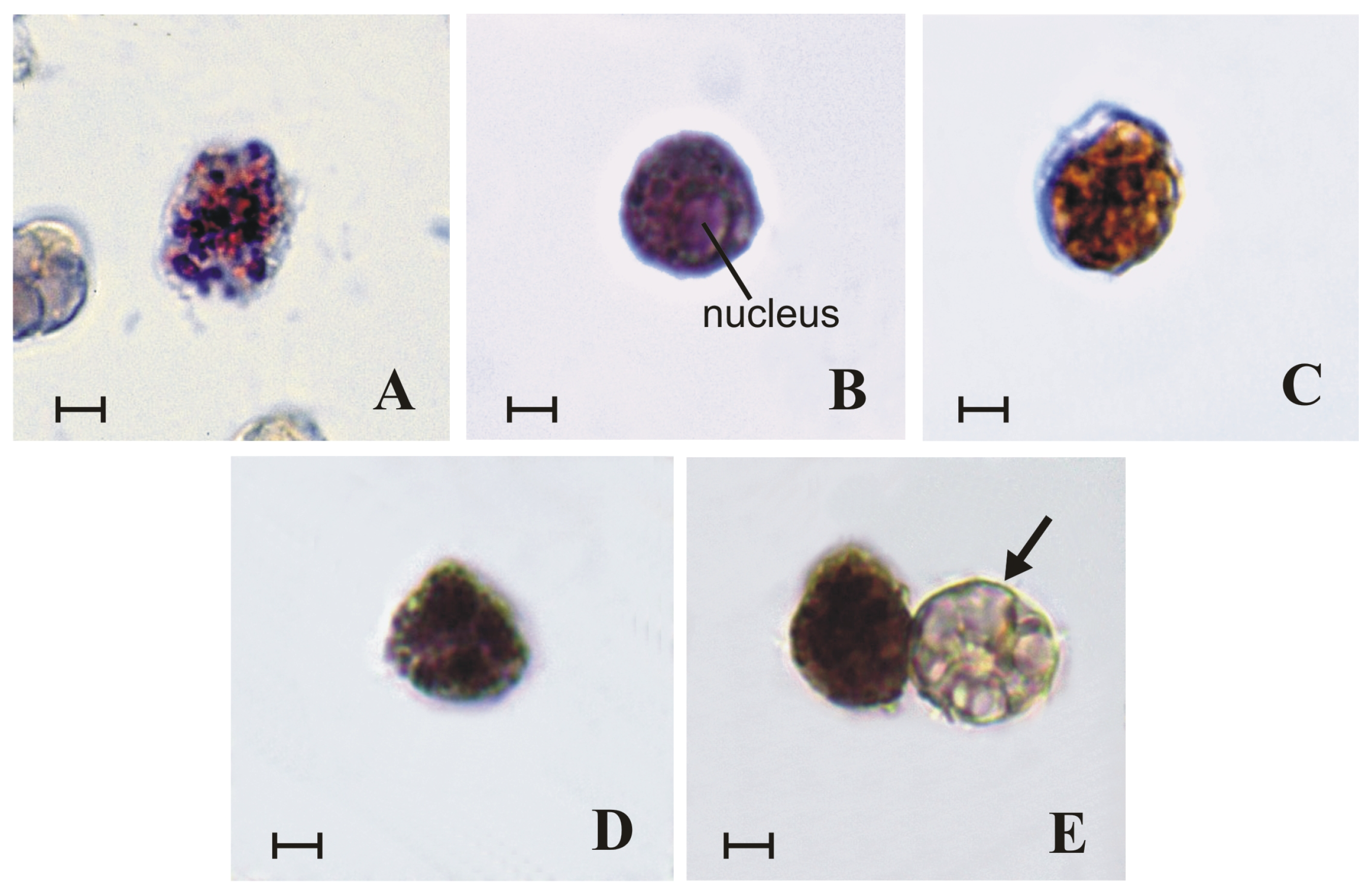
2.2. Granules of Granular Cells Contain Mast-Cell Hydrolases

Within the granules, enzymatic assays revealed the activities of various hydrolases, such as β-glucuronidase (Figure 5A), chloroacetyl esterase (Figure 5B), arylsulphatase (Figure 5C), and serine proteases, the latter represented by both tryptase (Figure 5D) and chymase (Figure 5E).

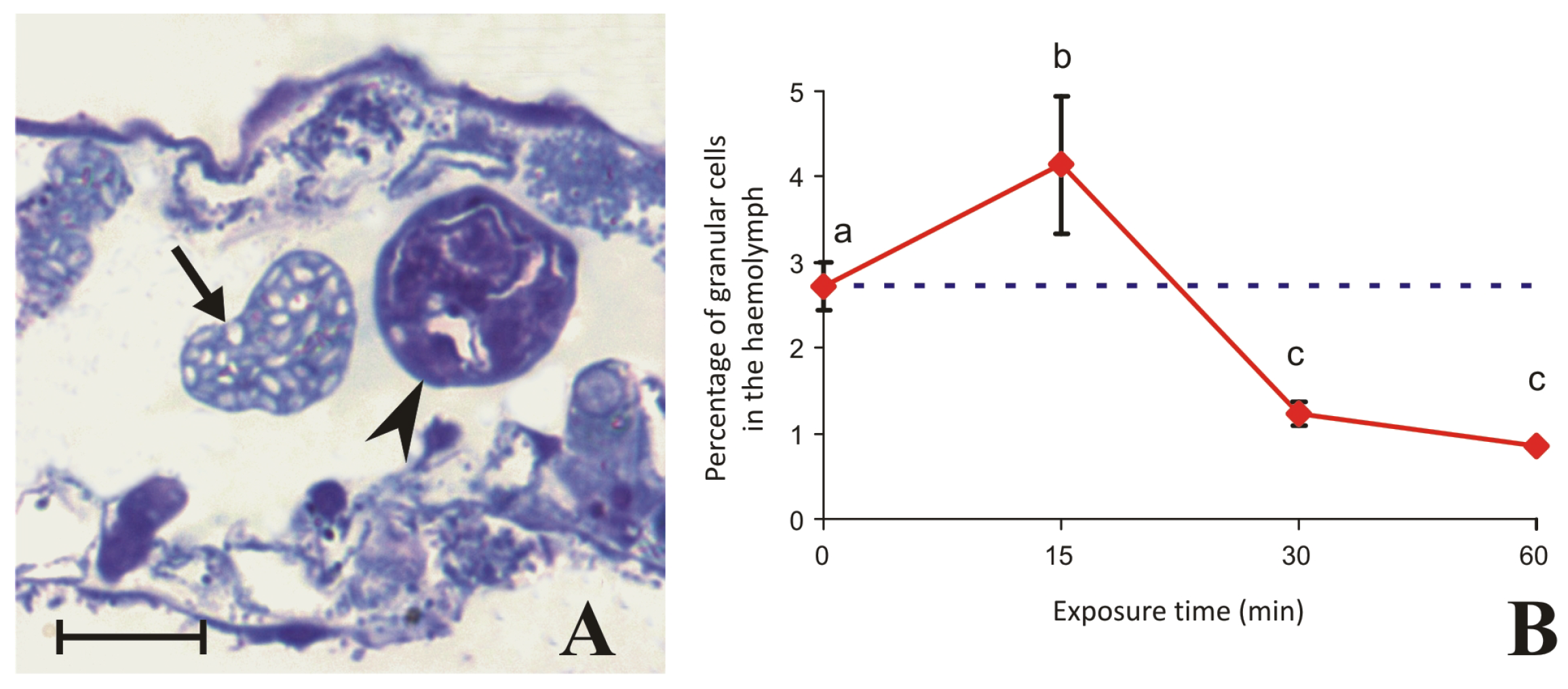
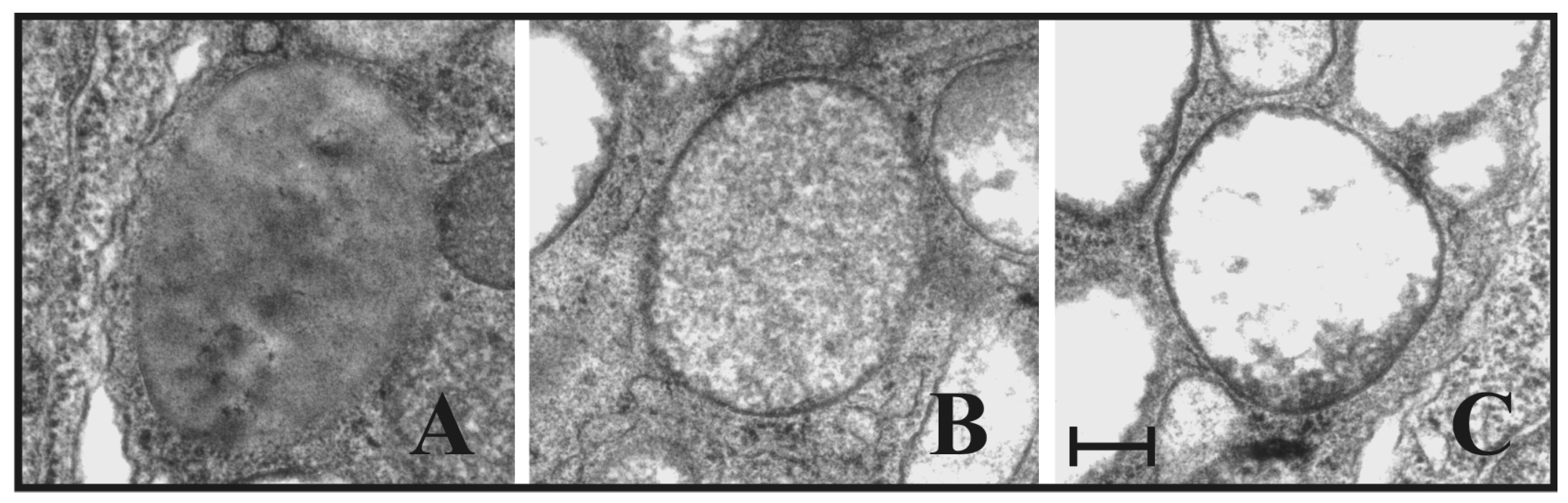
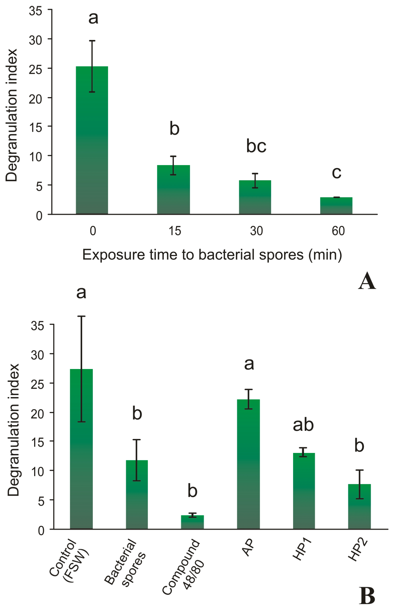
2.3. Granular Cells Have the Ability to Infiltrate Tissues and Degranulate in Response to a Stimulus

Experiments were carried out on genetically identical subclones of filter-feeding individuals from the same colony at different times of exposure to bacterial spores of Bacillus clausii in seawater with the aim of testing whether the entry of bacteria into the lumen of the postbranchial digestive tract causes the mobility and infiltration of granular cells from the haemolymph into the monolayer gut epithelium. After an increase at 15 min, probably due to their mobilisation, the percentage of circulating granular cells (Figure 6A) had significantly decreased after 30 min (Figure 6B). In parallel, after 30 min of exposure, ultrathin sections showed crowding of granular cells in the perivisceral sinus surrounding the digestive tract, causing sinus occlusion (Figure 7A,B). Some granular cells rested directly on the basal lamina of the intestinal epithelium, as the circulatory system within the zooid is open and haemolymph circulates in the interorgan spaces, i.e., the sinuses and lacunae in the mantle. Other granular cells could be observed crossing the epithelium (Figure 7A). Under TEM, the process of infiltration clearly involved diapedesis, with the formation of podosomes initially contacting the basal lamina (Figure 7C,D) and then the passage of a granular cell between two cells of the epithelium by rupture and subsequent reconstitution of cell junctions (Figure 7E,F). During infiltration, the granular cells presented an amoeboid appearance, and degranulation occurred with the loss of most of the granule content, as evidenced by the different arrangements of the electron-dense material (Figure 8A–C).
In vitro exposure to bacterial spores caused a significant decrease in the percentage of haemocytes with fluorescent granules labelled with anti-histamine antibody, indicating the induction of the degranulation process for granular cells. The decrease in fluorescence corresponding to a release of histamine into the medium occurred after 15 min exposure (Figure 9A). Similar significant decreases in histamine content were observed after 15 min exposure to compound 48/80, i.e., a specific FcεRI-independent secretagogue for mast cells [49], or heterologous plasmas (Figure 9B). The latter were obtained from the haemolymph of two distinct incompatible donors and compared with the plasma obtained from the haemolymph of the same colony (autologous plasma).

3. Discussion

The granular cells of B. leachii are commonly found in the circulation and tend to increase in number—together with multinucleated cells—at the beginning of the hibernation (torpor) process, which characterises this species at mid-latitudes [43,45,50,51] and during experimentally induced whole-body regeneration [44]. These cells appear to be involved primarily in inflammatory immune functions and share affinity for various histochemical and immunohistochemical staining methods with vertebrate mast cells [52], such as dye metachromasia, c-kit protein expression, and hydrolytic enzyme activities of β-glucuronidase, chloroacetyl esterase, arylsulphatase, and serine proteases. Like vertebrate mast cells, this cell type is able to localise at the interface of body surfaces exposed to the external environment [53], as in the case of gut epithelia, representing a haemocyte of pivotal importance in the defence response to external pathogens.
B. leachii granular cells have been observed to cross the basal membrane and infiltrate the gut epithelium after exposure of colonies to bacterial spores. A role in immunosurveillance of the alimentary tract, similar to that exerted by mammalian mucosal mast cells able to infiltrate the gastric mucosa [54], has been hypothesised. Initially, an increase in the number of circulating granular cells in the haemolymph occurred as a rapid inflammatory response to the stimulus.
This feature is very interesting and suggests an intriguing scenario for the precursors of the granular cell line. Haemopoietic tissues are unknown in this species, and the observed increase in granular cells in the haemolymph could be interpreted as rapid proliferation and differentiation of circulating stem cells, i.e., the haemoblasts, or mobilisation of the differentiated cells from storage sites, such as the peripheral blind-sac vessels of the colony, namely, ‘ampullae’ [43]. The fact that the number of circulating granular cells decreases with increasing exposure time to bacterial spores suggests that they massively leave the bloodstream to infiltrate the postbranchial digestive tract. This finding was supported by ultrastructural observations that revealed that the process has the characteristics of diapedesis [55]. This phenomenon involves all cells that, in response to inflammatory chemical signals, leave the bloodstream by adhering to epithelial cells and then make their way between the junctions of adjacent cells, which close as they pass. All the diapedesis phases have been identified in stimulated colonies of B. leachii, from the phase of approaching and adhering to the basal lamina to the phase of crossing the epithelium of the digestive tract, which occurs between the junctions of adjacent cells. Moreover, these cells clearly undergo a process of degranulation. In a similar manner, mammalian mast cells can cross the gastric wall and reach the stomach lumen, releasing part of the content of their granules. This response, called ‘gastric leukopedesis’, occurs in response to an increased bacterial load in the lumen [56,57]. However, the degranulation observed on TEM in the stimulated granular cells appears very peculiar, because no exocytosis of content or release of whole granules into the extracellular environment was observed, as is usually the case in mast cells, but the cytoplasmic granules retained the same size and gradually lost their content. Indeed, among the different types of degranulation known in mast cells, ‘piecemeal degranulation’ has recently been described as a process of mediator release without membrane fusion [15,58,59]. This type of degranulation is a gradual process involving the formation of small vesicles within the granules, which selectively transport mediators towards the plasmalemma until the granules, which maintain their original size, are emptied. This process allows a more modulated and prolonged inflammatory response over time. It is possible that a similar mechanism of mediator release also occurs in the granular cells of B. leachii, which should be investigated further.
In vitro exposure to various stimuli, such as bacterial spores, compound 48/80, and heterologous plasma, has been shown to induce rapid degranulation, which occurs within 15 min. The positive results of the PAS assay for polysaccharides [42] and Geyer’s assay indicate that the basophilic granules contain GAGs rich in sulphate groups. The metachromatic reaction of DMB, a method previously used to detect heparinoids in the pharyngeal tissues of S. plicata [60], and the labelling by the anti-heparin antibody, which disappears after treatment with heparinase I, but not in the presence of heparinase III, support the hypothesis that the GAG present in the granules is heparin and not heparan sulphate.
Previous studies on the solitary ascidian S. plicata are of particular interest because this species belongs to the same family, Styelidae, as B. leachii. In S. plicata, heparin has been extracted and purified from haemocytes, and immunohistochemistry has shown that heparin and histamine colocalise inside granules of circulating basophil-like cells [37]. In B. leachii, immunohistochemistry via TEM confirmed the colocalisation of heparin and histamine within the same granules, a feature that is also shared with vertebrate mast cells and basophils (Table 1).
Histamine is a biogenic amine that, within the granules of vertebrate mast cells, binds to heparin by ionic bonding between its positively charged amine groups and the sulphate and carboxylic negatively charged groups of heparin. Its detachment from heparin upon degranulation is facilitated by pH changes in the external medium [61,62,63]. In the tunicate S. plicata, histamine is secreted after a pathogenic stimulus, which decreases phagocytic ability and promotes vasoconstriction in tunic explants, supporting the hypothesis of its involvement in the regulation of the inflammatory response [64]. Heparin has been reported to have ten times less anticoagulant activity in this species than it does in mammals [60,65]. Even in mammalian mast cells, the main role of heparin does not appear to be as an anticoagulant, although this was the first function highlighted. This GAG acts as a binder for histamine, enzymes, cytokines, and growth factors, and plays a role in modulating the release of biologically active mediators in the granules, such as preventing their degradation and contributing to their modulation of inflammatory responses [66,67,68,69,70]. Therefore, the presence of heparin in granular cells suggests that it may play a similar binding-modulatory role in ascidian innate immunity.
The types of hydrolases found in the granules of B. leachii granular cells are another feature that should be considered owing to their common presence in granules of mast cells, but not in granules of basophils of vertebrates (Table 1). This reflects the origin of mast cells and basophils from different progenitors in the bone marrow [71,72], i.e., the basophil promyelocyte (or probasophil) and the immature mast cell (or promastocyte), respectively [6]. In addition, mast cells mature in tissues, whereas basophils fully mature in the bone marrow before being released into the bloodstream [73,74]. Therefore, the definition of tunicate granular cells as ‘basophils’ or ‘basophil-like cells’ should be used with great caution, as this would lead to confusion about the origin and the immune role of these cells. Indeed, the content in hydrolytic enzymes seems to be shared only among ascidian granular cells and vertebrate mast cells, as they are probably part of functional aspects and mechanisms conserved in chordates, the understanding of which can be inferred by looking at what is known about vertebrate mast cells. In mammalian mast cells, the acid hydrolase β-glucuronidase is responsible for the catalytic cleavage of β-D-glucuronides in heparin. Its activity appears to be correlated with inflammation, since the structure and activity of heparin are significantly modulated by this enzyme, which is upregulated during inflammatory conditions [75]. Chloroacetyl esterase contributes to mast cell degranulation in the inflammatory response [76], interacts with other enzymes of the granules such as proteases to modulate the immune response [77], and facilitates the release of inflammatory mediators from the granules such as histamine and leukotrienes [78]. Arylsulfatase is a universal enzyme that hydrolyses aromatic sulphates. In vertebrate mast cells, it is involved mainly in the degradation of sulphated proteoglycans [79] and contributes to the modulation of the inflammatory response by degrading extracellular matrix components [80]. Serine proteases are proteolytic enzymes that use a serine residue in their active site to catalyse the hydrolysis of peptide bonds [78]. The most important serine proteases of mast cells are tryptase, chymase, and carboxypeptidase A [81]. During inflammation, they activate cytokines and growth factors that modulate the immune response, degrade extracellular matrix proteins, and activate protease-activated receptors (PARs) [77], the presence of which has not yet been demonstrated in tunicates. In vertebrates, there are differences in the levels of tryptase and chymase between mast cells and basophils (Table 1): mast cells contain significant amounts of both enzymes, whereas basophils contain mainly tryptase [82,83]. Even in the ascidian S. plicata, the test cell, a component of the ovular envelope, shows a heparin-histamine content inside cytoplasmic granules and releases tryptase during degranulation experimentally induced by exposure to compound 48/80. In this study, tryptase was considered to be involved in the protection of oocytes from invading parasites [84,85].
The c-kit receptor, a tyrosine kinase receptor of vertebrates, is also known as CD117 and SCF-R. The specific labelling of anti-c-kit antibody to granular cell plasmalemma suggests the presence of this receptor in B. leachii, which should be confirmed by molecular analysis. This receptor is highly expressed on the plasmalemma of mast cells, but not basophils; thus, since detection by immunohistochemistry is a valuable biomarker for mast cell-specific recognition in vertebrates [52,86,87], it still supports the hypothesis that more features of B. leachii granular cells are shared with mast cells than with basophils. In mast cells, development and differentiation from haemopoietic precursors [88], survival through antiapoptotic signalling [89], proliferation in response to stem cell factor (SCF) [90], great migration in peripheral tissues [91], and activation with subsequent degranulation in response to stimuli [92] are all dependent on this receptor. Interestingly, although it does not directly induce degranulation, the activation of this receptor can enhance the degranulation response induced by other stimuli by acting in synergy with other tyrosine kinase receptors, such as FcεRI, of adaptive immunity [93]. The cascade of reactions that occurs after FcεRI activation stimulates phospholipase C (PLC), which catalyses the formation of inositol 1,4,5-trisphosphate (IP3). The latter binds to Ca2+ channel receptors on the membranes of the endoplasmic reticulum, and this binding causes the channels to open. The resulting release of calcium ions into the cytoplasm triggers degranulation [94]. However, the activation of c-kit can increase intracellular calcium mobilisation by a signal transduction mechanism without the involvement of immunoglobulins. In this case, the PLC is directly activated [95]. This leads to two considerations. First, a similar mechanism of calcium mobilisation cannot be excluded in animals without adaptive immunity such as ascidians. Second, there are degranulating mechanisms in mast cells that are independent of adaptive immunity and are probably related to mechanisms conserved in the innate immunity of all chordates.
A supporting example of this phenomenon is in vitro exposure to compound 48/80, a potent mast cell secretagogue widely used to induce degranulation that is able to achieve this with histamine depletion in B. leachii granular cells. It acts as a G protein-coupled receptor (GPCR) agonist on mast cells independently of FcεRI receptor activation by directly activating G proteins and thus stimulating PLC. Even in this case, the final result is the mobilisation of intracellular calcium, which causes degranulation [96,97,98], the latter of which is evidenced by a significant release of histamine [99].
In conclusion, there are several morphological, biochemical and functional aspects that B. leachii granular cells have in common with mast cells, much more so than with basophils, providing important insights into the origin and phylogeny of mast cells within the chordate phylum (Table 1). In vertebrates, a progressive separation of the functions and competence of different immunocytes could have occurred, which could have been integrated into mechanisms of adaptive immunity, giving rise to more complex immune systems than those of basal chordates [100,101,102,103]. The characteristics of the ancestral precursors of mast cells could still be present in present-day tunicates, retaining many of the ancestral morpho-functional characteristics typical of basal chordates. This opens the way for important considerations about the origin of mast cells in vertebrates and the immunomodulatory role of their mediators. Further analysis is needed to fully understand the differentiation pathway and role of this haemocyte within the immune system of B. leachii from an evolutionary viewpoint. In particular, it would be interesting to look for the presence of preformed and de novo synthesised mediators typical of vertebrate mast cells, such as proinflammatory cytokines, since recent studies have shown an upregulation of interleukin 17 and tumour necrosis alpha genes in haemocytes of C. intestinalis after an inflammatory stimulus [104,105].
Table 1. Principal preformed mediators within granules of basophils and mast cells of vertebrates and granular cells of B. leachii.
Basophils
(Vertebrates)
Mast Cells
(Vertebrates)
Granular Cells
(B. leachii)
1. GAGs
Heparin+++
2. Biogenic amines
Histamine+++
3. Hydrolytic enzymes
β-glucuronidase++
Chloroacetyl esterase++
Arylsulphatase++
Tryptase+++
Chymase++

4. Materials and Methods

4.1. Animals

Colonies of B. leachii collected near the marine station of the Department of Biology, University of Padova, in Chioggia (Southern Lagoon of Venice, Italy) were attached to glass slides and transferred to aquaria at a constant temperature of 17 °C with 12 h:12 h light–dark. Seawater (pH: 8.0 ± 0.1; salinity: 35 psu) was aerated with oxygenators and renewed three times a week on alternate days. Colonies were fed daily with a mixture of unicellular microalgae (Tetraselmis chuii and Isochrysis galbana) at a concentration of 1.4 × 10⁵ cells mL−1.

4.2. Haemocyte Collection

For each experiment, haemolymph was collected from the peripheral vessels of colonies, previously blotted dry, after making a small hole with a fine tungsten needle. Flowing haemolymph was collected with a glass micropipette and transferred to a 1.5 mL Eppendorf tube at a 1:1 ratio with 0.38% sodium citrate in filtered seawater (FSW) to prevent clotting. FSW was obtained through filtration in 0.45 µm cellulose acetate membranes (Sartorius Stedim Biotech GmbH, Goettingen, Germany). After sampling, the haemolymph samples were centrifuged at 780× g for 15 min to separate the haemocytes from the plasma. The supernatant was discarded, whereas the pellet was resuspended in FSW at a final concentration of 8 × 10⁶ cells mL−1.

4.3. Fixation of Haemocyte Monolayers

Sixty microlitres of haemocyte suspension was placed at the centre of SuperfrostTM Plus adhesion microscope slides (Thermo Scientific, Braunschweig, Germany) with an electrostatically adhesive surface, on which the suspended cells were allowed to adhere spontaneously for 30 min. After adhesion, the haemocytes were fixed for 30 min at 4 °C with a marine invertebrate fixative mixture consisting of 4% paraformaldehyde (MP Biomedicals, Eschwege, Germany) and 0.2% glutaraldehyde (AppliChem GmbH, Darmstadt, Germany) in 0.2 M sodium cacodylate buffer, pH 7.4, with the addition of 1% NaCl and 1% sucrose [106]. After rapid washes in 0.1 M phosphate-buffered saline (PBS: NaCl 8 g L−1, KCl 0.2 g L−1, KH2PO4 0.2 g L−1, Na2HPO4 1.15 g L−1, pH 7.2), monolayers were used for the various histochemical assays reported below. Finally, they were mounted with aqueous mounting medium (Acquovitrex, Carlo Erba, Milan, Italy) and observed under an Olympus CX31 light microscope (LM) equipped with a Lumenera Infinity 2 digital camera and Infinity Capture and Analyze Application software (version 5.0.0, Lumenera Co., 2002-2009, Ottawa, ON, Canada) as well as an Amplified Fluorescence by Transmitted Excitation of Radiation (AFTER) LED fluorescence module (Fraen Corp., Corsico, Milan, Italy), with excitation sources at 365 nm, 470 nm, and 535 nm.

4.4. Histochemical Assays

4.4.1. Staining Methods for Heparin Detection

The following assays were performed on fixed monolayers according to methods reported in detail by Suvarna and collaborators [107], with modifications as indicated.
(i).
Metachromatic reaction of toluidine blue. The monolayers were incubated for 20 min in a solution of 0.1% toluidine blue O (Fluka Chemie GmbH, Steinheim, Germany) in 30% ethanol and then washed in 95% ethanol. After quick washing in distilled water, the slides were mounted and observed under an LM. Basophilic substances were stained purple.
(ii).
Metachromatic reaction of 1,9-dimethylmethylene blue (DMB). The monolayers were incubated for 30 min in an aqueous solution containing 0.05 M DMB (Sigma-Aldrich, St. Louis, MO, USA), 0.1 M HCl, 0.04 mM glycine, and 0.04 M NaCl and then washed in distilled water. Basophilic substances, such as acidic mucopolysaccharides and heparinoids, were stained violet.
(iii).
Ehrlich’s triacid mixture. The monolayers were incubated in Ehrlich’s triacid mixture (12 vol saturated orange G aqueous solution, 8 vol saturated acid fuchsin solution, 10 vol saturated methyl green aqueous solution, 30 distilled water, 18 vol absolute ethanol and 5 vol glycerine) for 15 min and then washed in distilled water. The basophilic granules containing heparinoid substances were stained light green, the acidophilic granules were stained copper red, and the neutrophilic granules were stained violet.
(iv).
Csaba’s staining. The monolayers were incubated for 15 min in a solution of 0.36% Alcian blue, 0.18% safranin O, and 0.48% ammonium ferric sulphate in 0.1 M sodium acetate, pH 1.42. Non-sulphated heparin precursor was stained light blue, and highly N-sulphated heparin was stained red.
(v).
Geyer’s method for sulphates. The monolayers were incubated for 30 min in an aqueous solution of Fast Blue B (Fluka) (50 mg in 10 mL of 5% acetic acid). The monolayers were then rinsed for 1 min in distilled water at 4 °C and subsequently incubated for 5 min in a cold (4 °C) saturated solution of 1-naphthol (Sigma-Aldrich) in 0.1 M borax buffer, pH 9.4. The monolayers were then rinsed in distilled water for 1 min. The sulphate groups of the glycosaminoglycans were stained brown.
(vi).
Berlin and Enerbäck’s method [108] with berberine sulphate. The monolayers were incubated for 20 min in an aqueous solution containing 0.02% berberine sulphate (Sigma-Aldrich) adjusted to pH 4.0 with the addition of 1% citric acid. After being quickly washed in distilled water and mounted, the monolayers were observed under an LM with a UV light excitation source (365 nm). Sulphate-containing polyanions such as heparinoid substances emitted intense blue-green fluorescence.

4.4.2. Assay for Discrimination of Heparin from Heparan Sulphate

Fixed haemocyte monolayers were incubated overnight at 37 °C in aqueous solutions containing 0.05 U mL−1 heparin lyase, which specifically hydrolyses heparin (heparinase I, EC 4.2.2. 7, Sigma-Aldrich) or heparan sulphate (heparinase III, EC 4.2.2.8, Sigma-Aldrich) in 0.1 M Tris–HCl buffer at pH 7.0 and 7.6, respectively. Finally, the samples were subjected to fluorescence immunohistochemistry (see Section 4.6) to visualise the heparin content under an LM. The percentage of haemocytes labelled by the anti-heparin antibody compared with the total was obtained by counting the number of cells in ten optical fields at 1000× per slide, corresponding to approximately 300 cells.

4.5. Histoenzymatic Assays

Cytolocalisation of hydrolytic enzyme activities under an LM was performed with a simultaneous azo-coupling detection method for β-glucuronidase, chloroacetyl esterase and proteases based on the diazonium salts hexazonium-p-rosaniline, Fast Blue B, and Fast Blue BB (Fluka), respectively, as coupling reagents and a metal salt procedure for arylsulfatase detection according to methods extensively reported by Cima [109].
(i).
β-glucuronidase. Fixed monolayers were washed in sodium acetate buffer 0.1 M, pH 5.2, for 10 min at 37 °C for 2 h, after which 4 mg of naphthol AS-BI β-glucuronide, which was previously dissolved in 0.25 mL of dimethylformamide (DMF) in 20 mL of buffered hexazonium-p-rosaniline, was added. The monolayers were then washed in distilled water and mounted in Acquovitrex. Positive sites for the enzymatic reaction inside haemocytes were stained magenta.
(ii).
Chloroacetyl esterase. Fixed monolayers were washed in PBS, pH 6.5, for 10 min and incubated for 1 h at 20 °C in a reaction mixture consisting of 6 mg of naphthol chloroacetate (Sigma-Aldrich) previously dissolved in 1 mL of DMF and added to 20 mL of PBS containing 20 mg of Fast Blue B. Monolayers were then washed in distilled water and mounted in Acquovitrex. The positive sites inside the haemocytes were stained blue.
(iii).
Proteases. Fixed monolayers were incubated for 60 min at 37 °C in a reaction mixture containing 4 mg of the synthetic substrate Z-Ala-Ala-Lys-4-methoxy-2-naphthylamide (Z-AAK-mna, MP Biomedicals), which is specific for tryptase, or Suc-Ala-Ala-Phe-4-methoxy-2-naphthylamide (S-AAF-mna, MP Biomedicals), which is specific for chymase, previously dissolved in 0.5 mL of DMF and then added to 10 mL of 0.1 M Tris–HCl buffer, pH 7.0 for tryptase and 7.8 for chymase, containing 10 mg of Fast Blue BB. This was followed by incubation for 5 min in 1% copper sulphate. Finally, the monolayers were rinsed in distilled water and then mounted with Acquovitrex. The positive sites inside the haemocytes were stained dark blue or black.
(iv).
Arylsulphatase. Fixed monolayers were washed in sodium acetate buffer for 10 min and incubated for 2 h at 37 °C in the following reaction mixture: 0.16 g p-nitrocatecholsulphate (Sigma-Aldrich), 4 mL distilled water, 12 mL sodium acetate buffer, and 4 mL of 8% aqueous solution of lead nitrate. After incubation, the monolayers were washed with distilled water and then with an ammonium sulphide solution (diluted 1:100 in distilled water) for 2 min. Finally, the monolayers were washed with distilled water and mounted in Acquovitrex. The positive sites inside the haemocytes were stained brownish black.
Controls were made by omitting the substrate or preincubating the monolayers for 15 min in appropriate buffers with the following inhibitors: 100 mM mucic acid for β-glucuronidase, 10 mM sodium fluoride for chloroacetyl esterase, 100 µM chloromethyl ketone (Tosyl-Lys-CMK, MP Biomedicals) dissolved in 0.5 mL of DMF for serine proteases and 100 mM sodium sulphate for arylsulphatase.

4.6. Immunohistochemical Assays for Heparin, Histamine, and Stem Cell Factor Receptor

The adhered haemocyte monolayers were fixed as described in Section 2.3 and permeabilised for 5 min with 0.1% Triton X-100 in PBS. To block possible nonspecific interactions during subsequent treatments with antibodies, incubation was carried out for 30 min in a 10% solution of normal goat serum (Vector) in PBS anti-heparin and anti-histamine antibodies and in a 10% solution of rabbit serum (Vector) in the case of anti-stem cell factor receptor (SCF-R, CD117 or c-kit) antibodies. In all cases, primary antibodies were omitted in the controls.
For the heparin and histamine localisation assays, the samples were incubated for 15 h at 4 °C with a mouse monoclonal anti-heparin antibody (MAB570 clone A7.10, Chemicon International, Temecula, CA, USA) or a rabbit polyclonal anti-histamine antibody developed from synthetic histamine conjugated to succinylated KLH as an immunogen (H7403, Sigma-Aldrich), both at concentrations of 10 μg mL−1 in PBS. The monolayers were subsequently rinsed in PBS and incubated with a biotinylated goat anti-mouse IgG secondary antibody (401216, Calbiochem, EMD Biosciences Inc., San Diego, CA, USA) for 60 min, followed by incubation for 30 min with streptavidin–Cy3 (S6402, Sigma-Aldrich) at a concentration of 20 μg mL−1 in PBS to detect the presence of heparin. The Cy3 fluorochrome has an emission spectrum at 565 nm (red fluorescence). The haemocytes were incubated for 60 min with a goat anti-rabbit IgG secondary antibody conjugated with fluorescein isothiocyanate (FITC, 401314, Calbiochem) to detect the presence of histamine. The FITC fluorochrome has an emission spectrum at 520 nm (green fluorescence). Finally, the monolayers were washed in distilled water and mounted with FluorSave Reagent (Calbiochem), a mounting medium that decreases fluorescence fading. Observations were performed under an LM with green (535 nm) and blue (470 nm) LED light excitation sources for Cy3 and FITC, respectively.
Goat anti-human-SCF-R antibody (S2562, Sigma-Aldrich) was developed from a purified recombinant human SCF-R extracellular domain (UniProt ID P10721). The monolayers were incubated with the primary antibody at a concentration of 1 µg mL−1 and then incubated with a biotinylated rabbit anti-goat IgG secondary antibody (Vector) at a concentration of 15 µg mL−1 in PBS before being incubated for 30 min in streptavidin–FITC (S3762, Sigma-Aldrich) at a concentration of 20 µg mL−1 in PBS.

4.7. Electron Microscopy

Selected subclones of colonies not in regression were fixed in a solution of 1.5% glutaraldehyde in saline buffer (0.2 M sodium cacodylate buffer, pH 7.4, containing 1.7% NaCl and 1% saccharose) for 2 h at 4 °C. The samples were then rinsed in saline buffer, fixed for 45 min in 1% osmium tetroxide in cacodylate buffer, dehydrated in an increasing ethanol series, and embedded in Epon 812 epoxy resin (Sigma-Aldrich). Semithin sections (1 µm) were collected on slides, hot-stained with 1% toluidine blue and 1% borax in distilled water, and observed under an LM. Ultrathin sections (60 nm) were collected on copper grids.
For immunocytochemical analyses, selected subclones were fixed in a marine invertebrate fixative mixture (see Section 4.3), dehydrated in increasing ethanol concentrations, and embedded in London Resin White (LRW, Polyscience, Warrington, PA, USA). Ultrathin sections, once collected on gold grids, were incubated for 10 min in 10% goat serum, washed in PBS, and incubated overnight with primary anti-heparin and anti-histamine antibodies in a humidity chamber at the concentrations reported in Section 4.6. After washing, the grids were incubated for one hour at 20 °C with secondary antibodies conjugated with colloidal gold at a concentration of 10 µg mL−1 in PBS. For the anti-heparin antibody, ultrathin sections on grids were exposed to a goat anti-mouse IgG antibody conjugated with colloidal gold beads 15 nm in diameter (BBI Solutions, EM. GMHL15, Portland, ME, USA). For the anti-histamine antibody, a goat antirabbit IgG antibody conjugated with beads of colloidal gold 25 nm in diameter (Jackson ImmunoResearch, Ely, UK) was used.
Finally, ultrathin sections on both copper and gold grids were stained with uranyl acetate, and lead citrate and observed under an FEI Tecnai 12 transmission electron microscope (TEM) at 75 kV equipped with a Tietz high-resolution digital camera.

4.8. Degranulation Assay

After adhesion, the haemocyte monolayers were incubated for 15 min at 25 °C in 60 μL of a 4 × 10⁸ suspension of bacterial spores (Bacillus clausii, strain Enterogermina®, Opella Health Care, Milan, Italy)/mL of FSW, previously centrifuged at 12,000× g for 10 min and resuspended in an equal volume of FSW. Other monolayers were exposed to a 0.2 mM solution of compound 48/80 (Sigma-Aldrich) in FSW, which specifically activated mammalian mast cell degranulation through non-IgE-dependent stimulation. Other monolayers were incubated in autologous plasma from the same colony or heterologous plasma (HP) from incompatible donors. To obtain plasma, colonies of approximately 80 zooids were blotted dry, and marginal tunic vessels were lacerated with fine tungsten needles. Haemolymph was collected with a glass micropipette and centrifuged at 780× g for 15 min, after which the supernatant was collected.
Finally, the monolayers were fixed and processed to visualise histamine within the granules under an LM by fluorescence immunohistochemistry (see Section 4.6). The degranulation index was assessed by calculating the percentage of haemocytes lacking fluorescence for histamine compared with the total for ten optical fields at 1000×, corresponding to approximately 300 cells per slide.

4.9. Exposure of Colonies to Bacteria

One large colony was divided into sixteen subclones of at least three systems each. The subclones were formed of groups of zooids that were genetically identical and all kept at the same developmental stage because they were connected by a vascular network that extended throughout the common tunic enveloping the colony [50]. Eight subclones were used as controls. Another eight subclones were fasted for 24 h and then placed in a beaker containing 250 mL of a suspension of 8 × 10⁷ B. clausii spores per mL of FSW. Then, two subclones were chosen after 0 (control), 15, 30, and 60 min of exposure. For each pair, one subclone was used to collect haemocytes, and after adhesion and fixation (see Section 4.3) to count the percentage of granular cells in relation to the total number of haemocytes in 10 optical fields at 1000× by LM. The other subclone was fixed and used for TEM (see Section 4.7).

4.10. Statistical Analysis

The data are expressed as the means of at least three different biological samples (n = 3 slides) ± standard deviations. Statistical analyses were performed with PRIMER 6 software (PRIMER-E Ltd., Plymouth Marine Laboratory, Plymouth, UK). The data obtained were compared using a χ2 test followed by Dunnett’s test for multiple comparisons, and the level of significance was set at p < 0.05.

Author Contributions

Conceptualisation, N.B., S.D.P. and F.C.; supervision, F.C.; investigation, methodology, data curation, formal analysis, N.B. and S.D.P.; writing—original draft preparation and editing, F.C. All authors have read and agreed to the published version of the manuscript.

Funding

This research was supported by the Department of Biology, University of Padova, with PRID 2023 (BIRD231875) to FC.

Data Availability Statement

All data generated or analysed during this study are included in this published article.

Acknowledgments

The authors wish to thank Federico Caicci, manager of DeBio Imaging Facility at the Department of Biology, for TEM images.

Conflicts of Interest

The authors declare no conflicts of interest. The funders had no role in the design of the study; in the collection, analyses, or interpretation of data; in the writing of the manuscript; or in the decision to publish the results.

References

  1. Baccari, G.C.; Pinelli, C.; Santillo, A.; Minucci, S.; Kumar, R.R. Mast cells in nonmammalian vertebrates: An overview. Int. Rev. Cell Mol. Biol. 2011, 290, 1–53. [Google Scholar] [CrossRef] [PubMed]
  2. Dileepan, K.N.; Raveendran, V.V.; Sharma, R.; Abraham, H.; Barua, R.; Singh, V.; Sharma, R.; Sharma, M. Mast cell-mediated immune regulation in health and disease. Front. Med. 2023, 10, 1213320. [Google Scholar] [CrossRef] [PubMed]
  3. Crivellato, E.; Beltrami, C.; Mallardi, F.; Ribatti, D. Paul Ehrlich’s doctoral thesis: A milestone in the study of mast cells. Br. J. Haematol. 2003, 123, 19–21. [Google Scholar] [CrossRef]
  4. Ghably, J.; Saleh, H.; Vyas, H.; Peiris, E.; Misra, N.; Krishnaswamy, G. Paul Ehrlich’s mastzellen: A historical perspective of relevant developments in mast cell biology. In Mast Cell: Methods and Protocols; Torres, A., Ed.; Methods in Molecular Biology; Humana Press, Springer Science: New York, NY, USA, 2015; Volume 1220, pp. 3–10. [Google Scholar] [CrossRef]
  5. Valent, P.; Akin, C.; Hartmann, K.; Nilsson, G.; Reiter, A.; Hermine, O.; Sotlar, K.; Sperr, W.R.; Escribano, L.; George, T.I.; et al. Mast cells as a unique hematopoietic lineage and cell system: From Paul Ehrlich’s visions to precision medicine concepts. Theranostics 2020, 10, 10743–10768. [Google Scholar] [CrossRef]
  6. Atiakshin, D.; Samoilova, V.; Buchwalow, I.; Boecker, W.; Tiemann, M. Characterization of mast cell populations using different methods for their identification. Histochem. Cell Biol. 2017, 147, 683–694. [Google Scholar] [CrossRef]
  7. St. John, A.L.; Rathore, A.P.S.; Ginhoux, F. New perspectives on the origins and heterogeneity of mast cells. Nat. Rev. Immunol. 2023, 23, 55–68. [Google Scholar] [CrossRef]
  8. da Silva, E.Z.M.; Jamur, M.C.; Oliver, C. Mast cell function: A new vision of an old cell. J. Histochem. Cytochem. 2014, 62, 698–738. [Google Scholar] [CrossRef]
  9. Metz, M.; Siebenhaar, F.; Maurer, M. Mast cell functions in the innate skin immune system. Immunobiology 2008, 213, 251–269. [Google Scholar] [CrossRef]
  10. Staby, A.; Sand, M.B.; Hansen, R.G.; Jacobsen, J.H.; Andersen, L.A.; Gerstenberg, M.; Bruus, U.K.; Jensen, I.H. Comparison of chromatographic ion-exchange resins IV. Strong and weak cation-exchange resins and heparin resins. J. Chromatogr. A 2005, 1069, 65–77. [Google Scholar] [CrossRef]
  11. Bolten, S.N.; Rinas, U.; Scheper, T. Heparin: Role in protein purification and substitution with animal-component free material. Appl. Microbiol. Biotechnol. 2018, 102, 8647–8660. [Google Scholar] [CrossRef]
  12. Brody, D.; Metcalfe, D.D. Mast cells: A unique and functional diversity. Clin. Exp. Allergy 1998, 28, 1167–1170. [Google Scholar] [CrossRef]
  13. Kobayashi, H.; Ishizuka, T.; Okayama, Y. Human mast cells and basophils as sources of cytokines. Clin. Exp. Allergy 2000, 30, 1205–1212. [Google Scholar] [CrossRef]
  14. Krystel-Whittemore, M.; Dileepan, K.N.; Wood, J.G. Mast cell: A multi-functional master cell. Front. Immunol. 2016, 6, 620. [Google Scholar] [CrossRef]
  15. Mukai, K.; Tsai, M.; Saito, H.; Galli, S.J. Mast cells as sources of cytokines, chemokines, and growth factors. Immunol. Rev. 2018, 282, 121–150. [Google Scholar] [CrossRef]
  16. Moon, T.C.; Befus, A.D.; Kulka, M. Mast cell mediators: Their differential release and the secretory pathways involved. Front. Immunol. 2014, 5, 569. [Google Scholar] [CrossRef]
  17. Chen, Y.C.; Chang, Y.C.; Chang, H.A.; Lin, Y.S.; Tsao, C.W.; Shen, M.R.; Chiu, W.T. Differential Ca2+ mobilization and mast cell degranulation by FcεRI- and GPCR-mediated signaling. Cell Calcium 2017, 67, 31–39. [Google Scholar] [CrossRef]
  18. Ritter, U.; Meissner, A.; Ott, J.; Kömer, H. Analysis of the maturation process of dendritic cells deficient for TNF and lymphotoxin-α reveals a essential role for TNF. J. Leukoc. Biol. 2003, 74, 216–222. [Google Scholar] [CrossRef]
  19. Hershko, A.Y.; Rivera, J. Mast cell and T cell communication; Amplification and control of adaptive immunity. Immunol. Lett. 2010, 128, 98–104. [Google Scholar] [CrossRef]
  20. Merluzzi, S.; Frossi, B.; Gri, G.; Parusso, S.; Tripodo, C.; Pucillo, C. Mast cells enhance proliferation of B lymphocytes and drive their differentiation toward IgA-secreting plasma cells. Blood 2010, 115, 2810–2817. [Google Scholar] [CrossRef]
  21. Mekori, Y.A.; Metcalfe, D.D. Mast cells in innate immunity. Immunol. Rev. 2000, 173, 131–140. [Google Scholar] [CrossRef]
  22. Féger, F.; Varadaradjalou, S.; Gao, Z.; Abraham, S.N.; Arock, M. The role of mast cells in host defense and their subversion by bacterial pathogens. Trends Immunol. 2002, 23, 151–157. [Google Scholar] [CrossRef]
  23. Von Köckritz-Blickwede, M.; Goldmann, O.; Thulin, P.; Heinemann, K.; Norrby-Teglund, A.; Rohde, M.; Medina, E. Phagocytosis-independent antimicrobial activity of mast cells by means of extracellular trap formation. Blood 2008, 111, 3070–3080. [Google Scholar] [CrossRef]
  24. Delsuc, F.; Brinkmann, H.; Chourrout, D.; Philippe, H. Tunicates and not cephalochordates are the closest living relatives of vertebrates. Nature 2006, 439, 965–968. [Google Scholar] [CrossRef]
  25. Delsuc, F.; Philippe, H.; Tsagkogeorga, G.; Simion, P.; Tilak, M.K.; Turon, X.; López-Legentil, S.; Piette, J.; Lemaire, P.; Douzery, E.J.P. A phylogenomic framework and timescale for comparative studies of tunicates. BMC Biol. 2018, 16, 39. [Google Scholar] [CrossRef]
  26. Fodor, A.; Liu, J.; Turner, L.; Swalla, B.J. Transitional chordates and vertebrate origins: Tunicates. Curr. Top. Dev. Biol. 2021, 141, 149–171. [Google Scholar] [CrossRef]
  27. Chen, J.Y.; Huang, D.Y.; Peng, Q.Q.; Chi, H.M.; Wang, X.Q.; Feng, M. The first tunicate from the Early Cambrian of South China. Proc. Natl. Acad. Sci. USA 2003, 100, 8314–8318. [Google Scholar] [CrossRef]
  28. Nanglu, K.; Lerosey-Aubril, R.; Weaver, J.C.; Ortega-Hernández, J. A mid-Cambrian tunicate and the deep origin of the ascidiacean body plan. Nat. Commun. 2023, 14, 3832. [Google Scholar] [CrossRef]
  29. Donaghy, L.; Hong, H.K.; Park, K.I.; Nobuhisa, K.; Youn, S.H.; Kang, C.K.; Choi, K.S. Flow cytometric characterization of hemocytes of the solitary ascidian, Halocynthia roretzi. Fish Shellfish Immunol. 2017, 66, 289–299. [Google Scholar] [CrossRef]
  30. Undritz, E. Les Cellules Sanguines de L’homme et Dans La Serie Animale. Schweiz. Med. Wochenschr. 1946, 76, 1477. [Google Scholar]
  31. Steinemann-Hussein, A. Morphologie des Evertebratenblutes Mit Spezieller Berücksichtigung der Ascidien. Ph.D. Thesis, Inaugural Diss, Basel, Switzerland, 1963. [Google Scholar]
  32. Undritz, E.; Steinemann, A. Die Blutkörperchen der Ascidien. Schweiz. Med. Wochenschr. 1963, 93, 1477. [Google Scholar]
  33. Pearse, A.G.E. Histochemistry, Theoretical and Applied, 2nd ed.; Churchill Livingstone: Edinburgh, UK, 1960; p. 998. [Google Scholar]
  34. Zhang, H.; Sawada, T.; Cooper, E.L.; Tomonaga, S. Electron microscopic analysis of tunicate (Halocynthia roretzi) hemocytes. Zool. Sci. 1992, 9, 551–562. [Google Scholar] [CrossRef]
  35. Fuke, M.; Fukumoto, M. Correlative fine structural, behavioral, and histochemical analysis of ascidian blood cells. Acta Zool. 1993, 74, 61–71. [Google Scholar] [CrossRef]
  36. Sawada, T.; Zang, J.; Cooper, E.L. Classification and characterization of hemocytes in Styela clava. Biol. Bull. 1993, 184, 87–96. [Google Scholar] [CrossRef]
  37. de Barros, C.M.; Andrade, L.R.; Allodi, S.; Viskov, C.; Mourier, P.A.; Cavalcante, M.C.M.; Straus, A.H.; Tahahashi, H.K.; Pomin, V.H.; Carvalho, V.F.; et al. The hemolymph of the ascidian Styela plicata (Chordata-Tunicata) contains heparin inside basophil-like cells and a unique sulfated galactoglucan in the plasma. J. Biol. Chem. 2007, 282, 1615–1626. [Google Scholar] [CrossRef]
  38. Wong, G.W.; Zhuo, L.; Kimata, K.; Lam, B.K.; Satoh, N.; Stevens, R.L. Ancient origin of mast cells. Biochem. Biophys. Res. Commun. 2014, 451, 314–318. [Google Scholar] [CrossRef]
  39. Cima, F.; Peronato, A.; Ballarin, L. The haemocytes of the colonial aplousobranch ascidian Diplosoma listerianum: Structural, cytochemical and functional analyses. Micron 2017, 102, 51–64. [Google Scholar] [CrossRef]
  40. Cima, F.; Caicci, F.; Sordino, P. The haemocytes of the salp Thalia democratica (Tunicata, Thaliacea): An ultrastructural and histochemical study in the oozoid. Acta Zool. 2014, 95, 375–391. [Google Scholar] [CrossRef]
  41. Brunetti, R.; Mastrototaro, F. Ascidiacea of the European Waters; Fauna d’Italia; Calderini/Edagricole: Milan, Italy, 2017; Volume 51, p. 447. [Google Scholar]
  42. Cima, F.; Perin, A.; Burighel, P.; Ballarin, L. Morpho-functional characterisation of haemocytes of the compound ascidian Botrylloides leachi (Tunicata, Ascidiacea). Acta Zool. 2001, 82, 261–274. [Google Scholar] [CrossRef]
  43. Hyams, Y.; Paz, G.; Rabinowitz, C.; Rinkevich, B. Insights into the unique torpor of Botrylloides leachi, a colonial urochordate. Dev. Biol. 2017, 428, 101–117. [Google Scholar] [CrossRef]
  44. Blanchoud, S.; Zondag, L.; Lamare, M.D.; Wilson, M.J. Hematological analysis of the ascidian Botrylloides leachii (Savigny, 1816) during whole-body regeneration. Biol. Bull. 2017, 232, 143–157. [Google Scholar] [CrossRef]
  45. Hyams, Y.; Panov, J.; Taranenko, E.; Brodsky, L.; Rinkevich, Y.; Rinkevich, B. “Keep on rolling”: Circulating cells in a botryllid ascidian torpor. Front. Ecol. Evol. 2023, 11, 1196859. [Google Scholar] [CrossRef]
  46. Rinkevich, Y.; Rosner, A.; Rabinowitz, C.; Lapidot, Z.; Moiseeva, E.; Rinkevich, B. Piwi positive cells that line the vasculature epithelium, underlie whole body regeneration in a basal chordate. Dev. Biol. 2010, 345, 94–104. [Google Scholar] [CrossRef] [PubMed]
  47. Scelzo, M.; Alié, A.; Pagnotta, S.; Legeune, C.; Henry, P.; Gilletta, L.; Hiebert, L.S.; Mastrototaro, F.; Tiozzo, S. Novel budding mode in Polyandrocarpa zorritensis: A model for comparative studies on asexual development and whole body regeneration. EvoDevo 2019, 10, 7. [Google Scholar] [CrossRef] [PubMed]
  48. Hauswirth, A.W.; Florian, S.; Schernthaner, G.H.; Krauth, M.T.; Sonneck, K.; Sperr, W.R.; Valent, P. Expression of Cell Surface Antigens on Mast Cells. In Mast Cell Phenotyping: Methods and Protocols; Krishnaswamy, G., Chi, D.S., Eds.; Humana Press, Springer Science: New York, NY, USA, 2006; Volume 315, pp. 77–90. [Google Scholar] [CrossRef]
  49. Palomäki, V.A.; Laitinen, J.T. The basic secretagogue compound 48/80 activates G proteins indirectly via stimulation of phospholipase D-lysophosphatidic acid receptor axis and 5-HT1A receptors in rat brain sections. Br. J. Pharmacol. 2006, 147, 596–606. [Google Scholar] [CrossRef]
  50. Burighel, P.; Brunetti, R.; Zaniolo, G. Hibernation of the colonial ascidian Botrylloides leachi (Savigny): Histological observations. Boll. Zool. 1976, 43, 293–301. [Google Scholar] [CrossRef]
  51. Hyams, Y.; Panov, J.; Rosner, A.; Brodsky, L.; Rinkevich, Y.; Rinkevich, B. Transcriptome landscapes that signify Botrylloides leachi (Ascidiacea) torpor states. Dev. Biol. 2022, 490, 22–36. [Google Scholar] [CrossRef]
  52. Ribatti, D. The staining of mast cells: A historical overview. Int. Arch. Allergy Immunol. 2018, 176, 55–60. [Google Scholar] [CrossRef]
  53. Ribatti, D. Mast cells are at the interface between the external environment and the inner organism. Front. Med. 2024, 10, 1332047. [Google Scholar] [CrossRef]
  54. Lowe, J.S.; Anderson, P.G.; Stevens, A.S. Lowe’s Human Histology, 4th ed.; Elsevier-Mosby: Philadelphia, PA, USA, 2015. [Google Scholar]
  55. Filippi, M.D. Mechanism of diapedesis: Importance of the transcellular route. Adv. Immunol. 2016, 129, 25–53. [Google Scholar] [CrossRef]
  56. Cambel, P.; Conroy, C.E.; Sgouris, J.T. Gastric mast cell diapedesis in the albino rat. Science 1952, 115, 373–374. [Google Scholar] [CrossRef]
  57. De Winter, B.Y.; van den Wijngaard, R.M.; de Jonge, W.J. Intestinal mast cells in gut inflammation and motility disturbances. Biochim. Biophys. Acta 2012, 1822, 66–73. [Google Scholar] [CrossRef] [PubMed]
  58. Wernersson, S.; Pejler, G. Mast cell secretory granules: Armed for battle. Nat. Rev. Immunol. 2014, 14, 478–494. [Google Scholar] [CrossRef]
  59. Gaudenzio, N.; Sibilano, R.; Marichal, T.; Starkl, P.; Reber, L.L.; Cenac, N.; McNeil, B.D.; Dong, X.; Hernandez, J.D.; Sagi-Eisenberg, R.; et al. Different activation signals induce distinct mast cell degranulation strategies. J. Clin. Investig. 2016, 126, 3981–3998. [Google Scholar] [CrossRef]
  60. Cavalcante, M.C.M.; Allodi, S.; Valente, A.P.; Straus, A.; Takahashi, H.K.; Mourão, P.A.S.; Pavão, M.S.G. Occurence of heparin in the invertebrate Styela plicata is restricted to cell layers facing the outside environment: An ancient role in defense? J. Biol. Chem. 2000, 275, 36189–36196. [Google Scholar] [CrossRef]
  61. Jooyandeh, F.; Moore, J.S.; Davies, J.V. The mechanism of histamine binding to heparin. Int. J. Radiat. Biol. 1979, 35, 177–182. [Google Scholar] [CrossRef]
  62. Rabenstein, D.L.; Bratt, P.; Peng, J. Quantitative characterization of the binding of histamine by heparin. Biochemistry 1998, 37, 14121–14127. [Google Scholar] [CrossRef]
  63. Pejler, G.; Hu Frisk, J.M.; Sjöström, D.; Paivandy, A.; Öhrvik, H. Acidic pH is essential for maintaining mast cell secretory granule homeostasis. Cell Death Dis. 2017, 8, e2785. [Google Scholar] [CrossRef]
  64. García-García, E.; Gómez-González, N.E.; Meseguer, J.; García-Ayala, A.; Mulero, V. Histamine regulates the inflammatory response of the tunicate Styela plicata. Dev. Comp. Immunol. 2014, 46, 382–391. [Google Scholar] [CrossRef]
  65. Gandra, M.; Cavalcante, M.; Pavão, M. Anticoagulant sulfated glycosaminoglycans in the tissues of the primitive chordate Styela plicata (Tunicata). Glycobiology 2000, 10, 1333–1340. [Google Scholar] [CrossRef]
  66. Humphries, D.E.; Wong, G.W.; Friend, D.S.; Gurish, M.F.; Qui, W.T.; Huang, C.; Sharpe, A.H.; Stevens, R.L. Heparin is essential for the storage of specific granule proteases in mast cells. Nature 1999, 400, 769–772. [Google Scholar] [CrossRef]
  67. Zehnder, J.L.; Galli, S.J. Mast-cell heparin demystified. Nature 1999, 400, 714–715. [Google Scholar] [CrossRef] [PubMed]
  68. Lever, R.; Page, C.P. Novel drug development opportunities for heparin. Nat. Rev. Drug Discov. 2002, 1, 140–148. [Google Scholar] [CrossRef]
  69. Stevens, R.L.; Adachi, R. Protease-proteoglycan complexes of mouse and human mast cells and importance of their beta-tryptase-heparin complexes in inflammation and innate immunity. Immunol. Rev. 2007, 217, 155–167. [Google Scholar] [CrossRef]
  70. Young, E. The anti-inflammatory effects of heparin and related compounds. Thromb. Res. 2008, 122, 743–752. [Google Scholar] [CrossRef]
  71. Gauvreau, G.M.; Denburg, J.A. Human Mast Cells and Basophil/Eosinophil Progenitors. In Mast Cells: Methods and Protocols; Hughes, M., McNagny, K., Eds.; Methods in Molecular Biology; Humana Press, Springer Science: New York, NY, USA, 2015; Volume 1220, pp. 59–68. [Google Scholar] [CrossRef]
  72. Dahlin, J.S.; Hallgren, J. Mast cell progenitors: Origin, development and migration to tissues. Mol. Immunol. 2015, 63, 9–17. [Google Scholar] [CrossRef]
  73. Sasaki, H.; Kurotaki, D.; Tamura, T. Regulation of basophil and mast cell development by transcription factors. Allergol. Int. 2016, 65, 127–134. [Google Scholar] [CrossRef]
  74. Méndez-Enríquez, E.; Hallgren, J. Mast cells and their progenitors in allergic asthma. Front. Immunol. 2019, 10, 821. [Google Scholar] [CrossRef]
  75. Li, J.P.; Vlodavsky, I. Heparin, heparan sulfate and heparanase in inflammatory reactions. Thromb. Haemost. 2009, 102, 823–828. [Google Scholar] [CrossRef]
  76. Pejler, G.; Rönnberg, E.; Waern, I.; Wernersson, S. Mast cell proteases: Multifaceted regulators of inflammatory disease. Blood 2010, 115, 4981–4990. [Google Scholar] [CrossRef]
  77. Trivedi, N.N.; Caughey, G.H. Mast cell peptidases: Chameleons of innate immunity and host defense. Am. J. Respir. Cell Mol. Biol. 2010, 42, 257–267. [Google Scholar] [CrossRef]
  78. Caughey, G.H. Mast cell proteases as pharmacological targets. Eur. J. Pharmacol. 2016, 778, 44–55. [Google Scholar] [CrossRef] [PubMed]
  79. Metcalfe, D.D.; Baram, D.; Mekori, Y.A. Mast cells. Physiol. Rev. 1997, 77, 1033–1079. [Google Scholar] [CrossRef] [PubMed]
  80. Rönnberg, E.; Melo, F.R.; Pejler, G. Mast cell proteoglycans. J. Histochem. Cytochem. 2012, 60, 950–962. [Google Scholar] [CrossRef]
  81. Pejler, G.; Abrink, M.; Ringvall, M.; Wernersson, S. Mast cell proteases. Adv. Immunol. 2007, 95, 167–255. [Google Scholar] [CrossRef]
  82. Metcalfe, D.D.; Pawankar, R.; Ackerman, S.J.; Akin, C.; Clayton, F.; Falcone, F.H.; Gleich, G.J.; Irani, A.M.; Johansson, M.W.; Klion, A.D.; et al. Biomarkers of the involvement of mast cells, basophils and eosinophils in asthma and allergic diseases. World Allergy Organ. J. 2016, 9, 7. [Google Scholar] [CrossRef]
  83. Varricchi, G.; Raap, U.; Rivellese, F.; Marone, G.; Gibbs, B.F. Human mast cells and basophils—How are they similar how are they different? Immunol. Rev. 2018, 282, 8–34. [Google Scholar] [CrossRef]
  84. Cavalcante, M.C.M.; Mourãom, P.A.; Pavão, M.S. Isolation and characterization of a highly sulfated heparin sulfate from ascidian test cells. Biochim. Biophys. Acta 1999, 1428, 77–87. [Google Scholar] [CrossRef]
  85. Cavalcante, M.C.M.; De Aandrade, L.R.; Du Bocage Santos-Pinto, C.; Straus, A.H.; Takahashi, H.K.; Allodi, S.; Pavão, M.S.G. Colocalization of heparin and histamine in the intracellular granules of test cells from the invertebrate Styela plicata (chordata-tunicata). J. Struct. Biol. 2002, 137, 313–321. [Google Scholar] [CrossRef]
  86. Gilfillan, A.M.; Beaven, M.A. Regulation of mast cell responses in health and disease. Crit. Rev. Immunol. 2011, 31, 475–529. [Google Scholar] [CrossRef]
  87. Migalovich-Sheikhet, H.; Friedman, S.; Mankuta, D.; Levi-Schaffer, F. Novel identified receptors on mast cells. Front. Immunol. 2012, 3, 238. [Google Scholar] [CrossRef]
  88. Halova, I.; Rönnberg, E.; Draberova, L.; Vliagoftis, H.; Nilsson, G.P.; Draber, P. Changing the threshold-Signals and mechanisms of mast cell priming. Immunol. Rev. 2018, 282, 73–86. [Google Scholar] [CrossRef] [PubMed]
  89. Cildir, G.; Pant, H.; Lopez, A.F.; Tergaonkar, V. The transcriptional program, functional heterogeneity, and clinical targeting of mast cells. J. Exp. Med. 2017, 214, 2491–2506. [Google Scholar] [CrossRef]
  90. Migliaccio, G.; Migliaccio, A.R.; Valinsky, J.; Langley, K.; Zsebo, K.; Visser, J.W.; Adamson, J.W. Stem cell factor induces proliferation and differentiation of highly enriched murine hematopoietic cells. Proc. Natl. Acad. Sci. USA 1991, 88, 7420–7424. [Google Scholar] [CrossRef]
  91. Vliagoftis, H.; Worobec, A.S.; Metcalfe, D.D. The protooncogene c-kit and c-kit ligand in human disease. J. Allergy Clin. Immunol. 1997, 100, 435–440. [Google Scholar] [CrossRef]
  92. Dahlin, J.S.; Malinovschi, A.; Öhrvik, H.; Sandelin, M.; Janson, C.; Alving, K.; Hallgren, J. Lin- CD34hi CD117int/hi FcεRI+ cells in human blood constitute a rare population of mast cell progenitors. Blood 2016, 127, 383–391. [Google Scholar] [CrossRef]
  93. Hundley, T.R.; Gilfillan, A.M.; Tkaczyk, C.; Andrade, M.V.; Metcalfe, D.D.; Beaven, M.A. Kit and FcεRI mediate unique and convergent signals for release of inflammatory mediators from human mast cells. Blood 2004, 104, 2410–2417. [Google Scholar] [CrossRef]
  94. Arthur, G.K.; Ehrhardt-Humbert, L.C.; Snider, D.B.; Jania, C.; Tilley, S.L.; Metcalfe, D.D.; Cruse, G. The FcεRIβ homologue, MS4A4A, promotes FcεRI signal transduction and store-operated Ca2+ entry in human mast cells. Cell Signal. 2020, 71, 109617. [Google Scholar] [CrossRef]
  95. Gilfillan, A.M.; Tkaczyk, C. Integrated signalling pathways for mast-cell activation. Nat. Rev. Immunol. 2006, 6, 218–230. [Google Scholar] [CrossRef]
  96. Mousli, M.; Bronner, C.; Landry, Y.; Bockaert, J.; Rouot, B. Direct activation of GTP-binding regulatory proteins (G-proteins) by substance P and compound 48/80. FEBS Lett. 1990, 259, 260–262. [Google Scholar] [CrossRef]
  97. Chahdi, A.; Fraundorfer, P.F.; Beaven, M.A. Compound 48/80 activates mast cells phospholipase D via heterotrimeric GTP-binding proteins. J. Pharmacol. Exp. Ther. 2000, 292, 122–130. [Google Scholar] [CrossRef]
  98. Ferry, X.; Brehin, S.; Kamel, R.; Landry, Y. G protein-dependent activation of mast cell by peptides and basic secretagogues. Peptides 2002, 23, 1507–1515. [Google Scholar] [CrossRef]
  99. Koibuchi, Y.; Ichikawa, A.; Nakagawa, M.; Tomita, K. Histamine release induced from mast cells by active components of compound 4880. Eur. J. Pharmacol. 1985, 115, 163–170. [Google Scholar] [CrossRef]
  100. Crivellato, E.; Ribatti, D. The mast cell: An evolutionary perspective. Biol. Rev. 2010, 85, 347–360. [Google Scholar] [CrossRef]
  101. Crivellato, E.; Travan, L.; Ribatti, D. The phylogenetic profile of mast cells. In Mast Cells: Methods and Protocols; Hughes, M., McNagny, K., Eds.; Methods in Molecular Biology; Springer Protocols, Humana Press, Springer Science: New York, NY, USA, 2015; Volume 1220, pp. 11–27. [Google Scholar]
  102. Cardamone, C.; Parente, R.; Feo, G.D.; Triggiani, M. Mast cells as effector cells of innate immunity and regulators of adaptive immunity. Immunol. Lett. 2016, 178, 10–14. [Google Scholar] [CrossRef]
  103. Saccheri, P.; Travan, L.; Ribatti, D.; Crivellato, E. Mast cells, an evolutionary approach. Ital. J. Anat. Embryol. 2019, 124, 271–287. [Google Scholar] [CrossRef]
  104. Vizzini, A.; Di Falco, F.; Parrinello, D.; Sanfratello, M.A.; Mazzarella, C.; Parrinello, N.; Cammarata, M. Ciona intestinalis interleukin 17-like genes expression is upregulated by LPS challenge. Dev. Comp. Immunol. 2015, 48, 129–137. [Google Scholar] [CrossRef]
  105. Vizzini, A.; Parisi, M.G.; Cardinale, L.; Testasecca, L.; Cammarata, M. Evolution of Ciona intestinalis tumor necrosis factor alpha (CiTNFα): Polymorphism, tissues expression, and 3D modeling. Dev. Comp. Immunol. 2017, 67, 107–116. [Google Scholar] [CrossRef] [PubMed]
  106. Cima, F. Microscopy Methods for Morpho-Functional Characterisation of Marine Invertebrate haemocytes. In Microscopy: Science, Technology, Applications and Education; Mendez-Vilas, A., Alvarez, J.D., Eds.; Formatex Research Center: Badajoz, Spain, 2010; Volume 2, pp. 1100–1107. [Google Scholar]
  107. Suvarna, S.K.; Layton, C.; Bancroft, J.D. Bancroft’s Theory and Practice of Histological Techniques, 8th ed.; Elsevier: Amsterdam, The Netherlands, 2019. [Google Scholar]
  108. Berlin, G.; Enerbäck, L. Fluorescent berberine binding as a marker of secretory activity in mast cell. Int. Arch. Allergy Appl. Immunol. 1983, 71, 332–339. [Google Scholar] [CrossRef]
  109. Cima, F. Enzyme Histochemistry for Functional Histology in Invertebrates. In Single Molecule Histochemistry: Methods and Protocols; Pellicciari, C., Biggiogera, M., Eds.; Methods in Molecular Biology; Springer Protocols, Humana Press, Springer Science: New York, NY, USA, 2017; Volume 1560, pp. 69–90. [Google Scholar] [CrossRef]
Figure 1. Granular cells of B. leachii. (A,B) Living cells showing a globular (A) and an amoeboid (B) shape, with the cytoplasm filled with refractive clear granules. (C) Immunohistochemical labelling with an anti-c-kit antibody commonly used as a marker of mammalian mast cells. (D,E) TEM images of granular cells, in the bloodstream (D), and details of their densely packed membrane-bound granules (E) containing homogeneous finely granular content. N: nucleus; PC: pigmented cell. Scale bars: 5 µm (A,B); 4 µm (C); 0.8 µm (D); 0.2 µm (E).
Jmse 13 00811 g001
Figure 2. Histochemical characterisation of fixed granular cells under LM. (A) Metachromatic toluidine blue staining for acidic polysaccharides: cytoplasmic granules appear with purple metachromasia, while the nucleus remains blue. Note the absence of staining in the vacuoles of the morula cells (arrow). (B) Metachromatic staining of DMB for sulphated acid mucopolysaccharides: many granules appear violet. (C) Geyer’s reaction for sulphate groups: the granules are stained brown. (D) Ehrlich’s triacid staining mixture was used to highlight the basophilic nature of the granules because of their affinity for methyl green. Note the copper-red staining (acidophilic) taken up by a small granular amoebocyte (arrow), which is a precursor of the morula cells of the cytotoxic line. (E) Csaba’s histochemical staining for heparinoid compounds: the intense red indicates the presence of numerous sulphate groups. (F) Berberine sulphate reaction for heparin and observation with a UV excitation filter. Many cytoplasmic granules are marked by intense yellow–green fluorescence. Scale bars: 4 µm (A); 3.5 µm (BE); 3 µm (F).
Jmse 13 00811 g002
Figure 3. Immunohistochemistry for the localisation of histamine and heparin in granular cells. (A) Granules labelled with an anti-heparin antibody (Cy3 fluorochrome: red fluorescence). (B) Granules labelled with an anti-histamine antibody (FITC fluorochrome: green fluorescence). Note the absence of labelling in the nucleus. (C) TEM images of granule content labelled with anti-heparin antibody (arrows) and anti-histamine antibody (arrowheads). Scale bars: 3 µm (A,B); 0.5 µm (C).
Jmse 13 00811 g003
Figure 4. Percentage of fixed haemocytes showing fluorescent granules (marker: anti-heparin antibody) after incubation in seawater (control) and solutions containing two types of heparinase to discriminate between the presence of heparin (after digestion with heparinase I) or heparan sulphate (after digestion with heparinase III). The effect of enzyme digestion of the specific substratum is the decrease in haemocytes with fluorescence-labelled granules. Significant differences (p < 0.05) are indicated by different letters.
Jmse 13 00811 g004
Figure 5. Histoenzymatic assays of granular cells. (A) β-glucuronidase: the active sites within the granules appear magenta. (B) Chloroacetyl esterase: the active sites within the granules appear dark blue. (C) Arylsulphatase: the active sites within the granules appear brown. (D,E) Serine proteases, represented by tryptase (D) and chymase (E): the active sites within the granules appear black. Note the lack of staining within the vacuoles of the morula cell (arrow). Scale bars: 3.5 µm (AC); 2.8 µm (D); 4 µm (E).
Jmse 13 00811 g005
Figure 6. (A) Histological semithin section (toluidine blue) showing a granular cell (arrow) circulating with other cell types such as macrophage-like cells (arrowhead) in a haemolymph sinus. (B) Percentage of granular cells from haemolymph collected at various times of exposure to bacterial spores of actively filter-feeding subclones of the same colony (red line). Percentage of granular cells from unexposed subclones (blue dotted line). Significant differences (p < 0.05) are indicated by different letters. Scale bar: 25 µm (A).
Jmse 13 00811 g006
Figure 7. Postbranchial digestive tract sections of B. leachii subclones observed via LM (A) and TEM (BF). (A,B) Remarkable crowding of granular cells in the perivisceral sinus after 30 min of exposure of subclones to bacterial spores. A large number of granular cells appear packed below the intestinal epithelium (detail of (A) in (B)) blocking the haemolymph circulation in the perivisceral sinus. Some granular cells infiltrate the gut epithelium (arrow). (C) Granular cells in the perivisceral sinus (ps) adhering to the basal lamina (arrow, right) and infiltrating the mucous epithelial ciliated cells (arrow, left) with basal nuclei (N), heterophagic vacuoles (v) and an apical region facing the intestinal lumen (L) rich in cilia and microvilli. (D). Granular cell during the phase of contact and adhesion to the basal lamina (bl) between two adjacent epithelial cells (arrows) through the emission of podosomes (arrowheads). (E) Granular cell during the process of diapedesis between adjacent epithelial cells. Note the basal opening zone (arrowhead) and, in the inset, the unaffected junctional series in the apical region (arrow). mv: microvilli; sr: striated roots of cilia. (F) Granular cell during diapedesis, fully infiltrated and degranulating. Note the closed junctions after passage (arrows). bl: basal lamina; mt: mitochondrion; N: nucleus; RER: rough endoplasmic reticulum. Scale bars: 25 µm (A); 4 µm (B); 3 µm (C); 0.3 µm (D); 2.6 µm; 100 nm (inset) (E); 1.2 µm (F).
Jmse 13 00811 g007
Figure 8. Details of granule morphology via TEM at different stages of degranulation within an infiltrating cell in the gut epithelium. (A) Content of moderately electron-dense homogeneous granules. (B) Granules with disintegrating content and flocculent organisation. (C) Empty granules with residual material agglutinating below the membrane. Note the absence of changes in the size and shape of the granules. Scale bar: 0.4 µm.
Jmse 13 00811 g008
Figure 9. Degranulation index assessed in vitro as a decrease in the fluorescence of anti-histamine antibody (A) after various exposure times to bacterial spores and (B) after 15 min to bacterial spores, compound 48/80, and plasma from the same colony (autologous plasma, AP) and from two incompatible colonies (heterologous plasmas, HP1 and HP2). Significant differences (p < 0.05) are indicated by different letters.
Jmse 13 00811 g009
Disclaimer/Publisher’s Note: The statements, opinions and data contained in all publications are solely those of the individual author(s) and contributor(s) and not of MDPI and/or the editor(s). MDPI and/or the editor(s) disclaim responsibility for any injury to people or property resulting from any ideas, methods, instructions or products referred to in the content.

